Embed Size (px)
Citation preview

Molecular Basis of Formaldehyde Detoxification: Characterization of Two
S-Formylglutathione Hydrolases from Escherichia coli, FrmB and YeiG*
Claudio F. Gonzalez1, Michael Proudfoot1, Greg Brown1, Yurij Korniyenko1,
Hirotada Mori2, Alexei V. Savchenko1 and Alexander F. Yakunin1
1Banting and Best Department of Medical Research, University of Toronto, Toronto, Ontario M5G 1L6, Canada 2 Graduate School of Biological Sciences, Nara Institute of Science and Technology, 8916-5 Takayama, Ikoma, Nara 630-0101, Japan Running title: E. coli S-formylglutathione hydrolases Address correspondence to: Alexander Yakunin, tel: 416-946-0075; fax: 416-978-8528; E-mail: [email protected]
The E. coli genes frmB (yaiM) and yeiG encode two uncharacterized proteins that share 54% sequence identity and contain a serine esterase motif. We demonstrated that purified FrmB and YeiG have high carboxylesterase activity against the model substrates, p-nitrophenyl esters of fatty acids (C2 to C6) and α-naphthyl acetate. However, both proteins had highest hydrolytic activity toward S-formylglutathione, an intermediate of the glutathione-dependent pathway of formaldehyde detoxification. With this substrate, both proteins had similar affinity (Km = 0.41-0.43 mM), but FrmB was almost 5 times more active. Alanine replacement mutagenesis of YeiG demonstrated that Ser145, Asp233, and His256 are absolutely required for activity indicating that these residues represent a serine hydrolase catalytic triad in this protein and in other S-formylglutathione hydrolases. This was confirmed by inspecting the crystal structure of the Saccharomyces cerevisiae S-formylglutathione hydrolase YJG8 (PDB code 1pv1), which has 45% sequence identity to YeiG. The structure revealed a
canonical α/β-hydrolase fold and a classical serine hydrolase catalytic triad (Ser161, His276, Asp241). In E. coli cells, the expression of frmB was stimulated 45-75 times by the addition of formaldehyde to the growth medium, whereas YeiG was found to be a constitutive enzyme. The simultaneous deletion of both frmB and yeiG genes was required to increase the sensitivity of the growth of E. coli cells to formaldehyde suggesting that both FrmB and YeiG contribute to the detoxification of formaldehyde. Thus, FrmB and YeiG are S-formylglutathione hydrolases with a Ser-His-Asp catalytic triad involved in the detoxification of formaldehyde in E. coli. Formaldehyde is an extremely reactive chemical producing covalently cross-linked complexes with proteins and nucleic acids (1-3). It is a common environmental contaminant found in many industrial and medical products, as well as endogenously produced in all living organisms as a result of metabolism (methionine, histamine, methanol, methylamine), spontaneous dissociation of 5,10-mehylene tetrahydrofolate, or oxidative
1
http://www.jbc.org/cgi/doi/10.1074/jbc.M600996200The latest version is at JBC Papers in Press. Published on March 27, 2006 as Manuscript M600996200
Copyright 2006 by The American Society for Biochemistry and Molecular Biology, Inc.
by guest on April 30, 2019
http://ww
w.jbc.org/
Dow
nloaded from

demethylation of DNA and RNA (4-9). To prevent the lethal and mutagenic effects of formaldehyde, several repair mechanisms have evolved. Detoxification of formaldehyde can be carried out by enzymes like formaldehyde dismutase, methylformate synthase, or glutathione-independent formaldehyde dehydrogenase (10-12). However, these enzymes have been found in only a limited number of organisms, whereas a glutathione-dependent repair system appears to be widespread in nature and has been found in most prokaryotes (except for archaea) and all eukaryotes (7,13-15). Spontaneous reaction
GSH + CH2O GS-CH2-OH Much less information is available about
the prokaryotic FGHs. In Paracoccus denitrificans, the gene encoding FGH (fghA) has been identified and shown to be homologous to the human esterase D (15). The recent work on E. coli suggested that the three-gene operon frmR, frmA, frmB is likely to encode a complete pathway for degradation of formaldehyde (29). The expression of frmA and frmB in E. coli cells was induced by formaldehyde 20-100 fold over the level of uninduced cells. The frmR gene is likely to encode the transcriptional repressor of this operon, whereas frmA and frmB were predicted to encode S-hydroxymethylglutathione dehydrogenase and S-formylglutathione hydrolase, respectively (29).
FALDH FGH GS-CHO GSH + HCOOH NAD+ NADH H2O In this process (shown above), formaldehyde spontaneously reacts with reduced glutathione (GSH) to produce S-hydroxymethyl-glutathione, which is then oxidized by formaldehyde dehydrogenase (FALDH)1 to S-formylglutathione. Finally, this compound is hydrolyzed to formate and GSH by S-formylglutathione hydrolase (FGH). These functionally related enzymes, FALDH and FGH, also show genetic linkage, as their genes are adjacent in many bacterial genomes or even fused in some eukaryotes (16). FALDHs (class III alcohol dehydrogenases) were extensively characterized both structurally and biochemically (17-20), whereas FGHs have received far less attention. Only three eukaryotic FGHs (from the human liver, Saccharomyces cerevisiae, and Arabidopsis thaliana) (21-23) have been purified and partially characterized, but the homologous enzyme from bacteria has not been yet purified. The FGH enzymes from yeasts and A. thaliana are not strictly specific to S-formylglutathione and also show significant
carboxylesterase activity against model substrates α-naphthyl acetate and p-nitrophenyl (pNP) acetate (22, 23). Although the sequence analysis suggested that FGHs are serine hydrolases, it has been proposed that FGHs are cysteinyl hydrolases, because the A. thaliana FGH was highly sensitive to the inhibition by N-ethylmaleimide and heavy metals (23). In human, this enzyme (identical to esterase D) was highly expressed in liver and kidney, and its expression was stimulated by phenobarbital treatment (24) Genetic polymorphism of esterase D was discovered in various human populations, and the reduced enzymatic activity of this enzyme was found to be associated with the susceptibility to several pathological conditions (toxic liver cirrhosis, retinoblastoma, obesity, and autism) (25-28).
In this work, we have purified and biochemically characterized the E. coli FrmB and its paralog, YeiG, which shares 54% amino acid sequence identity with FrmB. Both proteins were shown to be active FGHs with a Ser-His-Asp catalytic triad. Our results demonstrated that YeiG is a constitutive enzyme in E. coli, whereas the expression of FrmB is greatly stimulated by formaldehyde, and that both proteins are involved in formaldehyde detoxification in vivo.
2
by guest on April 30, 2019
http://ww
w.jbc.org/
Dow
nloaded from

Materials and Methods Chemicals and strains – All chemicals were purchased from Sigma. S-formylglutathione was synthesized from formylthioglycolate and glutathione as described previously (30). Nitrosoglutathione was prepared according to Schapiro et al. (31). The primers, strains, and the plasmids used in this work are listed in the Supplementary Table S1. Cloning, overexpression and purification of FrmB and YeiG - The frmB open reading frame was PCR amplified using chromosomal DNA of the E. coli BW25113 strain and the cloning primers frmB-Clon-F and frmB-Clon-R (Supplementary Table S2). The restriction sites BamHI and NdeI were added to the primers and used to directionally clone the PCR product into pET15b (AmpR) as previously described (32). The recombinant plasmid was transformed into the E. coli BL21 (DE3) strain for over-expression. The YeiG over-expression plasmid was obtained from the Genobase collection (33). The cells were grown on LB medium at 37°C to an OD600 ~0.6, and the protein expression was induced by the addition of 0.7-1 mM IPTG. After addition of IPTG, the cells were incubated with shaking at 16 °C overnight. The cells were collected by centrifugation, resuspended in binding buffer (500 mM NaCl, 50 mM HEPES pH 7.5, 5 mM imidazole) and disrupted by sonication. Purification of 6His-tagged proteins was carried out using metal-chelate affinity chromatography on Ni-affinity resin (Qiagen) as previously described (32). Both proteins were well expressed and purified with a high yield (2-5 mg/L of culture) and purity (over 95% as verified by SDS gels and Coomassie staining). Purified enzymes were dialyzed against buffer Tris-HCl, 20 mM pH 8. When FrmB was dialyzed, 0.5 mM of TCEP was added to the buffer to prevent protein precipitation. Enzymatic assays - Esterase activity was measured spectrophotometrically using 2-10 µg/ml of purified enzyme at 37°C. Carboxylesterase activity was measured using p-nitrophenyl esters of various fatty acids (C2-C6) (34). The amount of p-nitrophenol released was quantified at A400 using the
extinction coefficient ε = 16,300 M-1 cm-1. Stock solutions of pNP-esters were prepared using acetonitrile as a solvent. Aryl esterase activity was determined by measuring the α-naphthol released from α-naphthyl acetate and α-naphthyl-butyrate at 310 nm (ε = 3000 M-1 cm-1). Stock solutions (100 mM) of α-naphthyl esters were prepared in acetone. Thioesterase activity was assayed using palmitoyl-CoA as previously described (35). Enzymatic activity against the glutathione derivatives (S-formylglutathione, S-lactoylglutathione, S-(1,2-dicarboxyethyl)glutathione, S-hexylglutathione, and S-(4-nitrobenzyl)glutathione) was followed continuously at 240nm (21). The reaction rate was calculated using the following extinction coefficients: S-formylglutathione, ε =3,000 M-
1 cm-1; S-lactoylglutathione ε = 3,100 M-1 cm-
1. For determination of the Km and Vmax, the assays contained substrates at concentrations of 0.005 – 1.5 mM. Kinetic parameters were determined by non-linear curve fitting from the Lineweaver-Burk plot using GraphPad Prism software (version 4.00 for Windows, GraphPad Software, San Diego, CA, www.graphpad.com). Construction of frmB and yeiG deletion mutants - Deletion mutants were generated by the methods described by Datsenko and Wanner (36). The Km cassette was PCR amplified from pKD4 by using specific primers for the frmB gene (frmB-KOF plus frmB-KOR) and for yeiG gene (yeiG-KOF plus yeiG-KOR). The PCR product was purified and used to transform competent cells of the E. coli BW25113 strain containing pKD46. To induce the λRed recombination proteins, the cells were grown on LB medium containing 1 mM arabinose. The competent cells were transformed by electroporation using a Gene Pulser (pulse controller at 200 Ω, capacitance at 250 µF, and voltage at 25 kV). Kanamycin resistant transformants were recovered on LB agar plates containing 25 µg/ml of Km. The insertion of the recombinant cassette was confirmed by PCR using a set of primers complementary to a region upstream and downstream of each
3
by guest on April 30, 2019
http://ww
w.jbc.org/
Dow
nloaded from

target gene. The set of primers used were (frmB-CF / frmB-CR) and (yeiG-CF / yeiG-CR) for frmB and yeiG respectively. Plasmid pKD46 was eliminated by growing the cells on LB plates at 37°C. The mutagenesis process was completed by deletion of Km cassette using pCP20 as described by Datsenko and Wanner (36). Quantitative RT-PCR - Bacterial cells were cultured in MOPS minimal media (37) with 0.05% glucose as carbon source with shaking at 37 °C. When the cells reached OD600 = 0.3, the cultures were split into three aliquots. Individual subcultures contained; no inducer, 0.25 mM formaldehyde and 250 mM methanol. Cells were collected by centrifugation 45 minutes later. Total RNA was subsequently isolated with the RNeasy Mini Kit (Qiagen) in accordance with the manufacturer's protocol. Residual DNA present in the RNA preparations was removed by RNase-free DNase (Fermentas). cDNAs were synthesized with the superscript first-strand synthesis kit (Invitrogen) in accordance with the manufacturer's instructions and stored at –20°C prior to use. Real-time quantitative PCR (qRT-PCR) was carried out on the Applied Biosystems 7300 apparatus (Applied Biosystems) using Platinum SYBR Green qPCR SuperMix UDG (Invitrogen) in accordance with the manufacturer's recommended protocol. Site directed mutagenesis - Site directed mutagenesis was performed using the Quickchange site directed mutagenesis kit (Stratagene) according to the manufacturer protocol. The yeiG ORF cloned on pCA24N expression vector was used as a template. Primers carrying specific mutations were 41-42 nucleotides in length; the target codon was flanked by approximately 20 bp each side. The amino acids selected to be mutated were all changed to alanine. The standard PCR mixture contained 50-100 ng template DNA and 150-250 ng each primer. The methylated plasmid was digested with DpnI (twice) and 4 µl of each reaction were used to transform competent E. coli DH5-α cells. Ampicillin resistant colonies were selected from LB plates and the specific mutations verified by DNA sequencing. The mutant proteins were
expressed on BW25113 and affinity purified using a nickel-chelate column as described previously (32). Growth experiments - Formaldehyde, methanol and methylglyoxal resistance tests were carried out in 20 ml glass tubes with 5 ml of MOPS glucose at 37 °C and 250 rpm (37). Growth parameters were determined in 250 ml bottles with 50 ml of MOPS plus 0.2% glucose. The growth rate was calculated from the exponential phase of growth by linear regression from three independent assays. Results and Discussion E. coli has two genes encoding putative S-formylglutathione hydrolases - In E. coli, the frmB (yaiM) gene encodes a putative S-formylglutathione hydrolase (FGH) and is part of an operon with frmR (encoding the putative transcriptional repressor for this operon) and frmA (encoding an S-hydroxymethylglutathione dehydrogenase). This operon is likely involved in the detoxification of formaldehyde (29). The BLAST search of the E. coli genome with the FrmB sequence revealed the presence of a paralog, the YeiG protein, which has 54% sequence identity to FrmB (Fig. 1). In contrast to frmB, the yeiG gene is a single gene operon located between folE and cirA and transcribed in the opposite direction to these genes. No paralogs of FrmR or FrmA were found in the E. coli genome. YeiG is annotated as an esterase (Swiss-Prot or EcoCyc databases) because in our recent work we demonstrated that YeiG possesses esterase activity in general enzymatic screens with model substrates (pNP-butyrate and palmitoyl-CoA) (38). Analysis of amino acid sequences of FGHs from sequenced genomes demonstrated that this enzyme is well conserved with sequences from bacteria and eukaryotes having 44% to 80% identity (Fig. 1). Interestingly, the BLAST analysis of archaeal and many Gram-positive bacterial genomes revealed no presence of either FGH homolog suggesting that these organisms use other
4
by guest on April 30, 2019
http://ww
w.jbc.org/
Dow
nloaded from

enzymes for the detoxification of formaldehyde. This scattered phylogenetic distribution of FGHs and rather high sequence similarity among FGHs from different organisms suggest a relatively recent evolutionary origin of this enzyme, which was probably spread mainly by the lateral gene transfer (39). Analysis of sequenced genomes revealed that besides three E. coli strains (K12, O157:H7, and O6), two FGH genes were only found in three environmental bacteria: Rhizobium (Sinorhizobium) meliloti, Shewanella oneidensis, and Photobacterium profundum. In contrast to E. coli, in R. meliloti and S. oneidensis genomes, both putative FGH genes show an operon association with genes encoding putative glutathione-dependent formaldehyde dehydrogenases suggesting that both operons might be involved in formaldehyde detoxification. Enzymatic activity of FrmB and YeiG – Purified FrmB and YeiG were screened for catalytic activity against a panel of 15 esterase substrates (carboxyl- and thio-esters). YeiG showed detectable activity (17.6 nmoles/min mg protein) against palmitoyl-CoA (C-S bond hydrolysis), high activity against α-naphthyl acetate, and significant activity toward several pNP-esters of short chain fatty acids (C-O bond hydrolysis) (Fig. 2). FrmB did not hydrolyze palmitoyl-CoA and showed significant esterase activity only toward two pNP-esters (pNP-acetate and pNP-propionate) and α-naphthyl acetate (Fig. 2). With pNP-propionate as substrate, both proteins had maximal activity at pH 7.5 – 8.8 (data not shown). Our experiments with purified FrmB and YeiG revealed high formylglutathione hydrolase activity in both proteins. This activity involves the hydrolysis of the C-S bond indicating that these proteins are promiscuous esterases capable of hydrolysis of two types of bonds: C-O and C-S. With S-formylglutathione as substrate, both proteins showed saturation kinetics with sigmoidal saturation curve (Hill coefficient nH = 2.2 ± 0.44) indicating positive cooperativity in S-formylglutathione binding. With other substrates (Table 1), both proteins exhibited
hyperbolic saturation curves. High concentrations of S-formylglutathione (> 1 mM) inhibited the activity of both proteins. FrmB and YeiG showed similar affinity to S-formylglutathione, but FrmB had 4-5 times higher specific activity (Table 1). For both proteins, S-formylglutathione was the best substrate indicating that this metabolite might be their natural substrate. The kinetic parameters of the E. coli FrmB and YeiG (Table 1) were similar to those of eukaryotic FGHs from yeast, A. thaliana, and human liver (21-23). Like for the A. thaliana FGH (23), the activity of both FrmB and YeiG was inhibited by the sulfhydryl inhibitors: N-ethylmaleimide, iodoacetate, ZnCl2, and CuCl2 (1 mM; data not shown) suggesting the importance of SH groups for catalysis in both proteins. In the available sequences of known and putative FGHs, there are two conserved Cys residues (Cys26 and Cys54 in YeiG; Fig. 1) that might be targeted by these sulfhydryl inhibitors. YeiG also showed significant activity against lactoylglutathione (Fig. 2). With this substrate, it had 20 times higher hydrolase activity than FrmB. No hydrolase activity was found toward S-(1,2-dicarboxyethyl)glutathione, S-hexylglutathione, or S-(4-nitrobenzyl)glutathione. S-lactoylglutathione is an intermediate of the detoxification of another aldehyde, methylglyoxal. In all organisms, methylglyoxal is produced spontaneously from the glycolytic intermediates (dihydroxyacetone phosphate or glyceraldehyde 3-phosphate) or by enzymes (methylglyoxal synthase, amino oxidases) (40-43). Like formaldehyde, methylglyoxal is very toxic and its elevated production results in rapid modification of proteins, nucleotides, and other substrates generating advanced glycation end products (AGEs) (44). The methylglyoxal-induced modifications may regulate gene expression and produce multiple negative effects in diabetes and other pathological conditions (45, 46). Methylglyoxal can be detoxified by the glutathione-independent glyoxalase III, or by the glutathione-dependent system comprising glyoxalase I and glyoxalase II, or by various
5
by guest on April 30, 2019
http://ww
w.jbc.org/
Dow
nloaded from

aldose and ketose reductases (47-50). Glyoxalase I produces S-lactoylglutathione, which is then hydrolyzed by glyoxalase II to D-lactate and glutathione (51). Although the presence of glyoxalase II (GloB) in E. coli has been reported, the biochemical characterization of this enzyme has not been published (52). However, the affinity and activity of the E. coli YeiG toward S-lactoylglutathione are within the range of these parameters of known glyoxalases from other organisms (0.05-3.0 mM; 1.34-2007 µmoles/min mg protein) (BRENDA database). The glyoxalase system of Gram-negative bacteria is thought to operate in the periplasmic space (42). YeiG may represent a cytoplasmic equivalent of glyoxalase II, specialized for detoxification of endogenous rather than extracellular methylglyoxal. In addition, high activity of YeiG toward α-naphthyl acetate might indicate that this enzyme is also involved in the degradation of a presently unknown naphthyl-containing metabolite in E. coli cells. Mutational studies of YeiG – Sequence alignment of the E. coli FrmB and YeiG with the characterized FGHs from eukaryotes revealed the presence of five large blocks of conserved residues and the classical signature of serine hydrolases, GHSMGG (Fig. 1). The active site of serine hydrolases is typically formed by three conserved residues creating a catalytic triad, Ser-His-Asp(Glu) (53, 54). Block-4 contains the classical signature of the serine nucleophile motif: Gly-Glu(His)-Ser-Xaa-Gly, whereas the block-5 resembles the His motif of this active site: Gly-Xaa-Xaa-His-Xaa-Xaa-Asp(Glu). Although no typical consensus sequence was identified for the acid residue of the catalytic triad, there are several conserved aspartates (Asp80, 199, 218, 223, and 255 in YeiG) that might be involved in the catalysis. Therefore, the sequence analysis indicates that both FrmB and YeiG are likely to be serine-dependent hydrolases. However, previous studies with the A. thaliana FGH revealed that this enzyme was inhibited by sulfhydryl alkylating agents, but was insensitive to serine hydrolase inhibitors suggesting that it is a cysteinyl hydrolase (23).
Since sulfhydryl inhibitors also inhibited both E. coli proteins, we performed the alanine replacement mutagenesis of YeiG to characterize the role of two conserved cysteines (Cys26, Cys54) and the residues from the putative serine hydrolase catalytic triad (Ser145, His256, Asp80, Asp199, Asp218, Asp223, Asp255) in the catalytic activity. The indicated amino acid residues of YeiG were replaced by Ala, and mutant proteins were over-expressed and purified. All proteins were properly folded and soluble. The enzymatic activity of mutant proteins was compared to the wild type YeiG using α-naphthyl acetate, which is a good substrate for this enzyme (Table 2). Both Cys26Ala and Cys54Ala mutants showed wild type activity indicating that these cysteines are not involved in catalysis. The Cys26Ala mutant also showed wild-type substrate affinity, which was slightly reduced in the Cys54Ala protein suggesting that this residue might contribute to substrate binding. On the contrary, mutations of Ser145 or His256 to alanine rendered this enzyme completely inactive suggesting that these residues represent a serine nucleophile and a general base, respectively. Since the mutation of Asp223 to Ala abolished all esterase activity of YeiG, this residue likely corresponds to the acid member of this catalytic triad. Like in typical serine hydrolases (55), the catalytic Ser145 of E. coli YeiG is located on the predicted tight turn between a β-strand and an α-helix. Likewise, Asp223 and His256 are also located on the loops connecting predicted α-helices and β-strands of YeiG. The lack of esterase activity in the Ser145Ala mutant was also confirmed using S-lactoylglutathione as a substrate. The alanine replacement of Asp80, Asp199, and Asp218 reduced the catalytic activity of YeiG without strong effect on the substrate affinity (Table 2). Surprisingly, the Asp255Ala mutant showed ~2 times lower activity, but 2 times higher affinity to α-naphthyl acetate. Crystal structure of FGH: S. cerevisiae YJG8 protein - The only available 3D structure of FGH, that of the S. cerevisiae YJG8 protein, was solved in the course of a structural genomics project (PDB code 1pv1). Although
6
by guest on April 30, 2019
http://ww
w.jbc.org/
Dow
nloaded from

YJG8 is annotated as a putative serine esterase, previous work identified the presence of S-formylglutathione hydrolase activity in this protein (22). YJG8 shows 45% and 41% of sequence identity to the E. coli YeiG and FrmB, respectively, and their sequence alignment demonstrated an absolute conservation of the serine hydrolase catalytic triad (Fig. 1). Because of this high sequence conservation between FGHs, we used the YJG8 structure to describe the FGH catalytic site. The crystal structure of YJG8 (1pv1) revealed a dimer of dimers in the crystallographic unit cell (Fig. 3A). This is consistent with the tetrameric form of the native E. coli YeiG demonstrated using analytical gel-filtration (121.2 kDa). Extensive dimer/dimer contacts contain comparable amounts of hydrophobic (5) and hydrophilic (6) residues. The two times smaller dimer/dimer contacts (seven residues) are more hydrophilic (Glu99, Ser101, Asp103, Lys200). The structure of the YJG8 monomer shows the general topology of the α/β hydrolase fold, with a central nine stranded β-sheet flanked by three helices on one side, and by ten helices on the other side (Fig. 3B). The canonical α/β hydrolase fold has an eight-stranded, mostly parallel β-sheet with the strand β2 antiparallel to the rest (strand order 12435678) (56, 57). The YJG8 monomer structure shows that it contains a canonical eight-stranded β-sheet to which an additional antiparallel strand (β1) has been added creating a nine-stranded β-sheet with the strand order 123546789 (Fig. 3B). The YJG8 β-sheet is highly twisted with the first and last strands oriented almost perpendicularly to one another. Using the Dali search (58), we compared the structure of YJG8 with other available protein structures. The five closest matches include dipeptidyl peptidase IV (1r9m; Z score 18.0; rmsd 3.3 Å), prolyl endopeptidase (1o6g; Z score 16.2; rmsd 3.8 Å), epoxide hydrolase 2 (1vj5; Z score 16.0; rmsd 2.9 Å), triacylglycerol lipase (1k8q; Z score 14.2; rmsd 3.5 Å), and serine carboxypeptidase KEX1 from yeast (1ac5; Z score 13.9; rmsd
3.5 Å). These proteins all have hydrolase activity against various substrates and show only 9-16% of total sequence identity to YJG8, but with the conservation of all three residues of the serine hydrolase catalytic triad (except for the catalytic serine in epoxide hydrolase 2). The overall topology of YJG8 (dimer of homodimers) is very similar to that of the tetrameric dipeptidyl peptidase IV (1r9m). The structure of YJG8 revealed the presence of the large cavity located near the dimer contacts on each monomer and holding the likely active site with the three residues of the catalytic triad arranged in a classical way of serine hydrolases (Fig 3C). The active site is formed by parts of helices α1, α6, α8, α13, strands β4, β6, and several flexible loops (β4-α1, β5-α3, β6-α6, β8-α12, β9-α13). The walls of the catalytic cavity are formed by the conserved residues from the sequence blocks #1, #4 (containing the serine nucleophile of the catalytic triad), and #5 (containing the histidine base of the catalytic triad) (Fig. 1). Most of the residues from two other blocks of conserved residues are located on the flexible loops and seems not to be directly involved in substrate coordination or protein oligomerization. Additional conserved residues identified in the catalytic cavity are Asn64, Lys68, Try102, Asp103, Try197, and Phe243. These residues are likely to be involved in substrate binding. The surface location of the FGH active site provides relatively easy access for substrates and is consistent with the promiscuous character of these enzymes. The length of the cavity is appr. 30.9 Å (from Gly105 to Val271), which fits the size of the glutathione molecule (appr. 25 Å) and explains the preference of the FGH enzymes to the short acyl chain substrates (Fig. 2). The catalytic Ser161 is located in the middle of this cavity (14.6 Å from the Gly105). The conserved Cys60, located in the center of the active site pocket, close to the catalytic Ser161 (8.5 Å), presumably accounts for the inhibition by thiol-specific reagents. Alanine mutagenesis of analogous Cys54 in the E. coli YeiG produced no effect on catalytic activity, but slightly reduced the substrate affinity of
7
by guest on April 30, 2019
http://ww
w.jbc.org/
Dow
nloaded from

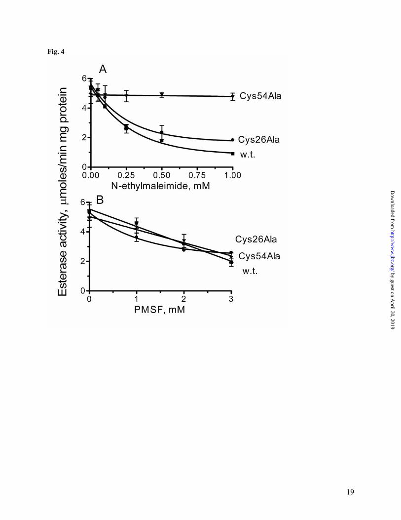
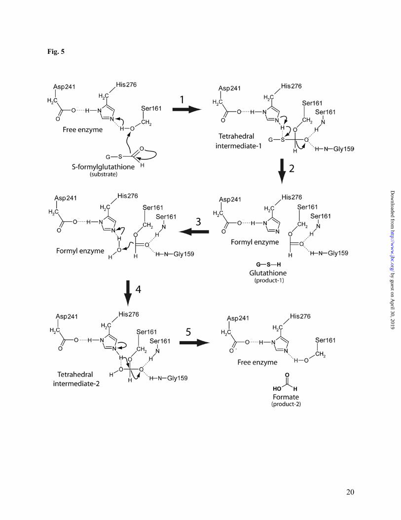
this protein and greatly increased its resistance to thiol inhibitors. As shown in Fig. 4, both the wild-type and Cys26Ala (another conserved Cys) YeiG proteins were strongly inhibited by 1 mM N-ethylmaleimide (Ki 101.5 µM and 91.3 µM, respectively), whereas the Cys54Ala mutant retained almost full activity. Phenylmethylsulfonyl fluoride (PMSF), which is known to react with the catalytic serine in hydrolases (59), produced similar levels of inhibition in all three YeiG proteins (Fig. 4). Thus, inhibiting effect of thiol inhibitors on the activity of FGHs can be explained by the binding of the inhibitor molecule to the conserved cysteine residue (Cys54 in YeiG or Cys60 in YJG8) located in the active site of this enzyme consequently blocking the binding of substrate to the active site of FGHs. The sequential and three-dimensional arrangement of the catalytic residues Ser161, His276, Asp241 corresponds to that of related α/β-hydrolases (55, 54, 56, 60). Ser161 is embedded in the sequence Gly159-His160-Ser161-Met162-Gly163-Gly163, conserved in all characterized FGHs (Fig. 1) and in many predicted esterases. This residue is located on a sharp turn (nucleophilic elbow), which connects strand β-6 and helix α-6. His276 is located at the end of an eight-residue loop situated between the strand β-9 and helix α-13 (Fig. 1). Its imidazole group is in the proper position to make hydrogen bonds with the catalytic Ser161 (3.65Å) and Asp241 (2.73Å) (Fig. 3C). Asp241 is located on the long loop (14 residues) between the strand β-8 and helix α-12 (Fig. 1). Its carboxylate groups form hydrogen bonds with the catalytic His276 (Fig. 3C). Analysis of the YJG8 active site suggests that like in the chymotrypsin-type α/β-hydrolases (60), its oxyanione hole is formed by the main chain NH groups of the catalytic Ser161 and Gly159. The lack of a side chain at position 159 (YJG8 numbering) allows correct positioning of the amido hydrogen to form the required H-bond with the oxyanion substrate. The present work allows a putative picture of the catalytic mechanism of the S-formylglutathione hydrolysis catalyzed by FGHs (Fig. 5). Consistent with previous
mechanistic studies on serine hydrolases (61), Ser161 of YJG8 is properly positioned for nucleophilic attack of the carboxyl carbon of the bound S-formylglutathione molecule. The importance of Ser161 during catalysis is supported by mutagenic studies of the E. coli YeiG showing that a Ser145Ala mutation results in an inactive enzyme (Table 2). His276 and Asp241, the other residues of the catalytic triad in the yeast YJG8 are positioned to assist in the general base-catalyzed attack of the serine nucleophile, and their significance for catalysis was also confirmed by mutational studies on YeiG (Table 2). The nucleophilic attack by the Ser161 hydroxyl group on the carbonyl carbon atom of S-formylglutathione leads to the formation of a tetrahedral intermediate that is stabilized through interactions with an oxyanione hole formed by the backbone NH groups of the catalytic Ser161 and Gly159 (Fig. 5). The tetrahedral intermediate breaks down by general acid catalysis to a formyl-enzyme intermediate with the formyl moiety covalently attached to Ser161. During the formylation step, the imidazole group of His276 transfers the proton of the serine hydroxyl to the sulfhydryl leaving group regenerating the reduced glutathione. The formyl-FGH is then deformylated through the reverse reaction pathway of formylation with formation of a second tetrahedral intermediate releasing free formate, but in this reaction a water molecule instead of the serine residue is the attacking nucleophile (Fig. 5). Deformylation produces the regenerated enzyme and the reaction product, formic acid. The molecular basis of genetic polymorphism of the human FGH (esterase D) has been characterized recently (62). The rarer polymorphic allele ESD*2 was found to contain Glu190 instead of the Gly190 present in the most common allele ESD*1. The ESD*7 allele (originated from ESD*2) has an Asp231Gly mutation, whereas the ESD*5 allele (originated from ESD*1) has a Gly257Asp mutation. The structure of YJG8 shows that Gly204 (Gly190 in human) and Glu246 (Asp231 in human) are located far away from the protein active site and, therefore, should have no significant influence
8
by guest on April 30, 2019
http://ww
w.jbc.org/
Dow
nloaded from

on activity. However, Gly273 (Gly257 in human) is a conserved residue present in all known FGHs and located just 2 residues before the catalytic His276. The replacement of this Gly to Asp (as in ESD*5 allele) will introduce strong negative charge close to the catalytic His276 and Asp241 and, therefore, can potentially affect the interaction of these residues. Moreover, in YJG8, Gly273 is one of 14 residues involved in the dimer intersubunit interaction (H-bonded to Ser8), and its replacement by Asp might also perturb the oligomerization of enzyme. Gene expression studies (real-time PCR) - To study the expression of the two FGHs in E. coli and their role in the dexification of formaldehyde, we designed two single deletion mutants (∆frmB and ∆yeiG) and a double mutant (∆frmB-∆yeiG) (see Materials and Methods). Using real-time PCR, we characterized the effect of different growth conditions and gene deletions on the expression of both FGHs in E. coli (Fig. 6). In the wild-type strain, the level of uninduced expression of frmB was ~5 times higher than that of yeiG. These basally expressed FGHs are perhaps involved in the detoxification of endogenous formaldehyde continuously produced by normal cellular metabolism or demethylation reactions. The addition of formaldehyde (as well as methanol, nitrosoglutathione, methylviologen, H2O2) to the growth medium had no effect on the expression of yeiG in both the wild-type and ∆frmB strains (Fig. 6B). However, formaldehyde greatly stimulated the expression of frmB: 45-fold in wild-type and 70-fold in ∆yeiG over the level of uninduced cells (Fig. 6A). This is consistent with previous studies that reported a 10- to 207-fold increase in the expression of the E. coli frmAB in response to formaldehyde addition (29, 63). Methylviologen or H2O2 did not affect the expression of frmB indicating that this gene is not regulated by oxidative stress. The higher level of the frmB induction by formaldehyde in the yeiG deletion mutant (Fig. 6A) might indicate that E. coli cells compensate for the absence of YeiG by increased synthesis of
FrmB and suggests that these two enzymes have overlapping functions in this organism. Thus, in E. coli, the expression of frmB is stimulated by exogenous formaldehyde or by deletion of yeiG, whereas the yeiG expression is not regulated by exogenous formaldehyde or methanol or by deletion of frmB. The uninduced (basal) expression of both FGHs in E. coli cells is perhaps associated with the degradation of endogenous formaldehyde produced by normal cellular metabolism or demethylation reactions, whereas exogenous formaldehyde induce additional synthesis of more active FGH (FrmB) in E. coli. E. coli resistance to formaldehyde - Both single deletion mutants (∆frmB and ∆yeiG) and a double mutant (∆frmB-∆yeiG) behaved as the wild-type strain under normal aerobic growth conditions on the minimal MOPS-glucose medium (µmax = 0.49-0.51 h-1) (Table 3). The addition of up to 0.4 mM formaldehyde to the medium produced no effect on the growth of ∆yeiG and small inhibition of the growth of the wild-type and ∆frmB strains (Table 3). However, the double ∆frmB-∆yeiG mutant was much more sensitive to formaldehyde, and its specific growth rate dropped to 43% of the wild type growth rate in the presence of 0.4 mM formaldehyde (Table 3). These results suggest that in single mutants the remaining FGH can compensate for the lack of another enzyme. Therefore, simultaneous inactivation of both frmB and yeiG genes is required to increase the sensitivity of E. coli cells to formaldehyde. The addition of methanol (7%), nitrosoglutathione (20 mM), or methylglyoxal (2mM) to the growth medium revealed no difference between the wild-type and mutant strains. Our observation that the sensitivity of the E. coli cells to exogenous methylglyoxal was not increased by the deletion of yeiG can be explained by the degradation of this compound by the glyoxalase system located in the periplasm (42), whereas YeiG might be involved in the detoxification of endogenous methylglyoxal. Thus, our data revealed that although the expression of yeiG was not stimulated by exogenous formaldehyde, this
9
by guest on April 30, 2019
http://ww
w.jbc.org/
Dow
nloaded from

protein is involved in its detoxification, and that both FrmB and YeiG are required for full resistance to exogenous formaldehyde in E. coli. Conclusion Thus, E. coli has two active S-formylglutathione hydrolases, inducible FrmB and constitutive YeiG. Both proteins are promiscuous serine hydrolases capable of hydrolysis of S-formylglutathione, S-lactoylglutathione, and the model substrates (pNP-esters and naphthyl acetate). FGHs from E. coli (and presumably from other organisms) use a classical serine hydrolase catalytic triad (Ser145, His256, and Asp223 in YeiG) for catalysis. Since FrmB has 5 times higher specific activity against S-formylglutathione, this protein is likely to be the main FGH in E. coli. YeiG seems also contribute to the detoxification of formaldehyde, and furthermore, it might be involved in the degradation of methylglyoxal and/or other aldehydes. In E. coli, there are over a dozen metabolic reactions producing various aldehydes (methylglyoxal, lactaldehyde, acetaldehyde, glycolaldehyde, glyceraldehydes 3-phosphate, succinate semialdehyde and other), whose concentrations must be kept low to prevent deleterious effects on cellular proteins and nucleic acids. These aldehydes also spontaneously react with intracellular glutathione reducing the intracellular pool of this important metabolite. The E. coli genome encodes over 30 putative esterases, and their biochemical characterization will likely reveal novel hydrolases involved in the detoxification of various aldehydes and in the maintenance of the intracellular pool of glutathione.
10
by guest on April 30, 2019
http://ww
w.jbc.org/
Dow
nloaded from

References 1. Bolt, H.M. (1987) J. Cancer Res. Clin. Oncol. 113, 305-309 2. Chaw, Y.F., Crane, L.E., Lange, P., and Shapiro, R. (1980) Biochemistry 19, 5525-5531 3. Heck, H.D., Casanova, M., and Starr, T.B. (1990) Crit. Rev. Toxicol. 20, 397-426 4. Ma, T.H., and Harris, M.M. (1988) Mutat. Res. 196, 37-59 5. Precious, E., Gunn, C.E., and Lyles, G.A. (1988) Biochem. Pharmacol. 37, 707-713 6. Fall, R., and Benson, A.A. (1996) Trends Plant Sci. 1, 296-310 7. Hanson, A.D., Gage, D.A., and Shachar-Hill, Y. (2000) Trends Plant Sci. 5, 206-213 8. Falnes, P.O., Johansen, R.F., and Seeberg, E. (2002) Nature 419, 178-182 9. Aas, P.A., Otterlei, M., Falnes, P.O., Vagbo, C.B., Skorpen, F., Akbari, M., Sundheim,
O., Bjoras, M., Slupphaug, G., Seeberg, E., and Krokan, H.E. (2003) Nature 421, 859-863
10. Bystrykh, L.V., Govorukhina, N.I., Van Ophem P.W., Hektor, H.J., Dijkhuizen, L., and Duine, J.A. (1993) J. Gen. Microbiol. 139, 1979-1985
11. Sakai, Y., Murdanoto, A.P., Sembiring, L., Tani, Y., and Kato, N. (1995) FEMS Microbiol. Lett. 127, 229-234
12. Ito, K., Takahashi, M., Yoshimoto, T., and Tsuru, D. (1994) J. Bacteriol. 176, 2483-2491 13. Uotila, L., and Koivusalo, M. (1979) Arch. Biochem. Biophys. 196, 33-45 14. Lee, W.-H., Wheatley, W., Benedict, W.F., Huang, C.M. and Lee, E.Y.-H. P (1986)
Proc. Natl. Acad. Sci. USA 83, 6790-6794 15. Harms, N., Ras, J., Reijnders, W.N., van Spanning, R.J., and Stouthamer, A.H. (1996) J.
Bacteriol. 178, 6296-6299 16. Stover, N.A., Cavalcanti, A.R., Li, A.J., Richardson, B.C., Landweber, L.F. (2005) Mol.
Biol. Evol. 22, 1539-1542 17. Moulis, J. M., Holmquist, B., and Vallee, B. L. (1991) Biochemistry 30, 5743-5749 18. Jensen, D. E., Belka, G. K., and Du Bois, G. C. (1998) Biochem. J. 331, 659-668 19. Liu, L., Hausladen, A., Zeng, M., Que, L., Heitman, J., and Stamler, J. S. (2001) Nature
410, 490-494 20. Sanghani, P.C., Bosron, W.F., and Hurley, T.D. (2002) Biochemistry 41, 15189-15194 21. Uotila, L., and Koivusalo, M. (1974) J. Biol. Chem. 249, 7664-7672 22. Degrassi, G., Uotila, L., Klima, R., and Venturi, V. (1999) Appl. Environ. Microbiol. 65,
3470-3472 23. Kordic, S., Cummins, I., and Edwards, R. (2002) Arch. Biochem. Biophys. 399, 232-238 24. Lee, E.Y-H.P., and Lee, W-H. (1986) Proc. Natl. Acad. Sci. USA 83, 6337-6341 25. Cowell, J.K., Rutland, P., Jay, M., and Hungerford, J. (1986) Hum. Genet. 74, 298-301 26. Ritvo, E.R., Mason-Brothers, A., Menkes, J.H., and Sparkes, R.S. (1988) Arch.Gen.
Psychiatry 45, 600 27. Spitsyn, V.A., Nafikova, A.Kh., Spitsyna, N.Kh., and Afanas’eva, I.S. (2001) Genetika
37, 698-707 28. Irizarry, K., Hu, G., Wong, M.L., Licino, J., and Lee, C.J. (2001) Pharmacogenomics J.
1, 193-203 29. Herring, C., and Blattner, F.R. (2004) J. Bacteriol. 186, 6714-6720 30. Uotila, L. (1981) Methods Enzymol. 77, 424-430 31. Schapiro, J.M., Libby, S.J. and Fang, F.C. (2003) Proc. Natl. Acad. Sci. USA 100, 8496-
8501 32. Zhang, R.G., Skarina, T., Katz, J.E., Beasley, S., Khachatryan, A., Vyas, S., Arrowsmith,
C.H., Clarke, S., Edwards, A., Joachimiak, A., Savchenko, A. (2001) Structure 9, 1095-1106
11
by guest on April 30, 2019
http://ww
w.jbc.org/
Dow
nloaded from

33. Kitagawa, M., Ara, T., Arifuzzaman, M., Ioka-Nakamichi, T., Inamoto, E., Toyonaga, H., and Mori, H. (2006) DNA Res. (in press)
34. Vorderwülbecke, T., Kieslich, K., and Erdmann, H. (1992) Enzyme Microb. Technol. 14, 631-639
35. Berge, R. K., and Farstad, M. (1981) Methods Enzymol. 71, 234–242 36. Datsenko, K.A., and Wanner, B.L. (2000) Proc. Natl. Acad. Sci. USA 97, 6640-6645 37. Neidhart, F.C., Bloch, P.L., and Smith, D.F. (1974) J.Bacteriol. 119, 736-47 38. Kuznetsova, E., Proudfoot, M., Sanders, S.A., Reinking, J., Savchenko, A., Arrowsmith,
C.H., Edwards, A.M., and Yakunin, A.F. (2005) FEMS Microbiol. Rev. 29, 263-279 39. Ochman, H., Lawrence, J.G., and Groisman, E.A. (2000) Nature 405, 299-304 40. Freedberg, W.B., Kistler, W.S., and Lin, E.C. (1971) J. Bacteriol. 108, 137-144 41. Richard, J.P. (1993) Biochem. Soc. Trans. 21, 549-553 42. Cooper, R.A. (1984) Annu. Rev. Microbiol. 38, 49–68 43. Kalapos, M.P. (1999) Toxicol. Lett. 110, 145-175 44. Shinohara, M., Thornalley, R.J., Giardino, I., Beisswenger, P., Thorpe, S.R., Onorato, J.,
and Brownlee, M. (1998) J. Clin. Invest. 101, 1142-1147 45. Ramasamy, R., Yan, S.F. and Schmidt, A.M. (2006) Cell 124, 258-260 46. Yao, D., Taguchi, T., Matsumura, T., Pestell, R., Edelstein. D., Giardino, I., Suske, G.,
Ahmed, N., Thornalley, P.J., Sarthy, V.P., Hammes, H-P., and Brownlee, M. (2006) Cell 124, 275-286
47. Thornalley, P.J. (1990) Biochem. J. 269, 1-11 48. Misra, K., Banerjee, A.B., Ray, S., and Ray, M, (1995) Biochem. J. 305, 999–1003 49. Misra, K., Banerjee, A.B., Ray, S., and Ray, M. (1996) Mol. Cell. Biochem. 156, 117-124 50. Inoue, Y., and Kimura, A. (1995) Adv. Microbiol. Physiol. 37, 177-227 51. Thornalley, P.J. (1995) Crit. Rev. Oncol. Hematol. 20, 99-128 52. Ferguson, G.P., Totemeyer, S., MacLean, M.J. and Booth, I.R. (1998) Arch. Microbiol
170, 209-218 53. Brenner, S. (1988) Nature 334, 528-530 54. Cygler, M., Schrag, J.D., Sussman, J.L., Harel, M., Silman, I., Gentry, M.K., and Doctor,
B.P. (1993) Proteins Sci. 2, 366-382 55. Ollis, D.L., Cheah, E., Cygler, M., Dijkstra, B., Frolow, F., Franken, S.M., Harel, M.,
Remington, S.J., Silman I., and Schrag, J. (1992) Protein Eng. 5, 197-211 56. Heikinheimo, P., Goldman, A., Jeffries, C., and Ollis, D.L. (1999) Structure 7, 141-146 57. Holmquist, M. (2000) Curr. Protein. Pept. Sci. 1, 209-235 58. Holm, L., and Sander, C. (1995) Trends Biochem. Sci. 20, 478-480 59. Gold, A.M. (1965) Biochemistry 4, 897-901 60. Polgar, L. (2005) Cell. Mol. Life Sci. 62, 2161-2172 61. Powers, J.C., Odake, S., Oleksyszyn, J., Hori, H., Ueda. T., Boduszek, B., and Kam, C.
(1993) Agents Actions Suppl. 42, 3-18 62. Yuasa, I., Umetsu, K., Tsuchida, S., Irizawa, Y., Henke, L., and Henke, J. (2004) Hum.
Biol. 76, 479-488 63. Gutheil, W.G., Kasimoglu, E., and Nicholson, P.C. (1997) Biochem. Biophys. Res.
Commun. 238, 693-696
12
by guest on April 30, 2019
http://ww
w.jbc.org/
Dow
nloaded from

Footnotes * This work was supported by Genome Canada through the Ontario Genomics Institute, the National Institutes of Health (GM62414-01) as well as by CREST (Japan Science and Technology) and the Inamori Foundation (H.M.). We thank all members of the Ontario Center for Structural Proteomics for help in conducting experiments. 1The abbreviations used are: FALDH, formaldehyde dehydrogenase; FGH, S-formylglutathione hydrolase; GSH, reduced glutathione; PMSF, phenylmethylsulfonyl fluoride; pNP, p- nitrophenyl.
Figure Legends Fig. 1. Multiple sequence alignment of the E. coli FrmB and YeiG with eukaryotic FGHs from S. cerevisiae, A. thaliana, and human. Highly conserved residues are highlighted in gray. The secondary structure elements derived from the yeast YJG8 are shown below its sequence. Residues mutated in this work are marked (*) above the alignment. Five blocks of conserved residues are boxed. The compared proteins are E. coli FrmB (P51025), E. coli YeiG (P33018), S. cerevisiae YJG8 (P40363), A. thaliana FGH (CAC87877), human esterase D (P10768). Fig. 2. Esterase activity (substrate profiles) of the E. coli FrmB (A) and YeiG (B) with various substrates. Experimental conditions were as described in Materials and Methods. Reaction mixtures contained 1 mM substrate and 2 µg of enzyme. Fig. 3. Structure of the yeast YJG8 (1pv1). (A), ribbon diagram of one subunit of YJG8. The monomer is composed of a single domain with an α/β-hydrolase fold. α-Helices are shown in red, β-strands – in yellow. Also shown as a thick wire model are the residues of the catalytic triad: Ser161, His276, Asp241. (B), the coordination of the side chains of the residues of the catalytic triad of YJG8. The pictures were drawn based on the PDB entry 1pv1 using the PyMOL program (http://www.pymol.org). Fig. 4. Effect of N-ethylmaleimide (A) and PMSF (B) on the hydrolysis of α-naphthyl acetate by the E. coli wild-type and mutant YieG. Reaction mixtures contained 1 mM α-naphthyl acetate 3 - 10µg of enzyme. Other reaction conditions were as described in Materials and Methods. Fig. 5. Possible reaction mechanism of the hydrolysis of S-formylglutathione by FGHs. The reaction proceeds through the following steps. (1), nucleophilic attack of Ser161 to form the first tetrahedral intermediate. (2), decomposition of the tetrahedral intermediate-1 and formation of the formyl enzyme intermediate. (3), release of reduced glutathione. (4), nucleophilic attack of water and the formation of the second tetrahedral intermediate. (5), release of the second product (formate) and formation of free enzyme. Fig. 6. Expression of the E. coli frmB (A) and yeiG (B) under different conditions as measured with real-time PCR. Wild-type (WT) and mutant strains were grown to an OD600 = 0.30 (as described in Materials and Methods). The cultures were split into three aliquots containing: no additions (uninduced); or 0.25 mM formaldehyde; or 250 mM methanol. After 45 min incubation, cells were collected by centrifugation to prepare cDNA (as described in Materials and Methods). In both figures, the values were normalized relative to the expression of the control gene, rrsC (arbitrarily set as 1). The data represent the average of three independent experiments ± standard deviations.
13
by guest on April 30, 2019
http://ww
w.jbc.org/
Dow
nloaded from

Table 1. Kinetic parameters of the E. coli FrmB and YeiG with several substrates
Substrate Km Vmax kcat kcat/Km
FrmB
pNP-acetate (C2)
(mM)
0.29 ± 0.02
(units/mg)a
0.58 ± 0.02
(s-1)
0.30 ± 0.01
(M-1 s-1)
1.03 x 10³
pNP-propionate (C3) 0.83 ± 0.04 0.27 ± 0.03 0.14 ± 0.02 0.17 x 10³
S-lactoylglutathione 0.60 ± 0.21 0.09 ± 0.02 0.05 ± 0.01 0.08 x 10³
S-formylglutathione 0.41 ± 0.03 55.0 ± 4.0 28.5 ± 2.07 6.90 x 10⁴
YeiG
pNP-acetate (C2)
0.45 ± 0.05
0.50 ± 0.08
0.26 ± 0.04
0.57 x 10³
pNP-propionate (C3) 0.48 ± 0.03 0.71 ± 0.01 0.37 ± 0.05 0.76 x 10³
pNP-butyrate (C4) 0.70 ± 0.03 0.70 ± 0.05 0.36 ± 0.03 0.51 x 10³
pNP-caproate (C6) 0.95 ± 0.01 0.64 ± 0.08 0.49 ± 0.04 0.52 x 10³
α-naphthyl acetate 1.03 ± 0.13 9.79 ± 0.56 5.09 ± 0.29 4.94 x 10³
S-lactoylglutathione 0.58 ± 0.18 1.97 ± 0.29 1.02 ± 0.15 1.75 x 10³
S-formylglutathione 0.43 ± 0.05 12.6 ± 1.30 6.51 ± 0.67 15.1 x 10⁴
a units = µmoles/min
14
by guest on April 30, 2019
http://ww
w.jbc.org/
Dow
nloaded from

Table 2. Enzymatic parameters of the wild type and mutant E. coli YeiG with α-naphthyl acetate as a substrate
YeiG Km Vmax kcat kcat/Km
1. Wild type
(mM)
1.03 ± 0.13
(units/mg)a
9.79 ± 0.56
(s-1)
5.09 ± 0.29
(M-1s-1)
4.94 x 10³
2. Cys26Ala 1.06 ± 0.27 9.83 ± 1.13 5.12 ± 0.58 3.20 x 10³
3. Cys54Ala 1.28 ± 0.17 9.80 ± 0.63 5.10 ± 0.32 3.98 x 10³
4. Asp80Ala 0.80 ± 0.12 4.17 ± 0.247 2.17 ± 0.12 2.71 x 10³
5. Ser145Ala n.d.b n.d. n.d. n.d.
6. Asp199Ala 1.01 ± 0.21 5.15 ± 0.48 2.96 ± 0.28 2.93 x 10³
7. Asp218Ala 1.30 ± 0.40 6.62 ± 1.08 3.81 ± 0.62 2.93 x 10³
8. Asp223Ala n.d. n.d. n.d. n.d.
9. Asp255Ala 0.54 ± 0.15 3.89 ± 0.43 2.23 ± 0.25 4.13 x 10³
10. His256Ala n.d. n.d. n.d. n.d.
a units = µmoles/min
b n.d., not detected
15
by guest on April 30, 2019
http://ww
w.jbc.org/
Dow
nloaded from

Table 3. Effect of formaldehyde on the growth rate (µmax) of the E. coli wild-type (WT) and mutant strains
Formaldehyde (mM)
Growth ratea
WT ∆frmB ∆yeiG ∆frmB,∆yeiG
No addition 0.51 ± 0.01 0.49 ± 0.02 0.51 ± 0.01 0.51 ± 0.02
0.10 0.48 ± 0.02 0.49 ± 0.01 0.50 ± 0.01 0.47 ± 0.01
0.35 0.46 ± 0.02 0.47 ± 0.01 0.50 ± 0.02 0.36 ± 0.02
0.40 0.47 ± 0.03 0.43 ± 0.05 0.50 ± 0.01 0.22 ± 0.05
a µmax, h-1
Fig. 1
16
by guest on April 30, 2019
http://ww
w.jbc.org/
Dow
nloaded from

Alexei V. Savchenko and Alexander F. YakuninClaudio F. Gonzalez, Michael Proudfoot, Greg Brown, Yurij Korniyenko, Hirotada Mori,
s-formylglutathione hydrolases from Escherichia coli, FrmB and YeiGMolecular basis of formaldehyde detoxification: Characterization of two
published online March 27, 2006J. Biol. Chem.
10.1074/jbc.M600996200Access the most updated version of this article at doi:
Alerts:
When a correction for this article is posted•
When this article is cited•
to choose from all of JBC's e-mail alertsClick here
Supplemental material:
http://www.jbc.org/content/suppl/2006/03/28/M600996200.DC1
by guest on April 30, 2019
http://ww
w.jbc.org/
Dow
nloaded from