Embed Size (px)
Citation preview
157
Characterization of surface roughness of new nanophotonic soft contact lenses using lacunarity and AFM method
Aleksandra Mitrovic1, Bozica Bojovic1, Dragomir Stamenkovic2, Dejana Popovic3
1University of Belgrade, Faculty of Mechanical Engineering, Belgrade, Serbia 2Optix, Belgrade, Serbia 3University of Belgrade, Vinča Institute of Nuclear Sciences, Belgrade, Serbia
Abstract
The aim of this study was to develop new soft contact lens (SCL) materials which would, after recommended and existing machining processes, improve surface roughness. Nanomaterials (fullerene, fullerol and methformin hydroxylate fullerene) were incorporated into commercial material for SCL (SL38) based on PHEMA, which were derived by the technology in the production lab of the company Soleko (Milan, Italy). Nanophotonic SCLs (SL38-A, SL38-B, SL38-C, respectively) were produced in the company Optix (Belgrade, Serbia) from the obtained materials. For the surface characterization of SCLs, AFM analysis and lacunarity method were performed. The results showed that for the SL38-B average roughness value is lower than those of SL38-A and SL38. The topography parameters of SL38-C were between the parameters of SL38-A and SL38-B. Lacunarity analysis of AFM images confirmed that SCLs surface state should belong to either group of adequate (slanted p-diagram) or inadequate (contorted p-diagram) roughness concerning tear film stability. Nanophotonic SCL SL38-C exibits more acceptable performance considering SCL surface functional behavior as compared to other SCLs. The positive result of incorporating nanomaterials into basic material for SCL is better quality of the nanophotonic SCLs surfaces. On the bases of these experiments, the assumption that incorporation of fullerene derivate will not increase surface roughness parameters is confirmed.
Keywords: soft contact lenses, nanophotonic contact lenses, fullerenes, AFM, lacunarity.
SCIENTIFIC PAPER
UDC 666.227.5: 620.3: 543.456
Hem. Ind. 72 (3) 157–166 (2018)
Available online at the Journal website: http://www.ache.org.rs/HI/
1. INTRODUCTION
Hydrogels, three-dimensional hydrophilic polymeric networks capable of absorbing large amounts of water or biological fluids, enable high hydrophilicity and biocompatibility due to peculiar network structure. Many common hydrogels such as poly (2-hydroxyethyl methacrylate) (PHEMA), poly (ethylene glycol) (PEG) or poly (vinyl alcohol) (PVA) have found wide use both in laboratory studies and clinical uses. Currently, hydrogels are used for manufacturing contact lenses, tissue engineering scaffolds, wound dressings, drug delivery systems and hygiene products [1-4]. Hydrogels based on 2-hydroxyethyl methacrylate (HEMA) copolymers are of a great interest because of their tunable chemical composition, hydrolytic stability, their excellent biocompatibility and physicochemical properties similar to those of living tissues [5-7]. 2-hydroxyethyl methacrylate was the original hydrophilic lens monomer and nowadays continues to be the most commonly used hydrophilic monomer for soft contact lenses [8,9].
Numerous studies have been conducted to modify PHEMA with the aim of improving its properties [2,4,5,10,11]. Also, a lot of attempts have been made to develop new contact lenses with better physical, chemical and mechanical properties [9,12-14]. Bauman et al. [15] disclosed a method for making soft contact lenses with a nano-textured surface, imitating the surface of human cornea. There have been attempts to combine the ease of processability of polymers with excellent properties associated with the buckyball [16] since the report of the synthesis of a C60-p-xylylene copolymer [17] has been published.
Fullerene C60 has stimulated intense interest in scientific, industrial and medical fields due to its unique structure, electronic and spectroscopic properties [18]. In addition, functionalized C60 can be copolymerized with other
Correspondence: Aleksandra D. Mitrovic, Faculty of Mechanical Engineering, Kraljice Marije 16, 11000 Belgrade, Serbia e-mail: [email protected]
Paper received: 24 September 2017
Paper accepted: 22 February 2018
https://doi.org/10.2298/HEMIND170924004M
A. MITROVIC et al.: SURFACE ROUGHNESS OF NEW NANOPHOTONIC SOFT CONTACT Hem. ind. 72 (3) 157–166 (2018)
158
monomers [16,19-22]. Researchers investigated the influence of nanomaterial addition, such as fullerenes and nanotubes, on properties of polymers and their compositions [23-25].
In accordance with specific biomedical applications of hydrogels as a soft contact lens material, a large number of tests must be implemented. In the field of optics and materials for soft contact lenses (SCLs), it is desirable to develop new materials that should improve performances of SCL after processing.
Several methods can depict the surface of soft contact lenses [26-29]. Atomic force microscopy (AFM) is a powerful technique that provides detailed surface characteristic information of contact lens materials [30–36]. Furthermore, the lacunarity analysis based on works of Mandelbrot [37], Voss [38] and Plotnick [39], regarding previous results [40-42], proves applicability of lacunarity analysis on various machining surfaces including lubrication aspect of surface functional behavior. Since Mandelbrot introduced the term lacunarity as a gappiness, the lacunarity application that is originally developed for further classification of fractals has become more generally and broadly applicable in natural sciences [43-45] as a very useful multi-scaled method for describing patterns of spatial dispersion. Therefore, SCL surface lacunarity analysis is our innovative approach in on-going project activities for surface topography characterization in addition to conventional AFM images analyses.
Every single SCL user provides a unique ambient condition in which these SCL surfaces have to function properly. SCL can lose functionality due to accumulated proteins, lipids and other tear components on outer SCL surface, despite routine cleaning activities. This is not valid in case of daily disposable SCL, but it is extremely important for extended wear SCL. In this study comparison of nanophotonic materials concerning the surface susceptibility for proteins and lipids deposits on tested SCLs was one of the tasks. The clinical performance of a SCL depends on roughness that is exposed to the deposit formation due to spoliation with tear residues and bacteria [46]. Thus, when characterizing a contact lens surface to complete the information on the finished quality, the surface roughness parameters and lacunarity have to be used to improve understanding of asperities and pits implications to the biocompatibility of the SCL nanophotonic materials [47]. Surface roughness, additionally, will affect changes on the surface of worn SCL [48] and have a large effect on the van der Waals attraction [49]. Therefore, surface roughness parameters and lacunarity analysis provide the usability distinction of nanophotonic material for SCL that will operate in the pre-lens tear film, which is 2–5 μm in thickness [50].
The purpose of this study was to provide surface characterization data of basic (commercial) and new nanophotonic SCLs which were obtained by incorporating the fullerene nanoparticles into the basic material for SCLs.
2. EXPERIMENTAL PART
2.1. Material
Materials for nanophotonic soft contact lenses were obtained according to the protocol described previously [25]. Fullerene (C60), fullerol (C60(OH)24) and methformin hydroxylate fullerene (C60(OH)12(OC4N5H10)12) (MER Corporation, SAD, ≥99%) were incorporated into the material for soft contact lens (SL38) based on PHEMA (SOLEKOTM, Milan, Italy). Nanophotonic soft contact lenses, SL38-A, SL38-B and SL38-C, were made from the obtained materials, respectively.
Fullerenes mixed with other polymers are a group of optical filters with remarkable properties such as easy fabrication, predictable wavelength tuning and excellent performance stability. One of the main disadvantages of fullerenes is their low solubility in water. To make them soluble, fullerenes have to be functionalized with polar groups such as –OH and –COOH. Fullerene unlike its derivate, fullerol and methformin hydroxylate fullerene, does not dissolve in water and certain solvents.
2.2. Method
2.2.1. Soft contact lenses micro machining
Machining process was the same for all three nanophotonic materials and the commercial one. The recommended process parameters that exist for commercial SCL material were chosen. These process parameters were already accompanied in machine control units which execute tool path during machining. Additionally, the contact lens manufacturers prefer to use in practice proven process parameters. Leading idea was to use process parameters and a diamond tool, which are suggested for the commercial SCL material on the existing production line. The angle of the diamond tool side-cutting edge was 60 degrees. SCL manufacturing process used for experimental validation was considered as micro-cutting regarding the cutting depth and diamond tool nose ratio. SCLs were manufactured by computer numerical controlled (CNC) three-axe lathe Politech 1800 Aspheric-Toric with an air bearing spindle and air operated collet, and closed loop control on linear (Z, R) and rotary (Theta) axes. The resolution of R axis was 0.02 µm, Z axis was 0.01 µm, and 1.62 s of Theta axis. All SCLs were manufactured in respect to recommended process parameters, as shown in Table 1.

A. MITROVIC et al.: SURFACE ROUGHNESS OF NEW NANOPHOTONIC SOFT CONTACT Hem. ind. 72 (3) 157–166 (2018)
159
Table 1. Process parameters for soft contact lens micro-turning
Parameter Coarse Fine
Depth of cut, mm 0.4 0.05
Angular feed rate, degrees/s 10 4
Nose radius, mm 0.5 0.2
Rotational speed, rev/min 8000
Determination of the surface roughness relation to process parameters was not intention of this study. Although process parameters have influence on surface roughness, the relation is established among three nanophotonic vs. basic material per se. Further research activities will include optimization of process parameters strictly in accordance to contact lens manufacturers.
2.2.2. Atomic Force Microscopy (AFM)
The surface topography changes were observed by AFM, while the changes in surface composition were detected using phase imaging AFM. All measurements were done on JEOL SPM 5200 (Japan) at room temperature. The microscope itself was isolated so that outside vibrations would be minimized. Used cantilever was manufactured by Nanosensors™ (type PPP-NCHAuD-10, stiffness 10-130 N/m). Resonant frequency of the cantilever was in the range 204-497 kHz. Imaging area for all tested SCLs was 10 µm x 10 µm. Images were analyzed in JEOL SPM Processing Software and WinSxM. Parameters used for SCL comparison were standard area roughness parameters Sa, Sq and Sz according to EN ISO 25178 standard.
2.2.3. Lacunarity analysis
Voss and Plotnick proposed “gliding box” method for lacunarity value determination. Based on it, the in-house made procedures in Matlab software were developed. Procedures were applied for various surfaces’ topography including tin metal surface [40] and RGP contact lens surfaces [41,42]. Gliding box was a window sliding systematically through the binary image which was obtained as a result of slicing surface topography image on levels. Binary image consisted of white pixels considered as binary 1 belonging to a surface and black pixels considered as binary 0 representing an empty space.
Detailed method description and mathematical representation could be found in the previous paper [40]. Except double logarithmic plots (Fig. 1a) providing visual information about the pixel entropy for visual lacunarity distinction done by Plotnick [39], parameter that can be used for AFM image lacunarity comparison was introduced [40]. In this study the parameter p was used as the slope value for every section and can serve as a parameter for surface section characterization. Additionally, the diagram of parameter p for every section shown in Figure 1b, can be considered as AFM image lacunarity signature.
a b
Figure 1. Plot of natural logarithms of L(r) against r for (a) arbitrary cutting level of engineering surface; (b) slope value p vs. cutting level n. Adapted from [42]. Copyright 2017 by Institute of Metals and Technology, Ljubljana. Reprinted with permission.
Therefore, the diagram which consisted of three sloped curve related to random distributed surface asperities generated by machining process was used for soft contact lenses comparison.
A. MITROVIC et al.: SURFACE ROUGHNESS OF NEW NANOPHOTONIC SOFT CONTACT Hem. ind. 72 (3) 157–166 (2018)
160
3. RESULTS AND DISCUSSION
For this study the convex surface of the SCL was chosen for the topography examination. Topography measurements were routinely conducted for the basic (SL38) and new nanophotonic soft contact lenses (SL38-A, SL38-B, SL38-C) in tapping mode using uniform scanning surface of 10 × 10 μm. The tapping mode was used due to its ability of non-destructive high-resolution imaging of soft and fragile samples in ambient environment. The key advantage of tapping mode is the elimination of the lateral shear forces present in contact AFM mode in order to prevent mechanical damage of the SCL surface [51]. Topography image yields information about surface shape and relative positions and dimensions. Phase imaging gives yet another way to distinguish different surface properties of SCLs made of heterogeneous polymers [52]. The phase image is generated by monitoring the phase angle of the oscillating probe relative to the phase angle of the signal that drives the probe in tapping mode. Differences in phase shifts indicate differences in material properties of the SCL. Measurements of the surface topography of SCLs revealed smooth areas suitable for indentation measurements.
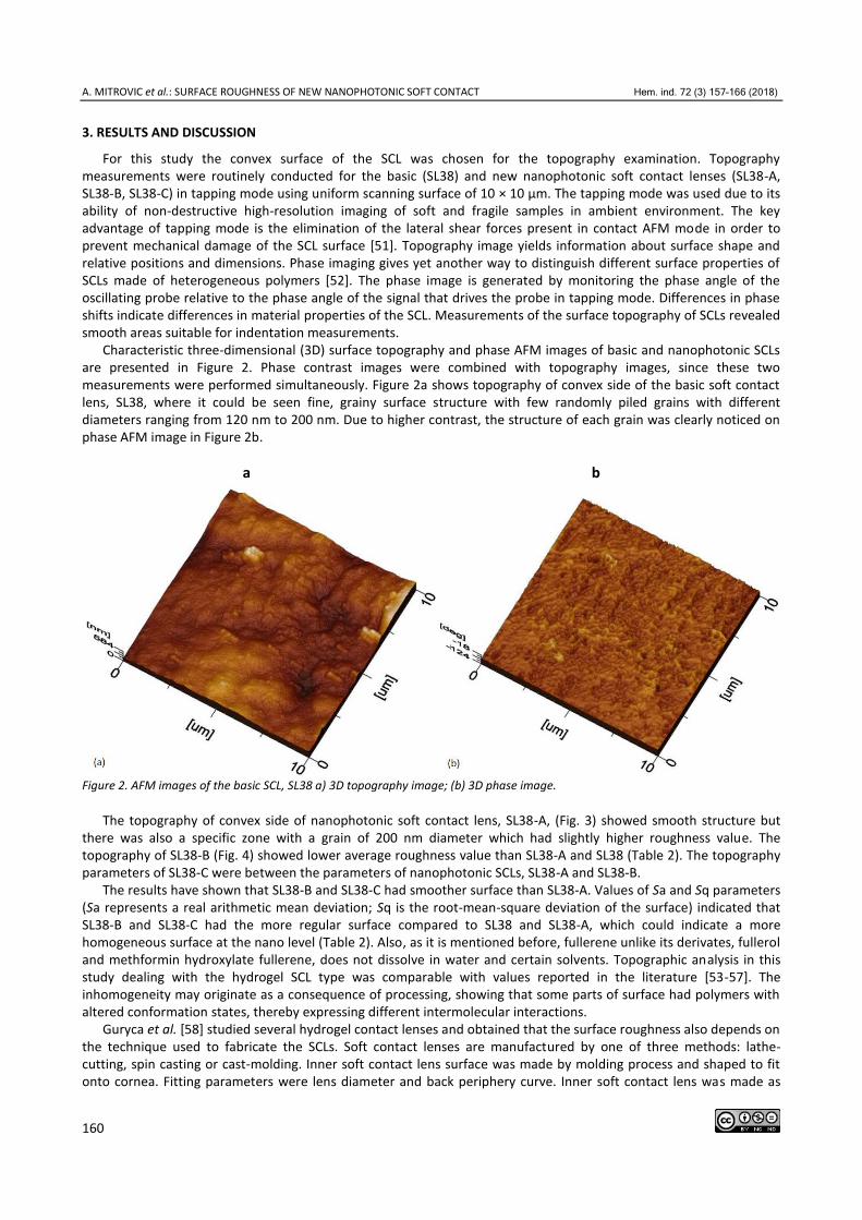
Characteristic three-dimensional (3D) surface topography and phase AFM images of basic and nanophotonic SCLs are presented in Figure 2. Phase contrast images were combined with topography images, since these two measurements were performed simultaneously. Figure 2a shows topography of convex side of the basic soft contact lens, SL38, where it could be seen fine, grainy surface structure with few randomly piled grains with different diameters ranging from 120 nm to 200 nm. Due to higher contrast, the structure of each grain was clearly noticed on phase AFM image in Figure 2b.
a b
Figure 2. AFM images of the basic SCL, SL38 a) 3D topography image; (b) 3D phase image.
The topography of convex side of nanophotonic soft contact lens, SL38-A, (Fig. 3) showed smooth structure but there was also a specific zone with a grain of 200 nm diameter which had slightly higher roughness value. The topography of SL38-B (Fig. 4) showed lower average roughness value than SL38-A and SL38 (Table 2). The topography parameters of SL38-C were between the parameters of nanophotonic SCLs, SL38-A and SL38-B.
The results have shown that SL38-B and SL38-C had smoother surface than SL38-A. Values of Sa and Sq parameters (Sa represents a real arithmetic mean deviation; Sq is the root-mean-square deviation of the surface) indicated that SL38-B and SL38-C had the more regular surface compared to SL38 and SL38-A, which could indicate a more homogeneous surface at the nano level (Table 2). Also, as it is mentioned before, fullerene unlike its derivates, fullerol and methformin hydroxylate fullerene, does not dissolve in water and certain solvents. Topographic analysis in this study dealing with the hydrogel SCL type was comparable with values reported in the literature [53-57]. The inhomogeneity may originate as a consequence of processing, showing that some parts of surface had polymers with altered conformation states, thereby expressing different intermolecular interactions.
Guryca et al. [58] studied several hydrogel contact lenses and obtained that the surface roughness also depends on the technique used to fabricate the SCLs. Soft contact lenses are manufactured by one of three methods: lathe-cutting, spin casting or cast-molding. Inner soft contact lens surface was made by molding process and shaped to fit onto cornea. Fitting parameters were lens diameter and back periphery curve. Inner soft contact lens was made as

A. MITROVIC et al.: SURFACE ROUGHNESS OF NEW NANOPHOTONIC SOFT CONTACT Hem. ind. 72 (3) 157–166 (2018)
161
separate process that is performed before outer surface machining. Fitting parameter for outer surface was front optic zone radius. Outer surface of soft contact lens was shaped to provide adequate optical power that was requested for vision correction. Inner surface roughness is not related to the outer surface roughness.
a b
Figure 3. AFM images of the nanophotonic SCL, SL38-A (a) 3D topography image; (b) 3D phase image.
a b
Figure 4. AFM images of the nanophotonic SCL, SL38-B (a) 3D topography image (b) 3D phase image.
a b
Figure 5. AFM images of the nanophotonic SCL, SL38-C (a) 3D topography image (b) 3D phase image.
Basic SL38 and nanophotonic SCLs, SL38-A, SL38-B and SL38-C were manufactured by lathe-cutting. This process uses a special lathe for cutting an anhydrous block of material into the required shape. This intermediate was subsequently hydrated to obtain a soft contact lens. Generally, AFM is indispensable for studies dealing with the influence of contact lens manufacturing on the surface character. In this study, the particular attention was paid to
A. MITROVIC et al.: SURFACE ROUGHNESS OF NEW NANOPHOTONIC SOFT CONTACT Hem. ind. 72 (3) 157–166 (2018)
162
imaging surface irregularities of SCLs below the submicron level. The convex surface of a SCL is in direct contact with the inner part of the upper eyelid. That contact would be painful if the convex surface was rough. The obtained values of roughness for new nanophotonic SCLs are in accordance with the results obtained in the literature [53,54], without negative influence on the comfort in contact lens wearing.
Also, the surface properties play an important role in the secondary effects like protein or bacteria adsorption causing negative ocular reactions [56,58]. Conformation states of polymers constituting SCL surface are changed during final stages of the manufacturing process (polishing), which presents a complex problem because surface molecules and their orientation influence the final level of surface quality.
Answering the questions of optimal processing parameters selection directly influences surface quality of SCLs which in turn reaches all aspects of its functionality: optical, medical and patient-comfort. We believe that our results could contribute to studies concerning these issues. The quality of polished surfaces, expressed by their roughness, for all three nanophotonic soft contact lenses completely satisfies the standards for contact lens production. None of the tested SCLs have irregular regions at their convex sides with height differences more than 615 nm. Thus, all irregularities at frontal surface will be smoothed by tear film when the contact lens is set on cornea [30,42]. The positive result of incorporating nanomaterials into basic material for SCL was better quality of the nanophotonic SCLs surfaces due to lower values of roughness parameters for all nanophotonic materials compared to those for basic material.
The comparison of all parameters is given in Table 2.
Table 2. Values of topography and phase parameters for SCLs
Topography Sample 1 Sample 2 Sample 3 Average value Standard deviation
SL 38
Sa / nm 81.00 42.00 66.50 63.17 19.71
Sq / nm 101.80 49.00 74.40 75.07 26.41
Sz / nm 445.00 189.20 255.00 296.40 132.80
SL 38-A
Sa / nm 152.90 89.60 94.60 112.40 35.19
Sq / nm 173.50 107.20 120.00 133.60 35.17
Sz / nm 567.40 425.80 435.80 476.30 79.02
SL 38-B
Sa / nm 8.88 6.65 10.80 8.78 2.08
Sq / nm 11.40 8.87 13.90 11.39 2.51
Sz / nm 59.20 57.90 72.70 63.27 8.19
SL 38-C
Sa / nm 27.50 18.80 21.30 22.53 4.48
Sq / nm 31.80 23.20 24.90 26.63 4.55
Sz / nm 134.20 98.70 92.30 108.40 22.57
Sa represents a real arithmetic mean deviation; Sq is the root-mean-square deviation of the surface; Sz is height difference
between the lowest and the highest point on topography image. It can be also concluded that incorporation of fullerene derivate can play a role in the prevention of an increase in
roughness due to lower values of roughness compared to basic SCL. For SCL lacunarity analysis, the topographic image of convex surfaces of SCL, made of basic and nanophotonic
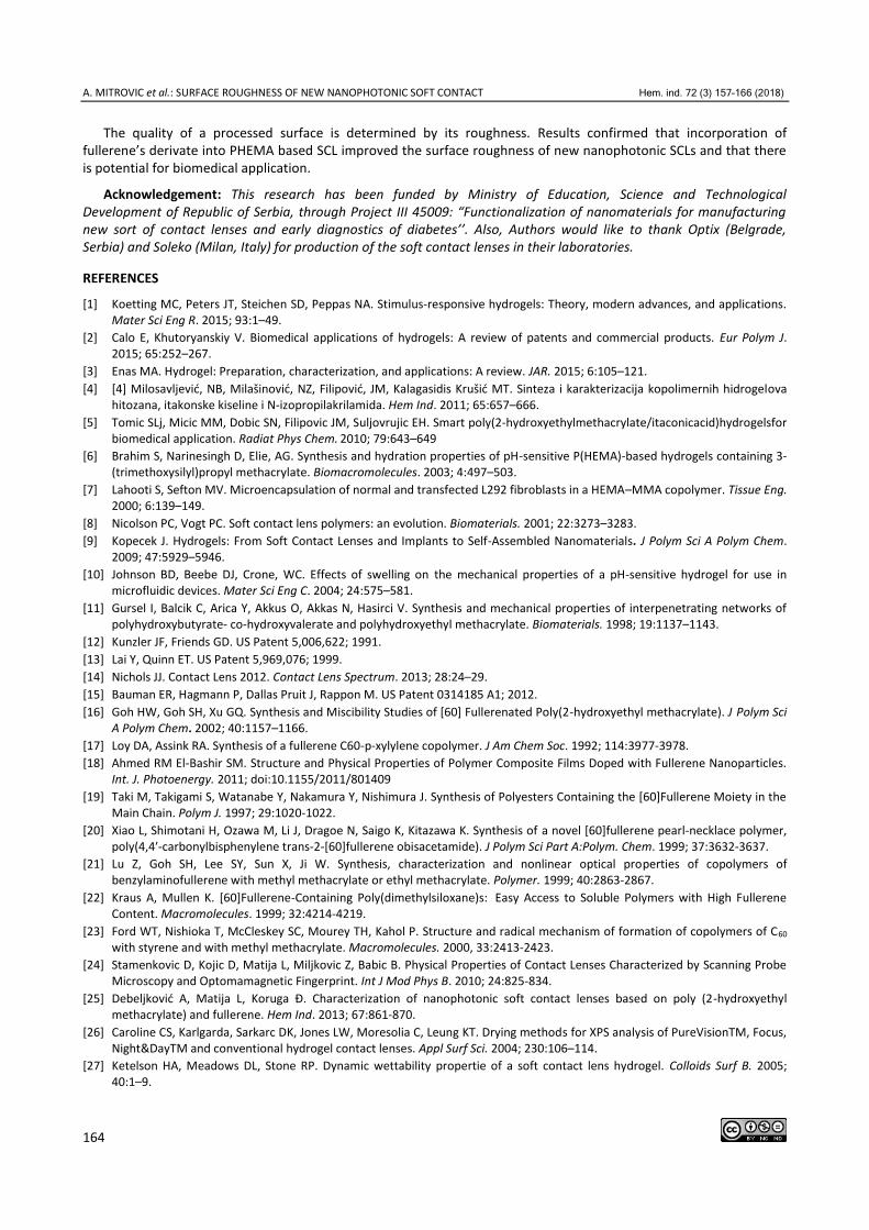
materials, were gathered. Four images were recorded by AFM and imported to Matlab procedures for further calculation. In order to make lacunarity perceptible for the integral contact lens surface, the parameter p was determined for every section that was made by slicing the 3D AFM image. Image maximum height was slicing on 100 sections denoted by n and every second was plotted in Figure 6. Diagram p vs. n for basic and nanophotonic SCLs were four distinctive curves that can be observed in Figure 6. Each p-diagram represents surface p parameter value variation over height reduction, caused by material accumulation and consequently empty space reduction that was obvious from top to the bottom of asperities.
Different forms of curves specified dissimilar surface appearances, which can be confirmed by 3D AFM images observation (Fig. 6). The p-diagram presents topology with regular distributed lower hills and this type of curve was
A. MITROVIC et al.: SURFACE ROUGHNESS OF NEW NANOPHOTONIC SOFT CONTACT Hem. ind. 72 (3) 157–166 (2018)
163
labeled as slanted p-diagram. This type of p-diagram corresponds to machined surface p-diagram from papers [41, 42]. P-diagrams for SL38-A and SL38-B had four slopes and this type of curve was labeled as contorted. The origins of the contorted p-diagram were bump-like entities that can be seen at 3D AFM images. For SL38-C, surface asperities distribution tended to be regular since bumps were multiplied and therefore the p-diagram was slanted.
Figure 6. Plot of parameter p vs. cutting level (n) for soft contact lens surface.
Three different line slopes for both slanted p-diagrams of SL38 and SL38-C had characteristic points at 24 % and 66 % of maximal height. In spite of equivalent position of slopes change, those two surfaces exhibited different behaviors. Two contorted p-diagrams with quite distinctive curve shapes belonged to SL38-A and SL38-B. P-diagram for SL38-B had a specific steep line slope between 40-50 % of the maximal height and the rest of it was gradually sloped. For SL38-A two steep line slopes were interrupted by a gradual one in the range of 50-60 % of the maximal height. All p-diagrams reached asymptotic zero value after certain level. Asymptotic curve part of SL38-C corresponded to the part of the image under 60 %. This occurred under 70 % in case of SL38 and SL38-B and 80 % for SL38-A.
4. CONCLUSION
New nanophotonic soft contact lens materials were developed to improve surface roughness after recommended and existing machining processes. Nanomaterials (fullerene, fullerol and methformin hydroxylate fullerene) were incorporated into commercial material for SCL (SL38) based on PHEMA. For the surface characterization of SCLs, AFM analysis and lacunarity method were performed. The results of SCLs outer surfaces have been compared at nano-scale and the conclusions were diverse. Our contribution presents topographical data for three new nanophotonic soft contact lenses. AFM measurements helped in evaluating surface quality of tested SCLs after the recommended machining processes were performed. These types of measurements on lenses are very useful for enhancing quality control capabilities. Furthermore, this type of information will help to speed the development of superior polymers and introduce it in SCL existing manufacturing production lines without deterioration of SCL surface roughness. Nondestructive methods, such as AFM, allow testing to be performed on the same sample by considering various parameters. All of these parameters, mentioned in this paper, are good candidates for quick and comparable test methods.
Lacunarity method showed that AFM images, which were associated to specific group diagrams, demonstrated similar surface topology. The origins of the contorted p-diagram were bump-like entities, which could be observed in adequate topography images pointing out that bumps significantly influenced surface lacunarity. This surface lacunarity signature indicated non-regular lubricant distribution. The surface lacunarity influences the tear film volume distribution and consequently, the contact lens surface lubrication. SCL surface lacunarity analysis confirmed SCLs surface state as belonging to either group adequate (slanted p-diagram) or inadequate (contorted p-diagram) roughness concerning tear film stability. In this study, nanophotonic soft contact lens SL38-C exhibits a more acceptable performance considering SCL surface functional behavior as compared to SL38-A and SL38-B.
A. MITROVIC et al.: SURFACE ROUGHNESS OF NEW NANOPHOTONIC SOFT CONTACT Hem. ind. 72 (3) 157–166 (2018)
164
The quality of a processed surface is determined by its roughness. Results confirmed that incorporation of fullerene’s derivate into PHEMA based SCL improved the surface roughness of new nanophotonic SCLs and that there is potential for biomedical application.
Acknowledgement: This research has been funded by Ministry of Education, Science and Technological Development of Republic of Serbia, through Project III 45009: “Functionalization of nanomaterials for manufacturing new sort of contact lenses and early diagnostics of diabetes’’. Also, Authors would like to thank Optix (Belgrade, Serbia) and Soleko (Milan, Italy) for production of the soft contact lenses in their laboratories.
REFERENCES
[1] Koetting MC, Peters JT, Steichen SD, Peppas NA. Stimulus-responsive hydrogels: Theory, modern advances, and applications. Mater Sci Eng R. 2015; 93:1–49.
[2] Calo E, Khutoryanskiy V. Biomedical applications of hydrogels: A review of patents and commercial products. Eur Polym J. 2015; 65:252–267.
[3] Enas MA. Hydrogel: Preparation, characterization, and applications: A review. JAR. 2015; 6:105–121.
[4] [4] Milosavljević, NB, Milašinović, NZ, Filipović, JM, Kalagasidis Krušić MT. Sinteza i karakterizacija kopolimernih hidrogelova hitozana, itakonske kiseline i N-izopropilakrilamida. Hem Ind. 2011; 65:657–666.
[5] Tomic SLj, Micic MM, Dobic SN, Filipovic JM, Suljovrujic EH. Smart poly(2-hydroxyethylmethacrylate/itaconicacid)hydrogelsfor biomedical application. Radiat Phys Chem. 2010; 79:643–649
[6] Brahim S, Narinesingh D, Elie, AG. Synthesis and hydration properties of pH-sensitive P(HEMA)-based hydrogels containing 3-(trimethoxysilyl)propyl methacrylate. Biomacromolecules. 2003; 4:497–503.
[7] Lahooti S, Sefton MV. Microencapsulation of normal and transfected L292 fibroblasts in a HEMA–MMA copolymer. Tissue Eng. 2000; 6:139–149.
[8] Nicolson PC, Vogt PC. Soft contact lens polymers: an evolution. Biomaterials. 2001; 22:3273–3283.
[9] Kopecek J. Hydrogels: From Soft Contact Lenses and Implants to Self-Assembled Nanomaterials. J Polym Sci A Polym Chem. 2009; 47:5929–5946.
[10] Johnson BD, Beebe DJ, Crone, WC. Effects of swelling on the mechanical properties of a pH-sensitive hydrogel for use in microfluidic devices. Mater Sci Eng C. 2004; 24:575–581.
[11] Gursel I, Balcik C, Arica Y, Akkus O, Akkas N, Hasirci V. Synthesis and mechanical properties of interpenetrating networks of polyhydroxybutyrate- co-hydroxyvalerate and polyhydroxyethyl methacrylate. Biomaterials. 1998; 19:1137–1143.
[12] Kunzler JF, Friends GD. US Patent 5,006,622; 1991.
[13] Lai Y, Quinn ET. US Patent 5,969,076; 1999.
[14] Nichols JJ. Contact Lens 2012. Contact Lens Spectrum. 2013; 28:24–29.
[15] Bauman ER, Hagmann P, Dallas Pruit J, Rappon M. US Patent 0314185 A1; 2012.
[16] Goh HW, Goh SH, Xu GQ. Synthesis and Miscibility Studies of [60] Fullerenated Poly(2-hydroxyethyl methacrylate). J Polym Sci A Polym Chem. 2002; 40:1157–1166.
[17] Loy DA, Assink RA. Synthesis of a fullerene C60-p-xylylene copolymer. J Am Chem Soc. 1992; 114:3977-3978.
[18] Ahmed RM El-Bashir SM. Structure and Physical Properties of Polymer Composite Films Doped with Fullerene Nanoparticles. Int. J. Photoenergy. 2011; doi:10.1155/2011/801409
[19] Taki M, Takigami S, Watanabe Y, Nakamura Y, Nishimura J. Synthesis of Polyesters Containing the [60]Fullerene Moiety in the Main Chain. Polym J. 1997; 29:1020-1022.
[20] Xiao L, Shimotani H, Ozawa M, Li J, Dragoe N, Saigo K, Kitazawa K. Synthesis of a novel [60]fullerene pearl-necklace polymer, poly(4,4′-carbonylbisphenylene trans-2-[60]fullerene obisacetamide). J Polym Sci Part A:Polym. Chem. 1999; 37:3632-3637.
[21] Lu Z, Goh SH, Lee SY, Sun X, Ji W. Synthesis, characterization and nonlinear optical properties of copolymers of benzylaminofullerene with methyl methacrylate or ethyl methacrylate. Polymer. 1999; 40:2863-2867.
[22] Kraus A, Mullen K. [60]Fullerene-Containing Poly(dimethylsiloxane)s: Easy Access to Soluble Polymers with High Fullerene Content. Macromolecules. 1999; 32:4214-4219.
[23] Ford WT, Nishioka T, McCleskey SC, Mourey TH, Kahol P. Structure and radical mechanism of formation of copolymers of C60 with styrene and with methyl methacrylate. Macromolecules. 2000, 33:2413-2423.
[24] Stamenkovic D, Kojic D, Matija L, Miljkovic Z, Babic B. Physical Properties of Contact Lenses Characterized by Scanning Probe Microscopy and Optomamagnetic Fingerprint. Int J Mod Phys B. 2010; 24:825-834.
[25] Debeljković A, Matija L, Koruga Đ. Characterization of nanophotonic soft contact lenses based on poly (2-hydroxyethyl methacrylate) and fullerene. Hem Ind. 2013; 67:861-870.
[26] Caroline CS, Karlgarda, Sarkarc DK, Jones LW, Moresolia C, Leung KT. Drying methods for XPS analysis of PureVisionTM, Focus, Night&DayTM and conventional hydrogel contact lenses. Appl Surf Sci. 2004; 230:106–114.
[27] Ketelson HA, Meadows DL, Stone RP. Dynamic wettability propertie of a soft contact lens hydrogel. Colloids Surf B. 2005; 40:1–9.
A. MITROVIC et al.: SURFACE ROUGHNESS OF NEW NANOPHOTONIC SOFT CONTACT Hem. ind. 72 (3) 157–166 (2018)
165
[28] Lopez-Alemany A, Compan V, Refojo MF. Porous structure of Purevisio (TM) versus Focus (R) Night & Day (TM) and conventional hydrogel contact lenses. J Biomed Mater Res. 2002; 63:319–325.
[29] Nakatomi R, Hayashida T, Fujimoto K, Tohyama K, Hashikawa T. Cryo-SEM and subsequent TEM examinations of identical neural tissue specimen. Brain Res Protoc. 2005; 14:100–106.
[30] Kim SH, Opdahl A, Marmo C, Somorjai GA. AFM and SFG studies of pHEMA-based hydrogel contact lens surfaces in saline solution: adhesion, friction, and the presence of non-crosslinked polymer chains at the surface. Biomaterials. 2002; 23:1657–1666.
[31] Opdahl A, Kim SH, Koffas TS, Marmo C, Somorjai GA. Surface mechanical properties of pHEMA contact lenses: viscoelastic and adhesive property changes on exposure to controlled humidity. J Biomed Mater Res Part A. 2003; 67A:350–356.
[32] Opdahl A, Koffas TS, Amitay-Sadovsky E, Kim J, Somorjai GA. Characterization of polymer surface structure and surface mechanical behaviour by sum frequency generation surface vibrational spectroscopy and atomic force microscopy. J Phys-Condens Mat. 2004; 16:R659–677.
[33] Kim SH, Marmo C, Somorjai GA. Friction studies of hydrogel contact lenses using AFM: non-crosslinked polymers of low friction at the surface. Biomaterials. 2001; 22:328594.
[34] Guryca V, Hobzova R, Pradny M, Sirc J, Michalek J. Surface morphology of contact lenses probed with microscopy technique. Cont Lens Anterior Eye. 2007; 30:215–222.
[35] Talu S. Characterization of Surface Roughness of Unworn Hydrogel Contact Lenses at a Nanometric Scale Using Methods of Modern Metrology. Polym Eng Sci. 2013; 53:2141–2150.
[36] Talu S, Stach S. Multifractal characterization of unworn hydrogel contact lens surfaces, Polym Eng Sci. 2014; 54: 1066–1080.
[37] Mandelbrot B. The Fractal Geometry of Nature. W.H. Freeman and Co, New York; 1982.
[38] Voss R.F. (1991) Random Fractals: characterization and measurement. In: Pynn R., Skjeltorp A. (eds) Scaling Phenomena in Disordered Systems. Springer, Boston, MA
[39] Plotnick RE, Gardner RH, Hargrove WW, Prestegaard K, Perlmutter M. Lacunarity analysis: A general technique for the analysis of spatial patterns. Phys Rev E. 1996; 53(5):5461-5468.
[40] Bojović B, Petrović M, Miljković Z, Babić B, Matija L. Lubrication prediction in digital manufacturing in Proceedings of 26th International Working Conference TQM. Belgrade, Serbia, 2011. pp. 475-480.
[41] Bojović B, Koruga Đ. Micro and nano lubricant behavior of tear film aqueous layer. Contemp Mater. 2012; III-1:55-62.
[42] Tomic M, Bojovic B, Stamenkovic D, Mileusnic I, Koruga Đ. Lacunarity Properties of Nanophotonic Materials Based on Poly(Methyl Methacrylate) for Contact Lenses. Materials and technology. 2017; 51(1):145-151.
[43] Borys P, Krasowska M, Grzywna ZJ, Djamgoz MBA, Mycielska ME. Lacunarity as a novel measure of cancer cells behavioр. Biosystems. 2008; 94(3):276-281.
[44] Hoechstetter S, Walz U, Thinh N. Adapting lacunarity techniques for gradient-based analyses of landscape surfaces. Ecol Complex. 2011; 8(3): 229-238.
[45] Gould D, Vadakkan T, Poche R, Dickinson M. Multifractal and Lacunarity Analysis of Microvascular Morphology and Remodeling, Microcirculation. 2011; 18(2):136–151.
[46] Muntz A, Subbaraman L. N, Sorbara L, Jones L. Tear exchange and contact lenses: A review. J. Optom. 2015; 8:2-11
[47] Giraldez MJ, Resua CG, Lira M, Real Oliveira ME, Magarinos B, Toranzo AE, Yebra-Pimentel E. Contact Lens Hydrophobicity and Roughness Effects on Bacterial Adhesion, Optom Vis Sci. 2010; 87: E426–E431
[48] Giraldez MJ, Serra C, Lira M, Real Oliveira ME, Yebra-Pimentel E. Soft Contact Lens Surface Profile by Atomic Force Microscopy. Optom Vis Sci. 2010; 87: E475–E481
[49] Lira M, Santos L, Azeredo J, Yebra-Pimentel E, Real Oliveira ME. Comparative Study of Silicone-Hydrogel Contact Lenses Surfaces Before and After Wear Using Atomic Force Microscopy. J Biomed Mater Res Part B: Appl Biomater. 2008, 85B: 361–367
[50] Sokoloff JB. Theory of Hydrostatic Lubrication for Polymer Hydrogel Coated Surfaces with Excess Salt. J Phys Chem. 2011; 115: 2709-2716
[51] Bojovic B, Miljković Z, Babić B, Koruga Đ. Fractal analysis for biosurface comparison and behaviour prediction, Hem Ind. 2009; 63 (3) 239–245.
[52] Kalagasidis Krušić M, Milosavljević N, Debeljković A, Üzüm ÖB, Karadağ E. Removal of Pb2+ Ions from Water by Poly(Acrylamide-co-Sodium Methacrylate) Hydrogels. Water Air Soil Pollut. 2012; 223:4355–4368.
[53] Bettuelli M, Trabattoni S, Fagnola M, Tavazzi S, Introzzi L. Farris S. Surface properties and wear performances of siloxane-hydrogel contact lenses. J. Biomed. Mater. Res. B. 2013;101: 1585–1593.
[54] Torrent-Burgués J, Sanz F. AFM in mode Peak Force applied to the study of un-worn contact lenses. Colloids Surf. B. 2014; 121:388–394.
[55] Caglayan MO, Atomic Force Microscopy as a Characterization Tool for Contact Lenses: Indentation Tests and Grain Analysis. Int J Polym Mater, 2013; 63:680–684
[56] Seong HK, Chris M, Gabor AS. Friction studies of hydrogel contact lenses using AFM: non-crosslinked polymers of low friction at the surface. Biomaterials. 2001; 22:3285-3294.
A. MITROVIC et al.: SURFACE ROUGHNESS OF NEW NANOPHOTONIC SOFT CONTACT Hem. ind. 72 (3) 157–166 (2018)
166
[57] Grobe GL, Valint PL, Ammon DM. Surface chemical structure for soft contact lenses as a function of polymer processing. J Biomed Mater Res. 1996;32:45–54.
[58] Guryca V, Hobzova R, Pradny M, Sirc J, Michalek J. Surface morphology of contact lenses probed with microscopy techniques, Multifractal Characterization of Unworn Hydrogel Contact Lens Surfaces. Cont Lens Anterior Eye. 2007; 30: 215–222.
SAŽETAK
KARAKTERIZACIJA HRAPAVOSTI POVRŠINE NOVIH NANOFOTONIČNIH MEKIH KONTAKTNIH SOČIVA MIKROSKOPIJOM ATOMSKIH SILA I METODOM LAGUNARNOSTI
Aleksandra Mitrović1, Božica Bojović1, Dragomir Stamenković2, Dejana Popović3
1Univerzitet u Beogradu, Mašinski fakultet, Beograd, Srbija 2Optix, Beograd, Srbija 3Univerzitet u Beogradu, Institut za nuklearne nauke Vinča, Beograd, Srbija
(Naučni rad)
U ovom radu predstavljeni su novi materijali za meka kontaktna sočiva (MKS), koja bi nakon preporučene mašinske obrade imala poboljšana svojstva hrapavosti površine. Nanomaterijali (fuleren, fulerol i metformin hidroksilata fulerena) su inkorporirani u bazni materijal za MKS (SL38), u čijoj je osnovi poli(2-hidroksietil metakrilat), pо tеhnоlоgiјi i u prоizvоdnim lаbоrаtоriјаmа kоmpаniје Soleko (Milano, Italija). Od dobijenih materijala napravljena su nanofotonična MKS (SL38-A, SL38-B i SL38-C, redom) u kompaniji Optix (Beograd, Srbija). Površinska karakterizacija izvršena je Miskorskopijom atomskih sila (MAS) i metodom lagunarnosti. Rezultati su pokazali da SL38-B ima niže vrednosti osnovnog parametra hrapavosti od SL38-A i SL38. Za SL38-C, vrednosti parametara topografije bili su između vrednosti parametara nanofotoničnih mekih kontaktnih sočiva SL38-A i SL38-B.
Analiza lagunarnosti MKS potvrđuje stanje površine sočiva ili kao adekvatno (nagnut p-dijagram) ili kao neadekvatno (iskrivljen p-dijagram), a s obzirom na naknadnu stabilnost suznog filma. Nanofotonično MKS u oznaci SL38-C ispoljava prihvatljivije performanse po pitanju funkcionalnog ponašanja u poređenju sa ostalim MKS.
Dobijeni su pozitivni rezultati inkorporiranja nanomaterijala u bazni material, koji pokazuju bolji kvalitet površina nanofotoničnih MKS. Na osnovu ovih eksperimenata potvrđeno je da inkorporiranje derivata fulerena ne utiče na porast parametara hrapavosti površine.
Ključne reči: meka kontaktna sočiva, nanofotonična meka kontaktna soči-va, fulereni, Mikroskopija atomskih sila, lagunarnost