Embed Size (px)
Citation preview
APPLIED GENETICS AND MOLECULAR BIOTECHNOLOGY
Characterization of self-generated variants in Pseudoalteromonaslipolytica biofilm with increased antifouling activities
Zhenshun Zeng1,2 & Xing-Pan Guo3 & Baiyuan Li1,2 & Pengxia Wang1 & Xingsheng Cai1 &
Xinpeng Tian1& Si Zhang1 & Jin-Long Yang3 & Xiaoxue Wang1
Received: 27 May 2015 /Revised: 8 July 2015 /Accepted: 20 July 2015 /Published online: 12 August 2015# The Author(s) 2015. This article is published with open access at Springerlink.com
Abstract Pseudoalteromonas is widespread in various ma-rine environments, and most strains can affect invertebratelarval settlement and metamorphosis by forming biofilms.However, the impact and the molecular basis of populationdiversification occurring in Pseudoalteromonas biofilms arepoorly understood. Here, we show that morphological diver-sification is prevalent in Pseudoalteromonas species duringbiofilm formation. Two types of genetic variants, wrinkled(frequency of 12 ± 5 %) and translucent (frequency of5 ± 3 %), were found in Pseudoalteromonas lipolyticabiofilms. The inducing activities of biofilms formed by thetwo variants on larval settlement and metamorphosis of themussel Mytilus coruscus were significantly decreased, sug-gesting strong antifouling activities. Using whole-genomere-sequencing combined with genetic manipulation, twogenes were identified to be responsible for the morphologyalternations. A nonsense mutation in AT00_08765 led to awrinkled morphology due to the overproduction of cellulose,
whereas a point mutation in AT00_17125 led to a translucentmorphology via a reduction in capsular polysaccharide pro-duction. Taken together, the results suggest that the microbialbehavior on larval settlement and metamorphosis in marineenvironment could be affected by the self-generated variantsgenerated during the formation of marine biofilms, therebyrendering potential application in biocontrol of marinebiofouling.
Keywords Pseudoalteromonas . Biofilm variant .
Exopolysaccharide . Antifouling
Introduction
Biofilm formation is an integral component of the bacteriallife cycle and a key factor for bacterial survival in diverseenvironments ( Hall-Stoodley et al. 2004). Marine biofilms(microbial mats, floating biofilms, and aggregates) are uniqueecological niches within which multiple organisms, such asinvertebrates, sessile plants, and animals, attach and grow on asurface submerged in the ocean via a dynamic process(Cooksey and Wigglesworthcooksey 1995; Lee et al. 2014).Marine biofilms play important roles in various biological andecological processes, including carbon cycling (Jiao et al.2014; Mounier et al. 2014), nitrogen fixation (Barlett and Leff2010), sulfate reduction (Santegoeds et al. 1998), and larvalrecruitment of marine invertebrates (Huang et al. 2007; Qianet al. 2007; Wang et al. 2012; Yang et al. 2013).
Marine Pseudoalteromonas, an important bacterial genusthat is found in oceans throughout the world, has gained at-tention due to its ecological significance and its capacity tosynthesize bioactive compounds by forming biofilms(Bernbom et al. 2011; Holmstrom and Kjelleberg 1999). Over90 Pseudoalteromonas genomes have been sequenced with
Electronic supplementary material The online version of this article(doi:10.1007/s00253-015-6865-x) contains supplementary material,which is available to authorized users.
* Jin-Long [email protected]
* Xiaoxue [email protected]
1 Key Laboratory of Tropical Marine Bio-resources and Ecology,Guangdong Key Laboratory of Marine Materia Medica, RNAMCenter for Marine Microbiology, South China Sea Institute ofOceanology, Chinese Academy of Sciences, Guangzhou 510301,People’s Republic of China
2 University of Chinese Academy of Sciences, Beijing 100049, China3 Shanghai Ocean University, Shanghai, China
Appl Microbiol Biotechnol (2015) 99:10127–10139DOI 10.1007/s00253-015-6865-x
the aim of exploring the adaptive strategies used by thesebacteria in various habitats (Medigue et al. 2005; Qin et al.2011; Thomas et al. 2008; Yu et al. 2013; Zeng et al. 2014).The attached lifestyle and exopolysaccharide (EPS) richnessof these strains suggest that they play important roles in ma-rine ecosystems due to their ability to form marine biofilms(Poli et al. 2010). In marine environments, the undesirableaccumulation of organic molecules and microorganism onsubmerged surfaces is termed biofouling (Patel et al. 2003).Marine invertebrates including mussels are typical membersof fouling communities throughout the world, mainly due tohigh tolerance to various environment conditions andenormous reproductive potential (Yang et al. 2008).For Pseudoalteromonas, several species have beenshown to induce or inhibit invertebrate larval settlementand metamorphosis of marine invertebrates (Dobretsovet al. 2006; Hadfield 2011; Holmstrom et al. 1996,2002; Shikuma et al. 2014; Yang et al. 2013). However,little attention has been given to explore the molecularbasis of bacterial mediation of larval settlement andmetamorphosis using isogenic Pseudoalteromonasspecies.
Studies of commensal and pathogenic bacteria have shownthat within-population diversification, especially diversifica-tion that occurs during biofilm formation, can help explain theadaptive strategies used by bacteria to cope with heteroge-neous environments (Devries and Ohman 1994; Moyanoet al. 2007; Rice et al. 2009;Webb et al. 2004). During biofilmformation, cells with diverse genotypes and phenotypes aregenerated (McElroy et al. 2014; Stewart and Franklin 2008).Much of this diversification occurring in biofilms (includinglaboratory-growing biofilms) can be explained by recognizingthe microscale heterogeneity in environmental factors that ispresent within a biofilm (Rainey and Travisano 1998). How-ever, studies of genetic basis of within-population diversifica-tion of Pseudoalteromonas and the ecological consequencesof diversification have been very limited, mainly due tothe difficulty of genetic manipulation of bacteria withinthis genus.
In this study, we found that the diversification of colonymorphology regularly occurs inmostPseudoalteromonas spe-cies during biofilm formation. Nine out of twelve species dis-play self-generated variation, including wrinkled or translu-cent variants, suggesting that diversification in colony mor-phology is common in this genus. Of the species studied,Pseudoalteromonas lipolytica produced the most distinctand diverse variants. We recently sequenced and assembledthe complete genome of P. lipolytica SCSIO 04301 (Zenget al. 2014) and have also successfully developed a gene de-letion and complementation system in this strain (Wang et al.2015). Therefore, we selected P. lipolytica as a model to in-vestigate the potential ecological significance of the variantsproduced during biofilm formation and to explore the genetic
changes that lead to the alterations of colony morphology bywhole-genome re-sequencing.
Materials and methods
Strains, plasmids, and growth conditions The bacterialstrains and plasmids used in this study are listed in Table 1,and the sequences of the primers used are listed in Table S1.P. lipolytica SCSIO 04301 was isolated from sediment takenfrom a depth of 63m in the South China Sea. In this part of theSouth China Sea, the temperature is generally 24–29 °C at adepth of 50–100 m and the salinity is approximately 33.2–34.2 ‰ (Huang 1988). P. lipolytica SCSIO 04301 has beendeposited in the GuangdongMicrobiology Culture Center un-der the accession number GIMCC 1.828. The whole-genomeshotgun project has been deposited at DDBJ/EMBL/GenBankunder the accession numbers JDVB00000000. Escherichiacoli strains were grown in LB at 37 °C, and 0.3 mM DAP(2,6-diamino-pimelic acid) was added to the culture mediumto culture E.coli WM3064. Pseudoalteromonas strains weregrown in 2216E or Seawater Luria-bertani (SW-LB medium)(1% tryptone and 0.5 % yeast extract dissolved in seawater) at25 °C. Chloramphenicol (30 μg/mL) was used to maintainpBBR1MCS-based plasmids, and kanamycin (50 μg/mL)and erythromycin (25 μg/mL) were used to maintainpK18mobsacB-ery-based plasmids in E. coli hosts and inPseudoalteromonas hosts, respectively.
Isolation of biofilm variants Biofilm was incubated withoutshaking to produce a spatially heterogeneous environment aspreviously reported (Armitano et al. 2014; Rainey andTravisano 1998). Liquid-air biofilms were grown in 2216Eor SW-LB medium in glass beakers and test tubes withoutshaking for an indicated time at 25 °C. Pellicles were assayedby visual inspection of the air-liquid interface of the standingculture. Morphology was observed and photographed everyday during the culturing. For the isolation and scoring of var-iants, biofilms were harvested and uniformly homogenizedand then diluted in 10-fold serial dilution steps into seawater.At the same time, planktonic cultures were maintained asbiofilms for comparison. The dilutions were plated on SW-LB agar plates so as to obtain 30–300 colonies on each plate.A total of 1000 colonies were examined and measured tocalculate the variation. At least three independent experimentswere conducted and evaluated.
Spawning and larval culture of mussels Adults of Mytiluscoruscus were collected from the coast of Shengsi, Zhoushan(122° 44′ E; 30° 73′ N), China. After spawning, M. coruscuslarvae were cultured as previously described (Wang et al.2012; Yang et al. 2008). Briefly, mussels were transferred toindividual 2-l glass beakers when they were ready to spawn.
10128 Appl Microbiol Biotechnol (2015) 99:10127–10139
Sperms and eggs were collected using a glass pipette and weretransferred to a beaker containing filtered seawater (FSW;acetate-fiber filter 1.2-μm pore size). Fertilization wasachieved by gently mixing eggs with a sperm suspension inFSW and maintained undisturbed for 20 min. Fertilized eggswere filtered onto a nylon plankton net (mesh size 20 μm) toremove excess sperm, washed thoroughly with FSW, and leftundisturbed for 2 days in an incubator maintained at 18 °C.After 2 days, swimming straight-hinge veliger larvaewere collected, washed gently with FSW, and culturedin 2- l g la s s beake r s a t an in i t i a l dens i ty o f5 larvae mL−1. Larvae were fed a diet of Chaetocerosgracilis at 5 × 10⁴ cells/mL/day. The culture water waschanged every 2 days and the temperature was main-tained at 18 °C. Larvae were cultured to the pediveligerstage of growth and were ready for use in settlementand metamorphosis bioassays.
Larval settlement andmetamorphosis bioassayBiofilms ofP. lipolytica and the two variants were prepared following apreviously described method (Yang et al. 2013). Briefly, eachstrain was cultured in 2216E at 25 °C for 48 h and then cellswere harvested by centrifugation at 1600g for 15 min. Cellpellet was washed three times by autoclaved filteredseawater (AFSW), and final cell density was adjustedto 106~107 colony forming unit (CFU)/mL. Cell suspen-sion was transferred to sterile glass Petri dishes, each ofwhich contained one piece of sterile glass slip (half of a
microscopic glass slide; 38 mm × 26 mm), and incubat-ed at 18 °C for 48 h to allow the bacteria to attach tothe dish surface. For each strain, 12 replicates wereused. Petri dishes were then emptied and rinsed threetimes gently with 60 ml of AFSW to remove unattachedcells. Bacteria that remained firmly attached on surfacesof glass slips were viewed as irreversible attached bac-terial biofilms. Next, twenty pediveliger larvae weret r ans fe r red in to indiv idua l g lass Pe t r i d i shes(Ø64 mm × 19 mm height) containing 20 mL AFSWand a monospecific bacterial biofilm. The inducing ac-tivity was evaluated by the percentage of metamor-phosed individuals (post-larvae) after 48 h. Post-larvaewere verified at ×100 magnification under an Olympusstereoscopic microscope. A negative control was includ-ed with a clean glass slip instead of the one with at-tached biofilm. Assays were conducted at 18 °C indarkness with six replicates for each condition.
Whole-genome re-sequencing The genomes of wrinkled andtranslucent variants were sequenced using the whole-genomeshotgun method by BGI Co., Ltd. (Shenzhen, GuangdongProvince, China) using the Illumina HiSeq 2000 sequencingplatform. Genomic DNA was extracted and randomlyfragmented using a Bioruptor. The overhangs resulting fromfragmentation were converted into blunt ends using T4 DNApolymerase, the Klenow fragment, and T4 polynucleotide ki-nase. After adding an ‘A’ base to the 3′ ends of the blunt
Table 1 Bacterial strains andplasmids used in this study Strains or plasmids Relevant characteristics Source
E.coli strains
WM3064 RP4(tra) in chromosome, DAP− (Dehio and Meyer 1997)
Isogenic mutants of Pseudoalteromonas lipolytica SCSIO 04301
Δ08765 In-frame deletion of AT00_08765 This study
Δ17125 In-frame deletion of AT00_17125 This study
Δ17170 In-frame deletion of AT00_17170 This study
Δ17220 In-frame deletion of AT00_17220 This study
Δ08765 ΔbcsZB In-frame deletion of AT00_08765, bcsZ, bcsB This study
Plasmid
pK18mobsacB-ery pK18mobsacB containing the erythromycin-resistant gene from pHT304, Kanr, Eryr
(Wang et al. 2015)
pK18mobsacB-ery-08765 Recombinant plasmid for deleting AT00_08765 This study
pK18mobsacB-ery-17125 Recombinant plasmid for deleting AT00_17125 This study
pK18mobsacB-ery-17170 Recombinant plasmid for deleting AT00_17170 This study
pK18mobsacB-ery-17220 Recombinant plasmid for deleting AT00_17220 This study
pK18mobsacB-ery-bcsZB Recombinant plasmid for deleting bcsZB This study
pBBR1MCS-Cm Broad-host-range vector containing thechloramphenicol-resistant gene from pWD2
This study
pBBR1MCS-08765 AT00_08765 cloned into pBBR1MCS-Cm This study
pBBR1MCS-17125 AT00_17125 cloned into pBBR1MCS-Cm This study
Erythromycin (25 μg/mL) and chloramphenicol (30 μg/mL) were used to maintain the pK18mobsacB-ery andpBBR1MCS-Cm plasmids, respectively
Appl Microbiol Biotechnol (2015) 99:10127–10139 10129
phosphorylated DNA fragments, adapters were ligated to theends of the DNA fragments. Fragments equal to or smallerthan 800 bp were purified by gel electrophoresis, selectivelyenriched and amplified by PCR. The index tag was introducedinto the adapter at the PCR stage, and a library quality test wasperformed. Finally, the qualified BS library was sequenced.The raw sequencing data were processed after filtering, andthe average depth and coverage ratio were calculated. Filteredshort reads were assembled using SOAP de novo (version1.05), and SNPs and InDels were detected based on thealigned result of the assembly sequence and the wild-typereference.
Construction of in-frame deletion mutants and expressionplasmids In-frame deletion mutants were constructed usingour recently developed conjugation-based gene deletionmeth-od (Wang et al. 2015). Briefly, the upstream and downstreamregions of the target gene open reading frame, whichcontained the restriction enzyme site at its 5′ ends, werePCR amplified. Recombination plasmids were then construct-ed by ligation of three DNA fragments. Integration of therecombinant plasmids into the P. lipolytica chromosome wascarried out by conjugal transfer from E.coliWM3064 harbor-ing the suicide plasmid to the strain P. lipolytica. The matingagar contained 0.5 % tryptone, 0.1 % yeast extract, half seawater and half distilled water, and 0.3 mM DAP. Afterallowing 2~5 days for mating, the bacteria were streaked on2216E medium containing 25 μg/mL erythromycin; positivecolonies were visible after 2~5 days. The colonies werethen verified by PCR using the Ery-F and Ery-Rprimers. The deletion mutants were screened by platingthe single-crossover strain on 2216E medium containing15 % sucrose. Further confirmation of the deletion mu-tant was carried out by PCR using four primer sets SF/SR, SF/LR, SR/LF, and LF/LR. The broad-host-rangevector pBBR1MCS was used to express the target genesin P. l ipoly t ica . Two genes , AT00_08765 andAT00_17125, were PCR amplified and ligated topBBR1MCs-cm after enzyme restriction. The resultingrecombinant plasmids were sequenced to confirm theiridentity using pBBR1MCS-f and pBBR1MCS-r. The re-combinant plasmids were transferred into P. lipolyticaby conjugal transfer from WM3064 harboring the ex-pression plasmid to the strain P. lipolytica. The conju-gation process was conducted in the same manner asthat described above with the exception that chloram-phenicol was used for screening.
Swimming motility assay The wild-type P. lipolytica, itswrinkled variant, and the Δ08765 mutant were grown in2216E or SW-LB medium at 25 °C for overnight. One micro-liter of the overnight cultures were inoculated onto 2216Emedium containing 0.3 % agar (Becton Dickinson, USA)
and placed at 25 °C for 16 h. Assays were performed withtwo independent cultures of each strain.
Congo red and calcofluor assay The Congo red bindingassay was performed according to a previously pub-lished method with minor modifications (Nielsen et al.2011). Colonies grown on 2216E plates were streakedonto 2216E plates containing 8 μg/mL Congo red andincubated at 25 °C for 3 days. The appearance of red orpink colonies on the Congo red plates indicated thatCongo red had bound the extracellular matrix material;an increasing depth of color indicated high productionlevels of the cellulose/curli matrix. For calcofluor assay,cells were collected from SW-LB agar plates and dilutedin 2216E broth to an OD600 at 4.0 for wild-type andΔ08765 strains. Calcofluor (15 μg/mL) was added toeach sample and was mixed vigorously at 30 °C for2 h. After 2 h, the mixture was centrifuged at 13,000 rpm for 15 min. Supernatant was collected andwas measured at OD350. A calcofluor calibration curvewas used to determine the cellulose concentration(Fig. S2). Assays were performed with two independentcultures of each strain.
Transmission electron microscopy Wild-type P. lipolytica,its translucent variant, and theΔ17125 mutant were grown in2216E or SW-LB medium at 25 °C. The resulting cultureswere collected and diluted in sterile Milli-Q filtered water toan OD600 between 0.5 and 1.0. The bacterial suspension wastransferred to a formvar-coated copper mesh membrane for2 min and then covered with 30 g/l phosphotungstic acid atpH 7.0 for another 2 min. After air drying the membrane, thecells were observed and photographed using a Hitachi H-7650microscope.
Colanic acid essay Colanic acid was quantified by measuringfucose according to a previously published method (Zhanget al. 2008). Cells were collected from SW-LB agar platesand diluted in sterile Milli-Q-filtered water to an OD600 at4.0 for the wild-type and the Δ17125 mutant. Each sample(1mL culture) was boiled for 10min and was then centrifugedat 13,000 rpm for 5 min. The supernatant (0.5 mL) wascollected, 2.25 mL H2SO4/H2O (6:1 v/v) was added, andthe mixture was heated at 100 °C for 20 min. The mixture(1 mL) was measured at OD396 and OD427. Next, freshcysteine hydrochloride (3 % m/v, 35 μL) was added to themixture (1.75 mL) and measured again at OD396 andOD427 after incubation at dark for 1 h. Fucose concentra-tion is calculated by differences of OD396 before and afteradding cysteine hydrochloride. A L-fucose calibrationcurve was used to determine the fucose concentration(Fig. S3). Assays were performed with two independentcultures of each strain.
10130 Appl Microbiol Biotechnol (2015) 99:10127–10139

Results
Diversification of colony morphology induced inPseudoalteromonas biofilms To investigate morphologicaldiversification of Pseudoalteromonas during biofilm forma-tion, single wild-type cells were propagated in nutrient-rich(SW-LB) medium in static cultures at 25 °C for 7 days andthen the biofilm cells were destructively sampled and platedon SW-LB agar plates. We tested twelve strains isolated fromdiverse habitats (Table S2). Of these, cells from biofilms ofP. atlantica, P. issachenkonii, P. spiralis, and P. sp. 11900showed changes in appearance from opaque to translucent(Fig. 1a–d), while cells from biofilms of P. translucida,P. arctica, P. nigrifaciens, and P. telluritireducens showedchanges in morphology from smooth to wrinkled (Fig. 1e–h). In particular, we observed a relatively high proportion ofmorphological changes during biofilm formation byP. lipolytica; in these cultures, the smooth and opaque wild-type cells generated colonies with wrinkled or translucentmorphologies (Fig. 1i). To investigate whether these traitsare heritable, at least 20 variants of each type were re-
inoculated into fresh medium for three rounds of overnightpassaging. None of the variants reverted to the morphologyof the wild-type strain, suggesting that the traits were pro-duced by genetic changes. In general, morphological diversi-fication during biofilm formation by Pseudoalteromonas wasrather common. Therefore, P. lipolytica was selected in thisstudy as the organism used to explore genetic basis of thewrinkled and translucent traits during biofilm formation andthe impact on larval settlement and metamorphosis.
Induction of diversified colony morphology in P. lipolyticabiofilms When P. lipolytica cells were incubated in SW-LBwithout shaking, they tended to form floating biofilms at theair-liquid interface (also known as Bpellicle^) and attachedbiofilms on solid surfaces (Fig. 2a). After 1 day, a thin pelliclethat lacked a complex structure began to form. A stable pelli-cle with a visible wrinkled surface and attached biofilm on thesolid surface were formed after 3 days of incubation (Fig. 2a).After 7 days, extensive morphological diversification was ob-served when biofilm cells were destructively sampled andplated on agar plates. Most of the phenotypic variants could
Fig. 1 Changes in colonymorphology of ninePseudoalteromonas strainsduring biofilm formation. Arrowspoint to the morphology variantsproduced by a 14-day-old biofilmat 25 °C in 2216E medium. Atleast two independentexperiments of each strain weretested, and only representativeimages are shown
Appl Microbiol Biotechnol (2015) 99:10127–10139 10131
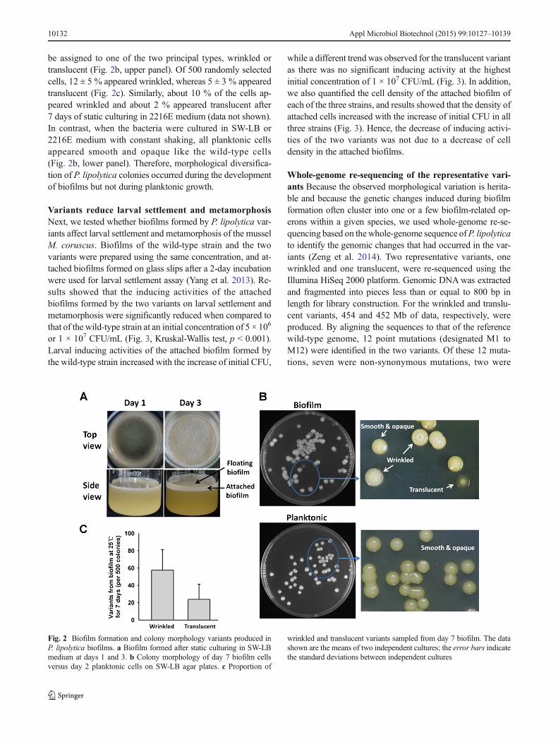
be assigned to one of the two principal types, wrinkled ortranslucent (Fig. 2b, upper panel). Of 500 randomly selectedcells, 12 ± 5 % appeared wrinkled, whereas 5 ± 3 % appearedtranslucent (Fig. 2c). Similarly, about 10 % of the cells ap-peared wrinkled and about 2 % appeared translucent after7 days of static culturing in 2216E medium (data not shown).In contrast, when the bacteria were cultured in SW-LB or2216E medium with constant shaking, all planktonic cellsappeared smooth and opaque like the wild-type cells(Fig. 2b, lower panel). Therefore, morphological diversifica-tion of P. lipolytica colonies occurred during the developmentof biofilms but not during planktonic growth.
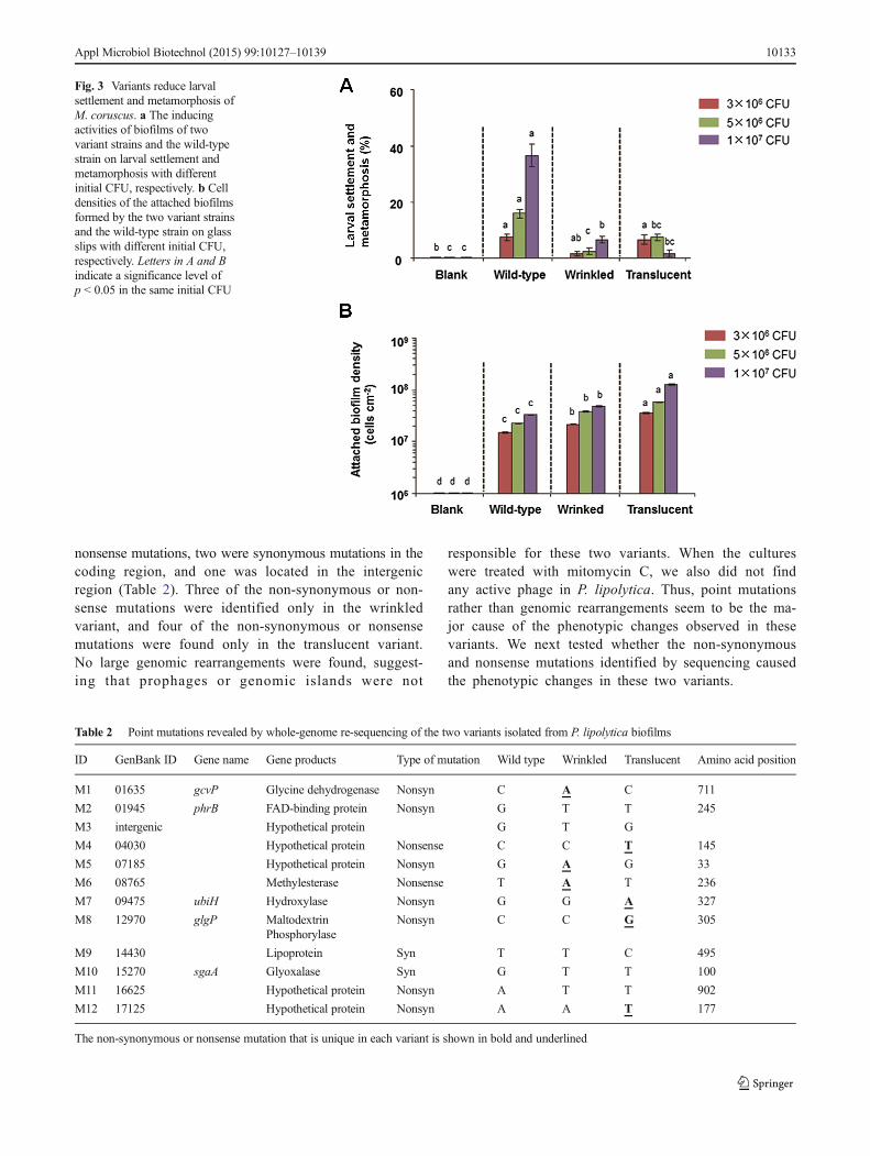
Variants reduce larval settlement and metamorphosisNext, we tested whether biofilms formed by P. lipolytica var-iants affect larval settlement and metamorphosis of the musselM. coruscus. Biofilms of the wild-type strain and the twovariants were prepared using the same concentration, and at-tached biofilms formed on glass slips after a 2-day incubationwere used for larval settlement assay (Yang et al. 2013). Re-sults showed that the inducing activities of the attachedbiofilms formed by the two variants on larval settlement andmetamorphosis were significantly reduced when compared tothat of the wild-type strain at an initial concentration of 5 × 10⁶
or 1 × 10⁷ CFU/mL (Fig. 3, Kruskal-Wallis test, p < 0.001).Larval inducing activities of the attached biofilm formed bythe wild-type strain increased with the increase of initial CFU,
while a different trend was observed for the translucent variantas there was no significant inducing activity at the highestinitial concentration of 1 × 10⁷ CFU/mL (Fig. 3). In addition,we also quantified the cell density of the attached biofilm ofeach of the three strains, and results showed that the density ofattached cells increased with the increase of initial CFU in allthree strains (Fig. 3). Hence, the decrease of inducing activi-ties of the two variants was not due to a decrease of celldensity in the attached biofilms.
Whole-genome re-sequencing of the representative vari-ants Because the observed morphological variation is herita-ble and because the genetic changes induced during biofilmformation often cluster into one or a few biofilm-related op-erons within a given species, we used whole-genome re-se-quencing based on thewhole-genome sequence ofP. lipolyticato identify the genomic changes that had occurred in the var-iants (Zeng et al. 2014). Two representative variants, onewrinkled and one translucent, were re-sequenced using theIllumina HiSeq 2000 platform. Genomic DNAwas extractedand fragmented into pieces less than or equal to 800 bp inlength for library construction. For the wrinkled and translu-cent variants, 454 and 452 Mb of data, respectively, wereproduced. By aligning the sequences to that of the referencewild-type genome, 12 point mutations (designated M1 toM12) were identified in the two variants. Of these 12 muta-tions, seven were non-synonymous mutations, two were
Fig. 2 Biofilm formation and colony morphology variants produced inP. lipolytica biofilms. a Biofilm formed after static culturing in SW-LBmedium at days 1 and 3. b Colony morphology of day 7 biofilm cellsversus day 2 planktonic cells on SW-LB agar plates. c Proportion of
wrinkled and translucent variants sampled from day 7 biofilm. The datashown are the means of two independent cultures; the error bars indicatethe standard deviations between independent cultures
10132 Appl Microbiol Biotechnol (2015) 99:10127–10139
nonsense mutations, two were synonymous mutations in thecoding region, and one was located in the intergenicregion (Table 2). Three of the non-synonymous or non-sense mutations were identified only in the wrinkledvariant, and four of the non-synonymous or nonsensemutations were found only in the translucent variant.No large genomic rearrangements were found, suggest-ing that prophages or genomic islands were not
responsible for these two variants. When the cultureswere treated with mitomycin C, we also did not findany active phage in P. lipolytica. Thus, point mutationsrather than genomic rearrangements seem to be the ma-jor cause of the phenotypic changes observed in thesevariants. We next tested whether the non-synonymousand nonsense mutations identified by sequencing causedthe phenotypic changes in these two variants.
Fig. 3 Variants reduce larvalsettlement and metamorphosis ofM. coruscus. a The inducingactivities of biofilms of twovariant strains and the wild-typestrain on larval settlement andmetamorphosis with differentinitial CFU, respectively. b Celldensities of the attached biofilmsformed by the two variant strainsand the wild-type strain on glassslips with different initial CFU,respectively. Letters in A and Bindicate a significance level ofp < 0.05 in the same initial CFU
Table 2 Point mutations revealed by whole-genome re-sequencing of the two variants isolated from P. lipolytica biofilms
ID GenBank ID Gene name Gene products Type of mutation Wild type Wrinkled Translucent Amino acid position
M1 01635 gcvP Glycine dehydrogenase Nonsyn C A C 711
M2 01945 phrB FAD-binding protein Nonsyn G T T 245
M3 intergenic Hypothetical protein G T G
M4 04030 Hypothetical protein Nonsense C C T 145
M5 07185 Hypothetical protein Nonsyn G A G 33
M6 08765 Methylesterase Nonsense T A T 236
M7 09475 ubiH Hydroxylase Nonsyn G G A 327
M8 12970 glgP Maltodextrin Nonsyn C C G 305Phosphorylase
M9 14430 Lipoprotein Syn T T C 495
M10 15270 sgaA Glyoxalase Syn G T T 100
M11 16625 Hypothetical protein Nonsyn A T T 902
M12 17125 Hypothetical protein Nonsyn A A T 177
The non-synonymous or nonsense mutation that is unique in each variant is shown in bold and underlined
Appl Microbiol Biotechnol (2015) 99:10127–10139 10133
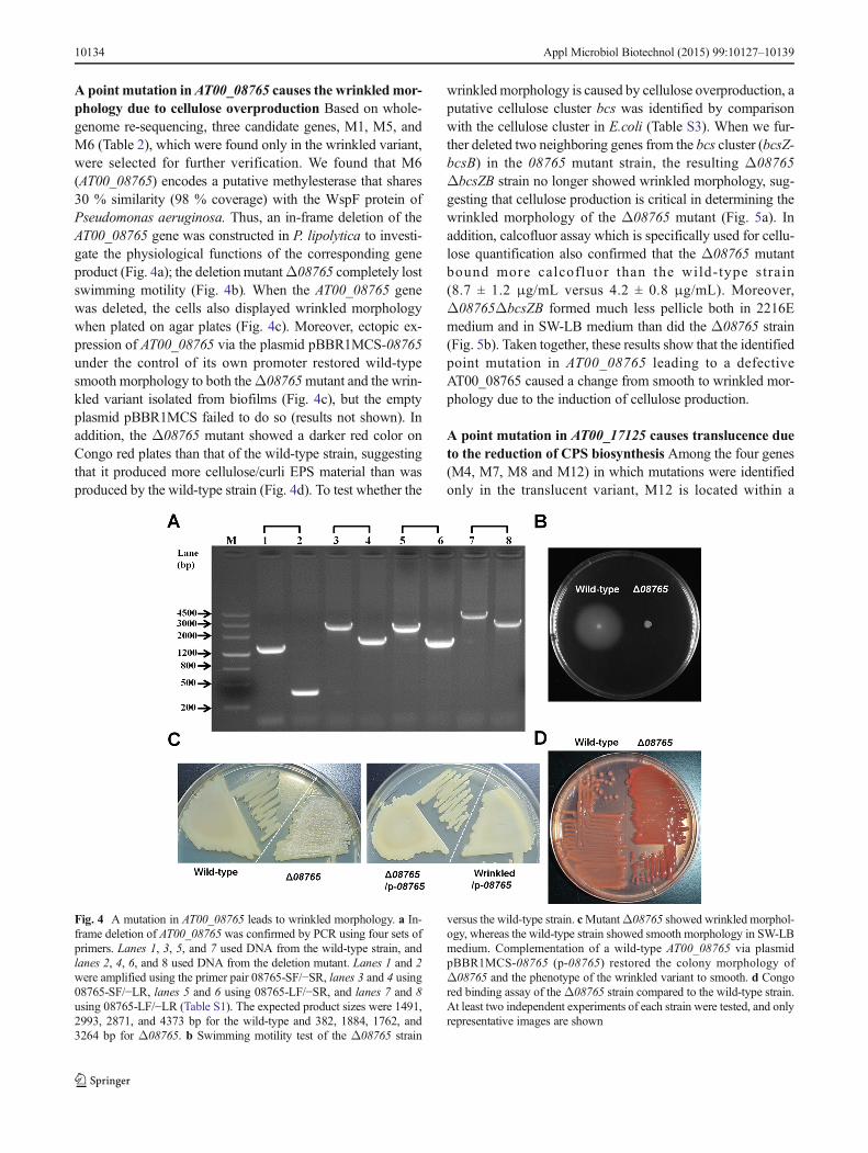
A point mutation inAT00_08765 causes the wrinkledmor-phology due to cellulose overproduction Based on whole-genome re-sequencing, three candidate genes, M1, M5, andM6 (Table 2), which were found only in the wrinkled variant,were selected for further verification. We found that M6(AT00_08765) encodes a putative methylesterase that shares30 % similarity (98 % coverage) with the WspF protein ofPseudomonas aeruginosa. Thus, an in-frame deletion of theAT00_08765 gene was constructed in P. lipolytica to investi-gate the physiological functions of the corresponding geneproduct (Fig. 4a); the deletion mutantΔ08765 completely lostswimming motility (Fig. 4b). When the AT00_08765 genewas deleted, the cells also displayed wrinkled morphologywhen plated on agar plates (Fig. 4c). Moreover, ectopic ex-pression of AT00_08765 via the plasmid pBBR1MCS-08765under the control of its own promoter restored wild-typesmooth morphology to both theΔ08765mutant and the wrin-kled variant isolated from biofilms (Fig. 4c), but the emptyplasmid pBBR1MCS failed to do so (results not shown). Inaddition, the Δ08765 mutant showed a darker red color onCongo red plates than that of the wild-type strain, suggestingthat it produced more cellulose/curli EPS material than wasproduced by the wild-type strain (Fig. 4d). To test whether the
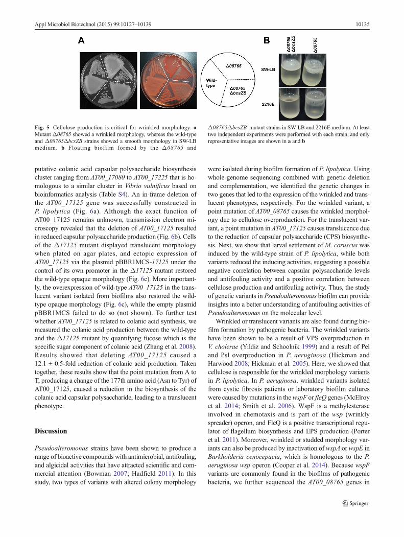
wrinkledmorphology is caused by cellulose overproduction, aputative cellulose cluster bcs was identified by comparisonwith the cellulose cluster in E.coli (Table S3). When we fur-ther deleted two neighboring genes from the bcs cluster (bcsZ-bcsB) in the 08765 mutant strain, the resulting Δ08765ΔbcsZB strain no longer showed wrinkled morphology, sug-gesting that cellulose production is critical in determining thewrinkled morphology of the Δ08765 mutant (Fig. 5a). Inaddition, calcofluor assay which is specifically used for cellu-lose quantification also confirmed that the Δ08765 mutantbound more calcofluor than the wild-type strain(8.7 ± 1.2 μg/mL versus 4.2 ± 0.8 μg/mL). Moreover,Δ08765ΔbcsZB formed much less pellicle both in 2216Emedium and in SW-LB medium than did the Δ08765 strain(Fig. 5b). Taken together, these results show that the identifiedpoint mutation in AT00_08765 leading to a defectiveAT00_08765 caused a change from smooth to wrinkled mor-phology due to the induction of cellulose production.
A point mutation in AT00_17125 causes translucence dueto the reduction of CPS biosynthesis Among the four genes(M4, M7, M8 and M12) in which mutations were identifiedonly in the translucent variant, M12 is located within a
Fig. 4 A mutation in AT00_08765 leads to wrinkled morphology. a In-frame deletion of AT00_08765 was confirmed by PCR using four sets ofprimers. Lanes 1, 3, 5, and 7 used DNA from the wild-type strain, andlanes 2, 4, 6, and 8 used DNA from the deletion mutant. Lanes 1 and 2were amplified using the primer pair 08765-SF/−SR, lanes 3 and 4 using08765-SF/−LR, lanes 5 and 6 using 08765-LF/−SR, and lanes 7 and 8using 08765-LF/−LR (Table S1). The expected product sizes were 1491,2993, 2871, and 4373 bp for the wild-type and 382, 1884, 1762, and3264 bp for Δ08765. b Swimming motility test of the Δ08765 strain
versus the wild-type strain. cMutantΔ08765 showed wrinkled morphol-ogy, whereas the wild-type strain showed smooth morphology in SW-LBmedium. Complementation of a wild-type AT00_08765 via plasmidpBBR1MCS-08765 (p-08765) restored the colony morphology ofΔ08765 and the phenotype of the wrinkled variant to smooth. d Congored binding assay of theΔ08765 strain compared to the wild-type strain.At least two independent experiments of each strain were tested, and onlyrepresentative images are shown
10134 Appl Microbiol Biotechnol (2015) 99:10127–10139
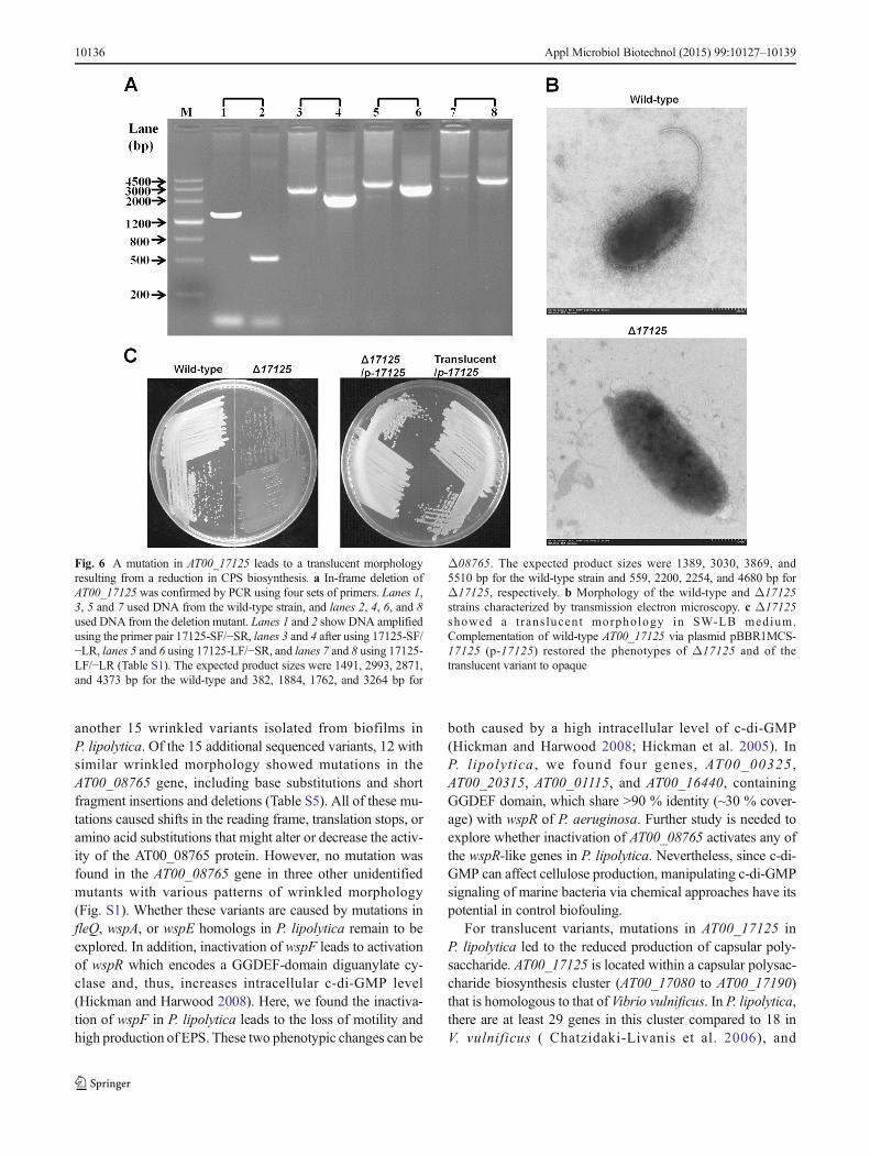
putative colanic acid capsular polysaccharide biosynthesiscluster ranging from AT00_17080 to AT00_17225 that is ho-mologous to a similar cluster in Vibrio vulnificus based onbioinformatics analysis (Table S4). An in-frame deletion ofthe AT00_17125 gene was successfully constructed inP. lipolytica (Fig. 6a). Although the exact function ofAT00_17125 remains unknown, transmission electron mi-croscopy revealed that the deletion of AT00_17125 resultedin reduced capsular polysaccharide production (Fig. 6b). Cellsof the Δ17125 mutant displayed translucent morphologywhen plated on agar plates, and ectopic expression ofAT00_17125 via the plasmid pBBR1MCS-17125 under thecontrol of its own promoter in the Δ17125 mutant restoredthe wild-type opaque morphology (Fig. 6c). More important-ly, the overexpression of wild-type AT00_17125 in the trans-lucent variant isolated from biofilms also restored the wild-type opaque morphology (Fig. 6c), while the empty plasmidpBBR1MCS failed to do so (not shown). To further testwhether AT00_17125 is related to colanic acid synthesis, wemeasured the colanic acid production between the wild-typeand the Δ17125 mutant by quantifying fucose which is thespecific sugar component of colanic acid (Zhang et al. 2008).Results showed that deleting AT00_17125 caused a12.1 ± 0.5-fold reduction of colanic acid production. Takentogether, these results show that the point mutation from A toT, producing a change of the 177th amino acid (Asn to Tyr) ofAT00_17125, caused a reduction in the biosynthesis of thecolanic acid capsular polysaccharide, leading to a translucentphenotype.
Discussion
Pseudoalteromonas strains have been shown to produce arange of bioactive compounds with antimicrobial, antifouling,and algicidal activities that have attracted scientific and com-mercial attention (Bowman 2007; Hadfield 2011). In thisstudy, two types of variants with altered colony morphology
were isolated during biofilm formation of P. lipolytica. Usingwhole-genome sequencing combined with genetic deletionand complementation, we identified the genetic changes intwo genes that led to the expression of the wrinkled and trans-lucent phenotypes, respectively. For the wrinkled variant, apoint mutation of AT00_08765 causes the wrinkled morphol-ogy due to cellulose overproduction. For the translucent var-iant, a point mutation in AT00_17125 causes translucence dueto the reduction of capsular polysaccharide (CPS) biosynthe-sis. Next, we show that larval settlement of M. coruscus wasinduced by the wild-type strain of P. lipolytica, while bothvariants reduced the inducing activities, suggesting a possiblenegative correlation between capsular polysaccharide levelsand antifouling activity and a positive correlation betweencellulose production and antifouling activity. Thus, the studyof genetic variants in Pseudoalteromonas biofilm can provideinsights into a better understanding of antifouling activities ofPseudoalteromonas on the molecular level.
Wrinkled or translucent variants are also found during bio-film formation by pathogenic bacteria. The wrinkled variantshave been shown to be a result of VPS overproduction inV. cholerae (Yildiz and Schoolnik 1999) and a result of Peland Psl overproduction in P. aeruginosa (Hickman andHarwood 2008; Hickman et al. 2005). Here, we showed thatcellulose is responsible for the wrinkled morphology variantsin P. lipolytica. In P. aeruginosa, wrinkled variants isolatedfrom cystic fibrosis patients or laboratory biofilm cultureswere caused bymutations in the wspF or fleQ genes (McElroyet al. 2014; Smith et al. 2006). WspF is a methylesteraseinvolved in chemotaxis and is part of the wsp (wrinklyspreader) operon, and FleQ is a positive transcriptional regu-lator of flagellum biosynthesis and EPS production (Porteret al. 2011). Moreover, wrinkled or studded morphology var-iants can also be produced by inactivation of wspA or wspE inBurkholderia cenocepacia, which is homologous to the P.aeruginosa wsp operon (Cooper et al. 2014). Because wspFvariants are commonly found in the biofilms of pathogenicbacteria, we further sequenced the AT00_08765 genes in
Fig. 5 Cellulose production is critical for wrinkled morphology. aMutant Δ08765 showed a wrinkled morphology, whereas the wild-typeand Δ08765ΔbcsZB strains showed a smooth morphology in SW-LBmedium. b Floating biofilm formed by the Δ08765 and
Δ08765ΔbcsZB mutant strains in SW-LB and 2216E medium. At leasttwo independent experiments were performed with each strain, and onlyrepresentative images are shown in a and b
Appl Microbiol Biotechnol (2015) 99:10127–10139 10135
another 15 wrinkled variants isolated from biofilms inP. lipolytica. Of the 15 additional sequenced variants, 12 withsimilar wrinkled morphology showed mutations in theAT00_08765 gene, including base substitutions and shortfragment insertions and deletions (Table S5). All of these mu-tations caused shifts in the reading frame, translation stops, oramino acid substitutions that might alter or decrease the activ-ity of the AT00_08765 protein. However, no mutation wasfound in the AT00_08765 gene in three other unidentifiedmutants with various patterns of wrinkled morphology(Fig. S1). Whether these variants are caused by mutations infleQ, wspA, or wspE homologs in P. lipolytica remain to beexplored. In addition, inactivation of wspF leads to activationof wspR which encodes a GGDEF-domain diguanylate cy-clase and, thus, increases intracellular c-di-GMP level(Hickman and Harwood 2008). Here, we found the inactiva-tion of wspF in P. lipolytica leads to the loss of motility andhigh production of EPS. These two phenotypic changes can be
both caused by a high intracellular level of c-di-GMP(Hickman and Harwood 2008; Hickman et al. 2005). InP. lipolytica , we found four genes, AT00_00325 ,AT00_20315, AT00_01115, and AT00_16440, containingGGDEF domain, which share >90 % identity (~30 % cover-age) with wspR of P. aeruginosa. Further study is needed toexplore whether inactivation of AT00_08765 activates any ofthe wspR-like genes in P. lipolytica. Nevertheless, since c-di-GMP can affect cellulose production, manipulating c-di-GMPsignaling of marine bacteria via chemical approaches have itspotential in control biofouling.
For translucent variants, mutations in AT00_17125 inP. lipolytica led to the reduced production of capsular poly-saccharide. AT00_17125 is located within a capsular polysac-charide biosynthesis cluster (AT00_17080 to AT00_17190)that is homologous to that of Vibrio vulnificus. In P. lipolytica,there are at least 29 genes in this cluster compared to 18 inV. vulnificus ( Chatzidaki-Livanis et al. 2006), and
Fig. 6 A mutation in AT00_17125 leads to a translucent morphologyresulting from a reduction in CPS biosynthesis. a In-frame deletion ofAT00_17125 was confirmed by PCR using four sets of primers. Lanes 1,3, 5 and 7 used DNA from the wild-type strain, and lanes 2, 4, 6, and 8used DNA from the deletion mutant. Lanes 1 and 2 showDNA amplifiedusing the primer pair 17125-SF/−SR, lanes 3 and 4 after using 17125-SF/−LR, lanes 5 and 6 using 17125-LF/−SR, and lanes 7 and 8 using 17125-LF/−LR (Table S1). The expected product sizes were 1491, 2993, 2871,and 4373 bp for the wild-type and 382, 1884, 1762, and 3264 bp for
Δ08765. The expected product sizes were 1389, 3030, 3869, and5510 bp for the wild-type strain and 559, 2200, 2254, and 4680 bp forΔ17125, respectively. b Morphology of the wild-type and Δ17125strains characterized by transmission electron microscopy. c Δ17125showed a t rans lucent morphology in SW-LB medium.Complementation of wild-type AT00_17125 via plasmid pBBR1MCS-17125 (p-17125) restored the phenotypes of Δ17125 and of thetranslucent variant to opaque
10136 Appl Microbiol Biotechnol (2015) 99:10127–10139
AT00_17125 is only present in P. lipolytica. The exact func-tion of AT00_17125 is currently unclear. Our TEM resultsshowed that translucent colonies contain markedly less capsu-lar polysaccharide, suggesting that AT00_17125 is either apositive regulator of capsular polysaccharide biosynthesis ora structural gene. To determine whether mutation of theAT00_17125 gene is commonly found in biofilm cells, wesequenced another ten translucent variants isolated frombiofilms; however, we did not find any mutations inAT00_17125 in those variants. In V. vulnificus, inactivationof wza, wzb, or wzc in the CPS cluster leads to a change inmorphology from opaque to translucent, enhances biofilmformation, and increases pathogenesis ( Chatzidaki-Livaniset al. 2006; Lee et al. 2013; Nakhamchik et al. 2008; Wrightet al. 2001). We also find homologs of these three genes in thegenome of P. lipolytica (AT00_17180, AT00_17175, andAT00_17170) (Table S4). Indeed, two additional single dele-tion mutants of AT00_17170 and AT00_17220, located withinthe colanic acid capsular polysaccharide, also showed a mor-phology change from opaque to translucent (data not shown).Thus, we reason that mutations in the rest of the genes withinthe CPS cluster could also cause translucent morphology.
Taken together, these results suggest that screening of var-iants using biofilm mode of growth in marine bacteria can beused for the selection of genetic mutants with favorable traits.Recent studies using whole-genome deep sequencing to iden-tify genetic variants in biofilms of P. aeruginosa (McElroyet al. 2014) and B. cenocepacia (Traverse et al. 2013) alsoshow that non-synonymous and positively selected mutationsgovern the within-population bacterial diversification. Asmany marine bacteria harbor multidrug resistance genes andabundant restriction-modification systems which make genet-ic manipulation rather difficult (Wang et al. 2015), this selec-tion approach eliminates the need for targeted genetic manip-ulation or random mutagenesis.
Last but not least, marine bacteria like Pseudoalteromonasliving in water columns, in sediments, or in association withanimal hosts inevitably face with changed environmental fac-tors such as oxygen, nutrients, chemicals, waste products, andsignaling molecules (Stocker 2012). Thus, the ocean can playa driving force for the generation of a large repertoire of ge-netic variants at the population level. Moreover, variants iso-lated from laboratory-cultured biofilms formed by pathogenicbacteria share similar mutations to those found in clinical iso-lates during infection (D’Argenio et al. 2007; Drenkard andAusubel 2002; Woo et al. 2012), suggesting that similarwithin-population diversification may also occur for marinebacteria living in their marine habitats. To improve under-s t and ing of the mic rob ia l behav io r o f va r iousPseudoalteromonas species, including those that survive inextreme marine environments (deep-sea or hydrothermalvents), further studies of the within-population variation ofother Pseudoalteromonas are warranted.
Conflict of interests The authors declare that they have no competinginterests.
Acknowledgments This work was supported by the National BasicResearch Program of China (2013CB955701), the National ScienceFoundation of China (31290233 and 41230962), and the Chinese Acad-emy of Sciences (XDA11030402). XW is the 1000-Youth Elite Programrecipient in China.
Open Access This article is distributed under the terms of the CreativeCommons At t r ibut ion 4 .0 In te rna t ional License (h t tp : / /creativecommons.org/licenses/by/4.0/), which permits unrestricted use,distribution, and reproduction in any medium, provided you giveappropriate credit to the original author(s) and the source, provide a linkto the Creative Commons license, and indicate if changes were made.
References
Armitano J, Mejean V, Jourlin-Castelli C (2014) Gram-negative bacteriacan also form pellicles. Environ Microbiol Rep 6(6):534–544
Barlett MA, Leff LG (2010) The effects of N:P ratio and nitrogen form onfour major freshwater bacterial taxa in biofilms. Can J Microbiol56(1):32–43
Bernbom N, Ng YY, Kjelleberg S, Harder T, Gram L (2011) Marinebacteria from danish coastal waters show antifouling activity againstthe marine fouling bacterium Pseudoalteromonas sp strain S91 andzoospores of the green alga Ulva australis independent of bacterio-cidal activity. Appl Environ Microbiol 77(24):8557–8567
Bowman JP (2007) Bioactive compound synthetic capacity and ecolog-ical significance of marine bacterial genus Pseudoalteromonas. MarDrugs 5(4):220–241
Chatzidaki-Livanis M, Jones MK,Wright AC (2006) Genetic variation inthe Vibrio vulnificus group 1 capsular polysaccharide operon. JBacteriol 188(5):1987–1998
Cooksey KE, Wigglesworthcooksey B (1995) Adhesion of bacteria anddiatoms to surfaces in the sea—a review. Aquat Microb Ecol 9(1):87–96
Cooper VS, Staples RK, Traverse CC, Ellis CN (2014) Parallel evolutionof small colony variants in Burkholderia cenocepacia biofilms.Genomics 104:447–452
D’Argenio DA, Wu M, Hoffman LR, Kulasekara HD, Deziel E, SmithEE, Nguyen H, Ernst RK, Larson Freeman TJ, Spencer DH,Brittnacher M, Hayden HS, Selgrade S, Klausen M, Goodlett DR,Burns JL, Ramsey BW, Miller SI (2007) Growth phenotypes ofPseudomonas aeruginosa lasR mutants adapted to the airways ofcystic fibrosis patients. Mol Microbiol 64(2):512–533
Dehio C, Meyer M (1997) Maintenance of broad-host-range incompati-bility group P and group Q plasmids and transposition of Tn5 inBartonella henselae following conjugal plasmid transfer fromEscherichia coli. J Bacteriol 179(2):538–540
Devries CA, Ohman DE (1994) Mucoid-to-nonmucoid conversion inalginate-producing Pseudomonas aeruginosa often results fromspontaneous mutations in algT, encoding a putative alternate sig-ma-factor, and shows evidence for autoregulation. J Bacteriol176(21):6677–6687
Dobretsov S, Dahms HU, Qian PY (2006) Inhibition of biofouling bymarine microorganisms and their metabolites. Biofouling22(1):43–54
Drenkard E, Ausubel FM (2002) Pseudomonas biofilm formation andantibiotic resistance are linked to phenotypic variation. Nature416(6882):740–743
Appl Microbiol Biotechnol (2015) 99:10127–10139 10137
Hadfield MG (2011) Biofilms and marine invertebrate larvae: what bac-teria produce that larvae use to choose settlement sites. Annu RevMar Sci 3:453–470
Hall-Stoodley L, Costerton JW, Stoodley P (2004) Bacterial biofilms:from the natural environment to infectious diseases. Nat RevMicrobiol 2(2):95–108
Hickman JW, Harwood CS (2008) Identification of FleQ fromPseudomonas aeruginosa as a c-di-GMP-responsive transcriptionfactor. Mol Microbiol 69(2):376–389
Hickman JW, Tifrea DF, Harwood CS (2005) A chemosensory systemthat regulates biofilm formation through modulation of cyclicdiguanylate levels. Proc Natl Acad Sci U S A 102(40):14422–14427
Holmstrom C, Kjelleberg S (1999) Marine Pseudoalteromonas speciesare associated with higher organisms and produce biologically ac-tive extracellular agents. FEMS Microbiol Ecol 30(4):285–293
Holmstrom C, James S, Egan S, Kjelleberg S (1996) Inhibition of com-mon fouling organisms by marine bacterial isolates with specialreference to the role of pigmented bacteria. Biofouling 10(1–3):251–259
Holmstrom C, Egan S, Franks A, McCloy S, Kjelleberg S (2002)Antifouling activities expressed by marine surface associatedPseudoalteromonas species. FEMS Microbiol Ecol 41(1):47–58
Huang YX (1988) Temperature and salinity distributions in the SouthChina sea and adjacent waters. Prog Oceanogr 21(3–4):493–501
Huang YL, Dobretsov S, Xiong HR, Qian PY (2007) Effect of biofilmformation by Pseudoalteromonas spongiae on induction of larvalsettlement of the polychaete Hydroides elegans. Appl EnvironMicrobiol 73(19):6284–6288
Jiao N, Robinson C, Azam F, Thomas H, Baltar F, Dang H, Hardman-Mountford NJ, JohnsonM, Kirchman DL, Koch BP, Legendre L, LiC, Liu J, Luo T, Luo YW, Mitra A, Romanou A, Tang K, Wang X,Zhang C, Zhang R (2014) Mechanisms of microbial carbon seques-tration in the ocean-future research directions. Biogeosciences11(19):5285–5306
Lee KJ, Kim JA, Hwang W, Park SJ, Lee KH (2013) Role of capsularpolysaccharide (CPS) in biofilm formation and regulation of CPSproduction by quorum-sensing in Vibrio vulnificus. Mol Microbiol90(4):841–857
Lee OO, ChungHC, Yang JK,WangY, Dash S,WangH,Qian PY (2014)Molecular techniques revealed highly diverse microbial communi-ties in natural marine biofilms on polystyrene dishes for invertebratelarval settlement. Microb Ecol 68(1):81–93
McElroy KE, Hui JG, Woo JK, Luk AW, Webb JS, Kjelleberg S, RiceSA, Thomas T (2014) Strain-specific parallel evolution drives short-term diversification during Pseudomonas aeruginosa biofilm for-mation. Proc Natl Acad Sci U S A 111(14):E1419–E1427
Medigue C, Krin E, Pascal G, Barbe V, Bernsel A, Bertin PN, Cheung F,Cruveiller S, D’Amico S, Duilio A, Fang G, Feller G, Ho C,Mangenot S, Marino G, Nilsson J, Parrilli E, Rocha EPC, Rouy Z,Sekowska A, Tutino ML, Vallenet D, von Heijne G, Danchin A(2005) Coping with cold: the genome of the versatile marineAntarctica bacterium Pseudoalteromonas haloplanktis TAC125.Genome Res 15(10):1325–1335
Mounier J, Camus A, Mitteau I, Vaysse PJ, Goulas P, Grimaud R,Sivadon P (2014) The marine bacterium Marinobacterhydrocarbonoclasticus SP17 degrades a wide range of lipids andhydrocarbons through the formation of oleolytic biofilms with dis-tinct gene expression profiles. FEMSMicrobiol Ecol 90(3):816–831
Moyano AJ, Lujan AM, Argarana CE, Smania AM (2007) MutS defi-ciency and activity of the error-prone DNA polymerase IV are cru-cial for determining mucA as the main target for mucoid conversionin Pseudomonas aeruginosa. Mol Microbiol 64(2):547–559
Nakhamchik A, Wilde C, Rowe-Magnus DA (2008) Cyclic-di-GMP reg-ulates extracellular polysaccharide production, biofilm formation,and rugose colony development by Vibrio vulnificus. ApplEnviron Microbiol 74(13):4199–4209
Nielsen L, Li X, Halverson LJ (2011) Cell-cell and cell-surfacei n t e r a c t i on s med i a t e d by c e l l u l o s e and a nove lexopolysaccharide contribute to Pseudomonas putida biofilmformation and fitness under water-limiting conditions.Environ Microbiol 13(5):1342–1356
Patel P, Callow ME, Joint I, Callow JA (2003) Specificity in the settle-ment - modifying response of bacterial biofilms towards zoosporesof the marine alga Enteromorpha. Environ Microbiol 5(5):338–349
Poli A, Anzelmo G, Nicolaus B (2010) Bacterial exopolysaccharidesfrom extreme marine habitats: production, characterization and bio-logical activities. Mar Drugs 8(6):1779–1802
Porter SL, Wadhams GH, Armitage JP (2011) Signal processing in com-plex chemotaxis pathways. Nat Rev Microbiol 9(3):153–165
Qian PY, Lau SC, Dahms HU, Dobretsov S, Harder T (2007) Marinebiofilms as mediators of colonization by marine macroorganisms:implications for antifouling and aquaculture. Mar Biotechnol (NY)9(4):399–410
Qin QL, Li Y, Zhang YJ, Zhou ZM, Zhang WX, Chen XL, Zhang XY,Zhou BC, Wang L, Zhang YZ (2011) Comparative genomics re-veals a deep-sea sediment-adapted life style of Pseudoalteromonassp. SM9913. ISME J 5(2):274–284
Rainey PB, Travisano M (1998) Adaptive radiation in a heterogeneousenvironment. Nature 394(6688):69–72
Rice SA, Tan CH, Mikkelsen PJ, Kung V, Woo J, Tay M, Hauser A,McDougald D, Webb JS, Kjelleberg S (2009) The biofilm life cycleand virulence of Pseudomonas aeruginosa are dependent on a fila-mentous prophage. ISME J 3(3):271–282
Santegoeds CM, Ferdelman TG, Muyzer G, de Beer D (1998) Structuraland functional dynamics of sulfate-reducing populations in bacterialbiofilms. Appl Environ Microbiol 64(10):3731–3739
Shikuma NJ, Pilhofer M, Weiss GL, Hadfield MG, Jensen GJ, NewmanDK (2014) Marine tubeworm metamorphosis induced by arrays ofbacterial phage tail-like structures. Science 343(6170):529–533
Smith EE, Buckley DG, Wu ZN, Saenphimmachak C, Hoffman LR,D’Argenio DA, Miller SI, Ramsey BW, Speert DP, MoskowitzSM, Burns JL, Kaul R, Olson MV (2006) Genetic adaptation byPseudomonas aeruginosa to the airways of cystic fibrosis patients.Proc Natl Acad Sci U S A 103(22):8487–8492
Stewart PS, Franklin MJ (2008) Physiological heterogeneity in biofilms.Nat Rev Microbiol 6(3):199–210
Stocker R (2012) Marine microbes see a sea of gradients. Science338(6107):628–633
Thomas T, Evans FF, Schleheck D,Mai-ProchnowA, Burke C, PenesyanA, Dalisay DS, Stelzer-Braid S, Saunders N, Johnson J, Ferriera S,Kjelleberg S, Egan S (2008) Analysis of the Pseudoalteromonastunicata genome reveals properties of a surface-associated life stylein the marine environment. PLoS One 3(9):e3252
Traverse CC, Mayo-Smith LM, Poltak SR, Cooper VS (2013) Tangledbank of experimentally evolved Burkholderia biofilms reflects se-lection during chronic infections. Proc Natl Acad Sci U S A 110(3):E250–E259
Wang C, Bao WY, Gu ZQ, Li YF, Liang X, Ling Y, Cai SL, Shen HD,Yang JL (2012) Larval settlement and metamorphosis of the musselMytilus coruscus in response to natural biofilms. Biofouling 28(3):249–256
Wang P, Yu Z, Li B, Cai X, Zeng Z, Chen X, Wang X (2015)Development of an efficient conjugation-based genetic manipula-tion system for Pseudoalteromonas. Microb Cell Factories 14(1):11
Webb JS, Lau M, Kjelleberg S (2004) Bacteriophage and phenotypicvariation in Pseudomonas aeruginosa biofilm development. JBacteriol 186(23):8066–8073
Woo JKK, Webb JS, Kirov SM, Kjelleberg S, Rice SA (2012)Biofilm dispersal cells of a cystic fibrosis Pseudomonasaeruginosa isolate exhibit variability in functional traits like-ly to contribute to persistent infection. FEMS Immunol MedMicrobiol 66(2):251–264
10138 Appl Microbiol Biotechnol (2015) 99:10127–10139
Wright AC, Powell JL, Kaper JB, Morris JG, Jr. (2001) Identification of agroup 1-like capsular polysaccharide operon for Vibrio vulnificus.Infect Immun 69(11):6893-6901
Yang JL, Satuito CG, Bao WY, Kitamura H (2008) Induction of meta-morphosis of pediveliger larvae of the mussel Mytilusgalloprovincialis Lamarck, 1819 using neuroactive compounds,KCl, NH4Cl and organic solvents. Biofouling 24(6):461–470
Yang JL, Shen PJ, Liang X, Li YF, Bao WY, Li JL (2013) Larvalsettlement and metamorphosis of the mussel Mytilus coruscusin response to monospecific bacterial biofilms. Biofouling29(3):247–259
Yildiz FH, Schoolnik GK (1999) Vibrio cholerae O1 El Tor: identifica-tion of a gene cluster required for the rugose colony type,
exopolysaccharide production, chlorine resistance, and biofilm for-mation. Proc Natl Acad Sci U S A 96(7):4028–4033
Yu M, Tang KH, Liu JW, Shi XC, Gulder TAM, Zhang XH (2013)Genome analysis of Pseudoalteromonas flavipulchra JG1 revealsvarious survival advantages in marine environment. BMCGenomics 14:707
Zeng Z, Dai S, Xie Y, Tian X, Li J, Wang X (2014) Genome sequences oftwo pseudoalteromonas strains isolated from the South China Sea.Genome A 2:e00305–e00314
Zhang XS, Garcia-Contreras R, Wood TK (2008) Escherichia coli tran-scription factor YncC (McbR) regulates colanic acid and biofilmformation by repressing expression of periplasmic protein YbiM(McbA). ISME J 2(6):615–631
Appl Microbiol Biotechnol (2015) 99:10127–10139 10139