Embed Size (px)
Citation preview
Characterization of new natural products from fungi
of the German Wadden Sea
Dissertation
zur Erlangung des Doktorgrades
der Mathematisch-Naturwissenschaftlichen Fakultät
der Christian-Albrechts-Universität
zu Kiel
vorgelegt von
Dipl.-Biol. Johanna Silber
Kiel, 2013

Referent: Prof. Dr. J. F. Imhoff
Korreferent: Prof. Dr. Peter Schönheit
Tag der mündlichen Prüfung: 21.02.2014
Zum Druck genehmigt: 21.02.2014
gez. Prof. Dr. Wolfgang J. Duschl, Dekan
PUBLICATIONS i
PUBLICATIONS
The results presented in this thesis have been published in the form of research papers
or will be published in the near future:
Silber, J., Ohlendorf, B., Labes, A., Näther, C., Imhoff, J. F. (2013). Calcaripeptides A–
C, cyclodepsipeptides from a Calcarisporium strain. J. Nat. Prod. 76(8), 1461-1467.
Silber, J., Ohlendorf, B., Labes, A., Erhard, A., Imhoff, J. F. (2013). Calcarides A–E,
antibacterial macrocyclic and linear polyesters from a Calcarisporium strain. Mar.
Drugs 11(9), 3309-3323.
Silber, J., Ohlendorf, B., Labes, A., Erhard, A., Näther, C., Imhoff, J. F. Malettinin E,
antimicrobial tropolone produced by a Cladosporium strain, to be submitted
TABLE OF CONTENTS ii
TABLE OF CONTENTS
ZUSAMMENFASSUNG ................................................................................................ 1
SUMMARY ..................................................................................................................... 3
ABBREVIATIONS ......................................................................................................... 5
INTRODUCTION .......................................................................................................... 8
Fungi – an Attunement .................................................................................................. 8
Fungal Natural Products ................................................................................................ 9
Biosynthesis of Fungal Natural Products .................................................................... 12
Need for New Natural Products .................................................................................. 16
Strategies to Discover New Fungal Natural Products ................................................. 18
Scope of the Thesis ..................................................................................................... 22
CHAPTER I .................................................................................................................. 24
Abstract ....................................................................................................................... 25
Introduction ................................................................................................................. 25
Results and Discussion ................................................................................................ 26
Experimental Section .................................................................................................. 34
Acknowledgments ....................................................................................................... 38
References ................................................................................................................... 39
CHAPTER II ................................................................................................................. 42
Abstract ....................................................................................................................... 43
Introduction ................................................................................................................. 43
Results and Discussion ................................................................................................ 45
Experimental Section .................................................................................................. 54

TABLE OF CONTENTS iii
Conclusions ................................................................................................................. 58
Acknowledgments ....................................................................................................... 58
References ................................................................................................................... 59
CHAPTER III ............................................................................................................... 61
Abstract ....................................................................................................................... 62
Introduction ................................................................................................................. 62
Material and Methods ................................................................................................. 63
Results ......................................................................................................................... 66
Discussion ................................................................................................................... 71
Acknowledgments ....................................................................................................... 72
References ................................................................................................................... 73
GENERAL DISCUSSION ........................................................................................... 76
REFERENCES .............................................................................................................. 83
ACKNOWLEDGMENTS/DANKSAGUNG .............................................................. 93
DECLARATION/ERKLÄRUNG ............................................................................... 94
APPENDIX .................................................................................................................... 95
Supporting Information for Chapter I ......................................................................... 95
Supporting Information for Chapter II ...................................................................... 105
Supporting Information for Chapter III ..................................................................... 115
ZUSAMMENFASSUNG 1
ZUSAMMENFASSUNG
Pilze sind hervorragende Produzenten von Naturstoffen, die für pharmazeutische und
agrarwirtschaftliche Anwendungen genutzt werden. Das Ziel der vorliegenden
Doktorarbeit war es, dieses außergewöhnliche Biosynthesepotential für die Suche nach
neuen Naturstoffen in Pilzisolaten aus dem deutschen Wattenmeer auszuschöpfen. Um
eine effiziente und zielgerichtete Screening-Strategie zu entwickeln, wurden vier
Kriterien zur Auswahl derjenigen Pilze herangezogen, die anschließend umfassend auf
ihre Fähigkeiten zur Biosynthese von Naturstoffen hin untersucht wurden: (i) Pilzisolate
von wenig erforschten Ökosystemen, (ii) weniger intensiv untersuchte Gattungen, (iii)
genetisches Potential anhand des Sequenznachweises für Polyketidsynthase- und
nichtribosomale Peptidsynthetase-Gene, (iv) stimulierende Effekte auf die Biosynthese
durch die Variation von Kulturbedingungen. Die Screening-Strategie zahlte sich aus,
denn es wurden neun neue Naturstoffe, die Calcaripeptide A–C, Calcaride A–E und ein
Tropolon-Derivat, entdeckt.
Die Calcaripeptide sind Cyclodepsipeptide, die von einem Calcarisporium sp. (Stamm
KF525) isoliert wurden. Die Strukturaufklärung mit Hilfe von ein- und
zweidimensionaler Kernspinresonanzspektroskopie wies einen ungewöhnlichen
Makrocyclus bestehend aus zwei Aminosäuren und eine nicht-peptidische Teilstruktur
für die Substanzen nach. Die absolute Konfiguration der Calcaripeptide wurde über
Röntgenstrukturanalysen in Kombination mit der erweiterten Marfey-Methode
bestimmt. Biosynthesestudien zu Calcaripeptid A zeigten, dass der nicht-peptidische
Strukturteil von Acetat und L-Methionin stammt, was einen biosynthetischen Ursprung
von einem Hybrid aus einer Polyketidsynthase und einer nichtribosomalen
Peptidsynthetase nahelegt. Trotz breit angelegter Tests zum Nachweis biologischer
Aktivitäten wiesen die Calcaripeptide keine inhibitorischen Eigenschaften auf.
Zusätzlich produzierte der Stamm KF525 eine zweite Klasse von Naturstoffen, welche
die neuen Calcaride A–E und fünf strukturverwandte Substanzen vom 15G256-Typ
umfasste. Bei den Substanzen handelt es sich um lineare und makrocyclische Polyester,
wobei die Calcaride methylierte Formen der Substanzen vom 15G256-Typ darstellen.
Die absolute Konfiguration der Metabolite wurde über Messungen der optischen
Rotation untersucht. Die Calcaride und Substanzen vom 15G256-Typ zeigten
antibakterielle Aktivitäten gegenüber Staphylococcus epidermidis, Xanthomonas
ZUSAMMENFASSUNG 2
campestris und Propionibacterium acnes, welche einer engen Struktur-Aktivitäts-
Beziehung unterlagen.
Von einem anderen Pilzstamm, Cladosporium sp. KF501, wurden Mitglieder der
Familie pilzlicher Tropolone isoliert. Unter den Substanzen befand sich ein neues
Derivat, das ein Stereoisomer der bekannten Tropolone des Pilzstamms darstellt. Die
relative Konfiguration des neuen Derivats wurde anhand einer Röntgenstrukturanalyse
an einem Einkristall bestimmt. Die isolierten Tropolone wiesen antibakterielle und
antifungale Eigenschaften in Testungen mit Xanthomonas campestris und Trichophyton
rubrum auf.
Insgesamt demonstrieren die Ergebnisse der Arbeit den hohen Wert, den Pilze aus
marinen Habitaten für die Entdeckung neuer bioaktiver Naturstoffe besitzen.
SUMMARY 3
SUMMARY
Fungi are prolific producers of natural products used for pharmaceutical and agricultural
applications. The scope of the present PhD study was to exploit this extraordinary
biosynthetic potential for the search for new natural products from fungal isolates of the
German Wadden Sea. In order to create a smart screening strategy, four criteria were
applied to select fungi for subsequent thorough investigation with regard to their natural
product biosynthetic abilities: (i) fungal isolates from a little-explored ecosystem, (ii)
less studied genera, (iii) genetic potential according to polyketide synthase (PKS) and
nonribosomal peptide synthetase (NRPS) gene sequences, (iv) stimulating effects on
biosynthesis by variation of culture conditions. The screening strategy was proven
effective as it led to the discovery of nine new natural products, namely, calcaripeptides
A–C, calcarides A–E and one tropolone derivative.
The calcaripeptides are cyclodepsipeptides that were isolated from a Calcarisporium sp.
(strain KF525). The structure elucidation employing one- and two-dimensional nuclear
magnetic resonance (NMR) spectroscopy techniques proved the compounds to possess
an unusual macrocycle composed of two amino acids and a nonpeptidic substructure.
The absolute configuration of the calcaripeptides was determined by X-ray
crystallography in combination with the advanced Marfey’s method. Biosynthetic
studies on calcaripeptide A showed that the nonpeptidic structural part of the compound
originated from acetate and L-methionine, suggesting a biosynthetic origin from a PKS–
NRPS hybrid. Despite broad testing for biological activities, the calcaripeptides did not
exhibit any inhibitory properties.
In addition, strain KF525 produced a second structural class of natural products
including the new calcarides A–E and five related compounds of the 15G256-type. The
compounds are linear and macrocyclic polyesters with the calcarides representing
methylated forms of the 15G256-type compounds. The absolute configuration of the
metabolites was investigated by measurements of the optical rotation. The calcarides
and 15G256-type compounds were found to possess antibacterial activities against
Staphylococcus epidermidis, Xanthomonas campestris and Propionibacterium acnes
underlying tight structure-activity relationships.
From another fungal strain, Cladosporium sp. KF501, members of the family of
tropolone fungal metabolites were isolated. Among the compounds was a new

SUMMARY 4
derivative that represents a stereoisomer of the known tropolones of the strain. Single-
crystal X-ray diffraction analysis was employed in order to determine the relative
configuration of the new derivative. The isolated tropolones showed antibacterial and
antifungal properties when profiled against Xanthomonas campestris and Trichophyton
rubrum.
Overall, the results of the study demonstrate the high value of fungi from marine
habitats for the discovery of new bioactive natural products.
ABBREVIATIONS 5
ABBREVIATIONS
(w/v) weight per volume
[α]20
D specific rotatory power; sodium D-line (589 nm); 20 °C
[α]22
D specific rotatory power; sodium D-line (589 nm); 22 °C
°C degrees Celsius
µ absorption coefficient (in connection with X-ray crystal data)
1D one-dimensional
2D two-dimensional
A adenylation
Å ångström
ACP acyl carrier protein
AT acyl transferase
ATP adenosine triphosphate
BLAST Basic Local Alignment Search Tool
br broad
c concentration
C condensation
calcd calculated
CoA coenzyme A
COSY correlation spectroscopy
CYC cyclase
d doublet
Dcalcd calculated density
DEPT distortionless enhancement by polarization transfer
DH dehydratase
DMSO dimethyl sulfoxide
DNA deoxyribonucleic acid
DSMZ German collection of microorganisms and cell cultures
Em emission
ER enoyl reductase
ESI electrospray ionization
ESIMS electrospray ionization mass spectrometry
EtOAc ethyl acetate
EtOH ethanol
ABBREVIATIONS 6
Ex excitation
FDVA Nα-(2,4-dinitro-5-fluorophenyl)-D-valinamide
g acceleration of gravity
g gram
h hour
HMBC heteronuclear multiple bond correlation
HPLC high-performance liquid chromatography
HRESIMS high-resolution electrospray ionization mass spectrometry
HSQC heteronuclear single-quantum coherence
Hz Hertz
IC50 half maximal inhibitory concentration
ITS internal transcribed spacer region
J spin-spin coupling constant
K Kelvin
KR ketoreductase
KS ketosynthase
L liter
m meter
M molar concentration
m/z mass-to-charge ratio
MeCN acetonitrile
MeOH methanol
MHz megahertz
MIC minimal inhibitory concentration
MICs minimal inhibitory concentrations
min minute
mp melting point
MRSA methicillin-resistant Staphylococcus aureus
MS mass spectrometry
MT methyl transferase
Mult. multiplicity
MW molecular weight
NMR nuclear magnetic resonance
NRPS nonribosomal peptide synthetase
ABBREVIATIONS 7
NRPSs nonribosomal peptide synthetases
OD600 optical density at 600 nm
PCP peptidyl carrier protein
PCR polymerase chain reaction
Phe phenylalanine
PKS polyketide synthase
PKSs polyketide synthases
ppm parts per million
Pro proline
PYG peptone yeast glucose medium
q quartet
Refl. reflections
rpm revolutions per minute
s singlet
SAM S-adenosylmethionine
sh shoulder
SI Supporting Information
T temperature
t triplet
TE thioesterase
tR retention time
UV ultraviolet
V unit cell volume
WHO World Health Organization
Z number of molecules per unit cell
δ NMR chemical shift [ppm]
ε molar extinction coefficient
λmax wavelength of maximum absorption
INTRODUCTION 8
INTRODUCTION
Fungi – an Attunement
Every year when days get shorter and autumn comes along it is the time that fungi
become apparent in the woods. Though edible mushrooms are often the first picture
coming to one’s mind when thinking of fungi, there is much more fungal existence in
our daily lives. Fungi have an impact on humans as yeasts are used for baking and
brewage. Fungi are the main pathogens of man’s crop plants. It is to their credit that
antibiotics were discovered, healing people on the one hand; on the other hand fungi
cause diseases such as athlete’s foots. A closer look reveals that fungi are present on
Earth almost everywhere, e.g., in air as spores, in soil, in ocean and fresh waters, on
rocks, on dead organic matter or in associations with plants, animals and microbes
including other fungi (Hawksworth and Mueller 2005, Fröhlich-Nowoisky et al. 2009).
This diverse distribution is reflected in an estimated number of 1.5 million fungi present
on Earth (Hawksworth 2001).
With respect to their biological characteristics, fungi are cell wall possessing, generally
non-motil, heterotroph organisms that represent an entire kingdom within the domain of
the Eukaryotes. They comprise unicellular forms such as yeasts. Yet the majority of
fungi are filamentous organisms possessing a mycelium, which is build up by filaments
called hyphae. In addition, some species have the ability to switch between uni- and
multicellular forms, a phenomenon referred to as dimorphism. Concerning
reproduction, fungi show a haploid, asexual life cycle and a sexual life cycle in which a
haploid phase alternates with a diploid phase. Characteristically, both types of
reproduction can be observed for most species with the respective forms called
teleomorphs (sexual reproductive state) and anamorphs (asexual reproductive state). In
the context of metabolic activities, fungi are able to convert a broad variety of complex
biological polymers. One outcome of their metabolic processes is the numerously
produced, digesting exoenzymes facilitating degradation, e.g., of wood, giving fungi a
key function as decomposers of organic material. Beside their primary metabolism,
fungi have evolved an extraordinary rich secondary metabolism, a feature they share
with plants and bacteria mainly (Bennett and Bentley 1989). The resulting secondary
metabolites, often referred to as natural products, were the main study subject of the
present PhD thesis.
INTRODUCTION 9
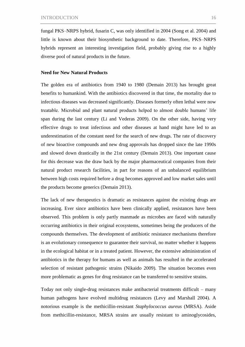
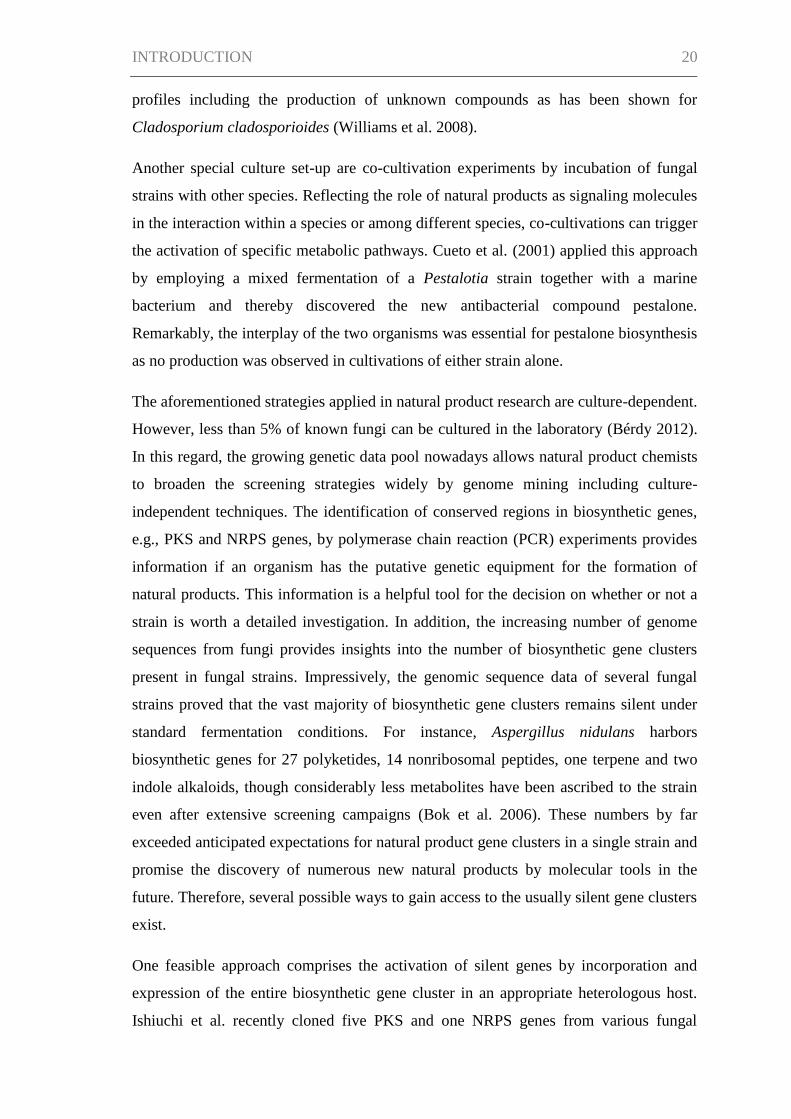
Fungal Natural Products
Systematic studies of fungal natural products were started in 1923 by Harold Raistrick
and colleagues, leading to the isolation of some 200 mold metabolites such as patulin,
penicillic acid and geodin (Raistrick 1949). Some years later when Alexander Fleming
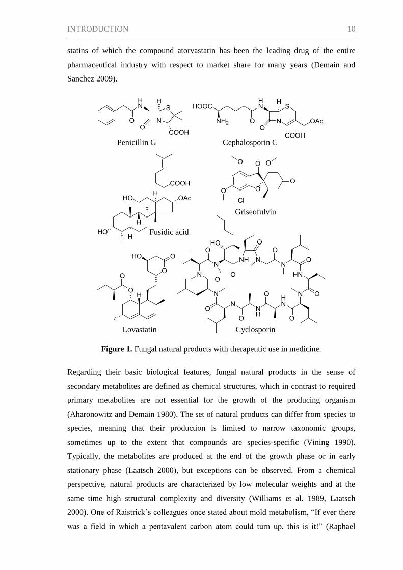
discovered penicillin G, a real success story of fungal natural products began (Figure 1).
Fleming’s discovery was an observation by chance as he found an undesired
contamination in one of his petri dishes containing a culture of staphylococci in 1928.
He noticed that the mold, which he identified as Penicillium notatum, inhibited the
growth of the bacteria. Fleming characterized penicillin as the mold’s active principle
which at the same time lacked toxicity on human blood cells and animals (Fleming
1945). It was not until ten years later that Howard W. Florey, Ernest Chain and
colleagues were able to prepare sufficient and stable penicillin suitable for clinical trials.
With these studies the compound was finally proved a very effective antibiotic
(Nicolaou and Montagnon 2008).
Stimulated by the discovery of penicillin G, a new epoch in research for biologically
active molecules was ushered in. It turned out that especially molds and actinomycetes
bacteria were potent natural product producers (Bérdy 2005). Further antibacterial drugs
of fungal origin were developed, e.g., cephalosporin C identified from Cephalosporium
acremonium (Newton and Abraham 1955) or fusidic acid, which was first isolated from
Fusidium coccineum, and still provides high activity against resistant Staphylococcus
strains (Godtfredsen et al. 1962, Aly et al. 2011). The more natural products from fungi
were described, the broader the panel of applications became. Griseofulvin from
Penicillium griseofulvum was one of the first antifungal metabolites used for therapy of
dermatophyte infections of skin, hair and nails (Grove et al. 1952, Aly et al. 2011).
Cyclosporin, obtained from Tolypocladium inflatum in 1971, was found to be a
suppressor of T-cells and for the first time provided transplantation surgery with a
selective immunosuppressive drug for administration after organ transplantations
(Dreyfuss et al. 1976, Grabley and Thiericke 1999). The statins, e.g., lovastatin, which
was originally isolated from Monascus ruber and later from Aspergillus terreus,
represent another important example among fungal natural products with use in
medicine (Alberts et al. 1980). Statins attracted attention because of their cholesterol-
lowering properties leading to the commercialization of lovastatin as an
antihypercholesterolemic drug in 1987. Further statins followed the way to the market,
especially semisynthetic and totally synthetic analogs derived from the fungal natural
INTRODUCTION 10
statins of which the compound atorvastatin has been the leading drug of the entire
pharmaceutical industry with respect to market share for many years (Demain and
Sanchez 2009).
Figure 1. Fungal natural products with therapeutic use in medicine.
Regarding their basic biological features, fungal natural products in the sense of
secondary metabolites are defined as chemical structures, which in contrast to required
primary metabolites are not essential for the growth of the producing organism
(Aharonowitz and Demain 1980). The set of natural products can differ from species to
species, meaning that their production is limited to narrow taxonomic groups,
sometimes up to the extent that compounds are species-specific (Vining 1990).
Typically, the metabolites are produced at the end of the growth phase or in early
stationary phase (Laatsch 2000), but exceptions can be observed. From a chemical
perspective, natural products are characterized by low molecular weights and at the
same time high structural complexity and diversity (Williams et al. 1989, Laatsch
2000). One of Raistrick’s colleagues once stated about mold metabolism, “If ever there
was a field in which a pentavalent carbon atom could turn up, this is it!” (Raphael
Griseofulvin
Cephalosporin C
Fusidic acid
Penicillin G
Cyclosporin Lovastatin
INTRODUCTION 11
1948), paraphrasing the seemingly unrestricted and partly unimaginable variety found in
chemical structures of fungal natural products.
On the basis of their structural diversity, the broad spectrum of biological activities of
fungal natural products can be explained. In addition to the above mentioned
antibacterial, antifungal, immunosuppressant and antihyperlipidemic examples, a
plethora of further fungal compounds showing biological properties has been isolated.
The activities range from antiviral, anti-insecticidal, antiprotozoal to antiparasitic (e.g.,
anthelmintic) effects. Furthermore, activities including cytotoxic, antitumoral,
mutagenic, carcinogenic, teratogenic properties and different enzyme inhibitory effects
have been assigned to fungal natural products (Keller et al. 2005). Therefore, natural
products of fungal origin have a broad field of application not only in human medicine,
but also in veterinary medicine and agriculture, e.g., in crop protection.
Though the practical benefit of natural products for humans is evident, their ecological
function and relevance in the natural environment often remains elusive. As fungi are
sessile organisms, the production of bioactive compounds can be a chemical defense
mechanism toward competitors rivaling for the same nutrients and habitats or as an anti-
predator adaptation (Kettering et al. 2004, Rohlfs et al. 2007). The fact that natural
products are frequently synthesized in a specific stage of developmental processes link
compounds and their function to the morphological differentiation of the producing
fungi. For instance, pigments known as melanins are associated with fungal spore
formation, contributing to the spores’ survival by the UV protecting features of
melanins (Calvo et al. 2002). Just as well natural products are recognized as mediators
of a number of symbiotic and parasitic interactions (Vining 1990). This relationship is
exemplified by secondary metabolites of endophytic fungi which are correlated to the
increased tolerance toward biotic stress in the fungi-hosting plants (Saikkonen et al.
1998).
For the majority of natural products, the precise ecological function has not been
understood though. This is especially true for those compounds that have no
demonstrated activity under laboratory conditions. The ecological role of natural
products has therefore always been matter of a controversial debate and it has even been
questioned whether the compounds are just the result of a “games room” of biochemical
evolution (Zähner 1979). However, given that the biosynthesis of natural products
involves complex enzyme systems (Laatsch 2000), which are encoded by genes
INTRODUCTION 12
accounting for many kilobases of DNA (Williams et al. 1989), it is energetically a
costly process that is logically only justified, if the compounds provide a benefit to the
producing organism.
Biosynthesis of Fungal Natural Products
Despite the chemical diversity of natural products, they are produced by only a few
biosynthetic pathways. Studies on the genes encoding fungal natural products revealed
that they are usually clustered on a single genetic locus, a feature that was formerly
thought to be exclusive of prokaryotic organisms (Keller and Hohn 1997). There are
four classes of fungal natural products with regard to their chemical scaffolds which in
turn reflect the metabolic route involved in biosynthesis: polyketides, nonribosomal
peptides, terpenes and indole alkaloids. The basic principles of polyketide and
nonribosomal peptide biosynthesis, being of particular interest in this study, shall be
described more in detail here.
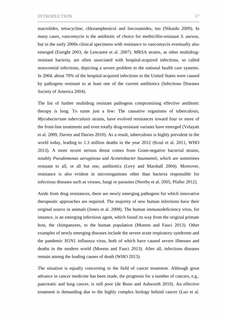
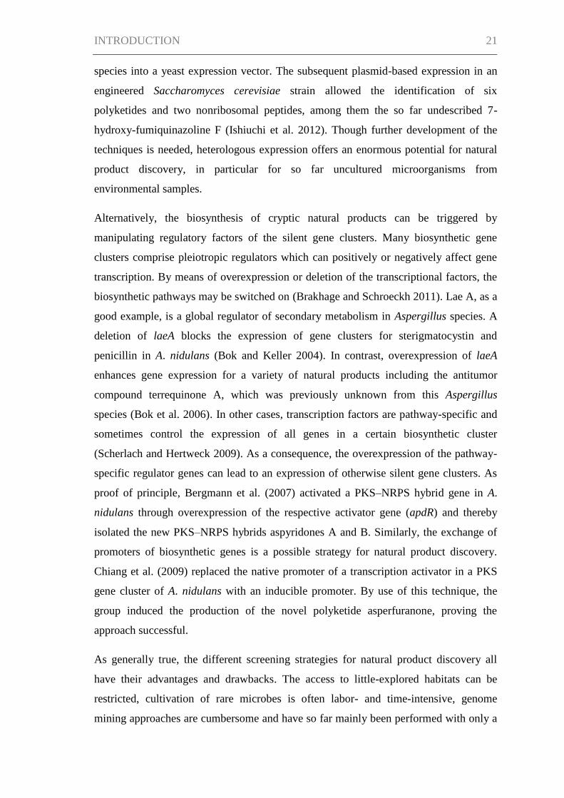
Polyketides
Polyketides are a diverse group of natural products, ranging from very simple structures
to highly complex ones, including polyphenols, macrolides, polyenes, enediynes and
polyethers (Hertweck 2009). The assembly of polyketides is catalyzed by large multi-
enzyme complexes called polyketide synthases (PKSs) which use short-chain
carboxylic acids, in fungi generally acetyl coenzyme A (CoA) and malonyl CoA, as
building blocks. During the enzymatic process, the substrates are successively
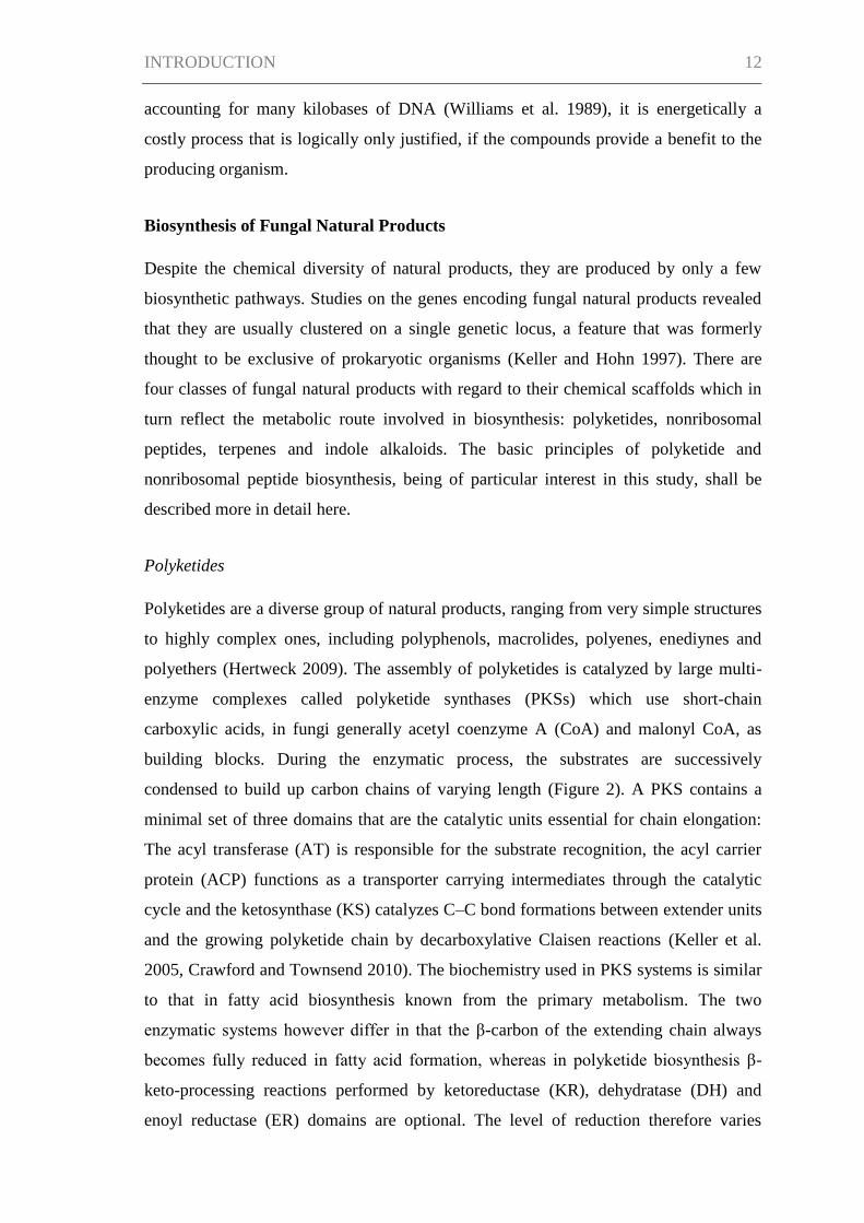
condensed to build up carbon chains of varying length (Figure 2). A PKS contains a
minimal set of three domains that are the catalytic units essential for chain elongation:
The acyl transferase (AT) is responsible for the substrate recognition, the acyl carrier
protein (ACP) functions as a transporter carrying intermediates through the catalytic
cycle and the ketosynthase (KS) catalyzes C–C bond formations between extender units
and the growing polyketide chain by decarboxylative Claisen reactions (Keller et al.
2005, Crawford and Townsend 2010). The biochemistry used in PKS systems is similar
to that in fatty acid biosynthesis known from the primary metabolism. The two
enzymatic systems however differ in that the β-carbon of the extending chain always
becomes fully reduced in fatty acid formation, whereas in polyketide biosynthesis β-
keto-processing reactions performed by ketoreductase (KR), dehydratase (DH) and
enoyl reductase (ER) domains are optional. The level of reduction therefore varies
INTRODUCTION 13
depending on the polyketide that is built. Additionally, the carbon chain can be
modified by cyclase (CYC) and methyl transferase (MT) activities (Schümann and
Hertweck 2006, Cox 2007). Despite the simple building blocks used in polyketidic
biosynthesis, the programming of their type and number, the number of reduction
reactions, extend of methylation, cyclization reactions as well as post-PKS steps give
rise to very different end products. The striking diversity of polyketides is hence a result
of the combination of these variables allowing innumerable structural possibilities
(Keller et al. 2005, Hertweck 2009).
Though the basic mechanisms of polyketide biosynthesis are applicable to all PKSs,
they are divided into three major classes according to the enzyme architecture. Type I
PKS are large multidomain enzyme systems comprising covalently fused domains that
can either show a modular arrangement or work iteratively. In the modular type I
system, typically found in bacteria, the catalytic domains form modules of which each is
usually responsible for only one elongation cycle. On the contrary, in the iterative type I
system, the domains are reused for the catalytic cycles (Hertweck 2009). The majority
of fungal PKS are iterative type I systems and are further subdivided into non-reducing,
partially reducing or highly reducing PKSs corresponding to the degree of reductive
processing that takes place during the assembly (Crawford and Townsend 2010). In type
II PKSs, the catalytic activities are present on dissociable proteins that associate to
Figure 2. Basic mechanisms of polyketide biosynthesis (modified according to
Crawford and Townsend 2010). KS, ketosynthase; AT, acyl transferase; ACP, acyl
carrier protein; KR, ketoreductase; DH, dehydratase; ER enoyl reductase; enz; enzyme.
INTRODUCTION 14
perform the enzymatic activities. Type II systems are exclusively found in bacteria, e.g.,
the formation of bacterial aromatic polyketides is catalyzed by type II iterative systems.
Finally, type III PKSs are single, iteratively working KS proteins that do not require
auxiliary enzymes and are found in plants, bacteria and fungi (Cox 2007, Crawford and
Townsend 2010).
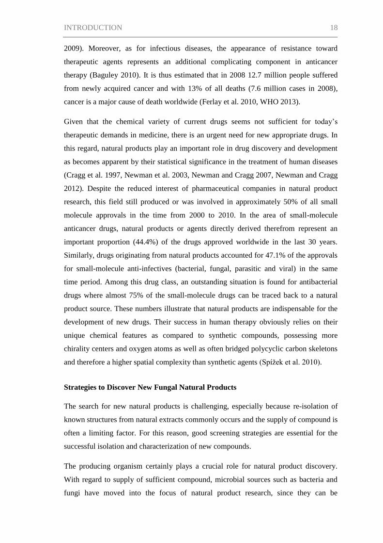
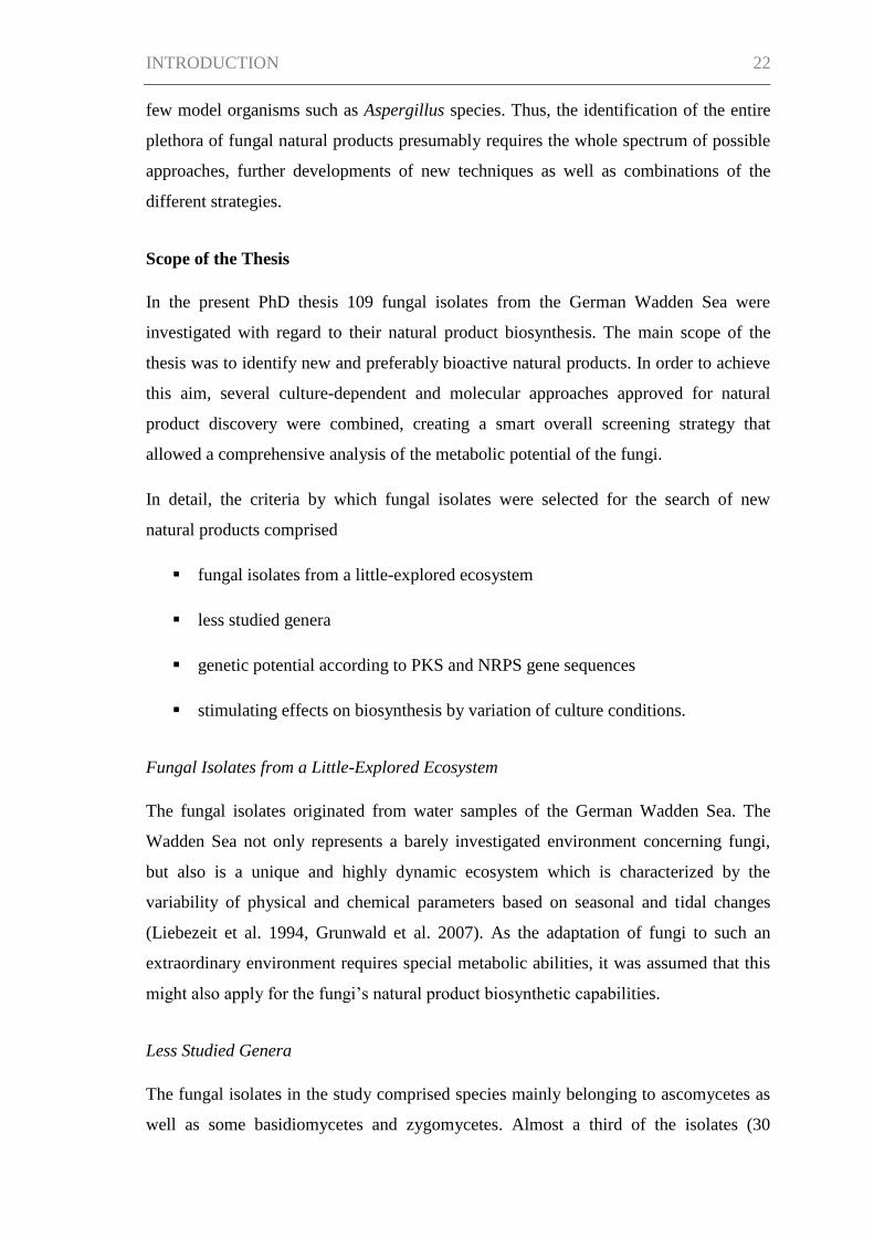
Nonribosomal Peptides
Nonribosomal peptides mainly originate from bacteria as well as fungi, comprising well
known natural products such as cyclosporins and β-lactam antibiotics like the penicillins
(Strieker et al. 2010). As the name suggests, the biosynthetic mechanisms of
nonribosomal peptides differ from the ubiquitous protein formation at the ribosomes in
primary metabolism. Unlike for ribosomal proteins, where the translation of the genetic
code allows the biosynthesis of different proteinogenic end products at a single catalytic
site, nonribosomal peptides are the result of product-specific enzymes called
nonribosomal peptide synthetases (NRPSs).
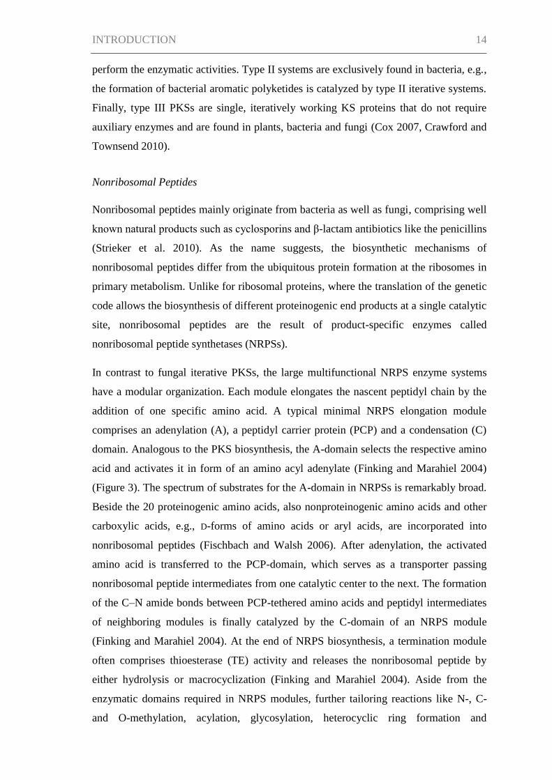
In contrast to fungal iterative PKSs, the large multifunctional NRPS enzyme systems
have a modular organization. Each module elongates the nascent peptidyl chain by the
addition of one specific amino acid. A typical minimal NRPS elongation module
comprises an adenylation (A), a peptidyl carrier protein (PCP) and a condensation (C)
domain. Analogous to the PKS biosynthesis, the A-domain selects the respective amino
acid and activates it in form of an amino acyl adenylate (Finking and Marahiel 2004)
(Figure 3). The spectrum of substrates for the A-domain in NRPSs is remarkably broad.
Beside the 20 proteinogenic amino acids, also nonproteinogenic amino acids and other
carboxylic acids, e.g., D-forms of amino acids or aryl acids, are incorporated into
nonribosomal peptides (Fischbach and Walsh 2006). After adenylation, the activated
amino acid is transferred to the PCP-domain, which serves as a transporter passing
nonribosomal peptide intermediates from one catalytic center to the next. The formation
of the C–N amide bonds between PCP-tethered amino acids and peptidyl intermediates
of neighboring modules is finally catalyzed by the C-domain of an NRPS module
(Finking and Marahiel 2004). At the end of NRPS biosynthesis, a termination module
often comprises thioesterase (TE) activity and releases the nonribosomal peptide by
either hydrolysis or macrocyclization (Finking and Marahiel 2004). Aside from the
enzymatic domains required in NRPS modules, further tailoring reactions like N-, C-
and O-methylation, acylation, glycosylation, heterocyclic ring formation and
INTRODUCTION 15
epimerization can be conducted by optional domains integrated in the NRPSs or stand-
alone enzymes (Mootz et al. 2002). According to these biosynthetic mechanisms, the
enormous structural diversity found among fungal nonribosomal peptides arises in the
wide range of building blocks employed, the length of peptides as well as the various
modifying tailoring reactions (Keller et al. 2005).
Due to the modular architecture of NRPS systems, the order of elongation modules
generally correlates with the order of amino acids incorporated (Hoffmeister and Keller
2007). In these cases, the genes encoding a specific NRPS allow the prediction of the
nonribosomal peptide product. However, there is increasing evidence of NRPSs for
which this co-linearity is not true, because modules or domains are used more than once
during peptide assembly. The NRPS for the fungal trimer enniatin, for instance,
comprises a reduced number of modules as the three repeated structural sequences are
catalyzed by only one set of modules in an iterative fashion (Glinski et al. 2002, Mootz
et al. 2002). A further example for deviation from the classical NRPS systems as
described above are a group of compounds called PKS–NRPS hybrids. In the
biosynthetic complexes for hybrids, enzymatic activities of PKS and NRPS domains act
together, resulting in the production of mixed peptide–polyketide compounds. The first
Figure 3. Basic mechanisms of nonribosomal peptide
biosynthesis (modified according to Strieker et al. 2010). A,
adenylation; PCP, peptidyl carrier protein; C, condensation; TE,
thioesterase; E, epimerase; aa, amino acid.
INTRODUCTION 16
fungal PKS–NRPS hybrid, fusarin C, was only identified in 2004 (Song et al. 2004) and
little is known about their biosynthetic background to date. Therefore, PKS–NRPS
hybrids represent an interesting investigation field, probably giving rise to a highly
diverse pool of natural products in the future.
Need for New Natural Products
The golden era of antibiotics from 1940 to 1980 (Demain 2013) has brought great
benefits to humankind. With the antibiotics discovered in that time, the mortality due to
infectious diseases was decreased significantly. Diseases formerly often lethal were now
treatable. Microbial and plant natural products helped to almost double humans’ life
span during the last century (Li and Vederas 2009). On the other side, having very
effective drugs to treat infectious and other diseases at hand might have led to an
underestimation of the constant need for the search of new drugs. The rate of discovery
of new bioactive compounds and new drug approvals has dropped since the late 1990s
and slowed down drastically in the 21st century (Demain 2013). One important cause
for this decrease was the draw back by the major pharmaceutical companies from their
natural product research facilities, in part for reasons of an unbalanced equilibrium
between high costs required before a drug becomes approved and low market sales until
the products become generics (Demain 2013).
The lack of new therapeutics is dramatic as resistances against the existing drugs are
increasing. Ever since antibiotics have been clinically applied, resistances have been
observed. This problem is only partly manmade as microbes are faced with naturally
occurring antibiotics in their original ecosystems, sometimes being the producers of the
compounds themselves. The development of antibiotic resistance mechanisms therefore
is an evolutionary consequence to guarantee their survival, no matter whether it happens
in the ecological habitat or in a treated patient. However, the extensive administration of
antibiotics in the therapy for humans as well as animals has resulted in the accelerated
selection of resistant pathogenic strains (Nikaido 2009). The situation becomes even
more problematic as genes for drug resistance can be transferred to sensitive strains.
Today not only single-drug resistances make antibacterial treatments difficult – many
human pathogens have evolved multidrug resistances (Levy and Marshall 2004). A
notorious example is the methicillin-resistant Staphylococcus aureus (MRSA). Aside
from methicillin-resistance, MRSA strains are usually resistant to aminoglycosides,
INTRODUCTION 17
macrolides, tetracycline, chloramphenicol and lincosamides, too (Nikaido 2009). In
many cases, vancomycin is the antibiotic of choice for methicillin-resistant S. aureus,
but in the early 2000s clinical specimens with resistance to vancomycin eventually also
emerged (Enright 2003, de Lencastre et al. 2007). MRSA strains, as other multidrug-
resistant bacteria, are often associated with hospital-acquired infections, so called
nosocomial infections, depicting a severe problem in the national health care systems.
In 2004, about 70% of the hospital-acquired infections in the United States were caused
by pathogens resistant to at least one of the current antibiotics (Infectious Diseases
Society of America 2004).
The list of further multidrug resistant pathogens compromising effective antibiotic
therapy is long. To name just a few: The causative organisms of tuberculosis,
Mycobacterium tuberculosis strains, have evolved resistances toward four or more of
the front-line treatments and even totally drug-resistant variants have emerged (Velayati
et al. 2009, Davies and Davies 2010). As a result, tuberculosis is highly prevalent in the
world today, leading to 1.3 million deaths in the year 2012 (Koul et al. 2011, WHO
2013). A more recent serious threat comes from Gram-negative bacterial strains,
notably Pseudomonas aeruginosa and Acinetobacter baumannii, which are sometimes
resistant to all, or all but one, antibiotics (Levy and Marshall 2004). Moreover,
resistance is also evident in microorganisms other than bacteria responsible for
infectious diseases such as viruses, fungi or parasites (Norrby et al. 2005, Pfaller 2012).
Aside from drug resistances, there are newly emerging pathogens for which innovative
therapeutic approaches are required. The majority of new human infections have their
original source in animals (Jones et al. 2008). The human immunodeficiency virus, for
instance, is an emerging infectious agent, which found its way from the original primate
host, the chimpanzees, to the human population (Morens and Fauci 2013). Other
examples of newly emerging diseases include the severe acute respiratory syndrome and
the pandemic H1N1 influenza virus, both of which have caused severe illnesses and
deaths in the modern world (Morens and Fauci 2013). After all, infectious diseases
remain among the leading causes of death (WHO 2013).
The situation is equally concerning in the field of cancer treatment. Although great
advance in cancer medicine has been made, the prognosis for a number of cancers, e.g.,
pancreatic and lung cancer, is still poor (de Bono and Ashworth 2010). An effective
treatment is demanding due to the highly complex biology behind cancer (Luo et al.
INTRODUCTION 18
2009). Moreover, as for infectious diseases, the appearance of resistance toward
therapeutic agents represents an additional complicating component in anticancer
therapy (Baguley 2010). It is thus estimated that in 2008 12.7 million people suffered
from newly acquired cancer and with 13% of all deaths (7.6 million cases in 2008),
cancer is a major cause of death worldwide (Ferlay et al. 2010, WHO 2013).
Given that the chemical variety of current drugs seems not sufficient for today’s
therapeutic demands in medicine, there is an urgent need for new appropriate drugs. In
this regard, natural products play an important role in drug discovery and development
as becomes apparent by their statistical significance in the treatment of human diseases
(Cragg et al. 1997, Newman et al. 2003, Newman and Cragg 2007, Newman and Cragg
2012). Despite the reduced interest of pharmaceutical companies in natural product
research, this field still produced or was involved in approximately 50% of all small
molecule approvals in the time from 2000 to 2010. In the area of small-molecule
anticancer drugs, natural products or agents directly derived therefrom represent an
important proportion (44.4%) of the drugs approved worldwide in the last 30 years.
Similarly, drugs originating from natural products accounted for 47.1% of the approvals
for small-molecule anti-infectives (bacterial, fungal, parasitic and viral) in the same
time period. Among this drug class, an outstanding situation is found for antibacterial
drugs where almost 75% of the small-molecule drugs can be traced back to a natural
product source. These numbers illustrate that natural products are indispensable for the
development of new drugs. Their success in human therapy obviously relies on their
unique chemical features as compared to synthetic compounds, possessing more
chirality centers and oxygen atoms as well as often bridged polycyclic carbon skeletons
and therefore a higher spatial complexity than synthetic agents (Spížek et al. 2010).
Strategies to Discover New Fungal Natural Products
The search for new natural products is challenging, especially because re-isolation of
known structures from natural extracts commonly occurs and the supply of compound is
often a limiting factor. For this reason, good screening strategies are essential for the
successful isolation and characterization of new compounds.
The producing organism certainly plays a crucial role for natural product discovery.
With regard to supply of sufficient compound, microbial sources such as bacteria and
fungi have moved into the focus of natural product research, since they can be
INTRODUCTION 19
fermented in large scales in the laboratory, yielding good quantities of compounds
without harming the natural environment. While mainly terrestrial fungi and bacteria,
e.g., from soil, have been of interest for natural product discovery for a long time, a
great fraction of the world’s microbial diversity remains to be explored (Harvey 2008,
Bérdy 2012). One strategy for the search of new natural products is hence the
investigation of microbes from little-explored environments. Marine habitats, for
instance, harbor a largely untouched number of microbes and those studied turned out to
be prolific producer of new bioactive compounds (Bhatnagar and Kim 2010, Imhoff et
al. 2011). The same is true for endophytic microorganisms and in particular endophytes
from rainforests represent a huge, unexplored source for natural products (Strobel et al.
2004, Bérdy 2012). In addition, only few taxonomic groups have been examined
thoroughly with regard to their bioactive metabolites production. In the case of fungi,
these are especially Penicillium, Aspergillus or Fusarium isolates, whereas many other
species have been less intensively studied and certainly bear an untapped potential for
the production of yet unknown compounds (Bérdy 2005, Dictionary of Natural Products
2012).
For cultivation of microorganisms, a stimulation of natural product biosynthesis by
systematically changing the culture conditions has been approved as an effective
screening strategy. The parameters can be easily accessible ones including media
composition, aeration, pH value, temperature or shape of culture vessels (Bode et al.
2002). The method has not only shown differences in quantities of metabolites
produced, but also induced the production of many different natural products from a
single strain. For example, the cultivation of Aspergillus ochraceus in different culture
flasks and the variation of agitation during cultivation (e.g., still cultures, shaken
cultures, Penicillium flasks, different fermenters) resulted in the formation of 15
additional compounds which were formerly unknown from the strain (Fuchser 1996).
Intensive studies on the influence of several culture conditions on microbial metabolism
enabled Zeeck and his colleagues to isolate more than 100 natural products belonging to
25 structural classes from just six microorganisms (Bode et al. 2002). Relating to the
variation of culture media, an addition of epigenetic modifiers has recently gained
increasing attention. Epigenetic modifiers regulate the level of e.g. acetylation and
methylation in histones and thereby control the transcriptional accessibility of
biosynthetic gene clusters of natural products. The use of culture media treated with
epigenetic modifying agents can hence result in a significant change of metabolic
INTRODUCTION 20
profiles including the production of unknown compounds as has been shown for
Cladosporium cladosporioides (Williams et al. 2008).
Another special culture set-up are co-cultivation experiments by incubation of fungal
strains with other species. Reflecting the role of natural products as signaling molecules
in the interaction within a species or among different species, co-cultivations can trigger
the activation of specific metabolic pathways. Cueto et al. (2001) applied this approach
by employing a mixed fermentation of a Pestalotia strain together with a marine
bacterium and thereby discovered the new antibacterial compound pestalone.
Remarkably, the interplay of the two organisms was essential for pestalone biosynthesis
as no production was observed in cultivations of either strain alone.
The aforementioned strategies applied in natural product research are culture-dependent.
However, less than 5% of known fungi can be cultured in the laboratory (Bérdy 2012).
In this regard, the growing genetic data pool nowadays allows natural product chemists
to broaden the screening strategies widely by genome mining including culture-
independent techniques. The identification of conserved regions in biosynthetic genes,
e.g., PKS and NRPS genes, by polymerase chain reaction (PCR) experiments provides
information if an organism has the putative genetic equipment for the formation of
natural products. This information is a helpful tool for the decision on whether or not a
strain is worth a detailed investigation. In addition, the increasing number of genome
sequences from fungi provides insights into the number of biosynthetic gene clusters
present in fungal strains. Impressively, the genomic sequence data of several fungal
strains proved that the vast majority of biosynthetic gene clusters remains silent under
standard fermentation conditions. For instance, Aspergillus nidulans harbors
biosynthetic genes for 27 polyketides, 14 nonribosomal peptides, one terpene and two
indole alkaloids, though considerably less metabolites have been ascribed to the strain
even after extensive screening campaigns (Bok et al. 2006). These numbers by far
exceeded anticipated expectations for natural product gene clusters in a single strain and
promise the discovery of numerous new natural products by molecular tools in the
future. Therefore, several possible ways to gain access to the usually silent gene clusters
exist.
One feasible approach comprises the activation of silent genes by incorporation and
expression of the entire biosynthetic gene cluster in an appropriate heterologous host.
Ishiuchi et al. recently cloned five PKS and one NRPS genes from various fungal
INTRODUCTION 21
species into a yeast expression vector. The subsequent plasmid-based expression in an
engineered Saccharomyces cerevisiae strain allowed the identification of six
polyketides and two nonribosomal peptides, among them the so far undescribed 7-
hydroxy-fumiquinazoline F (Ishiuchi et al. 2012). Though further development of the
techniques is needed, heterologous expression offers an enormous potential for natural
product discovery, in particular for so far uncultured microorganisms from
environmental samples.
Alternatively, the biosynthesis of cryptic natural products can be triggered by
manipulating regulatory factors of the silent gene clusters. Many biosynthetic gene
clusters comprise pleiotropic regulators which can positively or negatively affect gene
transcription. By means of overexpression or deletion of the transcriptional factors, the
biosynthetic pathways may be switched on (Brakhage and Schroeckh 2011). Lae A, as a
good example, is a global regulator of secondary metabolism in Aspergillus species. A
deletion of laeA blocks the expression of gene clusters for sterigmatocystin and
penicillin in A. nidulans (Bok and Keller 2004). In contrast, overexpression of laeA
enhances gene expression for a variety of natural products including the antitumor
compound terrequinone A, which was previously unknown from this Aspergillus
species (Bok et al. 2006). In other cases, transcription factors are pathway-specific and
sometimes control the expression of all genes in a certain biosynthetic cluster
(Scherlach and Hertweck 2009). As a consequence, the overexpression of the pathway-
specific regulator genes can lead to an expression of otherwise silent gene clusters. As
proof of principle, Bergmann et al. (2007) activated a PKS–NRPS hybrid gene in A.
nidulans through overexpression of the respective activator gene (apdR) and thereby
isolated the new PKS–NRPS hybrids aspyridones A and B. Similarly, the exchange of
promoters of biosynthetic genes is a possible strategy for natural product discovery.
Chiang et al. (2009) replaced the native promoter of a transcription activator in a PKS
gene cluster of A. nidulans with an inducible promoter. By use of this technique, the
group induced the production of the novel polyketide asperfuranone, proving the
approach successful.
As generally true, the different screening strategies for natural product discovery all
have their advantages and drawbacks. The access to little-explored habitats can be
restricted, cultivation of rare microbes is often labor- and time-intensive, genome
mining approaches are cumbersome and have so far mainly been performed with only a
INTRODUCTION 22
few model organisms such as Aspergillus species. Thus, the identification of the entire
plethora of fungal natural products presumably requires the whole spectrum of possible
approaches, further developments of new techniques as well as combinations of the
different strategies.
Scope of the Thesis
In the present PhD thesis 109 fungal isolates from the German Wadden Sea were
investigated with regard to their natural product biosynthesis. The main scope of the
thesis was to identify new and preferably bioactive natural products. In order to achieve
this aim, several culture-dependent and molecular approaches approved for natural
product discovery were combined, creating a smart overall screening strategy that
allowed a comprehensive analysis of the metabolic potential of the fungi.
In detail, the criteria by which fungal isolates were selected for the search of new
natural products comprised
fungal isolates from a little-explored ecosystem
less studied genera
genetic potential according to PKS and NRPS gene sequences
stimulating effects on biosynthesis by variation of culture conditions.
Fungal Isolates from a Little-Explored Ecosystem
The fungal isolates originated from water samples of the German Wadden Sea. The
Wadden Sea not only represents a barely investigated environment concerning fungi,
but also is a unique and highly dynamic ecosystem which is characterized by the
variability of physical and chemical parameters based on seasonal and tidal changes
(Liebezeit et al. 1994, Grunwald et al. 2007). As the adaptation of fungi to such an
extraordinary environment requires special metabolic abilities, it was assumed that this
might also apply for the fungi’s natural product biosynthetic capabilities.
Less Studied Genera
The fungal isolates in the study comprised species mainly belonging to ascomycetes as
well as some basidiomycetes and zygomycetes. Almost a third of the isolates (30
INTRODUCTION 23
strains) were affiliated to the genus Penicillium. Though Penicillia are prolific
producers of bioactive compounds, it is also one of the best investigated fungal genera
regarding natural products and therefore the probability to find already characterized
compounds is high. To avoid a re-isolation of known natural products, the focus of the
screening was set to other, less examined genera or isolates that did not show high
similarities to gene sequences deposited at GenBank, being putative new species.
Genetic Potential According to PKS and NRPS Gene Sequences
The fungi’s genetic potential to produce polyketides or nonribosomal peptides was
investigated by PCR amplification of the respective biosynthetic gene fragments and
subsequent sequencing. For PKS genes, degenerative primer pairs (LC1 and LC2c, LC3
and LC5c) for the amplification of the KS-domain were used (Bingle et al. 1999). The
primers for NRPS genes amplified fragments of the C-domain (Klotz 2010). On the
basis of the PCR and sequencing data, only PKS- and NRPS-positive strains were
selected for further investigations of chemical profiles.
Stimulating Effects on Biosynthesis by Variation of Culture Conditions
All 109 isolates were screened for their natural product formation in one standard
culture condition (modified Wickerham medium, still cultures for filamentous fungi and
shaken cultures for yeasts). In order to induce the activation of gene clusters that were
silent under the standard fermentation condition, a selection (29 strains) of less studied
genera and PKS-/NRPS-positive isolates was cultured in five different conditions, more
precisely, four different media (potato-carrot medium, modified malt extract medium,
Czapek medium, casamino acids glucose medium) and shaking conditions in modified
Wickerham medium (except for the yeasts of the selection).
The general methodologies employed for the analysis and dereplication of natural
products in the extracts from fungal cultures included HPLC, MS and bioactivity
screenings. Putative new bioactive compounds were purified by chromatographic means
and elucidated in structure on the basis of NMR spectra. The identification of new
natural products raised further questions concerning their stereochemistry, bioactivities
and biosynthesis which were subsequently investigated.
CHAPTER I 24
CHAPTER I
Calcaripeptides A–C, cyclodepsipeptides from a Calcarisporium strain
Johanna Silber, Birgit Ohlendorf, Antje Labes, Christian Näther and Johannes F. Imhoff
Published in 2013 as an article in J. Nat. Prod. 76(8), 1461-1467
Reprinted with permission from Silber, J., Ohlendorf, B., Labes, A., Näther, C., Imhoff,
J. F. (2013). Calcaripeptides A–C, cyclodepsipeptides from a Calcarisporium strain. J.
Nat. Prod. 76(8), 1461-1467. Copyright 2013 American Chemical Society.
CHAPTER I 25
Abstract
The isolation and structure elucidation of the novel calcaripeptides A (1), B (2), and C
(3) and studies on their biosynthetic origin are described. The calcaripeptides were
identified from Calcarisporium sp. strain KF525, which was isolated from the German
Wadden Sea. Compounds 1−3 are macrocyclic structures composed of a proline and a
phenylalanine residue as well as a nonpeptidic substructure. Structure elucidation was
achieved by applying one- and two-dimensional NMR spectroscopy supported by high-
resolution mass spectrometry. X-ray crystallography was performed to determine the
relative configuration of 1. The absolute configuration of 1 was assigned by HPLC of
the amino acids after hydrolysis of the molecule and derivatization with chiral agents.
Studies on the biosynthesis by feeding 13
C-labeled substrates revealed that the
nonpeptidic part of 1 originates from acetate and L-methionine. The involvement of a
hybrid between a polyketide synthase and a nonribosomal peptide synthetase in the
biosynthesis of the calcaripeptides is discussed.
Introduction
With the aim to discover new natural products, fungal strains from the German Wadden
Sea were analyzed regarding their metabolite profiles. In Wadden Sea habitats often
both true marine organisms and organisms from terrestrial and freshwater habitats
frequently occur together. The Calcarisporium sp. strain KF525 attracted attention
because it produced a set of metabolites that could not be identified by extensive
searches of the literature and databases. Fungi of the genus Calcarisporium show a
widespread occurrence on wood (Sutton 1973, Cooper 2005) and leaf litter (Rambelli et
al. 2004, Somrithipol and Jones 2006), in plants as endophytic fungi (Gong and Guo
2010, Ji et al. 2004), or in coal spoil tips (Evans 1971). Commonly, Calcarisporium
spp. are found as mycoparasites or symbionts of higher basidiomycetes and
ascomycetes (Sutton 1973, Barnett 1958, Barnett and Lilly 1958, Watson 1955, Carrión
and Rico-Gray 2002, Rombach and Roberts 1987). For some mycoparasitic strains of
Calcarisporium sp., the ability to reduce feed spoiling molds or fungal plant pathogens,
like those responsible for mildews, was observed. Hence, the use of Calcarisporium
culture filtrates as feed-protecting preservatives and as biocontrol agents in crop
protection has been discussed (Ji et al. 2004, Carrión and Rico-Gray 2002, Hijwegen
1989, Hijwegen and Verhaar 1993). It is well known that predatory interactions of
microbes are often mediated by enzymes or small-molecule compounds and toxins.

CHAPTER I 26
Even though this has also been assumed to be the case for the antifungal interplay of
Calcarisporium species (Hijwegen 1989), few natural products have been described for
the genus (Dictionary of Natural Products 2012). Among them are antifungal
compounds such as 15-azahomosterols (Chrisp et al. 1990), aurovertins inhibiting
mitochondrial ATP synthesis and ATPases (Baldwin et al. 1964, Mulheirn et al. 1974,
Osselton et al. 1974), and calcarisporins B1−B4, with calcarisporin B1 showing
cytotoxic activity (Yu et al. 2002a, Yu et al. 2002b).
The marine-derived isolate KF525 of Calcarisporium sp. was shown to produce a
metabolite spectrum different from those of other known Calcarisporium strains. The
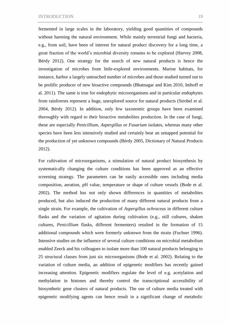
isolation of these metabolites from the fungal mycelium yielded the novel, structurally
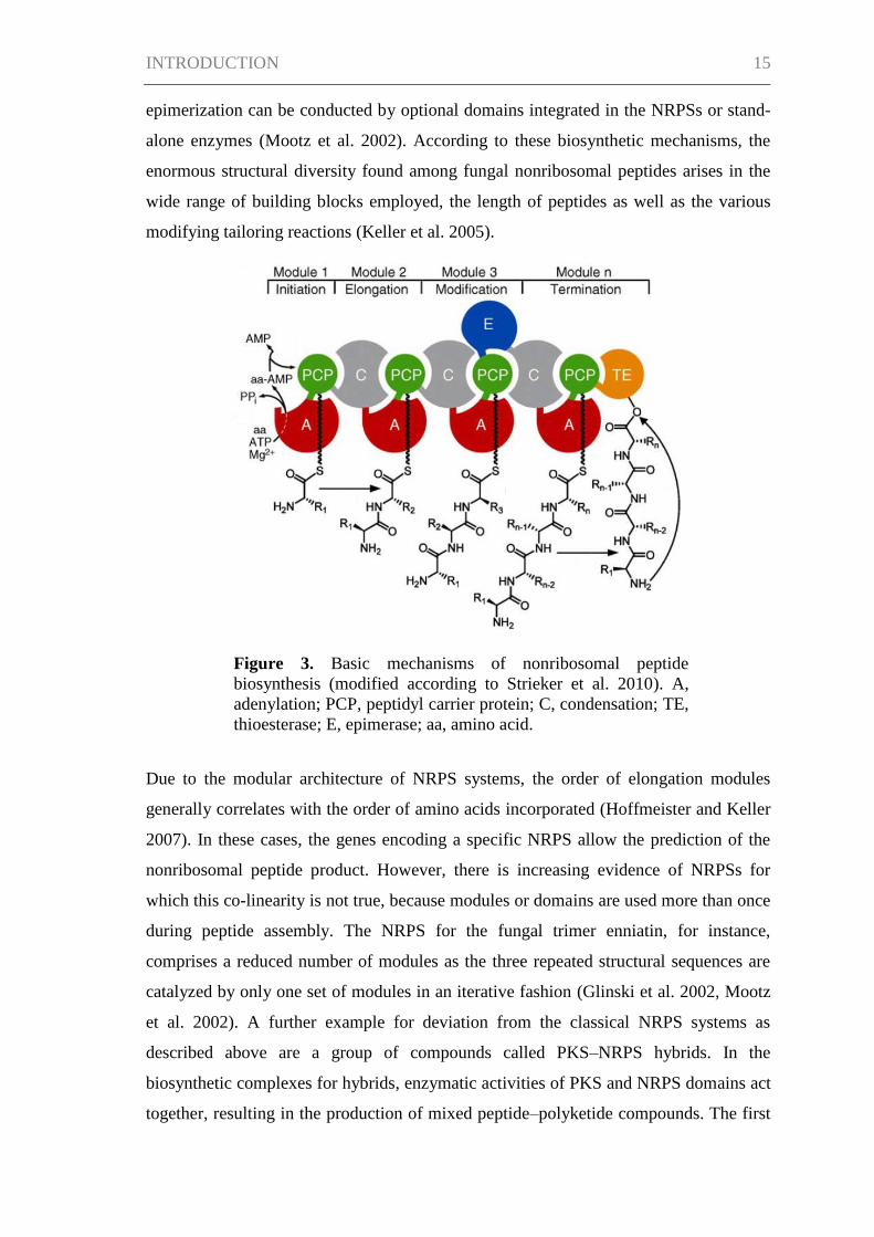
related calcaripeptides A (1), B (2), and C (3) described here (Figure I.1). Analysis of
the NMR spectroscopic data showed the compounds to be macrocyclic structures
consisting of two amino acids (proline and phenylalanine) and a nonpeptidic
substructure. The nonpeptidic chain varies in structure between the three compounds.
The absolute configuration of 1 was determined by X-ray crystallography in
combination with the configuration data of the amino acids obtained by HPLC analysis
after hydrolysis of the molecule and chemical derivatization. The biosynthesis of 1 was
investigated by feeding 13
C-labeled precursors.
Results and Discussion
Strain KF525 was identified as a Calcarisporium sp. based on morphological
characteristics, in particular microscopic examination of the conidia and conidiophores,
and on sequence analysis of the internal transcribed spacer region. As the variation of
culture conditions can often be reflected in altered metabolite patterns, the influence of
four different culture media as well as static and shaking cultivation conditions on the
metabolite production of KF525 was tested. As a response to these varied culture
conditions, the strain produced diverse metabolite profiles. While known compounds of
the genus Calcarisporium were not found, new metabolite spectra were identified. An
extract obtained from the mycelia of KF525 grown in modified casamino acids glucose
medium (Stevens 1974) under shaking conditions was fractionated by preparative
HPLC, yielding compounds 1−3.

CHAPTER I 27
O
O
N
O
O
NH
O
O
O
N
O
O
NH
O
O
O
N
O
O
NH
O
High-resolution ESIMS measurements along with the spectroscopic data gave a
molecular formula of C27H36N2O5 for 1, requiring 11 degrees of unsaturation. The
structure of 1 was established on the basis of one- and two-dimensional NMR spectra
(1H,
13C (
1H decoupled and DEPT), COSY, HSQC, and HMBC; see Table I.1). The
13C
NMR spectrum showed particularly intense signals at δC 129.8 and 130.7 accounting for
two magnetically equivalent carbons each (C-6″ + C-8″ and C- 5″ + C-9″). These
aromatic carbons together with the further aromatic methine signal of C-7″ (δC 128.3)
and the quaternary carbon C-4″ (δC 137.4) gave evidence of a monosubstituted benzene.
Four carbonyl functions, C-3 (δC 198.6), C-1 (δC 174.2), C-1′ (δC 171.2), and C-1″ (δC
171.8), were observed, of which the latter three had chemical shifts characteristic of
amide and ester carbonyl groups. Additionally, the 13
C NMR spectrum revealed the
presence of two olefinic carbons, C-4 (δC 136.6) and C-5 (δC 149.6), four methyl
groups, six methylene carbons, and five methine carbons.
4
1
2
3
1 2
3
5
6
7 8 9 1' 2' 3'
4'
5' 1"
2"
3" 4"
5" 9"
6" 8"
7"
Figure I.1. Structures of calcaripeptides A–C (1–3),
isolated from Calcarisporium sp. KF525.
CHAPTER I 28
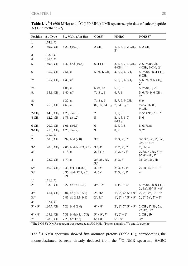
Table I.1. 1H (600 MHz) and
13C (150 MHz) NMR spectroscopic data of calcaripeptide
A (1) in methanol-d4.
Position δC, Type δH, Mult. (J in Hz) COSY HMBC NOESYa
1 174.2, C
2 49.7, CH 4.23, q (6.9) 2-CH3 1, 3, 4, 5, 2-CH3,
2''
5, 2-CH3
3 198.6, C
4 136.6, C
5 149.6, CH 6.42, br d (10.4) 6, 4-CH3 3, 4, 6, 7, 4-CH3,
6-CH3
2, 6, 7a/8a, 7b,
4-CH3, 6-CH3, 2''
6 35.2, CH 2.54, m 5, 7b, 6-CH3 4, 5, 7, 6-CH3 5, 7a/8a, 8b, 4-CH3,
6-CH3
7a 35.7, CH2 1.40, mb
5, 6, 8, 6-CH3 5, 6, 7b, 9, 6-CH3,
2''
7b 1.06, m 6, 8a, 8b 5, 8, 9 5, 7a/8a, 9, 2''
8a 35.9, CH2 1.40, mb
7b, 8b, 9 6, 7, 9 5, 6, 7b, 9, 6-CH3,
2''
8b 1.32, m 7b, 8a, 9 5, 7, 9, 9-CH3 6, 9
9 75.0, CH 4.65, m 8a, 8b, 9-CH3 7, 9-CH3, 1' 7a/8a, 7b, 8b,
9-CH3
2-CH3 14.3, CH3 1.28, d (6.9) 2 1, 2, 3 2, 5'' + 9'', 6'' + 8''
4-CH3 12.2, CH3 1.73, d (1.2) 5 3, 4, 5, 6, 7,
6-CH3
5, 6
6-CH3 20.7, CH3 1.01, d (6.6) 6 5, 6, 7, 8 5, 6, 7a/8a
9-CH3 21.0, CH3 1.20, d (6.2) 9 8, 9 9, 2''
1' 171.2, C
2' 60.5, CH 3.92, br d (7.0) 3b' 1', 3', 4', 5' 3a', 3b', 5a', 2'', 3a'',
3b'', 5'' + 9''
3a' 28.8, CH2 2.06, br dd (12.3, 7.0) 3b', 4' 1', 2', 4', 5' 2', 3b', 4'
3b' 1.13, m 2', 3a', 4' 1', 2', 4', 5' 2', 3a', 4', 5a', 5'' +
9'', 6'' + 8'', 7''
4' 22.7, CH2 1.79, m 3a', 3b', 5a',
5b'
2', 3', 5' 3a', 3b', 5a', 5b'
5a' 46.8, CH2 3.43, dt (11.8, 8.8) 4', 5b' 2', 3', 4', 1'' 2', 3b', 4', 5'' + 9''
5b' 3.36, ddd (12.2, 9.2,
3.2)
4', 5a' 2', 3', 4', 1'' 4'
1'' 171.8, C
2'' 53.8, CH 5.27, dd (9.1, 5.6) 3a'', 3b'' 1, 1'', 3'', 4'' 5, 7a/8a, 7b, 9-CH3,
2', 3a'', 3b'', 5'' + 9''
3a'' 41.4, CH2 3.04, dd (12.9, 5.6) 2'', 3b'' 1'', 2'', 4'', 5'' + 9'' 2', 2'', 3b'', 5'' + 9''
3b'' 2.86, dd (12.9, 9.1) 2'', 3a'' 1'', 2'', 4'', 5'' + 9'' 2', 2'', 3a'', 5'' + 9''
4'' 137.4, C
5'' + 9'' 130.7, CH 7.22, br d (8.4) 6'' + 8'' 2'', 3'', 7'', 5'' + 9'' 2-CH3, 2', 3b', 5a',
2'', 3a'', 3b''
6'' + 8'' 129.8, CH 7.31, br dd (8.4, 7.3) 5'' + 9'', 7'' 4'', 6'' + 8'' 2-CH3, 3b'
7'' 128.3, CH 7.25, br t (7.3) 6'' + 8'' 5'' + 9'' 3b' aThe NOESY NMR spectrum was recorded at 500 MHz.
bProton signals of 7a and 8a overlap.
The 1H NMR spectrum showed five aromatic protons (Table I.1), corroborating the
monosubstituted benzene already deduced from the 13
C NMR spectrum. HMBC
CHAPTER I 29
correlations of H2-3″ (δH 2.86 and 3.04) to the aromatic carbons (C-4″, C- 5″ + C-9″) as
well as to C-2″ and C-1″ established a phenylalanine residue as a partial structure of 1.
The COSY spectrum displayed couplings between H2-3′ (δH 1.13 and 2.06), H2-4′ (δH
1.79), and H2-5′ (δH 3.36 and 3.43) indicating three consecutive methylene groups,
characteristic of a proline residue. In addition, correlations of H-2′ (δH 3.92) to H-3b′ in
the COSY spectrum and to C-1′ in the HMBC spectrum evidenced that C-2′ was the
proline α-carbon. The amide linkage between the phenylalanine and proline residues
was proven by an HMBC correlation of H2-5′ to C-1″ and is consistent with the
chemical shift of C-1″ (δC 171.8). A coupling of H-9 to C-1′ indicated a connection
between CH-9 and C-1′, which was further characterized as an ester bond by the
respective chemical shifts (δC 75.0, δH 4.65, and δC 171.2). The COSY spectrum
revealed that H-9 was part of a proton spin system that reached from 9-CH3 (δH 1.20) to
H-5 (δH 6.42) including the methyl group 6-CH3. According to the chemical shifts, CH-
5 (δC 149.6, δH 6.42) was olefinic. The methyl-substituted, olefinic carbon C-4 had to be
located adjacent to CH-5 due to long-range H,C-couplings of H-5 to C-4 and 4-CH3 and
of 4-CH3 (δH 1.73) to C-4 and C-5. The E-configuration of the double bond was
determined on the basis of the NOESY spectrum, in which 4-CH3 showed a more
intense cross-peak with H-6 than with H-5. In addition, the configuration was supported
by a NOESY cross-peak between 6-CH3 and H-5. HMBC correlations of H-2 (δH 4.23)
to 2-CH3, C-1, and C-3 as well as correlations of H-5 and 4-CH3 to C-3 completed the
consecutive chain from C-1 to C-9. Finally, correlations of H-2 to C-2″ and H-2″ to C-1
linked C-1 to the phenylalanine residue via an amide bond.
The aromatic ring, the proline ring, the macrocycle, four carbonyl groups, and the
double bond Δ4,5
accounted for 11 degrees of unsaturation, as was required by the
molecular formula of 1. Thus, a cyclodepsipeptide structure was established for 1.
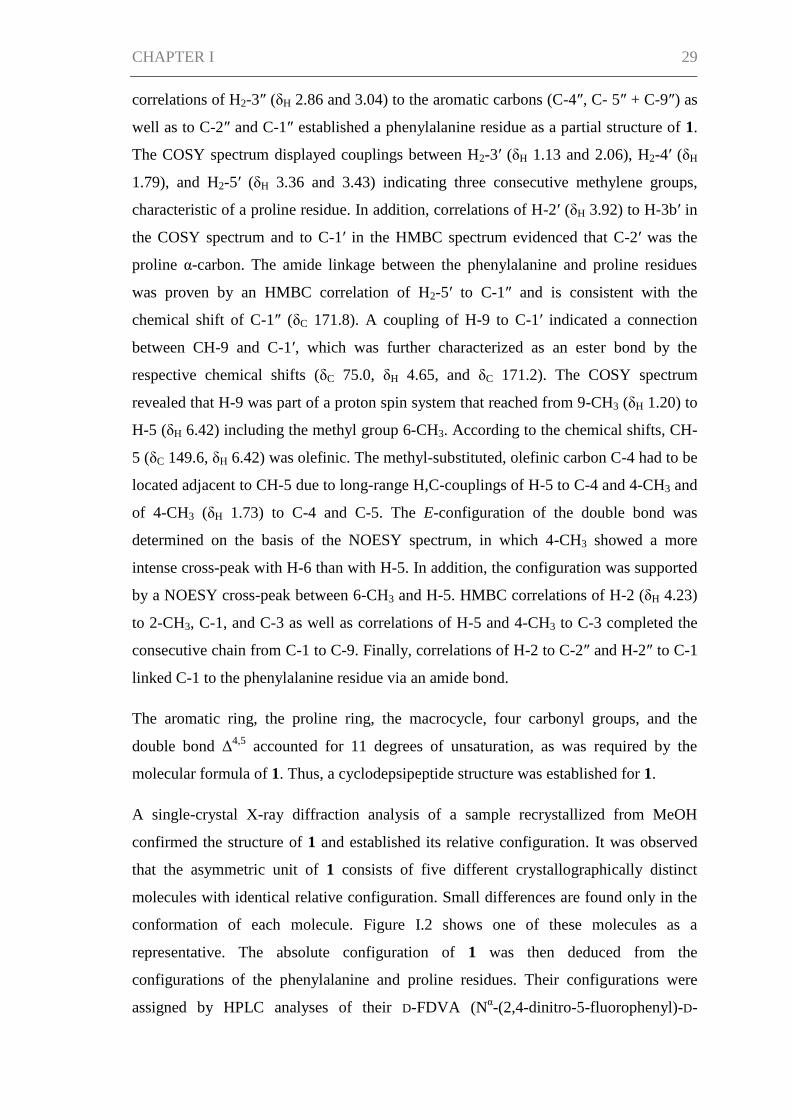
A single-crystal X-ray diffraction analysis of a sample recrystallized from MeOH
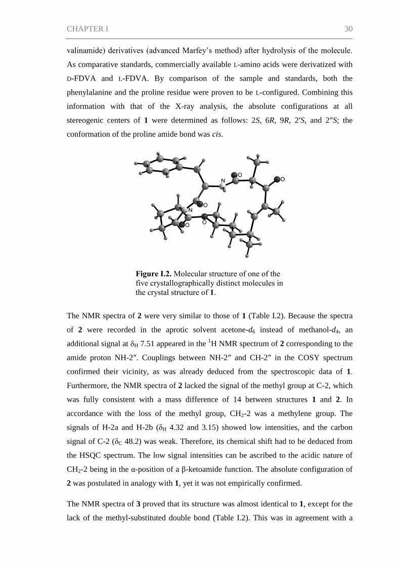
confirmed the structure of 1 and established its relative configuration. It was observed
that the asymmetric unit of 1 consists of five different crystallographically distinct
molecules with identical relative configuration. Small differences are found only in the
conformation of each molecule. Figure I.2 shows one of these molecules as a
representative. The absolute configuration of 1 was then deduced from the
configurations of the phenylalanine and proline residues. Their configurations were
assigned by HPLC analyses of their D-FDVA (Nα-(2,4-dinitro-5-fluorophenyl)-D-
CHAPTER I 30
valinamide) derivatives (advanced Marfey’s method) after hydrolysis of the molecule.
As comparative standards, commercially available L-amino acids were derivatized with
D-FDVA and L-FDVA. By comparison of the sample and standards, both the
phenylalanine and the proline residue were proven to be L-configured. Combining this
information with that of the X-ray analysis, the absolute configurations at all
stereogenic centers of 1 were determined as follows: 2S, 6R, 9R, 2′S, and 2″S; the
conformation of the proline amide bond was cis.
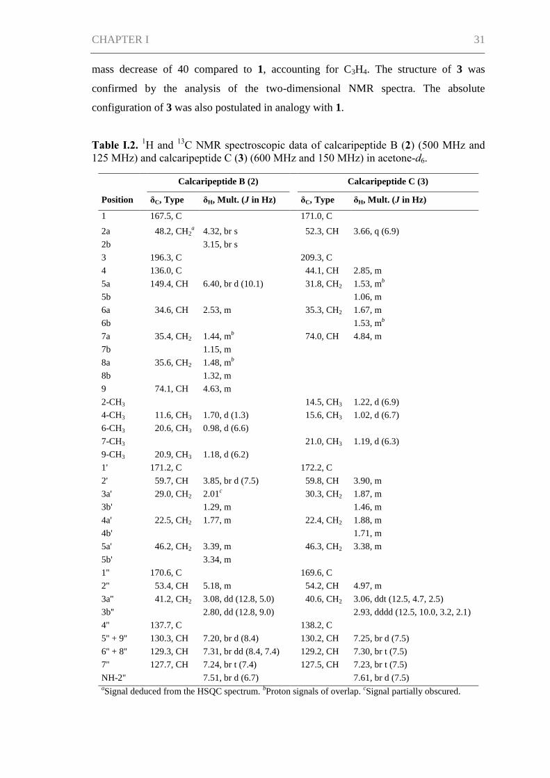
The NMR spectra of 2 were very similar to those of 1 (Table I.2). Because the spectra
of 2 were recorded in the aprotic solvent acetone-d6 instead of methanol-d4, an
additional signal at δH 7.51 appeared in the 1H NMR spectrum of 2 corresponding to the
amide proton NH-2″. Couplings between NH-2″ and CH-2″ in the COSY spectrum
confirmed their vicinity, as was already deduced from the spectroscopic data of 1.
Furthermore, the NMR spectra of 2 lacked the signal of the methyl group at C-2, which
was fully consistent with a mass difference of 14 between structures 1 and 2. In
accordance with the loss of the methyl group, CH2-2 was a methylene group. The
signals of H-2a and H-2b (δH 4.32 and 3.15) showed low intensities, and the carbon
signal of C-2 (δC 48.2) was weak. Therefore, its chemical shift had to be deduced from
the HSQC spectrum. The low signal intensities can be ascribed to the acidic nature of
CH2-2 being in the α-position of a β-ketoamide function. The absolute configuration of
2 was postulated in analogy with 1, yet it was not empirically confirmed.
The NMR spectra of 3 proved that its structure was almost identical to 1, except for the
lack of the methyl-substituted double bond (Table I.2). This was in agreement with a
Figure I.2. Molecular structure of one of the
five crystallographically distinct molecules in
the crystal structure of 1.
CHAPTER I 31
mass decrease of 40 compared to 1, accounting for C3H4. The structure of 3 was
confirmed by the analysis of the two-dimensional NMR spectra. The absolute
configuration of 3 was also postulated in analogy with 1.
Table I.2. 1H and
13C NMR spectroscopic data of calcaripeptide B (2) (500 MHz and
125 MHz) and calcaripeptide C (3) (600 MHz and 150 MHz) in acetone-d6.
Calcaripeptide B (2) Calcaripeptide C (3)
Position δC, Type δH, Mult. (J in Hz) δC, Type δH, Mult. (J in Hz)
1 167.5, C 171.0, C
2a 48.2, CH2a 4.32, br s 52.3, CH 3.66, q (6.9)
2b 3.15, br s
3 196.3, C 209.3, C
4 136.0, C 44.1, CH 2.85, m
5a 149.4, CH 6.40, br d (10.1) 31.8, CH2 1.53, mb
5b 1.06, m
6a 34.6, CH 2.53, m 35.3, CH2 1.67, m
6b 1.53, mb
7a 35.4, CH2 1.44, mb 74.0, CH 4.84, m
7b 1.15, m
8a 35.6, CH2 1.48, mb
8b 1.32, m
9 74.1, CH 4.63, m
2-CH3 14.5, CH3 1.22, d (6.9)
4-CH3 11.6, CH3 1.70, d (1.3) 15.6, CH3 1.02, d (6.7)
6-CH3 20.6, CH3 0.98, d (6.6)
7-CH3 21.0, CH3 1.19, d (6.3)
9-CH3 20.9, CH3 1.18, d (6.2)
1' 171.2, C 172.2, C
2' 59.7, CH 3.85, br d (7.5) 59.8, CH 3.90, m
3a' 29.0, CH2 2.01c 30.3, CH2 1.87, m
3b' 1.29, m 1.46, m
4a' 22.5, CH2 1.77, m 22.4, CH2 1.88, m
4b' 1.71, m
5a' 46.2, CH2 3.39, m 46.3, CH2 3.38, m
5b' 3.34, m
1'' 170.6, C 169.6, C
2'' 53.4, CH 5.18, m 54.2, CH 4.97, m
3a'' 41.2, CH2 3.08, dd (12.8, 5.0) 40.6, CH2 3.06, ddt (12.5, 4.7, 2.5)
3b'' 2.80, dd (12.8, 9.0) 2.93, dddd (12.5, 10.0, 3.2, 2.1)
4'' 137.7, C 138.2, C
5'' + 9'' 130.3, CH 7.20, br d (8.4) 130.2, CH 7.25, br d (7.5)
6'' + 8'' 129.3, CH 7.31, br dd (8.4, 7.4) 129.2, CH 7.30, br t (7.5)
7'' 127.7, CH 7.24, br t (7.4) 127.5, CH 7.23, br t (7.5)
NH-2'' 7.51, br d (6.7) 7.61, br d (7.5) aSignal deduced from the HSQC spectrum.
bProton signals of overlap.
cSignal partially obscured.
CHAPTER I 32
Structurally, the calcaripeptides are related to acremolides A and B (Ratnayake et al.
2008). The compounds share the feature of being cyclodepsipeptides containing an L-
proline-L-phenylalanine moiety that together with a nonpeptidic partial structure forms
the macrocycle. However, the nonpeptidic part of the acremolides and calcaripeptides
differs, and the acremolides possess an additional seven-membered alkyl side chain
connected to the ring. The 16- and 14-membered rings of 1, 2, and 3 are unusual for
natural products.
The calcaripeptides were tested for activities against five bacterial test strains, three
fungal test strains, one oomycete, and two cell lines as well as for inhibition of selected
enzyme targets (glycogen synthase kinase-3β, acetylcholinesterase, phosphodiesterase
4B2, and protein tyrosine phosphatase 1B). In addition, 1 was tested in further assays
including 24 cell lines. Despite the broad panel of 43 assays, neither antibacterial,
antifungal, and cytotoxic properties nor inhibition of the enzyme targets could be
detected for the calcaripeptides (data not shown; for information on test strains, cell
lines and enzymes see the Supporting Information (SI)).
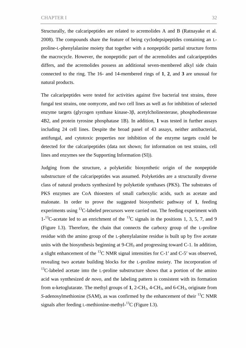
Judging from the structure, a polyketidic biosynthetic origin of the nonpeptide
substructure of the calcaripeptides was assumed. Polyketides are a structurally diverse
class of natural products synthesized by polyketide synthases (PKS). The substrates of
PKS enzymes are CoA thioesters of small carboxylic acids, such as acetate and
malonate. In order to prove the suggested biosynthetic pathway of 1, feeding
experiments using 13
C-labeled precursors were carried out. The feeding experiment with
1-13
C-acetate led to an enrichment of the 13
C signals in the positions 1, 3, 5, 7, and 9
(Figure I.3). Therefore, the chain that connects the carboxy group of the L-proline
residue with the amino group of the L-phenylalanine residue is built up by five acetate
units with the biosynthesis beginning at 9-CH3 and progressing toward C-1. In addition,
a slight enhancement of the 13
C NMR signal intensities for C-1′ and C-5′ was observed,
revealing two acetate building blocks for the L-proline moiety. The incorporation of
13C-labeled acetate into the L-proline substructure shows that a portion of the amino
acid was synthesized de novo, and the labeling pattern is consistent with its formation
from α-ketoglutarate. The methyl groups of 1, 2-CH3, 4-CH3, and 6-CH3, originate from
S-adenosylmethionine (SAM), as was confirmed by the enhancement of their 13
C NMR
signals after feeding L-methionine-methyl-13
C (Figure I.3).
CHAPTER I 33
Figure I.3. Biosynthetic origin of calcaripeptide
A (1) as determined by 13
C-labeling. For
detailed information on the calculation of the 13
C enrichment see SI.
O
O
N
O
O
NH
O
According to the feeding studies, a polyketidic origin of the nonpeptidic part of the
calcaripeptides was confirmed. The overall structure and the biosynthetic origin of the
building blocks support the involvement of a hybrid between a polyketide synthase and
nonribosomal peptide synthetase for the peptidic backbone (PKS−NRPS hybrid) in the
formation of the calcaripeptides. An increasing number of fungal compounds have
recently been shown to be synthesized by this type of enzyme, e.g., fusarin C,
aspyridone, or pseurotin (Collemare et al. 2008). The genome of Calcarisporium sp.
strain KF525 is currently under investigation with the aim of identifying genes encoding
the respective PKS−NRPS hybrids responsible for the biosynthesis of the
calcaripeptides. Reference sequences of the known hybrid PKS−NRPS genes from other
fungi are available for comparison.
Promiscuity with respect to the amino acid substrates has been shown for fungal NRPS
in vitro and in vivo (Qiao et al. 2011, Krause et al. 2001, Xu et al. 2007). As
promiscuity might thus be a possibility for the postulated NRPS component of the
biosynthesis of 1−3, it was tested whether a supply of structurally related, alternative
amino acids as substrates for the NRPS would lead to derivatives of 1. Supplementation
of L-tryptophan, L-tyrosine, or L-histidine to the culture broth of KF525 did not result in
feeding with enhancement of (enrichment)
1-13C-acetate C-1 (3.6%), C-3 (3.3%), C-5 (3.8%),
C-7 (3.0%), C-9 (3.2%), C-1' (1.4%),
C-5' (1.5%)
Me-13C-methionine 2-CH3 (63.7%), 4-CH3 (62.3%),
6-CH3 (60.5%)
1-13
C-acetate
SAM derived methyl group
CHAPTER I 34
their incorporation into 1 in place of L-proline or L-phenylalanine. Therefore, the
enzymes involved in the biosynthesis of the calcaripeptides seem to be specific for their
amino acid substrates. Additional supply of L-proline and L-phenylalanine as the
naturally incorporated amino acids led to an increased production of 1, displaying
potential for an optimization in the fermentation process.
Experimental Section
General Experimental Procedures
Melting points were determined on an Electrothermal melting point apparatus. Optical
rotation measurements were performed on a Perkin-Elmer model 241 polarimeter. UV
spectra were obtained on a Perkin-Elmer Lambda 2 spectrophotometer. NMR spectra
were recorded on Bruker DRX 500 (500 and 125 MHz for 1H and
13C NMR,
respectively) and Bruker AV 600 spectrometers (600 and 150 MHz for 1H and
13C
NMR, respectively), using the residual solvent signals as internal references (δH 3.31
and δC 49.0 for methanol-d4; δH 2.05 and δC 29.8 for acetone-d6). Measurements of
high-resolution mass spectra were conducted on a benchtop time-of-flight spectrometer
(micrOTOF II, Bruker) with positive ESI. Analytical reversed-phase HPLC-UV/MS
experiments were carried out on a VWR-Hitachi LaChrom Elite system consisting of an
L-2130 pump, an L-2450 diode array detector, an L-2200 autosampler, an L-2300
column oven, and a Phenomenex Onyx Monolithic column (C18, 100 × 3.00 mm)
applying an H2O (A)/ MeCN (B) gradient with 0.1% HCOOH added to both solvents
(gradient: 0 min 5% B, 4 min 60% B, 6 min 100% B; flow 2 mL min−1
). For mass
detections the HPLC system was coupled to an ESI-ion trap detector (Esquire4000,
Bruker Daltonics). Preparative HPLC was performed on a VWR LaPrep system
equipped with a P110 pump, a P311 UV detector, a Labocol Vario-2000 fraction
collector (LABOMATIC), a Smartline 3900 autosampler (Knauer), and a Phenomenex
Gemini-NX column (10 μ C18, 100A, Axia, 100 × 50.00 mm). Further compound
purifications were conducted on a Merck-Hitachi LaChrom Elite HPLC system
consisting of an L-7150 pump, an L-2450 diode array detector, an L-2200 autosampler,
and a Phenomenex Gemini-NX column (5 μ C18, 110A, Axia, 100 × 21.20 mm). The
eluents for all preparative HPLC separations were H2O (A) and MeCN (B) with 0.1%
HCOOH added to both solvents.
CHAPTER I 35
Isolation and Identification of the Fungal Strain
The strain KF525 was isolated from a water sample collected in the German Wadden
Sea. The DNA extraction, the amplification of the internal transcribed spacer region
(ITS), and the sequencing were performed using standard protocols (Wiese et al. 2011)
modified in that the centrifugation was carried out at 8000g for DNA extraction,
DreamTaq Green PCR Master Mix (2×) (Fermentas) was employed for amplification,
35 cycles instead of 30 were conducted for amplification, and the primer ITS4 was used
for sequencing. The DNA sequence was deposited in GenBank under the accession
number KC800713. A sequence analysis in GenBank using the Basic Local Alignment
Search Tool (BLAST) gave 91% similarity to Calcarisporium arbuscula. A
microscopic analysis of the strain showed structures typical of the genus
Calcarisporium. Taken together, the sequence and morphological data allowed the
identification of strain KF525 as a Calcarisporium sp., as was additionally confirmed
by the Centraalbureau voor Schimmelcultures (CBS, Utrecht, The Netherlands).
Fermentation
Cultivation experiments were performed in 16 2-L Erlenmeyer flasks, each containing
750 mL of modified casamino acids glucose medium (2.5 g of casein hydrolysate, 40 g
of glucose × H2O, 0.1 g of MgSO4 × 7H2O, 1.8 g of KH2PO4 per liter of distilled H2O,
pH 6.8) (Stevens 1974). Cultures were inoculated with a circular agar slant (1.8 cm in
diameter) of a preculture grown on solid modified Wickerham medium (3 g of malt
extract, 3 g of yeast extract, 5 g of peptone from soymeal, 10 g of glucose × H2O, 30 g
of NaCl, 15 g of agar per liter of distilled H2O, pH 6.25) (Wickerham 1951). The
preculture was incubated at room temperature in the dark for 11 days. The main cultures
were incubated at 22 °C under shaking conditions (120 rpm) in the dark for 24 days.
Purification of Calcaripeptides A−C
The culture broth of KF525 was separated into the culture supernatant and the
mycelium. The culture filtrate was extracted with EtOAc. The organic solvent was
evaporated to dryness in vacuo to give 0.92 g of extract. The extract was fractionated by
preparative HPLC on a VWR LaPrep system (gradient: 0 min 10% B, 17 min 60% B,
22 min 100% B; flow 100 mL min−1
; UV detection at 217 nm), yielding three fractions
that contained the calcaripeptides (tR 15.7, 16.6, and 17.6 min). These fractions were
CHAPTER I 36
further purified on a Merck-Hitachi LaChrom Elite system to give 117.9 mg of 1
(isocratic: 42% B; flow 18 mL min−1
; UV detection at 220 nm; tR 10.7 min), 6.2 mg of 2
(isocratic: 29% B; UV detection at 205 nm; tR 27.5 min), and 8.5 mg of a compound
mixture containing 3 (gradient: 0 min 40% B, 13 min 65% B; flow 18 mL min−1
; UV
detection at 205 nm; tR 6.2 min). Compound 3 was subjected to a third preparative
HPLC purification on the same system (gradient: 0 min 40% B, 13 min 80% B; flow 18
mL min−1
; UV detection at 205 nm; tR 5.8 min), resulting in a yield of 5.7 mg.
Calcaripeptide A (1): white needles or amorphous solid (MeOH); mp 199−201 °C;
[α]20
D −133 (c 1.4, MeOH); UV (MeOH) λmax (log ε) 201 (4.43), 230 (sh) (4.14) nm; 1D
and 2D NMR data, see Table I.1; HRESIMS m/z 469.2707 [M + H]+ (calcd for
C27H37N2O5, 469.2697).
Calcaripeptide B (2): colorless oil; [α]20
D −113 (c 0.31, MeOH); UV (MeOH) λmax (log
ε) 201 (4.35) nm, 230 (sh) (4.09); 1D and 2D NMR data, see Table I.2 and SI;
HRESIMS m/z 455.2538 [M + H]+ (calcd for C26H35N2O5, 455.2541).
Calcaripeptide C (3): colorless oil; [α]20
D −79 (c 0.2, MeOH); UV (MeOH) λmax (log ε)
201 (4.24) nm; 1D and 2D NMR data, see Table I.2 and SI; HRESIMS m/z 429.2379
[M + H]+ (calcd for C24H33N2O5, 429.2384).
X-ray Crystal Structure Determination
Data collection was performed using an imaging plate diffraction system (IPDS-2) from
STOE & CIE at 293 K using Mo Kα radiation (γ = 0.71073 Å). Formula: C27H36N2O5,
molecular weight: 468.58 g mol−1
, monoclinic, space group C2; unit cell dimensions: a
= 44.5917(12) Å, b = 9.6204(2) Å, c = 37.0616(10) Å, β = 122.453(2)°, V = 13416.1(6)
Å3, Z = 20, Dcalcd = 20, 1.160 Mg/m
3, μ = 0.080 mm
-1. The structure was solved with
methods using SHELXS-97, and refinement was performed against F2 using SHELXH.
All non-hydrogen atoms were refined anisotropically. The H atoms were positioned
with idealized geometry and refined isotropically with Uiso(H) = 1.2Ueq(C,N) using a
riding model (1.5 for methyl H atoms). A total of 27 668 reflections were measured in
the range of 2θ 1.2−24.6°, of which 11 536 are independent (Rint = 0.0347), 1531
parameters, GOF = 1.080, R1 for 8490 reflections with I > 2σ(I) = 0.0521, wR2 for all
reflections = 0.1205. Residual electron density = 0.326/0.164 e/Å3. In one of the five
independent molecules slightly enlarged anisotropic displacement parameters are
observed for some C atoms, indicating disorder that cannot be resolved successfully.
CHAPTER I 37
Because no strong anomalous scattering atoms are present, the absolute structure and
absolute configuration cannot be determined, and therefore, Friedel equivalents were
merged in the refinement.
Crystallographic data for 1 have been deposited with the Cambridge Crystallographic
Data Centre (CCDC-943694). Copies of the data can be obtained, free of charge, on
application to the Director, CCDC, 12 Union Road, Cambridge CB2 1EZ, UK (fax:
+44-(0)1223-336033 or e-mail: [email protected]).
Preparation and Analysis of D- and L-FDVA Derivatives
The hydrolysis of 1 was achieved by dissolving 2.7 mg of the compound in 1.5 mL of 6
N HCl and subsequent heating at 110 °C overnight. The reaction mixture was then
concentrated to dryness and redissolved in 250 μL of H2O. For the derivatization, 50 μL
of the hydrolysate solution was mixed with 100 μL of a 1% (w/v) solution of D-FDVA
(Nα-(2,4-dinitro-5-fluorophenyl)-D-valinamide) in acetone. After addition of 40 μL of 1
M NaHCO3 and 70 μL of DMSO, the mixture was incubated at 60 °C for 2 h. The
reaction was stopped by addition of 30 μL of 2 M HCl. The amino acid standards (50
mM L-proline and L-phenylalanine) were derivatized with D-FDVA and L-FDVA in the
above-described manner. Prior to analytical HPLC-UV/MS analyses the reaction
mixtures were diluted 100-fold with MeOH/H2O (1:1). The retention times (min) of the
amino acid standard derivatives were as follows: D-FDVA-L-Pro (3.69), L-FDVA-L-Pro
(3.48), D-FDVA-L-Phe (4.50), and L-FDVA-L-Phe (4.07). The HPLC analysis of the
hydrolysate D-FDVA derivatives showed peaks at 3.69 and 4.50 min. An additional
confirmation of the amino acid configuration was accomplished by spiking the
hydrolysate derivatives with the amino acid standard derivatives.
Biosynthetic Studies
For the feeding experiments, Calcarisporium sp. strain KF525 was cultivated as
described in the Fermentation Section above. Precultures were 7 to 15 days old. Each
feeding experiment was performed in one 2-L Erlenmeyer flask, containing 750 mL of
medium. After five days of cultivation 13
C-labeled compounds were added as sterile
filtered, aqueous solutions (500 mg of sodium acetate-1-13
C, Isotec or 250 mg of L-
methionine-methyl-13
C, Cambridge Isotope Laboratories). For the extraction, the
supernatant and the mycelium of the cultures were separated after 21 days of
CHAPTER I 38
incubation. The culture filtrate was extracted with EtOAc as described above, while the
mycelium was extracted with EtOH. The culture filtrate fed with 13
C-labeled acetate
was purified on a Merck-Hitachi LaChrom Elite system (gradient: 0 min 28% B, 30 min
61% B; flow 18 mL min−1
; UV detection at 230 nm; tR 17.9 min) to give 8.2 mg of 1.
In the feeding experiment with 13
C-labeled methionine, compound 1 was isolated from
the extracts of both the culture filtrate and the mycelium. Afterward, purified 1 was
combined for NMR spectroscopy studies. The extract of the culture filtrate was
separated on a Merck-Hitachi LaChrom Elite system (gradient: 0 min 28% B, 20 min
50% B; flow 18 mL min−1
; UV detection at 230 nm; tR 14.7 min), yielding 1.6 mg of 1.
The purification of compound 1 from the extract of the mycelium was conducted on the
same system (gradient: 0 min 35% B, 20 min 50% B, 20.5 min 70% B, flow 18 mL
min−1
; UV detection at 230 nm; tR 12.7 min) with a yield of 0.6 mg.
Feeding Alternative Amino Acids
Each amino acid experiment was performed in one 2-L Erlenmeyer flask, containing
750 mL of medium. Culture conditions and medium were the same as described in the
Fermentation Section above. After five days of cultivation the respective amino acid
was added as a sterile filtered, 50 mL aqueous solution (end concentration in culture
medium: 1 g L−1
). The added amino acids were L-tryptophan, L-tyrosine, L-histidine, L-
proline, L-phenylalanine, and L-proline plus L-phenylalanine. L-Tyrosine did not
dissolve completely, and only the dissolved portion was added to the culture. Two
cultures without additionally supplied amino acids served as controls in the experiment.
The culture filtrate was extracted with EtOAc as described above, while the mycelium
was extracted with EtOH. The extracts were analyzed by analytical HPLC-UV/MS.
Acknowledgments
We gratefully thank Dr. K. Schaumann for providing the strain KF525, A. Erhard and
the European ScreeningPort for bioactivity and enzyme assays, and G. Kohlmeyer-
Yilmaz, M. H ftmann, and r. F. S nnichsen for running and processing NMR
experiments. We also thank the Institute of Clinical Molecular Biology in Kiel for
providing Sanger sequencing as supported in part by the DFG Cluster of Excellence
“Inflammation at Interfaces” and “Future Ocean”. We thank the technicians S. Greve
and S. Arndt for technical support. This study was performed in the framework of
CHAPTER I 39
MARINE FUNGI, EU FP7 KBBE program, project no. 265926 at the Kieler Wirkstoff-
Zentrum (KiWiZ) at the GEOMAR Helmholtz Centre for Ocean Research Kiel.
References
Baldwin, C. L., Weaver, L. C., Brooker, R. M., Jacobsen, T. N., Osborne Jr, C. E.,
Nash, H. A. (1964). Biological and chemical properties of aurovertin: A metabolic
product of Calcarisporium abuscula. Lloydia 27(2), 88-95.
Barnett, H. L. (1958). A new Calcarisporium parasitic on other fungi. Mycologia 50(4),
497-500.
Barnett, H. L., Lilly, V. G. (1958). Parasitism of Calcarisporium parasiticum on
species of Physalospora and related fungi. West Virginia University Agricultural
Experiment Station, Morgantown, West Virginia, USA, pp. 37.
Carrión, G., Rico-Gray, V. (2002). Mycoparasites on the coffee rust in Mexico. Fungal
Diversity 11, 49-60.
Chrisp, P., Dewick, P. M., Boyle, F. T. (1990). Antifungal antibiotics from
Calcarisporium thermophilum: a new source of 15-azahomosterol derivatives. Z.
Naturforsch. C Biosci. 45(3-4), 179-186.
Collemare, J., Billard, A., Böhnert, H. U., Lebrun, M.-H. (2008). Biosynthesis of
secondary metabolites in the rice blast fungus Magnaporthe grisea: the role of hybrid
PKS–NRPS in pathogenicity. Mycol. Res. 112(2), 207-215.
Cooper, J. A. (2005). New Zealand hyphomycete fungi: additional records, new species,
and notes on interesting collections. N. Z. J. Bot. 43(1), 323-349.
Dictionary of Natural Products (2012). Chapman & Hall/CRC Press/Hampden Data
Services, Ltd.
Evans, H. C. (1971). Thermophilous fungi of coal spoil tips: II. Occurrence, distribution
and temperature relationships. Trans. Br. Mycol. Soc. 57(2), 255-266.
Gong, L.-J., Guo, S.-X. (2010). Endophytic fungi from Dracaena cambodiana and
Aquilaria sinensis and their antimicrobial activity. Afr. J. Biotechnol. 8(5), 731-736.
CHAPTER I 40
Hijwegen, T. (1989). Effect of culture filtrates of seventeen fungicolous fungi on
sporulation of cucumber powdery mildew. Neth. J. Plant Pathol. 95(Supplement 1), 95-
98.
Hijwegen, T., Verhaar, M. A. (1993). Induced resistance to Peronospora parasitica in
red cabbage. Neth. J. Plant Pathol. 99(Supplement 3), 103-107.
Ji, L. L., Song, Y. C., Tan, R. X. (2004). A potent feed preservative candidate produced
by Calcarisporium sp., an endophyte residing in stargrass (Cynodon dactylon). J. Appl.
Microbiol. 96(2), 352-358.
Krause, M., Lindemann, A., Glinski, M., Hornbogen, T., Bonse, G., Jeschke, P.,
Thielking, G., Gau, W., Kleinkauf, H., Zocher, R. (2001). Directed biosynthesis of new
enniatins. J. Antibiot. 54(10), 797-804.
Mulheirn, L. J., Beechey, R. B., Leworthy, D. P., Osselton, M. D. (1974). Aurovertin B,
a metabolite of Calcarisporium arbuscula. J. Chem. Soc., Chem. Commun., 874-876.
Osselton, M. D., Baum, H., Beechey, R. B. (1974). Isolation, purification and
characterization of aurovertin B. Biochem. Soc. Trans. 2, 200-202.
Qiao, K., Zhou, H., Xu, W., Zhang, W., Garg, N., Tang, Y. (2011). A fungal
nonribosomal peptide synthetase module that can synthesize thiopyrazines. Org. Lett.
13(7), 1758-1761.
Rambelli, A., Mulas, B., Pasqualetti, M. (2004). Comparative studies on microfungi in
tropical ecosystems in Ivory Coast forest litter: behaviour on different substrata. Mycol.
Res. 108(3), 325-336.
Ratnayake, R., Fremlin, L. J., Lacey, E., Gill, J. H., Capon, R. J. (2008). Acremolides
A− , lipodepsipeptides from an Australian marine-derived fungus, Acremonium sp. J.
Nat. Prod. 71(3), 403-408.
Rombach, M. C., Roberts, D. W. (1987). Calcarisporium ovalisporum, symbiotic with
the insect pathogen Hirsutella citriformis. Mycologia 79(1), 153-155.
Somrithipol, S., Jones, E. B. G. (2006). Calcarisporium phaeopodium sp. nov., a new
hyphomycete from Thailand. Sydowia 58(1), 133-140.
CHAPTER I 41
Stevens, R. B. (1974). Mycology guidebook. University of Washington Press, Seattle,
WA, USA, pp. 703.
Sutton, B. C. (1973). Hyphomycetes from Manitoba and Saskatchewan, Canada.
Commonwealth Mycological Institute, Kew, Surrey, UK, pp. 143.
Watson, P. (1955). Calcarisporium arbuscula living as an endophyte in apparently
healthy sporophores of Russula and Lactarius. Trans. Br. Mycol. Soc. 38(4), 409-414.
Wickerham, L. J. (1951). Taxonomy of yeasts. US Dept. of Agriculture, Washington, D.
C., USA, pp. 56.
Wiese, J., Ohlendorf, B., Blümel, M., Schmaljohann, R., Imhoff, J. F. (2011).
Phylogenetic identification of fungi isolated from the marine sponge Tethya aurantium
and identification of their secondary metabolites. Mar. Drugs 9(4), 561-585.
Xu, Y., Zhan, J., Wijeratne, E. M. K., Burns, A. M., Gunatilaka, A. A. L., Molnár, I.
(2007). Cytotoxic and antihaptotactic beauvericin analogues from precursor-directed
biosynthesis with the insect pathogen Beauveria bassiana ATCC 7159. J. Nat. Prod.
70(9), 1467-1471.
Yu, N.-J., Guo, S.-X., Lu, H.-Y. (2002a). Cytotoxic macrocyclic trichothecenes from
the mycelia of Calcarisporium arbuscula Preuss. J. Asian Nat. Prod. Res. 4(3), 179-
183.
Yu, N.-J., Guo, S.-X., Xiao, P.-G. (2002b). New sesquiterpene esters from the culture
filtrate of Calcarisporium arbuscula isolated from Ganoderma lucidum. Acta Bot. Sin.
44(7), 878-882.
CHAPTER II 42
CHAPTER II
Calcarides A–E, antibacterial macrocyclic and linear polyesters from a
Calcarisporium strain
Johanna Silber, Birgit Ohlendorf, Antje Labes, Arlette Erhard and Johannes F. Imhoff
Published in 2013 as an article in Mar. Drugs 11(9), 3309-3323
CHAPTER II 43
Abstract
Bioactive compounds were detected in crude extracts of the fungus, Calcarisporium sp.
KF525, which was isolated from German Wadden Sea water samples. Purification of
the metabolites from the extracts yielded the five known polyesters, 15G256α, α-2, β, β-
2 and π (1–5), and five new derivatives thereof, named calcarides A–E (6–10). The
chemical structures of the isolated compounds were elucidated on the basis of one- and
two-dimensional NMR spectroscopy supported by UV and HRESIMS data. The
compounds exhibited inhibitory activities against Staphylococcus epidermidis,
Xanthomonas campestris and Propionibacterium acnes. As the antibacterial activities
were highly specific with regard to compound and test strain, a tight structure-activity
relationship is assumed.
Introduction
Fungal species of the genus, Calcarisporium, have a widespread occurrence and are
frequently found as mycoparasites or symbiotes of higher fungi (Sutton 1973,
Somrithipol and Jones 2006, Ji et al. 2004, Kramer et al. 1959, Watson 1955). The
Calcarisporium sp. strain KF525 investigated in this study was isolated from a water
sample taken in the German Wadden Sea. It showed a diverse chemical profile,
including ten structurally closely related compounds of the 15G256-type known as, e.g.,
15G256α (1) or α-2 (2) (Figure II.1; to be differentiated from the structural class
comprising 15G256γ, δ and ε).
The 15G256-type compounds are produced by a number of fungi, such as ascomycetes,
like Hypoxylon, Penicillium, Talaromyces, Acremonium and Scedosporium species, as
well as by the basidiomycete, Albatrellus confluens (Schlingmann et al. 2002, Breinholt
et al. 1993, Ito et al. 1992a, Roy et al. 1996, Kondo et al. 1993, Arai et al. 2002, Zhou et
al. 2009). Since some metabolites were isolated independently from the different
producer strains, renaming occurred among the 15G256-type compounds. 15G256α (1)
is synonymous with BK233-A and NG-012, 15G256α-1 is a synonym of BK233-B and
probably NG-011 (discussed in Schlingmann et al. 2002), while 15G256β (3) was also
named orbuticin, BK233-C and BE-26263 (Schlingmann et al. 2002, Breinholt et al.
1993, Roy et al. 1996, Kondo et al. 1993, Ito et al. 1992b). With regard to the chemical
structures, the compounds are polyesters that occur either in closed ring structures as
macrocycles or as linear molecules (Figure II.1). Schlingmann et al. (2002) proposed a

CHAPTER II 44
Figure II.1. Compounds isolated from Calcarisporium sp. KF525:
15G256α, α-2, β, β-2 and π (1–5) and calcarides A–E (6–10).
OH
OH O O
O O OH
OHOO
OHO
OH
OH
OH O O
O O OH
OH
O
O
O
O O OR
OH
OH O O
O O OH
OHOO
OHO
O
O
OH
R
O
OH O O
O O OH
OH
O
OO O O
O
OH
O
OH O O
O O OH
OHOO
OHO
O
O
OH
R
O
OH O O
O O OH
OH
O
O
O
O O OR
1 2 3 4
5 7
9
11 13 15 17
21
23 26
29 31
32
15G256π (5)
15G256α (1) R = OH
15G256β (3) R = H
15G256α-2 (2) R = OH
15G256β-2 (4) R = H
Calcaride C (8)
Calcaride A (6) R = H
Calcaride B (7) R = OH
Calcaride D (9) R = OH
Calcaride E (10) R = H
biosynthesis route in which 6-hydroxymellein and β-hydroxybutyric acid (Figure II.2)
are the precursors of the 15G256 components, forming the acyclic polyesters in the first
instance. In later biosynthetic steps, the macrocyclic 15G256 compounds arise from the
linear polyesters by ring closure reactions (Schlingmann et al. 2002). Various biological
activities have been ascribed to the 15G256 agents, including antifungal, estrogenic and
cytotoxic properties, as well as they were shown to potentiate nerve growth factor-
induced neurite outgrowth (Schlingmann et al. 2002, Breinholt et al. 1993, Ito et al.
1992a, Roy et al. 1996, Kondo et al. 1993). For example, 15G256α (1), α-1 and β (3)
attracted attention in the field of crop protection, as they displayed antifungal properties
against important plant pathogenic fungi, e.g., Botrytis cinerea or Monilinia fructigena
(Breinholt et al. 1993).
Here, we report the isolation of five known (1–5) and five new (6–10) 15G256-type
compounds from Calcarisporium sp. strain KF525. Their structure elucidation and
antibacterial activities are described.
CHAPTER II 45
O
OH
OH O
O
O
OH O
OHOH
O
6-Hydroxymellein
β-Hydroxybutyric acid
6-Methoxymellein
Results and Discussion
Isolation and Structure Elucidation
The fungal strain KF525 is a Calcarisporium sp. that is a part of the marine fungal
strain collection of the Kiel Center for Marine Natural Products (Silber et al. 2013).
Extracts of cultures from the Calcarisporium sp. strain KF525 grown in modified
casamino acids glucose medium (Stevens 1974) showed antibacterial activities and
were investigated in detail. Fractionation of these extracts by preparative reversed-phase
HPLC yielded the known compounds, 15G256α, α-2, β, β-2 and π (1–5), as well as five
new derivatives thereof, calcarides A–E (6–10) (Figure II.1).
The structures of 1–5 were identified by comparison of their spectroscopic data (UV,
MS and 1H NMR spectra) with those described in the literature (Schlingmann et al.
2002). As the data agreed well with the values published, the structures of 1–5 were
assigned as shown in Figure II.1.
The molecular formula of calcaride A (6) was determined to be C33H40O14 by high-
resolution ESIMS measurements (measured 661.2503, calculated 661.2491 [M + H]+).
The structure of 6 was elucidated on the basis of one- and two-dimensional NMR
spectra (1H,
13C, DEPT, COSY, HSQC and HMBC, see Table II.1). The NMR spectra
of 6 were measured in acetone-d6, as it did not dissolve well in methanol-d4, whereas
NMR data of all other calcarides were recorded in methanol-d4. The analysis of the
NMR data of 6 revealed considerable structural similarities with 15G256β (3). The 1H
NMR spectrum, the 1H NMR coupling constants and the
13C NMR spectrum of 6
showed that, comparable to 3 (Schlingmann et al. 2002, Breinholt et al. 1993), the
compound was composed of two phenyl moieties with their two protons in meta
position to each other and five methyl, five methylene, five methine and five ester
carbonyl groups. The two-dimensional NMR spectra of 6 proved that the
Figure II.2. Proposed biosynthetic precursors of the calcarides
(based on the proposal of Schlingmann et al. 2002).
CHAPTER II 46
OH
OH O O
O O OH
OH
O
OO O O
O
OH
aforementioned functional groups were connected exactly as in 3, forming a cyclic
pentalactone. A mass difference of 14 between 6 and 3 indicated the presence of an
additional methyl group in 6, which was corroborated by additional signals for a methyl
group in the 1H and
13C NMR spectra. The chemical shifts of the respective proton and
carbon resonances (δH 3.82 ppm, δC 55.8 ppm) determined it to be a methoxy group.
The proton signal of the methoxy group correlated with C-9 in the HMBC NMR
spectrum. Thus, the structure of 6 was identified as the methylated derivative of 3, with
the methyl group attached to the hydroxyl group of C-9.
The molecular formula of calcaride B (7) was deduced to be C33H40O15 by high-
resolution ESIMS measurements (measured 677.2435, calculated 677.2440 [M + H]+).
UV and NMR spectra of 7 (Tables II.2 and II.3) strongly resembled those of 6. The
NMR spectra of 7 showed all resonances as those of 6, apart from one striking
difference: the signals in position 32 corresponded to a methylene instead of a methyl
group. The downfield shift of the proton and carbon signals of CH2-32 (δH 3.65 and
3.58 ppm, δC 63.6 ppm) suggested the attachment of a hydroxyl group to C-32 in 7. This
was in full accordance with a mass increase of 16 mass units in comparison to 6 and
determined 7 to be the methylated analog of the known compound, 15G256α (1).
Calcaride C (8) was found to have a molecular formula of C33H40O15 by means of high-
resolution ESIMS measurements (measured 677.2441, calculated 677.2440 [M + H]+).
Thus, 7 and 8 had the same molecular formula, showing that the closely related
compounds were structural isomers, as was confirmed by comparison of the NMR
spectra of the two compounds (Tables II.2 and II.3). For two of the known 15G256
components, 15G256α (1) and 15G256α-1 (Figure II.3), a comparable regioisomerism
was observed in which either β- or γ-hydroxyester linkages lead to the ring closure of the
compound (Schlingmann et al. 2002).
Figure II.3. Structure of 15G256 α-1 (Schlingmann et al. 2002).
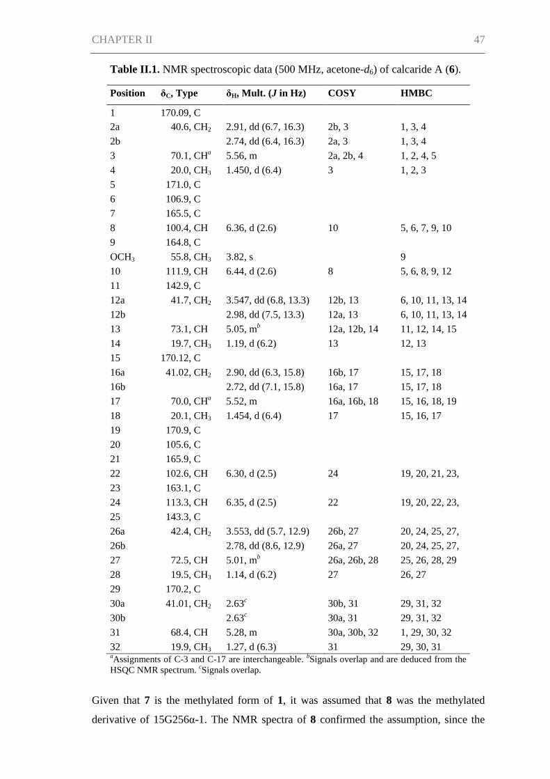
CHAPTER II 47
Table II.1. NMR spectroscopic data (500 MHz, acetone-d6) of calcaride A (6).
Position δC, Type δH, Mult. (J in Hz) COSY HMBC
1 170.09, C
2a 40.6, CH2 2.91, dd (6.7, 16.3) 2b, 3 1, 3, 4
2b 2.74, dd (6.4, 16.3) 2a, 3 1, 3, 4
3 70.1, CHa 5.56, m 2a, 2b, 4 1, 2, 4, 5
4 20.0, CH3 1.450, d (6.4) 3 1, 2, 3
5 171.0, C
6 106.9, C
7 165.5, C
8 100.4, CH 6.36, d (2.6) 10 5, 6, 7, 9, 10
9 164.8, C
OCH3 55.8, CH3 3.82, s 9
10 111.9, CH 6.44, d (2.6) 8 5, 6, 8, 9, 12
11 142.9, C
12a 41.7, CH2 3.547, dd (6.8, 13.3) 12b, 13 6, 10, 11, 13, 14
12b 2.98, dd (7.5, 13.3) 12a, 13 6, 10, 11, 13, 14
13 73.1, CH 5.05, mb 12a, 12b, 14 11, 12, 14, 15
14 19.7, CH3 1.19, d (6.2) 13 12, 13
15 170.12, C
16a 41.02, CH2 2.90, dd (6.3, 15.8) 16b, 17 15, 17, 18
16b 2.72, dd (7.1, 15.8) 16a, 17 15, 17, 18
17 70.0, CHa 5.52, m 16a, 16b, 18 15, 16, 18, 19
18 20.1, CH3 1.454, d (6.4) 17 15, 16, 17
19 170.9, C
20 105.6, C
21 165.9, C
22 102.6, CH 6.30, d (2.5) 24 19, 20, 21, 23,
24 23 163.1, C
24 113.3, CH 6.35, d (2.5) 22 19, 20, 22, 23,
26 25 143.3, C
26a 42.4, CH2 3.553, dd (5.7, 12.9) 26b, 27 20, 24, 25, 27,
28 26b 2.78, dd (8.6, 12.9) 26a, 27 20, 24, 25, 27,
28 27 72.5, CH 5.01, mb 26a, 26b, 28 25, 26, 28, 29
28 19.5, CH3 1.14, d (6.2) 27 26, 27
29 170.2, C
30a 41.01, CH2 2.63c
30b, 31 29, 31, 32
30b 2.63c
30a, 31 29, 31, 32
31 68.4, CH 5.28, m 30a, 30b, 32 1, 29, 30, 32
32 19.9, CH3 1.27, d (6.3) 31 29, 30, 31 aAssignments of C-3 and C-17 are interchangeable.
bSignals overlap and are deduced from the
HSQC NMR spectrum. cSignals overlap.
Given that 7 is the methylated form of 1, it was assumed that 8 was the methylated
derivative of 15G256α-1. The NMR spectra of 8 confirmed the assumption, since the
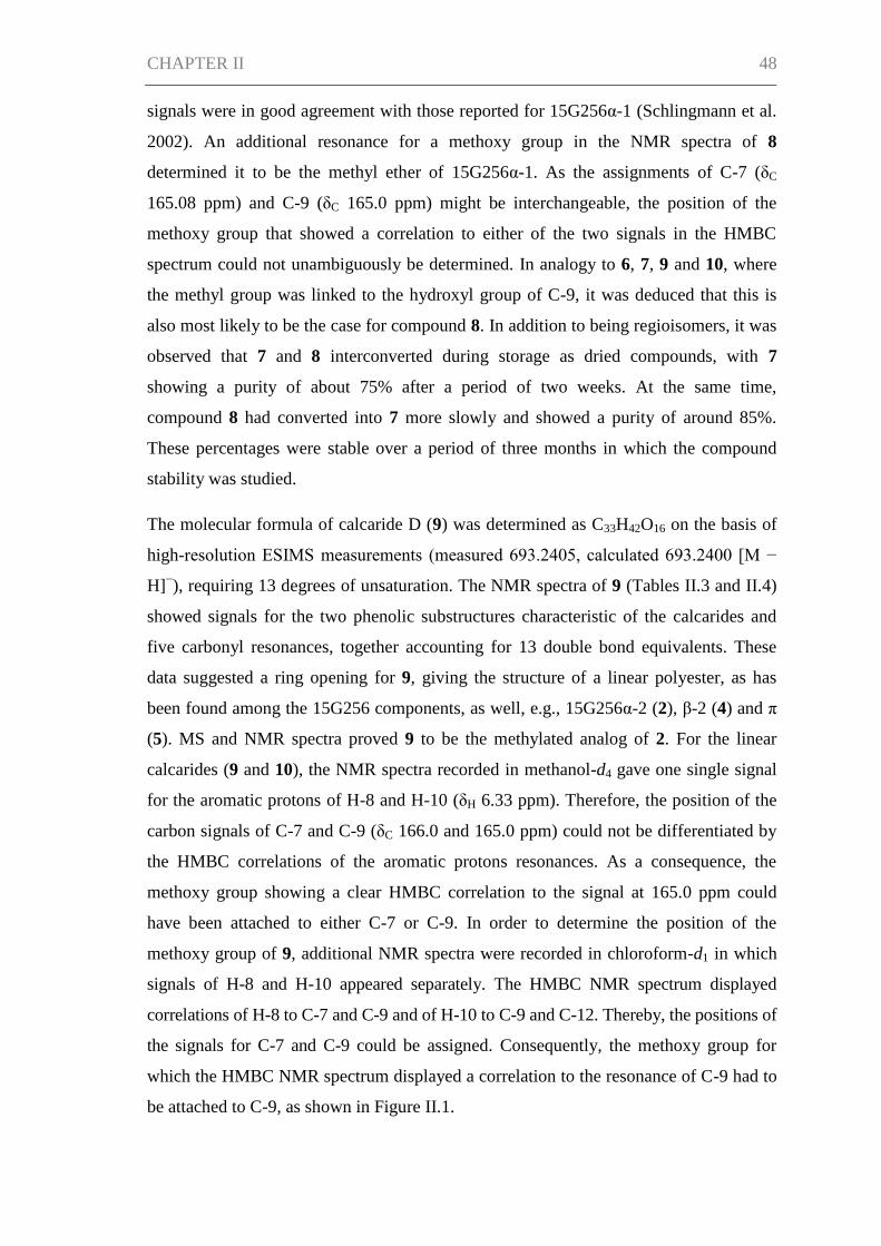
CHAPTER II 48
signals were in good agreement with those reported for 15G256α-1 (Schlingmann et al.
2002). An additional resonance for a methoxy group in the NMR spectra of 8
determined it to be the methyl ether of 15G256α-1. As the assignments of C-7 (δC
165.08 ppm) and C-9 (δC 165.0 ppm) might be interchangeable, the position of the
methoxy group that showed a correlation to either of the two signals in the HMBC
spectrum could not unambiguously be determined. In analogy to 6, 7, 9 and 10, where
the methyl group was linked to the hydroxyl group of C-9, it was deduced that this is
also most likely to be the case for compound 8. In addition to being regioisomers, it was
observed that 7 and 8 interconverted during storage as dried compounds, with 7
showing a purity of about 75% after a period of two weeks. At the same time,
compound 8 had converted into 7 more slowly and showed a purity of around 85%.
These percentages were stable over a period of three months in which the compound
stability was studied.
The molecular formula of calcaride D (9) was determined as C33H42O16 on the basis of
high-resolution ESIMS measurements (measured 693.2405, calculated 693.2400 [M −
H]−), requiring 13 degrees of unsaturation. The NMR spectra of 9 (Tables II.3 and II.4)
showed signals for the two phenolic substructures characteristic of the calcarides and
five carbonyl resonances, together accounting for 13 double bond equivalents. These
data suggested a ring opening for 9, giving the structure of a linear polyester, as has
been found among the 15G256 components, as well, e.g., 15G256α-2 (2), β-2 (4) and π
(5). MS and NMR spectra proved 9 to be the methylated analog of 2. For the linear
calcarides (9 and 10), the NMR spectra recorded in methanol-d4 gave one single signal
for the aromatic protons of H-8 and H-10 (δH 6.33 ppm). Therefore, the position of the
carbon signals of C-7 and C-9 (δC 166.0 and 165.0 ppm) could not be differentiated by
the HMBC correlations of the aromatic protons resonances. As a consequence, the
methoxy group showing a clear HMBC correlation to the signal at 165.0 ppm could
have been attached to either C-7 or C-9. In order to determine the position of the
methoxy group of 9, additional NMR spectra were recorded in chloroform-d1 in which
signals of H-8 and H-10 appeared separately. The HMBC NMR spectrum displayed
correlations of H-8 to C-7 and C-9 and of H-10 to C-9 and C-12. Thereby, the positions of
the signals for C-7 and C-9 could be assigned. Consequently, the methoxy group for
which the HMBC NMR spectrum displayed a correlation to the resonance of C-9 had to
be attached to C-9, as shown in Figure II.1.
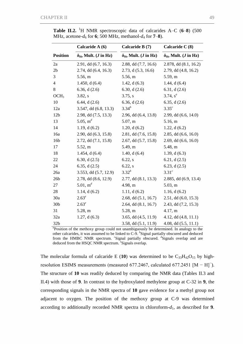
CHAPTER II 49
Table II.2. 1H NMR spectroscopic data of calcarides A–C (6–8) (500
MHz, acetone-d6 for 6; 500 MHz, methanol-d4 for 7–8).
Calcaride A (6) Calcaride B (7) Calcaride C (8)
Position δH, Mult. (J in Hz) δH, Mult. (J in Hz) δH, Mult. (J in Hz)
2a 2.91, dd (6.7, 16.3) 2.88, dd (7.7, 16.6) 2.878, dd (8.1, 16.2)
2b 2.74, dd (6.4, 16.3) 2.73, d (5.3, 16.6) 2.79, dd (4.8, 16.2)
3 5.56, m 5.56, m 5.59, m
4 1.450, d (6.4) 1.42, d (6.3) 1.44, d (6.4)
8 6.36, d (2.6) 6.30, d (2.6) 6.31, d (2.6)
OCH3 3.82, s 3.75, s 3.74, sa
10 6.44, d (2.6) 6.36, d (2.6) 6.35, d (2.6)
12a 3.547, dd (6.8, 13.3) 3.34b 3.35
c
12b 2.98, dd (7.5, 13.3) 2.96, dd (6.4, 13.8) 2.99, dd (6.6, 14.0)
13 5.05, md 5.07, m 5.16, m
14 1.19, d (6.2) 1.20, d (6.2) 1.22, d (6.2)
16a 2.90, dd (6.3, 15.8) 2.81, dd (7.6, 15.8) 2.85, dd (6.6, 16.0)
16b 2.72, dd (7.1, 15.8) 2.67, dd (5.7, 15.8) 2.69, dd (6.6, 16.0)
17 5.52, m 5.49, m 5.48, m
18 1.454, d (6.4) 1.40, d (6.4) 1.39, d (6.3)
22 6.30, d (2.5) 6.22, s 6.21, d (2.5)
24 6.35, d (2.5) 6.22, s 6.23, d (2.5)
26a 3.553, dd (5.7, 12.9) 3.32b 3.31
c
26b 2.78, dd (8.6, 12.9) 2.77, dd (8.1, 13.3) 2.885, dd (6.9, 13.4)
27 5.01, md 4.98, m 5.03, m
28 1.14, d (6.2) 1.11, d (6.2) 1.16, d (6.2)
30a 2.63e
2.68, dd (5.1, 16.7) 2.51, dd (6.0, 15.3)
30b 2.63e
2.64, dd (8.1, 16.7) 2.43, dd (7.2, 15.3)
31 5.28, m 5.28, m 4.17, m
32a 1.27, d (6.3) 3.65, dd (4.5, 11.9) 4.12, dd (4.8, 11.1)
32b 3.58, dd (5.1, 11.9) 4.08, dd (5.5, 11.1) aPosition of the methoxy group could not unambiguously be determined. In analogy to the
other calcarides, it was assumed to be linked to C-9. bSignal partially obscured and deduced
from the HMBC NMR spectrum. cSignal partially obscured.
dSignals overlap and are
deduced from the HSQC NMR spectrum. eSignals overlap.
The molecular formula of calcaride E (10) was determined to be C33H42O15 by high-
resolution ESIMS measurements (measured 677.2467, calculated 677.2451 [M − H]−).
The structure of 10 was readily deduced by comparing the NMR data (Tables II.3 and
II.4) with those of 9. In contrast to the hydroxylated methylene group at C-32 in 9, the
corresponding signals in the NMR spectra of 10 gave evidence for a methyl group not
adjacent to oxygen. The position of the methoxy group at C-9 was determined
according to additionally recorded NMR spectra in chloroform-d1, as described for 9.
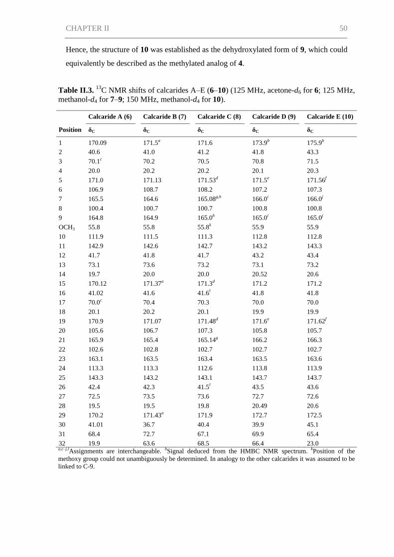
CHAPTER II 50
Hence, the structure of 10 was established as the dehydroxylated form of 9, which could
equivalently be described as the methylated analog of 4.
Table II.3. 13
C NMR shifts of calcarides A–E (6–10) (125 MHz, acetone-d6 for 6; 125 MHz,
methanol-d4 for 7–9; 150 MHz, methanol-d4 for 10).
Calcaride A (6) Calcaride B (7) Calcaride C (8) Calcaride D (9) Calcaride E (10)
Position δC δC δC δC δC
1 170.09 171.5a 171.6 173.9
b 175.9
b
2 40.6 41.0 41.2 41.8 43.3
3 70.1c 70.2 70.5 70.8 71.5
4 20.0 20.2 20.2 20.1 20.3
5 171.0 171.13 171.53d 171.5
e 171.56
f
6 106.9 108.7 108.2 107.2 107.3
7 165.5 164.6 165.08g,h
166.0i 166.0
j
8 100.4 100.7 100.7 100.8 100.8
9 164.8 164.9 165.0h 165.0
i 165.0
j
OCH3 55.8 55.8 55.8k 55.9 55.9
10 111.9 111.5 111.3 112.8 112.8
11 142.9 142.6 142.7 143.2 143.3
12 41.7 41.8 41.7 43.2 43.4
13 73.1 73.6 73.2 73.1 73.2
14 19.7 20.0 20.0 20.52 20.6
15 170.12 171.37a 171.3
d 171.2 171.2
16 41.02 41.6 41.6l 41.8 41.8
17 70.0c 70.4 70.3 70.0 70.0
18 20.1 20.2 20.1 19.9 19.9
19 170.9 171.07 171.48d 171.6
e 171.62
f
20 105.6 106.7 107.3 105.8 105.7
21 165.9 165.4 165.14g 166.2 166.3
22 102.6 102.8 102.7 102.7 102.7
23 163.1 163.5 163.4 163.5 163.6
24 113.3 113.3 112.6 113.8 113.9
25 143.3 143.2 143.1 143.7 143.7
26 42.4 42.3 41.5l 43.5 43.6
27 72.5 73.5 73.6 72.7 72.6
28 19.5 19.5 19.8 20.49 20.6
29 170.2 171.43a 171.9 172.7 172.5
30 41.01 36.7 40.4 39.9 45.1
31 68.4 72.7 67.1 69.9 65.4
32 19.9 63.6 68.5 66.4 23.0 a,c–j,l
Assignments are interchangeable. bSignal deduced from the HMBC NMR spectrum.
kPosition of the
methoxy group could not unambiguously be determined. In analogy to the other calcarides it was assumed to be
linked to C-9.
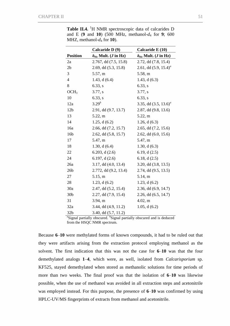
CHAPTER II 51
Table II.4. 1H NMR spectroscopic data of calcarides D
and E (9 and 10) (500 MHz, methanol-d4 for 9; 600
MHZ, methanol-d4 for 10).
Calcaride D (9) Calcaride E (10)
Position δH, Mult. (J in Hz) δH, Mult. (J in Hz)
2a 2.767, dd (7.5, 15.8) 2.72, dd (7.8, 15.4)
2b 2.69, dd (5.3, 15.8) 2.61, dd (5.9, 15.4)a
3 5.57, m 5.58, m
4 1.43, d (6.4) 1.43, d (6.3)
8 6.33, s 6.33, s
OCH3 3.77, s 3.77, s
10 6.33, s 6.33, s
12a 3.29b 3.35, dd (3.5, 13.6)
a
12b 2.91, dd (9.7, 13.7) 2.87, dd (9.8, 13.6)
13 5.22, m 5.22, m
14 1.25, d (6.2) 1.26, d (6.3)
16a 2.66, dd (7.2, 15.7) 2.65, dd (7.2, 15.6)
16b 2.62, dd (5.8, 15.7) 2.62, dd (6.0, 15.6)
17 5.47, m 5.47, m
18 1.30, d (6.4) 1.30, d (6.3)
22 6.203, d (2.6) 6.19, d (2.5)
24 6.197, d (2.6) 6.18, d (2.5)
26a 3.17, dd (4.0, 13.4) 3.20, dd (3.8, 13.5)
26b 2.772, dd (9.2, 13.4) 2.74, dd (9.5, 13.5)
27 5.15, m 5.14, m
28 1.23, d (6.2) 1.23, d (6.2)
30a 2.47, dd (5.2, 15.4) 2.36, dd (6.9, 14.7)
30b 2.27, dd (7.9, 15.4) 2.26, dd (6.5, 14.7)
31 3.94, m 4.02, m
32a 3.44, dd (4.9, 11.2) 1.05, d (6.2)
32b 3.40, dd (5.7, 11.2) aSignal partially obscured.
bSignal partially obscured and is deduced
from the HSQC NMR spectrum.
Because 6–10 were methylated forms of known compounds, it had to be ruled out that
they were artifacts arising from the extraction protocol employing methanol as the
solvent. The first indication that this was not the case for 6–10 was that the four
demethylated analogs 1–4, which were, as well, isolated from Calcarisporium sp.
KF525, stayed demethylated when stored as methanolic solutions for time periods of
more than two weeks. The final proof was that the isolation of 6–10 was likewise
possible, when the use of methanol was avoided in all extraction steps and acetonitrile
was employed instead. For this purpose, the presence of 6–10 was confirmed by using
HPLC-UV/MS fingerprints of extracts from methanol and acetonitrile.
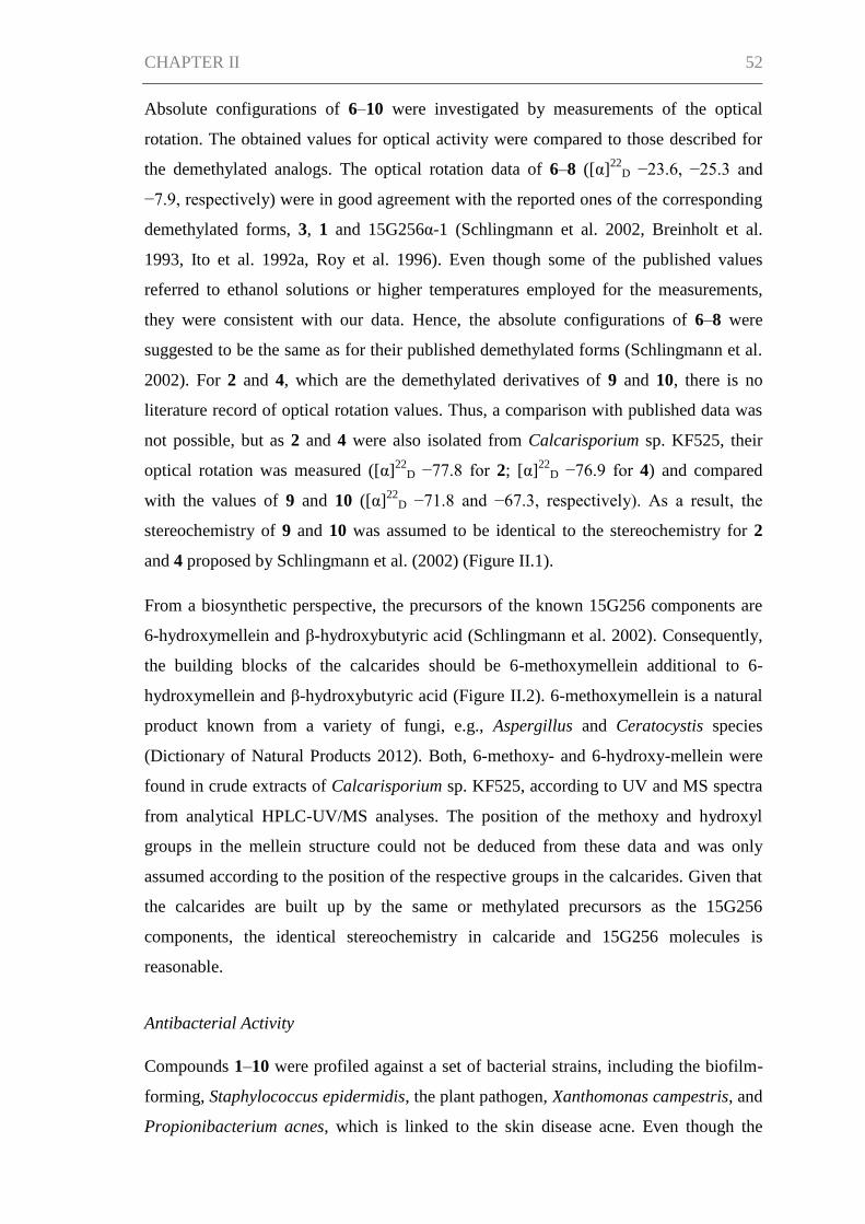
CHAPTER II 52
Absolute configurations of 6–10 were investigated by measurements of the optical
rotation. The obtained values for optical activity were compared to those described for
the demethylated analogs. The optical rotation data of 6–8 ([α]22
D −23.6, −25.3 and
−7.9, respectively) were in good agreement with the reported ones of the corresponding
demethylated forms, 3, 1 and 15G256α-1 (Schlingmann et al. 2002, Breinholt et al.
1993, Ito et al. 1992a, Roy et al. 1996). Even though some of the published values
referred to ethanol solutions or higher temperatures employed for the measurements,
they were consistent with our data. Hence, the absolute configurations of 6–8 were
suggested to be the same as for their published demethylated forms (Schlingmann et al.
2002). For 2 and 4, which are the demethylated derivatives of 9 and 10, there is no
literature record of optical rotation values. Thus, a comparison with published data was
not possible, but as 2 and 4 were also isolated from Calcarisporium sp. KF525, their
optical rotation was measured ([α]22
D −77.8 for 2; [α]22
D −76.9 for 4) and compared
with the values of 9 and 10 ([α]22
D −71.8 and −67.3, respectively). As a result, the
stereochemistry of 9 and 10 was assumed to be identical to the stereochemistry for 2
and 4 proposed by Schlingmann et al. (2002) (Figure II.1).
From a biosynthetic perspective, the precursors of the known 15G256 components are
6-hydroxymellein and β-hydroxybutyric acid (Schlingmann et al. 2002). Consequently,
the building blocks of the calcarides should be 6-methoxymellein additional to 6-
hydroxymellein and β-hydroxybutyric acid (Figure II.2). 6-methoxymellein is a natural
product known from a variety of fungi, e.g., Aspergillus and Ceratocystis species
(Dictionary of Natural Products 2012). Both, 6-methoxy- and 6-hydroxy-mellein were
found in crude extracts of Calcarisporium sp. KF525, according to UV and MS spectra
from analytical HPLC-UV/MS analyses. The position of the methoxy and hydroxyl
groups in the mellein structure could not be deduced from these data and was only
assumed according to the position of the respective groups in the calcarides. Given that
the calcarides are built up by the same or methylated precursors as the 15G256
components, the identical stereochemistry in calcaride and 15G256 molecules is
reasonable.
Antibacterial Activity
Compounds 1–10 were profiled against a set of bacterial strains, including the biofilm-
forming, Staphylococcus epidermidis, the plant pathogen, Xanthomonas campestris, and
Propionibacterium acnes, which is linked to the skin disease acne. Even though the
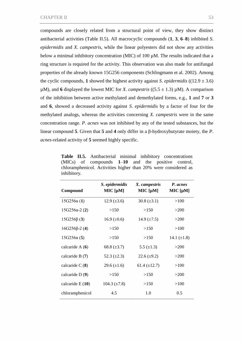
CHAPTER II 53
compounds are closely related from a structural point of view, they show distinct
antibacterial activities (Table II.5). All macrocyclic compounds (1, 3, 6–8) inhibited S.
epidermidis and X. campestris, while the linear polyesters did not show any activities
below a minimal inhibitory concentration (MIC) of 100 µM. The results indicated that a
ring structure is required for the activity. This observation was also made for antifungal
properties of the already known 15G256 components (Schlingmann et al. 2002). Among
the cyclic compounds, 1 showed the highest activity against S. epidermidis ((12.9 ± 3.6)
µM), and 6 displayed the lowest MIC for X. campestris ((5.5 ± 1.3) µM). A comparison
of the inhibition between active methylated and demethylated forms, e.g., 1 and 7 or 3
and 6, showed a decreased activity against S. epidermidis by a factor of four for the
methylated analogs, whereas the activities concerning X. campestris were in the same
concentration range. P. acnes was not inhibited by any of the tested substances, but the
linear compound 5. Given that 5 and 4 only differ in a β-hydroxybutyrate moiety, the P.
acnes-related activity of 5 seemed highly specific.
Table II.5. Antibacterial minimal inhibitory concentrations
(MICs) of compounds 1–10 and the positive control,
chloramphenicol. Activities higher than 20% were considered as
inhibitory.
Compound
S. epidermidis
MIC [µM]
X. campestris
MIC [µM]
P. acnes
MIC [µM]
15G256α (1) 12.9 (±3.6) 30.8 (±3.1) >100
15G256α-2 (2) >150 >150 >200
15G256β (3) 16.9 (±0.6) 14.9 (±7.5) >200
16G256β-2 (4) >150 >150 >100
15G256π (5) >150 >150 14.1 (±1.8)
calcaride A (6) 68.8 (±3.7) 5.5 (±1.3) >200
calcaride B (7) 52.3 (±2.3) 22.6 (±9.2) >200
calcaride C (8) 29.6 (±1.6) 61.4 (±12.7) >100
calcaride D (9) >150 >150 >200
calcaride E (10) 104.3 (±7.8) >150 >100
chloramphenicol 4.5 1.0 0.5
CHAPTER II 54
15G256 components are known to possess antifungal activities, estrogenic effects or
cytotoxic properties with regard to cellular ATP (viability) and chemiluminescence
(phagocytosis) in a mouse peritoneal macrophage assay, as well as they are described to
be potentiators of the nerve growth factor in PC12 cells (Schlingmann et al. 2002,
Breinholt et al. 1993, Ito et al. 1992a, Roy et al. 1996, Kondo el. 1993). However, no
antibacterial activities had been observed, even when tested. Here, the 15G256
compounds and the new methylated derivatives thereof were found to display
antibacterial activities that are moderate in potency, but have a tight structure-activity
relationship. Even a slight structural variation, like a methylation, as seen for the
calcarides, influenced compound activities significantly. This was found to be true for
other natural products (Walle et al. 2007, Liu et al. 2010). For instance, the methylation
of the cholesterol-lowering agent, lovastatin, resulted in the improved drug, simvastatin
(Li and Vederas 2009). Regarding simvastatin, the introduction of a methyl group was
used as a tool, but similarly, the isolation of derivatives of known compounds can be
used as a strategy to obtain improved lead structures. As shown for the calcarides, fungi
are a good source for this purpose, because they often produce a bunch of structurally
related compounds, e.g., resulting from the promiscuity of the biosynthetic enzymes
(Grüschow et al. 2011). From the fungus’ point of view, the production of closely-
related components might be of advantage as altered activities emerge.
Experimental Section
General Experimental Procedures
Optical rotation measurements were obtained on a Perkin Elmer model 241 polarimeter.
UV spectra were measured on a Perkin Elmer Lambda 2 photometer. NMR spectra
were recorded on a Bruker DRX 500 (500 and 125 MHz for 1H and
13C NMR,
respectively) and a Bruker AV 600 spectrometer (600 and 150 MHz for 1H and
13C
NMR, respectively). The residual solvent signals were used as internal references for
NMR analyses (δH 3.31 and δC 49.0 ppm for methanol-d4; δH 2.05 and
δC 29.8 ppm for acetone-d6, δH 7.26 and δC 77.2 ppm for chloroform-d1). High-
resolution mass spectra were measured with a Bruker micrOTOF II, applying positive
or negative ESI mode. Analytical reversed-phase HPLC-UV/MS experiments were
conducted on a VWR-Hitachi LaChrom Elite system equipped with an L-2130 pump,
an L-2450 diode array detector, an L-2200 autosampler, an L-2300 column oven and a
CHAPTER II 55
Phenomenex Onyx Monolithic column (C18, 100 × 3.00 mm). A gradient with 0.1%
formic acid in H2O (A) and 0.1% formic acid in acetonitrile (B) as solvents was applied:
0 min 5% B, 4 min 60% B, 6 min 100% B; flow 2 mL min−1
. For mass detection, the
analytical HPLC system was coupled to a Bruker esquire4000 ion-trap detector.
Preparative HPLC was performed on a VWR LaPrep system consisting of a P110
pump, a P311 UV detector, a Labocol Vario-2000 fraction collector (LABOMATIC), a
Smartline 3900 autosampler (Knauer) and a Phenomenex Gemini-NX column (10 μ
C18, 100A, Axia, 100 × 50.00 mm). Subsequent compound purifications were
performed on a Merck-Hitachi LaChrom Elite HPLC system comprised of an L-7150
pump, an L-2450 diode array detector and an L-2200 autosampler employing a
reversed-phase Phenomenex Gemini-NX column (5 μ C18, 110A, Axia, 100 × 21.20
mm). 0.1% of formic acid in H2O (A) and 0.1% formic acid in acetonitrile (B) were the
solvents used in preparative HPLC separations.
Fungal Material
Calcarisporium sp. KF525 is a part of the marine fungal strain collection of the Kiel
Center for Marine Natural Products. The strain was cryo-conserved using the
Microbank system (Pro-Lab).
Cultivation
Calcarisporium sp. KF525 was cultivated in 12 L of casamino acids glucose medium
(0.25% casein hydrolysate, 4% glucose × H2O, 0.01% MgSO4 × 7H2O, 0.18% KH2PO4,
pH 6.8) (Stevens 1974). 2-L Erlenmeyer flasks filled with 0.75 L medium were used for
the culture experiments, which were performed for 24 days at 22 °C under shaking
conditions (120 rpm) in the dark. A circular agar slant (1.8 cm in diameter) of a
preculture served as the inoculum. The preculture was grown on agar plates of modified
Wickerham medium (0.3% malt extract, 0.3% yeast extract, 0.5% peptone from
soymeal, 1% glucose × H2O, 3% NaCl, 1.5% agar, pH 6.25) (Wickerham 1951) over a
period of 11 days at room temperature in the dark.
Isolation Procedure
The cultures of Calcarisporium sp. KF525 were separated into mycelium and culture
supernatant. An extraction of the culture supernatant with EtOAc yielded a crude extract
of 0.92 g. The extract was subjected to the first purification step using a VWR LaPrep
CHAPTER II 56
HPLC system (gradient: 0 min 10% B, 17 min 60% B, 22 min 100% B; flow 100 mL
min−1
; UV detection at 217 nm). The purification gave 8 fractions containing the
15G256 and calcaride components (tR 12.4, 13.2, 14.5, 15.7, 16.2, 17.6, 18.5 and 20.6
min) of which fraction 8 comprised pure 6 in a yield of 17.7 mg. All other fractions
were further purified on a Merck-Hitachi LaChrom Elite HPLC system applying
reversed-phase chromatography. The purification of fraction 1 gave 17.8 mg of 2
(gradient: 0 min 30% B, 13 min 40% B; flow 18 mL min−1
; UV detection at 220 nm; tR
6.7 min) and fraction 2 afforded 11.8 mg of 5 (gradient: 0 min 25% B, 13 min 50% B;
flow 18 mL min−1
; UV detection at 220 nm; tR 9.2 min). The chromatography of
fraction 3 (gradient: 0 min 30% B, 15 min 37% B; flow 18 mL min−1
; UV detection at
220 nm) resulted in 14 mg of purified 4 (tR 11.1 min) and semi-pure 9 (tR 12.0 min),
which was isolated in two additional rounds of purification to give a yield of 23.2 mg
(1st, gradient: 0 min 30% B, 13 min 45% B; flow 18 mL min−1
; UV detection at 220
nm; tR 9.2 min. 2nd, isocratic: 25% B; flow 18 mL min−1
; UV detection at 210 nm; tR
23.2 min). Pure 1 (17.5 mg) was obtained from fraction 4 by applying two consecutive
steps of preparative HPLC (1st, 0 min 40% B, 13 min 65% B, flow 18 mL min−1
; UV
detection at 205 nm; tR 5.2 min. 2nd, gradient: 0 min 30% B, 10 min 61% B; flow 18
mL min−1
; UV detection at 220 nm; tR 7.6 min). Fraction 5 was subjected to two further
purification steps yielding 2.9 mg of 10 (1st, isocratic: 29% B, flow 18 mL min−1
; UV
detection at 220 nm; tR 21.7 min. 2nd, gradient: 0 min 27% B, 15 min 50% B; flow 18
mL min−1
; UV detection at 220 nm; tR 12.4 min). Fraction 6 contained compounds 7
and 8, which were initially eluted isocratically with 42% B (flow 18 mL min−1
; UV
detection at 220 nm; tR 11.8 min). A subsequent round of preparative HPLC (isocratic:
40%, flow 18 mL min−1
; UV detection at 210 nm) gave purified 7 (tR 12.8 min) and 8
(tR 16.0 min) in yields of 14.6 and 5.7 mg, respectively. 1.8 mg of compound 3 were
isolated from fraction 7 by employing two chromatographic steps (1st, gradient: 0 min
50% B, 13 min 80% B; flow 18 mL min−1
; UV detection at 220 nm; tR 6.0 min. 2nd,
isocratic: 45% B, flow 18 mL min−1
; UV detection at 220 nm; tR 7.3 min.).
Calcaride A (6): Pale yellow solid, [α]22
D −23.6 (c 0.195, MeOH), UV (MeOH) λmax
(log ε) 216 (4.57), 263 (4.27), 302 (3.92) nm; for 1D and 2D NMR data see Table II.1;
HRESIMS m/z 661.2503 [M + H]+ (calcd for C33H40O14, 661.2491).
CHAPTER II 57
Calcaride B (7): White solid, [α]22
D −25.3 (c 0.45, MeOH), UV (MeOH) λmax (log ε)
216 (4.58), 263 (4.27), 302 (3.92) nm; for 1D and 2D NMR data see Tables II.2, II.3
and SI; HRESIMS m/z 677.2435 [M + H]+ (calcd for C33H40O15, 677.2440).
Calcaride C (8): White solid, [α]22
D −7.9 (c 0.793, MeOH), UV (MeOH) λmax (log ε)
216 (4.59), 263 (4.30), 302 (3.94) nm; for 1D and 2D NMR data see Tables II.2, II.3
and SI; HRESIMS m/z 677.2441 [M + H]+ (calcd for C33H40O15, 677.2440).
Calcaride D (9): White solid, [α]22
D −71.8 (c 0.98, MeOH), UV (MeOH) λmax (log ε)
216 (4.60), 263 (4.35), 302 (3.99) nm; for 1D and 2D NMR data see Tables II.3, II.4
and SI; HRESIMS m/z 693.2405 [M − H]− (calcd for C33H42O16, 693.2400).
Calcaride E (10): White solid, [α]22
D −67.3 (c 0.05, MeOH), UV (MeOH) λmax (log ε)
216 (4.61), 263 (4.35), 302 (3.98) nm; for 1D and 2D NMR data see Tables II.3, II.4
and SI; HRESIMS m/z 677.2467 [M − H]− (calcd for C33H42O15, 677.2451).
The process of interconversion of 7 and 8 was studied by periodical HPLC-UV/MS
analyses using the mass chromatograms for quantification of the compounds’ purities.
For the experiments, purified and dried 7 and 8 were stored at 4 °C over a time period of
three months.
Antibacterial Assays
Antibacterial assays were performed using the test strains, Staphylococcus epidermidis
(DSM20044), Xanthomonas campestris (DSM 2405) and Propionibacterium acnes
(DSM1897).
S. epidermidis and X. campestris were cultivated in trypticase soy broth (1.2%
trypticase soy broth, 0.5% NaCl) overnight and diluted to an OD600 of 0.01–0.03. 10
mM DMSO solutions of the compounds were diluted with medium to gain desired test
concentrations. 10.5 µL of the compound solutions and 200 µL of the respective test
strain cell suspension were transferred into a 96-well microtiter plate. Microtiter plates
with S. epidermidis were incubated for 5 h at 37 °C in the dark, while microtiter plates
with X. campestris were incubated for 6 h at 28 °C in the dark. After the addition of 10
µL of a resazurin solution as the detective reagent (0.2 mg mL−1
in phosphate-buffered
saline), the incubation was continued for another 5–30 min. To evaluate cell viability,
the reduction of resazurin to resorufin was determined by measuring the intensity of
fluorescence at 560Ex/590Em in a Tecan Infinite M200 plate reader.
CHAPTER II 58
P. acnes was grown anaerobically (Anaerocult A mini, Merck) in modified PYG
medium (DSMZ medium 104) for 48 h at 37 °C in the dark. 2-day old cultures were
diluted to an OD600 of 0.02. A volume of 200 µL of the cell suspension was transferred
to a microtiter plate and mixed with 10.5 µL of the test compounds that were treated as
described above. After an anaerobic incubation for 48 h at 37 °C in the dark, 10 µL of
bromocresol purple (1 mg mL−1
) were added, and the acid production of
P. acnes was determined by measuring the absorbance at 590 nm (reference 690 nm)
using the Infinite M200 plate reader.
The resulting values of the antibacterial assays were compared with a positive control
(10 µM chloramphenicol for S. epidermidis or X. campestris and 1 µM chloramphenicol
for P. acnes) and a negative control (no compound). Minimal inhibitory concentrations
(MICs) were defined as more than 20% inhibition of metabolic activity or acid
production in the test strains compared to non-inhibited metabolism in the negative
control.
Conclusions
Five known 15G256 components and five methylated analogs thereof were isolated
from Calcarisporium sp. strain KF525: 15G256α, α-2, β, β-2, π (1–5) and calcarides A–
E (6–10). For the first time, antibacterial properties were detected for this type of
natural product. The compounds showed inhibitory bioactivities against Staphylococcus
epidermidis, Xanthomonas campestris and Propionibacterium acnes, underlying a tight
structure-activity relationship. 15G256α (1) displayed the strongest inhibition against S.
epidermidis, with a MIC of (12.9 ± 3.6) µM, and calcaride A (6) exhibited the lowest
MIC for X. campestris with (5.5 ± 1.3) µM. P. acnes was specifically inhibited by the
compound, 15G256π (5), having a MIC of (14.1 ± 1.8) µM.
Acknowledgments
We gratefully thank K. Schaumann for providing the strain, KF525, and G. Kohlmeyer-
Yilmaz, M. Höftmann, as well as F. Sönnichsen for running and processing NMR
experiments. This study was performed in the framework of the MARINE FUNGI, EU
FP7 KBBE program, project no. 265926 at the Kieler Wirkstoff-Zentrum (KiWiZ) at
the GEOMAR Helmholtz Centre for Ocean Research, Kiel.
CHAPTER II 59
References
Arai, M., Tomoda, H., Okuda, T., Wang, H., Tabata, N., Masuma, R., Yamaguchi, Y.,
Omura, S. (2002). Funicone-related compounds, potentiators of antifungal miconazole
activity, produced by Talaromyces flavus FKI-0076. J. Antibiot. 55(2), 172-180.
Breinholt, J., Jensen, G. W., Nielsen, R. I., Olsen, C. E., Frisvad, J. C. (1993).
Antifungal macrocyclic polylactones from Penicillium verruculosum. J. Antibiot. 46(7),
1101-1108.
Dictionary of Natural Products (2012). Chapman & Hall/CRC Press/Hampden Data
Services, Ltd.
Grüschow, S., Rackham, E. J., Goss, R. J. M. (2011). Diversity in natural product
families is governed by more than enzyme promiscuity alone: establishing control of the
pacidamycin portfolio. Chem. Sci. 2(11), 2182-2186.
Ito, M., Maruhashi, M., Sakai, N., Mizoue, K., Hanada, K. (1992a). NG-011 and NG-
012, novel potentiators of nerve growth factor. I. Taxonomy, isolation, and physico-
chemical and biological properties. J. Antibiot. 45(10), 1559-1565.
Ito, M., Tsuchida, Y., Mizoue, K., Hanada, K. (1992b). NG-011 and NG-012, novel
potentiators of nerve growth factor. II. The structure determination of NG-011 and NG-
012. J. Antibiot. 45(10), 1566-1572.
Ji, L. L., Song, Y. C., Tan, R. X. (2004). A potent feed preservative candidate produced
by Calcarisporium sp., an endophyte residing in stargrass (Cynodon dactylon). J. Appl.
Microbiol. 96(2), 352-358.
Kondo, H., Kurama, M., Nakajima, S., Osada, K., Ookura, A., Suda, H. (1993).
Estrogenic BE-26263 and its manufacture with Scedosporium apiospermum. Jpn. Kokai
Tokkyo Koho, JP 05032658.
Kramer, C. L., Pady, S. M., Rogerson, C. T., Ouye, L. G. (1959). Kansas aeromycology
II. Materials, methods, and general results. Trans. Kans. Acad. Sci. 62(3), 184-199.
Li, J. W.-H., Vederas, J. C. (2009). Drug discovery and natural products: End of an era
or an endless frontier? Science 325(5937), 161-165.
CHAPTER II 60
Liu, Y., Salvador, L. A., Byeon, S., Ying, Y., Kwan, J. C., Law, B. K., Hong, J.,
Luesch, H. (2010). Anticolon cancer activity of largazole, a marine-derived tunable
histone deacetylase inhibitor. J. Pharmacol. Exp. Ther. 335(2), 351-361.
Roy, K., Chatterjee, S., Deshmukh, S. K., Vijayakumar, E. K. S., Ganguli, B. N.,
Fehlhaber, H.-W. (1996). Orbuticin, a new secondary metabolite from Acremonium
butyri. J. Antibiot. 49(11), 1186-1187.
Schlingmann, G., Milne, L., Carter, G. T. (2002). Isolation and identification of
antifungal polyesters from the marine fungus Hypoxylon oceanicum LL-15G256.
Tetrahedron 58(34), 6825-6835.
Silber, J., Ohlendorf, B., Labes, A., Näther, C., Imhoff, J. F. (2013). Calcaripeptides A–
C, cyclodepsipeptides from a Calcarisporium strain. J. Nat. Prod. 76(8), 1461-1467.
Somrithipol, S., Jones, E. B. G. (2006). Calcarisporium phaeopodium sp. nov., a new
hyphomycete from Thailand. Sydowia 58(1), 133-140.
Stevens, R. B. (1974). Mycology guidebook. University of Washington Press, Seattle,
WA, USA, pp. 703.
Sutton, B. C. (1973). Hyphomycetes from Manitoba and Saskatchewan, Canada.
Commonwealth Mycological Institute, Kew, Surrey, UK, pp. 143.
Walle, T., Ta, N., Kawamori, T., Wen, X., Tsuji, P. A., Walle, U. K. (2007). Cancer
chemopreventive properties of orally bioavailable flavonoids - methylated versus
unmethylated flavones. Biochem. Pharmacol. 73(9), 1288-1296.
Watson, P. (1955). Calcarisporium arbuscula living as an endophyte in apparently
healthy sporophores of Russula and Lactarius. Trans. Br. Mycol. Soc. 38(4), 409-414.
Wickerham, L. J. (1951). Taxonomy of yeasts. US Dept. of Agriculture, Washington, D.
C., USA, pp. 56.
Zhou, Z.-Y., Liu, R., Jiang, M.-Y., Zhang, L., Niu, Y., Zhu, Y.-C., Dong, Z.-J., Liu, J.-
K. (2009). Two new cleistanthane diterpenes and a new isocoumarine from cultures of
the basidiomycete Albatrellus confluens. Chem. Pharm. Bull. 57(9), 975-978.
CHAPTER III 61
CHAPTER III
Malettinin E, antimicrobial tropolone produced by a Cladosporium
strain
Johanna Silber, Birgit Ohlendorf, Antje Labes, Arlette Erhard, Christian Näther and
Johannes F. Imhoff
to be submitted
CHAPTER III 62
Abstract
The isolation and structure elucidation of malettinins A–C (1–3) along with the new
malettinin E (4) are described. The compounds were produced by the fungus
Cladosporium sp. strain KF501, which was isolated from the German Wadden Sea. The
malettinins are built up of tropolone/dihydropyran ring structures linked to a furan ring.
The structure elucidation of the isolated compounds was achieved by means of one- and
two-dimensional NMR spectroscopy supported by mass and UV data. The relative
configuration of 4 was determined on the basis of single-crystal X-ray diffraction
analysis. 1–4 exhibited antibacterial and antifungal activities when profiled against
Xanthomonas campestris and Trichophyton rubrum. The influence of the chemical
structure of the furan ring and of configurational changes on biological activities was
observed.
Introduction
The genus Cladosporium represents one of the largest and most heterogeneous genera
of the Hyphomycetes (Bensch et al. 2012), showing ubiquitous occurrence. Species of
the genus often have pathogenic or saprophytic lifestyles and are frequently found in
air, soil, on foods, on plant materials or as endophytes (Stevens 1974, Samson et al.
2000). Occurring as airborne fungi, some species of Cladosporium are of clinical
importance as they can cause allergies such as allergic asthma (Simon-Nobbe et al.
2008). Concerning marine habitats, a minimum of three Cladosporium species were
described from marine sources (Kirk and Clipson 2013). Cladosporium species have
been shown to possess the ability to produce a variety of natural products, among them
the melanins which are pigments giving the fungal colonies their typical dark colored
appearance. Other natural products isolated from Cladosporium species are bioactive
compounds such as the antifungal cladosporides (Hosoe et al. 2000, Hosoe et al. 2001),
the plant growth factors cotylenins (Sassa 1971, Sassa et al. 1975), calphostins which
specifically inhibit the protein kinase C (Kobayashi et al. 1989) and cladosporin
exhibiting a broad activity spectrum including antifungal, antibacterial, insecticidal,
phytotoxic and immunosuppressive properties (Scott et al. 1971, Anke et al. 1978,
Grove and Pople 1981, Springer et al. 1981, Fujimoto et al. 1999).
In this study, a marine-derived Cladosporium strain was shown to produce the new
compound malettinin E (4) along with the known malettinins A–C (1–3) (Figure III.1),
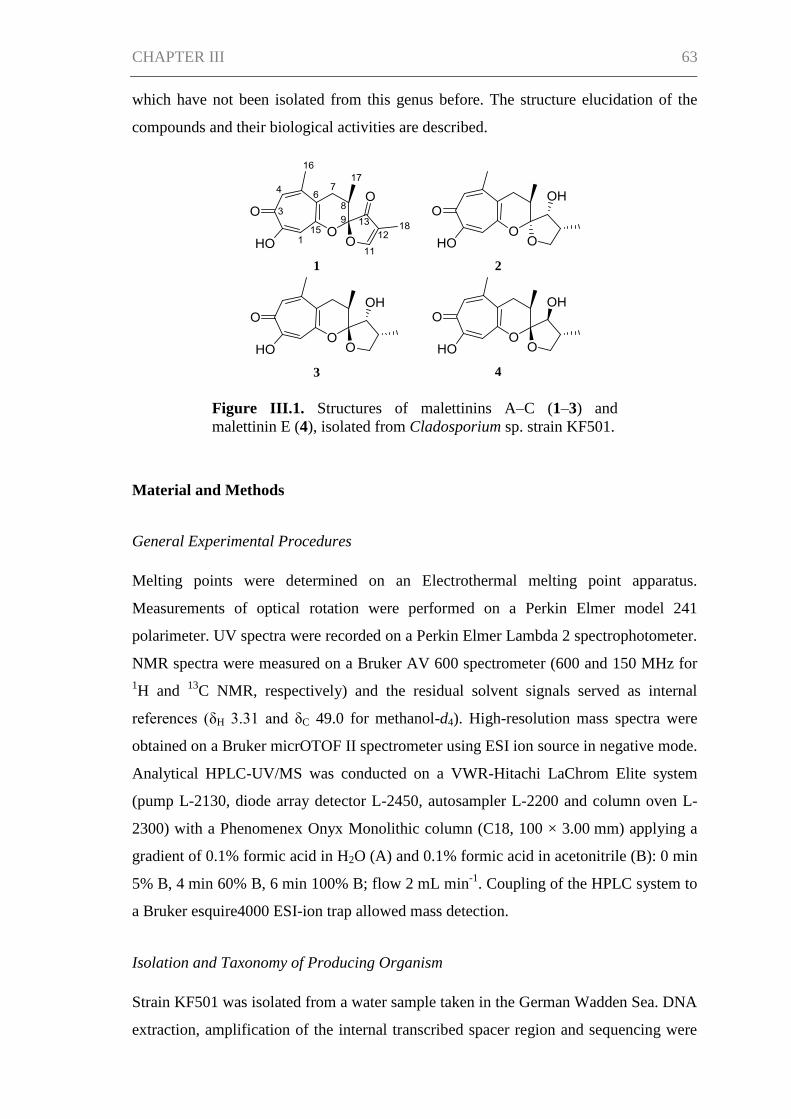
CHAPTER III 63
which have not been isolated from this genus before. The structure elucidation of the
compounds and their biological activities are described.
Material and Methods
General Experimental Procedures
Melting points were determined on an Electrothermal melting point apparatus.
Measurements of optical rotation were performed on a Perkin Elmer model 241
polarimeter. UV spectra were recorded on a Perkin Elmer Lambda 2 spectrophotometer.
NMR spectra were measured on a Bruker AV 600 spectrometer (600 and 150 MHz for
1H and
13C NMR, respectively) and the residual solvent signals served as internal
references (δH 3.31 and δC 49.0 for methanol-d4). High-resolution mass spectra were
obtained on a Bruker micrOTOF II spectrometer using ESI ion source in negative mode.
Analytical HPLC-UV/MS was conducted on a VWR-Hitachi LaChrom Elite system
(pump L-2130, diode array detector L-2450, autosampler L-2200 and column oven L-
2300) with a Phenomenex Onyx Monolithic column (C18, 100 × 3.00 mm) applying a
gradient of 0.1% formic acid in H2O (A) and 0.1% formic acid in acetonitrile (B): 0 min
5% B, 4 min 60% B, 6 min 100% B; flow 2 mL min-1
. Coupling of the HPLC system to
a Bruker esquire4000 ESI-ion trap allowed mass detection.
Isolation and Taxonomy of Producing Organism
Strain KF501 was isolated from a water sample taken in the German Wadden Sea. DNA
extraction, amplification of the internal transcribed spacer region and sequencing were
1 2
3 4
Figure III.1. Structures of malettinins A–C (1–3) and
malettinin E (4), isolated from Cladosporium sp. strain KF501.
CHAPTER III 64
performed as described by Wiese et al. (2011) with slight modifications, centrifugation
of the DNA at 8000g and 35 cycles of DNA amplification. The DNA sequence was
deposited in GenBank under the accession number KF923800. Cryo-conserved stock
cultures of strain KF501 were kept at -100 °C using the Microbank system (Pro-Lab).
Fermentation and Compound Isolation
The fungal isolate was cultivated in the following media: Casamino acids glucose
medium (casein hydrolysate 0.25%, glucose × H2O 4%, MgSO4 × 7H2O 0.01%,
KH2PO4 0.18%, pH 6.8) (Stevens 1974), Czapek medium (sucrose 3%, NaNO3 0.3%,
K2HPO4 0.1%, KCl 0.05%, MgSO4 × 7H2O 0.05%, FeSO4 × 7H2O 0.001%, pH 6.2)
(Samson et al. 2000), modified malt extract medium (malt extract 3%, NaCl 1.5%, pH
5.5) (Samson et al. 2000), potato-carrot medium (potatoes 40 g, carrots 40 g, each
boiled in 1 L of H2O and filtered off, 250 mL of potato extract and 250 mL of carrot
extract were filled up with 500 mL of distilled H2O) (Samson et al. 2000) and modified
Wickerham medium (malt extract 0.3%, yeast extract 0.3%, peptone from soymeal
0.5%, glucose × H2O 1%, NaCl 3%, agar 1.5%, pH 6.25) (Wickerham 1951).
For isolation of the malettinins, Cladosporium sp. KF501 was cultivated in 9.75 L of
casamino acids glucose medium. The cultivation experiments were performed as
surface cultures at 20 °C in the dark in 2-L Erlenmeyer flasks, each containing 750 mL
medium. The cultures were inoculated with an agar slant (2.6 cm in diameter) of a seven
days old preculture, which was grown on solid, modified Wickerham medium at room
temperature in the dark. The mycelium of strain KF501 was harvested after 40 days of
incubation by extraction with EtOH. As the obtained extract was very greasy, fats were
frozen out at -20 °C yielding 1.2 g of extract.
The malettinins were purified from the mycelium extract by preparative HPLC. The
separations were performed on a VWR LaPrep system (pump P110, UV detector P311
UV, fraction collector Labocol Vario-2000 from LABOMATIC, autosampler Smartline
3900 from Knauer) using a Phenomenex Gemini-NX column (10 µ C18, 100A, Axia,
100 × 50.00 mm). A gradient of 0.1% formic acid in H2O (A) and 0.1% formic acid in
acetonitrile (B) (0.0 min 20% B, 17.5 min 40% B, 21.0 min 60% B; flow 100 mL min-1
)
was applied to yield 17.5 mg of 2 (tR 8.6-9.0 min), 6.1 mg of 3 (tR 9.2-9.6 min), 4.6 mg
of 4 (tR 9.9-10.3 min) and 15.1 mg of 1 (tR 12.2-18.0 min).
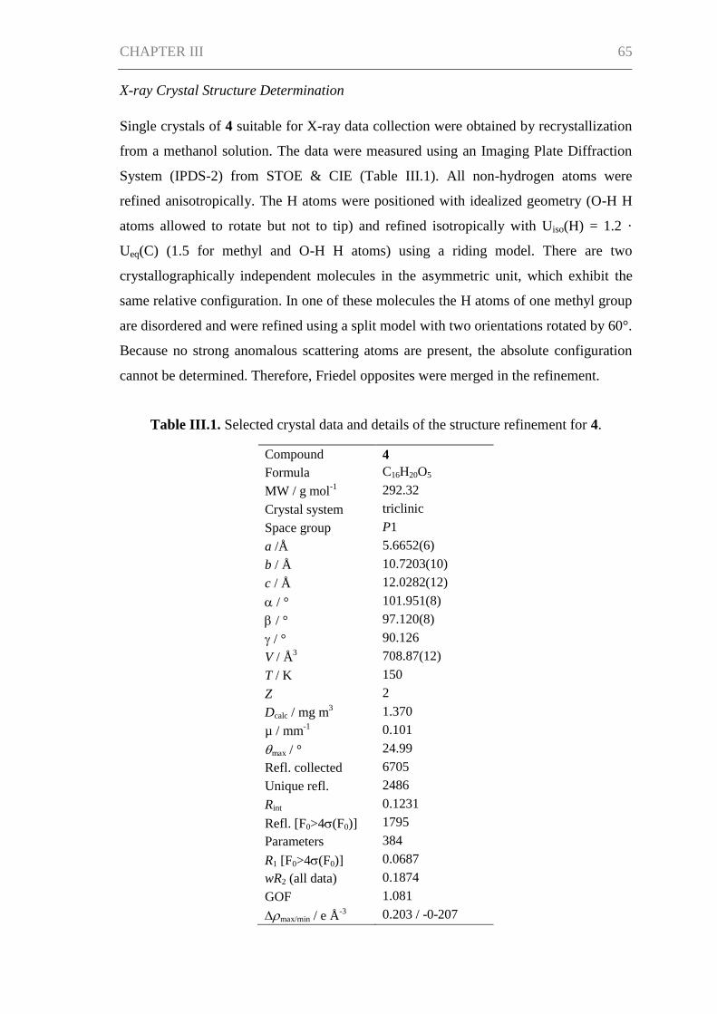
CHAPTER III 65
X-ray Crystal Structure Determination
Single crystals of 4 suitable for X-ray data collection were obtained by recrystallization
from a methanol solution. The data were measured using an Imaging Plate Diffraction
System (IPDS-2) from STOE & CIE (Table III.1). All non-hydrogen atoms were
refined anisotropically. The H atoms were positioned with idealized geometry (O-H H
atoms allowed to rotate but not to tip) and refined isotropically with Uiso(H) = 1.2 ·
Ueq(C) (1.5 for methyl and O-H H atoms) using a riding model. There are two
crystallographically independent molecules in the asymmetric unit, which exhibit the
same relative configuration. In one of these molecules the H atoms of one methyl group
are disordered and were refined using a split model with two orientations rotated by 60°.
Because no strong anomalous scattering atoms are present, the absolute configuration
cannot be determined. Therefore, Friedel opposites were merged in the refinement.
Table III.1. Selected crystal data and details of the structure refinement for 4.
Compound 4
Formula C16H20O5
MW / g mol-1
292.32
Crystal system triclinic
Space group P1
a /Å 5.6652(6)
b / Å 10.7203(10)
c / Å 12.0282(12)
/ ° 101.951(8)
/ ° 97.120(8)
/ ° 90.126
V / Å3 708.87(12)
T / K 150
Z 2
Dcalc / mg m3 1.370
µ / mm-1
0.101
max / ° 24.99
Refl. collected 6705
Unique refl. 2486
Rint 0.1231
Refl. [F0>4(F0)] 1795
Parameters 384
R1 [F0>4(F0)] 0.0687
wR2 (all data) 0.1874
GOF 1.081
max/min / e Å-3
0.203 / -0-207
CHAPTER III 66
Antimicrobial Assays
Antimicrobial assays with the test strains Xanthomonas campestris and Staphylococcus
epidermidis were performed as described by Silber et al. (2013). Bacillus subtilis and
Candida albicans were tested as described by Ohlendorf et al. (2012). For antifungal
assays using Trichophyton rubrum, cultivation was carried out in modified Sabouraud
medium (peptone 1%, glucose 2%, pH 5.6). A spore solution containing 5 × 10⁴ spores
of T. rubrum per mL medium was prepared. 10 mM DMSO solutions of compounds
were diluted with medium to achieve the desired test concentrations. For assaying, 200
µL of the spore solutions were mixed with 10.5 µL of compound solutions in 96-well
microtiter plates and incubated for 72 h at 28 °C in the dark. The cell growth of T.
rubrum was evaluated by measuring the optical densities with a Tecan Infinite M200
plate reader. The resulting values were compared with a positive control (0.1 and 0.5
µM clotrimazole) and a negative control (no compound). IC50 values were determined
according to the inhibition of metabolic activity or cell growth in the presence of a
compound as compared to the non-inhibited negative control.
Results
Taxonomy of Producing Organism
Strain KF501 was isolated from water samples taken in the German Wadden Sea. The
strain grew well in casamino acids glucose, Czapek and malt extract medium, slowly in
potato-carrot medium and did not grow in liquid modified Wickerham medium. In
general, the growth of strain KF501 was relatively slow by comparison with other
fungal isolates.
Macroscopic and microscopic morphological features, colony color and structure of
conidiophores were characteristic of Cladosporium spp. (Samson et al. 2000). This
classification was corroborated by the comparison of the ITS DNA sequence of strain
KF501 with sequences available at GenBank employing the Basic Local Alignment
Search Tool (BLAST) (Altschul et al. 1990). The search resulted in 100% similarity to
Cladosporium cladosporioides, C. pseudocladosporioides, C. uredinicola, C. bruhnei
and C. colombiae. Hence, strain KF501 could be identified as a member of the genus
Cladosporium, but identification to the species level was not possible.
CHAPTER III 67
Structure Elucidation
The new malettinin E (4) along with the known malettinins A–C (1–3) was obtained
from the mycelia extracts of Cladosporium sp. strain KF501 grown in casamino acids
glucose medium. The compounds were isolated by preparative reversed-phase HPLC.
The structures of compounds 1–3 were readily identified by spectroscopic methods
including 1H NMR, UV and MS spectra (Angawi et al. 2003, Angawi et al. 2005).
Table III.2. Physico-chemical properties of malettinin E (4).
Malettinin E (4)
Appearance Orange crystals or amorphous solid (MeOH)
Molecular formula C16H20O5
Molecular weight 292
HRESIMS (m/z)
Measured [M + H]+
293.1391
Calculated [M + H]+ 293.1384
UV λmax (MeOH) nm (log ε) 253 (4.45), 328 (3.68), 359 (3.91)
Mp (°C) 181–183 °C
[α]22
D +58 (c 0.2, MeOH)
Malettinin E (4) was assigned a molecular formula of C16H20O5 based upon HRESIMS.
The physico-chemical properties of 4 are summarized in Table III.2. The molecular
formula of 4 being the same as for 2 and 3 suggested the compounds to be isomeric. In
addition, the one- and two-dimensional NMR spectra of 4 (1H,
13C, DEPT, COSY,
HSQC and HMBC; see Table III.3 and Supporting Information) revealed the presence
of considerable structural similarities with 2 and 3 (Table III.4) (Angawi et al. 2005).
The 1H and
13C NMR data of 4 showed resonances for three methyl, two methylene and
two aliphatic methine groups, one oxygenated methine and one carbonyl group as well
as signals for six aromatic carbons and one oxygenated quaternary carbon. Thus, all
structural groups and atoms present in 2 or 3 were also observed for 4. Correlations of
the aromatic protons in the HMBC NMR spectrum (Figure III.2) established the same
tropolone moiety for 4 as found in the known malettinins. In addition to the tropolone
unit, 4 contained a dihydropyran ring identical to that of 2 and 3. The proton and carbon
chemical shifts of the corresponding signals were in good agreement with those
reported for 2 and 3, with the proton signals agreeing particularly well to the structure
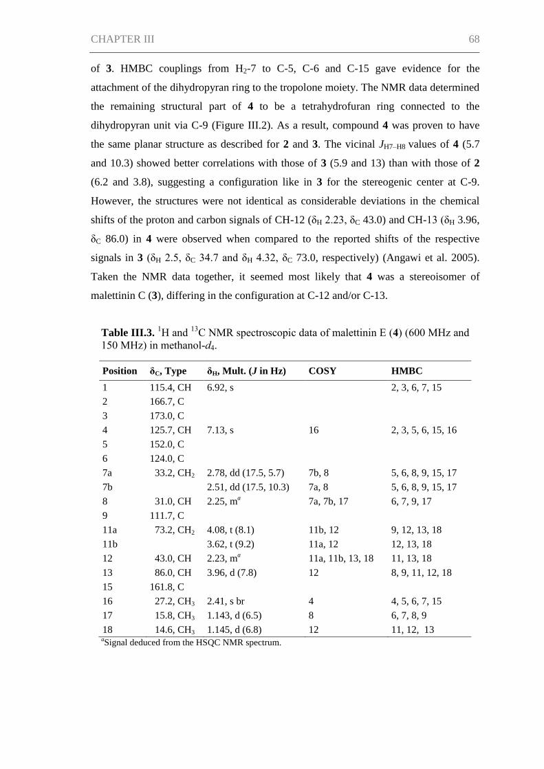
CHAPTER III 68
of 3. HMBC couplings from H2-7 to C-5, C-6 and C-15 gave evidence for the
attachment of the dihydropyran ring to the tropolone moiety. The NMR data determined
the remaining structural part of 4 to be a tetrahydrofuran ring connected to the
dihydropyran unit via C-9 (Figure III.2). As a result, compound 4 was proven to have
the same planar structure as described for 2 and 3. The vicinal JH7–H8 values of 4 (5.7
and 10.3) showed better correlations with those of 3 (5.9 and 13) than with those of 2
(6.2 and 3.8), suggesting a configuration like in 3 for the stereogenic center at C-9.
However, the structures were not identical as considerable deviations in the chemical
shifts of the proton and carbon signals of CH-12 (δH 2.23, δC 43.0) and CH-13 (δH 3.96,
δC 86.0) in 4 were observed when compared to the reported shifts of the respective
signals in 3 (δH 2.5, δC 34.7 and δH 4.32, δC 73.0, respectively) (Angawi et al. 2005).
Taken the NMR data together, it seemed most likely that 4 was a stereoisomer of
malettinin C (3), differing in the configuration at C-12 and/or C-13.
Table III.3. 1H and
13C NMR spectroscopic data of malettinin E (4) (600 MHz and
150 MHz) in methanol-d4.
Position δC, Type δH, Mult. (J in Hz) COSY HMBC
1 115.4, CH 6.92, s 2, 3, 6, 7, 15
2 166.7, C
3 173.0, C
4 125.7, CH 7.13, s 16 2, 3, 5, 6, 15, 16
5 152.0, C
6 124.0, C
7a 33.2, CH2 2.78, dd (17.5, 5.7) 7b, 8 5, 6, 8, 9, 15, 17
7b
2.51, dd (17.5, 10.3) 7a, 8 5, 6, 8, 9, 15, 17
8 31.0, CH 2.25, ma 7a, 7b, 17 6, 7, 9, 17
9 111.7, C
11a 73.2, CH2 4.08, t (8.1) 11b, 12 9, 12, 13, 18
11b
3.62, t (9.2) 11a, 12 12, 13, 18
12 43.0, CH 2.23, ma 11a, 11b, 13, 18 11, 13, 18
13 86.0, CH 3.96, d (7.8) 12 8, 9, 11, 12, 18
15 161.8, C
16 27.2, CH3 2.41, s br 4 4, 5, 6, 7, 15
17 15.8, CH3 1.143, d (6.5) 8 6, 7, 8, 9
18 14.6, CH3 1.145, d (6.8) 12 11, 12, 13 aSignal deduced from the HSQC NMR spectrum.
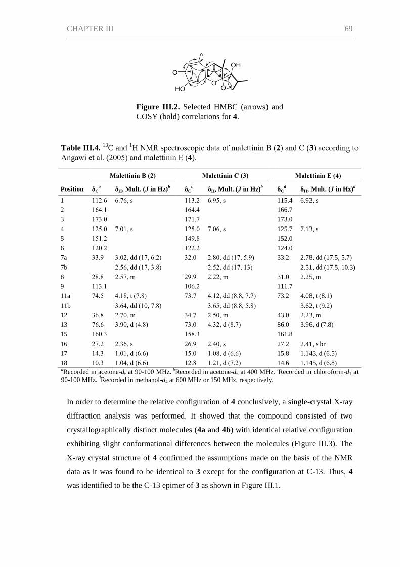
CHAPTER III 69
Figure III.2. Selected HMBC (arrows) and
COSY (bold) correlations for 4.
Table III.4. 13
C and 1H NMR spectroscopic data of malettinin B (2) and C (3) according to
Angawi et al. (2005) and malettinin E (4).
Malettinin B (2)
Malettinin C (3)
Malettinin E (4)
Position δCa δH, Mult. (J in Hz)
b δC
c δH, Mult. (J in Hz)
b δC
d δH, Mult. (J in Hz)
d
1 112.6 6.76, s
113.2 6.95, s
115.4 6.92, s
2 164.1
164.4
166.7
3 173.0
171.7
173.0
4 125.0 7.01, s
125.0 7.06, s
125.7 7.13, s
5 151.2
149.8
152.0
6 120.2
122.2
124.0
7a 33.9 3.02, dd (17, 6.2)
32.0 2.80, dd (17, 5.9)
33.2 2.78, dd (17.5, 5.7)
7b
2.56, dd (17, 3.8)
2.52, dd (17, 13)
2.51, dd (17.5, 10.3)
8 28.8 2.57, m
29.9 2.22, m
31.0 2.25, m
9 113.1
106.2
111.7
11a 74.5 4.18, t (7.8)
73.7 4.12, dd (8.8, 7.7)
73.2 4.08, t (8.1)
11b
3.64, dd (10, 7.8)
3.65, dd (8.8, 5.8)
3.62, t (9.2)
12 36.8 2.70, m
34.7 2.50, m
43.0 2.23, m
13 76.6 3.90, d (4.8)
73.0 4.32, d (8.7)
86.0 3.96, d (7.8)
15 160.3
158.3
161.8
16 27.2 2.36, s
26.9 2.40, s
27.2 2.41, s br
17 14.3 1.01, d (6.6)
15.0 1.08, d (6.6)
15.8 1.143, d (6.5)
18 10.3 1.04, d (6.6) 12.8 1.21, d (7.2) 14.6 1.145, d (6.8) aRecorded in acetone-d6 at 90-100 MHz.
bRecorded in acetone-d6 at 400 MHz.
cRecorded in chloroform-d1 at
90-100 MHz. dRecorded in methanol-d4 at 600 MHz or 150 MHz, respectively.
In order to determine the relative configuration of 4 conclusively, a single-crystal X-ray
diffraction analysis was performed. It showed that the compound consisted of two
crystallographically distinct molecules (4a and 4b) with identical relative configuration
exhibiting slight conformational differences between the molecules (Figure III.3). The
X-ray crystal structure of 4 confirmed the assumptions made on the basis of the NMR
data as it was found to be identical to 3 except for the configuration at C-13. Thus, 4
was identified to be the C-13 epimer of 3 as shown in Figure III.1.
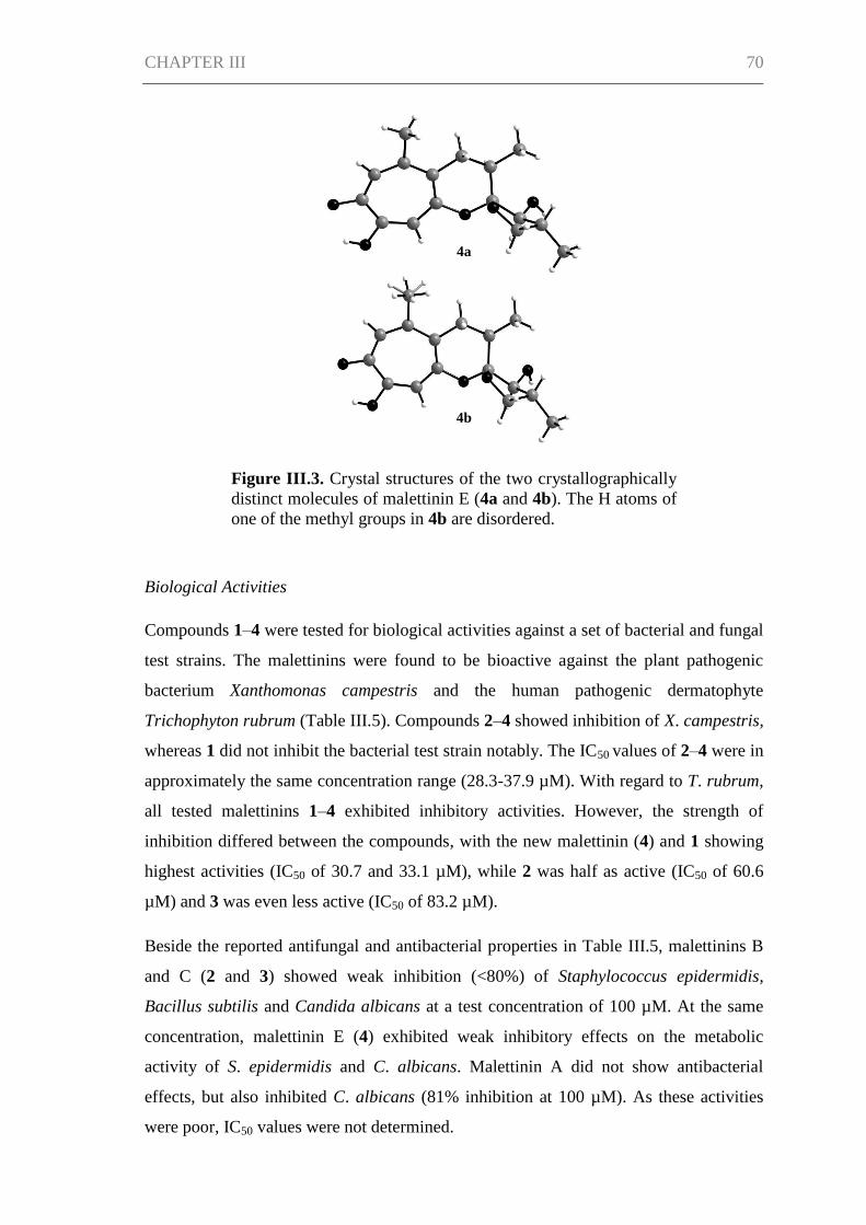
CHAPTER III 70
Biological Activities
Compounds 1–4 were tested for biological activities against a set of bacterial and fungal
test strains. The malettinins were found to be bioactive against the plant pathogenic
bacterium Xanthomonas campestris and the human pathogenic dermatophyte
Trichophyton rubrum (Table III.5). Compounds 2–4 showed inhibition of X. campestris,
whereas 1 did not inhibit the bacterial test strain notably. The IC50 values of 2–4 were in
approximately the same concentration range (28.3-37.9 µM). With regard to T. rubrum,
all tested malettinins 1–4 exhibited inhibitory activities. However, the strength of
inhibition differed between the compounds, with the new malettinin (4) and 1 showing
highest activities (IC50 of 30.7 and 33.1 µM), while 2 was half as active (IC50 of 60.6
µM) and 3 was even less active (IC50 of 83.2 µM).
Beside the reported antifungal and antibacterial properties in Table III.5, malettinins B
and C (2 and 3) showed weak inhibition (<80%) of Staphylococcus epidermidis,
Bacillus subtilis and Candida albicans at a test concentration of 100 µM. At the same
concentration, malettinin E (4) exhibited weak inhibitory effects on the metabolic
activity of S. epidermidis and C. albicans. Malettinin A did not show antibacterial
effects, but also inhibited C. albicans (81% inhibition at 100 µM). As these activities
were poor, IC50 values were not determined.
Figure III.3. Crystal structures of the two crystallographically
distinct molecules of malettinin E (4a and 4b). The H atoms of
one of the methyl groups in 4b are disordered.
4a
4b
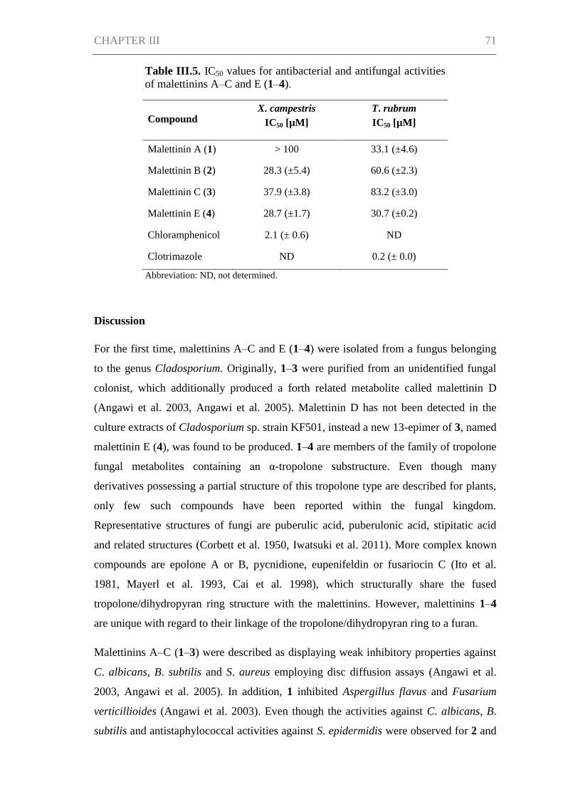
CHAPTER III 71
Table III.5. IC50 values for antibacterial and antifungal activities
of malettinins A–C and E (1–4).
Compound X. campestris
IC50 [µM]
T. rubrum
IC50 [µM]
Malettinin A (1) > 100 33.1 (±4.6)
Malettinin B (2) 28.3 (±5.4) 60.6 (±2.3)
Malettinin C (3) 37.9 (±3.8) 83.2 (±3.0)
Malettinin E (4) 28.7 (±1.7) 30.7 (±0.2)
Chloramphenicol 2.1 (± 0.6) ND
Clotrimazole ND 0.2 (± 0.0)
Abbreviation: ND, not determined.
Discussion
For the first time, malettinins A–C and E (1–4) were isolated from a fungus belonging
to the genus Cladosporium. Originally, 1–3 were purified from an unidentified fungal
colonist, which additionally produced a forth related metabolite called malettinin D
(Angawi et al. 2003, Angawi et al. 2005). Malettinin D has not been detected in the
culture extracts of Cladosporium sp. strain KF501, instead a new 13-epimer of 3, named
malettinin E (4), was found to be produced. 1–4 are members of the family of tropolone
fungal metabolites containing an α-tropolone substructure. Even though many
derivatives possessing a partial structure of this tropolone type are described for plants,
only few such compounds have been reported within the fungal kingdom.
Representative structures of fungi are puberulic acid, puberulonic acid, stipitatic acid
and related structures (Corbett et al. 1950, Iwatsuki et al. 2011). More complex known
compounds are epolone A or B, pycnidione, eupenifeldin or fusariocin C (Ito et al.
1981, Mayerl et al. 1993, Cai et al. 1998), which structurally share the fused
tropolone/dihydropyran ring structure with the malettinins. However, malettinins 1–4
are unique with regard to their linkage of the tropolone/dihydropyran ring to a furan.
Malettinins A–C (1–3) were described as displaying weak inhibitory properties against
C. albicans, B. subtilis and S. aureus employing disc diffusion assays (Angawi et al.
2003, Angawi et al. 2005). In addition, 1 inhibited Aspergillus flavus and Fusarium
verticillioides (Angawi et al. 2003). Even though the activities against C. albicans, B.
subtilis and antistaphylococcal activities against S. epidermidis were observed for 2 and
CHAPTER III 72
3 in this study as well, higher activities were found when profiled against the bacterium
X. campestris and the fungus T. rubrum. X. campestris causes a variety of plant
diseases, e.g., black rot in cabbage which is difficult to treat and therefore leads to high
crop losses (Roohie and Umesha 2012). T. rubrum is responsible for clinical pictures
like tinea pedis (athlete’s foot) and likewise treatments are challenging, in part due to
the toxicity in long-term treatments (Ramsey et al. 2013). New bioactive compounds for
better treatments in plant protection as well as in clinical aspects are hence highly
desirable. Comparing activities though shows that the malettinins inhibit X. campestris
with a 13-fold higher IC50 than the antibiotic chloramphenicol used as positive control.
Thus, their activities can be considered as moderate, only. The inhibition of the
malettinins against T. rubrum is rather poor given that the IC50 of the positive control
clotrimazole is lower by a factor of greater than 170.
The activities regarding X. campestris showed that all stereoisomeric compounds (2–4)
inhibited the test strain in comparable concentration ranges, while 1, possessing a very
slightly varied furan ring, did not show inhibition. This observation suggests that the
furan ring of the malettinins is critical for antibacterial properties against X. campestris.
With regard to activities against T. rubrum, apparently configurational changes of the
malettinins determine the strength of activity, since the distinct stereoisomers exhibited
different IC50 values.
Acknowledgments
We gratefully thank Dr. K. Schaumann for providing strain KF501, G. Kohlmeyer-
Yilmaz, M. Höftmann and Dr. F. Sönnichsen for running and processing NMR
experiments and R. Schmied for help with measurements of the melting point. We also
thank the Institute of Clinical Molecular Biology in Kiel for providing Sanger
sequencing as supported in part by the FG Cluster of Excellence “Inflammation at
Interfaces” and "Future Ocean". We thank the technicians S. Greve and S. Arndt for
technical support. This study was performed in the framework of the EU project
MARINE FUNGI, which was funded within the European Union Seventh Framework
Programme (FP7/2007-2013 under grant agreement number 265926).
CHAPTER III 73
References
Altschul, S. F., Gish, W., Miller, W., Myers, E. W., Lipman, D. J. (1990). Basic Local
Alignment Search Tool. J. Mol. Biol. 215(3), 403-410.
Angawi, R. F., Swenson, D. C., Gloer, J. B., Wicklow, D. T. (2003). Malettinin A: a
new antifungal tropolone from an unidentified fungal colonist of Hypoxylon stromata
(NRRL 29110). Tetrahedron Lett. 44(41), 7593-7596.
Angawi, R. F., Swenson, D. C., Gloer, J. B., Wicklow, D. T. (2005). Malettinins B–D:
new polyketide metabolites from an unidentified fungal colonist of Hypoxylon stromata
(NRRL 29110). J. Nat. Prod. 68(2), 212-216.
Anke, H., Zähner, H., König, W. A. (1978). Metabolic products of microorganisms.
170. On the antibiotic activity of cladosporin. Arch. Microbiol. 116(3), 253-257.
Bensch, K., Braun, U., Groenewald, J. Z., Crous, P. W. (2012). The genus
Cladosporium. Stud. Mycol. 72(1), 1-401.
Cai, P., Smith, D., Cunningham, B., Brown-Shimer, S., Katz, B., Pearce, C., Venables,
D., Houck, D. (1998). Epolones: novel sesquiterpene-tropolones from fungus OS-
F69284 that induce erythropoietin in human cells. J. Nat. Prod. 61(6), 791-795.
Corbett, R. E., Hassall, C. H., Johnson, A. W., Todd, A. R. (1950). 1. Puberulic and
puberulonic acids. Part I. The molecular formula of puberulonic acid and consideration
of possible benzenoid structures for the acids. J. Chem. Soc., 1-6.
Fujimoto, H., Fujimaki, T., Okuyama, E., Yamazaki, M. (1999). Immunomodulatory
constituents from an ascomycete, Microascus tardifaciens. Chem. Pharm. Bull. 47(10),
1426-1432.
Grove, J. F., Pople, M. (1981). The insecticidal activity of some fungal
dihydroisocoumarins. Mycopathologia 76(2), 65-67.
Hosoe, T., Okada, H., Itabashi, T., Nozawa, K., Okada, K., de Campos Takaki, G. M.,
Fukushima, K., Miyaji, M., Kawai, K.-I. (2000). A new pentanorlanostane derivative,
cladosporide A, as a characteristic antifungal agent against Aspergillus fumigatus,
isolated from Cladosporium sp. Chem. Pharm. Bull. 48(10), 1422-1426.
CHAPTER III 74
Hosoe, T., Okamoto, S., Nozawa, K., Kawai, K.-I., Okada, K., de Campos Takaki, G.
M., Fukushima, K., Miyaji, M. (2001). New pentanorlanostane derivatives,
cladosporide B–D, as characteristic antifungal agents against Aspergillus fumigatus,
isolated from Cladosporium sp. J. Antibiot. 54(9), 747-750.
Ito, T., Arai, T., Ohashi, Y., Sasada, Y. (1981). Structure of fusariocin C, a cytotoxic
metabolite from Fusarium moniliforme. Agric. Biol. Chem. 45(7), 1689-1692.
Iwatsuki, M., Takada, S., Mori, M., Ishiyama, A., Namatame, M., Nishihara-
Tsukashima, A., Nonaka, K., Masuma, R., Otoguro, K., Shiomi, K., Omura, S. (2011).
In vitro and in vivo antimalarial activity of puberulic acid and its new analogs, viticolins
A–C, produced by Penicillium sp. FKI-4410. J. Antibiot. 64(2), 183-188.
Kirk, P., Clipson, N. (2013). Cladosporium Link, 1816. In: Index Fungorum Partnership
(2013) Index Fungorum. Accessed through: World Register of Marine Species at
http://www.marinespecies.org/aphia.php?p=taxdetails&id=100218 on 2013-11-27.
Kobayashi, E., Ando, K., Nakano, H., Iida, T., Ohno, H., Morimoto, M., Tamaoki, T.
(1989). Calphostins (UCN-1028), novel and specific inhibitors of protein kinase C. I.
Fermentation, isolation, physico-chemical properties and biological activities. J.
Antibiot. 42(10), 1470-1474.
Mayerl, F., Gao, Q., Huang, S., Klohr, S. E., Matson, J. A., Gustavson, D. R., Pirnik, D.
M., Berry, R. L., Fairchild, C., Rose, W. C. (1993). Eupenifeldin, a novel cytotoxic
bistropolone from Eupenicillium brefeldianum. J. Antibiot. 46(7), 1082-1088.
Ohlendorf, B., Schulz, D., Erhard, A., Nagel, K., Imhoff, J. F. (2012).
Geranylphenazinediol, an acetylcholinesterase inhibitor produced by a Streptomyces
species. J. Nat. Prod. 75(7), 1400-1404.
Ramsey, J. P., Mercurio, A., Holland, J. A., Harris, R. N., Minbiole, K. P. C. (2013).
The cutaneous bacterium Janthinobacterium lividum inhibits the growth of
Trichophyton rubrum in vitro. Int. J. Dermatol. doi: 10.1111/ijd.12217.
Roohie, R. K., Umesha, S. (2012). Development of multiplex PCR for the specific
detection of Xanthomonas campestris pv. campestris in cabbage and correlation with
disease incidence. J. Plant Pathol. Microb. 3(127), doi: 10.4172/2157-7471.1000127.
CHAPTER III 75
Samson, R. A., Hoekstra, E. S., Frisvad, J. C., Filtenborg, O. (2000). Introduction to
food-and airborne fungi. Centraalbureau voor Schimmelcultures (CBS), Utrecht, The
Netherlands, pp. 389.
Sassa, T. (1971). Cotylenins, leaf growth substances produced by a fungus. Agric. Biol.
Chem. 35(9), 1415-1418.
Sassa, T., Togashi, M., Kitaguchi, T. (1975). The structures of cotylenins A, B, C, D
and E. Agric. Biol. Chem. 39(9), 1735-1744.
Scott, P. M., van Walbeek, W., MacLean, W. M. (1971). Cladosporin, a new antifungal
metabolite from Cladosporium cladosporioides. J. Antibiot. 24(11), 747-755.
Silber, J., Ohlendorf, B., Labes, A., Erhard, A., Imhoff, J. F. (2013). Calcarides A–E,
antibacterial macrocyclic and linear polyesters from a Calcarisporium strain. Mar.
Drugs 11(9), 3309-3323.
Simon-Nobbe, B., Denk, U., Pöll, V., Rid, R., Breitenbach, M. (2008). The spectrum of
fungal allergy. Int. Arch. Allergy Immunol. 145(1), 58-86.
Springer, J. P., Cutler, H. G., Crumley, F. G., Cox, R. H., Davis, E. E., Thean, J. E.
(1981). Plant growth regulatory effects and stereochemistry of cladosporin. J. Agric.
Food Chem. 29(4), 853-855.
Stevens, R. B. (1974). Mycology guidebook. University of Washington Press, Seattle,
WA, USA, pp. 703.
Wickerham, L. J. (1951). Taxonomy of yeasts. US Dept. of Agriculture, Washington, D.
C., USA, pp. 56.
Wiese, J., Ohlendorf, B., Blümel, M., Schmaljohann, R., Imhoff, J. F. (2011).
Phylogenetic identification of fungi isolated from the marine sponge Tethya aurantium
and identification of their secondary metabolites. Mar. Drugs 9(4), 561-585.
GENERAL DISCUSSION 76
GENERAL DISCUSSION
Natural products isolated from fungi have proved very valuable for mankind, owing to
their use, e.g., in medicine and agriculture. However, the low-hanging fruits in natural
product discovery have been picked and it becomes an increasingly difficult
undertaking to find new bioactive compounds. Lower hit rates and the re-discovery of
known compounds, amongst others, drive up the costs for the discovery and
development of drugs derived from natural products. Pharmaceutical companies have
thus changed their strategic orientation und turned their focus to combinatorial
chemistry. With the synthesis of new organic compounds by the combination of a set of
smaller chemical structures, libraries comprising millions of compounds can be
generated within a short time and are screened for bioactivities in high-throughput
screenings. Yet the success of this method has not materialized to date as only one de
novo drug (sorafenib) has emerged from synthetic-combinatorial libraries in the last 30
years (Newman and Cragg 2012). Thus, in comparison, 0.001% of compounds
synthesized by combinatorial chemistry became accepted drugs, while of the known
microbial metabolites about 0.2–0.3% became a therapeutic product and about the same
number served as lead compounds for the development of new drugs. Therefore,
microbial natural products show a more than two order of magnitude higher efficacy
than compounds originating from combinatorial chemistry (Bérdy 2005). These figures
paired with the increasing resistances toward current drugs and newly emerging
pathogens demand concerted efforts in the drug discovery from natural products.
The aim of the present PhD thesis was thus to find new fungal natural products by
creating a screening strategy that allows picking the “high-hanging fruits”. For this
purpose, different approaches approved for natural product discovery were combined
(fungal isolates from little-explored ecosystems, less studied genera, genetic potential
according to PKS and NRPS gene sequences and stimulating effects on biosynthesis by
variation of culture conditions) and used for a rational selection of fungi which were in
detail investigated with regard to their biosynthesized natural products. In this way,
three new cyclodepsipeptides, five new linear and macrocyclic polyesters and one new
tropolone structure were identified and characterized. Given that as a general rule up to
10,000 microbial extracts need to be analyzed to discover one to ten new bioactive
structures (Gräfe 1999), the respective ratio in the studies presented here is impressively
high. From only 243 extracts, nine new natural products including five active
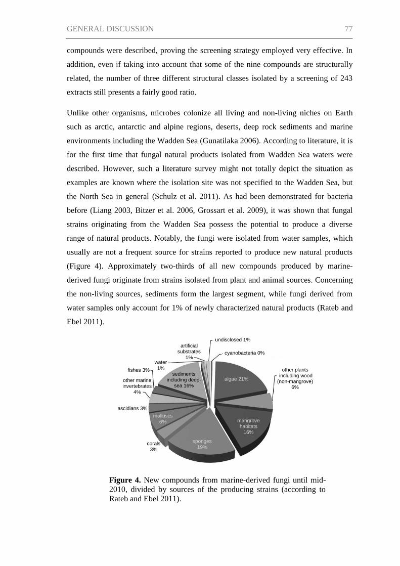
GENERAL DISCUSSION 77
undisclosed 1%
cyanobacteria 0%
algae 21%
other plants including wood (non-mangrove)
6%
mangrove habitats
16%
sponges
19% corals
3%
molluscs
6%
ascidians 3%
sediments including deep-
sea 16% other marine invertebrates
4%
water
1% fishes 3%
artificial substrates
1%
compounds were described, proving the screening strategy employed very effective. In
addition, even if taking into account that some of the nine compounds are structurally
related, the number of three different structural classes isolated by a screening of 243
extracts still presents a fairly good ratio.
Unlike other organisms, microbes colonize all living and non-living niches on Earth
such as arctic, antarctic and alpine regions, deserts, deep rock sediments and marine
environments including the Wadden Sea (Gunatilaka 2006). According to literature, it is
for the first time that fungal natural products isolated from Wadden Sea waters were
described. However, such a literature survey might not totally depict the situation as
examples are known where the isolation site was not specified to the Wadden Sea, but
the North Sea in general (Schulz et al. 2011). As had been demonstrated for bacteria
before (Liang 2003, Bitzer et al. 2006, Grossart et al. 2009), it was shown that fungal
strains originating from the Wadden Sea possess the potential to produce a diverse
range of natural products. Notably, the fungi were isolated from water samples, which
usually are not a frequent source for strains reported to produce new natural products
(Figure 4). Approximately two-thirds of all new compounds produced by marine-
derived fungi originate from strains isolated from plant and animal sources. Concerning
the non-living sources, sediments form the largest segment, while fungi derived from
water samples only account for 1% of newly characterized natural products (Rateb and
Ebel 2011).
Figure 4. New compounds from marine-derived fungi until mid-
2010, divided by sources of the producing strains (according to
Rateb and Ebel 2011).
GENERAL DISCUSSION 78
Though isolated from the Wadden Sea, it remains unclear whether the fungal isolates
investigated in the studies were obligate or facultative marine organisms or if they were
isolated as dormant spores from the water. The Wadden Sea as a land/ocean interface,
of course, receives input of terrestrial and freshwater fungi, for instance, by river inflow
or via air (especially important for fungal spores). According to Kohlmeyer and
Kohlmeyer (1979), obligate marine fungi are those that grow and sporulate exclusively
in a marine or estuarine habitat, whereas facultative marine fungi are those from
freshwater or terrestrial areas able to grow (and possibly also to sporulate) in the marine
environment. The fungal isolates described in chapters I–III could be facultative marine
organisms as they grew on modified casamino acids glucose medium not containing
sodium chloride as well as on modified Wickerham medium with sodium chloride
supplemented. Nevertheless, Calcarisporium and Cladosporium strains belong to
ubiquitous genera common to terrestrial habitats and no definite statement can be made
unless actual growth in sea water is tested (Shearer et al. 2007).
Furthermore, the results of the study show that it is worth to have a look for novel
natural products in less studied fungal genera. However, with only two strains, namely a
Calcarisporium sp. and a Cladosporium sp., it is a small number of organisms that
produced new natural products. In this regard, the fact that the initial collection of
fungal isolates lacked high diversity is considered an important reason. Even though the
total number of strains screened was large (109 strains), 30 isolates belonged to the
particularly well-investigated genus Penicillium and another 30 isolates were yeasts
which rarely produce bioactive compounds (Bérdy 2005). The search for new natural
products in not extensively investigated genera is a screening approach that is
presumably more promising when applied to a more diverse group of fungal isolates.
Therefore, the isolation procedure is of crucial importance, requiring the employment of
various different isolation techniques. In addition, the selection of fungi from
biodiversity “hotspots” such as mangrove habitats (Shearer et al. 2007) or from
sponges, which are supposed to host unique and diverse fungal communities (Bugni and
Ireland 2004, Wang 2006, Wiese et al. 2011), as an additional criterion for the screening
would probably enhance the screening output.
While microbial natural product discovery for decades was a rather coincidence-driven
culturing and screening of thousands of strains, the advances in genome sequencing
offer more directed possibilities. Schneemann (2011) showed for actinomycetes that
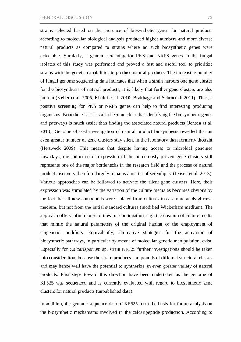
GENERAL DISCUSSION 79
strains selected based on the presence of biosynthetic genes for natural products
according to molecular biological analysis produced higher numbers and more diverse
natural products as compared to strains where no such biosynthetic genes were
detectable. Similarly, a genetic screening for PKS and NRPS genes in the fungal
isolates of this study was performed and proved a fast and useful tool to prioritize
strains with the genetic capabilities to produce natural products. The increasing number
of fungal genome sequencing data indicates that when a strain harbors one gene cluster
for the biosynthesis of natural products, it is likely that further gene clusters are also
present (Keller et al. 2005, Khaldi et al. 2010, Brakhage and Schroeckh 2011). Thus, a
positive screening for PKS or NRPS genes can help to find interesting producing
organisms. Nonetheless, it has also become clear that identifying the biosynthetic genes
and pathways is much easier than finding the associated natural products (Jensen et al.
2013). Genomics-based investigation of natural product biosynthesis revealed that an
even greater number of gene clusters stay silent in the laboratory than formerly thought
(Hertweck 2009). This means that despite having access to microbial genomes
nowadays, the induction of expression of the numerously proven gene clusters still
represents one of the major bottlenecks in the research field and the process of natural
product discovery therefore largely remains a matter of serendipity (Jensen et al. 2013).
Various approaches can be followed to activate the silent gene clusters. Here, their
expression was stimulated by the variation of the culture media as becomes obvious by
the fact that all new compounds were isolated from cultures in casamino acids glucose
medium, but not from the initial standard cultures (modified Wickerham medium). The
approach offers infinite possibilities for continuation, e.g., the creation of culture media
that mimic the natural parameters of the original habitat or the employment of
epigenetic modifiers. Equivalently, alternative strategies for the activation of
biosynthetic pathways, in particular by means of molecular genetic manipulation, exist.
Especially for Calcarisporium sp. strain KF525 further investigations should be taken
into consideration, because the strain produces compounds of different structural classes
and may hence well have the potential to synthesize an even greater variety of natural
products. First steps toward this direction have been undertaken as the genome of
KF525 was sequenced and is currently evaluated with regard to biosynthetic gene
clusters for natural products (unpublished data).
In addition, the genome sequence data of KF525 form the basis for future analysis on
the biosynthetic mechanisms involved in the calcaripeptide production. According to
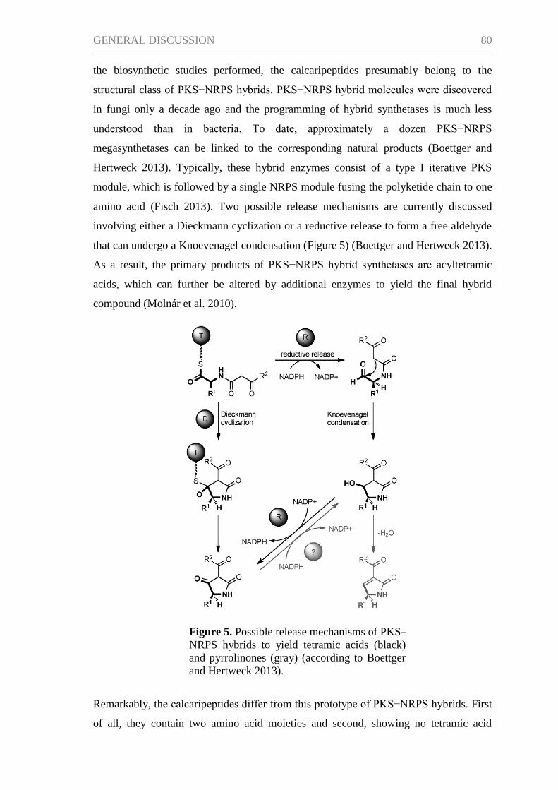
GENERAL DISCUSSION 80
the biosynthetic studies performed, the calcaripeptides presumably belong to the
structural class of PKS−NRPS hybrids. PKS−NRPS hybrid molecules were discovered
in fungi only a decade ago and the programming of hybrid synthetases is much less
understood than in bacteria. To date, approximately a dozen PKS−NRPS
megasynthetases can be linked to the corresponding natural products (Boettger and
Hertweck 2013). Typically, these hybrid enzymes consist of a type I iterative PKS
module, which is followed by a single NRPS module fusing the polyketide chain to one
amino acid (Fisch 2013). Two possible release mechanisms are currently discussed
involving either a Dieckmann cyclization or a reductive release to form a free aldehyde
that can undergo a Knoevenagel condensation (Figure 5) (Boettger and Hertweck 2013).
As a result, the primary products of PKS−NRPS hybrid synthetases are acyltetramic
acids, which can further be altered by additional enzymes to yield the final hybrid
compound (Molnár et al. 2010).
Remarkably, the calcaripeptides differ from this prototype of PKS−NRPS hybrids. First
of all, they contain two amino acid moieties and second, showing no tetramic acid
Figure 5. Possible release mechanisms of PKS−
NRPS hybrids to yield tetramic acids (black)
and pyrrolinones (gray) (according to Boettger
and Hertweck 2013).
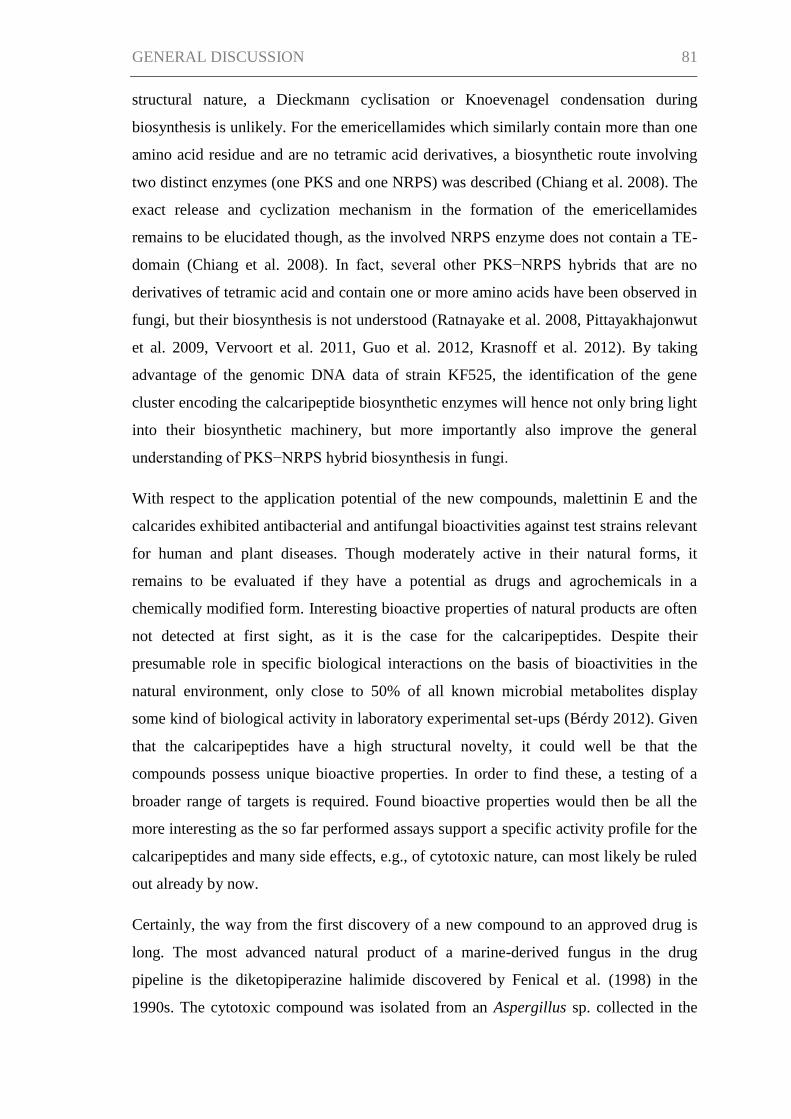
GENERAL DISCUSSION 81
structural nature, a Dieckmann cyclisation or Knoevenagel condensation during
biosynthesis is unlikely. For the emericellamides which similarly contain more than one
amino acid residue and are no tetramic acid derivatives, a biosynthetic route involving
two distinct enzymes (one PKS and one NRPS) was described (Chiang et al. 2008). The
exact release and cyclization mechanism in the formation of the emericellamides
remains to be elucidated though, as the involved NRPS enzyme does not contain a TE-
domain (Chiang et al. 2008). In fact, several other PKS−NRPS hybrids that are no
derivatives of tetramic acid and contain one or more amino acids have been observed in
fungi, but their biosynthesis is not understood (Ratnayake et al. 2008, Pittayakhajonwut
et al. 2009, Vervoort et al. 2011, Guo et al. 2012, Krasnoff et al. 2012). By taking
advantage of the genomic DNA data of strain KF525, the identification of the gene
cluster encoding the calcaripeptide biosynthetic enzymes will hence not only bring light
into their biosynthetic machinery, but more importantly also improve the general
understanding of PKS−NRPS hybrid biosynthesis in fungi.
With respect to the application potential of the new compounds, malettinin E and the
calcarides exhibited antibacterial and antifungal bioactivities against test strains relevant
for human and plant diseases. Though moderately active in their natural forms, it
remains to be evaluated if they have a potential as drugs and agrochemicals in a
chemically modified form. Interesting bioactive properties of natural products are often
not detected at first sight, as it is the case for the calcaripeptides. Despite their
presumable role in specific biological interactions on the basis of bioactivities in the
natural environment, only close to 50% of all known microbial metabolites display
some kind of biological activity in laboratory experimental set-ups (Bérdy 2012). Given
that the calcaripeptides have a high structural novelty, it could well be that the
compounds possess unique bioactive properties. In order to find these, a testing of a
broader range of targets is required. Found bioactive properties would then be all the
more interesting as the so far performed assays support a specific activity profile for the
calcaripeptides and many side effects, e.g., of cytotoxic nature, can most likely be ruled
out already by now.
Certainly, the way from the first discovery of a new compound to an approved drug is
long. The most advanced natural product of a marine-derived fungus in the drug
pipeline is the diketopiperazine halimide discovered by Fenical et al. (1998) in the
1990s. The cytotoxic compound was isolated from an Aspergillus sp. collected in the
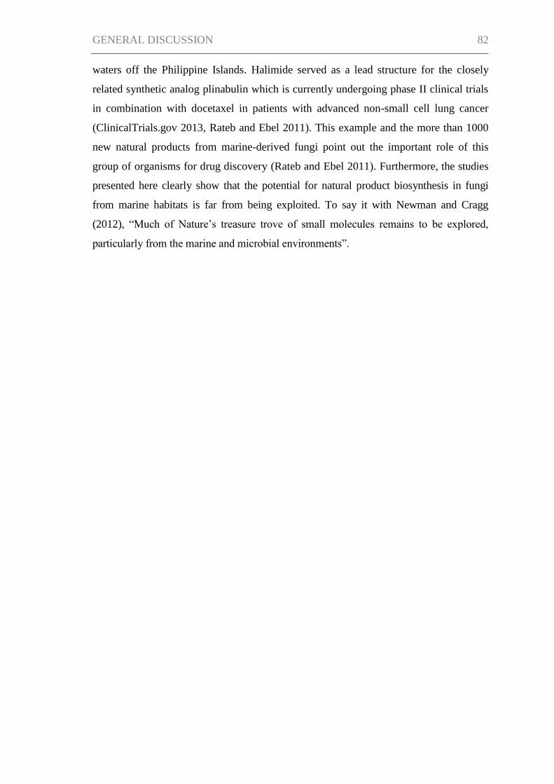
GENERAL DISCUSSION 82
waters off the Philippine Islands. Halimide served as a lead structure for the closely
related synthetic analog plinabulin which is currently undergoing phase II clinical trials
in combination with docetaxel in patients with advanced non-small cell lung cancer
(ClinicalTrials.gov 2013, Rateb and Ebel 2011). This example and the more than 1000
new natural products from marine-derived fungi point out the important role of this
group of organisms for drug discovery (Rateb and Ebel 2011). Furthermore, the studies
presented here clearly show that the potential for natural product biosynthesis in fungi
from marine habitats is far from being exploited. To say it with Newman and Cragg
(2012), “Much of Nature’s treasure trove of small molecules remains to be explored,
particularly from the marine and microbial environments”.
REFERENCES 83
REFERENCES
Aharonowitz, Y., Demain, A. L. (1980). Thoughts on secondary metabolism.
Biotechnol. Bioeng. 22(Suppl. 1), 5-9.
Alberts, A. W., Chen, J., Kuron, G., Hunt, V., Huff, J., Hoffman, C., Rothrock, J.,
Lopez, M., Joshua, H., Harris, E., Patchett, A., Monaghan, R., Currie, S., Stapley, E.,
Albers-Schönberg, G., Hensens, O., Hirshfield, J., Hoogsteen, K., Liesch, J., Springer,
J. (1980). Mevinolin: a highly potent competitive inhibitor of hydroxymethylglutaryl-
coenzyme A reductase and a cholesterol-lowering agent. Proc. Natl. Acad. Sci. USA
77(7), 3957-3961.
Aly, A. H., Debbab, A., Proksch, P. (2011). Fifty years of drug discovery from fungi.
Fungal Diversity 50(1), 3-19.
Baguley, B. C. (2010). Multiple drug resistance mechanisms in cancer. Mol. Biotechnol.
46(3), 308-316.
Bennett, J. W., Bentley, R. (1989). What's in a name? - Microbial secondary
metabolism. Adv. Appl. Microbiol. 34, 1-28.
Bérdy, J. (2005). Bioactive microbial metabolites. J. Antibiot. 58(1), 1-26.
Bérdy, J. (2012). Thoughts and facts about antibiotics: where we are now and where we
are heading. J. Antibiot. 65(8), 385-395.
Bergmann, S., Schümann, J., Scherlach, K., Lange, C., Brakhage, A. H., Hertweck, C.
(2007). Genomics-driven discovery of PKS–NRPS hybrid metabolites from Aspergillus
nidulans. Nat. Chem. Biol. 3(4), 213-217.
Bhatnagar, I., Kim, S.-K. (2010). Immense essence of excellence: marine microbial
bioactive compounds. Mar. Drugs 8(10), 2673-2701.
Bingle, L. E. H., Simpson, T. J., Lazarus, C. M. (1999). Ketosynthase domain probes
identify two subclasses of fungal polyketide synthase genes. Fungal Genet. Biol. 26(3),
209-223.
Bitzer, J., Große, T., Wang, L., Lang, S., Beil, W., Zeeck, A. (2006). New
aminophenoxazinones from a marine Halomonas sp.: fermentation, structure
elucidation, and biological activity. J. Antibiot. 59(2), 86-92.
REFERENCES 84
Bode, H. B., Bethe, B., Höfs, R., Zeeck, A. (2002). Big effects from small changes:
possible ways to explore nature's chemical diversity. ChemBioChem 3(7), 619-627.
Boettger, D., Hertweck, C. (2013). Molecular diversity sculpted by fungal PKS–NRPS
hybrids. ChemBioChem 14(1), 28-42.
Bok, J. W., Hoffmeister, D., Maggio-Hall, L. A., Murillo, R., Glasner, J. D., Keller, N.
P. (2006). Genomic mining for Aspergillus natural products. Chem. Biol. 13(1), 31-37.
Bok, J. W., Keller, N. P. (2004). LaeA, a regulator of secondary metabolism in
Aspergillus spp. Eukaryot. Cell 3(2), 527-535.
Brakhage, A. A., Schroeckh, V. (2011). Fungal secondary metabolites – strategies to
activate silent gene clusters. Fungal Genet. Biol. 48(1), 15-22.
Bugni, T. S., Ireland, C. M. (2004). Marine-derived fungi: a chemically and biologically
diverse group of microorganisms. Nat. Prod. Rep. 21(1), 143-163.
Calvo, A. M., Wilson, R. A., Bok, J. W., Keller, N. P. (2002). Relationship between
secondary metabolism and fungal development. Microbiol. Mol. Biol. Rev. 66(3), 447-
459.
Chiang, Y.-M., Szewczyk, E., Davidson, A. D., Keller, N., Oakley, B. R., Wang, C. C.
C. (2009). A gene cluster containing two fungal polyketide synthases encodes the
biosynthetic pathway for a polyketide, asperfuranone, in Aspergillus nidulans. J. Am.
Chem. Soc. 131(8), 2965-2970.
Chiang, Y.-M., Szewczyk, E., Nayak, T., Davidson, A. D., Sanchez, J. F., Lo, H.-C.,
Ho, W.-Y., Simityan, H., Kuo, E., Praseuth, A., Watanabe, K., Oakley, B. R., Wang, C.
C. C. (2008). Molecular genetic mining of the Aspergillus secondary metabolome:
discovery of the emericellamide biosynthetic pathway. Chem. Biol. 15(6), 527-532.
ClinicalTrials.gov (2013). www.clinicaltrials.gov/ct2/show?term=npi-2358&rank=1.
Cox, R. J. (2007). Polyketides, proteins and genes in fungi: programmed nano-machines
begin to reveal their secrets. Org. Biomol. Chem. 5(13), 2010-2026.
Cragg, G. M., Newman, D. J., Snader, K. M. (1997). Natural products in drug discovery
and development. J. Nat. Prod. 60(1), 52-60.
REFERENCES 85
Crawford, J. M., Townsend, C. A. (2010). New insights into the formation of fungal
aromatic polyketides. Nat. Rev. Microbiol. 8(12), 879-889.
Cueto, M., Jensen, P. R., Kauffman, C., Fenical, W., Lobkovsky, E., Clardy, J. (2001).
Pestalone, a new antibiotic produced by a marine fungus in response to bacterial
challenge. J. Nat. Prod. 64(11), 1444-1446.
Davies, J., Davies, D. (2010). Origins and evolution of antibiotic resistance. Microbiol.
Mol. Biol. Rev. 74(3), 417-433.
de Bono, J. S., Ashworth, A. (2010). Translating cancer research into targeted
therapeutics. Nature 467(7315), 543-549.
de Lencastre, H., Oliveira, D., Tomasz, A. (2007). Antibiotic resistant Staphylococcus
aureus: a paradigm of adaptive power. Curr. Opin. Microbiol. 10(5), 428-435.
Demain, A. L. (2013). Importance of microbial natural products and the need to
revitalize their discovery. J. Ind. Microbiol. Biotechnol. doi: 10.1007/s10295-013-1325-
z.
Demain, A. L., Sanchez, S. (2009). Microbial drug discovery: 80 years of progress. J.
Antibiot. 62(1), 5-16.
Dictionary of Natural Products (2012). Chapman & Hall/CRC Press/Hampden Data
Services, Ltd.
Dreyfuss, M., Härri, E., Hofmann, H., Kobel, H., Pache, W., Tscherter, H. (1976).
Cyclosporin A and C. Eur. J. Appl. Microbiol. Biotechnol. 3(2), 125-133.
Enright, M. C. (2003). The evolution of a resistant pathogen – the case of MRSA. Curr.
Opin. Pharmacol. 3(5), 474-479.
Fenical, W., Jensen, P. R., Cheng, X. C. (1998). Halimide, a cytotoxic marine natural
product, and derivatives thereof. US Patent, 6069146.
Ferlay, J., Shin, H.-R., Bray, F., Forman, D., Mathers, C., Parkin, D. M. (2010).
Estimates of worldwide burden of cancer in 2008: GLOBOCAN 2008. Int. J. Cancer
127(12), 2893-2917.
Finking, R., Marahiel, M. A. (2004). Biosynthesis of nonribosomal peptides. Annu. Rev.
Microbiol. 58, 453-488.
REFERENCES 86
Fisch, K. M. (2013). Biosynthesis of natural products by microbial iterative hybrid
PKS–NRPS. R. Soc. Chem. Adv. 3(40), 18228-18247.
Fischbach, M. A., Walsh, C. T. (2006). Assembly-line enzymology for polyketide and
nonribosomal peptide antibiotics: logic, machinery, and mechanisms. Chem. Rev.
106(8), 3468-3496.
Fleming, A. (1945). “Sir Alexander Fleming – Nobel Lecture: Penicillin".
Nobelprize.org. Nobel Media AB 2013. Web. 16 Sep 2013.
www.nobelprize.org/nobel_prizes/medicine/laureates/1945/fleming-lecture.html.
Fröhlich-Nowoisky, J., Pickersgill, D. A., Després, V. R., Pöschl, U. (2009). High
diversity of fungi in air particulate matter. Proc. Natl. Acad. Sci. USA 106(31), 12814-
12819.
Fuchser, J. (1996). Beeinflussung der Sekundärstoffbildung bei Aspergillus ochraceus
durch Variation der Kulturbedingungen sowie Isolierung, Strukturaufklärung und
Biosynthese der neuen Naturstoffe. PhD thesis, Universität Göttingen.
Glinski, M., Urbanke, C., Hornbogen, T., Zocher, R. (2002). Enniatin synthetase is a
monomer with extended structure: evidence for an intramolecular reaction mechanism.
Arch. Microbiol. 178(4), 267-273.
Godtfredsen, W. O., Jahnsen, S., Lorck, H., Roholt, K., Tybring, L. (1962). Fusidic
acid: a new antibiotic. Nature 193(4819), 987.
Grabley, S., Thiericke, R. (1999). The impact of natural products on drug discovery. In:
Grabley, S., Thiericke, R. (Eds.). Drug Discovery from Nature. Springer-Verlag, Berlin
Heidelberg, Germany, 3-37.
Gräfe, U. (1999). Secondary metabolites: from past to present. In: Grabley, S.,
Thiericke, R. (Eds.). Drug Discovery from Nature. Springer-Verlag, Berlin Heidelberg,
Germany, 117-123.
Grossart, H.-P., Thorwest, M., Plitzko, I., Brinkhoff, T., Simon, M., Zeeck, A. (2009).
Production of a blue pigment (glaukothalin) by marine Rheinheimera spp. Int. J.
Microbiol. doi: 10.1155/2009/701735.
Grove, J. F., MacMillan, J., Mulholland, T. P. C., Rogers, M. A. T. (1952).
Griseofulvin. Part IV. Structure. J. Chem. Soc., 3977-3987.
REFERENCES 87
Grunwald, M., Dellwig, O., Liebezeit, G., Schnetger, B., Reuter, R., Brumsack, H.-J.
(2007). A novel time-series station in the Wadden Sea (NW Germany): first results on
continuous nutrient and methane measurements. Mar. Chem. 107(3), 411-421.
Gunatilaka, A. A. L. (2006). Natural products from plant-associated microorganisms:
distribution, structural diversity, bioactivity, and implications of their occurrence. J.
Nat. Prod. 69(3), 509-526.
Guo, J.-P., Zhu, C.-Y., Zhang, C.-P., Chu, Y.-S., Wang, Y.-L., Zhang, J.-X., Wu, D.-K.,
Zhang, K.-Q., Niu, X.-M. (2012). Thermolides, potent nematocidal PKS–NRPS hybrid
metabolites from thermophilic fungus Talaromyces thermophilus. J. Am. Chem. Soc.
134(50), 20306-20309.
Harvey, A. L. (2008). Natural products in drug discovery. Drug Discov. Today 13(19-
20), 894-901.
Hawksworth, D. L. (2001). The magnitude of fungal diversity: the 1.5 million species
estimate revisited. Mycol. Res. 105(12), 1422-1432.
Hawksworth, D. L., Mueller, G. M. (2005). Fungal communities: their diversity and
distribution. In: Dighton, J., White, J. F., Oudemans P. (Eds.). The fungal community:
its organization and role in the ecosystem. CRC Press, Boca Raton, FL, USA, 27-37.
Hertweck, C. (2009). The biosynthetic logic of polyketide diversity. Angew. Chem. Int.
Ed. 48(26), 4688-4716.
Hertweck, C. (2009). Hidden biosynthetic treasures brought to light. Nat. Chem. Biol.
5(7), 450-452.
Hoffmeister, D., Keller, N. P. (2007). Natural products of filamentous fungi: enzymes,
genes, and their regulation. Nat. Prod. Rep. 24(2), 393-416.
Imhoff, J. F., Labes, A., Wiese, J. (2011). Bio-mining the microbial treasures of the
ocean: new natural products. Biotechnol. Adv. 29(5), 468-482.
Infectious Diseases Society of America (2004). Bad bugs, no drugs. As antibiotic
discovery stagnates…A public health crisis brews, pp. 35. Accessed through:
http://www.idsociety.org/Search.aspx?&lcid=9&q=Bad%20Bugs%2C%20No%20Drug
s.%20&tz=Europe%2FBerlin on 2013-12-16.
REFERENCES 88
Ishiuchi, K., Nakazawa, T., Ookuma, T., Sugimoto, S., Sato, M., Tsunematsu, Y.,
Ishikawa, N., Noguchi, H., Hotta, K., Moriya, H., Watanabe, K. (2012). Establishing a
new methodology for genome mining and biosynthesis of polyketides and peptides
through yeast molecular genetics. ChemBioChem 13(6), 846-854.
Jensen, P. R., Chavarria, K. L., Fenical, W., Moore, B. S., Ziemert, N. (2013).
Challenges and triumphs to genomics-based natural product discovery. J. Ind.
Microbiol. Biotechnol. doi: 10.1007/s10295-013-1353-8.
Jones, K. E., Patel, N. G., Levy, M. A., Storeygard, A., Balk, D., Gittleman, J. L.,
Daszak, P. (2008). Global trends in emerging infectious diseases. Nature 451(7181),
990-994.
Keller, N. P., Hohn, T. M. (1997). Metabolic pathway gene clusters in filamentous
fungi. Fungal Genet. Biol. 21(1), 17-29.
Keller, N. P., Turner, G., Bennett, J. W. (2005). Fungal secondary metabolism - from
biochemistry to genomics. Nat. Rev. Microbiol. 3(12), 937-947.
Kettering, M., Weber, D., Sterner, O., Anke, T. (2004). Sekundärmetabolite aus Pilzen -
Funktionen und Anwendungen. BIOspektrum 10(2), 147-149.
Khaldi, N., Seifuddin, F. T., Turner, G., Haft, D., Nierman, W. C., Wolfe, K. H.,
Fedorova, N. D. (2010). SMURF: genomic mapping of fungal secondary metabolite
clusters. Fungal Genet. Biol. 47(9), 736-741.
Klotz, H. (2010). Biosynthese von Sekundärmetaboliten in "marine derived" Pilzen:
Kulturbasierte und molekularbiologische Ansätze. Bachelor thesis, Fachhochschule
Aachen.
Kohlmeyer, J., Kohlmeyer, E. (1979). Marine mycology. The higher fungi. Academic
Press, New York, USA, pp. 690.
Koul, A., Arnoult, E., Lounis, N., Guillemont, J., Andries, K. (2011). The challenge of
new drug discovery for tuberculosis. Nature 469(7331), 483-490.
Krasnoff, S. B., Englich, U., Miller, P. G., Shuler, M. L., Glahn, R. P., Donzelli, B. G.
G., Gibson, D. M. (2012). Metacridamides A and B, macrocycles from conidia of the
entomopathogenic fungus Metarhizium acridum. J. Nat. Prod. 75(2), 175-180.
REFERENCES 89
Laatsch, H. (2000). Mikroorganismen als biologische Quelle neuer Wirkstoffe. In:
Kayser, O., Müller, R. H. (Eds.). Pharmazeutische Biotechnologie. Wissenschaftliche
Verlagsges., Stuttgart, Germany, 13-43.
Levy, S. B., Marshall, B. (2004). Antibacterial resistance worldwide: causes, challenges
and responses. Nat. Med. 10(12), 122-129.
Li, J. W.-H., Vederas, J. C. (2009). Drug discovery and natural products: end of an era
or an endless frontier? Science 325(5937), 161-165.
Liang, L. (2003). Investigation of secondary metabolites of North Sea bacteria:
fermentation, isolation, structure elucidation and bioactivity. PhD thesis, Universität
Göttingen.
Liebezeit, G., Kraul, T., Everts, B. (1994). Bulk chemical characterization of particulate
material from the Jade Bay, Lower Saxonian Wadden Sea. Neth. J. Aquat. Ecol. 28(3-
4), 365-370.
Luo, J., Solimini, N. L., Elledge, S. J. (2009). Principles of cancer therapy: oncogene
and non-oncogene addiction. Cell 136(5), 823-837.
Molnár, I., Gibson, D. M., Krasnoff, S. B. (2010). Secondary metabolites from
entomopathogenic Hypocrealean fungi. Nat. Prod. Rep. 27(9), 1241-1275.
Mootz, H. D., Schwarzer, D., Marahiel, M. A. (2002). Ways of assembling complex
natural products on modular nonribosomal peptide synthetases. ChemBioChem 3(6),
490-504.
Morens, D. M., Fauci, A. S. (2013). Emerging infectious diseases: threats to human
health and global stability. PLoS Pathog. 9(7), 1-3.
Newman, D. J., Cragg, G. M. (2007). Natural products as sources of new drugs over the
last 25 years. J. Nat. Prod. 70(3), 461-477.
Newman, D. J., Cragg, G. M. (2012). Natural products as sources of new drugs over the
30 years from 1981 to 2010. J. Nat. Prod. 75(3), 311-335.
Newman, D. J., Cragg, G. M., Snader, K. M. (2003). Natural products as sources of new
drugs over the period 1981-2002. J. Nat. Prod. 66(7), 1022-1037.
REFERENCES 90
Newton, G. G. F., Abraham, E. P. (1955). Cephalosporin C, a new antibiotic containing
sulphur and D-α-aminoadipic acid. Nature 175 (4456), 548.
Nicolaou, K. C., Montagnon, T. (2008). Molecules that changed the world. Wiley-
VCH, Weinheim, Germany, pp. 366.
Nikaido, H. (2009). Multidrug resistance in bacteria. Annu. Rev. Biochem. 78, 119-146.
Norrby, S. R., Nord, C. E., Finch, R. (2005). Lack of development of new antimicrobial
drugs: a potential serious threat to public health. Lancet Infect. Dis. 5(2), 115-119.
Pfaller, M. A. (2012). Antifungal drug resistance: mechanisms, epidemiology, and
consequences for treatment. Am. J. Med. 125(1), S3-S13.
Pittayakhajonwut, P., Usuwan, A., Intaraudom, C., Khoyaiklang, P., Supothina, S.
(2009). Torrubiellutins A–C, from insect pathogenic fungus Torrubiella luteorostrata
BCC 12904. Tetrahedron 65(31), 6069-6073.
Raistrick, H. (1949). Bakerian lecture. A region of biosynthesis. P. Roy. Soc. Lond. A
Mat. 199(1057), 141-168.
Raphael, R. A. (1948). Mould metabolic products. The Scientific Journal of the Royal
College of Science 18, 42-53.
Rateb, M. E., Ebel, R. (2011). Secondary metabolites of fungi from marine habitats.
Nat. Prod. Rep. 28(2), 290-344.
Ratnayake, R., Fremlin, L. J., Lacey, E., Gill, J. H., Capon, R. J. (2008). Acremolides
A− , lipodepsipeptides from an Australian marine-derived fungus, Acremonium sp. J.
Nat. Prod. 71(3), 403-408.
Rohlfs, M., Albert, M., Keller, N. P., Kempken, F. (2007). Secondary chemicals protect
mould from fungivory. Biol. Lett. 3(5), 523-525.
Saikkonen, K., Faeth, S. H., Helander, M., Sullivan, T. J. (1998). Fungal endophytes: a
continuum of interactions with host plants. Annu. Rev. Ecol. Syst. 29, 319-343.
Scherlach, K., Hertweck, C. (2009). Triggering cryptic natural product biosynthesis in
microorganisms. Org. Biomol. Chem. 7(9), 1753-1760.
REFERENCES 91
Schneemann, I. (2011). Nachweis von Biosynthesegenen des bakteriellen
Sekundärstoffwechsels sowie Isolierung und Strukturaufklärung von Naturstoffen aus
ausgewählten Actinomyceten. PhD thesis, Christian-Albrechts-Universität zu Kiel.
Schulz, D., Ohlendorf, B., Zinecker, H., Schmaljohann, R., Imhoff, J. F. (2011).
Eutypoids B–E produced by a Penicillium sp. strain from the North Sea. J. Nat. Prod.
74(1), 99-101.
Schümann, J., Hertweck, C. (2006). Advances in cloning, functional analysis and
heterologous expression of fungal polyketide synthase genes. J. Biotechnol. 124(4),
690-703.
Shearer, C. A., Descals, E., Kohlmeyer, B., Kohlmeyer, J., Marvanová, L., Padgett, D.,
Porter, D., Raja, H. A., Schmit, J. P., Thorton, H. A., Voglymayr, H. (2007). Fungal
biodiversity in aquatic habitats. Biodivers. Conserv. 16(1), 49-67.
Song, Z., Cox, R. J., Lazarus, C. M., Simpson, T. J. (2004). Fusarin C biosynthesis in
Fusarium moniliforme and Fusarium venenatum. ChemBioChem 5(9), 1196-1203.
Spížek, J., Novotná, J., Řezanka, T., emain, A. L. (2010). o we need new
antibiotics? The search for new targets and new compounds. J. Ind. Microbiol.
Biotechnol. 37(12), 1241-1248.
Strieker, M., Tanović, A., Marahiel, M. A. (2010). Nonribosomal peptide synthetases:
structures and dynamics. Curr. Opin. Struct. Biol. 20(2), 234-240.
Strobel, G., Daisy, B., Castillo, U., Harper, J. (2004). Natural products from endophytic
microorganisms. J. Nat. Prod. 67(2), 257-268.
Velayati, A. A., Masjedi, M. R., Farnia, P., Tabarsi, P., Ghanavi, J., ZiaZarifi, A. H.,
Hoffner, S. E. (2009). Emergence of new forms of totally drug-resistant tuberculosis
bacilli: super extensively drug-resistant tuberculosis or totally drug-resistant strains in
Iran. Chest 136(2), 420-425.
Vervoort, H. C., rašković, M., Crews, P. (2011). Histone deacetylase inhibitors as a
tool to up-regulate new fungal biosynthetic products – Isolation of EGM-556, a
cyclodepsipeptide, from Microascus sp. Org. Lett. 13(3), 410-413.
Vining, L. C. (1990). Functions of secondary metabolites. Annu. Rev. Microbiol. 44(1),
395-427.
REFERENCES 92
Wang, G. (2006). Diversity and biotechnological potential of the sponge-associated
microbial consortia. J. Ind. Microbiol. Biotechnol. 33(7), 545-551.
WHO (2013). www.who.int/en/.
Wiese, J., Ohlendorf, B., Blümel, M., Schmaljohann, R., Imhoff, J. F. (2011).
Phylogenetic identification of fungi isolated from the marine sponge Tethya aurantium
and identification of their secondary metabolites. Mar. Drugs 9(4), 561-585.
Williams, D. H., Stone, M. J., Hauck, P. R., Rahman, S. K. (1989). Why are secondary
metabolites (natural products) biosynthesized? J. Nat. Prod. 52(6), 1189-1208.
Williams, R. B., Henrikson, J. C., Hoover, A. R., Lee, A. E., Cichewicz, R. H. (2008).
Epigenetic remodeling of the fungal secondary metabolome. Org. Biomol. Chem. 6(11),
1895-1897.
Zähner, H. (1979). What are secondary metabolites? Folia Microbiol. 24(5), 435-443.
ACKNOWLEDGMENTS/DANKSAGUNG 93
ACKNOWLEDGMENTS/DANKSAGUNG
Herrn Prof. Dr. J. F. Imhoff danke ich, dass er mir die Möglichkeit gegeben hat, die
Doktorarbeit in seiner Arbeitsgruppe durchzuführen und für die Vergabe des
interessanten Themas.
Mein besonderer Dank gilt Antje und Birgit für die tolle Betreuung während der
Doktorarbeit. Ich danke euch aber auch für die schönen Stunden und guten Gespräche
beim Pilze sammeln, Doppelkopf und an so manchem Kneipenabend!
Arlette Erhard danke ich für die zuverlässige Durchführung der Aktivitätstests.
Nils, Franz und Anne danke ich für die lustige Zeit in unserem Doktorandenbüro.
Niiiiix hat sonst mehr Spaß gemacht.
Bei allen Mitarbeitern der Marinen Mikrobiologie möchte ich mich für das tolle
Arbeitsklima und die netten, gemeinsamen Stunden bedanken.
DECLARATION/ERKLÄRUNG 94
DECLARATION/ERKLÄRUNG
Hiermit erkläre ich, dass ich die vorliegende Dissertation selbständig und ohne
unerlaubte Hilfe angefertigt habe und dass sie nach Form und Inhalt meine eigene
Arbeit ist. Sie wurde weder im Ganzen noch zum Teil einer anderen Stelle im Rahmen
eines Prüfungsverfahrens vorgelegt und entstand unter Einhaltung der Regeln guter
wissenschaftlicher Praxis der Deutschen Forschungsgemeinschaft.
….………………………..
(Johanna Silber)
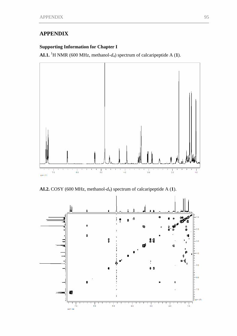
APPENDIX 95
APPENDIX
Supporting Information for Chapter I
AI.1. 1H NMR (600 MHz, methanol-d4) spectrum of calcaripeptide A (1).
AI.2. COSY (600 MHz, methanol-d4) spectrum of calcaripeptide A (1).
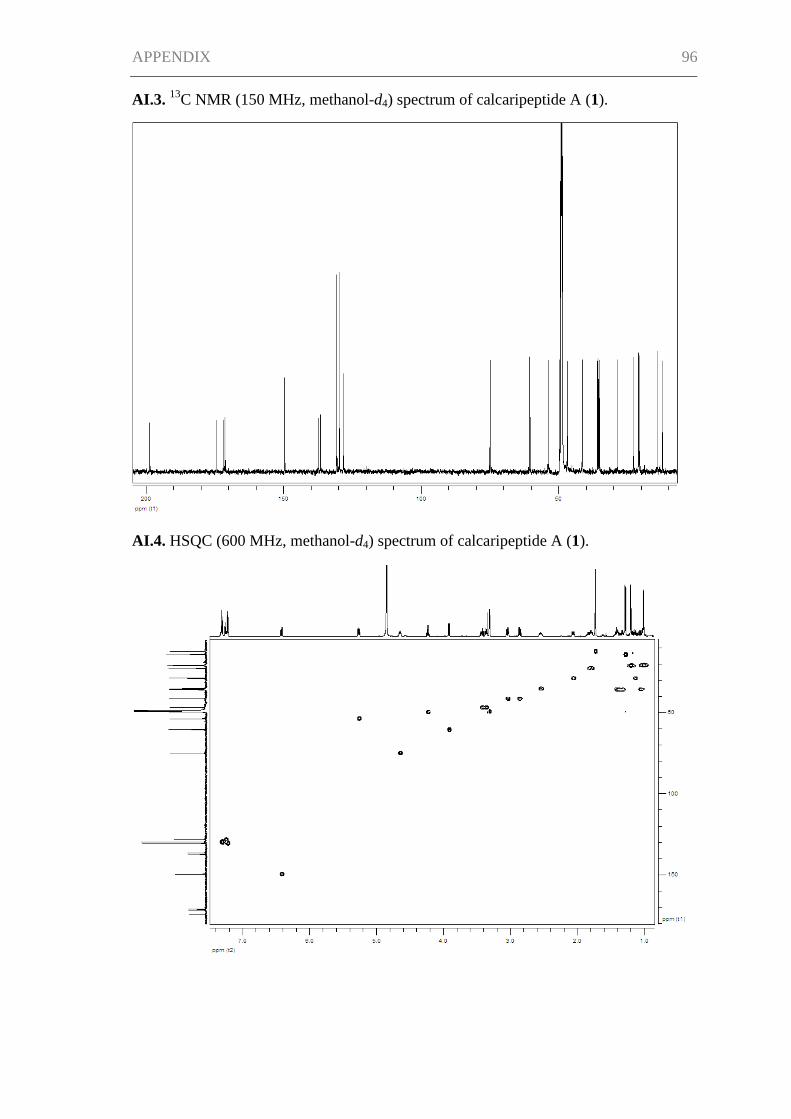
APPENDIX 96
AI.3. 13
C NMR (150 MHz, methanol-d4) spectrum of calcaripeptide A (1).
AI.4. HSQC (600 MHz, methanol-d4) spectrum of calcaripeptide A (1).
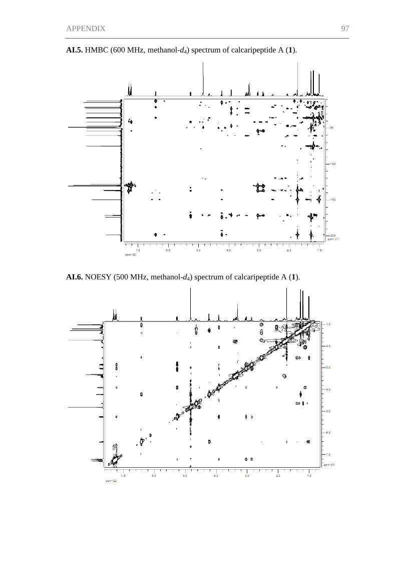
APPENDIX 97
AI.5. HMBC (600 MHz, methanol-d4) spectrum of calcaripeptide A (1).
AI.6. NOESY (500 MHz, methanol-d4) spectrum of calcaripeptide A (1).
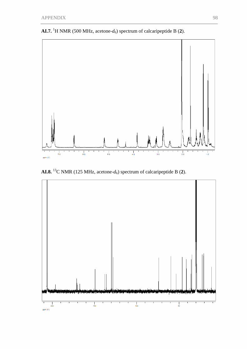
APPENDIX 98
AI.7. 1H NMR (500 MHz, acetone-d6) spectrum of calcaripeptide B (2).
AI.8. 13
C NMR (125 MHz, acetone-d6) spectrum of calcaripeptide B (2).
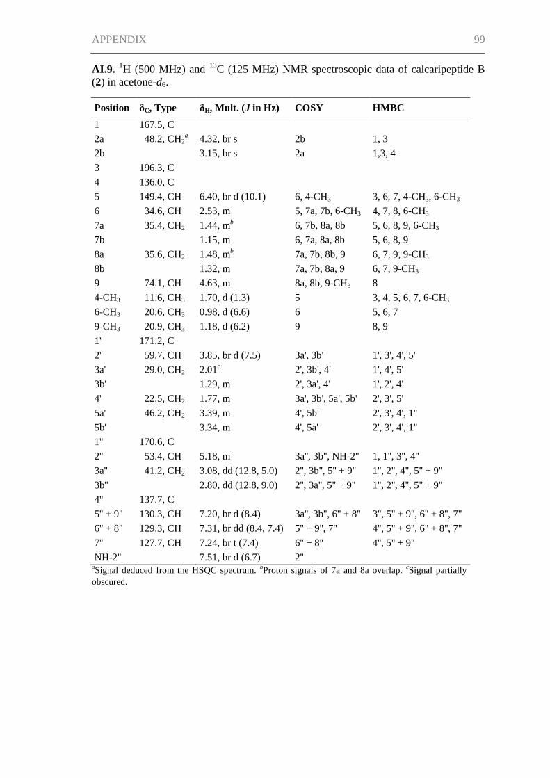
APPENDIX 99
AI.9. 1H (500 MHz) and
13C (125 MHz) NMR spectroscopic data of calcaripeptide B
(2) in acetone-d6.
Position δC, Type δH, Mult. (J in Hz) COSY HMBC
1 167.5, C
2a 48.2, CH2a 4.32, br s 2b 1, 3
2b 3.15, br s 2a 1,3, 4
3 196.3, C
4 136.0, C
5 149.4, CH 6.40, br d (10.1) 6, 4-CH3 3, 6, 7, 4-CH3, 6-CH3
6 34.6, CH 2.53, m 5, 7a, 7b, 6-CH3 4, 7, 8, 6-CH3
7a 35.4, CH2 1.44, mb 6, 7b, 8a, 8b 5, 6, 8, 9, 6-CH3
7b 1.15, m 6, 7a, 8a, 8b 5, 6, 8, 9
8a 35.6, CH2 1.48, mb 7a, 7b, 8b, 9 6, 7, 9, 9-CH3
8b 1.32, m 7a, 7b, 8a, 9 6, 7, 9-CH3
9 74.1, CH 4.63, m 8a, 8b, 9-CH3 8
4-CH3 11.6, CH3 1.70, d (1.3) 5 3, 4, 5, 6, 7, 6-CH3
6-CH3 20.6, CH3 0.98, d (6.6) 6 5, 6, 7
9-CH3 20.9, CH3 1.18, d (6.2) 9 8, 9
1' 171.2, C
2' 59.7, CH 3.85, br d (7.5) 3a', 3b' 1', 3', 4', 5'
3a' 29.0, CH2 2.01c 2', 3b', 4' 1', 4', 5'
3b' 1.29, m 2', 3a', 4' 1', 2', 4'
4' 22.5, CH2 1.77, m 3a', 3b', 5a', 5b' 2', 3', 5'
5a' 46.2, CH2 3.39, m 4', 5b' 2', 3', 4', 1''
5b' 3.34, m 4', 5a' 2', 3', 4', 1''
1'' 170.6, C
2'' 53.4, CH 5.18, m 3a'', 3b'', NH-2'' 1, 1'', 3'', 4''
3a'' 41.2, CH2 3.08, dd (12.8, 5.0) 2'', 3b'', 5'' + 9'' 1'', 2'', 4'', 5'' + 9''
3b'' 2.80, dd (12.8, 9.0) 2'', 3a'', 5'' + 9'' 1'', 2'', 4'', 5'' + 9''
4'' 137.7, C
5'' + 9'' 130.3, CH 7.20, br d (8.4) 3a'', 3b'', 6'' + 8'' 3'', 5'' + 9'', 6'' + 8'', 7''
6'' + 8'' 129.3, CH 7.31, br dd (8.4, 7.4) 5'' + 9'', 7'' 4'', 5'' + 9'', 6'' + 8'', 7''
7'' 127.7, CH 7.24, br t (7.4) 6'' + 8'' 4'', 5'' + 9''
NH-2'' 7.51, br d (6.7) 2'' aSignal deduced from the HSQC spectrum.
bProton signals of 7a and 8a overlap.
cSignal partially
obscured.
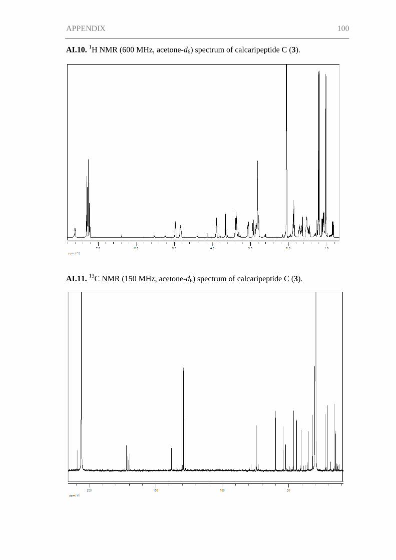
APPENDIX 100
AI.10. 1H NMR (600 MHz, acetone-d6) spectrum of calcaripeptide C (3).
AI.11. 13
C NMR (150 MHz, acetone-d6) spectrum of calcaripeptide C (3).
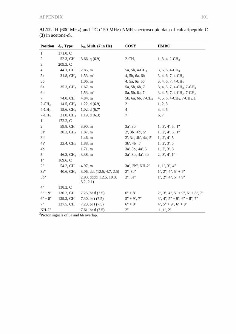
APPENDIX 101
AI.12. 1H (600 MHz) and
13C (150 MHz) NMR spectroscopic data of calcaripeptide C
(3) in acetone-d6.
Position δC, Type δH, Mult. (J in Hz) COSY HMBC
1 171.0, C
2 52.3, CH 3.66, q (6.9) 2-CH3 1, 3, 4, 2-CH3
3 209.3, C
4 44.1, CH 2.85, m 5a, 5b, 4-CH3 3, 5, 6, 4-CH3
5a 31.8, CH2 1.53, ma 4, 5b, 6a, 6b 3, 4, 6, 7, 4-CH3
5b 1.06, m 4, 5a, 6a, 6b 3, 4, 6, 7, 4-CH3
6a 35.3, CH2 1.67, m 5a, 5b, 6b, 7 3, 4, 5, 7, 4-CH3, 7-CH3
6b 1.53, ma 5a, 5b, 6a, 7 3, 4, 5, 7, 4-CH3, 7-CH3
7 74.0, CH 4.84, m 5b, 6a, 6b, 7-CH3 4, 5, 6, 4-CH3, 7-CH3, 1'
2-CH3 14.5, CH3 1.22, d (6.9) 2 1, 2, 3
4-CH3 15.6, CH3 1.02, d (6.7) 4 3, 4, 5
7-CH3 21.0, CH3 1.19, d (6.3) 7 6, 7
1' 172.2, C
2' 59.8, CH 3.90, m 3a', 3b' 1', 3', 4', 5', 1''
3a' 30.3, CH2 1.87, m 2', 3b', 4b', 5' 1', 2', 4', 5', 1''
3b' 1.46, m 2', 3a', 4b', 4a', 5' 1', 2', 4', 5'
4a' 22.4, CH2 1.88, m 3b', 4b', 5' 1', 2', 3', 5'
4b' 1.71, m 3a', 3b', 4a', 5' 1', 2', 3', 5'
5' 46.3, CH2 3.38, m 3a', 3b', 4a', 4b' 2', 3', 4', 1''
1'' 169.6, C
2'' 54.2, CH 4.97, m 3a'', 3b'', NH-2'' 1, 1'', 3'', 4''
3a'' 40.6, CH2 3.06, ddt (12.5, 4.7, 2.5) 2'', 3b'' 1'', 2'', 4'', 5'' + 9''
3b'' 2.93, dddd (12.5, 10.0,
3.2, 2.1)
2'', 3a'' 1'', 2'', 4'', 5'' + 9''
4'' 138.2, C
5'' + 9'' 130.2, CH 7.25, br d (7.5) 6'' + 8'' 2'', 3'', 4'', 5'' + 9'', 6'' + 8'', 7''
6'' + 8'' 129.2, CH 7.30, br t (7.5) 5'' + 9'', 7'' 3'', 4'', 5'' + 9'', 6'' + 8'', 7''
7'' 127.5, CH 7.23, br t (7.5) 6'' + 8'' 4'', 5'' + 9'', 6'' + 8''
NH-2'' 7.61, br d (7.5) 2'' 1, 1'', 2'' aProton signals of 5a and 6b overlap.
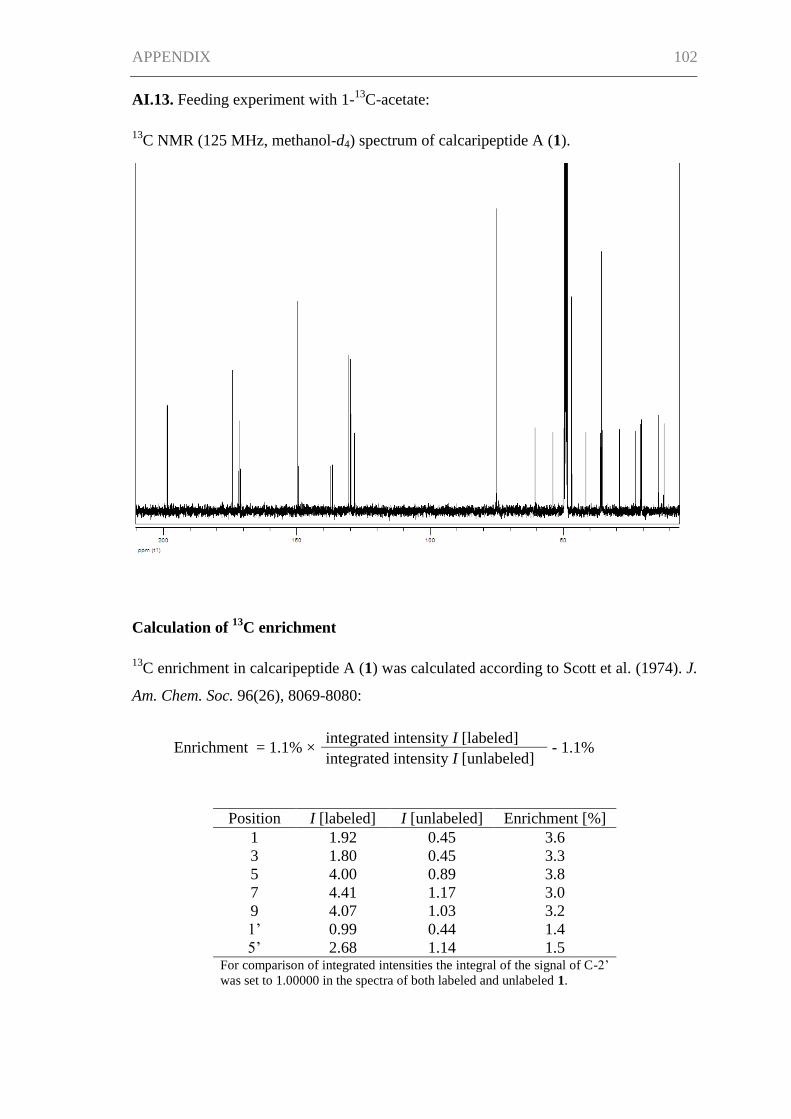
APPENDIX 102
AI.13. Feeding experiment with 1-13
C-acetate:
13C NMR (125 MHz, methanol-d4) spectrum of calcaripeptide A (1).
Calculation of 13
C enrichment
13C enrichment in calcaripeptide A (1) was calculated according to Scott et al. (1974). J.
Am. Chem. Soc. 96(26), 8069-8080:
Enrichment = 1.1% × integrated intensity I [labeled]
- 1.1% integrated intensity I [unlabeled]
Position I [labeled] I [unlabeled] Enrichment [%]
1 1.92 0.45 3.6
3 1.80 0.45 3.3
5 4.00 0.89 3.8
7 4.41 1.17 3.0
9 4.07 1.03 3.2
1’ 0.99 0.44 1.4
5’ 2.68 1.14 1.5 For comparison of integrated intensities the integral of the signal of C-2’
was set to 1.00000 in the spectra of both labeled and unlabeled 1.
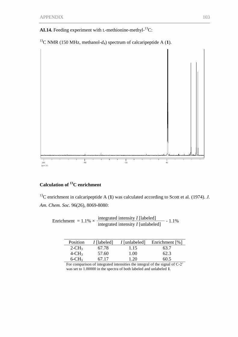
APPENDIX 103
AI.14. Feeding experiment with L-methionine-methyl-13
C:
13C NMR (150 MHz, methanol-d4) spectrum of calcaripeptide A (1).
Calculation of 13
C enrichment
13C enrichment in calcaripeptide A (1) was calculated according to Scott et al. (1974). J.
Am. Chem. Soc. 96(26), 8069-8080:
Enrichment = 1.1% × integrated intensity I [labeled]
- 1.1% integrated intensity I [unlabeled]
Position I [labeled] I [unlabeled] Enrichment [%]
2-CH3 67.78 1.15 63.7
4-CH3 57.60 1.00 62.3
6-CH3 67.17 1.20 60.5 For comparison of integrated intensities the integral of the signal of C-2’
was set to 1.00000 in the spectra of both labeled and unlabeled 1.
APPENDIX 104
AI.15. Activity assays - test strains, cell lines and enzymes.
Calcaripeptides A–C (1-3) were tested for antibacterial, antifungal and cytotoxic
properties and for inhibition of selected enzyme targets. The tested strains, cell lines and
enzymes were as follows (compound concentration in assay): Bacillus subtilis DSM 347
(100 µM), Staphylococcus lentus DSM 6672 (100 µM), Staphylococcus epidermidis
DSM 20044 (100 µM), Xanthomonas campestris DSM 2405 (100 µM),
Propionibacterium acnes DSM 1897 (100 µM), Candida albicans DSM 1386 (100
µM), Septoria tritici (100 µM), Phytophthora infestans (50 µM), Trichophyton rubrum
(100 µM), HepG2 cell line (50 µM), KIF5 fibroblasts (50 µM), glycogen synthase
kinase-3β (10 µM), acetylcholinesterase (10 µM), phosphodiesterase 4B2 (10 µM) and
protein tyrosine phosphatase 1B (10 µM). Moreover, calcaripeptide A (1) was tested
negative for activities against Ralstonia solanacearum DSM 9544 (100 µM), in enzyme
assays for reverse transcriptase (10 µM), histone deacetylase 6 (100 µM), histone
deacetylase 8 (100 µM) and against the cell lines 786-0, MCF-7, M14, HL-60(TB),
CCRF-CEM, K-562, MOLT-4, SR, A549/ATCC, COLO 205, HT29, KM12, SW-620,
SF-268, SF-539, SK-MEL-5, OVCAR-3, OVCAR-8, NCI/ADR-RES, SK-OV-3, PC-3,
T-47D as well as HEK-293 and PBMC (50 µM in all cell lines).
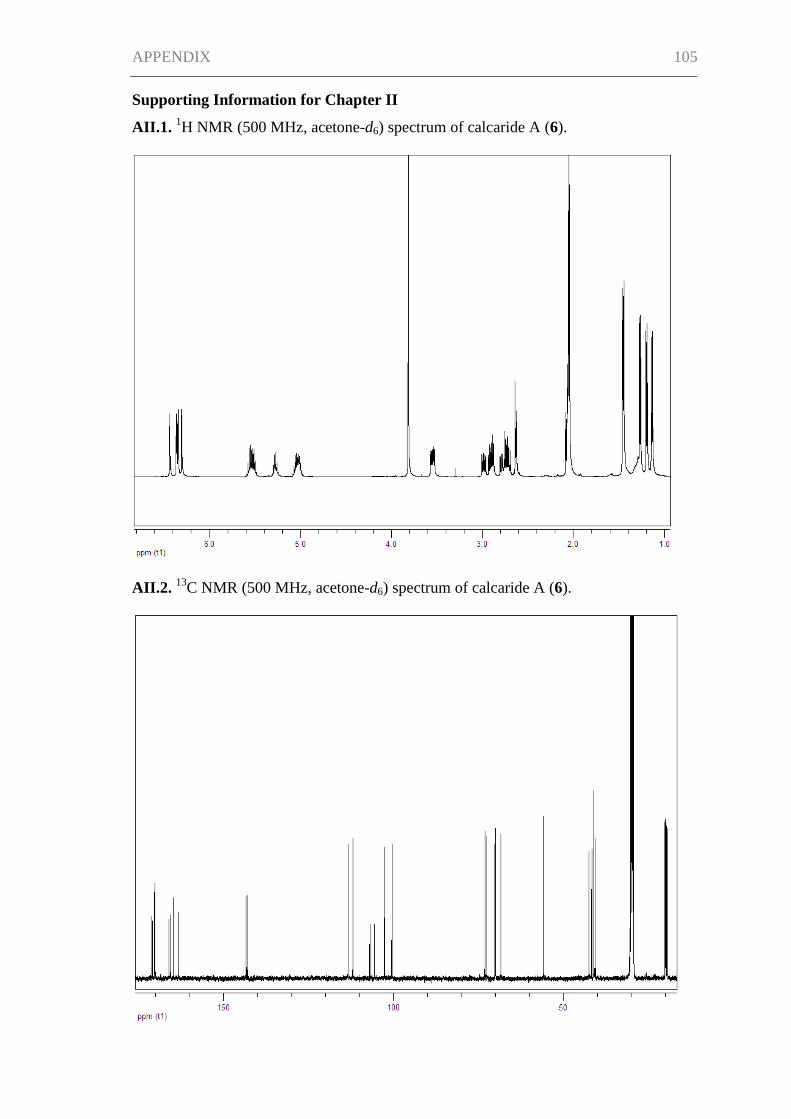
APPENDIX 105
Supporting Information for Chapter II
AII.1. 1H NMR (500 MHz, acetone-d6) spectrum of calcaride A (6).
AII.2. 13
C NMR (500 MHz, acetone-d6) spectrum of calcaride A (6).
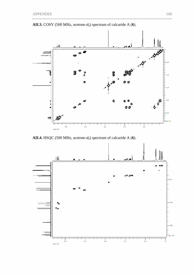
APPENDIX 106
AII.3. COSY (500 MHz, acetone-d6) spectrum of calcaride A (6).
AII.4. HSQC (500 MHz, acetone-d6) spectrum of calcaride A (6).
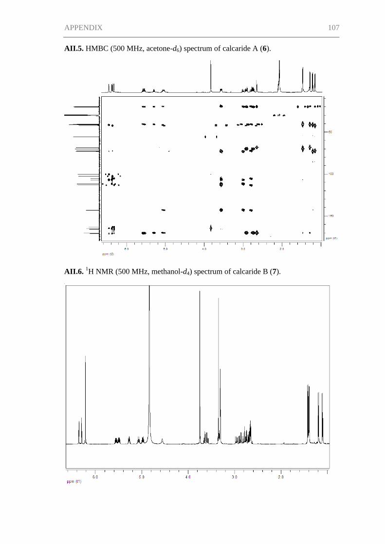
APPENDIX 107
AII.5. HMBC (500 MHz, acetone-d6) spectrum of calcaride A (6).
AII.6. 1H NMR (500 MHz, methanol-d4) spectrum of calcaride B (7).
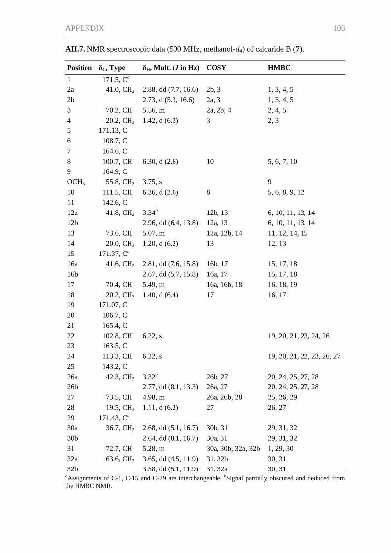
APPENDIX 108
AII.7. NMR spectroscopic data (500 MHz, methanol-d4) of calcaride B (7).
Position δC, Type δH, Mult. (J in Hz) COSY HMBC
1 171.5, Ca
2a 41.0, CH2 2.88, dd (7.7, 16.6) 2b, 3 1, 3, 4, 5
2b
2.73, d (5.3, 16.6) 2a, 3 1, 3, 4, 5
3 70.2, CH 5.56, m 2a, 2b, 4 2, 4, 5
4 20.2, CH3 1.42, d (6.3) 3 2, 3
5 171.13, C
6 108.7, C
7 164.6, C
8 100.7, CH 6.30, d (2.6) 10 5, 6, 7, 10
9 164.9, C
OCH3 55.8, CH3 3.75, s
9
10 111.5, CH 6.36, d (2.6) 8 5, 6, 8, 9, 12
11 142.6, C
12a 41.8, CH2 3.34b 12b, 13 6, 10, 11, 13, 14
12b
2.96, dd (6.4, 13.8) 12a, 13 6, 10, 11, 13, 14
13 73.6, CH 5.07, m 12a, 12b, 14 11, 12, 14, 15
14 20.0, CH3 1.20, d (6.2) 13 12, 13
15 171.37, Ca
16a 41.6, CH2 2.81, dd (7.6, 15.8) 16b, 17 15, 17, 18
16b
2.67, dd (5.7, 15.8) 16a, 17 15, 17, 18
17 70.4, CH 5.49, m 16a, 16b, 18 16, 18, 19
18 20.2, CH3 1.40, d (6.4) 17 16, 17
19 171.07, C
20 106.7, C
21 165.4, C
22 102.8, CH 6.22, s
19, 20, 21, 23, 24, 26
23 163.5, C
24 113.3, CH 6.22, s
19, 20, 21, 22, 23, 26, 27
25 143.2, C
26a 42.3, CH2 3.32b 26b, 27 20, 24, 25, 27, 28
26b
2.77, dd (8.1, 13.3) 26a, 27 20, 24, 25, 27, 28
27 73.5, CH 4.98, m 26a, 26b, 28 25, 26, 29
28 19.5, CH3 1.11, d (6.2) 27 26, 27
29 171.43, Ca
30a 36.7, CH2 2.68, dd (5.1, 16.7) 30b, 31 29, 31, 32
30b
2.64, dd (8.1, 16.7) 30a, 31 29, 31, 32
31 72.7, CH 5.28, m 30a, 30b, 32a, 32b 1, 29, 30
32a 63.6, CH2 3.65, dd (4.5, 11.9) 31, 32b 30, 31
32b 3.58, dd (5.1, 11.9) 31, 32a 30, 31 aAssignments of C-1, C-15 and C-29 are interchangeable.
bSignal partially obscured and deduced from
the HMBC NMR.
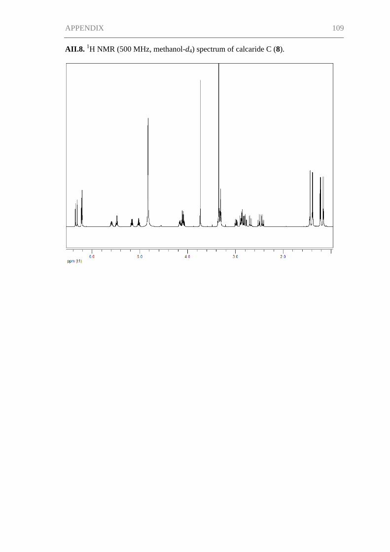
APPENDIX 109
AII.8. 1H NMR (500 MHz, methanol-d4) spectrum of calcaride C (8).
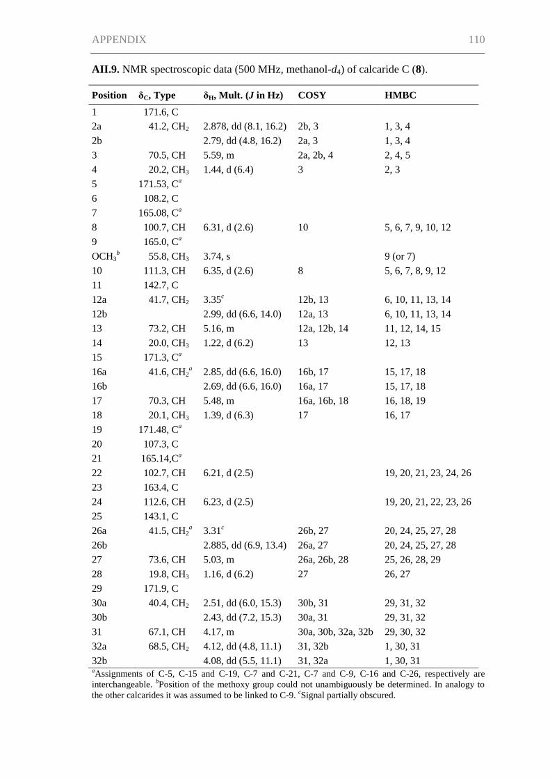
APPENDIX 110
AII.9. NMR spectroscopic data (500 MHz, methanol-d4) of calcaride C (8).
Position δC, Type δH, Mult. (J in Hz) COSY HMBC
1 171.6, C
2a 41.2, CH2 2.878, dd (8.1, 16.2) 2b, 3 1, 3, 4
2b
2.79, dd (4.8, 16.2) 2a, 3 1, 3, 4
3 70.5, CH 5.59, m 2a, 2b, 4 2, 4, 5
4 20.2, CH3 1.44, d (6.4) 3 2, 3
5 171.53, Ca
6 108.2, C
7 165.08, C
a
8 100.7, CH 6.31, d (2.6) 10 5, 6, 7, 9, 10, 12
9 165.0, Ca
OCH3
b 55.8, CH3 3.74, s
9 (or 7)
10 111.3, CH 6.35, d (2.6) 8 5, 6, 7, 8, 9, 12
11 142.7, C
12a 41.7, CH2 3.35c 12b, 13 6, 10, 11, 13, 14
12b
2.99, dd (6.6, 14.0) 12a, 13 6, 10, 11, 13, 14
13 73.2, CH 5.16, m 12a, 12b, 14 11, 12, 14, 15
14 20.0, CH3 1.22, d (6.2) 13 12, 13
15 171.3, Ca
16a 41.6, CH2
a 2.85, dd (6.6, 16.0) 16b, 17 15, 17, 18
16b
2.69, dd (6.6, 16.0) 16a, 17 15, 17, 18
17 70.3, CH 5.48, m 16a, 16b, 18 16, 18, 19
18 20.1, CH3 1.39, d (6.3) 17 16, 17
19 171.48, Ca
20 107.3, C
21 165.14,C
a
22 102.7, CH 6.21, d (2.5)
19, 20, 21, 23, 24, 26
23 163.4, C
24 112.6, CH 6.23, d (2.5)
19, 20, 21, 22, 23, 26
25 143.1, C
26a 41.5, CH2a 3.31
c 26b, 27 20, 24, 25, 27, 28
26b
2.885, dd (6.9, 13.4) 26a, 27 20, 24, 25, 27, 28
27 73.6, CH 5.03, m 26a, 26b, 28 25, 26, 28, 29
28 19.8, CH3 1.16, d (6.2) 27 26, 27
29 171.9, C
30a 40.4, CH2 2.51, dd (6.0, 15.3) 30b, 31 29, 31, 32
30b
2.43, dd (7.2, 15.3) 30a, 31 29, 31, 32
31 67.1, CH 4.17, m 30a, 30b, 32a, 32b 29, 30, 32
32a 68.5, CH2 4.12, dd (4.8, 11.1) 31, 32b 1, 30, 31
32b 4.08, dd (5.5, 11.1) 31, 32a 1, 30, 31 aAssignments of C-5, C-15 and C-19, C-7 and C-21, C-7 and C-9, C-16 and C-26, respectively are
interchangeable. bPosition of the methoxy group could not unambiguously be determined. In analogy to
the other calcarides it was assumed to be linked to C-9. cSignal partially obscured.
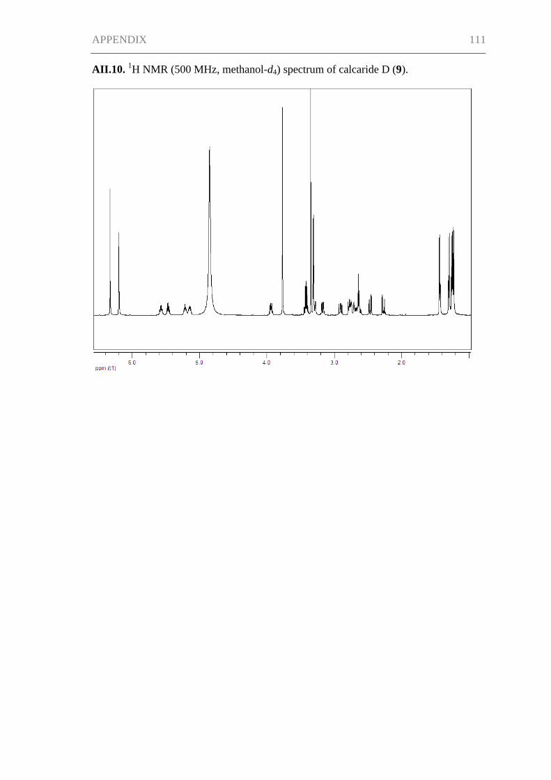
APPENDIX 111
AII.10. 1H NMR (500 MHz, methanol-d4) spectrum of calcaride D (9).
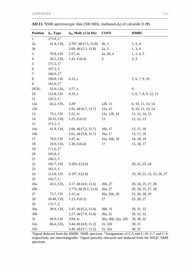
APPENDIX 112
AII.11. NMR spectroscopic data (500 MHz, methanol-d4) of calcaride D (9).
Position δC, Type δH, Mult. (J in Hz) COSY HMBC
1 173.9, Ca
2a 41.8, CH2 2.767, dd (7.5, 15.8) 2b, 3 1, 3, 4
2b
2.69, dd (5.3, 15.8) 2a, 3 1, 3, 4
3 70.8, CH 5.57, m 2a, 2b, 4 1, 2, 4, 5
4 20.1, CH3 1.43, d (6.4) 3 2, 3
5 171.5, Cb
6 107.2, C
7 166.0, C
b
8 100.8, CH 6.33, s
5, 6, 7, 9, 10
9 165.0, Cb
OCH3 55.9, CH3 3.77, s
9
10 112.8, CH 6.33, s
5, 6, 7, 8, 9, 12, 13
11 143.2, C
12a 43.2, CH2 3.29c 12b, 13 6, 10, 11, 13, 14
12b
2.91, dd (9.7, 13.7) 12a, 13 6, 10, 11, 13, 14
13 73.1, CH 5.22, m 12a, 12b, 14 11, 12, 14, 15
14 20.52, CH3 1.25, d (6.2) 13 11, 12, 13
15 171.2, C
16a 41.8, CH2 2.66, dd (7.2, 15.7) 16b, 17 15, 17, 18
16b
2.62, dd (5.8, 15.7) 16a, 17 15, 17, 18
17 70.0, CH 5.47, m 16a, 16b, 18 16, 18, 19
18 19.9, CH3 1.30, d (6.4) 17 15, 16, 17
19 171.6, Cb
20 105.8, C
21 166.2, C
22 102.7, CH 6.203, d (2.6)
20, 21, 23, 24
23 163.5, C
24 113.8, CH 6.197, d (2.6)
19, 20, 21, 22, 23, 26, 27
25 143.7, C
26a 43.5, CH2 3.17, dd (4.0, 13.4) 26b, 27 20, 24, 25, 27, 28
26b
2.772, dd (9.2, 13.4) 26a, 27 20, 24, 25, 27, 28
27 72.7, CH 5.15, m 26a, 26b, 28 25, 26, 28, 29
28 20.49, CH3 1.23, d (6.2) 27 25, 26, 27
29 172.7, C
30a 39.9, CH2 2.47, dd (5.2, 15.4) 30b, 31 29, 31, 32
30b
2.27, dd (7.9, 15.4) 30a, 31 29, 31, 32
31 69.9, CH 3.94, m 30a, 30b, 32a, 32b 29, 30, 32
32a 66.4, CH2 3.44, dd (4.9, 11.2) 31, 32b 30, 31
32b 3.40, dd (5.7, 11.2) 31, 32a 30, 31 aSignal deduced from the HMBC NMR spectrum.
bAssignments of C-5 and C-19, C-7 and C-9,
respectively are interchangeable. cSignal partially obscured and deduced from the HSQC NMR
spectrum.
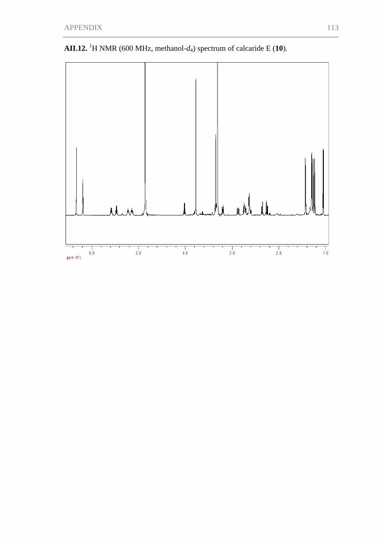
APPENDIX 113
AII.12. 1H NMR (600 MHz, methanol-d4) spectrum of calcaride E (10).
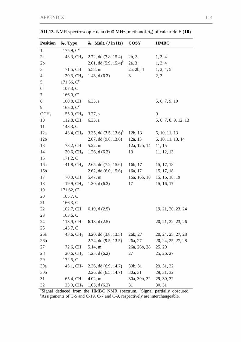
APPENDIX 114
AII.13. NMR spectroscopic data (600 MHz, methanol-d4) of calcaride E (10).
Position δC, Type δH, Mult. (J in Hz) COSY HMBC
1 175.9, Ca
2a 43.3, CH2 2.72, dd (7.8, 15.4) 2b, 3 1, 3, 4
2b
2.61, dd (5.9, 15.4)b 2a, 3 1, 3, 4
3 71.5, CH 5.58, m 2a, 2b, 4 1, 2, 4, 5
4 20.3, CH3 1.43, d (6.3) 3 2, 3
5 171.56, Cc
6 107.3, C
7 166.0, C
c
8 100.8, CH 6.33, s
5, 6, 7, 9, 10
9 165.0, Cc
OCH3 55.9, CH3 3.77, s
9
10 112.8, CH 6.33, s
5, 6, 7, 8, 9, 12, 13
11 143.3, C
12a 43.4, CH2 3.35, dd (3.5, 13.6)b 12b, 13 6, 10, 11, 13
12b
2.87, dd (9.8, 13.6) 12a, 13 6, 10, 11, 13, 14
13 73.2, CH 5.22, m 12a, 12b, 14 11, 15
14 20.6, CH3 1.26, d (6.3) 13 11, 12, 13
15 171.2, C
16a 41.8, CH2 2.65, dd (7.2, 15.6) 16b, 17 15, 17, 18
16b
2.62, dd (6.0, 15.6) 16a, 17 15, 17, 18
17 70.0, CH 5.47, m 16a, 16b, 18 15, 16, 18, 19
18 19.9, CH3 1.30, d (6.3) 17 15, 16, 17
19 171.62, Cc
20 105.7, C
21 166.3, C
22 102.7, CH 6.19, d (2.5)
19, 21, 20, 23, 24
23 163.6, C
24 113.9, CH 6.18, d (2.5)
20, 21, 22, 23, 26
25 143.7, C
26a 43.6, CH2 3.20, dd (3.8, 13.5) 26b, 27 20, 24, 25, 27, 28
26b
2.74, dd (9.5, 13.5) 26a, 27 20, 24, 25, 27, 28
27 72.6, CH 5.14, m 26a, 26b, 28 25, 29
28 20.6, CH3 1.23, d (6.2) 27 25, 26, 27
29 172.5, C
30a 45.1, CH2 2.36, dd (6.9, 14.7) 30b, 31 29, 31, 32
30b
2.26, dd (6.5, 14.7) 30a, 31 29, 31, 32
31 65.4, CH 4.02, m 30a, 30b, 32 29, 30, 32
32 23.0, CH3 1.05, d (6.2) 31 30, 31 aSignal deduced from the HMBC NMR spectrum.
bSignal partially obscured.
cAssignments of C-5 and C-19, C-7 and C-9, respectively are interchangeable.
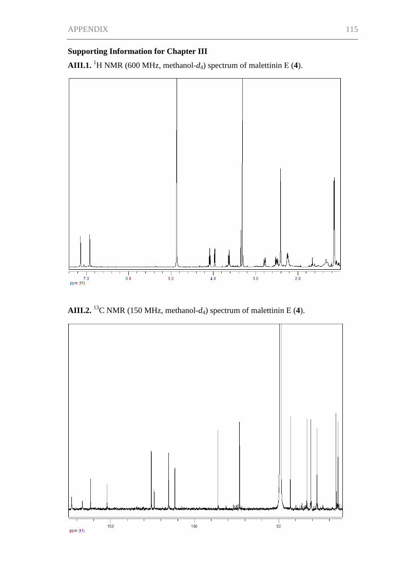
APPENDIX 115
Supporting Information for Chapter III
AIII.1. 1H NMR (600 MHz, methanol-d4) spectrum of malettinin E (4).
AIII.2. 13
C NMR (150 MHz, methanol-d4) spectrum of malettinin E (4).