Embed Size (px)
Citation preview
CHARACTERIZATION OF MYCOBACTERIAL
ESTERASES/LIPASES USING COMBINED
BIOCHEMICAL AND COMPUTATIONAL ENZYMOLOGY
ANKIT SHUKLA
(Bachelor of Technology, Bioinformatics)
SRM University, India
A THESIS SUBMITTED FOR THE DEGREE OF
MASTER OF SCIENCE
IN
INFECTIOUS DISEASES, VACCINOLOGY AND DRUG DISCOVERY
DEPARTMENT OF MICROBIOLOGY
NATIONAL UNIVERSITY OF SINGAPORE
AND
SWISS TROPICAL AND PUBLIC HEALTH INSTITUTE,
BIOZENTRUM, UNIVERSITÄT BASEL, SWITZERLAND
2013

i
DECLARATION
I hereby declare that the thesis is my original work and it has been written by
me in its entirety. I have duly acknowledged all the sources of information
which have been used in the thesis.
This thesis has also not been submitted for any degree in any university
previously.
______________
Ankit Shukla
28 December 2012
ii
ACKNOWLEDGMENTS
I would like to take this opportunity to specially thank my supervisor Prof. Markus Wenk
for giving me the opportunity to pursue my masters project in his lab in the highly
interesting field of lipidomics, his exceptional scientific support and understanding has
been instrumental throughout my project and has made my stay at NUS a good learning
and memorable experience which is bound to have positive implications for my future
career.
I’m highly grateful to my mentor Madhu Sudhan Ravindran who has always been willing
to teach me all experimental techniques right from scratch his valuable suggestions
throughout helped shape my project.
I would like to thank all the members of the Journal club for their honest comments on
the project and also giving me the opportunity to present my work in the scientific arena.
I’m really thankful to all the members of Markus’s lab who made my stay at NUS a
scientific as well as an unforgettable personal experience. In particular I express my
gratitude to Husna, Federico, Khanh Nagyuen, Jacklyn, Sudar, Pradeeep, Shentong,
Shareef, Phylis and Chrisitna who provided me with unending support and friendship.
I’m almost certain without their motivation and friendship it would have been possible to
achieve my goals three cheers for you guys!
I’m highly grateful to my parents whose love and support has always been with me in all
my endeavours. A special thanks goes out to my close friend Menorca who has been
always been with me and kept me up and going at all times. Lastly, I would like to thank
all my colleagues and everyone who has helped me in this project in some way or the
other from professional to personal space.

iii
Table of Contents
Declaration………………………………………………………….…………i
Acknowledgments…………………………………………………………….ii
Table of contents……………………………………………………….…….iii
Summary…………………………………………………………………….vii
List of Tables……………………………………………………………….viii
List of Figures………………………………………………………………………1
List of Abbreviations……………………………………………………………….3
1. INTRODUCTION………………………………………………………...........4
1.1. Esterases/Lipases……………………………………………………….....4
1.1.1 Esterases/Lipases in Infectious Diseases…………………………..5
1.1.2 Biology of Mycobacteria……………………………………………8
1.1.3 Mycobacterial Esterases/Lipases………….…………..…..…….9
1.1.4 Role of Esterases/Lipases in Mycobacterial Infection Cycle..…10
1.2. Esterases/Lipases in Physiopathology and Disease Progression….14
1.3. Esterases/Lipases Enzyme Classification System………………….16
1.3.1 Hydrolases (EC 3.)…………………………...…………………16
1.3.2 Carboxyl ester hydrolases (EC 3.1.1)………...………………....17
1.3.3 Carboxyl esterases (EC3.1.1.1)………………………...……….18

iv
1.3.4 Triacylglycerol (TAG) Lipases (EC 3.1.1.3) …………………....18
1.4. Alpha/Beta (α/β) Hydrolase Fold Family …………………...............18
1.4.1 Alpha/Beta (α/β) Hydrolase fold family in Mycobacteria…..........20
1.4.2 Mycobacterial lipase gene family…………………………...........28
1.4.2.1 Hormone-sensitive lipase sub-family ………………………….29
1.5. Issues and Problems with functional characterization of
Mycobacterial putative Esterases/Lipases. ……………………………...29
1.6. Tetrahydrolipstatin (Orlistat)………..………………………..…......29
1.6.1 An FDA approved anti-obesity drug………..……………………30
1.6.2 An anti-cancer agent…………………………..….....….....….......31
1.6.3 An anti-mycobacterial agent..…………………..….....….....….....31
1.7. Aims and Objectives………………………………..….....….....…......31
2. MATERIALS AND METHODS….……….................................................33
2.1. Molecular Modelling.…… ….………..................................................33
2.1.1 Sequence Analysis.……….………......................................................33
2.1.2 Comparative Modelling.……… ………........................................33
2.2. Molecular Dynamics Simulations……….............................................35
2.3. Virtual Ligand Screening.……… ………............................................35

v
2.4. Bacterial Strains and Cultures……….…………………...................38
2.4.1 Bacterial Strains………………………………………….............38
2.4.2 Bacterial Culture Media……………………………………….....38
2.4.3 Glycerol Stock of Bacteria……………………………….…........39
2.5. Cloning Procedures...............................................................................39
2.5.1 Genomic DNA Isolation (Mycobacteria)………………………...39
2.5.2 Preparation of Over-expression Plasmids…………………….......40
2.5. Mycobacterium bovis BCG Competent cell preparation,
Transformation and Selection of transformants………………........41
2.6. Preparation of Whole Cell Lysate (WCL)……………………..........41
2.7. Fluorescent Click Chemistry……………………………………........42
2.8. Gel Electrophoresis………………...…………………………...….....42
2.9. Enzymatic Assays…………………………………...……………........43
2.10. Inhibitor Assays…………………………...…………………...…….44
3. RESULTS………………………………………………………...………….46
3.1. Molecular Modelling of Protein 3D Structures………………............46
3.2. Molecular Structure Based Ligand Screening……………..…...........52
3.2.1. Evaluation of Ligand Screening Results……………..…..……….53
vi
3.3. Over Expressing Mycobacterial putative Esterases/Lipases………....57
3.4. Biochemical Characterization………………..…………………….......58
3.4.1 Enzymatic Assays………….………………………….……..................58
3.4.2 BCG_1460c (lipH probable lipase) shows short-chain esterase
activity…………………………….………………………..…..…........59
3.5. Tetrahydrolipstatin (THL) strongly inhibits short-chain esterase
activity of BCG_1460c (lipH)………………………………....…..........60
3.6. Predicted binding mode model of THL inhibition in BCG_1460c
(lipH) 3D Structure…………………………………………..................62
4. DISCUSSION...................................................................................................65
4.1. In silico Studies.........................................................................................65
4.1.1 Role of Virtual Screening in Antibacterial Drug Discovery....................65
4.1.2 Concepts, Feasibility and Drawbacks of Virtual screening.....................68
4.1.3 Molecular 3D structure Modelling and Virtual Ligand
Screening…………………………………………………......................68
4.1.4 Predicted binding mode model of THL inhibition in BCG_1460c (lipH)
3D Structure………………………………………….…………............70
4.2. In vitro Studies..........................................................................................70
4.2.1 BCG_1460c (lipH probable lipase) is a short-chain Carboxyl
esterase…………………………...…………………………..……........70

vii
4.2.2 Tetrahydrolipstatin (THL) strongly inhibits short-chain Carboxyl esterase
activity of BCG_1460c (lipH probable lipase)……………....................72
4.2.3 Possible Functions of BCG_1460c (lipH probable lipase)….……..........73
5. CONCLUSIONS AND FUTURE DIRECTIONS.……................................73
REFERENCES…….……....................................................................................79
Summary
Esterases/Lipases are evolutionarily related enzymes mostly belonging to
hydrolases superfamily sharing a common α/β-hydrolase protein fold. Recent
studies have suggested that they play a pivotal role in disease manifestation due to
disruption of lipid metabolizing enzymes and their pathways, yet only very few
have been functionally annotated.
This study focuses on Mycobacterial lipolytic/non-lipolytic enzymes comprising
of 31 putative lipases and/or esterases belonging to α/β-hydrolase fold family. In
silico molecular modeling, sequence analysis and ligand docking experiments
provide insights into molecular structure of these classes of enzymes. We
performed a unique structure based virtual ligand screening to predict natural
substrates of 4 putative Mycobacterial esterases/lipases namely BCG_1460c (lipH
probable lipase), BCG_2991c (lipN probable lipase/esterase), BCG_2950 (tesA
probable thioesterase) and BCG_3229 (lipV possible lipase).

viii
The information obtained from molecular docking and molecular dynamics (MD)
simulations was preceded with an in vitro study, where we performed enzymatic
assays with whole cell lysates of Mycobacterium bovis BCG over-expressing
lipases and/or esterases using synthetic substrates. We provide evidence at
structural and biochemical level that the short-chain esterase activity of
BCG_1460c (lipH probable lipase) is inhibited by tetrahydrolipstatin (THL) an
FDA approved drug. Overall, the in silico and in vitro analysis show high
correlation and provide an effective approach to characterize and distinguish
mycobacterial lipolytic/non-lipolytic enzymes.
List of Tables
Table 1.1 Effects of infectious pathogens on host lipid metabolism
Table 1.2 Probable functions and identities of Mycobacterial lip gene family
enzymes
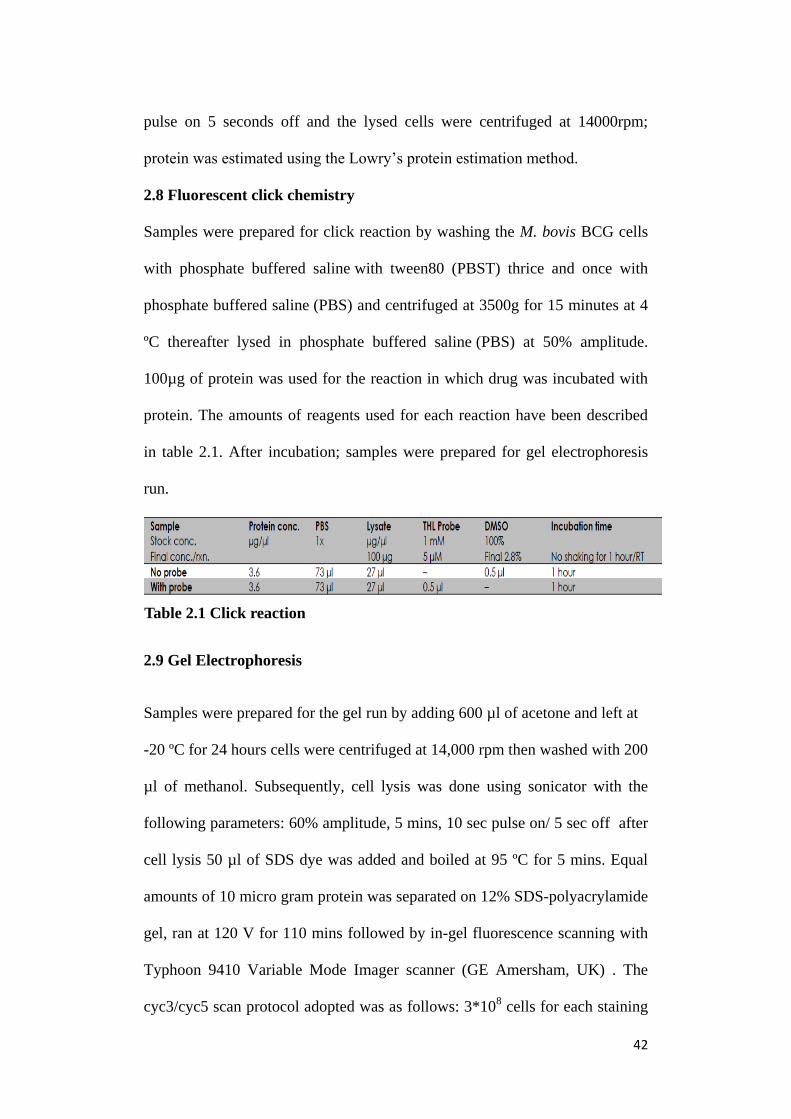
Table 2.1 Click reaction
Table 2.2 Concentrations of reagents used
Table 3.1 Summary of potential substrates ligand screening results
Table 3.2 Summary of potential inhibitors ligand screening results
Table 4.1 BCG_1460c (lipH) homologs in bacterial species

1
List of Figures
Figure 1.1 (a)Hydrolysis of carboxylic ester catalysed by carboxyl
esterase
(b)Hydrolysis of a triacylglycerol substrate catalysed by
TAG lipase enzyme
Figure 1.2 Functional classifcation of Mycobacterium tuberculosis
genome
Figure 1.3 Mycobacterium tuberculosis infection cycle
Figure 1.4 (a) Typical foamy macrophage having lipid bodies (LBs)
(b) LB surrounded by several M. tuberculosis bacilli
intracytoplasmic lipid (ILIs)
(c) Intracytoplasmic lipid within M. tuberculosis bacilli
Figure 1.5 Esterases enzyme classification system
Figure 1.6 Topology diagram of α/β hydrolase fold enzymes
Figure 1.7 (a) 3D structure of Pseudomonas fluorescens carboxyl
esterase
(b) Superposition of conserved α/β hydrolase core of 9
representative α/β hydrolase fold enzymes
Figure 1.8 (a) Total M. tuberculosis functionally annotated genes
(b) 94 α/β hydrolase fold distribution in lipid enzymes
Figure 1.9 Chemical Structure of Tetrahydrolipstatin (Orlistat)
Figure 2.1 Steps involved in Molecular modelling
Figure 2.2 Computational Docking
Figure 2.3 para-nitrophenol (pNP) assay
Figure 2.4 Template design of a 96 well plate used for Inhibitor assays
Figure 3.1 3D structure and Cα – backbone overlay of FAS-TE
modelled over know FAS-TE structure

2
Figure 3.2 3D structure models
Figure 3.3 Structure validation report
Figure 3.4 Ramachandran plot statistics of backbone dihedral angle
distribution
Figure 3.5 Protein structure analysis
Figure 3.6 Molecular dynamic simulations
Figure 3.7 Short (C2) to long (>C12) acyl chain ester ligands screening
results
Figure 3.8 SDS-PAGE fluorescent gel image showing M. bovis BCG
over expressing BCG_1460c (lipH) and BCG_2950 (tesA)
Figure 3.9 Enzymatic assay with whole cell lysate of overexpressed
BCG_1460c (lipH) and BCG_2950 (tesA)
Figure 3.10 Enzymatic assay of over expressed BCG_1460c (lipH)
with varying acyl chain length substrates
Figure 3.11 THL (Tetrahydrolipstatin) inhibitor assay (IC50)
Figure 3.12 E600 (diethylparanitrophenyl phosphate) inhibitor assay
(IC50)
Figure 3.13 THL (Tetrahydrolipstatin) binding mode model in
BCG_1460c (lipH) 3D structure
Figure 4.1 Pincipal antibacterial drug discovery strategies
Figure 4.2 State of the art in Virtual Screening
Figure 4.3 Detailed workflow of a high throughput virtual screening
(VHTS)
Figure 4.4 SDS-PAGE fluorescent gel image showing M. smegmatis
over expressing lipH (BCG_1460c)
Figure 4.5 Enzymatic assay with M. smegmatis
over expressed lipH (BCG_1460c)

3
Lists of Abbreviations
THL: Tetrahydrolipstatin
pNP: para-nitrophenol
PDB: Protein Data Bank
PMSF: phenylmethanesulfonylfluoride
E600: diethyl-p-nitrophenylphosphate
EC : Enzyme classifier
TAGs: Triacylglycerol
MTB: Mycobacterium tuberculosis
LB: Lipid bodies
ILI: Intracytoplasmic lipid
OD: Optical Density
SBDD: Structure Based Drug Design
VHTS: Virtual High Throughput Screening
FBDD: Fragment Based Drug Design
3DQSAR: Three Dimensional Quantitative Structure Activity Relationship

4
1. Introduction
1.1. Esterases/Lipases
The biological relevance and coexisting variability of lipids has led to the
development of wide range of lipid metabolizing enzymes. Esterases (EC 3.1)
are widely distributed amongst bacteria, fungi, plants and animals defined by
their ability to catalyse the formation and cleavage of ester bonds. They are
classified based on the nature of the ester bonds (carboxyl ester, thio ester,
phosphomonoester, etc.) they catalyse. Among them, carboxyl ester
hydrolases (EC 3.1.1) are enzymes that catalyses ester bond hydrolysis of
carboxylic esters (Figure 1.1a). Lipases/TAG lipases (EC 3.1.1.3) are lipolytic
enzymes which constitute a special sub-class of carboxyl ester hydrolases (EC
3.1.1) (Ali, Verger et al. 2012) capable of releasing long-chain fatty acids from
natural water-insoluble esters such as lipids (Figure 1.1b).
(b)
Triacylglycerol Water Fatty Acid
TAG Lipase
Carboxyl ester
hydrolase
Glycerol
Carboxylic ester Water Carboxylic acid Alcohol
Figure 1.1: (a) Hydrolysis of a carboxylic ester catalysed by carboxyl esterase
enzyme. (b) Hydrolysis of a triacylglycerol substrate catalysed by TAG
lipase enzyme (Source: Thomson, Delaquis et al. 1999)
(a)

5
1.1.1 Esterases/Lipases in Infectious Diseases
Several studies on lipid metabolism have been undertaken and the outcome of
these studies has opened up new ways and avenues to analyse and characterize
a host of diseases as well as providing newer insights and approaches into the
mechanisms involved when cells start functioning abnormally and hence
enable establishing of an important link between lipid metabolism and the
disease. The concept and hypothesis was further extended to infectious
diseases which are known to be accompanied by an altered host lipid
metabolism via a unique sequence i.e. the disruption of lipid metabolizing
enzymes (esterases/lipases) and their pathways and there also exists a close
and significant inter-relationship between lipid metabolism and host responses
to infection.
It is indeed important to explain the mechanism and the factors by which the
lipid metabolism is regulated. The frontline and exclusive studies have shown
that the lipid metabolism is regulated by lipid metabolizing enzymes
(esterases/lipases) which are considered to be one of the known virulence
factors in many bacteria such as Pseudomonas cepacia, Staphylococcus
aureus (Lonon, Woods et al. 1988 and Rollof, Braconier et al. 1988) and
fungal species like Candida albicans, Fusarium gramearium. Further insight
reveals that the lipid metabolizing enzymes of Propionibacterium acnes and
Staphylococcus epidermis are probably involved in incidence of commonly
prevalent human skin infections where they help triggering colonization and
subsequent persistence of bacteria on the human skin.

6
Table 1.1: Effects of infectious pathogens on host lipid metabolism
(Source: (Beisel and Fiser 1970))
According to some hypothesis it has been suggested that these enzymes may
also be responsible in contributing to the invasiveness and proliferation by
inducing the destruction of the host tissues thereby supplying hydrolysed
material as nutrient to the microorganisms. Lipid precursors needed for the
replication of the invading pathogens (bacteria, viruses and protozoans) are
derived and supplied from the source metabolic pools within the host by lipid
EFFECTS OF INFECTION ON LIPID METABOLISM OF HOST
Presence of invading microorganism
Secondary effects due to infection
1. Direct Effects
Decreased dietary intake of
fats. Utilization of host lipids required
by replicating microorganisms.
Disruption of host cell metabolism
by intracellular microorganisms.
Minimal interference with
intestinal absorption of fats.
Localized destruction of fat cells at
sites of an infectious process.
Altered lipid metabolism
within host cells.
2. Indirect Effects
Altered rates of hormone-
mediated lipolysis within fat
depots to supply increased
metabolic demands.
Alterations in host lipid metabolism
caused by bacterial exotoxins,
endotoxins or enzymes.
Activation of lipase and other
lysosomal enzymes within host
phagocytes.
Altered rates of lipid
synthesis within the liver
Release of mediator substances
from host cells, that is, endogenous
pyrogen, interferon.
Altered rates of fat
utilization by peripheral
tissues.

7
metabolizing enzymes thereby altering the host lipid metabolism. In the
subsequent process of progression and infection may allow for the re-
distribution of nutrients to cells which are considered extremely important in
ensuring host defence or tissue repair.
It has now been well established and recognized that certain pathogens have
the inherent capabilities to co-opt the lipid metabolism and some illustrative
examples can be listed as:
a.) The ability of Mycobacterium tuberculosis to catabolize cholesterol
as an energy source which might as a result facilitate and regulate
its ability to survive within the macrophages.
b.) Utilization of host cholesterol by Toxoplasma gondii for its
persistence, growth and proliferation.
c.) Heliobacter pylori being unable to synthesize cholesterol and
hence requires exogenous cholesterol from the host.
d.) Ebola virus (EBOV) in lysosomal compartments binds to
cholesterol transporter protein Niemann-Pick C1 (NPC1).
Fatty metamorphosis of cells particularly from liver, kidney and heart is a
common histologic finding during a host of bacterial infectious diseases. An
increase in esterified fatty acids has been observed in viral hepatitis whereas
free fatty acids and triacylglycerol (TAGs) have been reported to be elevated
in gram-negative bacillus infections in humans. Since the action of hormone-
sensitive lipase in adipose tissue is a major event contributing to free fatty
acids to blood, such infection-related hormonal responses may have a pivotal
role to play in altering/affecting the rates of lipolysis or fatty acid utilization.

8
1.1.2 Biology of Mycobacteria
It has now been reported that the genus Mycobacterium is known to comprise
of more than 100 species (Tortoli 2006). The cultivable (grown in lab)
members of Mycobacterium are clinically grouped either as the Mtb complex
or the non-tuberculous mycobacteria. The M. leprae, which is responsible for
causing leprosy is an obligate parasite and therefore not cultivable in vitro
(van Beers, de Wit et al. 1996). On the other hand, diseases caused by
members of Mtb complex include M. tuberculosis, M. bovis, M. microti, M.
africanum and M. Canettii subspecies they are known to possess and
demonstrate very similar clinical features. Pulmonary diseases caused by M.
tuberculosis and M. bovis are clinically, radiologically and pathologically
indistinguishable. However, M. bovis appears to have a diminished propensity
and potentiality to reactivate and spread from person to person (O'Reilly and
Daborn 1995) through human chain. Calmette and Guérin (BCG) attenuated a
strain of M. bovis to generate BCG which is used as a vaccine by continuous
passaging through culture media. Mycobacteria are aerobic and non-motile rod
(bacillus) shaped that are identified to be weakly Gram-positive and acid-fast
by Ziehl-Neelsen staining. The bacilli belong to the actinobacterium family
and all Mycobacterium species are known to share a characteristic cell wall
architecture which is relatively much thicker than other bacteria and are
known to be hydrophobic, rich in mycolic acids. In the laboratory, Mtb can be
grown, in vitro, on the agar-based Middlebrook medium or the egg based
Lowenstein-Jensen medium (Parrish, Dick et al. 1998). Considering that it is a
relatively slow growing bacteria, it takes a time period of around 4-6 weeks to

9
have visual bacterial colonies formed on these solid media (Parrish, Dick et al.
1998).
1.1.3 Mycobacterial Esterases/Lipases
In Mycobacterium such as Mycobacterium tuberculosis lipids play a vital role
wherein it is stated that a large fraction of the genome encodes putative
enzymes said to be involved in lipid metabolism (Cole, Brosch et al.
1998). Indeed, Mycobacterium tuberculosis genome contains 250 genes
encoding putative enzymes involved in the synthesis or degradation of lipids
compared to 50 genes in Escherichia coli, which is known to have a similar
genome size. This feature, combined with the extremely large quantum of
lipids representing 30–40% of the dry weight of M. tuberculosis tends to
suggest that lipids and lipid metabolizing enzymes play an important role in
the mycobacterial life cycle and perhaps also in virulence. In the study
conducted by (Deb, Daniel et al. 2006) group reported the expression status of
all the twenty-four putative lipase/esterase genes of lipase gene family of M.
tuberculosis H37Rv in Escherichia coli BL21.
In silico analysis has identified the presence of around 31 putative genes
encoding lipid metabolizing enzymes (enzymes involved in lipids degradation)
including 24 lipid/ester hydrolases belonging to the so called “Lip family”
(LipC to LipZ). These have been annotated as putative esterases or lipases
based on the presence of the consensus sequence GXSXG which is considered
to be the characteristic feature of the α/β hydrolase-fold family members
(Ollis, Cheah et al. 1992). The functional classification of Mycobacterium
tuberculosis genes has been depicted in fig.1.2

10
Figure1.2: Functional classifcation of Mycobacterium tuberculosis genome (Source: data from (Camus, Pryor et al. 2002))
1.1.4 Role of Esterases/Lipases in Mycobacterial Infection Cycle
There are a large number of mycobacterial species such as
M. tuberculosis (Garton, Christensen et al. 2002), (Schue, Maurin et al. 2010),
(Peyron, Vaubourgeix et al. 2008), (Daniel, Deb et al. 2004), (Deb, Daniel et
al. 2006), (McKinney, Bentrup et al. 2000), Mycobacterium bovis BCG (Low,
Rao et al. 2009 and Low, Shui et al. 2010), Mycobacterium leprae (Mattos,
D'Avila et al. 2010) and Mycobacterium smegmatis (Garton, Christensen et al.
2002 and Dhouib, Ducret et al. 2011) which predominantly demonstrate the
accumulation of lipids derived from host cells. In addition, the consumption
pathways involving lipid metabolizing enzymes (esterases/lipases) have also

11
been identified and expressed. In particular, the tubercule bacilli enter the
body by inhalation of aerosol route and reach the lungs where they are
phagocytosed by the frontline pulmonary alveolar macrophages.
Subsequently, host response ensues which consists of recruitment of
lymphocytes, macrophages and dendritic cells leading to the formation of a
highly organised structure termed as ‘granuloma’ a major histopathological
hallmark of tuberculosis (Singh, et al. 2010). In these granuloma macrophages
containing bacilli accumulates intra-cytoplasmic lipid inclusion bodies (LB)
which are predominantly composed of neutral lipids surrounded by a
phospholipid layer that reveals and assigns the macrophage their foamy
appearance within the foamy macrophage, phagocytised bacteria preferentially
metabolize lipids rather than carbohydrates (Wheeler and Ratledge 1988), a
view point that is supported by an evidence showing up-regulation of several
mycobacteria genes involved in lipid metabolism (McKinney, Honer zu
Bentrup et al. 2000). At this stage of progression, the intra phagosomal
bacteria acquire and accumulate intra cytoplasmic lipid inclusion (ILIs) in
their cytoplasm (Figure 1.4(B) and 1.4(C)) and persist in a non-replicating
state ultimately and eventually leading to dormancy i.e. latent infection. It has
been demonstrated in an in vitro model of human granulomas (Peyron,
Vaubourgeix et al. 2008) that these lipid bodies (LB and ILIs) serve as sources
of carbon and energy for dormant bacilli. The infection cycle of
Mycobacterium tuberculosis has been shown in fig.1.3

12
Figure 1.3: Mycobacterium tuberculosis infection cycle
(Russell 2007)
within granulomas and as a consequence aiding in reactivation that can
ultimately lead to an active tuberculosis infection (Parrish, Dick et al. 1998).
The typical foamy macrophages having lipid bodies, LB surrounded by Mtb

13
bacilli and intracytoplasmic lipid within M. tuberculosis bacilli have been
shown in fig 1.4
Figure 1.4: (A) Typical foamy macrophage having lipid bodies (LBs) and
M. tuberculosis containing phagosomes: arrows depict phagosomal membrane
around bacterium. (B) LB surrounded by several M. tuberculosis bacilli
intracytoplasmic lipid (ILIs) (C) Enlarged view of (B) showing large
intracytoplasmic lipid within M. tuberculosis bacilli [Adapted from (Peyron,
Vaubourgeix et al. 2008)]
In addition to attention drawn on the foamy macrophages, ILI accumulation
has also been reported in M. tuberculosis infected adipocytes as well as in
Mycobacterium leprae infected macrophages and Schwann cells (Mattos, Lara
et al. 2011). Further biochemical analysis and associated experimentation has
revealed that M. tuberculosis lipid inclusion bodies mainly comprise of
triacylglycerol (TAGs). These TAGs are derived from free fatty acids that may

14
be imported from host or result from denovo synthesis (Daniel, Maamar et al.
2011). The pattern indicated that Triacylglycerol (TAGs) accumulate during
mycobacterial growth and the amount of intracellular TAGs peak in the late
exponential growth phase (Kremer, de Chastellier et al. 2005) and non-
replicating phase (Daniel, Deb et al. 2004).
Further it has also been shown that the expression of M. tuberculosis specific
lipase gene family is significantly elevated during dormancy (Deb, Daniel et
al. 2006) and that in the re-activated bacilli, a reduction in triacylglycerol
(TAG) levels coincides with an increase triacylglycerol (TAG) lipase activity
(Low, Rao et al. 2009). Thus lipid metabolizing enzymes (esterases/lipases)
appear to play an important central role and associate with important
physiological functions and also contribute to the extraordinary capacity of
survival of M. tuberculosis within the infected host. These enzymes are
peculiar molecules that provide a metabolic turnover of lipids and can be
defined as essential biocatalysts for the hydrolysis of esters containing long
chainfattyacids.
1.2. Esterases/Lipases in physiopathology and disease progression
Pathogenic bacteria have been known to follow a number of mechanisms and
pathways to cause and allow subsequent persistence of diseases in human
hosts. The molecular strategies used by the bacteria to interact with the host
can be unique and characteristic to specific pathogens, and follow conserved
pattern across several different species. Hydrolytic enzymes like
esterases/lipases contribute to invasiveness and proliferation by causing

15
destruction of the host tissue thereby supplying hydrolysed material to the
organisms as nutrients. These esterases/lipases are one of the known and
critical virulence factors in a host of bacterial species such as Pseudomonas
cepacia, Staphylococcus aureus (Lonon, Woods et al. 1988, Rollof, Braconier
et al. 1988) and also in fungal species like Alternaria brassicicola, Candida
albicans and Fusarium graminearum (Berto, Commenil et al. 1999). M.
tuberculosis is a bacterial pathogen that can persist in for decades in an
infected patient in dormant state without causing symptoms or disease with
clinically evident features. In fact, prior to entering into dormancy, it has been
hypothesized that the bacteria accumulate lipids originating from the host cell
membrane degradation as precursors and re-synthesize complex lipid
molecules. Fluorescence studies (Garton, Christensen et al. 2002) have shown
that large amounts of intracellular lipids forming inclusion bodies can be
detected in the cytoplasm (Anuchin, Mulyukin et al. 2009) supporting the
view that bacteria tends to accumulate lipids. The presence of lipid inclusions
confers and indirectly correlates the existence of lipid metabolizing
esterases/lipases. During the re-activation phase of the bacteria, these stored
lipids are hydrolysed and the infection process acquires further impetus to
demonstrate its detectable occurrence (Cotes, Bakala et al. 2008). What is
interesting to mention here is that a critical link between storage-lipid
accumulation and development of phenotypic drug resistance in M.
tuberculosis has also been established and the findings of several studies on
non-mycobacterial pathogens suggested the involvement of lipid metabolizing
enzymes in pathogenicity.

16
In pathogenic bacteria, for example it has been shown that Mycobacterium
tuberculosis Rv3097c (lipY) is able to hydrolyse long-chain triacylglycerol
(TAG). The role of esterase Rv3487c (lipF) has been implicated in
pathogenesis (Zhang, Wang et al. 2005). In addition, Rv0220 (lipC) has been
reported to be an immunogenic cell-surface esterase actively involved in
modulation of the host immune response (Shen, Singh et al. 2012). This entity
Rv0220 (lipC) is also known to be capable of stimulating pro-inflammatory
cytokines and chemokines in macrophages as well as to pulmonary epithelium
cells (Shen, Singh et al. 2012). It is also important to mention that the lipase
Rv0183 has been identified as a monoglyceride lipase involved in degradation
of host cell lipids and may strongly induce immune responses of the host (Xu,
Jia et al. 2010). Based on these facts, it can therefore be categorically stated
that lipid metabolizing enzymes (esterases/lipases) are involved throughout the
life-cycle of the pathogen and they assume important physiological role during
dormancy and reactivation i.e. during the course of entire infection process.
The released fatty acids by these enzymes are then taken up by intracellular
mycobacteria and stored in the form of triacylglycerol (TAGs) to be
subsequently used as sources of carbon during the persistence stage.
Conversely, intracellular triacylglycerol hydrolases maybe required for
assimilation of intra-cytoplasmic lipid inclusions to exit dormancy.
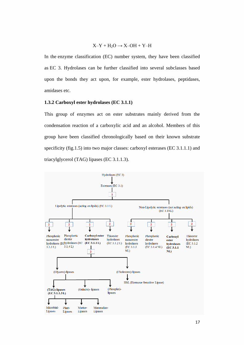
1.3 Esterases/Lipases Enzyme Classification System
1.3.1 Hydrolases (EC 3.)
Are group of enzymes which catalyse the hydrolysis of a chemical bond. In
general an enzyme capable of catalysing the following reaction is a hydrolase:
17
X–Y + H2O → X–OH + Y–H
In the enzyme classification (EC) number system, they have been classified
as EC 3. Hydrolases can be further classified into several subclasses based
upon the bonds they act upon, for example, ester hydrolases, peptidases,
amidases etc.
1.3.2 Carboxyl ester hydrolases (EC 3.1.1)
This group of enzymes act on ester substrates mainly derived from the
condensation reaction of a carboxylic acid and an alcohol. Members of this
group have been classified chronologically based on their known substrate
specificity (fig.1.5) into two major classes: carboxyl esterases (EC 3.1.1.1) and
triacylglycerol (TAG) lipases (EC 3.1.1.3).

18
(from previous page) Figure 1.5: Esterases’ classification based on
1 : Physico-chemical; 2 : chemical criteria (L means lipolytic those
enzymes capable of acting on lipid while NL: non-lipolytic those
enzymes which do not act on lipids ) (EC is the enzyme classifier )
[Source: adapted from (Ali, Verger et al. 2012)]
1.3.3 Carboxyl esterases (EC 3.1.1.1)
This group of enzymes shows diverse substrate specificity and catalyse the
hydrolysis of ester bond of acyl chain esters forming a carboxylic acid and an
alcohol.
RCOOR + H2O → R–COOH + R–OH
Most members of this group are hydrolases especially those involved in the
hydrolytic cleavage of carboxylic ester bonds are found to share a common
alpha/beta (α/β) hydrolase folding pattern. Enzymatic assays using
chromogenic substrates such as acyl esters of p-nitrophenol (pNP) allow for
the spectroscopic and calorimetric determination of esterase activity.
1.3.4 Triacylglycerol (TAG) Lipases (EC 3.1.1.3)
They constitute a special class of carboxyl esterases (Ali, Verger et al. 2012)
capable of releasing long-chain fatty acids from natural water-insoluble esters
(lipids) as depicted below:
Triacylglycerol Water Glycerol
Carboxylic ester Water Carboxylic Acid Alcohol
Fatty Acid
TAG Lipase

19
In bacterial species such as Mycobacteria, Pseudomonas, Burkholderia TAG
lipases have been shown to completely hydrolyse triacylglycerol substrates
although ester bonds are more preferable (Jaeger et al., 1994) and they possess
both lipolytic as well as esterolytic activity. Bacterial TAG lipases have also
been found to share a common alpha/beta (α/β) hydrolase folding pattern. A
large number of enzymatic assay methods using fluorescent substrates allow
for the fluorimetric and spectroscopic detection of lipase activity.
1.4 Alpha/Beta (α/β) Hydrolase fold family
The alpha/beta (α/β) hydrolase fold is considered to be a common
characteristic to a number of hydrolase enzymes of largely different
phylogenetic origin and catalytic function (Ollis, Cheah et al. 1992). Each
enzyme has a conserved alpha/beta (α/β) hydrolase core (fig.7b) consisting of
alpha/beta sheet having 8 strands connected by helices. They all have a similar
arrangement of a catalytic triad composed of nucleophilic serine charge relay
network aspartate and proton carrier histidine (shown in fig.1. 6) which are the
best-conserved structural features in the fold. The canonical α/β hydrolase fold
is an eight-stranded and mostly parallel α/β structure (figure 1.6), (1.7a&b)
(Ollis, Cheah et al. 1992).
Figure 1.6: Topology diagram of α/β hydrolase fold enzymes α Helices
and β strands are represented by black spheres and arrows,
respectively while catalytic triad members are highlighted by
black star and triangles.
H2N
COOH
β
4
β
3
β
5
β
6
β
7
β
8
β
1
β
2
Ser His
Asp

20
The enzymes adopting alpha/beta hydrolase fold share no significant sequence
similarity suggestive of a divergent evolution from a common ancestor. The
members of alpha/beta (α/β) hydrolase fold family include: hydrolases,
esterases, lipases, proteases, peroxidases, dehalogenases.
1.4.1 Alpha/Beta (α/β) Hydrolase fold family in Mycobacteria
Most members of the alpha/beta (α/β) hydrolase fold family are esterase/lipase
enzymes that catalyse ester hydrolysis reactions (Schrag et al., 1997, Nardini
et al., 1999). In Mycobacterium tuberculosis, out of the 250 genes encoding
putative enzymes involved in lipid metabolism, 94 gene products would have
the characteristic alpha/beta (α/β) hydrolase fold (Hotelier, Renault et al.
2004), ESTHER database http://bioweb.ensam.inra.fr/esther) of which 47 are
Figure 1.7: (a) 3D structure of Pseudomonas fluorescens carboxyl esterase (PfCES) belonging to
α/β hydrolase fold family revealing mostly-parallel β sheets. (b) Superposition of
the conserved α/β hydrolase core of 9 representative α/β hydrolase fold enzymes
(Source: Heikinheimo et. al, 1999)

21
annotated as esterases (ester hydrolases) and also including 24 lipid/ester
hydrolase belonging to lip family (lipC to lipZ) (Cole, Brosch et al. 1998). The
total M. tuberculosis functionally annotated genes and α/β hydrolase fold
distribution in M. tuberculosis are shown in fig.1.8 (a&b).
Overall alpha/beta (α/β) hydrolase fold family members in Mycobacteria are
mainly esterases/lipases suggesting an important structural link of this family
of enzymes in mycobacterial lipid metabolism.
M. tuberculosis annotated genes
M. tuberculosis α/β hydrolase fold
(94 annotated genes)
Figure 1.8: (a) Total M. tuberculosis functionally annotated genes.
(b) 94 α/β hydrolase fold distribution in enzymes involved in mycobacterial lipid
metabolism having 250 lipid encoding genes [http://bioweb.ensam.inra.fr/esther].
(a)
(b)

22
1.4.2 Mycobacterial Lipase gene family
This sub-family of alpha/beta (α/β) hydrolase fold family comprises of 24
genes annotated as putative esterases/lipases (lipC to LipZ) genes (Cole,
Brosch et al. 1998) and all pose a catalytic triad with active site as
nucleophilic serine showing a characteristic G-X-S-X-G sequence motif and
are typically involved in various biological processes. Their probable function
has been represented and listed in:
Protein
Gene Mol
wt.
Activity
type
Active
site
Biological
function
References
lipC BCG
0257
(Rv02
20)
44.3 Probable
esterase
(77% αβ-
hydrolase)
GCSAG Low TG lipase
activity, induced
under hypoxic
resuscitation
Carboxyl esterase
type B,
Upregulated in
starvation
Located in cell wall
and capsule, elicit
strong immune
response, expresses
only during active
tuberculosis,
hydrolyze short
chain esters
(Deb,
Daniel et al.
2006)
(Singh,
Singh et al.
2010)
lipD BCG
1962
(Rv19
23)
47.2 Probable
esterase/β-
lactamase
(69% β-
lactamase)
A hydrolase lipase
similar to esterases
and beta-
lactamases
Role in defence
Table 1.2 Probable functions and identities of Mycobacterial lipase gene family enzymes
23
lipE BCG
3837
(Rv37
75)
45.3 Caboxyl
esterase
(79% β-
lactamase)
Lipolytic enzyme
involved in cellular
metabolism
Defense
mechanism,
Induced under
hypoxic
resuscitation
(Deb,
Daniel et al.
2006)
lipF BCG
3551c
(Rv34
87c)
29.4 Caboxyl
esterase
(75% αβ-
hydrolase)
GDSAG Member of
Hormone sensitive
lipase family.
Non-lipolytic
hydrolase,
hydrolyze short
chain esters
Induced at low pH,
related to virulence
No TG lipase
activity,
intermediary
metabolism and
respiration
Important for
bacilli persistence
Membrane protein,
hydrolyzes short
chain esters
/phosphatidylcholi
ne
(Zhang,
Wang et al.
2005)
(Richter
and Saviola
2009)
(Camacho,
Ensergueix
et al. 1999)
(Deb,
Daniel et al.
2006)
lipG BCG
0695c
(Rv06
46c)
32.9 Probable
hydrolase
(80% αβ-
hydrolase)
GASMG Lipolytic enzyme,
involved in cellular
metabolism, highly
similar to various
hydrolases,
especially lipases
from Acinetobacter
calcoaceti
(Deb,
Daniel et al.
2006)

24
lipH BCG
1460c
(Rv13
99c)
33.9 Possible
lipase
(70% αβ-
hydrolase)
GWSLG Member of
Hormone sensitive
lipase family.
lipid transport and
metabolism
Non-lipolytic
esterase
(Deb,
Daniel et al.
2006)
(Canaan,
Maurin et
al. 2004)
lipI BCG
1461c
(Rv14
00c)
34.0 Probable
lipase
(69%
carboxyles
terase
family)
GDSAG A probable lipase
invloved
intermediary
metabolism and
respiration,
Member of
Hormone sensitive
lipase family.
Intermediary
metabolism and
respiration
lipJ BCG
1939c
(Rv19
00c)
49.7 Putative
lignin
peroxidase
(75% αβ-
hydrolase)
Alkaloid
biosynthesis II
lipK mbtJ,
BCG
2399
(Rv23
85)
32.9 Probable
acetyl
hydrolase
(71% αβ-
hydrolase)
Intermediary
metabolism, Low
TG lipase activity
(Deb,
Daniel et al.
2006)
lipL BCG
1560
(Rv14
97)
45.8 Probable
esterase
(70% β-
lactamase)
Transpeptidase,
Beta-lactamase
class C
Intermediary
metabolism and
respiration, Low
TG lipase activity
(Deb,
Daniel et al.
2006)

25
lipM BCG
2299
(Rv22
84)
46.7 Probable
esterase
(78% αβ-
hydrolase)
GGSAG Involved in
intermediary
metabolism and
respiration,
predicted
transmembrane
protein
Lipid transport and
metabolism
(Gu, Chen
et al. 2003)
lipN BCG
2991c
(Rv29
70c)
40.1 Lipase like
enzyme
(77% αβ-
hydrolase)
GDSAG Member of
Hormone sensitive
lipase family.
Lipid transport and
metabolism
lipO BCG
1487c
(Rv14
26c)
46.1 Probable
esterase
(79% αβ-
hydrolase)
GGSAG Lipid transport and
metabolism
lipP BCG
2483
(Rv24
63)
42.8 Probable
esterase
(76% β-
lactamase)
Involved in defense
mechanism
lipQ BCG
2503c
(Rv24
85c)
45.2 Carboxyle
sterase
(74% αβ-
hydrolase)
GGSAG Intermediary
metabolism and
respiration
lipR BCG
3109
(Rv30
84)
32.6 Probable
acetyl-
hydrolase/
esterase
(68% αβ-
hydrolase)
GDSAG Member of
Hormone sensitive
lipase family.
Based on sequence
analysis belongs to
'GDXG' family of
lipolytic enzymes,
Domain search
reveals it contains a
partial Thioesterase
(Fisher,
Plikaytis et
al. 2002)

26
lipS mesT
a,
BCG3
201c
(Rv31
76c)
35.2 Probable
epoxide
hydrolase
(99%
amidase)
Virulence,
detoxification and
adaptation
lipT BCG
2064c
(Rv20
45c)
56.1 Probable
carboxyles
terase
(71%
carboxyles
terase)
GESAG Converts unknown
esters to
correspondinf free
acid and alcohol, a
probable
carboxyesterase,
Contains
Carboxylesterases
type-B serine
active site.
Member of
Hormone sensitive
lipase family.
Lipid transport and
metabolism,
Upregulated in
starvation
Induced under
hypoxic
resuscitation
(Betts,
Lukey et al.
2002)
(Deb,
Daniel et al.
2006)
lipU BCG
1134
(Rv10
76)
31.7 Probable
lipase
(76% αβ-
hydrolase)
GDSAG Member of
Hormone sensitive
lipase family.
αβ-hydrolase,
Upregulated in
starvation
(Betts,
Lukey et al.
2002)

27
lipV BCG
3229
(Rv32
03)
23.6 Probable
lipase
(71% αβ-
hydrolase)
GHSFG Presumed to be a
lipolytic enzyme,
Contains serine
active site
signature of
lipases. Contains
TAG lipase
signature as well.
Intermediary
metabolism and
respiration
lipW BCG
0254c
(Rv02
17c)
32.2 Esterase
(75% αβ-
hydrolase)
GASAG Lipolytic enzyme
involved in cellular
metabolism,
Possible esterase,
showing similarity
with others
esterases
Alkaloid
biosynthesis II
lipX PE11,
BCG
1232c
(Rv11
69c)
10.8 PE family
protein,
Esterase/li
pase (73%
αβ-
hydrolase)
Hydrolase or
acyltransferase,
Upregulated in
starvation
(Betts,
Lukey et al.
2002)
lipY PE30,
BCG
3122c
(Rv30
97c)
45.0 PE-PGRS
family,
Membrane
associated
TG lipase
(99% PE-
PGRS
family)
GDSAG Member of
Hormone sensitive
lipase family.
TG lipase activity,
mutant has less TG
degradation, Lipid
transport and
metabolism,
induced under
hypoxic
resuscitation
(Mishra, de
Chastellier
et al. 2008)

28
lipZ BCG
1869
(Rv18
34)
31.6 Probable
hydrolase
(76% αβ-
hydrolase
Unknown function (Deb,
Daniel et al.
2006)
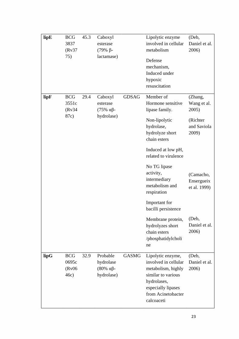
1.4.2.1 Hormone-sensitive lipase sub-family (HSL)
The hormone sensitive lipases are also known as triacylglycerol (TAG) lipases
wherein their main function is to hydrolyse first fatty acid of triacylglycerol
thereby yielding a diacylglycerol and a free fatty acid. They are, in turn, highly
regulated enzymes catalysing the hydrolysis of lipids in adipocytes. In silico
sequence analysis of mycobacterial lipase gene family members show
significant sequence homology with hormone-sensitive lipase family having a
characteristic HGG motif and a conserved active-site motif GDSAG. There
are also 12 mycobacterial lipolytic enzymes which belong to the hormone-
sensitive lipase family of which 8 are derived from lip gene family namely
lipF (Rv3487c), lipH (Rv1399c), lipI (Rv1400), lipN (Rv2970), lipR
(Rv3084), lipT (Rv2045c), lipU (Rv1076), lipY (Rv3097c) and are of high
functional importance.
1.5 Issues and Problems with functional characterization of
Mycobacterial putative Esterases/Lipases
A literature survey of the studies conducted in the past revealed that they were
aimed at the functional characterization of mycobacterial esterases/lipases and
reflects that the following issues and problems need to be addressed:

29
Expression of mycobacterial esterase/lipase enzymes in their non-
natural (non-mycobacterial) expression systems such as E. coli.(Deb,
Daniel et al. 2006)
Active conformation of enzymes lost after the purification step and
consequently re-folding in E. coli (Canaan, Maurin et al. 2004).
Mycobacterial esterase/lipase enzymes such as Rv1399c (lipH) and
Rv1400c (lipI) are insoluble and reported to form inclusion bodies
(Canaan, Maurin et al. 2004).
Poor solubility of mycobacterial esterase/lipase makes it difficult to
obtain an X-ray crystal structure. By far, only one 3D structure has
been solved of mycobacterial esterase (lipW) (PDB ID: 3QH4)
belonging to the lipase gene family.
1.6 Tetrahydrolipstatin (Orlistat)
Commonly called Orlistat and marketed by Roche pharmaceutical company
under the name of Xenical is a semisynthetic hydrogenated derivative of
naturally occurring lipase inhibitor lipstatin produced from Streptomyces
toxytricini (Hochuli, Kupfer et al. 1987). It has been well characterized as an
irreversible inhibitor of serine esterases (Hadvary, Lengsfeld et al. 1988)
covalently modifying its biological target. It was identified originally as a
specific inhibitor of pancreatic lipases and later developed as an anti-obesity
drug. The inhibitor has a reactive β- lactone ring (Fig.1.9) leading to an ester

30
Serine hydroxyl group of the catalytic triad of esterase/ lipase (Hadvary et al.,
1988).
1.6.1 An FDA approved anti-obesity drug
Orlistat was introduced in the pharmaceutical market by Roche under the
name of ‘Xenical’ launched in the year 1998, was approved by the Food and
Drug Administration (FAD), USA in 1999; it was represented as a ‘magic
medicine’ for control of obesity for the sole reason that it inhibits potently and
specifically breakdown of dietary triglycerides into absorbable fatty acids and
monoglycerides (Cudrey, van Tilbeurgh et al. 1993) thereby resulting into
30% fat absorption and hence consequential weight loss.
1.6.2 An anti-cancer agent
Recent studies have demonstrated that tetrahydrolipstatin (Orlistat) possess
antitumor properties to prostate cancer cells due to its ability to induce
inhibition of thioesterase domain of human fatty acid synthase (FAS)
lipogenic activity which is found to be significantly up-regulated in many
tumors and is an indicator of poor prognosis (Menendez, Vellon et al. 2005)
Figure 1.9: Chemical Structure of Tetrahydrolipstatin (Orlistat): arrow
depicting the β lactone ring in the chemical structure
3-hydroxytetradeca-5,8-dienoic acid
N-formylleucine
Hexyl-malonic acid

31
(Menendez et al., 2005). For example, 50% of breast cancers exhibit high
expression levels of FAS. THL eliminates tumour cells by inducing apoptosis.
1.6.3 An anti-mycobacterial agent
In mycobacteria it has been shown that tetrahydrolipstatin (Orlistat) interferes
with the cell wall formation of mycobacteria by decreasing mycolic acid
synthesis (Kremer, de Chastellier et al. 2005), leading to a defective
mycobacterial cell wall. Tetrahydrolipstatin is shown to inhibit Rv3802c an
essential cell wall lipase enzyme which is probably involved in the mycolic
acid biosynthetic pathway. Therefore tetrahydrolipstatin (THL) eliminates
both mycobacterial and tumor cells by interfering with the lipid metabolism.
1.7 Aims and Objectives of the present study:
Carboxyl ester hydrolases (EC 3.1.1) comprising of evolutionarily related
enzymes mostly belonging to hydrolases superfamily sharing a common α/β-
hydrolase protein fold and even though recent studies have revealed the
findings suggestive of their pivotal role in disease manifestation by way of
disruption of lipid metabolizing enzymes esterases/lipases and their pathways,
yet only very few have been functionally annotated. This study therefore
focuses attention on 4 putative mycobacterial lipases/esterase namely
BCG_1460c (lipH probable lipase), BCG_2991c (lipN probable
lipase/esterase), BCG_2950 (tesA probable thioesterase) and BCG_3229 (lipV
possible lipase) with the following objectives:

32
1. To explore computational enzymology as an effective tool for
In silico characterization of mycobacterial putative esterases/lipases and
identifying potential inhibitors.
2. To investigate structural features of mycobacterial putative
esterases/lipases and distinguishing lipolytic and non-lipolytic enzymes
at the structural level itself.
3. In vitro biochemical characterization of Mycobacterial esterases/lipases
and experimental validation of in silico predictions.
33
2. Materials and Methods
2.1 Molecular modelling
2.1.1 Sequence analysis:
The sequences of BCG_1460c (lipH probable lipase) (Uniprot Id: A1KII8),
BCG_2991c (lipN probable lipase/esterase) (Uniprot Id: A1KMW4),
BCG_2950 (tesA probable thioesterase) (Uniprot Id: A1KMS3) and
BCG_3229 (lipV possible lipase) (Uniprot Id: A1KNK2) and their
representative homologs were collected from the Uniprot database and
multiple sequence alignments were performed using ClustalW (Larkin,
Blackshields et al. 2007). This was found on the website
[http://www.ebi.ac.uk/Tools/msa/clustalw2/] to identify the catalytic triad
position, motifs and amino acid conservation.
2.1.2 Comparative modelling:
Structural homologs were identified using protein BLAST (Basic Local
Alignment Search Tool) [ http://blast.ncbi.nlm.nih.gov/] against Protein Data
Bank (PDB) database and were selected as templates based on the highest
sequence identity presented. Swiss PDB viewer was used to thread the protein
sequence on its structural homologs using MUSCLE package of Swiss PDB
viewer (Guex and Peitsch 1997) and thereafter initial structural alignments
were generated. Most favourable rotamers were added to the structure using
the rotamer library embedded in Swiss PDB viewer and a modelling request
was submitted to Swiss model server [http://swissmodel.expasy.org/]. There
were top ten models generated for each protein and validated using 4 different

34
validation soft wares on NIH MBI Laboratory for Structural Genomics and
Proteomics [http://nihserver.mbi.ucla.edu/SAVES/]. The evaluation of native
protein fold on validated 3D structure models was performed using ProSA
program (Wiederstein and Sippl 2007) and were further refined by fragment-
guided molecular dynamic simulations FG-MD program (Zhang, Liang et al.
2011) located at [http://zhanglab.ccmb.med.umich.edu/FG-MD/]. The refined
model was energy minimized with GROMACS 4.0.7 package
[http://www.gromacs.org/]. The resulting models were found to be
energetically stable. The schematic representation of the steps involved in
comparative modelling has been shown in figure 2.1
Figure 2.1: Schematic diagram of steps involved in Molecular modelling
(Source: adapted from http://swift.cmbi.ru.nl/teach/EMHOMX/EMBMOD_1.html)
35
2.2 Molecular Dynamics Simulations:
Protein stability of the energy minimized models in a solvent system was
assessed using molecular dynamics simulations and periodic boundary
conditions were applied to the structures in three dimension. By adding the
sodium ions and replacing the water molecules that are 3.5 Å from the protein
surface, the net charge of the system was neutralized. Simulations were run for
100 pico seconds with the solvent being equilibrated by a harmonic force
constant 100 KJ nm-2
and the solute atoms were restrained. A production run
of 10 nano second was used to check for the stability of protein models which
is a plot of root mean square deviation of the backbone structure versus time.
All simulations were performed at 300K temperature with velocity rescaling
thermostat using Parrinello-Rahman barostat. The pressure was maintained at
1 atmosphere and the long-range electrostatic interaction with cut-off 12 Å
was calculated using Ewald (PME) summation method while the hydrogen
bonds in the atoms were constrained with the linear constraint solver (LINCS)
algorithm (Hess, Bekker et al. 1997) and (Hess 2007).
2.3 Virtual Ligand Screening:
Computational docking of ligands in protein 3D structure model was
performed using Autodock Vina (Trott and Olson 2010). All essential
hydrogen atoms, solvation parameters and the united atom charges were added
using the help of AutoDock tools (Morris, Goodsell et al. 1998). Affinity grid
maps were constructed via and with the aid of Autogrid program. The Vander
Waals and electrostatic calculations were done using distance dependent

36
dielectric functions and parameters set functions of Autodock Vina.
Computational docking simulations have been performed using the
Lamarckian genetic algorithm and local search method (Solis and Wets,
1981). The initial orientations, position, torsion angles of ligands were set
randomly and during docking all rotatable torsions were released. For every
docking experiment, 500 independent docking runs were performed involving
30000 maximum energy evaluations and the population size was set to 200.
The ligand 2D-structures were drawn in ChemDraw and the 3D structures of
ligands were constructed with BUILDER module of molecular modelling
program Insight-II assigning standard geometric parameters. The atomic
charges were assigned to ligand using Amber potential. These ligands were
then subjected to force field until the root mean square energy gradient
became less than 0.005 Kcal/mol. All hydrogen atoms were added to the 3D
structure models and Amber all-atom charges were assigned to the whole
protein.
The active site was then analysed using the CASTp program (Dundas, Ouyang
et al. 2006) to recognize and characterize accessible pockets in the protein 3D
model based on two parameters: the solvent accessible surface and the
molecular surface. The active site was defined within a radius of 25 Å which
covered 94% of the total residues in the protein ensuring that the size of the
active site is sufficiently large enough to accommodate the whole ligand
binding pocket and also allow for rotation and translation of ligands. The
schematics of computational docking are shown in fig.2.2

37
Figure 2.2: Schematic diagram of Computational Docking
(Source: (Jacob, Andersen et al. 2012))

38
2.4 Bacterial strains and cultures
2.4.1 Bacterial Strains
M. bovis BCG Pasteur strain (ATCC 35734) and M. smegmatis (mc2155) were
used in the experiments.
2.4.2 Bacterial Culture Media
Cultures of mycobacteria were grown under aerobic conditions. Inoculum was
prepared from colony of growing cells and pre-culture was grown in 7H11
media. Aerobic M. bovis BCG were grown in roller bottles with initial OD
(optical density) measured at 600nm (OD600) of 0.5. The roller culture bottles
were rotated at 1rpm for three days.
Middlebrook 7H11: The mycobacterial cultures were grown using this culture
media. Middlebrook 7H11 (Biomed Diagnostics, BD Difco Mycobacteria
7H11 Agar, Catalogue No. 283810) supplemented with 10% oleic acid-
dextrose-albumin-catalase enrichment and 0.5% of glycerol (Biomed
Diagnostics, BD Difco Middlebrook OADC Enrichment catalogue no.212351)
at 37 °C. Whenever required, antibiotics were added to the M. bovis BCG
culture media with the following concentrations: kanamycin (Sigma-Aldrich,
USA) at 25 µg/ml and hygromycin (Roche,Germany) at 80 µg/ml .
Middlebrook 7H9 broth: This liquid media was used for growing
mycobacterial cultures. The Middlebrook 7H9 (Biomed Diagnostics, BD
Difco Mycobacteria 7H9 Broth, Catalogue No. 2713100) supplemented with
0.2% glycerol, 0.05% Tween-80 and 10% (v/v) Albumin-Dextrose-Saline
(ADS: 950ml dH20, 8.1g NaCl, 50g Bovine Serum Albumin Fraction V, 20g
39
D-dextrose). The whole medium was filter sterilized with o.22µM filter and
stored at 4°C until further use.
2.4.3 Glycerol stock of bacteria
M.bovis BCG and M. smegmatis: Glycerol stocks of cultures were prepared by
re-suspending in Middlebrook 7H9 broth containing 25% glycerol (v/v) and
stored as 0.5ml aliquots at -80ºC.
2.5 Cloning Procedures
2.5.1 Genomic DNA Isolation (Mycobacteria)
The mycobacterial cells were grown to an OD of 0.8 (measured at 600nm),
cells were then harvested by centrifuging at 3000g for 10mins at 4°C, washed
with PBS-T (0.05% of Tween-80). The pellet obtained was re-suspended in
600µL buffer having 3% SDS, 1mM Tris-HCL with 100Mm sodium chloride.
Cells were transferred to tubes having silica beads (0.1mm in diameter) and
then displaced using bead beater at 50rpm for 5mins at 4°C. Mixture of
phenol/isoamyl alcohol/chloroform in the ratio (49:2:49 v/v/v) was added and
then final volume made up to 2 X. Upon centrifuging at 16,000g for 5mins at
4°C the aqueous phase was collected then washed using 500µl of 70% ethyl
alcohol. Supernatant was then recovered pellet was dried at room temperature
for 10mins. Precipitated DNA pellets were then re-suspended in 100µl of TE
buffer and then storing it at 20°C until further use.

40
2.5.1 Preparation of Over-expression Plasmids
The putative esterases and/or lipases genes namely, BCG_1460c (lipH
probable lipase), BCG_2991c (lipN probable lipase/esterase), BCG_2950
(tesA probable thioesterase) and BCG_3229 (lipV possible lipase) were
amplified by using PCR with genomic DNA isolated from M. tuberculosis
H37Rv with primer details as given below:
Note: Restriction sites are underlined: forward BglII; reverse HindIII
The BglII and HindIII sites of the E.coli-Mycobacterium shuttle vector
pMV262 were inserted with PCR fragments under a constitutive HSP60
promoter.
Primers used for gene cloning and amplification
BCG_1460c
(lipH)
Forward GCAGATCTATGACCAAGAGTCTGCCAGGT
GCAAGCTTTTATGCGTGCAACGCCCTCTT Reverse
BCG_2991c
(lipN)
Forward GCAGATCTATGACCAAGAGTCTGCCAGGT
Reverse GCAAGCTTTCAAACCCGGCTAAGGTGCGC
BCG_2950
(tesA)
Forward
Reverse
GCAGATCTATGCTGGCCCGTCACGGACCA
GCAAGCTTCTAAGCTCGATCATGCCATTG
BCG_3229
(lipV)
Forward
Reverse
GCAGATCTTTGCCCGAAATCCCCATCGCC
GCAAGCTTCTAGCGCGGACCCAGTCGACT

41
2.6 Mycobacterium bovis BCG competent cell preparation, transformation
(electroporation) and selection of transformants
Inoculum from growing BCG cells was grown in 7H9 broth media (no
antibiotics) the culture was grown to ~0.8 optical density (OD600). The culture
was centrifuged at 3700rpm and cells were re-suspended in 1ml of 0.05%
tween80. 200 µl aliquot was stored at -80ºC. M.bovis BCG cells grown up to
an OD600. Cuvettes (Biorad ) with an electrode gap of 2mm were used for
electroporation and 1µl of plasmids was added to each cuvette and mixed
gently. Using Biorad gene pulser Xcell and pulsed at 2500V, 800 ohms, 25µF
transformants carrying the plasmids were selected on 7H11 plates containing
suitable antibiotic (hygromycin) the plates were left at 37 ºC to allow for
colonies to appear and thereafter an incubation period was allowed for four
weeks to have them grown until a visual turbidity was reached.
2.7 Preparation of Whole cell lysate (WCL)
The mycobacterial cultures were grown on Middlebrook 7H11 (Biomed
Diagnostics, BD Difco Mycobacteria 7H11 Agar, Catalogue No. 283810)
supplemented with 10% oleic acid-dextrose-albumin-catalase enrichment and
0.5% of glycerol) (Biomed Diagnostics, BD Difco Middlebrook OADC
Enrichment catalogue no.212351) for four weeks. Bacterial colonies were
harvested and washed thrice with phosphate buffered saline with tween80
(PBST) and cells were vortexed. Bacterial cells were lysed using probe
sonicator with the following parameters: 60% amplitude, 5 mins, 10 seconds
42
pulse on 5 seconds off and the lysed cells were centrifuged at 14000rpm;
protein was estimated using the Lowry’s protein estimation method.
2.8 Fluorescent click chemistry
Samples were prepared for click reaction by washing the M. bovis BCG cells
with phosphate buffered saline with tween80 (PBST) thrice and once with
phosphate buffered saline (PBS) and centrifuged at 3500g for 15 minutes at 4
ºC thereafter lysed in phosphate buffered saline (PBS) at 50% amplitude.
100µg of protein was used for the reaction in which drug was incubated with
protein. The amounts of reagents used for each reaction have been described
in table 2.1. After incubation; samples were prepared for gel electrophoresis
run.
2.9 Gel Electrophoresis
Samples were prepared for the gel run by adding 600 µl of acetone and left at
-20 ºC for 24 hours cells were centrifuged at 14,000 rpm then washed with 200
µl of methanol. Subsequently, cell lysis was done using sonicator with the
following parameters: 60% amplitude, 5 mins, 10 sec pulse on/ 5 sec off after
cell lysis 50 µl of SDS dye was added and boiled at 95 ºC for 5 mins. Equal
amounts of 10 micro gram protein was separated on 12% SDS-polyacrylamide
gel, ran at 120 V for 110 mins followed by in-gel fluorescence scanning with
Typhoon 9410 Variable Mode Imager scanner (GE Amersham, UK) . The
cyc3/cyc5 scan protocol adopted was as follows: 3*108 cells for each staining
Table 2.1 Click reaction

43
and washed once with phosphate buffered saline with 0.05%tween80 (PBST)
and centrifuged at 3000g for 5mins at 4 ºC. Subsequently washed with
phosphate buffered saline (PBS) after which 10ng of LD540 dye was added
and incubated for 10 mins at room temperature. 500µl of 4% formaldehyde
was added and tubes were centrifuged at 3000g for 5mins at 4 ºC. Cells were
again washed with phosphate buffered saline (PBS) and permealized with 500
µl of 0.25% Triton-X 100 in PBS for 15 mins .
2.10 Enzymatic Assays
The enzymatic assays were performed with whole cell lysates of BCG cells
overexpressing proteins BCG_2991c (lipN probable lipase/esterase),
BCG_2950 (tesA probable thioesterase) and BCG_3229 (lipV possible lipase)
using p-nitrophenyl (pNP) esters (Sigma Aldrich) with carbon chain lengths
of C2, C4 and C16. The pNP release was measured at 400nm by using a 96 well
plate spectrophotometer. The enzymatic reaction was performed with 225µl of
NaDC tris buffer (Tris 15.14mg, Nacl-0.8766g, Sodium deoxycholate -1mM
(20.7mg) measured and dissolved in 25ml of milliq water and the pH of the
whole medium was adjusted to pH 8.0). This was added to each reaction along
with 2.5µg/µl of protein concentration and 1mM of pnP-C2/C4/C16 substrate
incubated for 30mins at 37 ºC. The final volume of each well was kept at 250
µl and the absorption was measured at 400nm using a 96 well plate
spectrophotometer and readings were recorded at 0, 1, 2.5, 5 and 10 minutes
time points. All reactions were performed in triplicates. The schematics of
pNP assay are depicted in fig.2.3
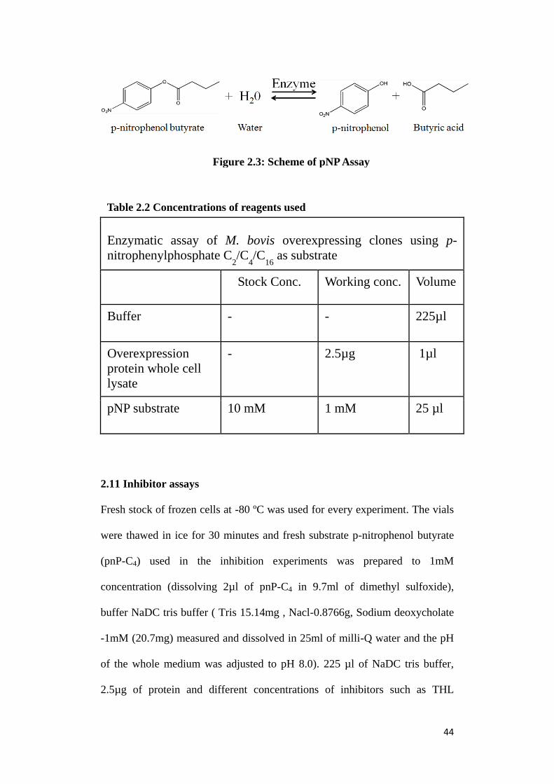
44
2.11 Inhibitor assays
Fresh stock of frozen cells at -80 ºC was used for every experiment. The vials
were thawed in ice for 30 minutes and fresh substrate p-nitrophenol butyrate
(pnP-C4) used in the inhibition experiments was prepared to 1mM
concentration (dissolving 2µl of pnP-C4 in 9.7ml of dimethyl sulfoxide),
buffer NaDC tris buffer ( Tris 15.14mg , Nacl-0.8766g, Sodium deoxycholate
-1mM (20.7mg) measured and dissolved in 25ml of milli-Q water and the pH
of the whole medium was adjusted to pH 8.0). 225 µl of NaDC tris buffer,
2.5µg of protein and different concentrations of inhibitors such as THL
Enzymatic assay of M. bovis overexpressing clones using p-
nitrophenylphosphate C2/C
4/C
16 as substrate
Stock Conc. Working conc. Volume
Buffer - - 225µl
Overexpression
protein whole cell
lysate
- 2.5µg 1µl
pNP substrate 10 mM 1 mM 25 µl
Table 2.2 Concentrations of reagents used
Figure 2.3: Scheme of pNP Assay

45
(tetrahydrolipstatin) E600 (diethyl-p-nitrophenylphosphate) was used from
.01µM to 100µM (volume kept at 1µl) were added to each well of 96 well
plate. DMSO (dimethyl sulfoxide) concentration was maintained at 0.004% in
each well, incubation time was 30 mins at 37 ºC and the absorption was
measured at 400nm using a 96 well plate spectrophotometer. Measurement
readings were recorded at 0, 1, 2.5, 5 and 10 minutes time points. All reactions
were performed in triplicates. The template indicating the component of each
well of the 96 well plate has been shown in figure 2.4
Figure 2.4: Template design of a 96 well plate used for Inhibitor assays
46
3. Results
3.1 Molecular modelling of protein 3D structures
In order to understand the insight as well as structural details and characterize
four putative esterases/lipases belonging to (α/β) hydrolase family viz.
BCG_1460c (lipH probable lipase), BCG_2991c (lipN probable
lipase/esterase), BCG_2950 (tesA probable thioesterase) and BCG_3229 (lipV
possible lipase), a computational prediction of their ligand specificity for
catalytic triad was performed. 3D model structures were generated using
molecular modelling protocol, at first we validated the molecular modelling
protocol by predicting in silico the 3D structure of protein named thioesterase
domain of fatty acid synthase (Human FAS-TE) (Protein Databank ID: 2PX6)
whose X-ray 3D structure is well known this acted as our positive control.
Molecular overlay of the modelled 3D structure with known 3D structure of
FAS-TE (figure 3.1 (a)) revealed that in silico modelled FAS-TE structure
could mimic the known FAS-TE structure reasonably well with a carbon alpha
backbone (Cα) root mean square deviation (rmsd) of 0.192Å (figure 3.1b) and
an acceptable statistics in Ramachandaran plot (Suplatov, Besenmatter et al.
2012). The 3D model structure of BCG_1460c (lipH probable lipase) was
generated based on structural homologues of 3 thermophilic esterases with
significant sequence identity in the range of 40 to 44% namely
Alicyclobacillus acidocaldarius (carboxyl esterase Est2) (Protein data bank
ID: 1U4N) with 44% sequence identity, Archaeoglobus fulgidus (carboxyl
esterase AFEST) (Protein data bank ID: 1JJI) with 41% sequence identity and
bacterial acetyl esterase (HerE) (Protein data bank ID: 1LZL) had 40%

47
sequence identity. It was also revealed that BCG_2950 (tesA probable
thioesterase) has 2 bacterial thioesterases structural homologues with sequence
identity in the range of 31-33% namely Streptomyces coelicolor (Thioesterase)
(Protein data bank ID: 3QMV) having 33% sequence identity and thioesterase
from Amycolactopsis mediterranei (Protein Data Bank ID: 3FLB) had 31%
sequence identity with protein sequence of BCG_2950 (tesA probable
thioesterase). For BCG_2991c (lipN probable lipase/esterase) the BLAST
searches found that it has structural homologs in 2 carboxyl esterases with
sequence identity ranging from 39% to 41% : Hormone sensitive lipase-like
Carboxylesterase from Sulfolobus Tokodaii (Protein data bank ID: 3AIO)
(41%) , carboxyl esterase (Est2) Alicyclobacillus acidocaldarius (Protein data
bank ID: 2HM7) (39%) while BCG_3229 (lipV possible lipase) shows
structural homologs in the sequence identity range of 27% to 31% with two
esterases: thioesterase domain from Curacin biosynthetic pathway (Protein
data bank ID: 2HM7) (31%) and Rv0045c from Mycobacterium tuberculosis
(Protein Data Bank ID: 3P2M).
Figure3.1: (a) 3D structure Overlay of FAS-TE modelled structure over FAS-TE known X-ray
3D structure in protein data bank (2PX6) (b) Cα – backbone overlay r.m.s.d 0.192Å (red: in
silico modelled 3D structure, yellow: X-ray known 3D structure Protein Databank ID: 2PX6)
(a) (b)

48
M
Figure3.2 3D structure models: (a) BCG_2950 (tesA probable thioesterase) (b) BCG_1460c
(lipH probable lipase) (c) BCG_2991c (lipN probable lipase/esterase) (d) BCG_3229 (lipV
possible lipase)
(b)
(d) (c)
(a)

49
The 3D structure models (figure 3.2) indicated that all 4 putative
esterases/lipases namely BCG_1460c (lipH probable lipase), BCG_2991c
(lipN probable lipase/esterase), BCG_2950 (tesA probable thioesterase) and
BCG_3229 (lipV possible lipase) are globular proteins similar to other
alpha/beta (α/β) hydrolase enzymes and consist of 11 α- helices and a central
β-sheet core containing 6 parallel β- strands. The GXSXG motif characteristic
of esterases/lipases was found to be conserved in all 4 enzymes viz.
BCG_1460c (lipH probable lipase), BCG_2991c (lipN probable
lipase/esterase), BCG_2950 (tesA probable thioesterase) and BCG_3229
(lipV possible lipase) whereas the invariant motif HGG of hormone sensitive
lipase family was conserved in BCG_1460c (lipH probable lipase),
BCG_2991c (lipN probable lipase/esterase). The catalytic triad was known to
be composed of nucleophilic serine which is the active site, a charge relay
network aspartate and proton carrier histidine. The structure validation report
from four different validation soft wares on NIH MBI laboratory
[http://nihserver.mbi.ucla.edu/SAVES/] for structural genomics and
proteomics are shown in fig.3.3 whereas angle distribution of amino acids in
3D structure models is shown in fig.3.4
Figure 3.3: Structure validation report from 4 different validation soft wares
on NIH MBI Laboratory for Structural Genomics and Proteomics
[http://nihserver.mbi.ucla.edu/SAVES/]

50
Figure 3.4: Ramachandran plot statistics of backbone dihedral angle distribution
of amino acids in the 3D structure model having no disallowed regions.

51
Fig. 3.5 illustrates protein structure analysis from PROSA (protein structural
analysis) web server as depicted below whereas fig 3.6 illustrates molecular
dynamic simulation on validated 3D structure models.
Figure 3.5: Protein structure analysis from PROSA (protein structural
analysis) web server shows 3D structure models have Z-score -6.47 in the
acceptable range for near native conformation of X-ray crystal structures
Figure 3.6: Molecular dynamic simulations of modelled 3D structures
show that the energy minimized 3D model structures are energetically stable

52
3.2 Molecular Structure based Ligand Screening
In order to identify natural substrates/potential inhibitors and to characterize
the substrate specificity of BCG_1460c (lipH probable lipase), BCG_2991c
(lipN probable lipase/esterase), BCG_2950 (tesA probable thioesterase) and
BCG_3229 (lipV possible lipase), a structure based virtual ligand screening
was performed. At first a in silico library of potential ligands was constructed
based on literature review of substrate preferences of well characterized
carboxyl esterases in mycobacterial species. This resulted into a in silico
ligand library of natural substrates such vinyl esters of varying acyl chain
lengths from short chain vinyl acetate (C2) to long chain palmitate (C16) along
with triacylglycerol substrates (TAGs) varying from short chain TAGs such as
tripropionate (C3) to long chain TAGs trioleate (C18) with potential inhibitors
of serine hydrolases such as THL, PMSF and E600.
The in silico potential ligand library was thereafter screened using molecular
docking engine Autodock (Trott and Olson 2010) into the active site of 3D
structures of BCG_1460c (lipH probable lipase), BCG_2991c (lipN probable
lipase/esterase), BCG_2950 (tesA probable thioesterase) and BCG_3229 (lipV
possible lipase) and results were subjected to further analysis and
interpretation.

53
3.2.1 Evaluation of Ligand Screening Results
The ligand screening results were evaluated based on two scoring functions:
a.) Ligand binding affinity/energy towards its target enzyme.
b.) Distance to active site (serine in all cases) which describes a near
attack conformation (NAC) (Hur, Bruice et al. 2003) of the ligand in
the binding site.
The enzyme-ligand docked structures were ranked by frequency binding in
near attack conformation obtained from 500 independent docking runs (p-
value <0.05). The ligand specificity to its target enzyme was confirmed based
on the highest binding affinity and the least distance to active site (serine). A
threshold for an acceptable near attack conformation of ligand distance to
active site should be less than 3.5Å (Suplatov, Besenmatter et al. 2012) while
the ligand binding affinity should fall within the range of -4 Kcal/mol to -8
Kcal/mol (fig 3.7 a,b,c,d).

54
Figure 3.7: Short (C2) to long (>C12) acyl chain ester ligands screening results, highlighted
circle in red show most favourable ligand (highest ligand specificity) based on its lowest
energy and least distance to active site (Serine). Graphs showing the ligand specificity
pattern for (a) lipH (b) lipN (c) lipV (d) tesA
(a) (b)
(c) (d)
lipH lipN
lipV tesA
Table 3.1 Summary of potential substrates screening results

55
Based on the two criteria evaluation of structure based virtual screening,
results revealed that BCG_1460c (lipH probable lipase) favours docked near
attack conformations with short acyl chain esters from vinyl butyrate (C4) up
to vinyl laurate (C12) and short chain TAGs such as tripropionate having
identical fatty acyl chains (3:0/3:0/3:0) and tri butyrate (4:0/4:0/4:0) (table
3.1) with highest ligand specificity for vinyl caproate (C6) and a binding
affinity of -5.11 Kcal/mol and 2.5Å distance to active site (serine) (figure
3.7(a)). For BCG_2991c (lipN probable lipase/esterase), it had a similar
binding affinity towards short acyl chain vinyl esters and TAGs ranging from
vinyl butyrate (C4) up to vinyl lauraterate (C12) and tripropionate having
identical fatty acyl chains (3:0/3:0/3:0) to tri butyrate (4:0/4:0/4:0) (table 3.1)
with highest ligand specificity for vinyl butyrate (C4) and a binding affinity of
-5.9 Kcal/mol with 2.62 Å distance to active site (serine) (figure 3.7(b)). On
the other hand BCG_3229 (lipV possible lipase) also depicted ligand
preference towards short chain vinyl esters such as vinyl butyrate (C4) up to
(C14) and TAGs ranging from tripropionate having identical fatty acyl chains
(3:0/3:0/3:0) to tricaproate (6:0/6:0/6:0) (table 3.1) with highest ligand
specificity for vinyl caproate (C6) having a binding affinity of -6.0 Kcal/mol
and 1.92Å distance to active site (serine) (figure 3.7(c)). However,
interestingly BCG_2950 (tesA probable thioesterase) showed preference for
very long chain lengths (>C16) with the highest ligand specificity for
phthiocerol (C28) having a binding affinity of -7.2 Kcal/mol and 1.02Å
distance to active site (serine) (figure 3.7(d)).

56
The screening for potential inhibitors resulted in the prediction of THL
(tetrahydrolipstatin) as the most potent inhibitor of BCG_1460c (lipH probable
lipase), BCG_2991c (lipN probable lipase/esterase), BCG_2950 (tesA
Protein Ligand Ligand
function
Distance to
active site
Binding
affinity/energy
(Kcal/mol)
lipH THL Lipase
inhibitor
1.82 Å -7.10
lipH E600 Lipase
inhibitor
2.02 Å -6.70
lipH PMSF Protease
Inhibitor
2.12 Å -5.60
lipN THL Lipase
inhibitor
2.08 Å -6.5
lipN E600 Lipase
inhibitor
2.23 Å -5.2
lipN PMSF Protease
Inhibitor
2.32 Å -4.9
tesA THL Lipase
inhibitor
1. 91Å -7.2
tesA E600 Lipase
inhibitor
2.37 Å -5.6
tesA PMSF Protease
Inhibitor
2.38 Å -5.4
lipV THL Lipase
inhibitor
1. 84Å -7.6
lipV E600 Lipase
inhibitor
2.06 Å -5.2
lipV PMSF Protease
Inhibitor
2.62 Å -4.4
Table 3.2 Summary of potential inhibitors ligand screening results

57
probable thioesterase) and BCG_3229 (lipV possible lipase) as compared to
E600 and PMSF as shown above in table 3.2.
3.3 Over expressing mycobacterial putative esterases/lipases.
BCG_1460c (lipH), BCG_2991c (lipN), BCG_2950 (tesA) and BCG_3229
(lipV) were found to be over expressed and successfully transformed. The
fluorescent gel image (fig.3.8) clearly demonstrates an over-expression in
protein levels of BCG_1460c (lipH probable lipase) and BCG_2950 (tesA
probable thioesterase) having molecular weight of 34.0 kDa and 29.0 kDa
respectively were seen as thick bands compared to wild type M. bovis BCG
(non-over expressed).
Figure 3.8: SDS-PAGE fluorescent gel image showing change in protein
levels of M. bovis BCG over expressing lipH and tesA

58
3.4 Biochemical Characterization
3.4.1 Enzymatic Assays
To characterize the activities of BCG_1460c (lipH) and BCG_2950 (tesA)
enzymes, the whole cell lysates of over expressed BCG_1460c (lipH) and
BCG_2950 (tesA) were assayed with synthetic vinyl ester substrate para-
nitrophenol-butyrate (C4). A ~2.5 times fold change was observed in the
absorption (measured at 400nm) for BCG_1460c (lipH) as compared to wild
type M. bovis BCG (non-over expressed) (fig.3.9)
Figure 3.9: Enzymatic assay with whole cell lysate of overexpressed
BCG_1460c (lipH) and BCG_2950 (tesA) using substrate para-nitro
phenol-butyrate (C4) (** n=2 biological replicates)

59
Within 2.5 mins of the enzyme reaction almost 80% of substrate para-
nitrophenol-butyrate (C4) was converted into products i.e. butyric acid and
para-nitrophenol the latter showing maximum absorption at 400 nm. However,
overexpressed BCG_2950 (tesA) indicated no change in absorption and was
lesser compared to the wild type (non-overexpressed) (fig3.9). In order to
ensure adequate accuracy, the enzymatic assay was performed with two
biological and two technical replicates.
3.4.2 BCG_1460c (lipH probable lipase) shows short-chain esterase
activity
To further investigate the substrate specificity of BCG_1460c (lipH), enzyme
assay was performed using synthetic substrates of vinyl ester substrates
including short–chain esters such as para-nitrophenol-acetate (C2) and para-
nitrophenol-butyrate (C4) and long-chain ester para-nitrophenol-palmitate
(C16) with whole cell lysate of over expressed BCG_1460c (lipH).
The assay results revealed that BCG_1460c (lipH probable lipase) shows a
short-chain esterase activity towards para-nitrophenol-acetate (C2) and para-
nitrophenol-butyrate (C4) (fig3.10) and there was no activity observed towards
long chain-vinly esters (C16) i.e. para-nitrophenol-palmitate (C16) as shown in
fig3.10.

60
3.5 THL (Tetrahydrolipstatin) strongly inhibits short-chain esterase
activity of BCG_1460c (lipH)
Inhibition experiments were performed using well known lipase inhibitor
tetrahydrolipstatin (THL) and E600 (diethylparanitrophenyl phosphate) the
latter being a specific inhibitor of catalytic site serine were assayed with whole
cell lysate of over expressed BCG_1460c (lipH) with the choice of substrate
being p-nitrophenol butyrate (pNP-C4) since all our previous enzymatic
reactions of over expressed BCG_1460c (lipH) were most stable with pNP-C4.
Tetrahydrolipstatin (THL) was found to strongly inhibit the short-chain
Figure 3.10: Enzymatic assay of over expressed BCG_1460c (lipH) with
varying acyl chain length substrates para-nitrophenol-acetate (C2),
butyrate (C4), palmitate (C16) (** n=2 biological replicates)

61
esterase activity of BCG_1460c (lipH) as indicated by its IC50 value of
0.12±0.035µM (fig3.11) and E600 (diethylparanitrophenyl phosphate)
inhibitor having an IC50 value of 0.399±0.172µM (fig3.12). All the inhibition
assays were performed with 2 biological and 2 technical replicates. Of note,
IC50 values are expected to be even lower for purified BCG_1460c (lipH)
enzyme since we used whole cell lysates for our inhibition assays.
Figure 3.11: THL (Tetrahydrolipstatin) inhibitor assay (IC50) towards
BCG overexpressing BCG_1460c (lipH) from log phase culture (*** n=3)
Figure 3.12: E600 (diethylparanitrophenyl phosphate) inhibitor assay
(IC50) towards BCG overexpressing BCG_1460c (lipH) from log phase
culture (*** n=3)

62
3.6 Predicted binding mode model of THL inhibition in BCG_1460c (lipH)
3D structure
To understand at the structural level THL inhibition of short-chain esterase
activity of BCG_1460c ligand docking experiments were performed. 3D
structure model of lipH is shown in fig.3.13 (a) with its potential docked
substrate (Vinyl caproate) and inhibitor (THL) predicted from in silico ligand
screening results. Interactions of substrate (Vinyl caproate) within binding
pocket have been shown in fig.3.13 (a) while interactions of THL
(tetrahydrolipstatin) within the binding pocket shown in fig. 3.13(b) revealed a
near attack conformation of β-lactone ring from the active site serine further
the molecular overlay of catalytic triad (serine, histidine, aspartate) from
substrate bound over inhibitor (THL) bound shown in fig. 3.13(c) revealed
Amino acid interaction Ligand binding pocket (a) lipH + substrate
Area 298.1 Å Volume 348.4 Å Length 14.21Å Width 9.32Å
Ile210
Asp260
His290
Gly291
Asn294
Tyr190
His100 Asp161
Ser162 Gly89
Gly90
Tyr86
Vinyl caproate
Ser162 His290
Asp260 Vinyl caproate
Binding Affinity/Energy
-5.11 Kcal/mol
Distance to active site
(Serine) 1.76 Å

63
Ser162
His290
Asp260 THL
Figure 3.13 (a & b). Three dimensional model of lipH structure depicting the core α/β-
hydrolase fold with catalytic triad (yellow). The 3D model is docked with lipH probable
substrate, vinyl caproate (blue) and probable inhibitor, THL (red). The insert (middle)
depicts the catalytic triad and other amino acids interacting with docked ligand. And the
binding energy of ligand binding pocket is represented on far right to show that area,
volume and width of the pocket decide the specificity of ligand conformation and the
folding pattern. (c) Overlay of catalytic triad docked with substrate (blue) and with THL
(red). The change in C—C atom r.m.s.d value of lipH catalytic site triad with substrate and
THL is mentioned at the bottom of the figure.
(c) lipH catalytic site overlay
(b) lipH + THL Amino acid interaction Ligand binding pocket
Binding
Affinity/Energy
-7.10 Kcal/mol
Distance to active
site (Serine) 1.82 Å
Area 533.2 Å Volume 579.5 Å Length 26.05 Å Width 9.56 Å
Ser162 His290 Asp260
rmsd 0.567Å rmsd 1.012Å rmsd 0.392Å
Arg38
Ile210
Leu39
Asp260
His290 Phe295
Asn294
His100
Tyr86
Asp161
Ser162 Gly89
Thr192
Gly90 Gly91
Val215
Ile216 Phe219
Trp92
Leu220
Thr99
THL

64
that there is only a very small root mean square deviation (difference) of the
catalytic triad restudies with active site serine having root mean square
deviation of 0.567 Å, histidine 0.392 Å and aspartate 1.012 Å (fig. 3.13(c)).
Therefore, it was predicted that THL inhibitor binds in the substrate pocket of
BCG_1460c (lipH) where the hexanoyl (C6) arm of THL binding to the
substrate pocket may well be mimicking the short-chain substrate (vinyl
caproate C6) binding in the ligand binding pocket of BCG_1460c (lipH) this
hypothesis has been discussed in details in the discussion section which would
now follow.
4. Discussion
Mycobacterium tuberculosis incorporates an unusually high number of genes
in its lipid metabolism (Cole et al., 1998) and it is known that there are 250
genes encoding putative lipid synthesis/degrading enzymes as compared to
only 50 genes in E.coli having a similar genome size. Recent studies have
suggested that success of Mycobacterium tuberculosis pathogen relies on its
exceptional capacity to latently infect and re-infect its host which is mainly
attributed to its lipid metabolism. The mycobacteria prior to entering into non-
replicating state have been shown to accumulate intracellular lipids (TAGs)
(Garton, Christensen et al. 2002). The studies of Coles group, in 2008 (Cotes,
Bakala N'goma J et al. 2008) have reported that during the mycobacterial re-
infection cycle i.e. exit from non-replicating state, the bacteria hydrolyses its
intracellular lipid deposits (TAGs) using hydrolytic enzymes like
esterases/lipases. Sequence analysis has revealed 31 of such putative lipolytic

65
enzymes out of which 24 have been classified under lip family as putative
esterases/lipases but very few have been functionally annotated and the 3D
structure of only one esterase lipW (PDB ID: 3QH4) has been reported to have
been established so far. Our results from this study conducted in
Mycobacteirum bovis BCG provides insights into molecular structure of 4
putative mycobacterial esterases/lipases namely BCG_1460c (lipH probable
lipase), BCG_2991c (lipN probable lipase/esterase), BCG_2950 (tesA
probable thioesterase) and BCG_3229 (lipV possible lipase) and predicting
natural substrates and a potent inhibitor THL (tetrahydrolipstatin) by structure
based virtual ligand screening apart from biochemical characterization of
short-chain esterase activity BCG_1460c (lipH probable lipase) inhibited by
THL (tetrahydrolipstatin). However, detailed results would now be discussed
in following sections
4.1 In silico Studies:
4.1.1 Role of Virtual Screening in Antibacterial Drug Discovery
The golden era of antibacterial chemotherapy began in the 1950s and by the
advent of 1970s almost all major classes of antibacterial agents currently in
use (Simmons, Chopra et al. 2010) were discovered this was a major scientific
and medical achievement that had enormous benefits for treatment of deadly
infectious diseases (caused by bacteria) affecting the human race. However,
the emergence of widespread global occurrence of bacteria resistant to the
antibiotics and synthetic drugs threatens to reverse our ability to treat
infectious diseases (Chopra 2013). Furthermore the new drugs in
pharmaceutical pipelines are mostly derivatives of older classes of
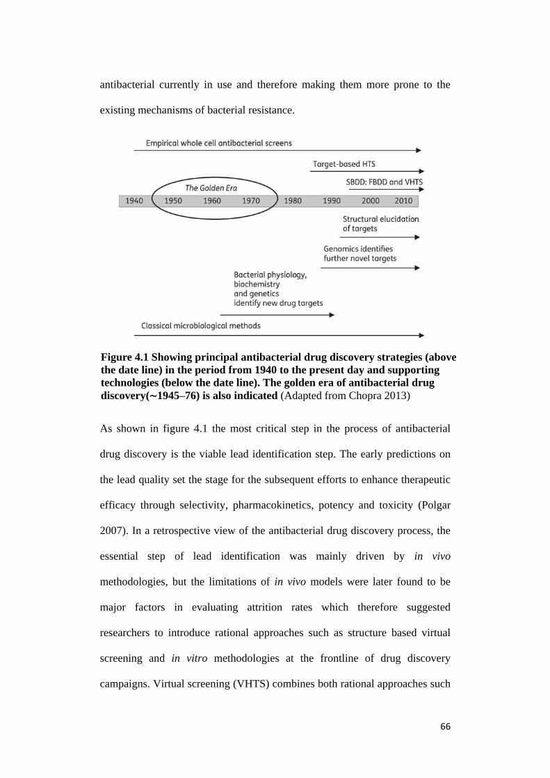
66
antibacterial currently in use and therefore making them more prone to the
existing mechanisms of bacterial resistance.
As shown in figure 4.1 the most critical step in the process of antibacterial
drug discovery is the viable lead identification step. The early predictions on
the lead quality set the stage for the subsequent efforts to enhance therapeutic
efficacy through selectivity, pharmacokinetics, potency and toxicity (Polgar
2007). In a retrospective view of the antibacterial drug discovery process, the
essential step of lead identification was mainly driven by in vivo
methodologies, but the limitations of in vivo models were later found to be
major factors in evaluating attrition rates which therefore suggested
researchers to introduce rational approaches such as structure based virtual
screening and in vitro methodologies at the frontline of drug discovery
campaigns. Virtual screening (VHTS) combines both rational approaches such
Figure 4.1 Showing principal antibacterial drug discovery strategies (above
the date line) in the period from 1940 to the present day and supporting
technologies (below the date line). The golden era of antibacterial drug
discovery(∼1945–76) is also indicated (Adapted from Chopra 2013)

67
as structure-based screening with high throughput approaches (HTS). The
typical workflow of a high throughput virtual screening has been depicted in
fig. 4.2
Figure 4.3 Detailed workflow of a high throughput virtual screening (VHTS)
(Source: http://www.molfunction.com/virtual.htm )
Fig. 4.2 State of the art in Virtual Screening

68
4.1.2 Concepts, Feasibility and Drawbacks of Virtual Screening
Virtual screening is a strategy aimed at bringing together a more focused
approach to high throughput screening by applying rational computational
analysis to a subset of compounds considered to be appropriate for a given
receptor. It is quite clear that this strategy implies that there is some starting
point information which is available pertaining to either the nature of the
binding site of receptor and/or ligand type that would be expected to
favourably bind within the active site of the receptor. However, it should be
realized that virtual screening itself combines a variety of computational
screens, which encompasses simplistic to sophisticated algorithms which
explore different types of information describing receptor. Similarly, it can be
used to produce either a highly focused compound subset (for instance if only
the close structural analogs of a lead compound are of interest) or a highly
open ended subset (for instance a constraint on size, described by molecular
weight, may be applied), although this largely depends on the objectives and
interests of the HTS project.
4.1.3 Molecular 3D Structure Modelling and Virtual Ligand Screening
In our in silico study, we have demonstrated 3D structure models of
BCG_1460c (lipH probable lipase), BCG_2991c (lipN probable
lipase/esterase), BCG_2950 (tesA probable thioesterase) and BCG_3229 (lipV
possible lipase), with an accuracy of >95% having near native conformation
of energetically stable and validated 3D structure models.

69
Partially modelled 3D structure of Rv1399c (BCG_1460c lipH) in
Mycobacterium tuberculosis has been reported in the study of Canaan group in
2004 (Canaan, Maurin et al. 2004) and Rv2928 (BCG_2950 tesA) from
Chavadi group in 2011 (Chavadi, Edupuganti et al. 2011) whereas in the
present study we have predicted complete domain modelling of BCG_1460c
(lipH probable lipase), BCG_2950 (tesA probable thioesterase), BCG_2991c
(lipN probable lipase/esterase), and BCG_3229 (lipV possible lipase).
We have uniquely extended the concept of molecular modelling to a structure
based ligand screening to predict and understand the ligand specificities
particularly those pertaining to BCG_1460c (lipH probable lipase),
BCG_2950 (tesA probable thioesterase), BCG_2991c (lipN probable
lipase/esterase), and BCG_3229 (lipV possible lipase). Our results suggest that
BCG_1460c (lipH probable lipase), BCG_2991c (lipN probable
lipase/esterase) and BCG_3229 (lipV possible) show an inclined tendency of
ligand affinity towards short-chain vinyl esters such as vinyl butyrate (C4) and
vinly caproate (C6) whereas for triacylglycerol (TAGs) substrates were found
to be tripropionate having identical fatty acyl chains (3:0/3:0/3:0) to
tricaproate having identical fatty acyl chains (6:0/6:0/6:0). Our results were
performed with 500 independent docking runs involving 300000 maximum
energy evaluations to achieve desired statistical significance (p-value<0.05).
Further our molecular modelling and ligand screening (docking) protocols
were validated to serve as effective computational tools towards in silico
characterization of mycobacterial putative esterases/lipases.

70
4.1.4 Predicted binding mode model of THL inhibition in BCG_1460c
(lipH) 3D structure
Our results predict the binding mode and ligand interactions of THL
(tetrahydrolipstatin) within the ligand binding pocket of BCG_1460c (lipH)
(fig.3.13). 3D structure and molecular overlay of catalytic site in fig.3.13(c)
shows a very small root mean square deviation (difference) of substrate bound
and inhibitor bound catalytic triad residues with active site serine having an
root mean square deviation of 0.567 Å, histidine 0.392 Å and aspartate 1.012
Å. This observation tends to suggest that THL binds within the substrate
pocket of BCG_1460c (lipH). Further it was also found that β-lactone ring of
THL known to interact covalently with the active site serine of
esterases/lipases adopts near attack conformation within the substrate binding
pocket of BCG_1460c (lipH) and is located at a distance of 1.82 Å from active
site serine (hydroxyl group-OH). This again is a favourable indicator of a
possible nucleophilic attack by active serine on the carbonyl carbon of β-
lactone ring. Further comparisons between ligand interactions of short-chain
substrate vinyl caproate (C6) and inhibitor THL tempted us to predict that
hexanoyl (C6) arm of THL might well be mimicking short-chain vinyl
caproate (C6) substrate specificity of BCG_1460c (lipH) within its substrate
pocket.
4.2 In vitro Studies:
4.2.1 BCG_1460c (lipH probable lipase) is a short-chain carboxyl esterase
Our in vitro biochemical assays suggest a short-chain esterase activity of
BCG_1460c (lipH probable lipase) (fig 3.13) which correlates well with the

71
findings of a study conducted by Canaan et al., 2004 in Mycobacterium
tuberculosis on Rv1399C( lipH putative lipase) where they have cloned,
expressed and folded the enzyme in E.coli. This is not the natural environment
(non-mycobacterial) and the enzyme was found to be insoluble, expressed as
inclusion bodies and the active state was lost after the purification step and
was consequently re-folded in E.coli using an alternative in vitro protocol. In
this study we performed enzymatic assays with whole cell lysates of
Mycobacterium bovis BCG over-expressing BCG_1460c (lipH probable
lipase) using synthetic substrates and results suggest a short-chain esterase
activity of BCG_1460c (lipH probable lipase) with para-nitrophenyl actetae
(C2) and para-nitrophenyl actetae (C4) (fig.4.1). We also transformed
Mycobacterium smegmatis MC2 with BCG_1460c (lipH probable lipase)
over-expression clone and our biochemical assays again showed a short-chain
esterase activity of BCG_1460c (lipH probable lipase) in Mycobacterium
smegmatis MC2 (fig.4.2)
Figure 4.4: SDS-PAGE fluorescent gel image showing change in protein
levels of M. smegmatis MC2 over expressing lipH (BCG_1460c)
mc2
wild
Molecular
weight 34 kDA
mc2
wild pMV lipH Marker pMV lipH
Marker

72
4.2.2 THL (Tetrahydrolipstatin) strongly inhibits short-chain carboxyl
esterase activity of BCG_1460c (lipH probable lipase)
Further we found that this short-chain esterase activity of BCG_1460c (lipH
probable lipase) was inhibited by THL (tetrahydrolipstatin) as indicated by
IC50 value of 0.12±0.035µM (fig.3.11) while E600 (diethylparanitrophenyl
phosphate) inhibitor having an IC50 value of 0.399±0.172µM (fig.3.11). These
values are expected to be even lower for purified enzyme since we performed
Figure 4.5: Enzymatic assay with whole cell lysate of M. smegmatis
MC2 over expressing lipH (BCG_1460c) using substrate Pnp-C4
para-nitrophenol-butyrate (C4) (** n=2 biological replicates)

73
our enzymatic assays using whole cell lysates. The 3D protein structure model
supports our biochemical data wherein at the structural level our data suggests
a near attack conformation of β-lactone ring from the active site serine and a
favourable nucleophilic attack on the carbonyl carbon of β-lactone ring
therefore opening the β-lactone ring cycle.
4.2.3 Possible functions of BCG_1460c (lipH probable lipase)
The mycobacterial lip family comprises of 24 putative esterases/lipases of
which hormone sensitive lipase (HSL) family is a sub-family. BCG_1460c
(lipH probable lipase) is a member of hormone sensitive lipase (HSL) family
in this family Rv3097c (lipY) has been reported previously in studies
conducted by Deb and Mishra groups (Deb. et al., 2006 and Mishra et al.,
2008) that Rv3097c (lipY) hydrolyses long-chain TAGs releasing long-chain
fatty acids which are used in the glyoxylate cycle while other HSL member
enzymes such as BCG_1460c (lipH probable lipase) and it is contemplated
that these maybe involved in short-chain substrate pathways such as
regulation, detoxification, signalling or support to the membrane and may
provide mycobacteria an alternate source of carbon. A non-redundant database
search from BLAST results shown in (table1) reveals that BCG_1460c (lipH
probable lipase) gene although a non-essential gene, is found to be
significantly well conserved right from non-pathogenic soil mycobacteria to
human pathogenic Mycobacterium tuberculosis as shown in table and
suggesting that this gene maybe be conditionally important for the bacteria.

74
Table 4.1 BCG_1460c (lipH) homologs in bacterial species
lipH homologs (BLAST non-redundant database search)
Organism Gene name Coverage Identity
Mycobacterium
tuberculosis SUMu001 lipase lipH 100% 100%
Mycobacterium bovis
BCG str. Pasteur 1173P2,
Mycobacterium bovis
BCG str. Tokyo 172,
Mycobacterium bovis
BCG str. Mexico,
Mycobacterium bovis
BCG str. Moreau
lipase lipH
100%
100%
Mtb. H37Rv,
Mycobacterium bovis
AF2122/97
,Mycobacterium H37Ra,
Mycobacterium
tuberculosis F11,
Mycobacterium
tuberculosis KZN 1435,
Mycobacterium
tuberculosis CPHL_A,
Mtb K85 ,Mtb. T85, Mtb.
210, Mtb KZN 4207, Mtb.
KZN R506
lipase lipH
100%
100%
Mycobacterium
marinum lipase lipH 99% 79%
Mycobacterium
ulcerans Agy99 lipH gene product 99% 79%
uncultured organism hypothetical
protein 98% 79%

75
Mycobacterium
parascrofulaceum lipase lipH 99% 79%
Mycobacterium
tuberculosis '98-R604
INH-RIF-EM' putative lipase 76% 99%
Mycobacterium
colombiense
putative lipase
LIPH 98% 76%
Mycobacterium
rhodesiae NBB3 esterase/lipase 97% 65%
Mycobacterium
xenopi RIVM700367 putative lipase 97% 63%
Mycobacterium
rhodesiae JS60
Triacylglycerol
lipase 93% 67%
Mycobacterium
canettii CIPT 140010059
putative lipase
LIPH 97% 63%
Mycobacterium bovis
AF2122/97,
Mycobacterium bovis
BCG str.
Tokyo172,Mycobacterium
africanum
lipase/esterase ,
lipase lipH 97% 63%
Mycobacterium
thermoresistibile ATCC
19527 esterase 97% 64%
Mycobacterium
gilvum Spyr1 esterase/lipase 96% 63%
Mycobacterium
smegmatis str. MC2 155 esterase 97% 62%
Mycobacterium
avium subsp. avium
ATCC 25291
α/β hydrolase
domain-containing
protein
97%
63%
Mycobacteriu Abs. lipase LipH 97% 60%

76
Mycobacterium
intracellulare MOTT-64 lipase LipI 97% 61%
Mycobacterium
marinum M lipase LipI 97% 58%
Mycobacterium
ulcerans Agy99 lipase LipI 97% 58%
Mycobacterium sp.
MCS α/β hydrolase 97% 62%
Segniliparus rugosus
ATCC BAA-974] α/β hydrolase 93% 57%
The data pertaining to cloning of BCG_1460c (lipH probable lipase),
BCG_2991c (lipN probable lipase/esterase), BCG_2950 (tesA probable
thioesterase) and BCG_3229 (lipV possible lipase) were derived from the
doctoral thesis work of Mr. Ravindran in Prof. Wenk’s lab. The molecular
modelling and biochemical assay data (figures) for BCG_1460c (lipH
probable lipase) were also included in the manuscript under preparation for
research publication.
77
5. Conclusions and Future directions:
The present study was undertaken with pointed objectives as enumerated right
in the beginning. Both In silico as well as In vitro experimental approach were
adopted to demonstrate the convergence and correlation of the results. The
outcome provided for the evidence at structural and biochemical level that the
short-chain esterase activity of BCG_1460c (lipH) was inhibited by
tetrahydrolipstatin (THL). As pointed out in silico and in vitro analysis
correlate and the data so obtained provided an effective approach to
characterize and distinguish lipolytic/non-lipolytic enzymes. To summarize
the conclusive findings of the present study following points need to be
emphasized:
In the present study, it has emerged that the 3D structure models of 4
mycobcaterial putative esterases/lipases namely BCG_1460c (lipH probable
lipase), BCG_2991c (lipN probable lipase/esterase), BCG_2950 (tesA
probable thioesterase) and BCG_3229 (lipV possible lipase) were well
validated achieving the degree of accuracy to the tune of >95%. In addition
complete domain modelling of all structures and their respective ligand
specificities was also undertaken. It is to be pointed out that this was a unique
feature particularly in view of the fact that only a partial modelling of lipH
(Rv1399c) and tesA (Rv2928c) was so far done in the studies reported by
(Canaan et. al.2004) and (Chavadi et. al. 2006) respectively. Further for the
first time this study validated models for BCG_3229 (lipV possible lipase) and
BCG_2991c (lipN probable lipase/esterase) 3D structures.

78
In order to experimentally characterize these and also validate the in
silico ligand specificity predictions an in vitro biochemical characterization
was carried out with whole cell lysates of BCG overexpressing BCG_1460c
(lipH probable lipase), BCG_2991c (lipN probable lipase/esterase),
BCG_2950 (tesA probable thioesterase) and BCG_3229 (lipV possible lipase).
Out of these four, one BCG_1460c (lipH probable lipase) was shown to have a
short chain esterase activity and further inhibitor studies indicated that this
short chain esterase activity was strongly inhibited by a well-known FDA
approved drug called THL (Tetrahydrolipstatin).
In silico molecular modelling predictions put forth were observed to be
in line with biochemical experimental data making it an effective and
validated characterization approach to mycobacterial esterases/lipases.
Future work in continuation to the present work would preferably be to
understand the exact functional role of BCG_1460c (lipH probable lipase) and
the pathways in which this enzyme acts in mycobacteria so as to understand its
exact functional role. At this point of time we can only hypothesize its
probable function in mycobacteria that it might be acting on short-chain
substrate pathways such as regulation, detoxification, signalling or support to
the membrane and may provide mycobacteria an alternate source of carbon.
However, it would be worthwhile to design and undertake a systematic study
to unfold the truth of the current hypothesis.

79
References
Ali, Y. B., R. Verger and A. Abousalham (2012). "Lipases or esterases: does it
really matter? Toward a new bio-physico-chemical classification." Methods
Mol Biol 861: 31-51.
Canaan, S., D. Maurin, H. Chahinian, B. Pouilly, C. Durousseau, F.
Frassinetti, L. Scappuccini-Calvo, C. Cambillau and Y. Bourne (2004).
"Expression and characterization of the protein Rv1399c from Mycobacterium
tuberculosis. A novel (lipH probable lipase) carboxyl esterase structurally
related to the HSL family." Eur J Biochem 271(19): 3953-3961.
Daniel, J., C. Deb, V. S. Dubey, T. D. Sirakova, B. Abomoelak, H. R.
Morbidoni and P. E. Kolattukudy (2004). "Induction of a novel class of
diacylglycerol acyltransferases and triacylglycerol accumulation in
Mycobacterium tuberculosis as it goes into a dormancy-like state in culture." J
Bacteriol 186(15): 5017-5030.
Daniel, J., H. Maamar, C. Deb, T. D. Sirakova and P. E. Kolattukudy (2011).
"Mycobacterium tuberculosis uses host triacylglycerol to accumulate lipid
droplets and acquires a dormancy-like phenotype in lipid-loaded
macrophages." PLoS Pathog 7(6): e1002093.
Deb, C., J. Daniel, T. D. Sirakova, B. Abomoelak, V. S. Dubey and P. E.
Kolattukudy (2006). "A novel lipase belonging to the hormone-sensitive lipase
family induced under starvation to utilize stored triacylglycerol in
Mycobacterium tuberculosis." J Biol Chem 281(7): 3866-3875.
Dhouib, R., A. Ducret, P. Hubert, F. Carriere, S. Dukan and S. Canaan (2011).
"Watching intracellular lipolysis in mycobacteria using time lapse
fluorescence microscopy." Biochim Biophys Acta 1811(4): 234-241.
Garton, N. J., H. Christensen, D. E. Minnikin, R. A. Adegbola and M. R.
Barer (2002). "Intracellular lipophilic inclusions of mycobacteria in vitro and
in sputum." Microbiology 148(Pt 10): 2951-2958.
Kremer, L., C. de Chastellier, G. Dobson, K. J. Gibson, P. Bifani, S. Balor, J.
P. Gorvel, C. Locht, D. E. Minnikin and G. S. Besra (2005). "Identification
and structural characterization of an unusual mycobacterial
monomeromycolyl-diacylglycerol." Mol Microbiol 57(4): 1113-1126.
Lonon, M. K., D. E. Woods and D. C. Straus (1988). "Production of lipase by
clinical isolates of Pseudomonas cepacia." J Clin Microbiol 26(5): 979-984.
Low, K. L., P. S. Rao, G. Shui, A. K. Bendt, K. Pethe, T. Dick and M. R.
Wenk (2009). "Triacylglycerol utilization is required for regrowth of in vitro
hypoxic nonreplicating Mycobacterium bovis bacillus Calmette-Guerin." J
Bacteriol 191(16): 5037-5043.

80
Low, K. L., G. Shui, K. Natter, W. K. Yeo, S. D. Kohlwein, T. Dick, S. P. Rao
and M. R. Wenk (2010). "Lipid droplet-associated proteins are involved in the
biosynthesis and hydrolysis of triacylglycerol in Mycobacterium bovis bacillus
Calmette-Guerin." J Biol Chem 285(28): 21662-21670.
Mattos, K. A., H. D'Avila, L. S. Rodrigues, V. G. Oliveira, E. N. Sarno, G. C.
Atella, G. M. Pereira, P. T. Bozza and M. C. Pessolani (2010). "Lipid droplet
formation in leprosy: Toll-like receptor-regulated organelles involved in
eicosanoid formation and Mycobacterium leprae pathogenesis." J Leukoc Biol
87(3): 371-384.
Mattos, K. A., F. A. Lara, V. G. Oliveira, L. S. Rodrigues, H. D'Avila, R. C.
Melo, P. P. Manso, E. N. Sarno, P. T. Bozza and M. C. Pessolani (2011).
"Modulation of lipid droplets by Mycobacterium leprae in Schwann cells: a
putative mechanism for host lipid acquisition and bacterial survival in
phagosomes." Cell Microbiol 13(2): 259-273.
McKinney, J. D., K. Honer zu Bentrup, E. J. Munoz-Elias, A. Miczak, B.
Chen, W. T. Chan, D. Swenson, J. C. Sacchettini, W. R. Jacobs, Jr. and D. G.
Russell (2000). "Persistence of Mycobacterium tuberculosis in macrophages
and mice requires the glyoxylate shunt enzyme isocitrate lyase." Nature
406(6797): 735-738.
Parrish, N. M., J. D. Dick and W. R. Bishai (1998). "Mechanisms of latency in
Mycobacterium tuberculosis." Trends Microbiol 6(3): 107-112.
Peyron, P., J. Vaubourgeix, Y. Poquet, F. Levillain, C. Botanch, F. Bardou, M.
Daffe, J. F. Emile, B. Marchou, P. J. Cardona, C. de Chastellier and F. Altare
(2008). "Foamy macrophages from tuberculous patients' granulomas constitute
a nutrient-rich reservoir for M. tuberculosis persistence." PLoS Pathog 4(11):
e1000204.
Rollof, J., J. H. Braconier, C. Soderstrom and P. Nilsson-Ehle (1988).
"Interference of Staphylococcus aureus lipase with human granulocyte
function." Eur J Clin Microbiol Infect Dis 7(4): 505-510.
Russell, D. G. (2007). "Who puts the tubercle in tuberculosis?" Nat Rev
Microbiol 5(1): 39-47.
Schue, M., D. Maurin, R. Dhouib, J. C. Bakala N'Goma, V. Delorme, G.
Lambeau, F. Carriere and S. Canaan (2010). "Two cutinase-like proteins
secreted by Mycobacterium tuberculosis show very different lipolytic
activities reflecting their physiological function." FASEB J 24(6): 1893-1903.
Shen, G., K. Singh, D. Chandra, C. Serveau-Avesque, D. Maurin, S. Canaan,
R. Singla, D. Behera and S. Laal (2012). "LipC (Rv0220) is an immunogenic
cell surface esterase of Mycobacterium tuberculosis." Infect Immun 80(1):
243-253.
81
Singh, G., G. Singh, D. Jadeja and J. Kaur (2010). "Lipid hydrolizing enzymes
in virulence: Mycobacterium tuberculosis as a model system." Crit Rev
Microbiol 36(3): 259-269.
Wheeler, P. R. and C. Ratledge (1988). "Use of carbon sources for lipid
biosynthesis in Mycobacterium leprae: a comparison with other pathogenic
mycobacteria." J Gen Microbiol 134(8): 2111-2121.
Ali, Y. B., R. Verger and A. Abousalham (2012). "Lipases or esterases: does it
really matter? Toward a new bio-physico-chemical classification." Methods
Mol Biol 861: 31-51.
Anuchin, A. M., A. L. Mulyukin, N. E. Suzina, V. I. Duda, G. I. El-Registan
and A. S. Kaprelyants (2009). "Dormant forms of Mycobacterium smegmatis
with distinct morphology." Microbiology 155(Pt 4): 1071-1079.
Beisel, W. R. and R. H. Fiser, Jr. (1970). "Lipid metabolism during infectious
illness." Am J Clin Nutr 23(8): 1069-1079.
Berto, P., P. Commenil, L. Belingheri and B. Dehorter (1999). "Occurrence of
a lipase in spores of Alternaria brassicicola with a crucial role in the infection
of cauliflower leaves." FEMS Microbiol Lett 180(2): 183-189.
Betts, J. C., P. T. Lukey, L. C. Robb, R. A. McAdam and K. Duncan (2002).
"Evaluation of a nutrient starvation model of Mycobacterium tuberculosis
persistence by gene and protein expression profiling." Mol Microbiol 43(3):
717-731.
Camacho, L. R., D. Ensergueix, E. Perez, B. Gicquel and C. Guilhot (1999).
"Identification of a virulence gene cluster of Mycobacterium tuberculosis by
signature-tagged transposon mutagenesis." Mol Microbiol 34(2): 257-267.
Camus, J. C., M. J. Pryor, C. Medigue and S. T. Cole (2002). "Re-annotation
of the genome sequence of Mycobacterium tuberculosis H37Rv."
Microbiology 148(Pt 10): 2967-2973.
Canaan, S., D. Maurin, H. Chahinian, B. Pouilly, C. Durousseau, F.
Frassinetti, L. Scappuccini-Calvo, C. Cambillau and Y. Bourne (2004).
"Expression and characterization of the protein Rv1399c from Mycobacterium
tuberculosis. A novel carboxyl esterase structurally related to the HSL
family." Eur J Biochem 271(19): 3953-3961.
Chavadi, S. S., U. R. Edupuganti, O. Vergnolle, I. Fatima, S. M. Singh, C. E.
Soll and L. E. N. Quadri (2011). "Inactivation of tesA Reduces Cell Wall
Lipid Production and Increases Drug Susceptibility in Mycobacteria." Journal
of Biological Chemistry 286(28): 24616-24625.
Chopra, I. (2013). "The 2012 Garrod Lecture: Discovery of antibacterial drugs
in the 21st century." Journal of Antimicrobial Chemotherapy 68(3): 496-505.

82
Cole, S. T., R. Brosch, J. Parkhill, T. Garnier, C. Churcher, D. Harris, S. V.
Gordon, K. Eiglmeier, S. Gas, C. E. Barry, 3rd, F. Tekaia, K. Badcock, D.
Basham, D. Brown, T. Chillingworth, R. Connor, R. Davies, K. Devlin, T.
Feltwell, S. Gentles, N. Hamlin, S. Holroyd, T. Hornsby, K. Jagels, A. Krogh,
J. McLean, S. Moule, L. Murphy, K. Oliver, J. Osborne, M. A. Quail, M. A.
Rajandream, J. Rogers, S. Rutter, K. Seeger, J. Skelton, R. Squares, S.
Squares, J. E. Sulston, K. Taylor, S. Whitehead and B. G. Barrell (1998).
"Deciphering the biology of Mycobacterium tuberculosis from the complete
genome sequence." Nature 393(6685): 537-544.
Cotes, K., C. Bakala N'goma J, R. Dhouib, I. Douchet, D. Maurin, F. Carriere
and S. Canaan (2008). "Lipolytic enzymes in Mycobacterium tuberculosis."
Appl Microbiol Biotechnol 78(5): 741-749.
Cudrey, C., H. van Tilbeurgh, Y. Gargouri and R. Verger (1993). "Inactivation
of pancreatic lipases by amphiphilic reagents 5-(dodecyldithio)-2-nitrobenzoic
acid and tetrahydrolipstatin. Dependence upon partitioning between micellar
and oil phases." Biochemistry 32(50): 13800-13808.
Daniel, J., C. Deb, V. S. Dubey, T. D. Sirakova, B. Abomoelak, H. R.
Morbidoni and P. E. Kolattukudy (2004). "Induction of a novel class of
diacylglycerol acyltransferases and triacylglycerol accumulation in
Mycobacterium tuberculosis as it goes into a dormancy-like state in culture." J
Bacteriol 186(15): 5017-5030.
Daniel, J., H. Maamar, C. Deb, T. D. Sirakova and P. E. Kolattukudy (2011).
"Mycobacterium tuberculosis uses host triacylglycerol to accumulate lipid
droplets and acquires a dormancy-like phenotype in lipid-loaded
macrophages." PLoS Pathog 7(6): e1002093.
Deb, C., J. Daniel, T. D. Sirakova, B. Abomoelak, V. S. Dubey and P. E.
Kolattukudy (2006). "A novel lipase belonging to the hormone-sensitive lipase
family induced under starvation to utilize stored triacylglycerol in
Mycobacterium tuberculosis." J Biol Chem 281(7): 3866-3875.
Dhouib, R., A. Ducret, P. Hubert, F. Carriere, S. Dukan and S. Canaan (2011).
"Watching intracellular lipolysis in mycobacteria using time lapse
fluorescence microscopy." Biochim Biophys Acta 1811(4): 234-241.
Dundas, J., Z. Ouyang, J. Tseng, A. Binkowski, Y. Turpaz and J. Liang
(2006). "CASTp: computed atlas of surface topography of proteins with
structural and topographical mapping of functionally annotated residues."
Nucleic Acids Res 34(Web Server issue): W116-118.
Fisher, M. A., B. B. Plikaytis and T. M. Shinnick (2002). "Microarray analysis
of the Mycobacterium tuberculosis transcriptional response to the acidic
conditions found in phagosomes." J Bacteriol 184(14): 4025-4032.

83
Garton, N. J., H. Christensen, D. E. Minnikin, R. A. Adegbola and M. R.
Barer (2002). "Intracellular lipophilic inclusions of mycobacteria in vitro and
in sputum." Microbiology 148(Pt 10): 2951-2958.
Gu, S., J. Chen, K. M. Dobos, E. M. Bradbury, J. T. Belisle and X. Chen
(2003). "Comprehensive proteomic profiling of the membrane constituents of
a Mycobacterium tuberculosis strain." Mol Cell Proteomics 2(12): 1284-1296.
Guex, N. and M. C. Peitsch (1997). "SWISS-MODEL and the Swiss-Pdb
Viewer: An environment for comparative protein modeling."
ELECTROPHORESIS 18(15): 2714-2723.
Hadvary, P., H. Lengsfeld and H. Wolfer (1988). "Inhibition of pancreatic
lipase in vitro by the covalent inhibitor tetrahydrolipstatin." Biochem J 256(2):
357-361.
Hess, B. (2007). "P-LINCS: A Parallel Linear Constraint Solver for Molecular
Simulation." Journal of Chemical Theory and Computation 4(1): 116-122.
Hess, B., H. Bekker, H. J. C. Berendsen and J. G. E. M. Fraaije (1997).
"LINCS: A linear constraint solver for molecular simulations." Journal of
Computational Chemistry 18(12): 1463-1472.
Hochuli, E., E. Kupfer, R. Maurer, W. Meister, Y. Mercadal and K. Schmidt
(1987). "Lipstatin, an inhibitor of pancreatic lipase, produced by Streptomyces
toxytricini. II. Chemistry and structure elucidation." J Antibiot (Tokyo) 40(8):
1086-1091.
Hotelier, T., L. Renault, X. Cousin, V. Negre, P. Marchot and A. Chatonnet
(2004). "ESTHER, the database of the alpha/beta-hydrolase fold superfamily
of proteins." Nucleic Acids Res 32(Database issue): D145-147.
Jacob, R. B., T. Andersen and O. M. McDougal (2012). "Accessible High-
Throughput Virtual Screening Molecular Docking Software for Students and
Educators." PLoS Comput Biol 8(5): e1002499.
Kremer, L., C. de Chastellier, G. Dobson, K. J. Gibson, P. Bifani, S. Balor, J.
P. Gorvel, C. Locht, D. E. Minnikin and G. S. Besra (2005). "Identification
and structural characterization of an unusual mycobacterial
monomeromycolyl-diacylglycerol." Mol Microbiol 57(4): 1113-1126.
Larkin, M. A., G. Blackshields, N. P. Brown, R. Chenna, P. A. McGettigan,
H. McWilliam, F. Valentin, I. M. Wallace, A. Wilm, R. Lopez, J. D.
Thompson, T. J. Gibson and D. G. Higgins (2007). "Clustal W and Clustal X
version 2.0." Bioinformatics 23(21): 2947-2948.
Lonon, M. K., D. E. Woods and D. C. Straus (1988). "Production of lipase by
clinical isolates of Pseudomonas cepacia." J Clin Microbiol 26(5): 979-984.

84
Low, K. L., P. S. Rao, G. Shui, A. K. Bendt, K. Pethe, T. Dick and M. R.
Wenk (2009). "Triacylglycerol utilization is required for regrowth of in vitro
hypoxic nonreplicating Mycobacterium bovis bacillus Calmette-Guerin." J
Bacteriol 191(16): 5037-5043.
Low, K. L., G. Shui, K. Natter, W. K. Yeo, S. D. Kohlwein, T. Dick, S. P. Rao
and M. R. Wenk (2010). "Lipid droplet-associated proteins are involved in the
biosynthesis and hydrolysis of triacylglycerol in Mycobacterium bovis bacillus
Calmette-Guerin." J Biol Chem 285(28): 21662-21670.
Mattos, K. A., H. D'Avila, L. S. Rodrigues, V. G. Oliveira, E. N. Sarno, G. C.
Atella, G. M. Pereira, P. T. Bozza and M. C. Pessolani (2010). "Lipid droplet
formation in leprosy: Toll-like receptor-regulated organelles involved in
eicosanoid formation and Mycobacterium leprae pathogenesis." J Leukoc Biol
87(3): 371-384.
Mattos, K. A., F. A. Lara, V. G. Oliveira, L. S. Rodrigues, H. D'Avila, R. C.
Melo, P. P. Manso, E. N. Sarno, P. T. Bozza and M. C. Pessolani (2011).
"Modulation of lipid droplets by Mycobacterium leprae in Schwann cells: a
putative mechanism for host lipid acquisition and bacterial survival in
phagosomes." Cell Microbiol 13(2): 259-273.
McKinney, J. D., K. Honer zu Bentrup, E. J. Munoz-Elias, A. Miczak, B.
Chen, W. T. Chan, D. Swenson, J. C. Sacchettini, W. R. Jacobs, Jr. and D. G.
Russell (2000). "Persistence of Mycobacterium tuberculosis in macrophages
and mice requires the glyoxylate shunt enzyme isocitrate lyase." Nature
406(6797): 735-738.
Menendez, J. A., L. Vellon and R. Lupu (2005). "Antitumoral actions of the
anti-obesity drug orlistat (XenicalTM) in breast cancer cells: blockade of cell
cycle progression, promotion of apoptotic cell death and PEA3-mediated
transcriptional repression of Her2/neu (erbB-2) oncogene." Ann Oncol 16(8):
1253-1267.
Mishra, K. C., C. de Chastellier, Y. Narayana, P. Bifani, A. K. Brown, G. S.
Besra, V. M. Katoch, B. Joshi, K. N. Balaji and L. Kremer (2008). "Functional
role of the PE domain and immunogenicity of the Mycobacterium tuberculosis
triacylglycerol hydrolase LipY." Infect Immun 76(1): 127-140.
Morris, G. M., D. S. Goodsell, R. S. Halliday, R. Huey, W. E. Hart, R. K.
Belew and A. J. Olson (1998). "Automated docking using a Lamarckian
genetic algorithm and an empirical binding free energy function." Journal of
Computational Chemistry 19(14): 1639-1662.
O'Reilly, L. M. and C. J. Daborn (1995). "The epidemiology of
Mycobacterium bovis infections in animals and man: a review." Tuber Lung
Dis 76 Suppl 1: 1-46.

85
Ollis, D. L., E. Cheah, M. Cygler, B. Dijkstra, F. Frolow, S. M. Franken, M.
Harel, S. J. Remington, I. Silman, J. Schrag and et al. (1992). "The alpha/beta
hydrolase fold." Protein Eng 5(3): 197-211.
Parrish, N. M., J. D. Dick and W. R. Bishai (1998). "Mechanisms of latency in
Mycobacterium tuberculosis." Trends Microbiol 6(3): 107-112.
Peyron, P., J. Vaubourgeix, Y. Poquet, F. Levillain, C. Botanch, F. Bardou, M.
Daffe, J. F. Emile, B. Marchou, P. J. Cardona, C. de Chastellier and F. Altare
(2008). "Foamy macrophages from tuberculous patients' granulomas constitute
a nutrient-rich reservoir for M. tuberculosis persistence." PLoS Pathog 4(11):
e1000204.
Polgar, T. (2007). "[The role of structure based virtual screening in the early
phase of drug discovery]." Acta Pharm Hung 77(4): 223-234.
Richter, L. and B. Saviola (2009). "The lipF promoter of Mycobacterium
tuberculosis is upregulated specifically by acidic pH but not by other stress
conditions." Microbiol Res 164(2): 228-232.
Rollof, J., J. H. Braconier, C. Soderstrom and P. Nilsson-Ehle (1988).
"Interference of Staphylococcus aureus lipase with human granulocyte
function." Eur J Clin Microbiol Infect Dis 7(4): 505-510.
Russell, D. G. (2007). "Who puts the tubercle in tuberculosis?" Nat Rev
Microbiol 5(1): 39-47.
Schue, M., D. Maurin, R. Dhouib, J. C. Bakala N'Goma, V. Delorme, G.
Lambeau, F. Carriere and S. Canaan (2010). "Two cutinase-like proteins
secreted by Mycobacterium tuberculosis show very different lipolytic
activities reflecting their physiological function." FASEB J 24(6): 1893-1903.
Shen, G., K. Singh, D. Chandra, C. Serveau-Avesque, D. Maurin, S. Canaan,
R. Singla, D. Behera and S. Laal (2012). "LipC (Rv0220) is an immunogenic
cell surface esterase of Mycobacterium tuberculosis." Infect Immun 80(1):
243-253.
Simmons, K. J., I. Chopra and C. W. G. Fishwick (2010). "Structure-based
discovery of antibacterial drugs." Nat Rev Micro 8(7): 501-510.
Singh, G., G. Singh, D. Jadeja and J. Kaur (2010). "Lipid hydrolizing enzymes
in virulence: Mycobacterium tuberculosis as a model system." Crit Rev
Microbiol 36(3): 259-269.
Suplatov, D. A., W. Besenmatter, V. K. Svedas and A. Svendsen (2012).
"Bioinformatic analysis of alpha/beta-hydrolase fold enzymes reveals
subfamily-specific positions responsible for discrimination of amidase and
lipase activities." Protein Eng Des Sel 25(11): 689-697.

86
Thomson, C. A., P. J. Delaquis and G. Mazza (1999). "Detection and
measurement of microbial lipase activity: a review." Crit Rev Food Sci Nutr
39(2): 165-187.
Tortoli, E. (2006). "The new mycobacteria: an update." FEMS Immunol Med
Microbiol 48(2): 159-178.
Trott, O. and A. J. Olson (2010). "AutoDock Vina: Improving the speed and
accuracy of docking with a new scoring function, efficient optimization, and
multithreading." Journal of Computational Chemistry 31(2): 455-461.
van Beers, S. M., M. Y. de Wit and P. R. Klatser (1996). "The epidemiology
of Mycobacterium leprae: recent insight." FEMS Microbiol Lett 136(3): 221-
230.
Wheeler, P. R. and C. Ratledge (1988). "Use of carbon sources for lipid
biosynthesis in Mycobacterium leprae: a comparison with other pathogenic
mycobacteria." J Gen Microbiol 134(8): 2111-2121.
Wiederstein, M. and M. J. Sippl (2007). "ProSA-web: interactive web service
for the recognition of errors in three-dimensional structures of proteins."
Nucleic Acids Res 35(Web Server issue): W407-410.
Xu, G., H. Jia, Y. Li, X. Liu, M. Li and Y. Wang (2010). "Hemolytic
phospholipase Rv0183 of Mycobacterium tuberculosis induces inflammatory
response and apoptosis in alveolar macrophage RAW264.7 cells." Can J
Microbiol 56(11): 916-924.
Zhang, J., Y. Liang and Y. Zhang (2011). "Atomic-Level Protein Structure
Refinement Using Fragment-Guided Molecular Dynamics Conformation
Sampling." Structure (London, England : 1993) 19(12): 1784-1795.
Zhang, M., J. D. Wang, Z. F. Li, J. Xie, Y. P. Yang, Y. Zhong and H. H.
Wang (2005). "Expression and characterization of the carboxyl esterase
Rv3487c from Mycobacterium tuberculosis." Protein Expr Purif 42(1): 59-66.
Solis, F. J., and Wets, R. J.-B. (1981) “Minimization by Random Search
Techniques,” Mathematics of Operations Research, 6, 19-30.
Sun Hur andThomas C. Bruice (2003) “The near attack conformation
approach to the study of the chorismate to prephenate reaction”,
PNAS 2003 100

87