Embed Size (px)
Citation preview
Characterization of Multidrug Resistance 1a/P-GlycoproteinKnockout Rats Generated by Zinc Finger Nucleases□S
Xiaoyan Chu, Zuo Zhang, Jocelyn Yabut, Sarah Horwitz, John Levorse, Xiang-qing Li,Lei Zhu, Harmony Lederman, Rachel Ortiga, John Strauss, Xiaofang Li, Karen A. Owens,Jasminka Dragovic, Thomas Vogt, Raymond Evers, and Myung K. ShinPharmacokinetics, Pharmacodynamics and Drug Metabolism (X.C., J.Y., Xiao. Li, K.A.O., J.D., R.E.), Genetically EngineeredModel (Z.Z., S.H., J.L., Xian. Li, L.Z., T.V., M.K.S.), Laboratory Animal Resources (H.L.), and Rahway Central Pharmacology(R.O., J.S.), Merck & Co. & Inc., Rahway, New Jersey
Received June 22, 2011; accepted November 2, 2011
ABSTRACTThe development of zinc finger nuclease (ZFN) technology hasenabled the genetic engineering of the rat genome. The abilityto manipulate the rat genome has great promise to augment theutility of rats for biological and pharmacological studies. AWistar Hannover rat model lacking the multidrug resistanceprotein Mdr1a P-glycoprotein (P-gp) was generated using a ratMdr1a-specific ZFN. Mdr1a was completely absent in tissues,including brain and small intestine, of the knockout rat. Phar-macokinetic studies with the Mdr1a P-gp substrates lopera-mide, indinavir, and talinolol indicated that Mdr1a was function-ally inactive in the blood-brain barrier and intestine in
Mdr1a(�/�) rats. To identify possible compensatory mecha-nisms in Mdr1a(�/�) rats, the expression levels of drug-me-tabolizing enzyme and transporter-related genes were com-pared in brain, liver, kidney, and intestine of male and femaleMdr1a(�/�) and control rats. In general, alterations in geneexpression of these genes in Mdr1a(�/�) rats seemed to bemodest, with more changes in female than in male rats. Takentogether, our studies demonstrate that the ZFN-generatedMdr1a(�/�) rat will be a valuable tool for central nervous sys-tem drug target validation and determining the role of P-gp indrug absorption and disposition.
IntroductionGene targeting technology using embryonic stem cells to
modify specific alleles in mice has been an invaluable tool toincrease the understanding of gene function and disease pro-cesses (Capecchi, 2005). However, its ability to specifically tar-get and manipulate the genome of other species has been lim-ited. Notwithstanding, several recent breakthroughs inmanipulating the rat genome hold great promise in generatingbetter animal models to study human disease (Hamra, 2010;Tong et al., 2010). The ability to generate genetically altered ratmodels would be particularly valuable in drug discovery be-cause rats are frequently used as pharmacology models forpharmacokinetic and toxicity studies (Aitman et al., 2008).
One of these promising methods for generating geneticallyengineered rat models is zinc finger nuclease (ZFN) technol-
ogy. The ZFN is created by linking zinc finger DNA bindingdomains (Klug, 2010) to a FokI nuclease domain. The engi-neered chimeric protein introduces a DNA double-strandbreak at a predesigned genomic locus (Urnov et al., 2010),which is then repaired by the error-prone nonhomologousend-joining (NHEJ) pathway or the accurate homologous re-combination (HR) pathway. The former mechanism results insmall deletions or insertions near the repaired region, fre-quently leading to frameshifts and the creation of a targetedknockout (KO) allele (Urnov et al., 2010). ZFN technology hasbeen successfully used to generate targeted KO rats (Geurtset al., 2009) in a shorter time frame than embryonic stemcell-based gene targeting (Le Provost et al., 2010) and hasbeen applied in other species (Geurts and Moreno, 2010;Urnov et al., 2010).
P-glycoprotein (P-gp, ABCB1) is localized in the plasmamembrane of cells where it mediates the ATP-dependentexport of drugs (Schinkel and Jonker, 2003; Eyal et al., 2009;Giacomini et al., 2010; Lee et al., 2010). Human MDR1 wasfirst discovered by its overexpression in multidrug-resistanttumor cell lines (Juliano and Ling, 1976), but it is also ex-
X.C. and Z.Z. contributed equally to this work.Article, publication date, and citation information can be found at
http://molpharm.aspetjournals.org.http://dx.doi.org/10.1124/mol.111.074179.□S The online version of this article (available at http://molpharm.
aspetjournals.org) contains supplemental material.
ABBREVIATIONS: ZFN, zinc finger nuclease; NHEJ, nonhomologous end-joining; HR, homologous recombination; KO, knockout; P-gp, P-gly-coprotein; MDR/Mdr. multidrug resistance; CNS, central nervous system; PCR, polymerase chain reaction; LC, liquid chromatography; MS/MS,tandem mass spectrometry; RT, reverse transcription; q, quantitative; OATP, organic anion-transporting polypeptide; AUC, area under the curve.
1521-0111/12/8102-220–227$25.00MOLECULAR PHARMACOLOGY Vol. 81, No. 2Copyright © 2012 The American Society for Pharmacology and Experimental Therapeutics 74179/3743188Mol Pharmacol 81:220–227, 2012
220
http://molpharm.aspetjournals.org/content/suppl/2011/11/02/mol.111.074179.DC1Supplemental material to this article can be found at:
at ASPE
T Journals on July 14, 2018
molpharm
.aspetjournals.orgD
ownloaded from
pressed in various tissues such as in the luminal membraneof the small intestine, the blood-brain barrier, the apicalmembrane of hepatocytes, and kidney proximal tubule epi-thelia (Raub, 2006; Kimura et al., 2007; Miller et al., 2008;Zhou, 2008). In the mouse and rat genome, there are twoparalogous genes encoding P-gp: Mdr1a and Mdr1b, theamino acid sequences of which are 83% identical (Devaultand Gros, 1990). On the basis of RNA analysis, mouse Mdr1ais predominantly expressed in intestine, brain, and testis,whereas the expression of Mdr1b is most prominent in theadrenal gland, ovaries, placenta, and kidneys. Both Mdr1aand Mdr1b are also expressed in liver and heart (Schinkel etal., 1994; Chen et al., 2003). P-gp has broad substrate spec-ificity; most of these substrates are neutral and amphipathicpositively charged compounds, including many commonlyprescribed drugs from various chemical and pharmacologicalclasses (Mahar Doan et al., 2002; Zhou, 2008; Lee et al.,2010).
Mdr1a(�/�) or Mdr1a/1b(�/�) mice and Mdr1a-deficientCF-1 mice have been widely used to assess the role of P-gp invivo (Schinkel et al., 1995, 1996; Schinkel, 1998; Chen et al.,2003). For instance, in vivo studies in Mdr1a(�/�) mice havedemonstrated the functional importance of P-gp in limitingbrain penetration of many drugs and have also established arole of Mdr1 in drug absorption and excretion into bile andurine (Schinkel et al., 1995, 1996; Schinkel, 1998; Jonker etal., 1999; Chen et al., 2003). Mdr1a(�/�) mice are a powerfultool for selecting non-P-gp substrates for central nervoussystem (CNS)-targeted drugs and establishing pharmacoki-netic/pharmacodynamic relationships. Because rats are fre-quently used in the drug development process, a rat modellacking Mdr1a would serve as an additional valuable in vivomodel.
In this study, we used ZFN technology to manipulate therat Mdr1a gene. The resulting Mdr1a(�/�) rats were molec-ularly and pharmacologically characterized. Our results sug-gest that the Mdr1a rat KO model would serve as a valuablein vivo model for pharmacological studies.
Materials and MethodsAnimals. Mdr1a(�/�) and wild-type Wistar Hannover rats
(10–12 weeks old) were used. Animals were kept in a temperature-controlled environment with a 12-h light/dark cycle and received foodand water ad libitum. All animal handling was according to AnimalProcedure Statements approved by the Merck Rahway InstitutionalAnimal Care and Use Committee.
Chemicals. Loperamide and indinavir were obtained fromSigma-Aldrich (St. Louis, MO). Talinolol was purchased from To-ronto Research Chemicals Inc. (North York, ON, Canada). All otherreagents were commercially obtained and of the highest analyticalpurity grade.
ZFN mRNA Preparation and Microinjections of Rat Em-bryos. ZFN constructs targeting the rat Mdr1a gene were designedand purchased from Sigma-Aldrich. The constructs have been de-scribed elsewhere (Carbery et al., 2010; Cui et al., 2010), and thetargeting site is shown in Fig. 1. ZFN expression plasmids werelinearized by XbaI site and transcribed using a MessageMax T7 kitand poly(A) tailing kit (Epicenter Technologies, Madison, WI) accord-ing to the manufacturer’s protocol. The final products were purifiedusing a MegaClean kit (Ambion, Austin, TX) and dissolved in water.RNA was quantified and adjusted to desired concentration (2 ng/�l)using Tris-EDTA buffer (10 mM Tris, pH 7.6, and 1 mM EDTA) forembryo injection.
All animal work was done in accordance with Merck institutionalanimal care guidelines. Wistar-Hannover rats purchased fromCharles River Laboratories, Inc. (Wilmington, MA) were housed instandard conditions under a 14-h/10-h light/dark cycle. Four- to5-week-old females were superovulated with 30 U of pregnant mareserum gonadotropin followed 48 h later by 40 U of human chorionicgonadotropin and placed with fertile males of the same strain. Fer-tilized oocytes were collected the next morning and microinjectedwith ZFN mRNA (2 ng/�l) into cytoplasm or pronucleus. Survivingoocytes were implanted into the oviducts of pseudopregnantSprague-Dawley females and allowed to go to term.
Genomic DNA Preparation and Targeted Allele Identifica-tion. Rat genomic DNA was extracted from the tail tip using aMammalian GenElute kit (Sigma-Aldrich) following the manufactur-er’s protocol. To detect small deletions generated by NHEJ, a Cel-1assay was performed as described elsewhere using a Surveyor mu-tation detection kit (Carbery et al., 2010). The sequences of PCRprimers of Mdr1a were TTGGCAAAACAAAACTGGCT and TTAG-CAAAAAGCATGAAATTGTG.
Western Blot Analysis. Rat tissue samples were collected andsnap-frozen in liquid nitrogen for future processing. Membrane frac-tions from different tissues were prepared using a Qproteome cellcompartment kit (QIAGEN, Hilden, Germany) following the manu-facturer’s protocol. Then 20 to 30 �g of protein for each sample wasloaded onto NuPAGE 4 to 12% SDS gel (Invitrogen, Carlsbad, CA)and transferred to a nitrocellulose membrane. Western blotting us-ing anti-Mdr1a (C219; Abcam Inc., Cambridge, MA) or anti-actin(Cell Signaling Technology, Danvers, MA) antibodies was performedusing a WesternBreeze chemiluminescent kit (Invitrogen) followingthe manufacturer’s protocol.
Analysis of Serum and Urine. Male and female Mdr1a(�/�)and wild-type rats were used in this study. The animals were fastedovernight with free access to water before the study. Blood sampleswere taken by cardiac puncture. The serum samples obtained wereused for analysis. Urine samples were collected overnight in meta-bolic cages. During urine collection, samples were kept on ice and inthe dark. Serum and urine chemistry parameters were determinedusing a Hitachi 911 clinical chemistry analyzer (Roche Diagnostics,Indianapolis, IN). Sodium, potassium, and chloride levels were de-termined using ion-specific electrodes. Other tests were performedby standard biochemical methods.
Loperamide and Indinavir Brain Penetration Studies. MaleMdr1a(�/�) and wild-type rats were used in these studies. Lopera-mide was dissolved in saline containing 5% ethanol at a concentra-tion of 0.2 mg/ml. Indinavir was dissolved in saline-propylene glycol-ethanol [50:40:10 (v/v)] at a concentration of 0.5 mg/ml. Afterintravenous administration of loperamide (1 mg/kg) or indinavir (2mg/kg) via a catheterized femoral vein, three rats from each groupwere sacrificed at 0.5, 2, and 4 h, and plasma and brain samples werecollected. All samples were kept at �80°C until LC-MS/MS analysis.
Talinolol Pharmacokinetic Studies. Male Mdr1a(�/�) andwild-type rats were used in these studies. The animals were fastedovernight with free access to water before the study. Talinolol, for-mulated in sodium phosphate buffer (0.2 M, pH 7.4)-propylene gly-col-ethanol [40:40:20 (v/v)] or polyethylene glycol 400-ethanol-water[25:15:60 (v/v)], was administered via intravenous injection into acatheterized femoral vein (1 mg/kg) or oral gavage (5 mg/kg). Multi-ple blood samples were collected from the femoral artery at desig-nated time points, and plasma samples were separated by centrifu-gation immediately. All samples were kept at �80°C until furtheranalysis.
Quantitative RT-PCR and PCR Array for mRNA Analysis.RNA was isolated from tissues of Mdr1a(�/�) and wild-type maleand female rats with three animals per group. Total RNA wasisolated using an RNeasy kit from QIAGEN, and cDNA was synthe-sized using a High-Capacity cDNA Archive kit (Applied Biosystems,Foster City, CA) as described previously (Chu et al., 2006). TaqManlow-density microarray cards were custom-made by Applied Biosys-
Mdr1a Knockout Rat 221
at ASPE
T Journals on July 14, 2018
molpharm
.aspetjournals.orgD
ownloaded from
tems and contained probes for the detection of 90 genes (Supplemen-tal Tables S1 and S2). Real-time quantitative PCR was performedusing an ABI PRISM 7900 Sequence Detection System (AppliedBiosystems). Changes in gene expression were determined by the��Ct method (Livak and Schmittgen, 2001). The cycle thresholdvalue (Ct) represents the PCR cycle at which the level of fluorescenceduring RT-PCR for a specific target gene exceeds the baseline thresh-old. Quantitation of the target cDNAs in all samples was normalizedto 18S ribosomal RNA (Cttarget � Ct18S � �Ct), and the difference inexpression for each target cDNA in the Mdr1a(�/�) rat was ex-pressed to the amount in the wild-type rat (�Ctwild-type ��CtMdr1a(�/�) � ��Ct). Fold changes in target gene expression weredetermined by taking 2 to the power of this value (2���Ct).
A 384-well format custom-designed PCR array was obtained fromSABiosciences (Frederick, MD). The custom rat 384-well array wasdesigned to combine four existing SABioscience 96-well arrays forDrug Metabolism and Transporters (Drug Metabolism: Phase I En-zymes, Drug Metabolism: Phase II Enzymes, Drug Transporters,and Drug Resistance and Metabolism PCR Array; http://www.sabiosciences.com/ArrayList.php?pline�PCRArray). A total of 372genes and 5 housekeeping genes were assayed. Genomic DNA con-tamination primer control and positive PCR controls were all in-cluded on the array as part of SABiosciences standard array setup.Then 0.5 to 2 �g of total RNA from each sample was used in 50 �l ofcDNA reaction by using a High-Capacity Archive cDNA kit. ThecDNA reaction was set up in a Biomek FX liquid handling system,and 10 �l of the PCR was loaded into each well of the PCR array
using a Biomek FX liquid handling system. Real-time PCR wasperformed on the 7900HT PCR system (Applied Biosystems) with 2�SYBR Fast PCR Master Mix (SABiosciences) and 2 �l of cDNA foreach reaction. The expression levels of mRNA for each gene werenormalized to the average of Actb, Hprt1, Ldha, Rplp1, and Rp113ain each sample. Genes with greater than 2-fold changes were furthervalidated by individual TaqMan (Applied Biosystems) qRT-PCR.
Quantification of Loperamide, Indinavir, and Talinolol inPlasma and Brain Samples. Plasma samples were thawed on ice.A 50-�l sample was taken, and 6 volumes of acetonitrile containing100 nM alprazolam or 40 ng/ml labetalol (Sigma-Aldrich) as internalstandard were added to the samples and standard solutions. Aftermixing, the contents were centrifuged at 1800g for 10 min. Thesupernatant was then transferred to a 96-well plate and analyzedusing liquid chromatography-tandem mass spectrometry. Brainsamples were weighed and placed in a 20-well plate, and a 2-foldamount of water and four stainless steel beads were added. Thebrains were then homogenized using the 2000 Geno/Grinder (SpexCertiPrep, Metuchen, NJ). A 100- to 150-�l sample of the homoge-nate of each brain sample was taken and placed in a 96-well plateand analyzed by LC-MS/MS. Chromatography was performed usingan Atlantis T3 column (2.1 � 50 mm, 3-�m particle size; Waters,Milford, MA) and an HPLC system consisting of Transcend Systempumps and an LX-2 (with an Aria OS 1.61 operating system) au-tosampler (Thermo Fisher Scientific, Waltham, MA), using a gradi-ent of water with 0.1% formic acid (A) and acetonitrile with 0.1%formic acid (B). The HPLC flow rate was 0.7 to 0.75 ml/min. Before
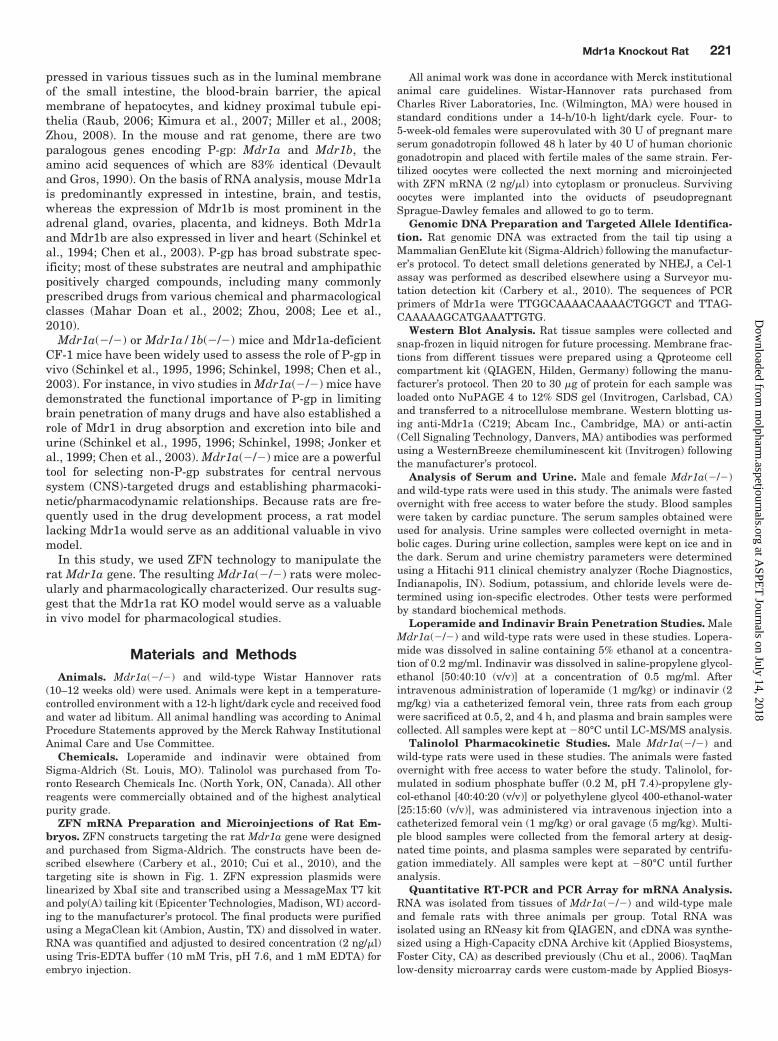
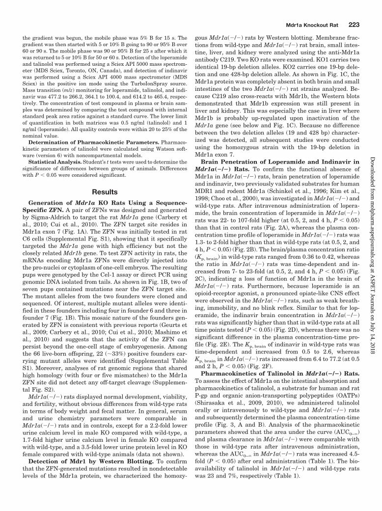
Fig. 1. Microinjection of Mdr1a ZFN mRNA into rat one-cell embryos specifically induces targeted gene mutations. A, schematic representations ofthe rat Mdr1a gene structure and the position of the ZFN targeting site selected. The two ZFN binding sequences are shown in opposite orientationin blue and red, respectively. The ZFN pair consists of two subunits, one with six and one with five fingers, respectively. When the ZFN pair bindsto the target site, the two FokI domains dimerize to generate a DNA double-strand break (DSB), which will be repaired by either NHEJ (major) orHR (minor) pathways. B, screening and analyses of mutant alleles in the rat offspring derived from ZFN-injected embryos. The positive result in theCel-1 assay indicates that six and seven pups may carry mutations near the Mdr1a ZFN target site. The genomic regions surrounding the ZFN sitefrom founder 6 and 7 were cloned and sequenced. Founder 6 contains four mutant alleles, with 175-, 115-, 105-, and 11-bp deletions, respectively.Founder 7 contains one wild-type (WT) allele and three mutant alleles with 8-, 35-, and 19-bp deletions, respectively. The open reading frame of Mdr1anear the ZFN site is shown (1272 amino acids). The deletions that cause frameshift and translational premature termination are highlighted in red.C, Western blots showing undetectable levels of Mdr1a protein in the brain and small intestine (S. Intestine) of KO rats. In the liver sample, the bandwith a size that was apparently similar to that of the Mdr1a protein in KO rats may represent the up-regulated Mdr1b protein because of thecross-reactivity of the antibody. The weak potential Mdr1b protein was detected in the kidney of KO rats as well but not as strongly as in liver.
222 Chu et al.
at ASPE
T Journals on July 14, 2018
molpharm
.aspetjournals.orgD
ownloaded from
the gradient was begun, the mobile phase was 5% B for 15 s. Thegradient was then started with 5 or 10% B going to 90 or 95% B over60 or 90 s. The mobile phase was 90 or 95% B for 25 s after which itwas returned to 5 or 10% B for 50 or 60 s. Detection of the loperamideand talinolol was performed using a Sciex API 5000 mass spectrom-eter (MDS Sciex, Toronto, ON, Canada), and detection of indinavirwas performed using a Sciex API 4000 mass spectrometer (MDSSciex) in the positive ion mode using the TurboIonSpray source.Mass transition (m/z) monitoring for loperamide, talinolol, and indi-navir was 477.2 to 266.2, 364.1 to 100.4, and 614.2 to 465.4, respec-tively. The concentration of test compound in plasma or brain sam-ples was determined by comparing the test compound with internalstandard peak area ratios against a standard curve. The lower limitof quantification in both matrices was 0.5 ng/ml (talinolol) and 1ng/ml (loperamide). All quality controls were within 20 to 25% of thenominal value.
Determination of Pharmacokinetic Parameters. Pharmaco-kinetic parameters of talinolol were calculated using Watson soft-ware (version 6) with noncompartmental models.
Statistical Analysis. Student’s t tests were used to determine thesignificance of differences between groups of animals. Differenceswith P � 0.05 were considered significant.
ResultsGeneration of Mdr1a KO Rats Using a Sequence-
Specific ZFN. A pair of ZFNs was designed and generatedby Sigma-Aldrich to target the rat Mdr1a gene (Carbery etal., 2010; Cui et al., 2010). The ZFN target site resides inMdr1a exon 7 (Fig. 1A). The ZFN was initially tested in ratC6 cells (Supplemental Fig. S1), showing that it specificallytargeted the Mdr1a gene with high efficiency but not theclosely related Mdr1b gene. To test ZFN activity in rats, themRNAs encoding Mdr1a ZFNs were directly injected intothe pro-nuclei or cytoplasm of one-cell embryos. The resultingpups were genotyped by the Cel-1 assay or direct PCR usinggenomic DNA isolated from tails. As shown in Fig. 1B, two ofseven pups contained mutations near the ZFN target site.The mutant alleles from the two founders were cloned andsequenced. Of interest, multiple mutant alleles were identi-fied in these founders including four in founder 6 and three infounder 7 (Fig. 1B). This mosaic nature of the founders gen-erated by ZFN is consistent with previous reports (Geurts etal., 2009; Carbery et al., 2010; Cui et al., 2010; Mashimo etal., 2010) and suggests that the activity of the ZFN canpersist beyond the one-cell stage of embryogenesis. Amongthe 66 live-born offspring, 22 (�33%) positive founders car-rying mutant alleles were identified (Supplemental TableS1). Moreover, analyses of rat genomic regions that sharedhigh homology (with four or five mismatches) to the Mdr1aZFN site did not detect any off-target cleavage (Supplemen-tal Fig. S2).
Mdr1a(�/�) rats displayed normal development, viability,and fertility, without obvious differences from wild-type ratsin terms of body weight and fecal matter. In general, serumand urine chemistry parameters were comparable inMdr1a(�/�) rats and in controls, except for a 2.2-fold lowerurine calcium level in male KO compared with wild-type, a1.7-fold higher urine calcium level in female KO comparedwith wild-type, and a 3.5-fold lower urine protein level in KOfemale compared with wild-type animals (data not shown).
Detection of Mdr1 by Western Blotting. To confirmthat the ZFN-generated mutations resulted in nondetectablelevels of the Mdr1a protein, we characterized the homozy-
gous Mdr1a(�/�) rats by Western blotting. Membrane frac-tions from wild-type and Mdr1a(�/�) rat brain, small intes-tine, liver, and kidney were analyzed using the anti-Mdr1aantibody C219. Two KO rats were examined. KO1 carries twoidentical 19-bp deletion alleles. KO2 carries one 19-bp dele-tion and one 428-bp deletion allele. As shown in Fig. 1C, theMdr1a protein was completely absent in both brain and smallintestines of the two Mdr1a(�/�) rat strains analyzed. Be-cause C219 also cross-reacts with Mdr1b, the Western blotsdemonstrated that Mdr1b expression was still present inliver and kidney. This was especially the case in liver whereMdr1b is probably up-regulated upon inactivation of theMdr1a gene (see below and Fig. 1C). Because no differencebetween the two deletion alleles (19 and 428 bp) character-ized was detected, all subsequent studies were conductedusing the homozygous strain with the 19-bp deletion inMdr1a exon 7.
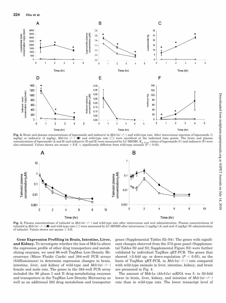
Brain Penetration of Loperamide and Indinavir inMdr1a(�/�) Rats. To confirm the functional absence ofMdr1a in Mdr1a(�/�) rats, brain penetration of loperamideand indinavir, two previously validated substrates for humanMDR1 and rodent Mdr1a (Schinkel et al., 1996; Kim et al.,1998; Choo et al., 2000), was investigated in Mdr1a(�/�) andwild-type rats. After intravenous administration of lopera-mide, the brain concentration of loperamide in Mdr1a(�/�)rats was 22- to 107-fold higher (at 0.5, 2, and 4 h, P � 0.05)than that in control rats (Fig. 2A), whereas the plasma con-centration time profile of loperamide in Mdr1a(�/�) rats was1.3- to 2-fold higher than that in wild-type rats (at 0.5, 2, and4 h, P � 0.05) (Fig. 2B). The brain/plasma concentration ratio(Kp, brain) in wild-type rats ranged from 0.36 to 0.42, whereasthe ratio in Mdr1a(�/�) rats was time-dependent and in-creased from 7- to 23-fold (at 0.5, 2, and 4 h, P � 0.05) (Fig.2C), indicating a loss of function of Mdr1a in the brain ofMdr1a(�/�) rats. Furthermore, because loperamide is anopioid-receptor agonist, a pronounced opiate-like CNS effectwere observed in the Mdr1a(�/�) rats, such as weak breath-ing, immobility, and no blink reflex. Similar to that for lop-eramide, the indinavir brain concentration in Mdr1a(�/�)rats was significantly higher than that in wild-type rats at alltime points tested (P � 0.05) (Fig. 2D), whereas there was nosignificant difference in the plasma concentration-time pro-file (Fig. 2E). The Kp, brain of indinavir in wild-type rats wastime-dependent and increased from 0.5 to 2.6, whereasKp, brain in Mdr1a(�/�) rats increased from 6.4 to 77.2 (at 0.5and 2 h, P � 0.05) (Fig. 2F).
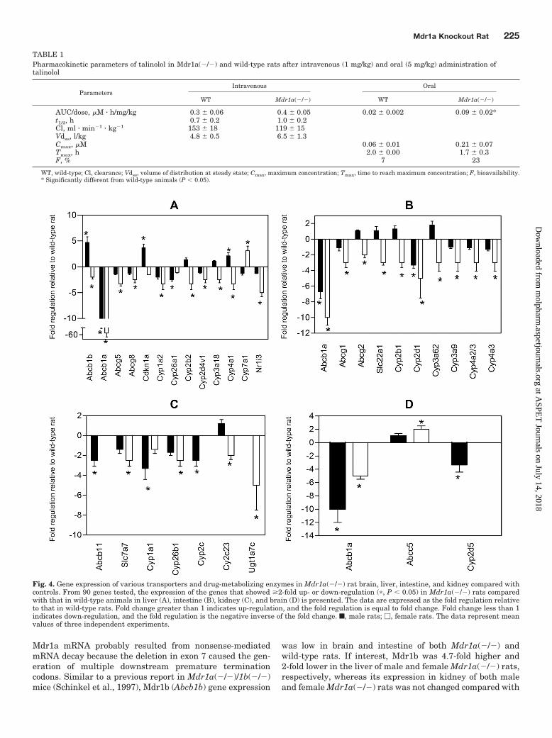
Pharmacokinetics of Talinolol in Mdr1a(�/�) Rats.To assess the effect of Mdr1a on the intestinal absorption andpharmacokinetics of talinolol, a substrate for human and ratP-gp and organic anion-transporting polypeptides (OATPs)(Shirasaka et al., 2009, 2010), we administered talinololorally or intravenously to wild-type and Mdr1a(�/�) ratsand subsequently determined the plasma concentration-timeprofile (Fig. 3, A and B). Analysis of the pharmacokineticparameters showed that the area under the curve (AUC0–�)and plasma clearance in Mdr1a(�/�) were comparable withthose in wild-type rats after intravenous administration,whereas the AUC0–� in Mdr1a(�/�) rats was increased 4.5-fold (P � 0.05) after oral administration (Table 1). The bio-availability of talinolol in Mdr1a(�/�) and wild-type ratswas 23 and 7%, respectively (Table 1).
Mdr1a Knockout Rat 223
at ASPE
T Journals on July 14, 2018
molpharm
.aspetjournals.orgD
ownloaded from
Gene Expression Profiling in Brain, Intestine, Liver,and Kidney. To investigate whether the loss of Mdr1a altersthe expression profile of other drug transporters and metab-olizing enzymes, we used 96-well TaqMan Low-Density Mi-croarrays (Micro Fluidic Cards) and 384-well PCR arrays(SABiosciences) to determine expression changes in brain,intestine, liver, and kidney of wild-type and Mdr1a(�/�)female and male rats. The genes in the 384-well PCR arrayincluded the 90 phase I and II drug-metabolizing enzymesand transporters in the TaqMan Low-Density Microarray aswell as an additional 282 drug metabolism and transporter
genes (Supplemental Tables S2–S4). The genes with signifi-cant changes observed from the 372-gene panel (Supplemen-tal Tables S2 and S3; Supplemental Figure S3) were furthervalidated by individual TaqMan qRT-PCR. The genes thatshowed �2-fold up- or down-regulation (P � 0.05), on thebasis of TaqMan qRT-PCR, in Mdr1a(�/�) rats comparedwith wild-type animals in liver, intestine, kidney, and brainare presented in Fig. 4.
The amount of Mdr1a (Abcb1a) mRNA was 5- to 50-foldlower in brain, liver, kidney, and intestine of Mdr1a(�/�)rats than in wild-type rats. The lower transcript level of
Fig. 2. Brain and plasma concentrations of loperamide and indinavir in Mdr1a(�/�) and wild-type rats. After intravenous injection of loperamide (1mg/kg) or indinavir (2 mg/kg), Mdr1a(�/�) (f) and wild-type rats (�) were sacrificed at the indicated time points. The brain and plasmaconcentrations of loperamide (A and B) and indinavir (D and E) were measured by LC-MS/MS. Kp, brain values of loperamide (C) and indinavir (F) werealso estimated. Values shown are means � S.E. �, significantly different from wild-type animals (P � 0.05).
Fig. 3. Plasma concentrations of talinolol in Mdr1a(�/�) and wild-type rats after intravenous and oral administration. Plasma concentrations oftalinolol in Mdr1a(�/�) (f) and wild-type rats (�) were measured by LC-MS/MS after intravenous (1 mg/kg) (A) and oral (5 mg/kg) (B) administrationof talinolol. Values shown are means � S.E.
224 Chu et al.
at ASPE
T Journals on July 14, 2018
molpharm
.aspetjournals.orgD
ownloaded from
Mdr1a mRNA probably resulted from nonsense-mediatedmRNA decay because the deletion in exon 7 caused the gen-eration of multiple downstream premature terminationcodons. Similar to a previous report in Mdr1a(�/�)/1b(�/�)mice (Schinkel et al., 1997), Mdr1b (Abcb1b) gene expression
was low in brain and intestine of both Mdr1a(�/�) andwild-type rats. If interest, Mdr1b was 4.7-fold higher and2-fold lower in the liver of male and female Mdr1a(�/�) rats,respectively, whereas its expression in kidney of both maleand female Mdr1a(�/�) rats was not changed compared with
Fig. 4. Gene expression of various transporters and drug-metabolizing enzymes in Mdr1a(�/�) rat brain, liver, intestine, and kidney compared withcontrols. From 90 genes tested, the expression of the genes that showed �2-fold up- or down-regulation (�, P � 0.05) in Mdr1a(�/�) rats comparedwith that in wild-type animals in liver (A), intestine (B), kidney (C), and brain (D) is presented. The data are expressed as the fold regulation relativeto that in wild-type rats. Fold change greater than 1 indicates up-regulation, and the fold regulation is equal to fold change. Fold change less than 1indicates down-regulation, and the fold regulation is the negative inverse of the fold change. f, male rats; �, female rats. The data represent meanvalues of three independent experiments.
TABLE 1Pharmacokinetic parameters of talinolol in Mdr1a(�/�) and wild-type rats after intravenous (1 mg/kg) and oral (5 mg/kg) administration oftalinolol
ParametersIntravenous Oral
WT Mdr1a(�/�) WT Mdr1a(�/�)
AUC/dose, �M � h/mg/kg 0.3 � 0.06 0.4 � 0.05 0.02 � 0.002 0.09 � 0.02*t1/2, h 0.7 � 0.2 1.0 � 0.2Cl, ml � min�1 � kg�1 153 � 18 119 � 15Vdss, l/kg 4.8 � 0.5 6.5 � 1.3Cmax, �M 0.06 � 0.01 0.21 � 0.07Tmax, h 2.0 � 0.00 1.7 � 0.3F, % 7 23
WT, wild-type; Cl, clearance; Vdss, volume of distribution at steady state; Cmax, maximum concentration; Tmax, time to reach maximum concentration; F, bioavailability.* Significantly different from wild-type animals (P � 0.05).
Mdr1a Knockout Rat 225
at ASPE
T Journals on July 14, 2018
molpharm
.aspetjournals.orgD
ownloaded from
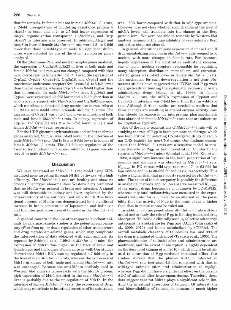
that for controls. In female but not in male Mdr1a(�/�) rats,a 2-fold up-regulation of multidrug resistance protein 5(Abcc5) in brain and a 2- to 2.5-fold lower expression ofAbcg1, organic cation transporter 1 (Slc22a1), and Bcrp(Abcg2) in intestine was observed. In addition, Abcg5 andAbcg8 in liver of female Mdr1a(�/�) rats were 2.5- to 3-foldlower than those in wild-type animals. No significant differ-ences were detected for any of the other transporter genesanalyzed.
Of the cytochrome P450 and nuclear receptor genes analyzed,the expression of Cyp3a1/Cyp3a23 in liver of both male andfemale Mdr1a(�/�) rats was not changed compared with thatin wild-type rats. In female Mdr1a(�/�) liver, the expression ofCyp1a2, Cyp2b2, Cyp2d4v1, Cyp3a18, and Cyp4a1 and theconstitutive androstane receptor (Nr1i3) was 2.5- to 5-fold lowerthan that in controls, whereas Cyp7a1 was 3-fold higher thanthat in controls. In male Mdr1a(�/�) liver, Cyp26a1 andCyp4a1 were expressed 2.5-fold lower and 2-fold higher than inwild-type rats, respectively. The Cyp3a9 and Cyp3a62 enzymes,which contribute to intestinal drug metabolism in rats (Aiba etal., 2005), were 3-fold lower in female Mdr1a(�/�) rats. Theexpression of Cyp2d1 was 3- to 5-fold lower in intestine of bothmale and female Mdr1a(�/�) rats. In kidney, expression ofCyp1a1 and Cyp26b1 was 2.5- to 3-fold lower in male andfemale Mdr1a(�/�) rats, respectively.
For the UDP-glucuronosyltransferase and sulfotransferasegenes analyzed, Sult1a1 was 3-fold lower in the intestine ofmale Mdr1a(�/�) rats. Ugt1a7c was 5-fold lower in kidney offemale Mdr1a(�/�) rats. The 3.7-fold up-regulation of theCdkn1a (cyclin-dependent kinase inhibitor 1) gene was ob-served in male Mdr1a(�/�) rats.
DiscussionWe have generated an Mdr1a(�/�) rat model using ZFN-
mediated gene targeting through NHEJ pathways with highefficiency. The Mdr1a(�/�) rats are healthy and show noobvious phenotypic abnormalities. Western blots confirmedthat no Mdr1a was present in brain and intestine. A signalwas still detectable in kidney and liver, explained by thecross-reactivity of the antibody used with Mdr1b. The func-tional absence of Mdr1a was demonstrated by a significantincrease in brain penetration of loperamide and indinavirand the intestinal absorption of talinolol in the Mdr1a(�/�)rats.
A general concern in the use of transporter knockout ani-mals for pharmacokinetic studies is the potential compensa-tory effect from up- or down-regulation of other transportersand drug metabolism-related genes, which may complicatethe interpretation of data obtained with such animals. Asreported by Schinkel et al. (1994) in Mdr1a(�/�) mice, theexpression of Mdr1b was higher in the liver of male andfemale mice and the kidney of male mice as well. Our studiesshowed that Mdr1b RNA was up-regulated 4.7-fold only inthe liver of male Mdr1a(�/�) rats, whereas the expression ofMdr1b in kidney of both male and female Mdr1a(�/�) ratswas unchanged. Because the anti-Mdr1a antibody used inWestern blot analysis cross-reacts with the Mdr1b protein,high expression of Mdr1 detected in the male Mdr1a(�/�)liver is probably due to the up-regulation of Mdr1b. In theintestine of female Mdr1a(�/�) rats, the expression of Bcrp,which may contribute to intestinal secretion of its substrates,
was �50% lower compared with that in wild-type animals.However, it is not clear whether such changes at the level ofmRNA levels will translate into the change at the Bcrpprotein level. We were not able to test this by Western blotanalysis because of the unavailability of very selective Bcrpantibodies (data not shown).
In general, alterations in gene expression of phase I and IIdrug-metabolizing enzymes in Mdr1a(�/�) rats seemed to bemodest, with more changes in female rats. For instance,hepatic expression of the constitutive androstane receptor,one of major nuclear receptors responsible for regulatingmany absorption, distribution, metabolism, and excretion-related genes was 5-fold lower in female Mdr1a(�/�) rats.The mechanism for such down-regulation is not clear. Nu-merous studies have suggested that CYP3A and P-gp worksynergistically in limiting the systematic exposure of orallyadministered drugs (Benet et al., 1999). In femaleMdr1a(�/�) rats, the mRNA expression of Cyp3a9 andCyp3a62 in intestine was 3-fold lower than that in wild-typerats. Although further studies are needed to confirm thatthese changes are resulting in altered enzyme activity, cau-tion should be exercised in interpreting pharmacokineticdata obtained in female Mdr1a(�/�) rats that are substratesfor Cyp3a9 or Cyp3a62.
One of the major applications of Mdr1a(�/�) rats will bestudying the role of P-gp in brain penetration of drugs, whichhas been critical for selecting CNS-targeted drugs or reduc-ing CNS toxicity for non-CNS drugs. Our findings demon-strate that Mdr1a(�/�) rats are a sensitive model to mea-sure the role of P-gp in brain penetration. Similar to thereports in Mdr1a(�/�) mice (Schinkel et al., 1996; Kim et al.,1998), a significant increase in the brain penetration of lop-eramide and indinavir was observed in Mdr1a(�/�) rats.Kp, brain in KO versus wild-type rats was 17- to 63-fold forloperamide and 9- to 30-fold for indinavir, respectively. Thisvalue is higher than that previously reported for Mdr1a(�/�)mice. The cause of this discrepancy is possibly the differencein analytical methods applied, because we measured Kp, brain
of the parent drugs loperamide or indinavir by LC-MS/MS,whereas only total radioactivity was measured in the exper-iments with Mdr1a(�/�) mice. As an alternative, the possi-bility that the activity of P-gp in the brain of rat is higherthan that in mouse cannot be ruled out.
In addition to brain penetration, Mdr1a(�/�) rats will be auseful tool to study the role of P-gp in limiting intestinal drugabsorption. Talinolol, a clinically used �1 selective adrenergicantagonist, is a substrate for P-gp and OATPs (Shirasaka etal., 2009, 2010) and is not metabolized by CYP3A4. Theoverall metabolic clearance of talinolol is low, and 99% ofthe drug is eliminated unchanged (Trausch et al., 1995). Thepharmacokinetics of talinolol after oral administration arenonlinear, and the extent of absorption is highly dependenton the dose level (Kagan et al., 2010), which might be attrib-uted to saturation of P-gp-mediated intestinal efflux. Ourstudies showed that the plasma AUC of talinolol inMdr1a(�/�) rats increased 4.5-fold compared with that inwild-type animals after oral administration (5 mg/kg),whereas P-gp did not have a significant effect on the plasmaAUC of talinolol after intravenous dosing. Therefore, thesedata suggest that rat Mdr1a plays a significant role in lim-iting the intestinal absorption of talinolol. Of interest, theoral bioavailability of talinolol in humans is much higher
226 Chu et al.
at ASPE
T Journals on July 14, 2018
molpharm
.aspetjournals.orgD
ownloaded from
than that in rats (�55% in humans at a 100-mg dose versus7% in rats at 5 mg/kg), although the dose level in rats in ourstudy was higher than that in humans (Trausch et al., 1995).This finding is probably due to potential species differencesin the functional activity of MDR1/Mdr1a and OATPs be-tween human and rats. Further investigations are needed toconfirm this hypothesis.
ZFN-mediated direct gene KO in rat embryos has recentlybeen proven to be successful in numerous reports (Geurts etal., 2010; Mashimo et al., 2010; Menoret et al., 2010). How-ever, genetic modification of target genes through HR iscritical to enable engineering of the rat genome in a flexibleway. A recent report has demonstrated the feasibility ofachieving HR directly in rat embryo facilitated by ZFN (Cuiet al., 2010), which may expand the application of this tech-nology and allow the generation of conditional KO/knock-inor humanized rat models. The robustness and full utility ofthis advancement need to be explored further.
Acknowledgments
We thank Xiaoli Ping, Loise Gichuru, Nina Jochnowitz, Chris Loe-wrigkeit, Carol Ann Keohane, and Alexandra Wickham for providingsurgical support. Thanks to Irene Capodanno, Christian N. Nunes, andPan Yi for technical assistance. We also thank Merck Research Labo-ratories New Technology Review and Licensing Committee.
Authorship Contributions
Participated in research design: Chu, Zhang, Vogt, Evers, andShin.
Conducted experiments: Chu, Zhang, Yabut, Horwitz, Levorse,Xiang-qing Li, Zhu, Lederman, Ortiga, Strauss, Xiaofang Li, Owens,and Dragovic.
Performed data analysis: Chu, Zhang, Yabut, Evers, and Shin.Wrote or contributed to the writing of the manuscript: Chu, Zhang,
Vogt, Evers, and Shin.
ReferencesAiba T, Yoshinaga M, Ishida K, Takehara Y, and Hashimoto Y (2005) Intestinal
expression and metabolic activity of the CYP3A subfamily in female rats. BiolPharm Bull 28:311–315.
Aitman TJ, Critser JK, Cuppen E, Dominiczak A, Fernandez-Suarez XM, Flint J,Gauguier D, Geurts AM, Gould M, Harris PC, et al. (2008) Progress and prospectsin rat genetics: a community view. Nat Genet 40:516–522.
Benet LZ, Izumi T, Zhang Y, Silverman JA, and Wacher VJ (1999) Intestinal MDRtransport proteins and P-450 enzymes as barriers to oral drug delivery. J ControlRelease 62:25–31.
Capecchi MR (2005) Gene targeting in mice: functional analysis of the mammaliangenome for the twenty-first century. Nat Rev Genet 6:507–512.
Carbery ID, Ji D, Harrington A, Brown V, Weinstein EJ, Liaw L, and Cui X (2010)Targeted genome modification in mice using zinc-finger nucleases. Genetics 186:451–459.
Chen C, Liu X, and Smith BJ (2003) Utility of Mdr1-gene deficient mice in assessingthe impact of P-glycoprotein on pharmacokinetics and pharmacodynamics in drugdiscovery and development. Curr Drug Metab 4:272–291.
Choo EF, Leake B, Wandel C, Imamura H, Wood AJ, Wilkinson GR, and Kim RB(2000) Pharmacological inhibition of P-glycoprotein transport enhances the distri-bution of HIV-1 protease inhibitors into brain and testes. Drug Metab Dispos28:655–660.
Chu XY, Strauss JR, Mariano MA, Li J, Newton DJ, Cai X, Wang RW, Yabut J,Hartley DP, Evans DC, et al. (2006) Characterization of mice lacking the multi-drug resistance protein Mrp2 (Abcc2). J Pharmacol Exp Ther 317:579–589.
Cui X, Ji D, Fisher DA, Wu Y, Briner DM, and Weinstein EJ (2010) Targetedintegration in rat and mouse embryos with zinc-finger nucleases. Nat Biotechnol29:64–67.
Devault A and Gros P (1990) Two members of the mouse mdr gene family confermultidrug resistance with overlapping but distinct drug specificities. Mol Cell Biol10:1652–1663.
Eyal S, Hsiao P, and Unadkat JD (2009) Drug interactions at the blood-brain barrier:fact or fantasy? Pharmacol Ther 123:80–104.
Geurts AM, Cost GJ, Freyvert Y, Zeitler B, Miller JC, Choi VM, Jenkins SS, Wood A,Cui X, Meng X, Vincent A, Lam S, Michalkiewicz M, Schilling R, Foeckler J,
Kalloway S, Weiler H, Menoret S, Anegon I, Davis GD, Zhang L, Rebar EJ,Gregory PD, Urnov FD, Jacob HJ, and Buelow R.. (2009) Knockout rats via embryomicroinjection of zinc-finger nucleases. Science 325(5939):433.
Geurts AM, Cost GJ, Remy S, Cui X, Tesson L, Usal C, Menoret S, Jacob HJ, AnegonI, and Buelow R (2010) Generation of gene-specific mutated rats using zinc-fingernucleases. Methods Mol Biol 597:211–225.
Geurts AM and Moreno C (2010) Zinc-finger nucleases: new strategies to target therat genome. Clin Sci (Lond) 119:303–311.
Hamra FK. (2010) Gene targeting: enter the rat. Nature 467:161–163.Giacomini KM, Huang SM, Tweedie DJ, Benet LZ, Brouwer KL, Chu X, Dahlin A,
Evers R, Fischer V, Hillgren KM, et al. (2010) Membrane transporters in drugdevelopment. Nat Rev Drug Discov 9:215–236.
Jonker JW, Wagenaar E, van Deemter L, Gottschlich R, Bender HM, Dasenbrock J,and Schinkel AH (1999) Role of blood-brain barrier P-glycoprotein in limiting brainaccumulation and sedative side-effects of asimadoline, a peripherally acting anal-gaesic drug. Br J Pharmacol 127:43–50.
Juliano RL and Ling V (1976) A surface glycoprotein modulating drug permeabilityin Chinese hamster ovary cell mutants. Biochim Biophys Acta 455:152–162.
Kagan L, Dreifinger T, Mager DE, and Hoffman A (2010) Role of p-glycoprotein inregion-specific gastrointestinal absorption of talinolol in rats. Drug Metab Dispos38:1560–1566.
Kim RB, Fromm MF, Wandel C, Leake B, Wood AJ, Roden DM, and Wilkinson GR(1998) The drug transporter P-glycoprotein limits oral absorption and brain entryof HIV-1 protease inhibitors. J Clin Invest 101:289–294.
Kimura Y, Morita SY, Matsuo M, and Ueda K (2007) Mechanism of multidrugrecognition by MDR1/ABCB1. Cancer Sci 98:1303–1310.
Klug A (2010) The discovery of zinc fingers and their applications in gene regulationand genome manipulation. Annu Rev Biochem 79:213–231.
Le Provost F, Lillico S, Passet B, Young R, Whitelaw B, and Vilotte JL (2010) Zincfinger nuclease technology heralds a new era in mammalian transgenesis. TrendsBiotechnol 28:134–141.
Lee CA, Cook JA, Reyner EL, and Smith DA (2010) P-glycoprotein related druginteractions: clinical importance and a consideration of disease states. Expert OpinDrug Metab Toxicol 6:603–619.
Livak KJ and Schmittgen TD (2001) Analysis of relative gene expression data usingreal-time quantitative PCR and the 2���CT method. Methods 25:402–408.
Mahar Doan KM, Humphreys JE, Webster LO, Wring SA, Shampine LJ, Serabjit-Singh CJ, Adkison KK, and Polli JW (2002) Passive permeability and P-glycopro-tein-mediated efflux differentiate central nervous system (CNS) and non-CNSmarketed drugs. J Pharmacol Exp Ther 303:1029–1037.
Mashimo T, Takizawa A, Voigt B, Yoshimi K, Hiai H, Kuramoto T, and Serikawa T(2010) Generation of knockout rats with X-linked severe combined immunodefi-ciency (X-SCID) using zinc-finger nucleases. PLoS One 5:e8870.
Menoret S, Iscache AL, Tesson L, Remy S, Usal C, Osborn MJ, Cost GJ, BruggemannM, Buelow R, and Anegon I (2010) Characterization of immunoglobulin heavychain knockout rats. Eur J Immunol 40:2932–2941.
Miller DS, Bauer B, and Hartz AM (2008) Modulation of P-glycoprotein at theblood-brain barrier: opportunities to improve central nervous system pharmaco-therapy. Pharmacol Rev 60:196–209.
Raub TJ (2006) P-glycoprotein recognition of substrates and circumvention throughrational drug design. Mol Pharm 3:3–25.
Schinkel AH (1998) Pharmacological insights from P-glycoprotein knockout mice. IntJ Clin Pharmacol Ther 36:9–13.
Schinkel AH and Jonker JW (2003) Mammalian drug efflux transporters of the ATPbinding cassette (ABC) family: an overview. Adv Drug Deliv Rev 55:3–29.
Schinkel AH, Smit JJ, van Tellingen O, Beijnen JH, Wagenaar E, van Deemter L,Mol CA, van der Valk MA, Robanus-Maandag EC, and te Riele HP (1994) Disrup-tion of the mouse mdr1a P-glycoprotein gene leads to a deficiency in the blood-brain barrier and to increased sensitivity to drugs. Cell 77:491–502.
Schinkel AH, Wagenaar E, Mol CA, and van Deemter L (1996) P-glycoprotein in theblood-brain barrier of mice influences the brain penetration and pharmacologicalactivity of many drugs. J Clin Invest 97:2517–2524.
Schinkel AH, Wagenaar E, van Deemter L, Mol CA, and Borst P (1995) Absence ofthe mdr1a P-glycoprotein in mice affects tissue distribution and pharmacokineticsof dexamethasone, digoxin, and cyclosporin A. J Clin Invest 96:1698–1705.
Shirasaka Y, Kuraoka E, Spahn-Langguth H, Nakanishi T, Langguth P, and TamaiI (2010) Species difference in the effect of grapefruit juice on intestinal absorptionof talinolol between human and rat. J Pharmacol Exp Ther 332:181–189.
Shirasaka Y, Li Y, Shibue Y, Kuraoka E, Spahn-Langguth H, Kato Y, Langguth P,and Tamai I (2009) Concentration-dependent effect of naringin on intestinal ab-sorption of �1-adrenoceptor antagonist talinolol mediated by P-glycoprotein andorganic anion transporting polypeptide (Oatp). Pharm Res 26:560–567.
Tong C, Li P, Wu NL, Yan Y, and Ying QL. (2010) Production of p53 gene knockoutrats by homologous recombination in embryonic stem cells. Nature 467:211–213.
Trausch B, Oertel R, Richter K, and Gramatte T (1995) Disposition and bioavail-ability of the �1-adrenoceptor antagonist talinolol in man. Biopharm Drug Dispos16:403–414.
Urnov FD, Rebar EJ, Holmes MC, Zhang HS, and Gregory PD (2010) Genome editingwith engineered zinc finger nucleases. Nat Rev Genet 11:636–646.
Zhou SF (2008) Structure, function and regulation of P-glycoprotein and its clinicalrelevance in drug disposition. Xenobiotica 38:802–832.
Address correspondence to: Dr. Myung Kyun Shin, Merck Sharp andDohme Corp., RY80Y-310, 126 East Lincoln Ave., Rahway, NJ 07065. E-mail:[email protected]
Mdr1a Knockout Rat 227
at ASPE
T Journals on July 14, 2018
molpharm
.aspetjournals.orgD
ownloaded from


![Arsenic Triglutathione [As(GS)3] Transport by Multidrug ...molpharm.aspetjournals.org/content/molpharm/90/2/127.full.pdf · Received February 2, 2016; accepted June 10, 2016 ABSTRACT](https://img.pdfslide.us/doc/110x75/5fc975e3ece17b339e60754b/arsenic-triglutathione-asgs3-transport-by-multidrug-received-february-2.jpg)