Embed Size (px)
Citation preview

Portland State UniversityPDXScholar
Dissertations and Theses Dissertations and Theses
Spring 6-5-2014
Characterization of Methylene Diphenyl Diisocyanate ProteinConjugatesMorgen MhikePortland State University
Let us know how access to this document benefits you.Follow this and additional works at: https://pdxscholar.library.pdx.edu/open_access_etds
Part of the Allergy and Immunology Commons, and the Chemistry Commons
This Dissertation is brought to you for free and open access. It has been accepted for inclusion in Dissertations and Theses by an authorizedadministrator of PDXScholar. For more information, please contact [email protected].
Recommended CitationMhike, Morgen, "Characterization of Methylene Diphenyl Diisocyanate Protein Conjugates" (2014). Dissertations and Theses. Paper1844.
10.15760/etd.1843

Characterization of Methylene Diphenyl Diisocyanate Protein Conjugates
by
Morgen Mhike
A dissertation submitted in partial fulfillment of the
requirements for the degree of
Doctor of Philosophy
in
Chemistry
Dissertation Committee:
Reuben H. Simoyi, Chair
Paul D. Siegel
Itai Chipinda
Niles Lehman
Shankar B. Rananavare
Robert Strongin
E. Kofi Agorsah
Portland State University
2014

© 2014 Morgen Mhike
i
ABSTRACT
Diisocyanates (dNCO) such as methylene diphenyl diisocyanate (MDI) are used
primarily as cross-linking agents in the production of polyurethane products such as
paints, elastomers, coatings and adhesives, and are the most frequently reported cause of
chemically induced immunologic sensitization and occupational asthma (OA). Immune
mediated hypersensitivity reactions to dNCOs include allergic rhinitis, asthma,
hypersensitivity pneumonitis and allergic contact dermatitis.
There is currently no simple diagnosis for the identification of dNCO asthma due
to the variability of symptoms and uncertainty regarding the underlying mechanisms.
Immunological sensitization due to dNCO exposure is traditionally thought to require
initial conjugation of the dNCO to endogenous proteins to generate neoantigens, which
trigger production of dNCO specific T lymphocytes and ultimately dNCO specific IgE.
Testing for dNCO-specific IgE, for diagnosis of dNCO asthma is however, only specific
(96–98%) but not sensitive (18–27%). The low prevalence of detectable dNCO specific
IgE has been attributed to both assay limitations and a potential IgE-independent dNCO
asthma mechanism(s). The identity of the conjugated proteins responsible for the
sensitization also remains unknown. It is also not clear whether dNCOs bind to
extracellular, cell membrane, or intracellular proteins as a way of triggering non-IgE
asthma. Standardization and optimization of immunoassays used to screen for dNCO
specific antibodies in sera is important if its utility as a dNCO asthma diagnostic tool is to
be achieved. This will potentially improve sensitivity and allow comparison of results
ii
across studies. Current studies on assays of dNCO-specific IgE and IgG lack or have
limited characterization of the conjugates used.
Diisocyanates bound to hemoglobin (Hb), human serum albumin (HSA), and
THP-1 proteins were quantified by HPLC with fluorescence detection. Proteomic tandem
mass spectrometry (MS) was used to delineate TDI and MDI specific amino acid binding
sites on Hb as well as identification of proteins from MDI exposed THP-1 cells. The
trinitrobenzene sulfonic acid assay (TNBS) and SDS gel electrophoresis were used to
evaluate extent of intra and intermolecular cross-linking in dNCO-HSA conjugates.
Binding of monoclonal antibodies (mAbs) to dNCO bound proteins in enzyme-linked
immunosorbent assay (ELISA) was used to evaluate antigenicity of dNCO-protein
conjugates.
The amount of dNCO binding to HSA and Hb increased with the concentration of
the dNCO used for conjugation. All the dNCOs reacted with HSA more than with Hb.
Eight binding sites were observed with both MDI and TDI on Hb. The N-terminal valines
of both the alpha and beta subunits on Hb, lysine 40 of the alpha subunit and lysine 61 of
the beta subunit were common binding sites for both TDI and MDI. Lysine 7 of the alpha
subunit and lysines 8, 65 and 66 of the beta subunit were unique to MDI. On the other
hand, lysines 11, and 16 of the alpha subunit and lysines 17 and 144 of the beta subunit
were unique to TDI. Protein bound MDI was detected in a dose-dependent manner in
membrane and cytoplasm fractions of MDI exposed THP-1 cells. MDI was also detected
in 11 of the 13 cytoplasmic protein bands. The extent of MDI intracellular protein
binding was not affected by cytochalasin D, a chemical that binds actin filaments and
iii
inhibits active uptake into cells. The extent of cross-linking shown using the TNBS assay
was found to increase with amount of dNCO used. Clear bands from both intra and
intermolecular cross-linking were observed on all dNCO-Hb/HSA SDS gels. Using
ELISA, both TDI-Hb and TDI-HSA conjugates were reactive to monoclonal antibodies
produced against TDI conjugated HSA indicating that dNCO-Hb is also antigenic.
The best characterization of dNCO-protein conjugates is achieved by the
quantitative determination of conjugated dNCO per mole of protein as well as
determining the extent of dNCO cross-linking. Although HSA is more reactive to dNCOs
than other serum proteins such as Hb, contribution from other serum proteins to
development of OA should not be overlooked as dNCO-Hb was found to be reactive to
dNCO specific mAbs. dNCO-conjugated proteins identified in the soluble fraction of
MDI exposed THP-1 cells were all of intracellular origin suggesting that MDI can cross
the cell membrane and react with intracellular proteins. The entry of MDI into live cells
is a passive process, as the extent of intracellular binding was not affected by
cytochalasin D. The present study support the potential involvement of dNCO-haptenated
membrane and intracellular proteins in development of non-IgE dNCO asthma.

iv
TO MY MOTHER AND LATE FATHER – I OWE EVERYTHING TO YOU
v
ACKNOWLEDGEMENTS
First, I would like to thank my three advisers, Dr. Reuben H. Simoyi, Dr. Paul D.
Siegel, and Dr. Itai Chipinda for their support throughout my studies. They introduced me
into the fascinating fields of chemical kinetics and diisocyanate induced immunological
sensitization. Words cannot explain how grateful I am for their mentorship and guidance
throughout my research.
I would also want to express my gratitude to all members of my dissertation
committee, Drs. Niles Lehman; Shankar B. Rananavare, Robert Strongin, and E. Kofi
Agorsah for their time and invaluable help in this work. It was a privilege to have them in
my committee. I wish to thank past and present members of the Simoyi Research Group
in particular Dr. Moshood K. Morakinyo, Dr. Risikat A. Adigun, Dr. Wilbes Mbiya, and
Kudzanai Chipiso.
My gratitude also goes to Dr. Don Beezhold and his ACIB team at CDC/NIOSH
Morgantown for hosting me for my PhD dissertation work. I should mention particularly
the following people, Dr. Justin M. Hettick, Toni A. Bledsoe, Brandon F. Law, Angela R.
Lemons, Dr. Ajay P. Nayak, and Dr. Brett J. Green. Their help with various assays,
constructive criticism and discussions, helped me get this work to this stage.
I could not have achieved this degree without the support and encouragement of
my family. Many thanks are due to my parents who always supported me to achieve my
dream and provided all the financial and moral support I needed to succeed.
vi
TABLE OF CONTENTS
ABSTRACT i
DEDICATION iv
ACKNOWLEDGEMENTS v
LIST OF TABLES ix
LIST OF FIGURES x
LIST OF ABBREVIATIONS xi
1. INTRODUCTION 1
1.1. Industrial and commercial uses of isocyanates 1
1.2. Chemistry of diisocyanates 3
1.3. Reaction pathways in the biological system 4
1.4. Health Effects of MDI Exposure 5
1.4.1. Inhalation Exposure 6
1.4.2. Dermal exposure 6
1.4.3. Hypersensitivity 6
1.5. Reaction mechanisms of protein binding 7
1.6. Development of Occupational asthma 9
1.7. Diagnosis of diisocyanate asthma 12
1.8. Methods for characterization of MDI-protein adducts 13
2. INSTRUMENTATION, MATERIALS AND METHODS 17
2.1. Instrumentation 17
2.1.1. Mass Spectrometry 17
2.1.2. Electrospray Ionization 17
2.1.3. High Performance Liquid Chromatography (HPLC) 18
2.1.4. Spectrophotometry 18
2.2. Materials 19
2.2.1. Chemicals 19
2.3. Methods 19
2.3.1. Preparation of dNCO–protein adducts 19
2.3.2. THP-1 MDI Conjugations 21
2.3.3. THP-1 exposure controls 22
2.3.4. Protein estimation 23
2.3.5. The TNBS assay 23
2.3.6. Hydrolysis and extraction 24
2.3.7. Derivatization and HPLC analysis 24
2.3.8. Delineation of MDI and TDI specific binding sites on Hb 25
vii
2.3.9. Identification of MDI bound proteins 27
2.3.10. Sodium dodecyl sulfate (SDS) gel electrophoresis 27
2.3.11. Enzyme-linked immunosorbent (ELISA) assays 28
3. DEVELOPMENT OF AN HPLC METHOD FOR QUANTIFICATION
OF DIISOCYANATES CONJUGATION TO HSA AND Hb 30
3.1 Introduction 30
3.2. The HPLC method 32
3.2.1. Hydrolysis 32
3.2.2. Extraction 33
3.2.3. Derivatization 34
3.3. Results and discussion 35
3.3.1. Optimization of the derivatization reaction 35
3.3.2. Determination of the optimum solvent system for derivatization 35
3.3.3. HPLC analysis of the derivatized MDA 37
3.3.4. Extending the derivatization to TDA and HDA 39
3.3.5. Direct derivatization calibration curves 39
3.3.6. Optimization of the extraction 41
3.3.7. Back extraction 42
3.3.8. Extracted calibration curve 42
3.4. Discussion and conclusion 43
4. CHARACTERIZATION OF METHYLENE DIPHENYL DIISOCYANATE
CONJUGATED HUMAN SERUM ALBUMIN AND HEMOGLOBIN 45
4.1. Introduction 45
4.2. Results and discussion 48
4.2.1. Mapping the binding sites of MDI on Hb 48
4.2.2. Quantification of MDI binding in Hb and HSA 51
4.2.3. Cross-linking in MDI–HSA- TNBS assay 52
4.2.4. Gel electrophoresis: qualitative assessment of crosslinking
In MDI–HSA and MDI–Hb 55
4.2.5. ELISA assessment of percentage of albumin conjugated 58
4.3. CONCLUSION 59
viii
5. COMPARATIVE ANALYSIS OF TOLUENE DIISOCYANATE AND
HEXAMETHYLENE DIISOCYANATE CONJUGATION TO HUMAN SERUM
ALBUMIN AND HEMOGLOBIN 62
5.1. Introduction 62
5.2. Results and discussion 63
5.2.1. Mapping TDI binding sites on Hb 63
5.2.2. Quantification of TDI and HDI binding on Hb and HSA 66
5.2.3. Cross-linking in TDI–HSA: TNBS assay 67
5.2.4. Gel electrophoresis: qualitative assessment of conjugation
and crosslinking In TDI/HDI–HSA and TDI/HDI–Hb 68
5.2.5. ELISA assessment of TDI-HSA /Hb 71
5.3. CONCLUSION 72
6. EVALUATING THE POTENTIAL FOR AN IgE INDEPENDENT
MECHANISM THROUGH WHICH DIISOCYANATE ASTHMA
DEVELOPS 74
6.1. Introduction 74
6.2. Results and discussion 76
6.2.1. Quantification of MDI in the cytoplasm 76
6.2.2. Quantification of MDI in the cell membrane fraction 78
6.2.3. Evaluating the effect of exposure time on the extent
of MDI binding 79
6.2.4. Control experiments 80
6.2.5. Detection of MDI in specific SDS gel bands from MDI
exposed THP-1 Cells 83
6.2.6. Identification proteins from bands with detectable MDI 85
6.3. CONCLUSION 88
REFERENCE LIST 90
ix
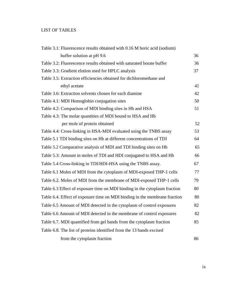
LIST OF TABLES
Table 3.1: Fluorescence results obtained with 0.16 M boric acid (sodium)
buffer solution at pH 9.6 36
Table 3.2: Fluorescence results obtained with saturated borate buffer 36
Table 3.3: Gradient elution used for HPLC analysis 37
Table 3.5: Extraction efficiencies obtained for dichloromethane and
ethyl acetate 41
Table 3.6: Extraction solvents chosen for each diamine 42
Table 4.1: MDI Hemoglobin conjugation sites 50
Table 4.2: Comparison of MDI binding sites in Hb and HSA 51
Table 4.3: The molar quantities of MDI bound to HSA and Hb
per mole of protein obtained 52
Table 4.4: Cross-linking in HSA-MDI evaluated using the TNBS assay 53
Table 5.1 TDI binding sites on Hb at different concentrations of TDI 64
Table 5.2 Comparative analysis of MDI and TDI binding sites on Hb 65
Table 5.3: Amount in moles of TDI and HDI conjugated to HSA and Hb 66
Table 5.4 Cross-linking in TDI/HDI-HSA using the TNBS assay. 67
Table 6.1 Moles of MDI from the cytoplasm of MDI-exposed THP-1 cells 77
Table 6.2. Moles of MDI from the membrane of MDI-exposed THP-1 cells 79
Table 6.3 Effect of exposure time on MDI binding in the cytoplasm fraction 80
Table 6.4. Effect of exposure time on MDI binding in the membrane fraction 80
Table 6.5 Amount of MDI detected in the cytoplasm of control exposures 82
Table 6.6 Amount of MDI detected in the membrane of control exposures 82
Table 6.7. MDI quantified from gel bands from the cytoplasm fraction 85
Table 6.8. The list of proteins identified from the 13 bands excised
from the cytoplasm fraction 86

x
LIST OF FIGURES
Figure 1.1: The general chemical reactions of isocyanates 3
Figure 1.2 Reaction pathways of MDI in biological systems. 5
Figure 1.3: Reaction products of human serum albumin with MDI. 8
Figure 1.4: Possible mechanism(s) leading to the development of hypersensitivity
Reactions and OA upon MDI exposure. 11
Figure 2.1: Chemical structures for the different diisocyanates used in this study 19
Figure 3.1: Structures of the amino acids that react with isocyanates. 331
Figure 3.2: Hydrolysis of MDI-protein conjugates in acid and high temperature 33
Figure 3.3: The reaction of a primary amine with fluorescamine 34
Figure 3.4: MDA chromatograms from HPLC analysis. 38
Figure 3.5: MDA chromatograms from HPLC analysis from 17-20 minutes. 39
Figure 3.6: Calibration curve obtained from the direct derivatization of MDA 40
Figure 3.7: Calibration curve obtained when MDA extracted from 0.5 mg/ml Hb 43
Figure 4.1: Tandem mass spectrum of the hemoglobin subunit β fragment 1-17
Conjugated to MDI on the N-terminal valine residue. 49
Figure 4.2: Total MDI bound per mole of HSA and the approximate number
of MDI That are cross-linked 54
Figure 4.3: An 8% denaturing gel of MDI: HSA 56
Figure 4.4: An 8% native gel of MDI: HSA 57
Figure 4.5: denaturing 4% to 20% gradient gel of MDI-Hb 58
Figure 4.6: Immunoassay of MDI haptenated HSA 59
Figure 5.1: denaturing 8% gel of TDI-HSA. 69
Figure 5.2: denaturing 12% gel of TDI-Hb 70
Figure 5.3: Immunoassay of TDI haptenated HSA and Hb 72
Figure 6.1. A 12 % denaturing gel of MDI exposed THP-1
cells’ cytoplasm fraction 84

xi
LIST OF ABBREVIATIONS
ACD allergic contact dermatitis
AP alkaline phosphatase
APC antigen presenting cell
ATCC American Type Culture Collection
CID collision-induced dissociation
CSN culture supernatant
DDI distilled deionized
DIY Do It Yourself
dNCO diisocyanates
ELISA enzyme linked lmmunosorbent assay
ESI electrospray ionization
Hb Hemoglobin
HDI 1, 6-hexamethylene diisocyanate
HMW High Molecular Weight
HPLC High Performance Liquid Chromatography
HPLC-Q-TOF high performance liquid chromatographic-
quadrupole-time of flight mass spectrometry
HSA human serum albumin
IgE Immunoglobulin E
IgG Immunoglobulin G

xii
IPDI isophorone diisocyanate
KLH keyhole limpet hemocyanin
LOD Limit of Detection
LMW low molecular weight
m/z mass to charge ratio
mAb monoclonal antibody
MDA 4, 4’-Methylene Dianiline
MDI 4,4’ methylene diphenyl diisocyanate
MHC1 major histocompatibility complex 1
MHC11 major histocompatibility complex 11
Mwt molecular weight
NCO isocyanate
NIOSH National Institute for Occupational Safety and
Health
NSBHR Nonspecific bronchial hyper responsiveness
OA occupational asthma
OCF One-component foam
PEFP Peak Expiration Flow Rates
PBS phosphate buffered saline
PBSTM phosphate buffered saline tween milk
PEG polyethylene glycol
RT room temperature
xiii
SBPT Specific bronco-provocation testing
SDS-PAGE sodium dodecyl sulphate-poly acrylamide gel
electrophoresis
SPF Spray Polyurethane Foam
2, 4-TDA 2, 4-toluene diamine
2, 4-TDI 2, 4-toluene diisocyanate
TNBS 2, 4, 6-trinitrobenzenesulphonic acid
TOF time of flight
UV/Vis ultra violet/visible
1
CHAPTER 1
INTRODUCTION
1.1 Industrial and commercial uses of isocyanates
Diisocyanates (dNCO), characterized by the very highly reactive N=C=O group, are
used primarily as cross-linkers in the production of polyurethane products such as
paints, elastomers, coatings, and adhesives1. The most commonly used monomeric
diisocyanates include methylene diphenyl diisocyanate (MDI), 2, 4 and 2,6-toluene
diisocyanate (TDI) , and hexamethylene diisocyanate (HDI)2 . Other less commonly
used diisocyanates include naphthalene diisocyanate (NDI), methylene bis-
cyclohexylisocyanate (HMDI) (hydrogenated MDI), and isophorone diisocyanate
(IPDI). Examples of widely used polyisocyanates include MDI and TDI based
polyisocyanates, HDI biuret, and HDI isocyanurate. MDI is increasingly replacing
TDI in the production of flexible and rigid foams, particle board and wood binders,
paints and coatings, adhesives, sealants, elastomers and spandex fibers 3. It is also used
for consolidating loose rock zones in coal mining or tunneling.
During production of polyurethane products, the NCO group reacts with hydroxyl
groups to form the polyurethane polymers until essentially all available NCO groups
have reacted. This process is called ‘curing’. Some products contain free NCO groups
and are meant to undergo the curing process during use. Adhesives are a good
example of dNCOs that will undergo curing during use. These are applied in an
uncured form and bind to wood as they cure. There are also certain products such as
2
mattresses, pillows, and bowling balls, which contain already cured NCO groups
before they are sold. Cured dNCOs are usually considered less toxic because they do
not contain free NCO groups 4. The curing reaction in most cases leaves sequestered
pockets of unreacted NCO groups, which potentially pose a risk of dNCO exposure.
Diisocyanates are used in settings, such as construction (adhesives, home insulation),
and the automotive industry (paints and truck bed liners), where an end-use curing
process is part of the functional performance of the product. These pose greater risk of
exposure to dNCO. There is an increasing availability of uncured dNCO to both the
professional applicator, as well as the do it yourself (DIY) consumer. The use of
polyurethane foam for insulation is increasing as federal and state government
programs create incentives for their use in increasing energy efficiency. Pour-in-place
(PIP), spray polyurethane foam (SPF) and one-component foam (OCF) cans are used
in the arts industry for sculpture and mold making. Auto refinishing and repair, marine
leisure craft maintenance and repair, and floor and deck maintenance and repair, all
use uncured polyurethane products. Polyurethane coatings are used to seal concrete
and refinish floors. Polyurethane sealants are used for glass installation of windshields
and side windows. Adhesives are used domestically for woodworking and
bookbinding. All these are dNCO-containing products that pose risk of exposure to
workers and end users. US federal agencies such as OSHA and NIOSH acknowledge
the health risks resulting from occupational exposure to dNCOs and have established
occupational exposure limits that are supposed to be protective to workers over an 8 hr
time weighted average working shift. The OSHA mandated permissible exposure
3
limits (PEL) for TDI and MDI are 0.02 ppm as ceiling levels. The NIOSH
recommended exposure limits (REL) for these two dNCOs are currently set as 5 ppb.
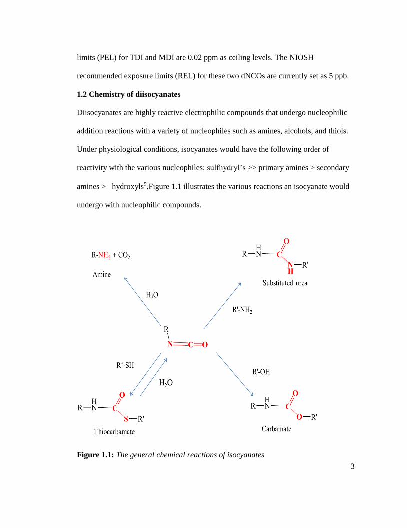
1.2 Chemistry of diisocyanates
Diisocyanates are highly reactive electrophilic compounds that undergo nucleophilic
addition reactions with a variety of nucleophiles such as amines, alcohols, and thiols.
Under physiological conditions, isocyanates would have the following order of
reactivity with the various nucleophiles: sulfhydryl’s >> primary amines > secondary
amines > hydroxyls5.Figure 1.1 illustrates the various reactions an isocyanate would
undergo with nucleophilic compounds.
Figure 1.1: The general chemical reactions of isocyanates
4
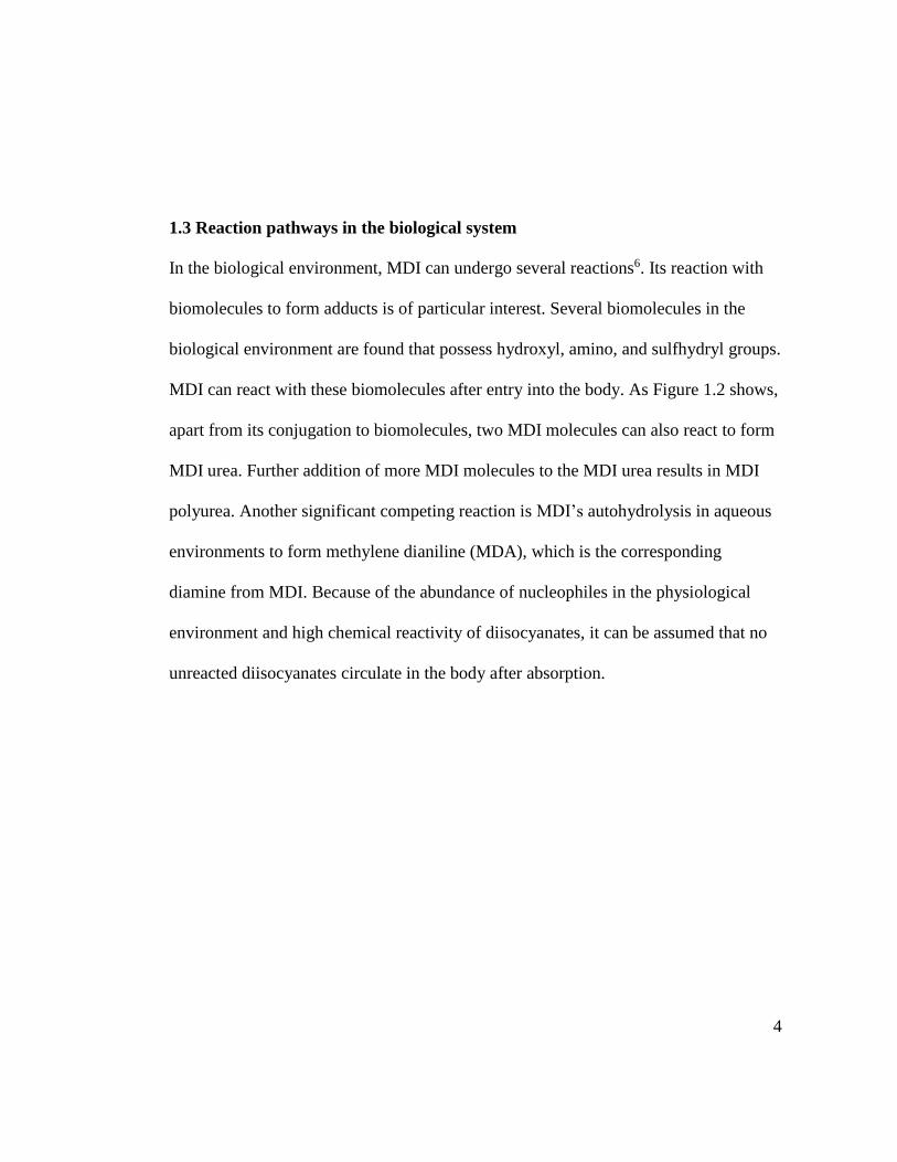
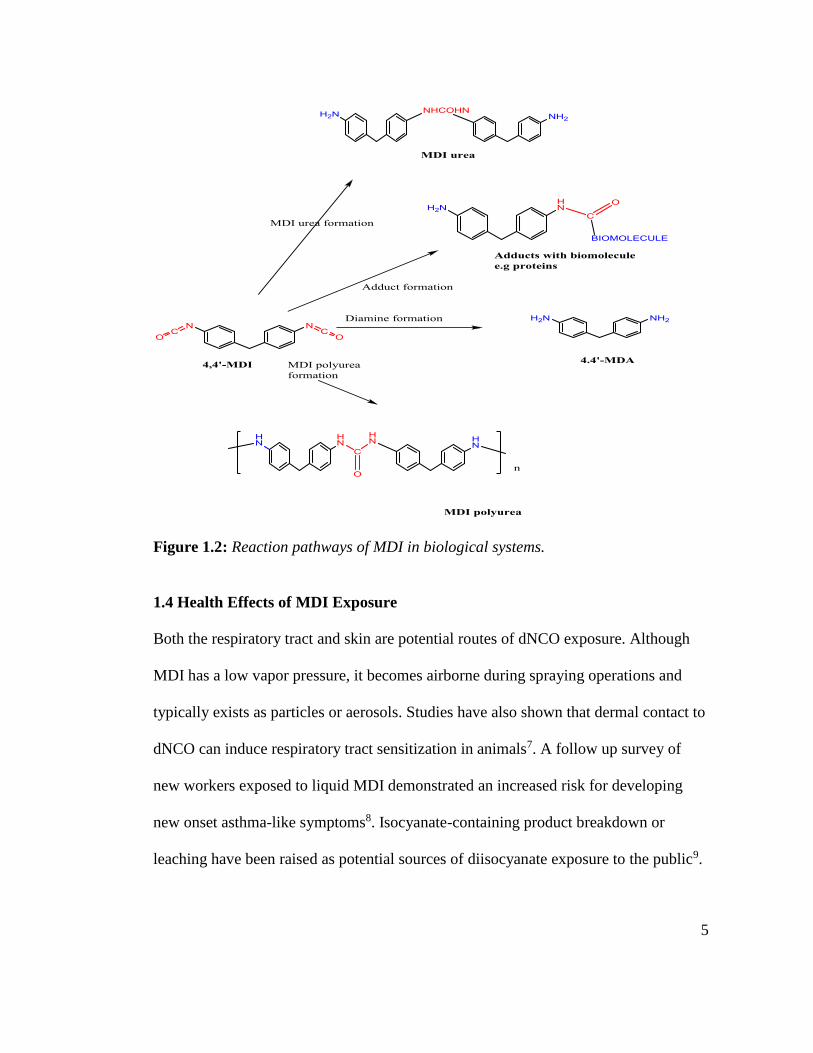
1.3 Reaction pathways in the biological system
In the biological environment, MDI can undergo several reactions6. Its reaction with
biomolecules to form adducts is of particular interest. Several biomolecules in the
biological environment are found that possess hydroxyl, amino, and sulfhydryl groups.
MDI can react with these biomolecules after entry into the body. As Figure 1.2 shows,
apart from its conjugation to biomolecules, two MDI molecules can also react to form
MDI urea. Further addition of more MDI molecules to the MDI urea results in MDI
polyurea. Another significant competing reaction is MDI’s autohydrolysis in aqueous
environments to form methylene dianiline (MDA), which is the corresponding
diamine from MDI. Because of the abundance of nucleophiles in the physiological
environment and high chemical reactivity of diisocyanates, it can be assumed that no
unreacted diisocyanates circulate in the body after absorption.
5
Figure 1.2: Reaction pathways of MDI in biological systems.
1.4 Health Effects of MDI Exposure
Both the respiratory tract and skin are potential routes of dNCO exposure. Although
MDI has a low vapor pressure, it becomes airborne during spraying operations and
typically exists as particles or aerosols. Studies have also shown that dermal contact to
dNCO can induce respiratory tract sensitization in animals7. A follow up survey of
new workers exposed to liquid MDI demonstrated an increased risk for developing
new onset asthma-like symptoms8. Isocyanate-containing product breakdown or
leaching have been raised as potential sources of diisocyanate exposure to the public9.
6
1.4.1 Inhalation Exposure
4, 4’-MDI is a solid at room temperature and has a very low vapor pressure (≤0.01
Pa). MDI containing polyurethane products that require mixing of the MDI with
polyols and then spraying at the site of application pose the greatest risk of inhalation
exposure 10,11.
1.4.2 Dermal exposure
Dermal exposure to MDI in workers has been well-documented 8,12,13. Dermal
sensitization to MDI has been reported in animal studies 14,15. In domestic settings,
dermal exposure to MDI from use of do it yourself (DIY) products is possible. Due to
the presence of MDI in polyurethane based paints, adhesives and coatings, dermal
exposure can occur during both spraying and other application type operations and at
times in domestic settings by coming in contact with a product before it completely
cures.
1.4.3 Hypersensitivity
Diisocyanates are the most frequently reported cause of chemically-induced
occupational asthma (OA) and are capable of producing both immune- and irritant-
mediated asthmas 16,17. The prevalence of diisocyanate asthma among exposed
workers ranges from 5 %-15 %17. Over 200,000 workers are directly employed in the
production and use of isocyanates worldwide in various industries18 and millions more
are potentially exposed through use of dNCO containing products. MDI is one of the
dNCOs known to cause occupational asthma. Common direct toxicities of dNCO
exposure include respiratory19, dermal and mucous membrane irritation. Immune-
7
mediated hypersensitivity reactions to dNCOs include allergic rhinitis20, asthma,
hypersensitivity pneumonitis21,22 and allergic contact dermatitis23. Despite MDI
accounting for approximately 60% of the dNCO global market, most of the published
biomedical dNCO research studies have used toluene diisocyanate (TDI). This is
possibly because of two reasons (i) from about 15 years ago and back, TDI was the
most used diisocyanate and so researchers were more interested in evaluating effects
of exposure to TDI. There was a shift in the market from TDI to MDI use because
MDI, being less volatile than TDI, was conceived to pose a lesser risk of exposure
than TDI, and (ii) MDI’s lower vapor pressure, not only made respiratory exposure
less likely, but is technically more challenging to conduct animal inhalation exposure
studies24,25. MDI is a known respiratory sensitizer based on reported animal and
human studies. In humans, health effects due to respiratory hypersensitivity to MDI
can range from asymptomatic, mild respiratory symptoms, to severe responses such as
hypersensitivity pneumonitis or severe asthma. Death from severe asthma in sensitized
subjects has been reported19.
1.5 Reaction mechanisms of protein binding.
Immunologic MDI sensitization and OA is commonly believed to require initial
conjugation of MDI to endogenous proteins to generate neoantigens26.These
complexes are recognized by antigen presenting cells; leading to the production of
dNCO-specific T-lymphocytes and some dNCO-specific B-lymphocytes that mature
to dNCO-specific IgE secreting plasma cells. However, the functional form of the
8
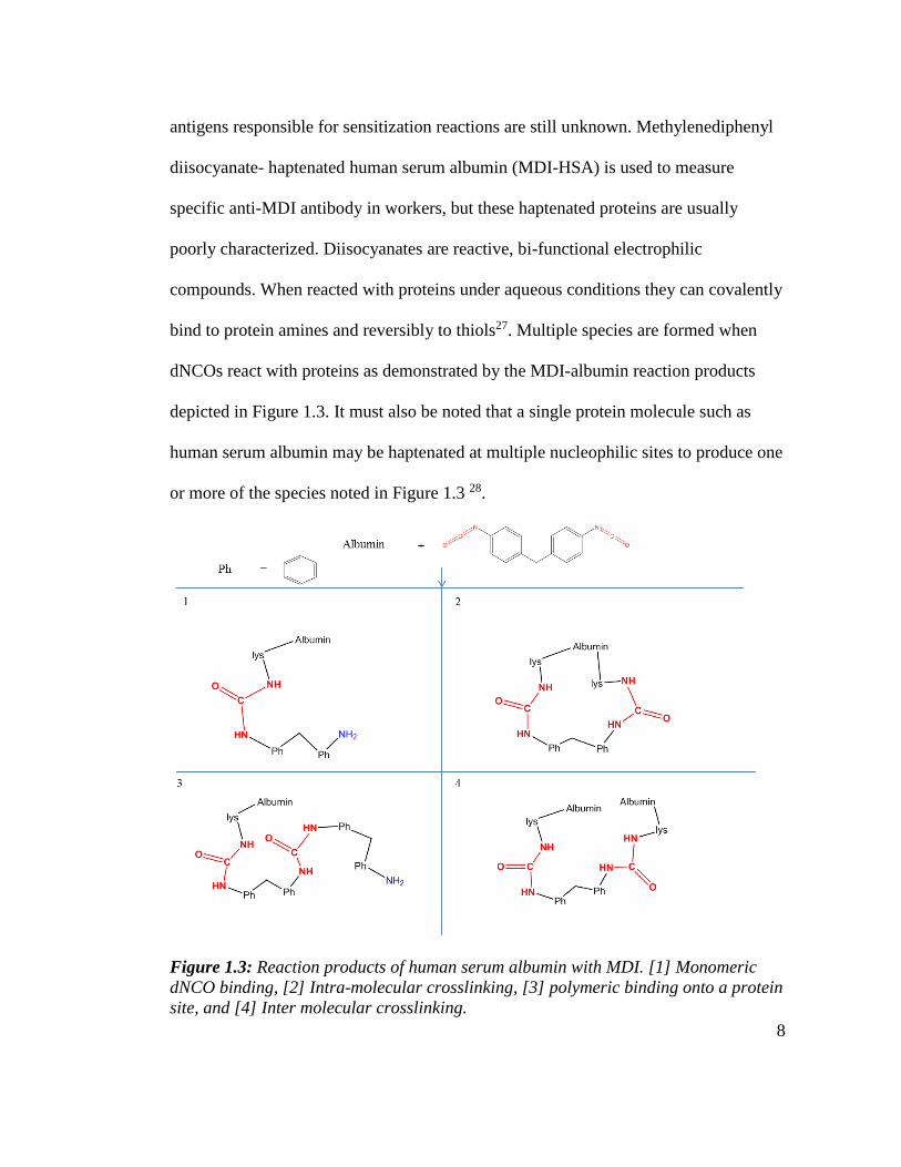
antigens responsible for sensitization reactions are still unknown. Methylenediphenyl
diisocyanate- haptenated human serum albumin (MDI-HSA) is used to measure
specific anti-MDI antibody in workers, but these haptenated proteins are usually
poorly characterized. Diisocyanates are reactive, bi-functional electrophilic
compounds. When reacted with proteins under aqueous conditions they can covalently
bind to protein amines and reversibly to thiols27. Multiple species are formed when
dNCOs react with proteins as demonstrated by the MDI-albumin reaction products
depicted in Figure 1.3. It must also be noted that a single protein molecule such as
human serum albumin may be haptenated at multiple nucleophilic sites to produce one
or more of the species noted in Figure 1.3 28.
Figure 1.3: Reaction products of human serum albumin with MDI. [1] Monomeric
dNCO binding, [2] Intra-molecular crosslinking, [3] polymeric binding onto a protein
site, and [4] Inter molecular crosslinking.
9
1.6 Development of occupational asthma
Occupational asthma (OA) is a complex disease with genetic, behavioral and
environmental factors 29. OA is a type of asthma that is caused by conditions
attributable to a particular work environment. There are essentially two types of OA:
immunologic OA which is characterized by an asymptomatic latency period of
exposure necessary for acquiring immunologic sensitization, and non-immunologic
OA, which has no latency period and occurs upon exposure to high concentrations of
irritants 30. Occupational sensitizers are either high molecular weight (HMW, e.g.
proteins) or low molecular weight (LMW) compounds which induce asthma by
producing specific IgE antibodies or possibly trigger other immune cell mediated
mechanisms. HMW agents act as complete antigens, whereas LMW chemicals, such
as MDI, must first react with autologous proteins to produce a functional allergen 21,31.
Agents that are of insufficient volume to be allergenic and must bind covalently to a
protein to become allergens are referred to as haptens. The fate of diisocyanate in the
body and the ultimate antigenic form(s) are unknown 32. Haptenation to proteins is
believed to be the initial step for these sensitization reactions.
Diisocyanates do not consistently induce specific IgE antibodies 33,34. Therefore
testing for dNCO-specific IgE for diagnosis of dNCO asthma is specific (96-98%), but
insensitive (18-27%). One possible explanation is that sensitization of the respiratory
tract by dNCO can be achieved by more than one immunologic mechanism, including
some that are IgE independent. Another possibility is that assay methods currently
10
being used are not sensitive enough to detect certain low levels of anti-dNCO-IgE.
There are several issues with immunoassays being used, (i) most of them do not report
characterization of the HSA-MDI conjugates used to detect MDI specific IgE, (ii)
most still do not report how the conjugates were prepared, and (iii) some misinterpret
results from the characterization of the HSA-MDI conjugates. It is worth noting also
that the circulating life of specific IgE is 2.5 days but after cessation of exposure, IgE
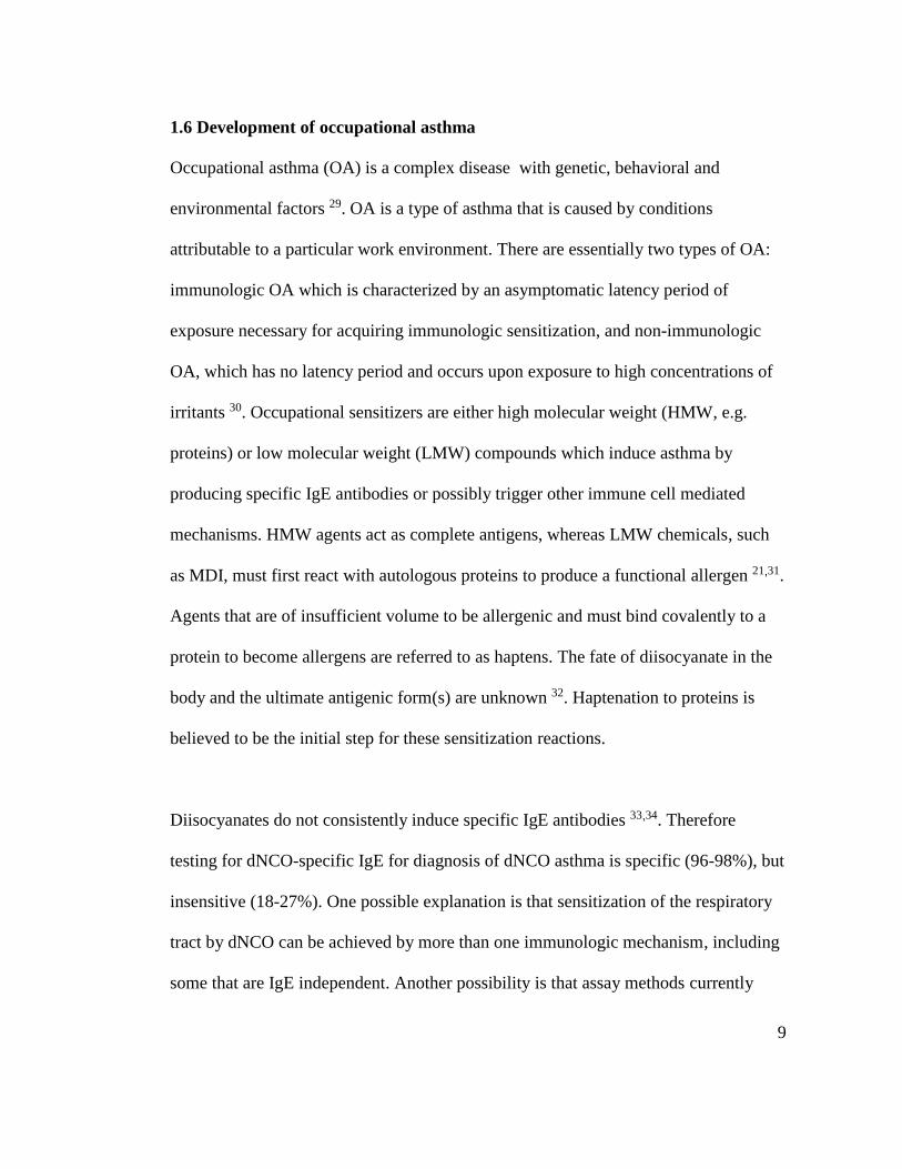
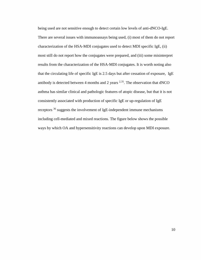
antibody is detected between 4 months and 2 years 2,35. The observation that dNCO
asthma has similar clinical and pathologic features of atopic disease, but that it is not
consistently associated with production of specific IgE or up-regulation of IgE
receptors 36 suggests the involvement of IgE-independent immune mechanisms
including cell-mediated and mixed reactions. The figure below shows the possible
ways by which OA and hypersensitivity reactions can develop upon MDI exposure.
11
Figure 1.4: Possible mechanism(s) leading to the development of hypersensitivity
reactions and OA upon MDI exposure.
OA can also develop through a non-immunologic mechanism (irritant-induced
asthma). This occurs when an individual is exposed to high concentrations of
respiratory irritants resulting in injury to the epithelial system. The main target of
injury, though not yet identified, could be bronchial epithelium which, when denuded,
exposes nerve endings resulting in neurogenic inflammation, loss of relaxing epithelial
factors, release of inflammatory mediators and pro-inflammatory cytokines, secretion
12
of growth factors for epithelial cells, smooth muscle cells, fibroblasts and matrix
degradation 37-39.
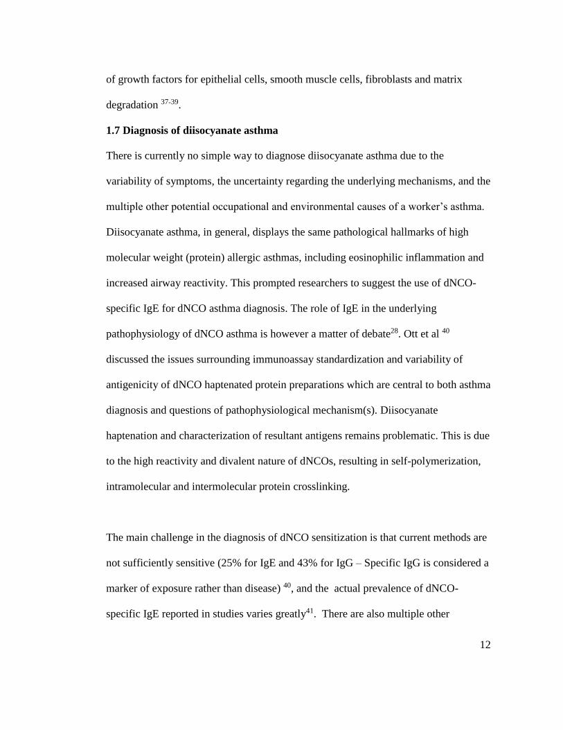
1.7 Diagnosis of diisocyanate asthma
There is currently no simple way to diagnose diisocyanate asthma due to the
variability of symptoms, the uncertainty regarding the underlying mechanisms, and the
multiple other potential occupational and environmental causes of a worker’s asthma.
Diisocyanate asthma, in general, displays the same pathological hallmarks of high
molecular weight (protein) allergic asthmas, including eosinophilic inflammation and
increased airway reactivity. This prompted researchers to suggest the use of dNCO-
specific IgE for dNCO asthma diagnosis. The role of IgE in the underlying
pathophysiology of dNCO asthma is however a matter of debate28. Ott et al 40
discussed the issues surrounding immunoassay standardization and variability of
antigenicity of dNCO haptenated protein preparations which are central to both asthma
diagnosis and questions of pathophysiological mechanism(s). Diisocyanate
haptenation and characterization of resultant antigens remains problematic. This is due
to the high reactivity and divalent nature of dNCOs, resulting in self-polymerization,
intramolecular and intermolecular protein crosslinking.
The main challenge in the diagnosis of dNCO sensitization is that current methods are
not sufficiently sensitive (25% for IgE and 43% for IgG – Specific IgG is considered a
marker of exposure rather than disease) 40, and the actual prevalence of dNCO-
specific IgE reported in studies varies greatly41. There are also multiple other

13
potential occupational and environmental causes of asthma. It can be difficult to
distinguish diisocyanate asthma from irritant aggravation of underlying asthma. The
major tools are clinical history including exposure questionnaire; spirometry and non-
specific inhalation challenge. Methacholine challenge is used to identify hyperactive
reactive airways, a hallmark of asthma. Lack of hyperreactive airways is used to rule
out asthma. False negatives are common if subjects are on corticosteroids. Although
documenting the relationship between symptoms and work can be helpful, it is more
reliable at excluding rather than confirming a diagnosis29. Specific broncho
provocation testing (SBPT) with a diisocyanate in a specialized inhalation challenge
laboratory is regarded as the gold standard for dNCO asthma diagnosis. This is,
however, an expensive and technically difficult technique that is mainly used for
research purposes in the United States and even so, the method is neither 100%
sensitive nor specific33,42,43. Measurements of peak expiratory flow rates (PEFR) at
and away from exposure environment has been used for diagnosis of dNCO asthma
30,31. However, sensitivity ranges from 70 %-100 % and specificity from 50 %-100 %
against SBPT32,34.
1.8 Methods for characterization of MDI-protein adducts
There are several methods that have been used to characterize dNCO-protein
conjugates 35,36 for standardization of dNCO-specific antibody detection
immunoassays and also for biomonitoring assays of dNCO exposure. Some high
performance liquid chromatographic (HPLC) and gas chromatographic (GC) methods
have been developed and used which involve use of electrochemical and mass
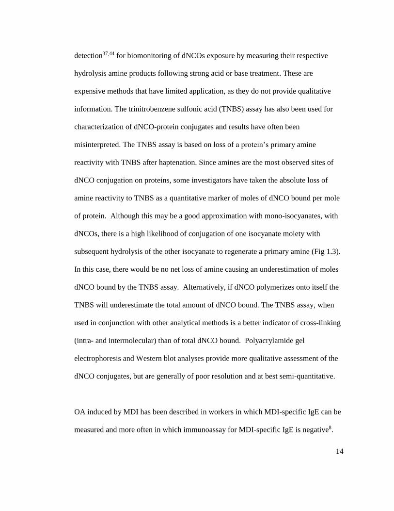
14
detection37,44 for biomonitoring of dNCOs exposure by measuring their respective
hydrolysis amine products following strong acid or base treatment. These are
expensive methods that have limited application, as they do not provide qualitative
information. The trinitrobenzene sulfonic acid (TNBS) assay has also been used for
characterization of dNCO-protein conjugates and results have often been
misinterpreted. The TNBS assay is based on loss of a protein’s primary amine
reactivity with TNBS after haptenation. Since amines are the most observed sites of
dNCO conjugation on proteins, some investigators have taken the absolute loss of
amine reactivity to TNBS as a quantitative marker of moles of dNCO bound per mole
of protein. Although this may be a good approximation with mono-isocyanates, with
dNCOs, there is a high likelihood of conjugation of one isocyanate moiety with
subsequent hydrolysis of the other isocyanate to regenerate a primary amine (Fig 1.3).
In this case, there would be no net loss of amine causing an underestimation of moles
dNCO bound by the TNBS assay. Alternatively, if dNCO polymerizes onto itself the
TNBS will underestimate the total amount of dNCO bound. The TNBS assay, when
used in conjunction with other analytical methods is a better indicator of cross-linking
(intra- and intermolecular) than of total dNCO bound. Polyacrylamide gel
electrophoresis and Western blot analyses provide more qualitative assessment of the
dNCO conjugates, but are generally of poor resolution and at best semi-quantitative.
OA induced by MDI has been described in workers in which MDI-specific IgE can be
measured and more often in which immunoassay for MDI-specific IgE is negative8.

15
Considerable findings from both human and animal studies suggest that an alternative
immune-mediated (possibly Th-1 or CD8+) mechanism(s) of dNCO asthma may be
involved, suggesting that the target haptenated proteins may be intracellular or
membrane bound. 45-48. Recent studies suggest involvement of T-lymphocytes in
diisocyanate asthma rather than through induction and suppression of IgE synthesis as
T-lymphocytes release potent cytokines causing direct inflammation 49,50. CD8+ T cell
numbers increase in the peripheral circulation of diisocyanate asthmatics following
workplace exposure 51. CD8+ T cell clones from two diisocyanate asthmatics
exhibited a mixed Th1/Th2-like phenotype and secreted high levels of both IFN-γ and
IL-5 but not IL-4, contrary to the profile of T cell clones that are preferentially
expanded from atopic individuals, which are primarily CD4+ and produce IL-4 and
IL-5 but not IFN- γ46,52.
As noted above, there are knowledge gaps with respect to the ultimate allergenic
haptenated proteins, and mechanisms of asthmatic sensitization. Better methods of
characterizing MDI-haptenated proteins used for antibody detection and evaluation of
the ability of MDI to bind both cell membrane and intracellular proteins would
provide data that would help address these knowledge gaps.
The specific objectives addressed in this thesis are (i) Development of sensitive and
specific method(s) for quantitative evaluation and characterization of MDI-protein
conjugates, (ii) Evaluation of historical methods for characterization of dNCO-protein
16
conjugates, (iii) Mapping out specific MDI and TDI binding sites on Hb using
proteomic analysis of tryptic peptides from MDI-protein conjugates, (iv)
Demonstration of MDI binding to proteins in cellular fractions of exposed THP-1
cells, (v) Identification of proteins from THP-1 cells.
17
CHAPTER 2
INSTRUMENTATION, MATERIALS AND METHODS
2.1 INSTRUMENTATION
2.1.1 Mass Spectrometry
Electrospray ionization mass spectrometry (ESI- MS) detects small mass shifts in large
biomolecules making it a very useful tool for studying protein haptenation. Tandem
mass spectrometry enables the acquisition and structural elucidation of both native and
modified proteins and peptides. The technique is also very useful for identification of
unknown proteins from complex protein mixtures. ESI-MS was employed in this
project for both identification and characterization of modified proteins due to dNCO
conjugation as well as identifying unknown proteins from dNCO exposed THP-1 cells.
2.1.2 Electrospray Ionization
The mass spectrometer used in these studies was a Micromass QTOF-II (Waters
Corporation, Milford, MA) quadrupole time-of-flight mass spectrometer (qTOF-MS).
Analyte ions were generated by positive-mode electrospray ionization (ESI). The ESI
involves droplet formation, droplet shrinkage and gaseous ion formation 53,54. Tryptic
peptides were separated on a Waters nanoACQUITY UPLC system (Waters
Corporation, Milford, MA, USA). Positive ion nanoelectrospray was performed using
10-µm Pico-Tip (Waters) emitters held at a potential of +3.5 kV. The cone voltage was
held constant at +40 V for all experiments. Dry nitrogen desolvation gas was supplied
to the instrument via a nitrogen generator (NitroFlowLab, Parker Hannifin, Haverhill,
MA, USA). [Glu]1-Fibrinopeptide B (100 fmol/µl in 75:25 A/B) was supplied to an
18
orthogonal reference probe55. Ultra-high purity (UHP) argon was used as collision gas.
The collision energy was set to 6 eV (1-s low energy scan) and a 15- to 30-eV ramp (1-s
high energy scan).
2.1.3 High Performance Liquid Chromatography (HPLC)
For quantification of MDI binding in pure proteins and THP-1 cell fractions, Reverse
Phase High Performance Liquid Chromatography (RP-HPLC) was employed.
Following hydrolysis, extraction and derivatization of the amine hydrolysis product, the
resulting fluorescent complex was injected onto a SUPELCO discovery C18 column at
1ml/min and analyzed on a Shimadzu Prominence HPLC system consisting of an online
vacuum degasser (model DGU-20A5), a quaternary pump (model LC-20AT), an auto
sampler (model SIL-10AD-VP) and a fluorescence detector (model RF-10AXL). The
HPLC system is controlled by EZStart software version 7.3. The resulting MDA-
fluorescamine complex was excited at 410 nm and emission measured at 510 nm.
2.1.4 Spectrophotometry
The extent of cross linking using the TNBS assay was evaluated using
ultraviolet/visible light (UV/Vis) on a Beckman Coulter spectrophotometer (model DU
800, Beckman Coulter, Somerset, NJ, USA). The spectrophotometer was interfaced to
an IBM PC computer and used the DU Series 800 Software (Fullerton, CA). Path length
of the cuvette was 1 cm.
19
2.2 MATERIALS
2.2.1 Chemicals
Unless otherwise specified, all reagents were acquired from Sigma–Aldrich (St. Louis,
MO, USA) and used without further purification. Ethyl acetate (reagent grade) was
purchased from J.T. Baker/ Avantor Performance Materials (Center Valley, PA, USA).
Sodium tetraborate, sodium hydroxide, hydrochloric acid, dialysis membranes,
molecular weight cut-off (MWCO) of 12,000–14,000, 98% sulfuric acid, and N acetyl
glycine were purchased from Fischer Scientific (Fair Lawn, NJ, USA).
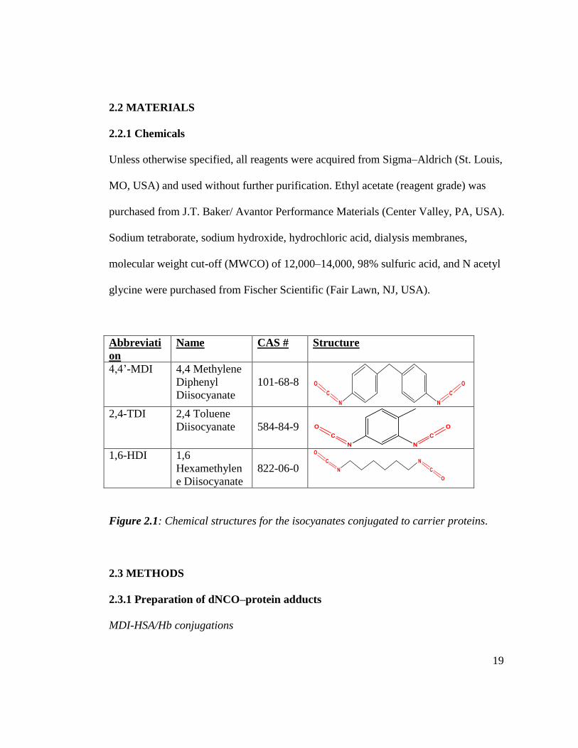
Abbreviati
on
Name CAS # Structure
4,4’-MDI 4,4 Methylene
Diphenyl
Diisocyanate
101-68-8
2,4-TDI 2,4 Toluene
Diisocyanate
584-84-9
1,6-HDI 1,6
Hexamethylen
e Diisocyanate
822-06-0
Figure 2.1: Chemical structures for the isocyanates conjugated to carrier proteins.
2.3 METHODS
2.3.1 Preparation of dNCO–protein adducts
MDI-HSA/Hb conjugations
20
MDI–protein adducts were prepared as described previously55. Briefly, protein solutions
were prepared in 0.1 M PBS (pH 7.4) at 0.5 mg/ml. MDI was dissolved in dry acetone
at 1.8, 9, 18, and 72 µg/ml for HSA conjugation and at 1.84, 9.2, 18.4, and 73.6 µg/ml
for Hb conjugation immediately before use. Each MDI solution was added at 34.5 µl to
5 ml of 0.5 mg/ml protein with mixing, resulting in MDI-protein molar ratios of 1:1,
5:1, 10:1, and 40:1. Samples were then incubated at room temperature (RT) for 1 h with
vortexing. Following incubation, samples were dialyzed for 18 h against 4 L of distilled
deionized water using 12,000 to 14,000 MWCO dialysis tubing (Sigma–Aldrich). The
samples were stored at 20˚C until analysis.
TDI-HSA/Hb conjugations
For preparation of TDI–protein adducts, protein solutions were prepared in 0.1 M PBS
(pH 7.4) at 0.5 mg/ml. 42.3 µl of TDI were dissolved in 1 ml dry acetone and diluted
ten-fold to make stock solution for the 40:1 TDI: protein. Serial dilutions of TDI in
acetone were prepared to make solutions for 10: 1, 5: 1 and 1: 1 TDI: protein. Each TDI
solution (50µl) was added to 5 ml of 0.5 mg/ml protein with mixing, resulting in TDI:
protein molar ratios of 1:1, 5:1, 10:1, and 40:1. Samples were incubated and dialyzed as
described in section 2.3.1 above.
HDI-HSA/Hb conjugations
For preparation of HDI–protein adducts, protein solutions were prepared in 0.1 M PBS
(pH 7.4) at 0.5 mg/ml. A 47.3 µl aliquot of HDI was dissolved in 1 ml dry acetone and
diluted ten times to make stock solution for the 40:1 HDI: protein. Serial dilutions of
HDI in acetone were prepared to make solutions for 10: 1, 5: 1 and 1: 1 HDI: protein.
21
50 µL of the HDI solution was added to 5 ml of 0.5 mg/ml protein with mixing,
resulting in HDI/protein molar ratios of 1:1, 5:1, 10:1, and 40:1. Samples were
incubated and dialyzed as described previously.
2.3.2 THP-1 MDI Conjugations
THP-1 cell culture and MDI exposure
THP-1 monocytic cells (ATCC, Rockville, MD) were cultured in RPMI 1640 (Life
Technologies) containing 4mM L-glutamine, 25mM HEPES, 50µM ß-mercaptoethanol,
10% FBS, and 1% antibiotic/antimycotic solution. After sufficient cell replication, the
cells were harvested by centrifuging at 300 g for 10 min. The RPMI culture media was
discarded and the cell pellet washed two times with Hanks buffer (+Glucose, + calcium,
+ magnesium) and the cells were resuspended in the same buffer. Viability and cell
count were determined before and after exposure to MDI and maintained above 75 %.
Viability was measured using trypan blue. Fifteen million cells suspended in 2 ml of
Hanks buffer were exposed to MDI 5 µM, 10 µM, 25 µM, 50 µM and 100 µM MDI
prepared in 20 µL of ethylene glycol dimethyl ether (EGDE). The MDI was added drop
wise with vortexing and samples were incubated at 37⁰ C and 5% CO2 (model 370
Steri-Cycle CO2 incubator, Forma Scientific Inc., Marietta, OH) for 4 hrs. Cells were
spun down to remove the incubation media (Hanks buffer) and washed two times with
Hanks buffer. Cells were stored as a pellet at -20⁰ C until further analysis.
22
Effect of exposure times on THP-1-MDI conjugations
The effect of exposure time on the extent of conjugation of THP-1 cells was studied by
exposing 15 million cells in a total volume of 2 ml Hanks buffer to 100 µM MDI
prepared in EGDE for different time durations (30 min, 1 hr, 2 hr, and 4 hr). MDI was
added drop wise with vortexing and samples were incubated at 37 ⁰ C and 5 % CO2.
Samples were processed post exposure as described previously.
2.3.3 THP-1 exposure controls
Different control experiments were performed to confirm whether MDI crosses cell
membranes and react with both membrane and intracellular proteins.
THP-1, Cytochalasin D and MDI controls
Cytochalasin D is a chemical that binds actin filaments and inhibits active uptake of
substances into cells. THP-1 cells were incubated with 10 µM cytochalasin D for 1 hr
and exposed to 50 µM MDI for 4 hours. Cells were processed as previously described.
THP-1 and MDA
THP-1 cells were exposed to 100µM MDA prepared in EGDE. This was performed to
assess the effectiveness of the washing process to get rid of any MDA that is not
conjugated to proteins. After incubation, samples were processed as previously
described.
23
THP-1 and EGDE
The effect of the organic solvent used for conjugation on cell viability was determined
by co-incubation of THP-1 cells with 1% EGDE for 4 hr at 37 ⁰C and 5 % CO2. After
incubation, cell count and viability were measured. Cells were processed as described
previously.
2.3.4 Protein estimation
Two approaches were used to extract proteins from MDI-exposed THP-1 cells,
differential centrifugation and use of protein extraction kits. Differential centrifugation
at 300 g separates the membrane fraction (debri) from the cytoplasm fraction
(supernatant). Membrane and cytoplasm protein extraction kits (Fischer scientific) were
also used to selectively extract proteins from the membrane and cytoplasm fractions.
Protein determinations were performed on the conjugates by measuring absorbance at
280 nm on a nano-drop spectrophotometer.
2.3.5 The TNBS assay
The TNBS reagent assesses, primarily, chemical adduction with primary amines of
amino acids 56,57 on the surface of proteins. TNBS produces an absorbance shift at 420
nm upon binding to a primary amine. The trinitrobenzene sulfonic acid (TNBS) assay
was performed on HSA and MDI–HSA conjugates. HSA and HSA conjugates were
diluted in 0.1 M sodium tetraborate (pH 9.3). TNBS (5%, w/v) was diluted 1:5.48 with
0.1 M borate buffer. To 500 µl of samples, 12.5 µl of TNBS was added, mixed well,
24
and left to react for 30 min. Absorbance at 420 nm was measured on a Beckman Coulter
spectrophotometer (model DU 800, Beckman Coulter, Somerset, NJ, USA). Hb has a
strong absorbance at 420 nm that prevents measurement of primary amine content by
TNBS.
2.3.6 Hydrolysis and extraction
dNCO-protein conjugates
Diisocyanate conjugated proteins were hydrolyzed by incubating in 3 M H2SO4 at 100
°C for 16 hr. MDA, TDA or HDA spiked proteins were run in parallel as external
calibration standards to the respective dNCO adducted proteins. Following hydrolysis,
samples and standards were cooled to room temperature and 5 mL of saturated sodium
hydroxide added. The samples were vortexed and incubated in an ice bath for 10
minutes. 6 mL of organic solvent, ethyl acetate (for MDI) or dichloromethane (for TDI
and HDI) were added to the samples, vortexed for 1 min and left standing at room
temperature for 1 hr. A 4 mL portion of the organic solvent was then collected and
evaporated to 1 mL at 40 °C under N2. To the organic solvent, 500 µL of 0.5 % H2SO4
were added and vortexed for 1 min. The sample was vortexed for an additional 10
minutes and left standing for 10 minutes. 400 µL of the 0.5 % H2SO4 were collected for
derivatization.
2.3.7 Derivatization and HPLC analysis
To derivatize the extracted diamines for HPLC analysis, 250 µl of saturated borate
buffer (pH 8.5) and 450 µl of acetonitrile were added to 250 µl of 0.5% H2SO4 extract
25
with MDA, TDA or HDA. This was vortexed for 1 min, and 50 µl of 14.4 mg/mL
fluorescamine in acetonitrile were added. This was vortexed for 1 min before 100 µl of
the resultant fluorescent solution was injected onto a Supelco LC-SI C18 column (25
cm x 4.6 mm, 5 µm, Supelco, Bellefonte, PA, USA). Samples and standards were
analyzed on a Shimadzu Prominence HPLC system (Shimadzu, Columbia, MD, USA).
2.3.8 Delineation of MDI and TDI specific binding sites on Hb
Trypsin digestion of hemoglobin samples
MDI and TDI conjugation sites of Hb were analyzed by ultra-performance liquid
chromatography quadrupole time-of-flight mass spectrometry (UPLC–qTOF MS). In
preparation for mass spec analysis, 200 µl aliquots of MDI–Hb and TDI-Hb samples
were treated with tributylphosphine for 30 min at RT to reduce the disulfide bonds. The
samples were alkylated with iodoacetamide for 1 h at room temperature. Alkylation was
quenched by further addition of tributylphosphine for 15 min at room temperature.
Porcine trypsin in 25 mM NH4HCO3 was then added at a 40:1 (protein/trypsin) ratio.
Samples were incubated overnight at 37⁰ C.
Ultra Performance Liquid Chromatography (UPLC)
Tryptic peptides of Hb, TDI-Hb and MDI–Hb were separated on a Waters
nanoACQUITY UPLC system (Waters, Milford, MA, USA). Aliquots (1 µl) of the
digest mixture were injected and trapped/desalted on a 5-µm Symmetry C18 trapping
column (180 µm x 20 mm) with 99.5/0.5 A/B (A: 0.1% formic acid; B: 0.1% formic
26
acid in acetonitrile) at a flow rate of 15 µl/min for 1 min. Separation was performed on
a 1.7-µm BEH130 C18 analytical column (100 µm x 100 mm) using gradient elution at
a flow rate of 400 nl/min and a gradient of 99:1 to 60:40 A/B over 90 min.
MS/MS of Hb peptides
The eluent from the UPLC system was directed to the nanoelectrospray source of a
Waters SYNAPT MS qTOF mass spectrometer. Positive ion nanoelectrospray was
performed using 10 µm Pico-Tip (Waters) emitters held at a potential of +3.5 kV. The
cone voltage was held constant at +40 V for all experiments. Dry nitrogen desolvation
gas was supplied to the instrument via a nitrogen generator (NitroFlowLab, Parker
Hannifin, Haverhill, MA, USA). [Glu]1 Fibrinopeptide B (100 fmol/µl in 75:25 A/B)
was supplied to an orthogonal reference probe, and the [M+2H]2+ ion (m/z 785.84265u)
was measured as an external calibrant at 30-s intervals. Ultra-high purity (UHP) argon
was used as collision gas. Spectra were acquired in an ‘‘MSe’’ fashion58. Alternating 1
sec mass spectra were acquired. The collision energy was set to 6 eV (1 sec low energy
scan) and a 15- to 30-eV ramp (1 sec high energy scan).
Data analysis for MDI binding sites on Hb
Data were analyzed with BioPharmaLynx version 1.2 (Waters), a software program for
analysis of peptide mass maps and identification of sites of modification on known
protein sequences. Default peptide mass map analysis criteria of 30 ppm mass error in
both low- and high-collision energy mode were specified. Trypsin was specified as the
27
digestion enzyme, and two missed cleavages were allowed. Identification of an
isocyanate binding site proceeded via a rigorous procedure that involved the following
steps: (i) observing a potential peptide–dNCO conjugation product with less than 30
ppm m/Dm mass error in the analyte peptide mass map, (ii) comparing analyte and
control peptide mass map from unmodified Hb showing that observed m/z and
chromatographic retention time are unique to analyte, and (iii) observing MS/MS data
containing bn- and yn-type ions consistent with the assigned sequence and modifier.
2.3.9 Identification of MDI bound proteins
SDS gel electrophoresis of the cytoplasm and membrane fractions were run and protein
bands with detectable MDA were excised. Trypsin digestion was performed on the
resultant proteins and the peptides analyzed by mass spectrometry to identify the
proteins in the gel bands as described previously.
2.3.10 Sodium dodecyl sulfate (SDS) polyacrylamide gel electrophoresis (PAGE)
dNCO-protein gels
For denaturing gels, proteins and dNCO–protein conjugates were mixed with 950 µl of
Laemmli sample buffer and 50 µl of 2- mercaptoethanol. Sodium dodecyl sulfate (SDS)
acrylamide gels (8 % and 12 %) were cast. Precast gradient gels (4–20%) were obtained
from Bio-Rad (Hercules, CA, USA). Samples were run on 8 %, 12 %, and 4 %-20%
gradient polyacrylamide gels. Following electrophoretic separation of proteins, the gels
were stained with imperial protein stain (Pierce, Rockford, IL, USA) and destained with

28
water. Unmodified protein and Bio-Rad prestained molecular weight markers were used
for relative molecular weight determination. For native gels, unmodified protein and
dNCO–protein samples were mixed with native sample buffer from Bio-Rad and run on
8 % native gels in parallel with unstained protein markers from Life Technologies
(Carlsbad, CA, USA). Gels were stained and destained as described previously59.
2.3.11 Enzyme-linked immunosorbent (ELISA) assays
Immunoassay for percentage of number of HSA conjugated.
Binding of a murine IgM 15D4 mAb to MDI-conjugated HSA was analyzed using a
sandwich ELISA60,61. Here, 96-well plates (Corning, Corning, NY, USA) were coated
with 4 µg/ml Affini-Pure goat anti-mouse IgM and µ chain-specific IgG (Jackson
ImmunoResearch Laboratories, West Grove, PA, USA) overnight at 4⁰ C. After
washing three times with PBST (PBS and Tween 20), wells were incubated on a shaker
for 1 h with 2 µg/ml 15D4 mAb at room temperature. The plates were then blocked
with 3% skim milk/PBS–Tween 20 (SMPBST) for 1 h at 37⁰ C. Duplicate MDI–HSA
conjugates were then added to the blocked plates at a concentration of 25 µg/ml and
incubated for 1 h at 37⁰ C. Plates were then washed three times with PBST and
incubated for 1 h at 37⁰ C with biotin-conjugated affinity-purified rabbit anti-HSA
(Rockland, Gilbertsville, PA, USA) diluted 1:5000 (v/v) in SMPBST. The plates were
then washed three times with PBST and incubated for 1 h at 37⁰ C with alkaline
phosphatase-conjugated streptavidin (Jackson Immuno- Research Laboratories) diluted
1:5000 (v/v) in SMPBST. Following incubation, the plates were washed with PBST and

29
binding of the 15D4 mAb to the conjugates was visualized using 0.5 mg/mL p-
nitrophenyl phosphate (Sigma–Aldrich) in alkaline phosphatase substrate. The optical
density was determined at 405 nm after 30 min.
ELISA for TDI-HSA and TDI-Hb
Binding of an IgG, 60G2 monoclonal antibody (mAb) to TDI-conjugated HSA and Hb
was analyzed using an indirect enzyme-linked immunosorbent assay (ELISA)61-63. 96-
well plates (Corning, Corning, NY, USA) were coated with TDI-protein conjugates
overnight at 4⁰ C. After washing three times with PBST (PBS and Tween 20), wells
were incubated on a shaker for 1 h with two µg/ml 60G2 mAb at RT. The plates were
then blocked with 3% skim milk/PBS–Tween 20 (SMPBST) for 1 h at 37⁰ C. Plates
were then washed three times with PBST and incubated for 1 h at 37⁰ C with alkaline
phosphatase conjugated affini-Pure goat, anti-mouse IgG(H+L) (Promega, Cat # S3721
USA) diluted 1:5000 (v/v) in SMPBST. Following incubation, the plates were washed 3
times with PBST and binding of the 60G2 mAb to the conjugates was visualized using
0.5 mg/ml p-nitrophenyl phosphate (Sigma–Aldrich) in alkaline phosphatase substrate.
The optical density was determined at 405 nm after 30 min.
30
CHAPTER 3
DEVELOPMENT OF AN HPLC METHOD FOR QUANTIFICATION
OF DIISOCYANATES CONJUGATION TO HSA AND Hb.
3.1 INTRODUCTION
Protein haptenation by dNCOs such as MDI, TDI and HDI is thought to be an
important step in the development of diisocyanate-specific allergic sensitization. The
fate of the dNCO in the body and the ultimate protein adduct responsible for
immunological sensitization currently remain unknown32. Apart from directly reacting
with proteins at the site of exposure, diisocyanates can react with glutathione or
protein thiols, forming labile thiocarbamate adducts that may possibly be transported
to sites distal to the site of exposure. It is not known whether the antigenicity of
proteins adducted by isocyanates regenerated from thiocarbamates is different from
that via direct haptenation. Diisocyanate-conjugated proteins are used to screen for
dNCO-specific antibodies in workers’ sera and the assay is commonly used as a tool
in dNCO asthma diagnosis, however, the assay has been reported to have poor
sensitivity in confirmed diisocyanate-asthma cases64. Diisocyanate-haptenated
albumins used for the screening of dNCO-specific antibodies in workers’ sera are
often poorly characterized. Recently, the need to develop standardized immunoassays
using a consistent, well-characterized dNCO-haptenated protein to screen for the
presence of diisocyanate-specific IgE and IgG from workers’ sera has been recognized
and emphasized65,66. Synthetic methods have been reported for amino acid conjugation

31
using thiocarbamates67, which may possibly find utility in haptenation of whole
proteins for specific antibody detection.
Isocyanates can react with the following amino acids under physiological conditions:
the α-amino group of all N-terminal amino acids, the sulfhydryl group of cysteine, the
hydroxyl groups of tyrosine and especially the ε-amino group of lysine and the
imidazole ring of histidine68,69. Figure 3.1 shows the structures of the four amino acids
(excluding those where the N-terminal has been reported bound) that have been
observed to react with diisocyanates. Isocyanate-specific adducts with the N-terminal
valine of hemoglobin with MDI have been quantified in rats70. Sepai et. al. found
hemoglobin adducts correlated with the administered dose of MDI71 while Sabbioni et.
al. demonstrated that MDA also binds to hemoglobin (through a metabolic
intermediate) 72.
Figure 3.1: Structures of the amino acids that have been found to react with
isocyanates.
32
There are several HPLC and GC mass spectrometry methods that have been developed
and used to characterize dNCO-protein conjugates for biomonitoring of dNCO
exposure 73,74. The TNBS assay has also been used for characterization of dNCO-
protein conjugates and results have often been over-interpreted75. Gel electrophoresis
and Western blot analyses provide more qualitative assessment of the dNCO
conjugates, but are generally of poor resolution and at best can be semi-quantitative
with respect to degree of haptenation.
In light of these limitations, it was important to develop a sensitive and simple method
of determining dNCO binding to proteins. The method would be useful for
characterization of dNCO conjugation to proteins.
3.2. The HPLC method.
The HPLC system used in this study was a reversed phase with the C-18 R-group of
the siloxane coating.
3.2.1 Hydrolysis
The HPLC method is based on the hydrolysis of dNCO-protein conjugates under high
acid concentrations at elevated temperature. This generates a complex mixture of the
dNCO hydrolysis products [methylene dianiline (MDA), toluene diamine (TDA) and
hexamethylene diamine (HDA)], amino acids and several hydrolysis products from
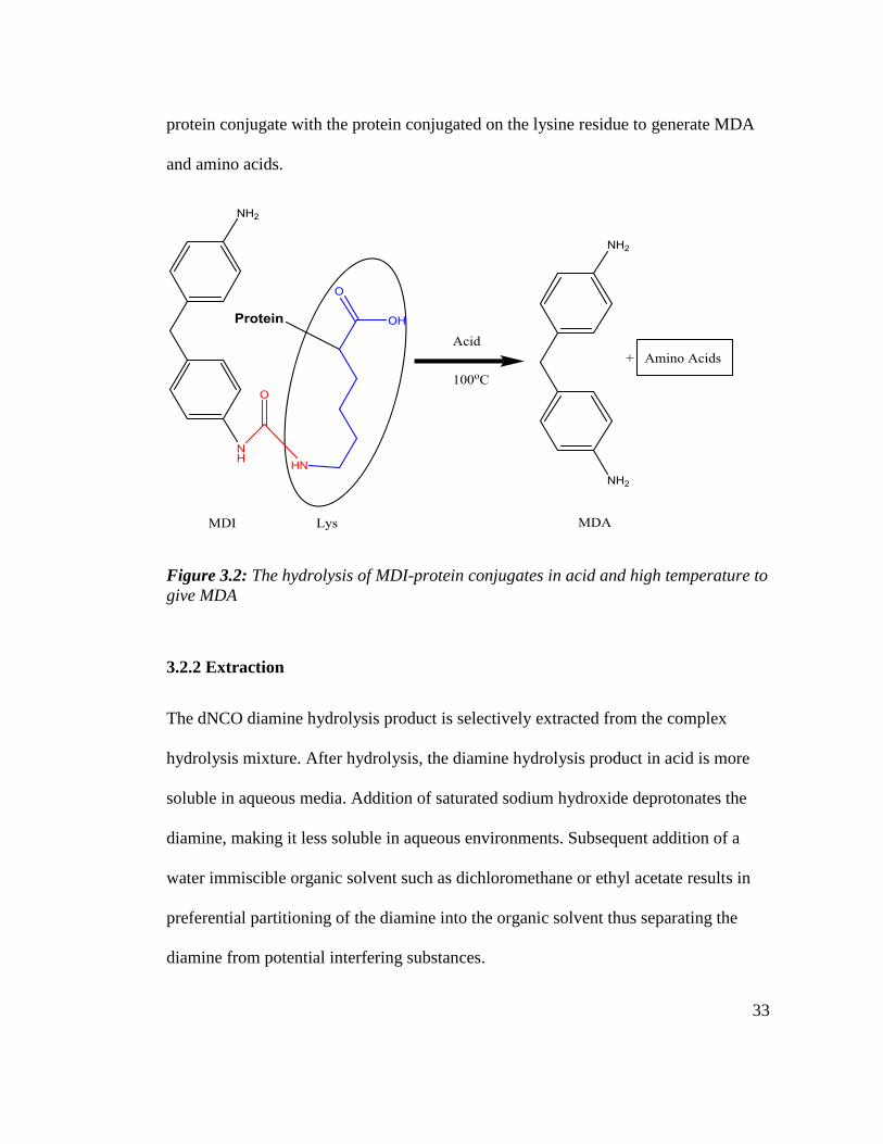
protein(s). Figure 3.2 below is a simplified scheme of the hydrolysis of an MDI-
33
protein conjugate with the protein conjugated on the lysine residue to generate MDA
and amino acids.
Figure 3.2: The hydrolysis of MDI-protein conjugates in acid and high temperature to
give MDA
3.2.2 Extraction
The dNCO diamine hydrolysis product is selectively extracted from the complex
hydrolysis mixture. After hydrolysis, the diamine hydrolysis product in acid is more
soluble in aqueous media. Addition of saturated sodium hydroxide deprotonates the
diamine, making it less soluble in aqueous environments. Subsequent addition of a
water immiscible organic solvent such as dichloromethane or ethyl acetate results in
preferential partitioning of the diamine into the organic solvent thus separating the
diamine from potential interfering substances.
34
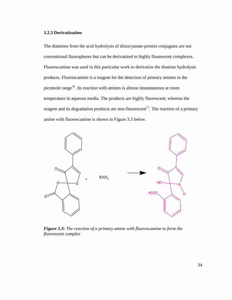
3.2.3 Derivatization
The diamines from the acid hydrolysis of diisocyanate-protein conjugates are not
conventional fluorophores but can be derivatized to highly fluorescent complexes.
Fluorescamine was used in this particular work to derivatize the diamine hydrolysis
products. Fluorescamine is a reagent for the detection of primary amines in the
picomole range76. Its reaction with amines is almost instantaneous at room
temperature in aqueous media. The products are highly fluorescent, whereas the
reagent and its degradation products are non-fluorescent77. The reaction of a primary
amine with fluorescamine is shown in Figure 3.3 below.
Figure 3.3: The reaction of a primary amine with fluorescamine to form the
fluorescent complex
35
3.3. RESULTS AND DISCUSSION.
3.3.1. Optimization of the derivatization reaction
The diamine-fluorescamine derivatization reaction was studied under different pH
conditions, in different buffer systems, and in different proportions of acetonitrile to
the buffer system. Fluorescence intensity was measured using a conventional
flourometer. Toker SE et al78 reported an HPLC method with fluorescence detection in
which they performed the fluorescamine derivatization of memantine extracted from
human plasma. The memantine residue was derivatized by reacting with 320 molar
excess fluorescamine in 500 mL of borate buffer (pH 8.5), 250 mL of fluorescamine
reagent solution and 250 mL of acetonitrile. Although memantine has a single primary
amine group, the conditions they employed were the basis for this optimization
process.
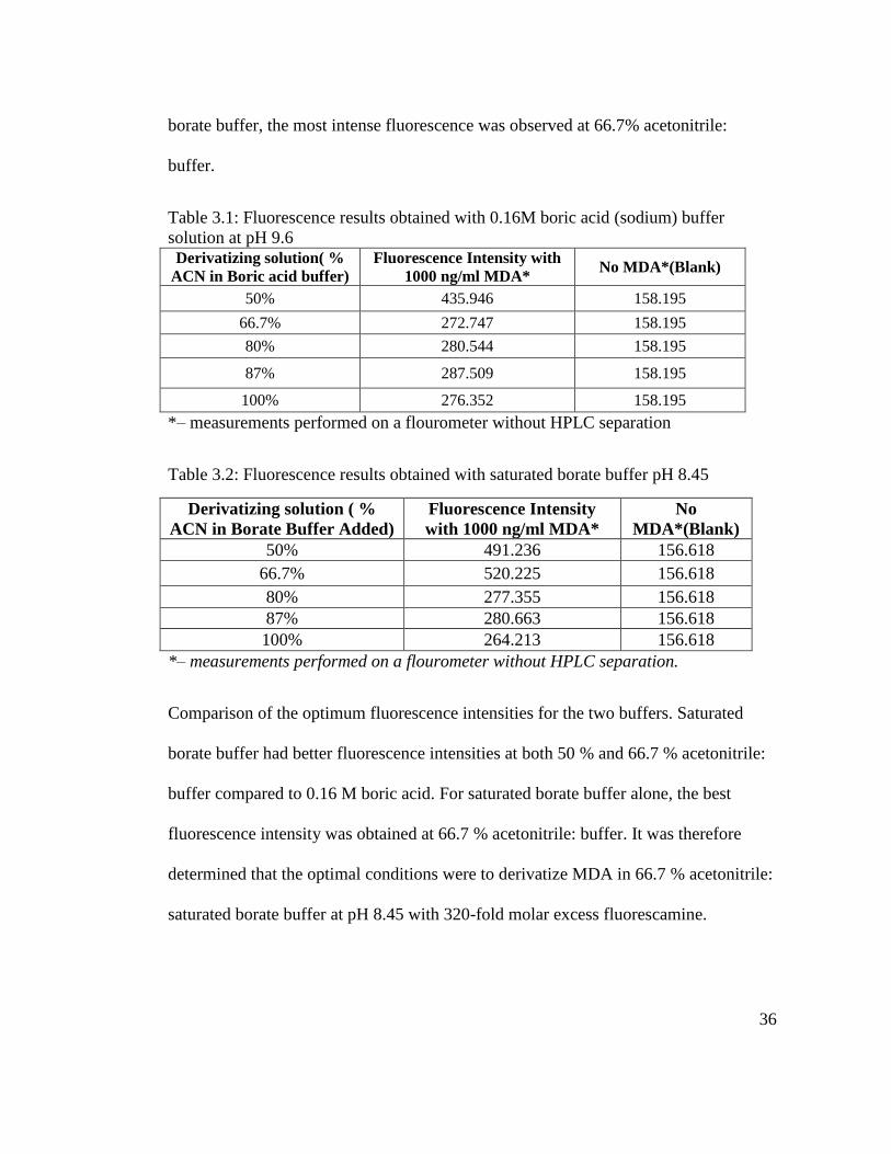
3.3.2. Determination of the optimum solvent system for derivatization
The solvent system that produced the best fluorescence was evaluated by derivatizing
MDA in different buffers with different proportions of buffer: acetonitrile and at
different pHs. Table 3.1 below shows the fluorescence intensities obtained when 1000
ng/ml MDA was derivatized in 0.16 M boric acid at pH 9.6 at different ratios of
acetonitrile to buffer. The highest fluorescence intensity was obtained when
derivatization was performed in a 50% acetonitrile-buffer mixture. Table 3.2 shows
fluorescence intensities obtained when the same experiment was performed in
saturated borate buffer at pH 8.45 at different ratios of acetonitrile: buffer. In saturated
36
borate buffer, the most intense fluorescence was observed at 66.7% acetonitrile:
buffer.
Table 3.1: Fluorescence results obtained with 0.16M boric acid (sodium) buffer
solution at pH 9.6
Derivatizing solution( %
ACN in Boric acid buffer)
Fluorescence Intensity with
1000 ng/ml MDA* No MDA*(Blank)
50% 435.946 158.195
66.7% 272.747 158.195
80% 280.544 158.195
87% 287.509 158.195
100% 276.352 158.195
*– measurements performed on a flourometer without HPLC separation
Table 3.2: Fluorescence results obtained with saturated borate buffer pH 8.45
Derivatizing solution ( %
ACN in Borate Buffer Added)
Fluorescence Intensity
with 1000 ng/ml MDA*
No
MDA*(Blank)
50% 491.236 156.618
66.7% 520.225 156.618
80% 277.355 156.618
87% 280.663 156.618
100% 264.213 156.618
*– measurements performed on a flourometer without HPLC separation.
Comparison of the optimum fluorescence intensities for the two buffers. Saturated
borate buffer had better fluorescence intensities at both 50 % and 66.7 % acetonitrile:
buffer compared to 0.16 M boric acid. For saturated borate buffer alone, the best
fluorescence intensity was obtained at 66.7 % acetonitrile: buffer. It was therefore
determined that the optimal conditions were to derivatize MDA in 66.7 % acetonitrile:
saturated borate buffer at pH 8.45 with 320-fold molar excess fluorescamine.
37
3.3.3. HPLC analysis of the derivatized MDA
After optimizing the derivatization reaction, MDA was dissolved in 0.5% H2SO4 and
derivatized as described previously. The resultant fluorescent complex was run on the
HPLC measured in the fluorometer at an excitation λ=410 nm and emission λ=510 nm.
The following parameters were used on the HPLC to run the MDA standards.
Parameters
1. Table 3.3: Gradient elution
Time Module Action ACN Value (%)
0.01 Controller Start
0.02 Pumps Pump B conc 10.0
13.00 Pumps Pump B conc 50.0
18.00 Pumps Pump B conc 50.0
19.00 Pumps Pump B conc 10.0
20.00 Controller Stop
NB: Pump B is acetonitrile and pump A is water.
2. Excitation wavelength: 410 nm, Emission wavelength: 510 nm
3. Flow rate 1 mL/min
4. Column C18, 25 cm x 4.4 mm, 5 µm particle size with guard column.
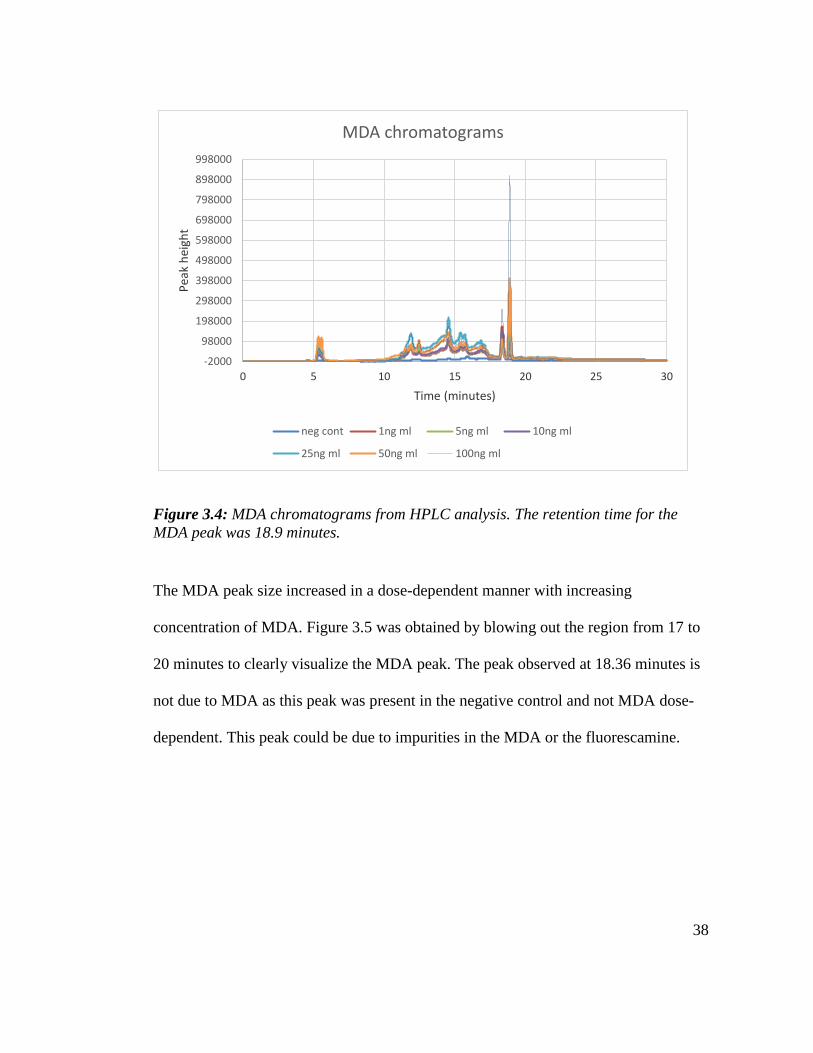
Figure 3.4 is an overlay of derivatized MDA chromatograms from 1-100 ng/ml. A
negative solvent control that had no MDA was also derivatized to evaluate the extent
of background fluorescence.
38
-2000
98000
198000
298000
398000
498000
598000
698000
798000
898000
998000
0 5 10 15 20 25 30
Pea
k h
eigh
t
Time (minutes)
MDA chromatograms
neg cont 1ng ml 5ng ml 10ng ml
25ng ml 50ng ml 100ng ml
Figure 3.4: MDA chromatograms from HPLC analysis. The retention time for the
MDA peak was 18.9 minutes.
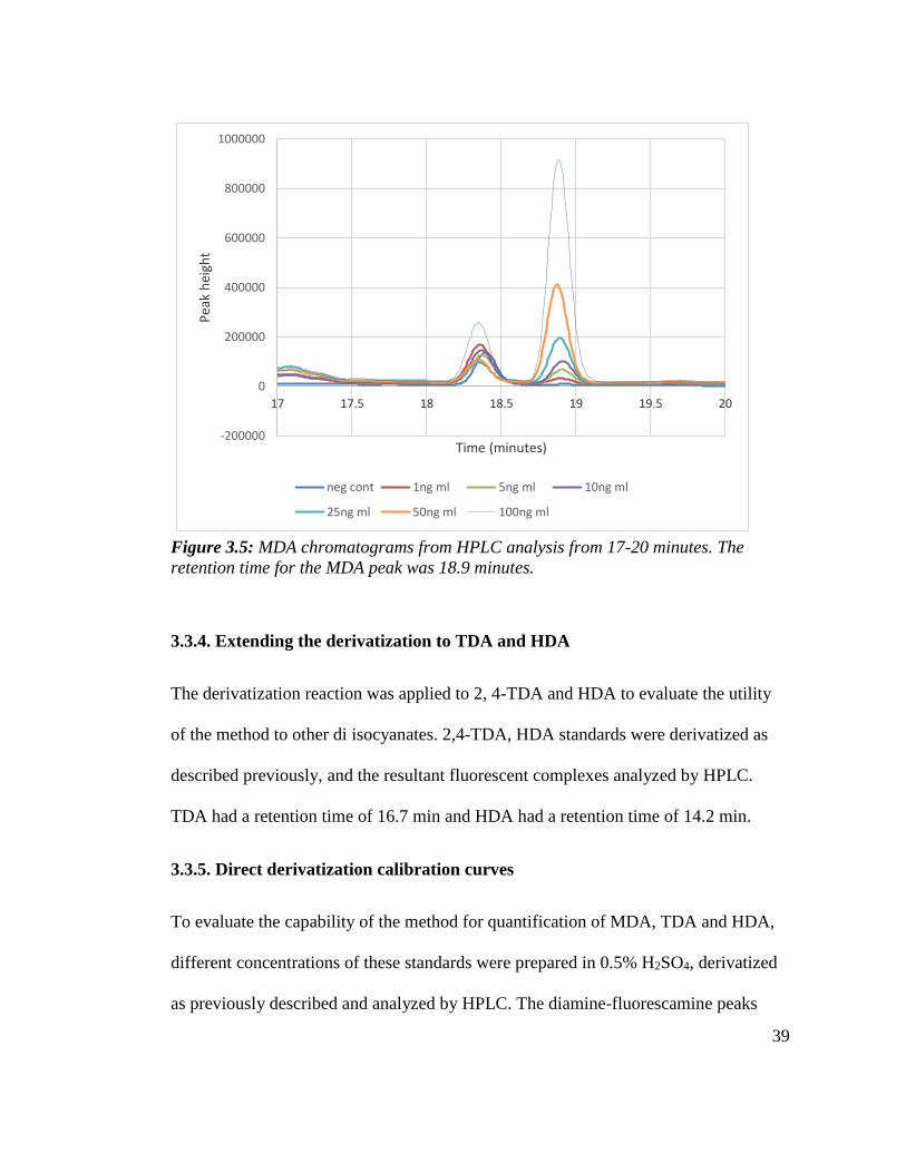
The MDA peak size increased in a dose-dependent manner with increasing
concentration of MDA. Figure 3.5 was obtained by blowing out the region from 17 to
20 minutes to clearly visualize the MDA peak. The peak observed at 18.36 minutes is
not due to MDA as this peak was present in the negative control and not MDA dose-
dependent. This peak could be due to impurities in the MDA or the fluorescamine.
39
-200000
0
200000
400000
600000
800000
1000000
17 17.5 18 18.5 19 19.5 20
Pea
k h
eigh
t
Time (minutes)
neg cont 1ng ml 5ng ml 10ng ml
25ng ml 50ng ml 100ng ml
Figure 3.5: MDA chromatograms from HPLC analysis from 17-20 minutes. The
retention time for the MDA peak was 18.9 minutes.
3.3.4. Extending the derivatization to TDA and HDA
The derivatization reaction was applied to 2, 4-TDA and HDA to evaluate the utility
of the method to other di isocyanates. 2,4-TDA, HDA standards were derivatized as
described previously, and the resultant fluorescent complexes analyzed by HPLC.
TDA had a retention time of 16.7 min and HDA had a retention time of 14.2 min.
3.3.5. Direct derivatization calibration curves
To evaluate the capability of the method for quantification of MDA, TDA and HDA,
different concentrations of these standards were prepared in 0.5% H2SO4, derivatized
as previously described and analyzed by HPLC. The diamine-fluorescamine peaks

40
were integrated using the EZstart software and the peak area was plotted against the
diamine concentration.
Figure 3.6 below is a calibration curve obtained when different concentrations of
MDA were analyzed by HPLC. There is a strong linear correlation between the peak
area and the MDA standard concentration with an r-square value of 0.9986. The limit
of detection was 1 ng/mL MDA with an injection volume of 100 µL.
Figure 3.6: Calibration curve obtained from the direct derivatization of MDA with
fluorescamine
When the same experiment was performed with 2, 4-TDA and HDA, the correlations
were good with correlation coefficients of 0.9947 and 0.9992 for 2, 4-TDA and HDA
respectively. It is therefore possible to quantify the amount of MDA, 2, 4-TDA or
HDA in a sample using this HPLC method.

41
3.3.6. Optimization of the extraction
After hydrolysis of the diisocyanate-protein conjugate, the diamine hydrolysis product
was selectively extracted prior to derivatization. Toluene, dichloromethane, and ethyl
acetate extraction solvents were compared. MDA was spiked into 0.5mg/ml of HSA
and Hb solutions, acid hydrolyzed, alkalinized, and extracted using the three solvents.
No MDA was detected when toluene was used for extraction. Solubility checks
indicated that MDA is barely soluble in toluene making toluene a poor choice for
extraction of MDA. Dichloromethane (DCM) and ethyl acetate (EA) were therefore
employed for extraction of MDA, 2, 4-TDA and HDA and extraction efficiencies of
the two solvents were tested by using each solvent to extract a 100ng/ml MDA, 2, 4-
TDA and HDA. Table 3.5 shows the extraction efficiencies of the two solvents
calculated by dividing the peak area of 100 ng/ml MDA, 2,4-TDA and HDA extracted
using DCM and EA by the peak area of directly derivatized 100 ng/ml MDA, 2, 4-
TDA and HDA. Table 3.6 shows solvents that were chosen for extraction of each
diamine based on superior extraction efficiencies.
Table 3.4: Extraction efficiencies obtained for DCM and EA
Solvent Ext efficiency (%)
for MDA
Ext efficiency (%)
for TDA
Ext efficiency (%)
for HDA
DCM 1 36.2 ± 17.0 (s.d.) 97.4 ± 3.2 (s.d.) 95.7 ± 3.9 (s.d.)
EA 1 96.2 ± 4.3 (s.d.) 88.3 ± 4.8 (s.d.) 89.2 ± 4.5 (s.d.)
42
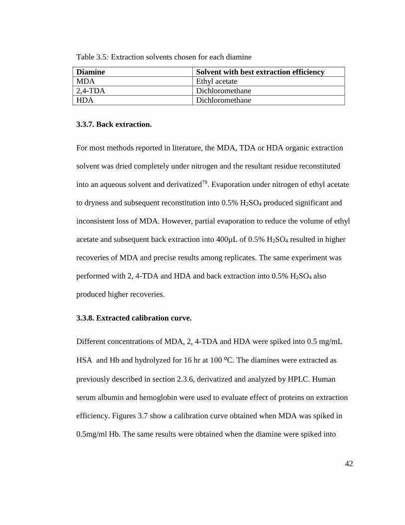
Table 3.5: Extraction solvents chosen for each diamine
Diamine Solvent with best extraction efficiency
MDA Ethyl acetate
2,4-TDA Dichloromethane
HDA Dichloromethane
3.3.7. Back extraction.
For most methods reported in literature, the MDA, TDA or HDA organic extraction
solvent was dried completely under nitrogen and the resultant residue reconstituted
into an aqueous solvent and derivatized79. Evaporation under nitrogen of ethyl acetate
to dryness and subsequent reconstitution into 0.5% H2SO4 produced significant and
inconsistent loss of MDA. However, partial evaporation to reduce the volume of ethyl
acetate and subsequent back extraction into 400µL of 0.5% H2SO4 resulted in higher
recoveries of MDA and precise results among replicates. The same experiment was
performed with 2, 4-TDA and HDA and back extraction into 0.5% H2SO4 also
produced higher recoveries.
3.3.8. Extracted calibration curve.
Different concentrations of MDA, 2, 4-TDA and HDA were spiked into 0.5 mg/mL
HSA and Hb and hydrolyzed for 16 hr at 100 ⁰C. The diamines were extracted as
previously described in section 2.3.6, derivatized and analyzed by HPLC. Human
serum albumin and hemoglobin were used to evaluate effect of proteins on extraction
efficiency. Figures 3.7 show a calibration curve obtained when MDA was spiked in
0.5mg/ml Hb. The same results were obtained when the diamine were spiked into
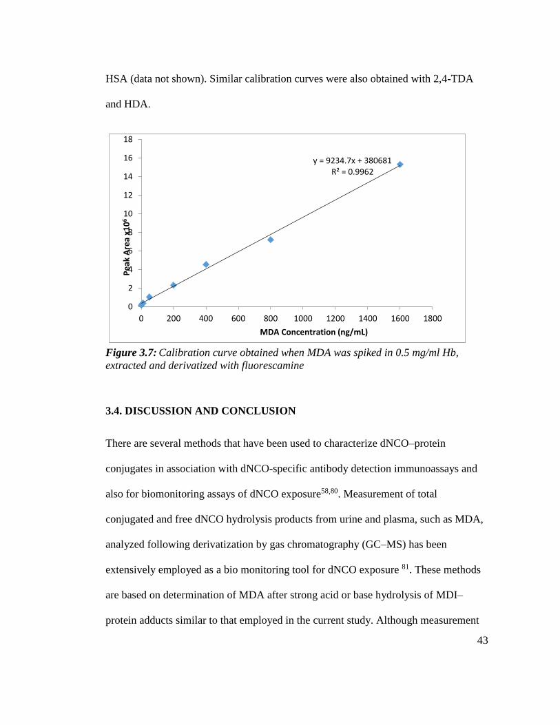
43
HSA (data not shown). Similar calibration curves were also obtained with 2,4-TDA
and HDA.
y = 9234.7x + 380681R² = 0.9962
0
2
4
6
8
10
12
14
16
18
0 200 400 600 800 1000 1200 1400 1600 1800
Pe
ak A
rea
x10
6
MDA Concentration (ng/mL)
Figure 3.7: Calibration curve obtained when MDA was spiked in 0.5 mg/ml Hb,
extracted and derivatized with fluorescamine
3.4. DISCUSSION AND CONCLUSION
There are several methods that have been used to characterize dNCO–protein
conjugates in association with dNCO-specific antibody detection immunoassays and
also for biomonitoring assays of dNCO exposure58,80. Measurement of total
conjugated and free dNCO hydrolysis products from urine and plasma, such as MDA,
analyzed following derivatization by gas chromatography (GC–MS) has been
extensively employed as a bio monitoring tool for dNCO exposure 81. These methods
are based on determination of MDA after strong acid or base hydrolysis of MDI–
protein adducts similar to that employed in the current study. Although measurement
44
of the diamine hydrolysis product has been used for biomonitoring, only one study has
reportedly applied this approach to characterize in vitro prepared HSA conjugates82.
Several solvents used for the extraction of diamines derived from dNCO hydrolysis
are commercially available. These include toluene, dichloromethane, and ethyl acetate
74,83. Toluene is the most commonly extraction solvent reported for both TDI derived
TDA and MDI derived MDA; however, MDA has poor solubility in toluene. Based on
extraction efficiencies, ethyl acetate was chosen as the extraction solvent for
extraction of MDA and dichloromethane for extraction of both 2, 4-TDA and HDA.
Loss of MDA was observed on complete solvent evaporation under nitrogen but
partial evaporation of ethyl acetate with subsequent back extraction into 0.5% H2SO4
produced high, consistent recoveries and was used to extract the diamine from ethyl
acetate into an aqueous solvent prior to derivatization.
This HPLC method based on fluorescence detection is applicable for absolute
quantification of diisocyanate conjugation to proteins by quantifying the diisocyanate
hydrolysis product following conjugate hydrolysis. This method is inexpensive,
simple, and sensitive (limit of detection [LOD] = 1 ng/ml). Mass spectral methods
incorporate an isotopic internal standard that may increase accuracy, especially where
matrix effects may alter extraction efficiency. However, the external standard
methodology used for quantification of laboratory haptenated proteins where there is a
uniform matrix provides sufficient accuracy for quantification of mass of dNCO-
protein conjugated.
45
CHAPTER 4
CHARACTERIZATION OF METHYLENE DIPHENYL DIISOCYANATE
CONJUGATED HUMAN SERUM ALBUMIN AND HEMOGLOBIN
4.1 INTRODUCTION
Diagnosis of dNCO-induced OA remains confounded by methodological limitations
84. Immunoassay standardization is critical for optimization of immunoassays used to
detect diisocyanate specific antibodies in workers’ sera as well as comparison of
results across studies 85. A number of factors that may affect results from these
immunoassays include the dNCO used, the carrier protein employed, dNCO–protein
reaction conditions, and post-reaction processing of the haptenated protein. The
variability of results obtained in these immunoassays may also be due in part to a lack
of standardization in conjugate preparation and characterization.
HSA is the most common carrier protein used for dNCO antibody immunoassays 80
due to its prevalence in plasma to form MDI adducts86. Other molecules, such as
keratin 18 , tubulin 87, and the peptide, glutathione 88 have been reported to be
modified by dNCO exposure. Hemoglobin–MDI haptenation occurs in vivo following
MDI exposure. Sabbioni and coworkers70 reported MDI binding to the N-terminal
valine of Hb in MDI exposed rats and proposed Hb–MDI as a biological marker of
MDI exposure. The same authors also found the N-terminal adduct with valine in
globin of a TDI-exposed worker and in two women with polyurethane-covered breast
implants 89. The immunogenicity of adducted proteins that have been identified other
than albumin has not been tested; however, the fact that haptenated keratin and tubulin
46
were identified immunochemically suggests that multiple haptenated protein species
formed following exposure may be antigens. Due to the lack of characterization of
protein–dNCO adducts, antibody reactivity toward other endogenous haptenated
proteins may be overlooked using conventional detection approaches. Other proteins
assessed as carrier proteins for dNCOs, however, have not been as effective at
detecting dNCO-specific antibody in the blood of exposed workers. It is not known
whether this is due to varying degrees of dNCO adduction to different proteins or
antibody recognition. Characterization of different dNCO–protein conjugates,
therefore, is important to further our understanding of dNCO haptenation and
adduction.
Although MDI accounts for almost 60 % of the global market of dNCO worldwide,
most of the published biomedical dNCO research studies have used toluene
diisocyanate (TDI), possibly because of MDI’s lower vapor pressure and lower
reactivity. Its lower volatility makes respiratory exposure less likely and harder to
generate into the air for animal studies. There was also a switch about 15 years ago
from TDI use to MDI use with respect to industrial applications. This could also
explain why there are not as many research studies reported on MDI.
Our previous research has been directed towards delineating the concentration-
dependent increase in specific dNCOs such as toluene diisocyanate (TDI) binding sites
on HSA90. In these studies, the predominant TDI binding sites on HSA were lysine
residues, although binding to the N-terminal arginine and to glutamine were also
observed. In another study performed in our laboratory 91, MDI was shown to be

47
bound to the same sites as TDI, but overall the reactivity was reduced. In this study,
MDI–HSA conjugates were characterized with the aim of developing a standardized
approach for screening for IgE- and IgG-specific antibodies. MDI reactivity toward
Hb was also examined and compared with HSA because dNCO-adducted hemoglobin
has been measured from the blood of exposed workers and used as a biological marker
of dNCO exposure 92.
Wong and coworkers 93 reported that acrylonitrile- adducted Hb was antigenic. In
another study, Pien and coworkers 94 found that rats exposed to the respiratory
allergen and inducer of late respiratory systemic syndrome, trimellitic anhydride
(TMA), by inhalation produced IgG that recognized both TMA-haptenated albumin
and Hb. They demonstrated, through cross-inhibition studies, that TMA–albumin and
TMA–Hb share antigenic determinants. Collectively, these studies suggest that
haptenated hemoglobin can be antigenic.
These experiments sought to (a) use the HPLC method developed to quantify amount
of MDI conjugation to both HSA and Hb, (b) evaluate other established techniques
used to characterize MDI conjugates that are formed with different proteins and (c)
suggest a set of methods and techniques that can be used to characterize conjugates
that are used for detection of MDI specific antibodies in workers’ sera.
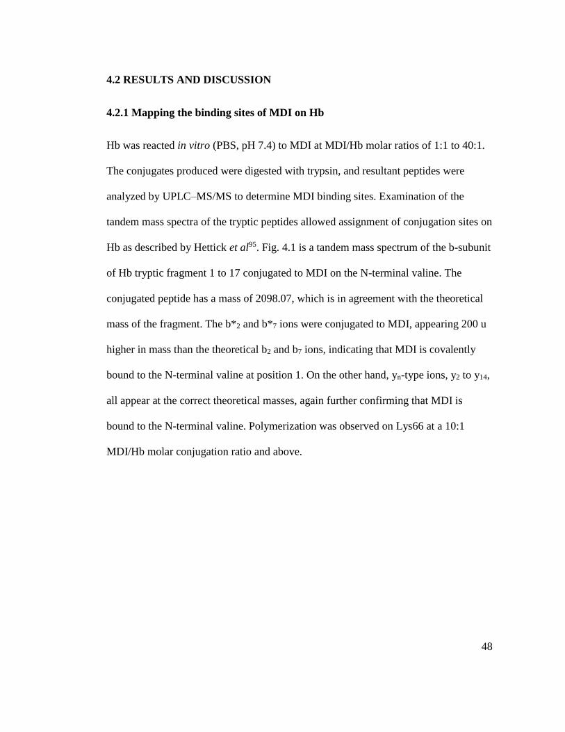
48
4.2 RESULTS AND DISCUSSION
4.2.1 Mapping the binding sites of MDI on Hb
Hb was reacted in vitro (PBS, pH 7.4) to MDI at MDI/Hb molar ratios of 1:1 to 40:1.
The conjugates produced were digested with trypsin, and resultant peptides were
analyzed by UPLC–MS/MS to determine MDI binding sites. Examination of the
tandem mass spectra of the tryptic peptides allowed assignment of conjugation sites on
Hb as described by Hettick et al95. Fig. 4.1 is a tandem mass spectrum of the b-subunit
of Hb tryptic fragment 1 to 17 conjugated to MDI on the N-terminal valine. The
conjugated peptide has a mass of 2098.07, which is in agreement with the theoretical
mass of the fragment. The b*2 and b*7 ions were conjugated to MDI, appearing 200 u
higher in mass than the theoretical b2 and b7 ions, indicating that MDI is covalently
bound to the N-terminal valine at position 1. On the other hand, yn-type ions, y2 to y14,
all appear at the correct theoretical masses, again further confirming that MDI is
bound to the N-terminal valine. Polymerization was observed on Lys66 at a 10:1
MDI/Hb molar conjugation ratio and above.
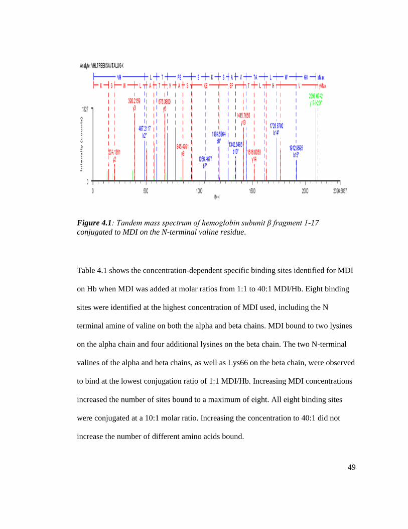
49
Figure 4.1: Tandem mass spectrum of hemoglobin subunit β fragment 1-17
conjugated to MDI on the N-terminal valine residue.
Table 4.1 shows the concentration-dependent specific binding sites identified for MDI
on Hb when MDI was added at molar ratios from 1:1 to 40:1 MDI/Hb. Eight binding
sites were identified at the highest concentration of MDI used, including the N
terminal amine of valine on both the alpha and beta chains. MDI bound to two lysines
on the alpha chain and four additional lysines on the beta chain. The two N-terminal
valines of the alpha and beta chains, as well as Lys66 on the beta chain, were observed
to bind at the lowest conjugation ratio of 1:1 MDI/Hb. Increasing MDI concentrations
increased the number of sites bound to a maximum of eight. All eight binding sites
were conjugated at a 10:1 molar ratio. Increasing the concentration to 40:1 did not
increase the number of different amino acids bound.

50
Table 4.1: MDI Hemoglobin conjugation sites
MDI:Hb Conjugation Ratios
Binding Site 1:1 5:1 10:1 40:1
Alpha subunit
Val 1 x x x x
Lys 7 x x x
Lys 40 x x x
Beta subunit
Val 1 x x x x
Lys 8 x x
Lys 61 x x
Lys 65 x x
Lys 66 x x x x
In contrast, MDI conjugates five different HSA lysines at 1:1 MDI/HSA91.
Conjugation to the N-terminal Asp in HSA was not observed until the MDI
concentration was raised to 5:1 MDI/HSA and two glutamine sites were bound at the
higher conjugation ratios 91. In addition, the number of different amino acid binding
sites increased through the entire MDI concentration range, with 20 different binding
sites conjugated at 40:1 MDI/HSA. Table 4.2 compares the number of observed
binding sites for Hb and HSA. HSA has more lysine binding sites accessible for MDI
binding compared with Hb through the entire MDI concentration range. However,
relative to the number of potential lysine binding sites in the two proteins, both
proteins showed relatively equivalent percentage binding, where the percentage
binding was calculated as the number of observed binding sites divided by the total
number of potential binding sites (lysines + N-terminal amines), expressed as a
percentage.
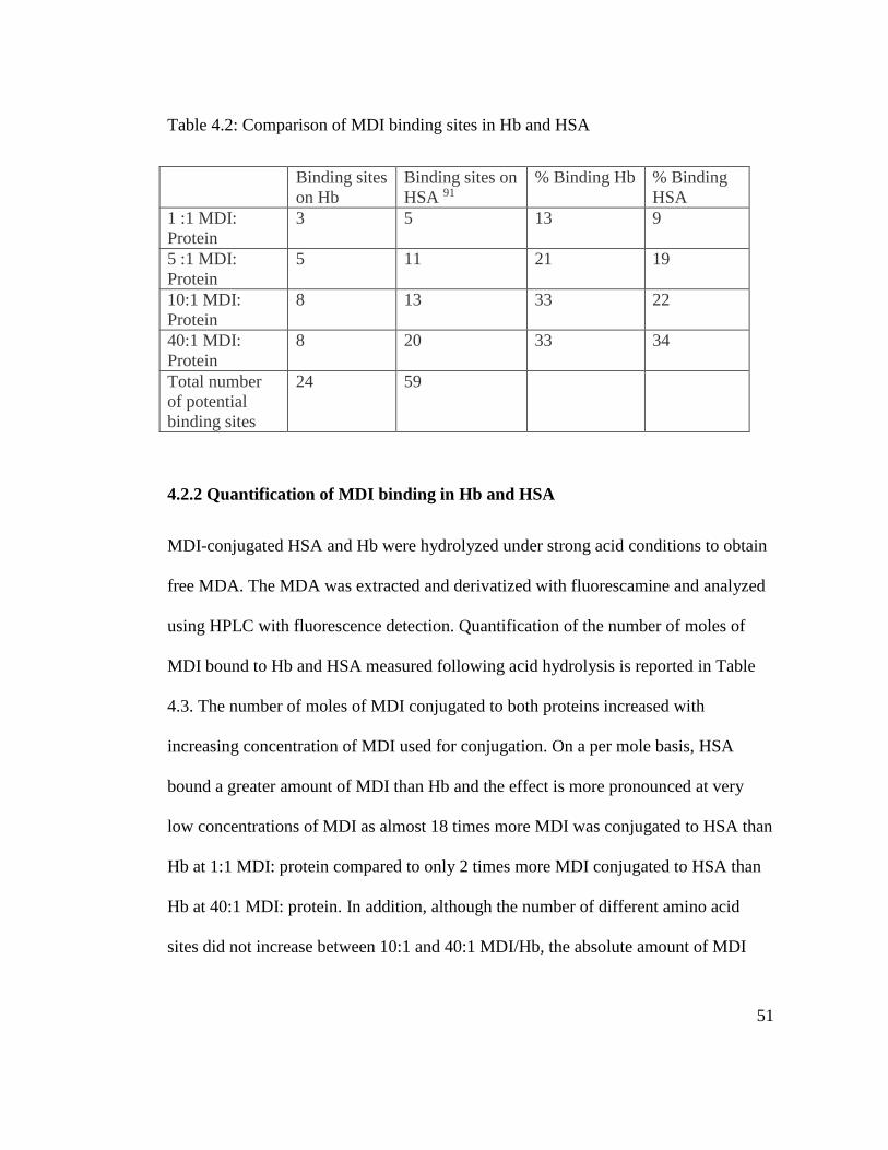
51
Table 4.2: Comparison of MDI binding sites in Hb and HSA
Binding sites
on Hb
Binding sites on
HSA 91
% Binding Hb % Binding
HSA
1 :1 MDI:
Protein
3 5 13 9
5 :1 MDI:
Protein
5 11 21 19
10:1 MDI:
Protein
8 13 33 22
40:1 MDI:
Protein
8 20 33 34
Total number
of potential
binding sites
24 59
4.2.2 Quantification of MDI binding in Hb and HSA
MDI-conjugated HSA and Hb were hydrolyzed under strong acid conditions to obtain
free MDA. The MDA was extracted and derivatized with fluorescamine and analyzed
using HPLC with fluorescence detection. Quantification of the number of moles of
MDI bound to Hb and HSA measured following acid hydrolysis is reported in Table
4.3. The number of moles of MDI conjugated to both proteins increased with
increasing concentration of MDI used for conjugation. On a per mole basis, HSA
bound a greater amount of MDI than Hb and the effect is more pronounced at very
low concentrations of MDI as almost 18 times more MDI was conjugated to HSA than
Hb at 1:1 MDI: protein compared to only 2 times more MDI conjugated to HSA than
Hb at 40:1 MDI: protein. In addition, although the number of different amino acid
sites did not increase between 10:1 and 40:1 MDI/Hb, the absolute amount of MDI
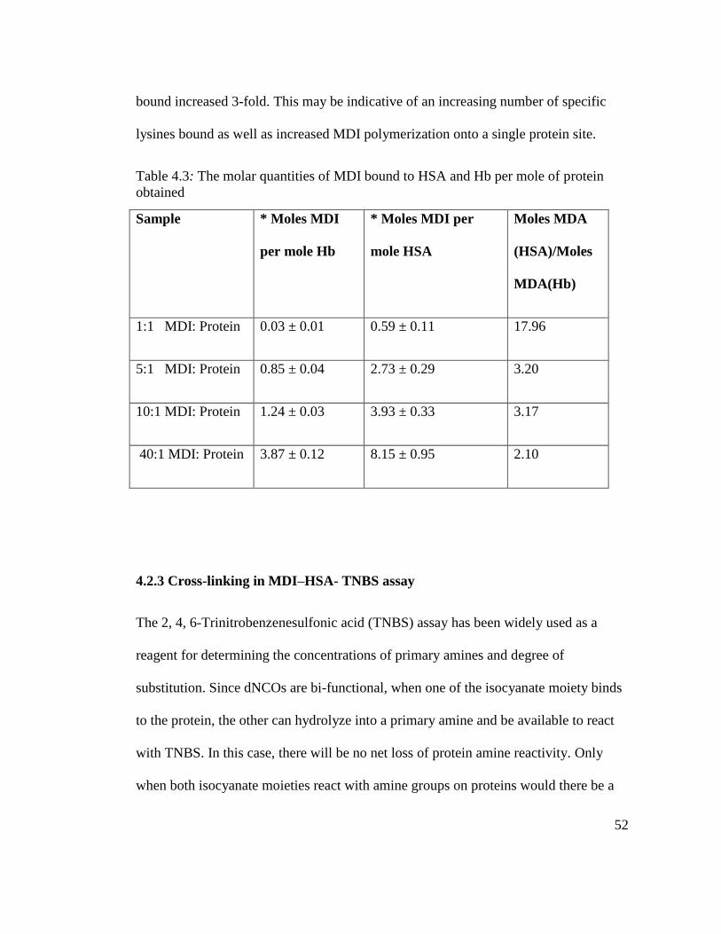
52
bound increased 3-fold. This may be indicative of an increasing number of specific
lysines bound as well as increased MDI polymerization onto a single protein site.
Table 4.3: The molar quantities of MDI bound to HSA and Hb per mole of protein
obtained
Sample * Moles MDI
per mole Hb
* Moles MDI per
mole HSA
Moles MDA
(HSA)/Moles
MDA(Hb)
1:1 MDI: Protein 0.03 ± 0.01 0.59 ± 0.11 17.96
5:1 MDI: Protein 0.85 ± 0.04 2.73 ± 0.29 3.20
10:1 MDI: Protein 1.24 ± 0.03 3.93 ± 0.33 3.17
40:1 MDI: Protein 3.87 ± 0.12 8.15 ± 0.95 2.10
4.2.3 Cross-linking in MDI–HSA- TNBS assay
The 2, 4, 6-Trinitrobenzenesulfonic acid (TNBS) assay has been widely used as a
reagent for determining the concentrations of primary amines and degree of
substitution. Since dNCOs are bi-functional, when one of the isocyanate moiety binds
to the protein, the other can hydrolyze into a primary amine and be available to react
with TNBS. In this case, there will be no net loss of protein amine reactivity. Only
when both isocyanate moieties react with amine groups on proteins would there be a
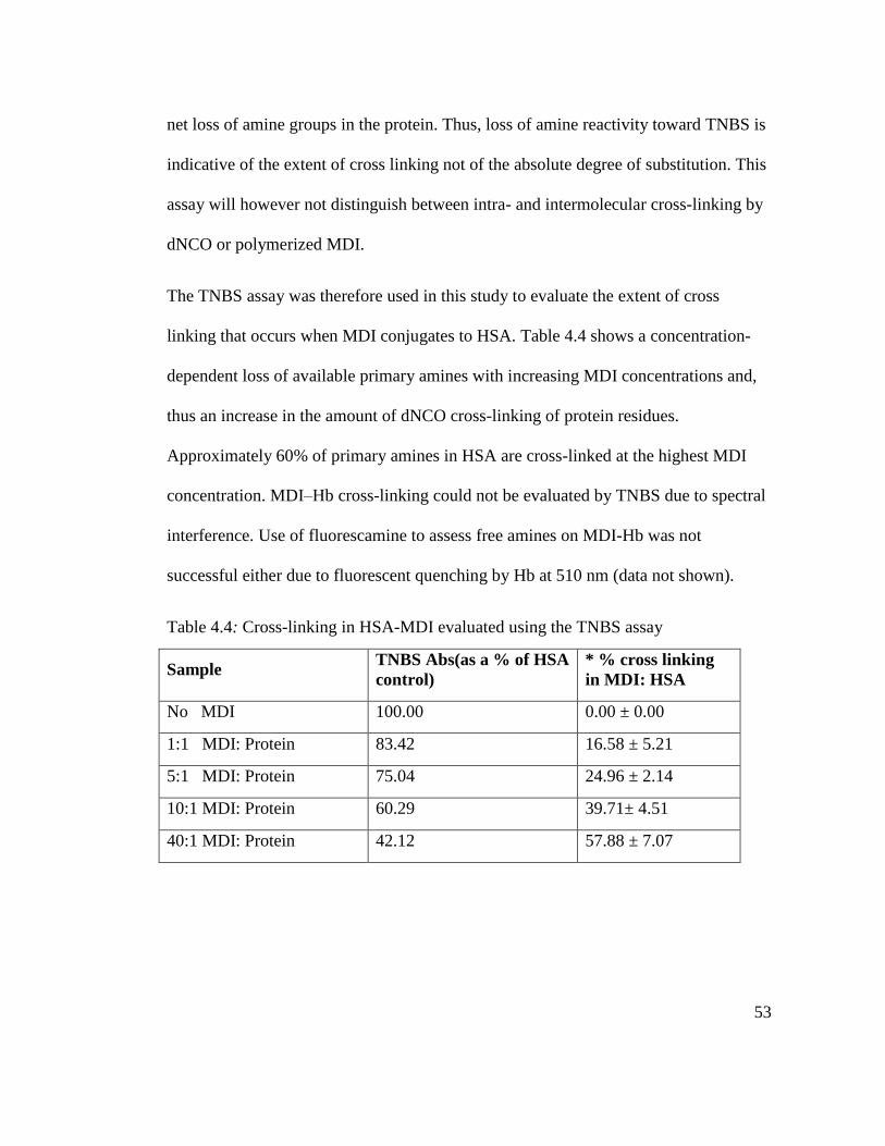
53
net loss of amine groups in the protein. Thus, loss of amine reactivity toward TNBS is
indicative of the extent of cross linking not of the absolute degree of substitution. This
assay will however not distinguish between intra- and intermolecular cross-linking by
dNCO or polymerized MDI.
The TNBS assay was therefore used in this study to evaluate the extent of cross
linking that occurs when MDI conjugates to HSA. Table 4.4 shows a concentration-
dependent loss of available primary amines with increasing MDI concentrations and,
thus an increase in the amount of dNCO cross-linking of protein residues.
Approximately 60% of primary amines in HSA are cross-linked at the highest MDI
concentration. MDI–Hb cross-linking could not be evaluated by TNBS due to spectral
interference. Use of fluorescamine to assess free amines on MDI-Hb was not
successful either due to fluorescent quenching by Hb at 510 nm (data not shown).
Table 4.4: Cross-linking in HSA-MDI evaluated using the TNBS assay
Sample TNBS Abs(as a % of HSA
control)
* % cross linking
in MDI: HSA
No MDI 100.00 0.00 ± 0.00
1:1 MDI: Protein 83.42 16.58 ± 5.21
5:1 MDI: Protein 75.04 24.96 ± 2.14
10:1 MDI: Protein 60.29 39.71± 4.51
40:1 MDI: Protein 42.12 57.88 ± 7.07
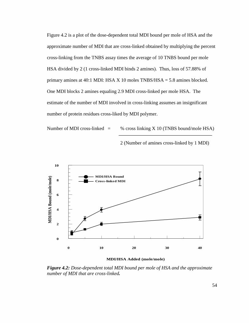
54
Figure 4.2 is a plot of the dose-dependent total MDI bound per mole of HSA and the
approximate number of MDI that are cross-linked obtained by multiplying the percent
cross-linking from the TNBS assay times the average of 10 TNBS bound per mole
HSA divided by 2 (1 cross-linked MDI binds 2 amines). Thus, loss of 57.88% of
primary amines at 40:1 MDI: HSA X 10 moles TNBS/HSA = 5.8 amines blocked.
One MDI blocks 2 amines equaling 2.9 MDI cross-linked per mole HSA. The
estimate of the number of MDI involved in cross-linking assumes an insignificant
number of protein residues cross-liked by MDI polymer.
Number of MDI cross-linked = % cross linking X 10 (TNBS bound/mole HSA)
2 (Number of amines cross-linked by 1 MDI)
MDI/HSA Bound
Cross-linked MDI
MDI/HSA Added (mole/mole)
0 10 20 30 40
MD
I/H
SA
Bou
nd
(m
ole/
mol
e)
0
2
4
6
8
10
Figure 4.2: Dose-dependent total MDI bound per mole of HSA and the approximate
number of MDI that are cross-linked.
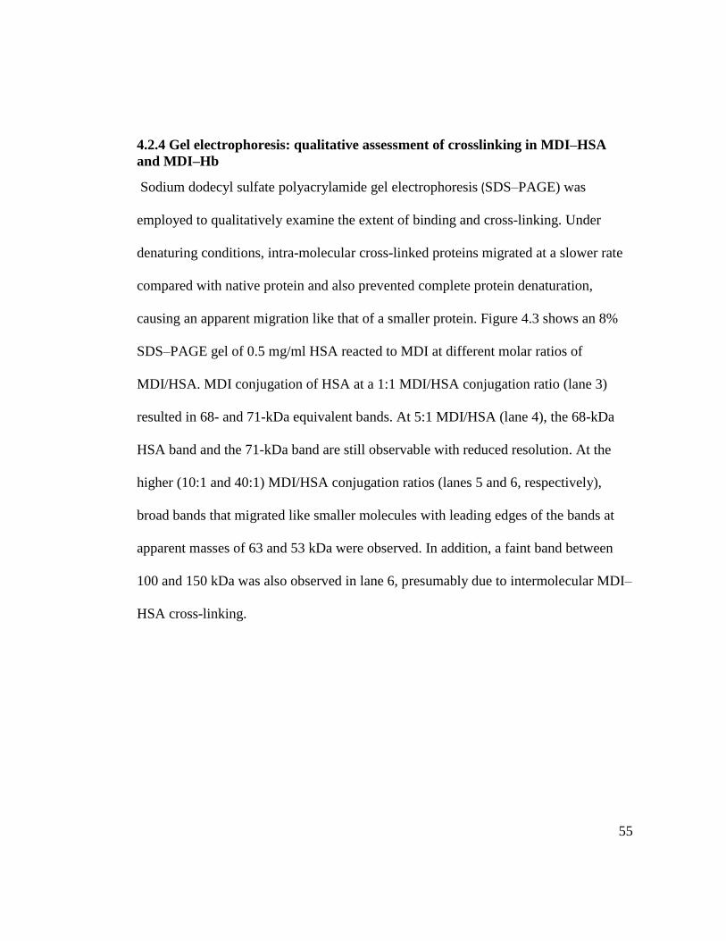
55
4.2.4 Gel electrophoresis: qualitative assessment of crosslinking in MDI–HSA
and MDI–Hb
Sodium dodecyl sulfate polyacrylamide gel electrophoresis (SDS–PAGE) was
employed to qualitatively examine the extent of binding and cross-linking. Under
denaturing conditions, intra-molecular cross-linked proteins migrated at a slower rate
compared with native protein and also prevented complete protein denaturation,
causing an apparent migration like that of a smaller protein. Figure 4.3 shows an 8%
SDS–PAGE gel of 0.5 mg/ml HSA reacted to MDI at different molar ratios of
MDI/HSA. MDI conjugation of HSA at a 1:1 MDI/HSA conjugation ratio (lane 3)
resulted in 68- and 71-kDa equivalent bands. At 5:1 MDI/HSA (lane 4), the 68-kDa
HSA band and the 71-kDa band are still observable with reduced resolution. At the
higher (10:1 and 40:1) MDI/HSA conjugation ratios (lanes 5 and 6, respectively),
broad bands that migrated like smaller molecules with leading edges of the bands at
apparent masses of 63 and 53 kDa were observed. In addition, a faint band between
100 and 150 kDa was also observed in lane 6, presumably due to intermolecular MDI–
HSA cross-linking.
56
Figure 4.3: An 8% denaturing gel of MDI: HSA. Lane 1is the molecular weight
marker, lane 2 is HSA, lanes 3, 4, 5, and 6 are 1:1, 5:1, 10:1, and 40:1 MDI: HSA
conjugates respectively.
Native gel electrophoresis of MDI–HSA conjugates was also assessed (Figure 4.4);
however, higher molecular weight bands 2- to 3-fold the mass of HSA were observed
in control samples (lane 2) and identified by digestion and UPLC–MS/MS to be HSA.
These results demonstrated the aggregation of HSA under the assay conditions and
prevented the observation of potential intramolecular cross-linking. An MDI
concentration dependent increase in migration of the conjugate band was also
observed in the MDI–HSA conjugates similar to that observed under denaturing
conditions, suggestive of intramolecular cross- linking. The faster migration (apparent
smaller size) may be possibly attributable to exclusion of intramolecular water(s),
effectively decreasing the size.

57
Figure 4.4: An 8% native gel of MDI: HSA. Lane 1 is the molecular weight marker,
lane 2 is HSA and lanes 3, 4, 5, and 6 are 1:1, 5:1, 10:1, and 40:1 MDI: HSA
respectively.
Figure 4.5 is a 4% to 20% gradient gel of 0.5 mg/ml Hb reacted to MDI at four
different mole ratios of MDI/Hb. Denaturation of Hb resulted in the dissociation of the
alpha and beta subunits that migrated at 15.2 and 15.9 kDa, respectively. Upon MDI
conjugation, there was emergence of a band at approximately 30 kDa, which is a result
of two crosslinked subunits. A dark unresolved smear of high-molecular-weight
compounds also emerged at 45 kDa and above, suggestive of extensive multiple inter-
subunit cross-linking, and the intensity of this band increased with increasing MDI
concentrations. A downward shift of the 15.2- and 15.9-kDa subunits with increased
MDI concentrations was also observed, similar to that found with the MDI–HSA
conjugates.
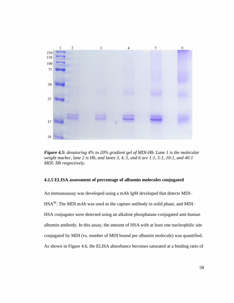
58
Figure 4.5: denaturing 4% to 20% gradient gel of MDI-Hb. Lane 1 is the molecular
weight marker, lane 2 is Hb, and lanes 3, 4, 5, and 6 are 1:1, 5:1, 10:1, and 40:1
MDI: Hb respectively.
4.2.5 ELISA assessment of percentage of albumin molecules conjugated
An immunoassay was developed using a mAb IgM developed that detects MDI–
HSA96. The MDI mAb was used as the capture antibody in solid phase, and MDI–
HSA conjugates were detected using an alkaline phosphatase-conjugated anti-human
albumin antibody. In this assay, the amount of HSA with at least one nucleophilic site
conjugated by MDI (vs. number of MDI bound per albumin molecule) was quantified.
As shown in Figure 4.6, the ELISA absorbance becomes saturated at a binding ratio of

59
20:1, with no further increase at 40:1. These results demonstrate that all HSA
molecules are conjugated to at least one MDI at the 20:1 MDI/HSA conjugation ratio.
MDI:HSA Molar Conjugation Ratio
Ab
sorb
ance
(40
5 n
m)
0.0
0.3
0.5
0.8
1.0
1.3
1.5
1.8
2.0
5:1 10:1 20:1 40:1
Figure 4.6: Immunoassay of MDI haptenated HSA.
4.3. CONCLUSION
In the current study, MS/MS was used to delineate specific MDI binding sites on Hb
and 8 binding sites were identified in Hb. Except for the N-terminal valines on both
the alpha and beta subunits, Lys66 was the only non-terminal binding site that was
observed at the lowest MDI concentration studied (1:1 MDI/Hb). Although additional
MDI binding sites were not observed when the binding ratio was increased from 10:1
to 40:1, the number of moles of MDI bound per mole Hb increased 3-fold.
Using the TNBS assay, a concentration-dependent increase in the amount of MDI
cross-linking of HSA was observed with loss of 58% of primary amine reactivity at
60
40:1 MDI/HSA translating to 2.9 moles MDI cross-linked per mole HSA. The
estimate of the number of MDI cross-linking assumes that an insignificant number of
protein residues are cross-linked by MDI polymer. Due to spectral interference from
Hb, crosslinking in MDI–Hb conjugates could not be assessed quantitatively.
Intermolecular cross-linking was also observed in both MDI haptenated HSA and Hb
at the higher conjugation ratios using polyacrylamide gel electrophoresis by the
appearance of bands approximately 2-fold the monomeric protein masses.
Immunoassays using a recently developed mAb (15D4) specific for MDI-conjugated
protein can be formatted in a variety of configurations to provide further information
to develop a standardized antigen for diisocyanate-specific antibody screening. The
ELISA format employed in the current study uses an anti-MDI–protein IgM as the
capture antibody and a labeled anti-HSA as the detection antibody and, thus, measures
the number of albumin with at least one MDI molecule covalently bound. No increase
in immuno-reactivity was observed at reaction ratios above 20:1 MDI/HSA,
suggesting that any increase in bound MDI was due to polymerization and reaction to
additional HSA nucleophilic sites versus reaction to unbound HSA. Direct binding of
the haptenated HSA to an ELISA plate with measurement of binding by an mAb (data
not shown) is also an approach that can be used to standardize the antigenicity of the
haptenated protein. Wisnewski and coworkers 97 reported that maximal MDI-specific
IgG reactivity of exposed workers’ sera against MDI–HSA in their immunoassay was
observed at a conjugation ratio of 200 µg MDI/mg HSA (~50:1). It must be noted that
the HSA concentration reacted to MDI was 10 times greater than that used in the
61
current study, which very possibly altered the final product given the potential for
greater intermolecular crosslinking and polymerization associated with these higher
protein and MDI concentrations.
Multiple methods should be used to fully characterize dNCO–HSA conjugates for use
in standardized immunoassays for screening dNCO-specific IgE and IgG from
workers’ sera. These analyses should include (i) measurement of the amount of
diamine/mole protein following acid hydrolysis, (ii) TNBS measurement of cross-
linking, (iii) denature gel electrophoresis for indication of inter- and intramolecular
crosslinking, and (iv) use of mAb for antigenicity standardization. Both qualitative and
quantitative differences in MDI binding were observed between Hb and HSA
conjugates, suggesting that normalization to conjugation ratio is not sufficient to
compare antigenicity of different diisocyanate-haptenated proteins.
62
CHAPTER 5
COMPARATIVE ANALYSIS OF TOLUENE DIISOCYANATE AND
HEXAMETHYLENE DIISOCYANATE CONJUGATION TO HUMAN
SERUM ALBUMIN AND HEMOGLOBIN
5.1 INTRODUCTION
Toluene diisocyanate (TDI) and hexamethylene diisocyanate (HDI) are widely used in
the manufacture of polyurethane products and both have been reported to cause
respiratory sensitization and occupational asthma (OA) in exposed workers98. Once
allergic sensitization to isocyanates occurs, asthmatic reactions may be triggered by
exceedingly minute concentrations99,100. Both TDI and HDI are relatively volatile and
exposure occurs predominantly through inhalation at workplaces17. However, dermal
exposure has also been reported to cause asthma-like symptoms.
There are differences in reactivity between aromatic isocyanates and aliphatic
isocyanates. In a study by Wisnewski et al101 in which albumin was conjugated to TDI
and HDI by GSH-TDI and GSH-HDI, respectively, the kinetics of GSH-HDI mediated
albumin carbomylation at neutral pH was substantially slower compared with those of
GSH-TDI. These researchers also found that GSH-HDI conjugates were more stable
compared to aromatic isocyanate-GSH conjugates which probably explain the slower
rate of HDI carbomylation by GSH-HDI. The hydrolysis of aliphatic isocyanates is
also much slower than aromatic isocyanates. The reactivity of HDI directly conjugated
to proteins may however confer different kinetics.
63
Diisocyanate-adducted hemoglobin in vivo immunogenicity or antigenicity has not yet
been evaluated. Wong and coworkers93 reported that acrylonitrile- adducted Hb was
antigenic. Pien and coworkers102 also found that rats exposed to the respiratory
allergen and inducer of late respiratory systemic syndrome, trimellitic anhydride
(TMA), by inhalation produced IgG that recognized both TMA-haptenated albumin
and Hb. They demonstrated, through cross inhibition studies, that TMA–albumin and
TMA–Hb share antigenic determinants. Collectively, these studies suggest that
haptenated hemoglobin can be antigenic.
Previous work was focused on developing an HPLC method for quantitative
characterization of dNCO-protein adducts as well as characterization of MDI-HSA
and MDI-Hb conjugates for immunoassay standardization59. The experiments reported
herein sought to (i) extend the characterization of the conjugates formed upon reaction
of TDI and HDI with HSA and Hb to determine the amount of diisocyanate bound, (ii)
utilize the TNBS assay and SDS gel electrophoresis to evaluate extent of intra- and
inter-molecular cross linking, (iii) delineate TDI binding sites on Hb and (iv) evaluate
the antigenicity of TDI and HDI conjugates using a mAb produced in our lab.
5.2 RESULTS AND DISCUSSION
5.2.1 Mapping TDI binding sites on Hb
Hb-TDI conjugates were digested with trypsin, and resultant peptides were analyzed
by UPLC–MS/MS to determine TDI binding sites. Examination of the tandem mass
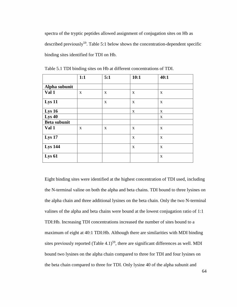
64
spectra of the tryptic peptides allowed assignment of conjugation sites on Hb as
described previously59. Table 5:1 below shows the concentration-dependent specific
binding sites identified for TDI on Hb.
Table 5.1 TDI binding sites on Hb at different concentrations of TDI.
1:1 5:1 10:1 40:1
Alpha subunit
Val 1 x x x x
Lys 11 x x x
Lys 16 x x
Lys 40 x
Beta subunit
Val 1 x x x x
Lys 17 x x
Lys 144 x x
Lys 61 x
Eight binding sites were identified at the highest concentration of TDI used, including
the N-terminal valine on both the alpha and beta chains. TDI bound to three lysines on
the alpha chain and three additional lysines on the beta chain. Only the two N-terminal
valines of the alpha and beta chains were bound at the lowest conjugation ratio of 1:1
TDI:Hb. Increasing TDI concentrations increased the number of sites bound to a
maximum of eight at 40:1 TDI:Hb. Although there are similarities with MDI binding
sites previously reported (Table 4.1)59, there are significant differences as well. MDI
bound two lysines on the alpha chain compared to three for TDI and four lysines on
the beta chain compared to three for TDI. Only lysine 40 of the alpha subunit and
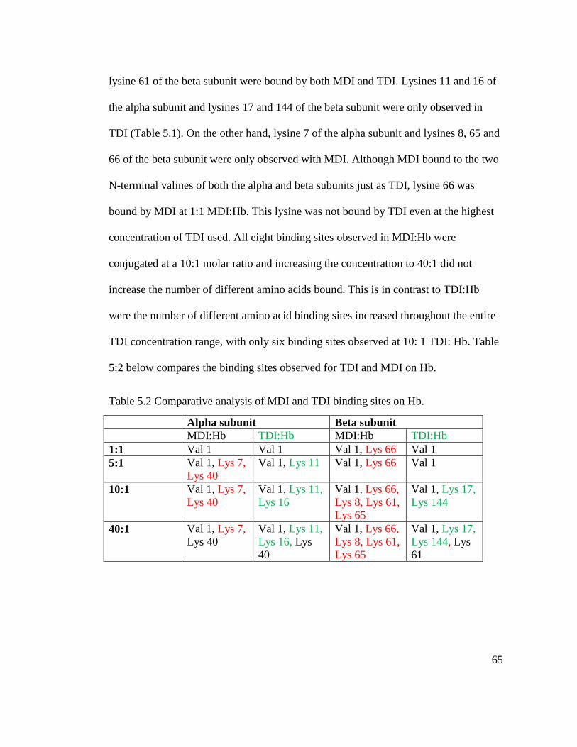
65
lysine 61 of the beta subunit were bound by both MDI and TDI. Lysines 11 and 16 of
the alpha subunit and lysines 17 and 144 of the beta subunit were only observed in
TDI (Table 5.1). On the other hand, lysine 7 of the alpha subunit and lysines 8, 65 and
66 of the beta subunit were only observed with MDI. Although MDI bound to the two
N-terminal valines of both the alpha and beta subunits just as TDI, lysine 66 was
bound by MDI at 1:1 MDI:Hb. This lysine was not bound by TDI even at the highest
concentration of TDI used. All eight binding sites observed in MDI:Hb were
conjugated at a 10:1 molar ratio and increasing the concentration to 40:1 did not
increase the number of different amino acids bound. This is in contrast to TDI:Hb
were the number of different amino acid binding sites increased throughout the entire
TDI concentration range, with only six binding sites observed at 10: 1 TDI: Hb. Table
5:2 below compares the binding sites observed for TDI and MDI on Hb.
Table 5.2 Comparative analysis of MDI and TDI binding sites on Hb.
Alpha subunit Beta subunit
MDI:Hb TDI:Hb MDI:Hb TDI:Hb
1:1 Val 1 Val 1 Val 1, Lys 66 Val 1
5:1 Val 1, Lys 7,
Lys 40
Val 1, Lys 11 Val 1, Lys 66 Val 1
10:1 Val 1, Lys 7,
Lys 40
Val 1, Lys 11,
Lys 16
Val 1, Lys 66,
Lys 8, Lys 61,
Lys 65
Val 1, Lys 17,
Lys 144
40:1 Val 1, Lys 7,
Lys 40
Val 1, Lys 11,
Lys 16, Lys
40
Val 1, Lys 66,
Lys 8, Lys 61,
Lys 65
Val 1, Lys 17,
Lys 144, Lys
61

66
The differences in the binding sites between MDI and TDI can give an insight into the
structural differences in the resultant conjugates, which potentially affects disease
mechanisms.
5.2.2 Quantification of TDI and HDI binding on Hb and HSA
TDI and HDI-conjugated HSA and Hb were hydrolyzed as previously described in
section 2.3.6 and the resultant TDA and HDA was extracted and derivatized with
fluorescamine and quantified using HPLC. The number of moles of TDI and HDI
bound to Hb and HSA is shown in Table 5:3 below.
Table 5.3: Amount in moles of TDI and HDI conjugated to HSA and Hb.
Ratio dNCO:
protein
dNCO: HSA dNCO: Hb
TDI: HSA HDI: HSA TDI: Hb HDI: Hb
1:1 0.51 ± 0.56 0.04 ± 0.41 0.15 ± 0.15 0.02 ± 0.21
5:1 2.65 ± 0.23 0.44 ± 0.45 0.57 ± 0.02 0.13 ± 0.23
10:1 5.06 ± 0.43 0.82 ± 0.36 1.48 ± 0.09 0.32 ± 0.60
40:1 12.86 ± 0.56 2.79 ± 0.40 4.31 ± 0.06 1.03 ± 0.59
The number of moles of TDI and HDI bound in both HSA and Hb increased with
increasing concentration of TDI and HDI used. On a per mole basis, HSA was three
times more reactive to both TDI and HDI than Hb at all dNCO/protein conjugation
ratios. These results agree with our earlier findings with MDI-HSA and MDI-Hb
conjugates, where HSA was more reactive to MDI than Hb. This could be a result of
the structural differences between HSA and Hb. Hemoglobin, with four subunits (two
alpha and two beta) and the iron containing porphyrin ring, may mask potential
binding sites thus affecting its reactivity with dNCOs. This contrasts sharply with
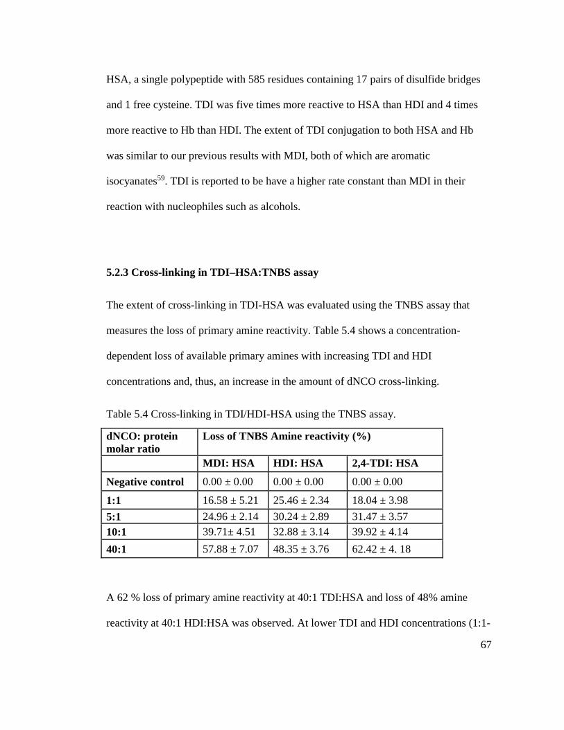
67
HSA, a single polypeptide with 585 residues containing 17 pairs of disulfide bridges
and 1 free cysteine. TDI was five times more reactive to HSA than HDI and 4 times
more reactive to Hb than HDI. The extent of TDI conjugation to both HSA and Hb
was similar to our previous results with MDI, both of which are aromatic
isocyanates59. TDI is reported to be have a higher rate constant than MDI in their
reaction with nucleophiles such as alcohols.
5.2.3 Cross-linking in TDI–HSA:TNBS assay
The extent of cross-linking in TDI-HSA was evaluated using the TNBS assay that
measures the loss of primary amine reactivity. Table 5.4 shows a concentration-
dependent loss of available primary amines with increasing TDI and HDI
concentrations and, thus, an increase in the amount of dNCO cross-linking.
Table 5.4 Cross-linking in TDI/HDI-HSA using the TNBS assay.
dNCO: protein
molar ratio
Loss of TNBS Amine reactivity (%)
MDI: HSA HDI: HSA 2,4-TDI: HSA
Negative control 0.00 ± 0.00 0.00 ± 0.00 0.00 ± 0.00
1:1 16.58 ± 5.21 25.46 ± 2.34 18.04 ± 3.98
5:1 24.96 ± 2.14 30.24 ± 2.89 31.47 ± 3.57
10:1 39.71± 4.51 32.88 ± 3.14 39.92 ± 4.14
40:1 57.88 ± 7.07 48.35 ± 3.76 62.42 ± 4. 18
A 62 % loss of primary amine reactivity at 40:1 TDI:HSA and loss of 48% amine
reactivity at 40:1 HDI:HSA was observed. At lower TDI and HDI concentrations (1:1-
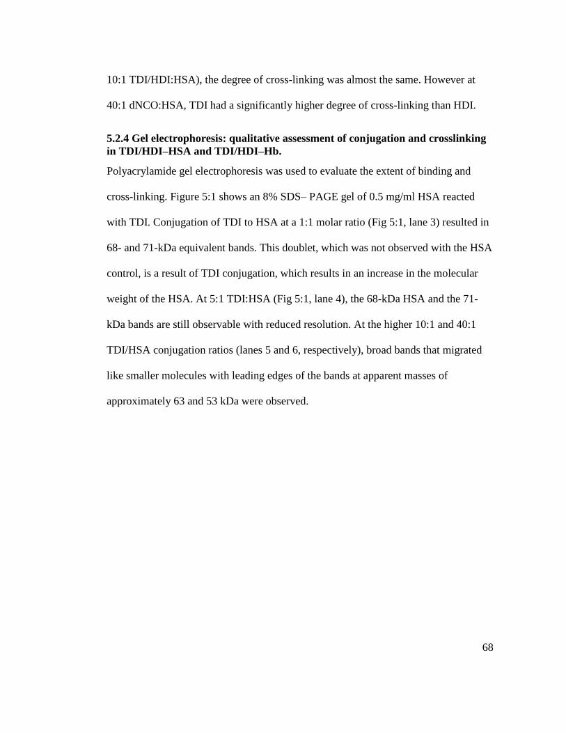
68
10:1 TDI/HDI:HSA), the degree of cross-linking was almost the same. However at
40:1 dNCO:HSA, TDI had a significantly higher degree of cross-linking than HDI.
5.2.4 Gel electrophoresis: qualitative assessment of conjugation and crosslinking
in TDI/HDI–HSA and TDI/HDI–Hb.
Polyacrylamide gel electrophoresis was used to evaluate the extent of binding and
cross-linking. Figure 5:1 shows an 8% SDS– PAGE gel of 0.5 mg/ml HSA reacted
with TDI. Conjugation of TDI to HSA at a 1:1 molar ratio (Fig 5:1, lane 3) resulted in
68- and 71-kDa equivalent bands. This doublet, which was not observed with the HSA
control, is a result of TDI conjugation, which results in an increase in the molecular
weight of the HSA. At 5:1 TDI:HSA (Fig 5:1, lane 4), the 68-kDa HSA and the 71-
kDa bands are still observable with reduced resolution. At the higher 10:1 and 40:1
TDI/HSA conjugation ratios (lanes 5 and 6, respectively), broad bands that migrated
like smaller molecules with leading edges of the bands at apparent masses of
approximately 63 and 53 kDa were observed.
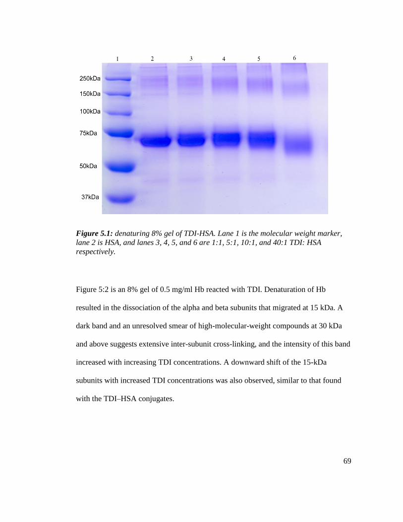
69
Figure 5.1: denaturing 8% gel of TDI-HSA. Lane 1 is the molecular weight marker,
lane 2 is HSA, and lanes 3, 4, 5, and 6 are 1:1, 5:1, 10:1, and 40:1 TDI: HSA
respectively.
Figure 5:2 is an 8% gel of 0.5 mg/ml Hb reacted with TDI. Denaturation of Hb
resulted in the dissociation of the alpha and beta subunits that migrated at 15 kDa. A
dark band and an unresolved smear of high-molecular-weight compounds at 30 kDa
and above suggests extensive inter-subunit cross-linking, and the intensity of this band
increased with increasing TDI concentrations. A downward shift of the 15-kDa
subunits with increased TDI concentrations was also observed, similar to that found
with the TDI–HSA conjugates.

70
Figure 5.2: denaturing 12% gel of TDI-Hb. Lane 1 is the molecular weight marker,
lane 2 is Hb, and lanes 3, 4, 5, and 6 are 1:1, 5:1, 10:1, and 40:1 TDI: Hb
respectively.
Intermolecular cross-linking was observed in both dNCO-HSA and dNCO-Hb by the
appearance of bands higher than the molecular weight of the native proteins.
Intramolecular cross-linking was also evidenced by a dNCO concentration-dependent
downward shift in the leading edge of the protein band. Extensive intramolecular
cross-linking increases conjugate mobility, presumably due to inhibition of complete
denaturation under denaturing conditions and exclusion of intramolecular water under
native conditions. The same observations were made with HDI-HSA and HDI-Hb
conjugates (results not shown).

71
5.2.5 ELISA assessment of TDI-HSA /Hb.
Binding of an IgG, 60G2 monoclonal antibody (mAb) raised against TDI-KLH to
TDI-conjugated HSA and Hb was analyzed using an indirect enzyme-linked
immunosorbent assay (ELISA) as described in section 2.3.11103,104. Figure 5:3 below
is a plot of absorbance readings at 10:1 and 40:1 of TDI conjugated HSA and Hb. As
expected, reactivity of conjugates to 60G2 was higher at 40:1 TDI-protein than 10:1 in
both HSA and Hb conjugates. TDI-HSA was almost three times more reactive to
60G2 than TDI-Hb at both 40:1 and 10:1 TDI-protein.
Both TDI-HSA and TDI-Hb reacted to the anti-TDI-protein mAb, demonstrating the
antigenicity of dNCO bound Hb. TDI-Hb reactivity was almost 30% lower than that
observed for TDI-HSA. This is in agreement with HPLC results where three times the
amount of TDI bound to HSA than it did to Hb. Hettick et al identified 37 TDI
binding sites on HSA. In our study, only 8 binding sites were identified on Hb. These
findings clearly indicate that the observed differences in antigenicity could be a
function of number of dNCO bound per mole of protein and or availability in sera for
dNCO conjugation.
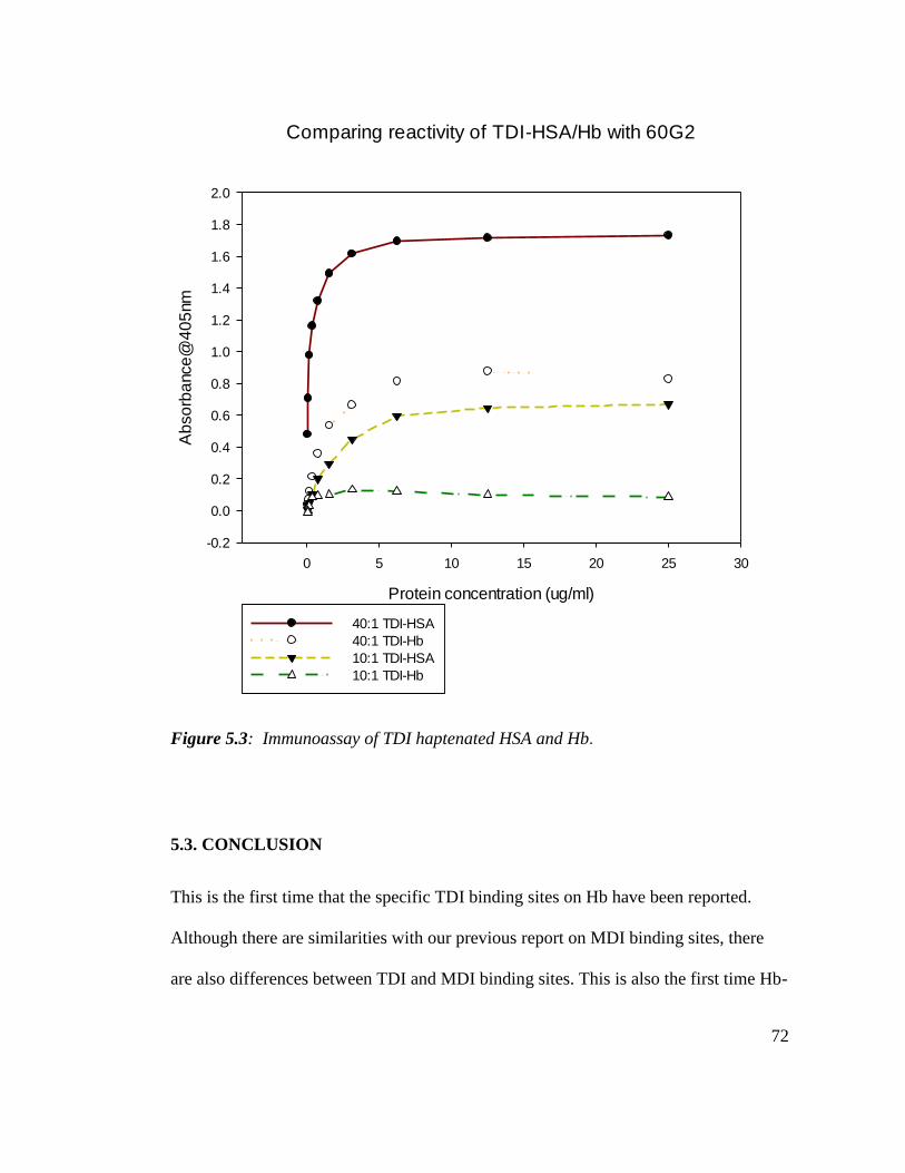
72
Comparing reactivity of TDI-HSA/Hb with 60G2
Protein concentration (ug/ml)
0 5 10 15 20 25 30
Ab
so
rba
nce
@4
05
nm
-0.2
0.0
0.2
0.4
0.6
0.8
1.0
1.2
1.4
1.6
1.8
2.0
40:1 TDI-HSA
40:1 TDI-Hb
10:1 TDI-HSA
10:1 TDI-Hb
Figure 5.3: Immunoassay of TDI haptenated HSA and Hb.
5.3. CONCLUSION
This is the first time that the specific TDI binding sites on Hb have been reported.
Although there are similarities with our previous report on MDI binding sites, there
are also differences between TDI and MDI binding sites. This is also the first time Hb-
73
dNCO conjugates were demonstrated to be antigenic. Although their involvement in
disease development can‘t be inferred from the observed antigenicity, it is important to
evaluate the antigenicity of potentially endogenously haptenated proteins. Both
qualitative and quantitative differences in TDI and HDI binding were observed
between Hb and HSA conjugates. This suggests that the choice of the isocyanate for
use in immunoassays has to be carefully considered.

74
CHAPTER 6
EVALUATING THE POTENTIAL FOR AN IgE INDEPENDENT
MECHANISM THROUGH WHICH DIISOCYANATE ASTHMA DEVELOPS
6.1 INTRODUCTION
Although the ultimate immunogenic forms that lead to dNCO asthma are still
unknown, dNCOs are low molecular weight chemical sensitizers that require
haptenation to biomolecules in vivo to produce dNCO-specific immune sensitization.
A number of biomolecules are targets for dNCO conjugation. Epithelial proteins are
the major targets upon inhalation exposure105,106 whereas skin proteins are targeted
upon skin exposure and both exposures can lead to OA. Kennedy et al107, using a rat
model demonstrated that serum albumin is the major target for TDI conjugation in the
blood.
The traditional mechanism for development of asthma involves recognition and
endocytosis of antigens by antigen presenting cells. These cells then digest the
antigens and associate the resultant peptides with major histocompatibility complex
molecules (MHC). The MHC-peptide complex is then expressed on the cell surface
together with co-stimulatory molecules, a requirement for successful CD4+ T-cell
(Th0) activation106. Differentiation of TH0 to TH1 or TH2 subtypes depends on the
nature of the antigen, cooperating cells, and cytokines. Allergenic antigens plus an
environment rich in IL-4 from mast cells encourage Th2 phenotype development that
leads to enhanced production of IL-4, IL-5, IL-3, and GM-CSF with minimal IFN-γ
production. The Th2 subtype is associated with IgE and eosinophilia and hence
allergy.
75
Only about 25 % of dNCO asthmatic workers have detectable levels of IgE to dNCO
HSA conjugates108. Although there is evidence that chemicals that cause respiratory
sensitization provoke immune responses of the TH2 type, the mechanism of dNCO
OA may possibly include cell-mediated and mixed reactions109,110. In a study by
Bottomly et al 111, female BALB/c mice sensitized to HDI developed HDI-specific
IgG as well as elevated levels of IL-5, IL-13 and IFN- γ on stimulation with HDI-
mouse serum albumin, indicating a mixed TH1/TH2 immune response. Most
importantly, these investigators observed that the sensitizing dose for antibody
production was higher than that for airway inflammation and cytokine production.
This suggests that different mechanisms dominate for the two outcomes. Del prete 112
and Maestrelli113 demonstrated that the majority of T-cell clones from patients with
TDI-induced asthma were CD8+ and capable of producing IL-5. David I. Bernstein et
al114 demonstrated that repeated antigenic stimulation of dNCO asthmatic peripheral
blood mononuclear cells (PBMC) in culture resulted in synthesis of tumor necrosis
factor-α (TNF- α), a non-IgE dependent pro-inflammatory cytokine, and monocyte
chemo-attractant protein-1, but not IL-4 or IL-5.
THP-1 cells were used in this study as a surrogate for antigen presenting cells and they
were derived from the peripheral blood of a 1-year-old male with acute monocytic
leukemia. They are single, round suspension cells with distinct monocytic markers115
with Fc and C3b receptors and lack surface and cytoplasmic immunoglobulins. THP-1
cells have advantages in studies over human primary monocytes-macrophages because

76
they have a homogeneous genetic background, which minimizes the degree of
variability in the cell phenotype116,117.
The detection of dNCO specific antibodies in only a subset of dNCO asthmatics has
been attributed to assay limitations and the potential for other non-IgE dependent
mechanisms. Our previous work has been focused on characterization of dNCO-
protein conjugates that are used in immunoassays for detection of dNCO specific
antibodies with a view to standardize these immunoassays. We have also evaluated
conjugates prepared in different conditions to evaluate the effect of preparative
conditions on the nature of the conjugates. In this work, we hypothesize that MDI,
apart from reacting with extracellular proteins, can also cross cell membranes intact
and react with intracellular proteins. This will result is formation of intracellular
conjugates which can possibly trigger cell mediated immune responses. To test this
hypothesis, the aims of this work were: (a) to expose THP-1 cells to different
concentrations of MDI, (b) use the HPLC method to analyze the membrane and
cytoplasm fractions for MDI conjugation, and (c) identify proteins from MDI-exposed
THP-1 cells using mass spectrometry.
6.2 RESULTS AND DISCUSSION
6.2.1 Quantification of MDI in the cytoplasm
THP-1 cells were incubated with different concentrations of MDI (dissolved in
ethylene glycol dimethyl ether) and exposures were performed in Hanks’ balanced
solution for 4 h at 37 ⁰C and 5 % CO2. After washing three times, the cells were lysed
using an ultra-sonicator and the cytoplasm fraction was extracted using cytoplasm
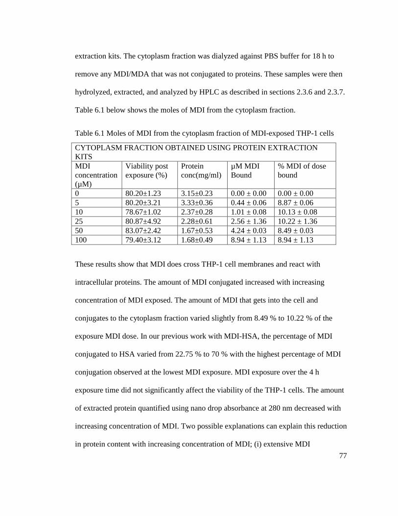
77
extraction kits. The cytoplasm fraction was dialyzed against PBS buffer for 18 h to
remove any MDI/MDA that was not conjugated to proteins. These samples were then
hydrolyzed, extracted, and analyzed by HPLC as described in sections 2.3.6 and 2.3.7.
Table 6.1 below shows the moles of MDI from the cytoplasm fraction.
Table 6.1 Moles of MDI from the cytoplasm fraction of MDI-exposed THP-1 cells
CYTOPLASM FRACTION OBTAINED USING PROTEIN EXTRACTION
KITS
MDI
concentration
(µM)
Viability post
exposure (%)
Protein
conc(mg/ml)
µM MDI
Bound
% MDI of dose
bound
0 80.20±1.23 3.15±0.23 0.00 ± 0.00 0.00 ± 0.00
5 80.20±3.21 3.33±0.36 0.44 ± 0.06 8.87 ± 0.06
10 78.67±1.02 2.37±0.28 1.01 ± 0.08 10.13 ± 0.08
25 80.87±4.92 2.28±0.61 2.56 ± 1.36 10.22 ± 1.36
50 83.07±2.42 1.67±0.53 4.24 ± 0.03 8.49 ± 0.03
100 79.40±3.12 1.68±0.49 8.94 ± 1.13 8.94 ± 1.13
These results show that MDI does cross THP-1 cell membranes and react with
intracellular proteins. The amount of MDI conjugated increased with increasing
concentration of MDI exposed. The amount of MDI that gets into the cell and
conjugates to the cytoplasm fraction varied slightly from 8.49 % to 10.22 % of the
exposure MDI dose. In our previous work with MDI-HSA, the percentage of MDI
conjugated to HSA varied from 22.75 % to 70 % with the highest percentage of MDI
conjugation observed at the lowest MDI exposure. MDI exposure over the 4 h
exposure time did not significantly affect the viability of the THP-1 cells. The amount
of extracted protein quantified using nano drop absorbance at 280 nm decreased with
increasing concentration of MDI. Two possible explanations can explain this reduction
in protein content with increasing concentration of MDI; (i) extensive MDI
78
conjugation may significantly reduce solubility of proteins in the extraction buffers
used. This phenomenon was observed with MDI conjugated HSA and Hb where the
conjugates tend to precipitate out of solution with time after conjugation, (ii) MDI-
conjugation may interfere with protein absorbance at 280 nm compromising the
protein quantification.
6.2.2. Quantification of MDI in the cell membrane fraction
The membrane fraction of MDI exposed THP-1 cells consist of the plasma membrane
and intracellular organelle membranes. These were extracted using membrane protein
extraction kits, dialyzed against PBS buffer using 12-14 000 MWCO membranes. The
dialyzed samples were hydrolyzed, extracted and analyzed for MDI using HPLC as
described in section 2.3.6 and 2.3.7. Table 6.2 shows the amount of MDI bound to the
membrane fraction. As expected, the amount of MDI bound to the membrane fraction
increased with increasing amount of MDI used. MDI binding in the membrane
fraction was less than that obtained from the cytoplasm fraction. It is also important to
note that the protein content was significantly lower in the membrane fraction
compared to the cytoplasm fraction as well.
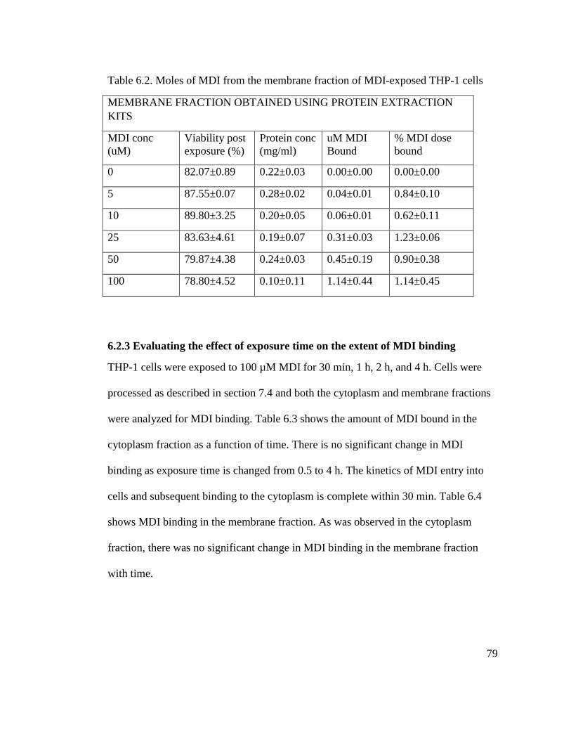
79
Table 6.2. Moles of MDI from the membrane fraction of MDI-exposed THP-1 cells
MEMBRANE FRACTION OBTAINED USING PROTEIN EXTRACTION
KITS
MDI conc
(uM)
Viability post
exposure (%)
Protein conc
(mg/ml)
uM MDI
Bound
% MDI dose
bound
0 82.07±0.89 0.22±0.03 0.00±0.00 0.00±0.00
5 87.55±0.07 0.28±0.02 0.04±0.01 0.84±0.10
10 89.80±3.25 0.20±0.05 0.06±0.01 0.62±0.11
25 83.63±4.61 0.19±0.07 0.31±0.03 1.23±0.06
50 79.87±4.38 0.24±0.03 0.45±0.19 0.90±0.38
100 78.80±4.52 0.10±0.11 1.14±0.44 1.14±0.45
6.2.3 Evaluating the effect of exposure time on the extent of MDI binding
THP-1 cells were exposed to 100 µM MDI for 30 min, 1 h, 2 h, and 4 h. Cells were
processed as described in section 7.4 and both the cytoplasm and membrane fractions
were analyzed for MDI binding. Table 6.3 shows the amount of MDI bound in the
cytoplasm fraction as a function of time. There is no significant change in MDI
binding as exposure time is changed from 0.5 to 4 h. The kinetics of MDI entry into
cells and subsequent binding to the cytoplasm is complete within 30 min. Table 6.4
shows MDI binding in the membrane fraction. As was observed in the cytoplasm
fraction, there was no significant change in MDI binding in the membrane fraction
with time.
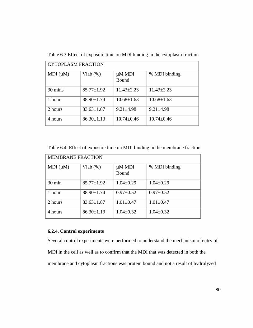
80
Table 6.3 Effect of exposure time on MDI binding in the cytoplasm fraction
CYTOPLASM FRACTION
MDI (µM) Viab (%) µM MDI
Bound
% MDI binding
30 mins 85.77±1.92 11.43±2.23 11.43±2.23
1 hour 88.90±1.74 10.68±1.63 10.68±1.63
2 hours 83.63±1.87 9.21±4.98 9.21±4.98
4 hours 86.30±1.13 10.74±0.46 10.74±0.46
Table 6.4. Effect of exposure time on MDI binding in the membrane fraction
MEMBRANE FRACTION
MDI (µM) Viab (%) µM MDI
Bound
% MDI binding
30 min 85.77±1.92 1.04±0.29 1.04±0.29
1 hour 88.90±1.74 0.97±0.52 0.97±0.52
2 hours 83.63±1.87 1.01±0.47 1.01±0.47
4 hours 86.30±1.13 1.04±0.32 1.04±0.32
6.2.4. Control experiments
Several control experiments were performed to understand the mechanism of entry of
MDI in the cell as well as to confirm that the MDI that was detected in both the
membrane and cytoplasm fractions was protein bound and not a result of hydrolyzed
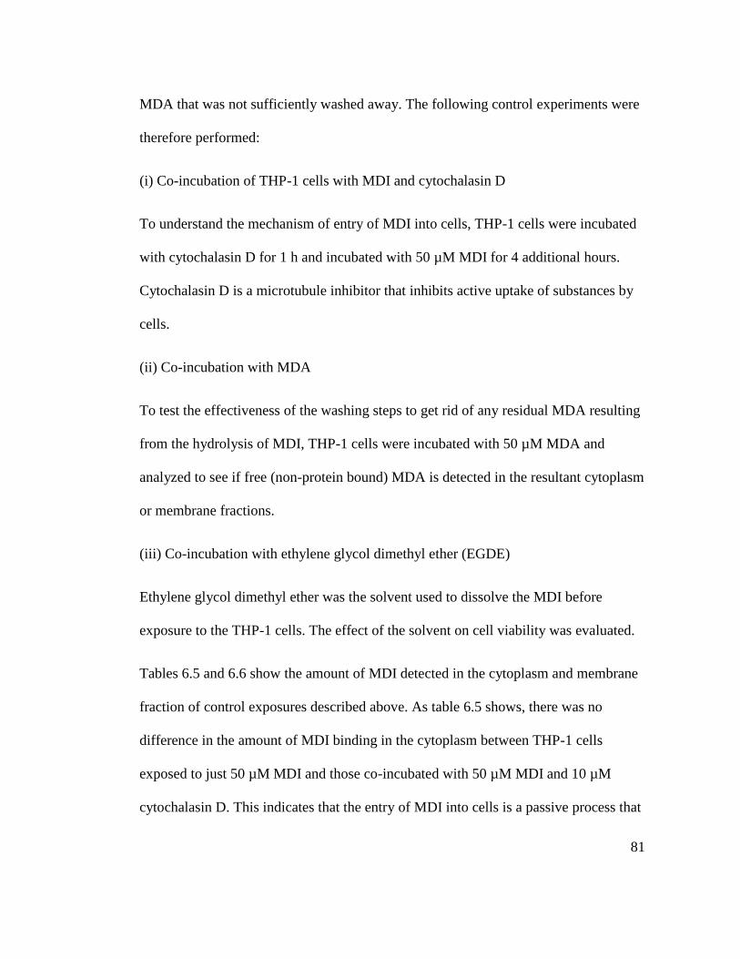
81
MDA that was not sufficiently washed away. The following control experiments were
therefore performed:
(i) Co-incubation of THP-1 cells with MDI and cytochalasin D
To understand the mechanism of entry of MDI into cells, THP-1 cells were incubated
with cytochalasin D for 1 h and incubated with 50 µM MDI for 4 additional hours.
Cytochalasin D is a microtubule inhibitor that inhibits active uptake of substances by
cells.
(ii) Co-incubation with MDA
To test the effectiveness of the washing steps to get rid of any residual MDA resulting
from the hydrolysis of MDI, THP-1 cells were incubated with 50 µM MDA and
analyzed to see if free (non-protein bound) MDA is detected in the resultant cytoplasm
or membrane fractions.
(iii) Co-incubation with ethylene glycol dimethyl ether (EGDE)
Ethylene glycol dimethyl ether was the solvent used to dissolve the MDI before
exposure to the THP-1 cells. The effect of the solvent on cell viability was evaluated.
Tables 6.5 and 6.6 show the amount of MDI detected in the cytoplasm and membrane
fraction of control exposures described above. As table 6.5 shows, there was no
difference in the amount of MDI binding in the cytoplasm between THP-1 cells
exposed to just 50 µM MDI and those co-incubated with 50 µM MDI and 10 µM
cytochalasin D. This indicates that the entry of MDI into cells is a passive process that
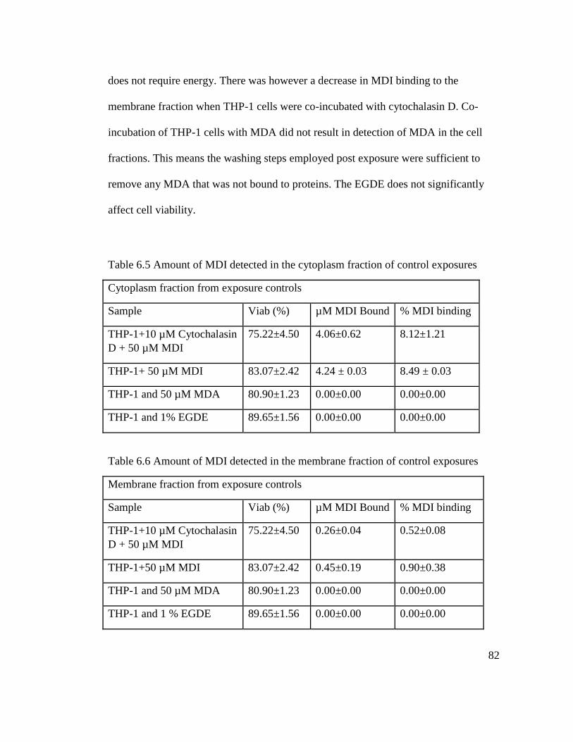
82
does not require energy. There was however a decrease in MDI binding to the
membrane fraction when THP-1 cells were co-incubated with cytochalasin D. Co-
incubation of THP-1 cells with MDA did not result in detection of MDA in the cell
fractions. This means the washing steps employed post exposure were sufficient to
remove any MDA that was not bound to proteins. The EGDE does not significantly
affect cell viability.
Table 6.5 Amount of MDI detected in the cytoplasm fraction of control exposures
Cytoplasm fraction from exposure controls
Sample Viab (%) µM MDI Bound % MDI binding
THP-1+10 µM Cytochalasin
D + 50 µM MDI
75.22±4.50 4.06±0.62 8.12±1.21
THP-1+ 50 µM MDI 83.07±2.42 4.24 ± 0.03 8.49 ± 0.03
THP-1 and 50 µM MDA 80.90±1.23 0.00±0.00 0.00±0.00
THP-1 and 1% EGDE 89.65±1.56 0.00±0.00 0.00±0.00
Table 6.6 Amount of MDI detected in the membrane fraction of control exposures
Membrane fraction from exposure controls
Sample Viab (%) µM MDI Bound % MDI binding
THP-1+10 µM Cytochalasin
D + 50 µM MDI
75.22±4.50 0.26±0.04 0.52±0.08
THP-1+50 µM MDI 83.07±2.42 0.45±0.19 0.90±0.38
THP-1 and 50 µM MDA 80.90±1.23 0.00±0.00 0.00±0.00
THP-1 and 1 % EGDE 89.65±1.56 0.00±0.00 0.00±0.00
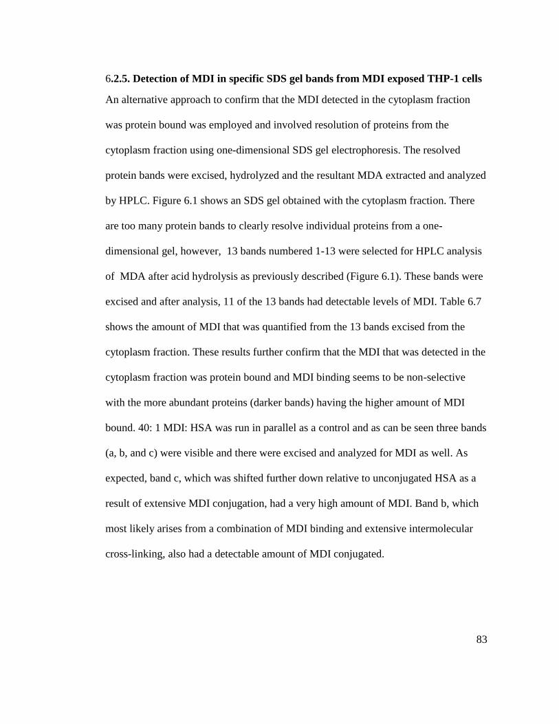
83
6.2.5. Detection of MDI in specific SDS gel bands from MDI exposed THP-1 cells
An alternative approach to confirm that the MDI detected in the cytoplasm fraction
was protein bound was employed and involved resolution of proteins from the
cytoplasm fraction using one-dimensional SDS gel electrophoresis. The resolved
protein bands were excised, hydrolyzed and the resultant MDA extracted and analyzed
by HPLC. Figure 6.1 shows an SDS gel obtained with the cytoplasm fraction. There
are too many protein bands to clearly resolve individual proteins from a one-
dimensional gel, however, 13 bands numbered 1-13 were selected for HPLC analysis
of MDA after acid hydrolysis as previously described (Figure 6.1). These bands were
excised and after analysis, 11 of the 13 bands had detectable levels of MDI. Table 6.7
shows the amount of MDI that was quantified from the 13 bands excised from the
cytoplasm fraction. These results further confirm that the MDI that was detected in the
cytoplasm fraction was protein bound and MDI binding seems to be non-selective
with the more abundant proteins (darker bands) having the higher amount of MDI
bound. 40: 1 MDI: HSA was run in parallel as a control and as can be seen three bands
(a, b, and c) were visible and there were excised and analyzed for MDI as well. As
expected, band c, which was shifted further down relative to unconjugated HSA as a
result of extensive MDI conjugation, had a very high amount of MDI. Band b, which
most likely arises from a combination of MDI binding and extensive intermolecular
cross-linking, also had a detectable amount of MDI conjugated.

84
Figure 6.1. Resolution of THP-1 Cytoplasm proteins. A 12 % denaturing gel of MDI
exposed THP-1 cells’ cytoplasm fraction. Lane 1 is the molecular weight marker, lane
2 is HSA, lanes 3 is 40:1 MDI: HSA, 4 is unconjugated THP-1 cytoplasm and lanes 5-
9 are replicates of the cytoplasm fraction exposed to 100 µM MDI.
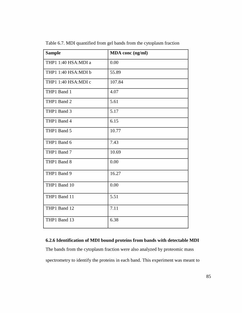
85
Table 6.7. MDI quantified from gel bands from the cytoplasm fraction
Sample MDA conc (ng/ml)
THP1 1:40 HSA:MDI a 0.00
THP1 1:40 HSA:MDI b 55.89
THP1 1:40 HSA:MDI c 107.84
THP1 Band 1 4.07
THP1 Band 2 5.61
THP1 Band 3 5.17
THP1 Band 4 6.15
THP1 Band 5 10.77
THP1 Band 6 7.43
THP1 Band 7 10.69
THP1 Band 8 0.00
THP1 Band 9 16.27
THP1 Band 10 0.00
THP1 Band 11 5.51
THP1 Band 12 7.11
THP1 Band 13 6.38
6.2.6 Identification of MDI bound proteins from bands with detectable MDI
The bands from the cytoplasm fraction were also analyzed by proteomic mass
spectrometry to identify the proteins in each band. This experiment was meant to
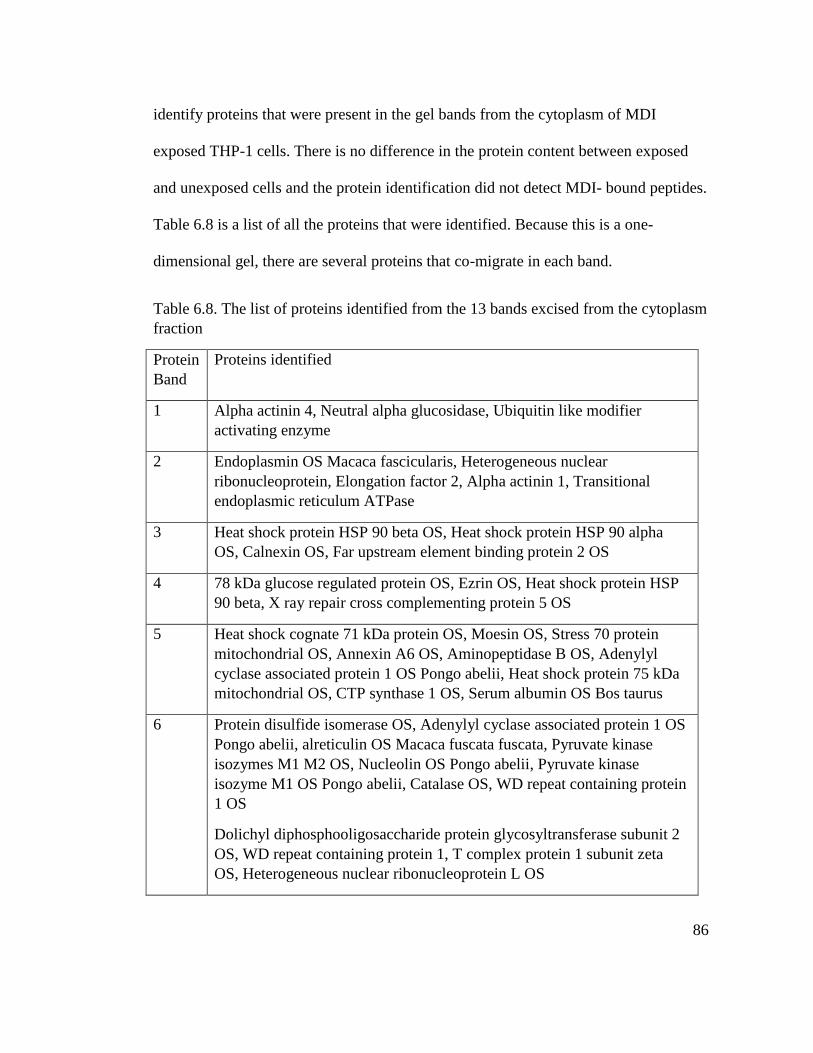
86
identify proteins that were present in the gel bands from the cytoplasm of MDI
exposed THP-1 cells. There is no difference in the protein content between exposed
and unexposed cells and the protein identification did not detect MDI- bound peptides.
Table 6.8 is a list of all the proteins that were identified. Because this is a one-
dimensional gel, there are several proteins that co-migrate in each band.
Table 6.8. The list of proteins identified from the 13 bands excised from the cytoplasm
fraction
Protein
Band
Proteins identified
1 Alpha actinin 4, Neutral alpha glucosidase, Ubiquitin like modifier
activating enzyme
2 Endoplasmin OS Macaca fascicularis, Heterogeneous nuclear
ribonucleoprotein, Elongation factor 2, Alpha actinin 1, Transitional
endoplasmic reticulum ATPase
3 Heat shock protein HSP 90 beta OS, Heat shock protein HSP 90 alpha
OS, Calnexin OS, Far upstream element binding protein 2 OS
4 78 kDa glucose regulated protein OS, Ezrin OS, Heat shock protein HSP
90 beta, X ray repair cross complementing protein 5 OS
5 Heat shock cognate 71 kDa protein OS, Moesin OS, Stress 70 protein
mitochondrial OS, Annexin A6 OS, Aminopeptidase B OS, Adenylyl
cyclase associated protein 1 OS Pongo abelii, Heat shock protein 75 kDa
mitochondrial OS, CTP synthase 1 OS, Serum albumin OS Bos taurus
6 Protein disulfide isomerase OS, Adenylyl cyclase associated protein 1 OS
Pongo abelii, alreticulin OS Macaca fuscata fuscata, Pyruvate kinase
isozymes M1 M2 OS, Nucleolin OS Pongo abelii, Pyruvate kinase
isozyme M1 OS Pongo abelii, Catalase OS, WD repeat containing protein
1 OS
Dolichyl diphosphooligosaccharide protein glycosyltransferase subunit 2
OS, WD repeat containing protein 1, T complex protein 1 subunit zeta
OS, Heterogeneous nuclear ribonucleoprotein L OS
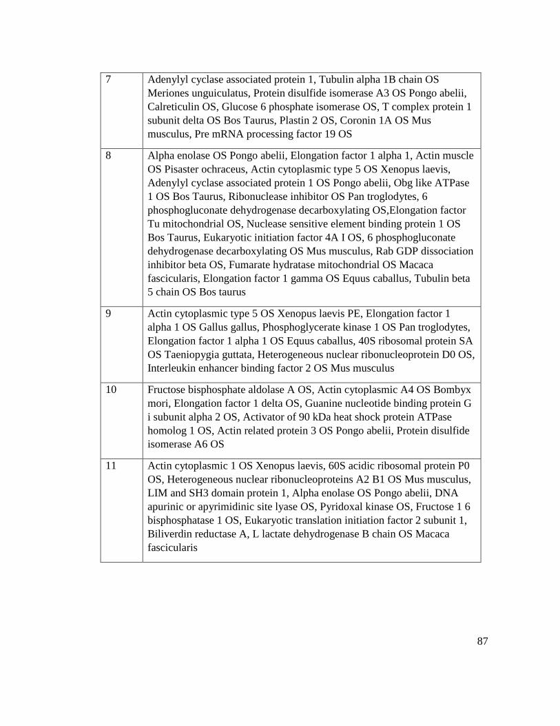
87
7 Adenylyl cyclase associated protein 1, Tubulin alpha 1B chain OS
Meriones unguiculatus, Protein disulfide isomerase A3 OS Pongo abelii,
Calreticulin OS, Glucose 6 phosphate isomerase OS, T complex protein 1
subunit delta OS Bos Taurus, Plastin 2 OS, Coronin 1A OS Mus
musculus, Pre mRNA processing factor 19 OS
8 Alpha enolase OS Pongo abelii, Elongation factor 1 alpha 1, Actin muscle
OS Pisaster ochraceus, Actin cytoplasmic type 5 OS Xenopus laevis,
Adenylyl cyclase associated protein 1 OS Pongo abelii, Obg like ATPase
1 OS Bos Taurus, Ribonuclease inhibitor OS Pan troglodytes, 6
phosphogluconate dehydrogenase decarboxylating OS,Elongation factor
Tu mitochondrial OS, Nuclease sensitive element binding protein 1 OS
Bos Taurus, Eukaryotic initiation factor 4A I OS, 6 phosphogluconate
dehydrogenase decarboxylating OS Mus musculus, Rab GDP dissociation
inhibitor beta OS, Fumarate hydratase mitochondrial OS Macaca
fascicularis, Elongation factor 1 gamma OS Equus caballus, Tubulin beta
5 chain OS Bos taurus
9 Actin cytoplasmic type 5 OS Xenopus laevis PE, Elongation factor 1
alpha 1 OS Gallus gallus, Phosphoglycerate kinase 1 OS Pan troglodytes,
Elongation factor 1 alpha 1 OS Equus caballus, 40S ribosomal protein SA
OS Taeniopygia guttata, Heterogeneous nuclear ribonucleoprotein D0 OS,
Interleukin enhancer binding factor 2 OS Mus musculus
10 Fructose bisphosphate aldolase A OS, Actin cytoplasmic A4 OS Bombyx
mori, Elongation factor 1 delta OS, Guanine nucleotide binding protein G
i subunit alpha 2 OS, Activator of 90 kDa heat shock protein ATPase
homolog 1 OS, Actin related protein 3 OS Pongo abelii, Protein disulfide
isomerase A6 OS
11 Actin cytoplasmic 1 OS Xenopus laevis, 60S acidic ribosomal protein P0
OS, Heterogeneous nuclear ribonucleoproteins A2 B1 OS Mus musculus,
LIM and SH3 domain protein 1, Alpha enolase OS Pongo abelii, DNA
apurinic or apyrimidinic site lyase OS, Pyridoxal kinase OS, Fructose 1 6
bisphosphatase 1 OS, Eukaryotic translation initiation factor 2 subunit 1,
Biliverdin reductase A, L lactate dehydrogenase B chain OS Macaca
fascicularis
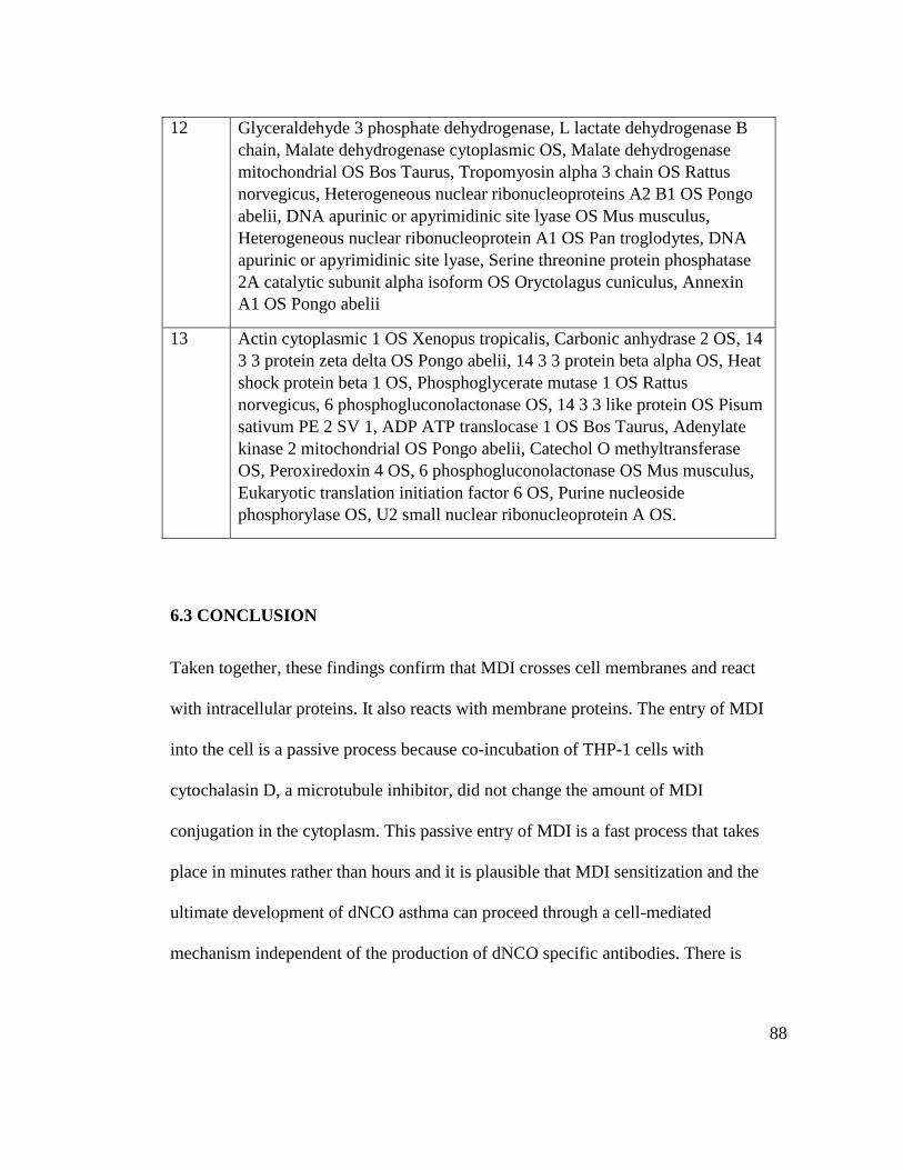
88
12 Glyceraldehyde 3 phosphate dehydrogenase, L lactate dehydrogenase B
chain, Malate dehydrogenase cytoplasmic OS, Malate dehydrogenase
mitochondrial OS Bos Taurus, Tropomyosin alpha 3 chain OS Rattus
norvegicus, Heterogeneous nuclear ribonucleoproteins A2 B1 OS Pongo
abelii, DNA apurinic or apyrimidinic site lyase OS Mus musculus,
Heterogeneous nuclear ribonucleoprotein A1 OS Pan troglodytes, DNA
apurinic or apyrimidinic site lyase, Serine threonine protein phosphatase
2A catalytic subunit alpha isoform OS Oryctolagus cuniculus, Annexin
A1 OS Pongo abelii
13 Actin cytoplasmic 1 OS Xenopus tropicalis, Carbonic anhydrase 2 OS, 14
3 3 protein zeta delta OS Pongo abelii, 14 3 3 protein beta alpha OS, Heat
shock protein beta 1 OS, Phosphoglycerate mutase 1 OS Rattus
norvegicus, 6 phosphogluconolactonase OS, 14 3 3 like protein OS Pisum
sativum PE 2 SV 1, ADP ATP translocase 1 OS Bos Taurus, Adenylate
kinase 2 mitochondrial OS Pongo abelii, Catechol O methyltransferase
OS, Peroxiredoxin 4 OS, 6 phosphogluconolactonase OS Mus musculus,
Eukaryotic translation initiation factor 6 OS, Purine nucleoside
phosphorylase OS, U2 small nuclear ribonucleoprotein A OS.
6.3 CONCLUSION
Taken together, these findings confirm that MDI crosses cell membranes and react
with intracellular proteins. It also reacts with membrane proteins. The entry of MDI
into the cell is a passive process because co-incubation of THP-1 cells with
cytochalasin D, a microtubule inhibitor, did not change the amount of MDI
conjugation in the cytoplasm. This passive entry of MDI is a fast process that takes
place in minutes rather than hours and it is plausible that MDI sensitization and the
ultimate development of dNCO asthma can proceed through a cell-mediated
mechanism independent of the production of dNCO specific antibodies. There is

89
ongoing work that involves running 2-dimensional gel electrophoresis and western
blot analysis using an MDI specific monoclonal antibody (IgG).
90
Reference List
1. Munn, S. J.; Hansen, B. G. EU risk assessment: science and policy. Toxicology
2002, 181-182, 281-285.
2. Piirila, P. L.; Nordman, H.; Keskinen, H. M.; Luukkonen, R.; Salo, S. P.;
Tuomi, T. O.; Tuppurainen, M. Long-term follow-up of hexamethylene
diisocyanate-, diphenylmethane diisocyanate-, and toluene
diisocyanate-induced asthma. Am. J. Respir. Crit Care Med. 2000, 162
(2 Pt 1), 516-522.
3. Frick, M.; Bjorkner, B.; Hamnerius, N.; Zimerson, E. Allergic contact
dermatitis from dicyclohexylmethane-4,4'-diisocyanate. Contact
Dermatitis 2003, 48 (6), 305-309.
4. Krone, C. A.; Klingner, T. D. Isocyanates, polyurethane and childhood asthma.
Pediatr. Allergy Immunol. 2005, 16 (5), 368-379.
5. Ulrich, H. Chemistry and technology of isocyanates; Wiley New York: 1996.
6. Clark, R. L.; Bugler, J.; Paddle, G. M.; Chamberlain, J. D.; Allport, D. C. A
17-year epidemiological study on changes in lung function in toluene
diisocyanate foam workers. Int. Arch. Occup. Environ. Health 2003, 76
(4), 295-301.
7. Karol, M. H.; Hauth, B. A.; Riley, E. J.; Magreni, C. M. Dermal contact with
toluene diisocyanate (TDI) produces respiratory tract hypersensitivity
in guinea pigs. Toxicol. Appl. Pharmacol. 1981, 58 (2), 221-230.
8. Petsonk, E. L.; Wang, M. L.; Lewis, D. M.; Siegel, P. D.; Husberg, B. J.
Asthma-like symptoms in wood product plant workers exposed to
methylene diphenyl diisocyanate. Chest 2000, 118 (4), 1183-1193.
9. Krone, C. A. Diisocyanates and nonoccupational disease: a review. Arch.
Environ. Health 2004, 59 (6), 306-316.
10. LeSage, J.; Stanley, J.; Karoly, W. J.; Lichtenberg, F. W. Airborne methylene
diphenyl diisocyanate (MDI) concentrations associated with the
application of polyurethane spray foam in residential construction. J.
Occup. Environ. Hyg. 2007, 4 (2), 145-155.
11. Crespo, J.; Galan, J. Exposure to MDI during the process of insulating
buildings with sprayed polyurethane foam. Ann. Occup. Hyg. 1999, 43
(6), 415-419.
91
12. Liljelind, I.; Norberg, C.; Egelrud, L.; Westberg, H.; Eriksson, K.; Nylander-
French, L. A. Dermal and inhalation exposure to methylene bisphenyl
isocyanate (MDI) in iron foundry workers. Ann. Occup. Hyg. 2010, 54
(1), 31-40.
13. Robert, A.; Ducos, P.; Francin, J. M.; Marsan, P. Biological monitoring of
workers exposed to 4,4'-methylenediphenyl diisocyanate (MDI) in 19
French polyurethane industries. Int. Arch. Occup. Environ. Health
2007, 80 (5), 412-422.
14. Wisnewski, A. V.; Xu, L.; Robinson, E.; Liu, J.; Redlich, C. A.; Herrick, C. A.
Immune sensitization to methylene diphenyl diisocyanate (MDI)
resulting from skin exposure: albumin as a carrier protein connecting
skin exposure to subsequent respiratory responses. J. Occup. Med.
Toxicol. 2011, 6, 6.
15. Kimber, I.; Basketter, D. Thresholds, dose-response relationships and dose
metrics in allergic contact dermatitis. Br. J. Dermatol. 2008, 159 (6),
1380-1381.
16. Mapp, C. E.; Boschetto, P.; Maestrelli, P.; Fabbri, L. M. Occupational asthma.
Am. J. Respir. Crit Care Med. 2005, 172 (3), 280-305.
17. Redlich, C. A.; Karol, M. H. Diisocyanate asthma: clinical aspects and
immunopathogenesis. Int. Immunopharmacol. 2002, 2 (2-3), 213-224.
18. Ott, M. G.; Diller, W. F.; Jolly, A. T. Respiratory effects of toluene
diisocyanate in the workplace: a discussion of exposure-response
relationships. Crit Rev. Toxicol. 2003, 33 (1), 1-59.
19. Karol, M. H. Respiratory effects of inhaled isocyanates. Crit Rev. Toxicol.
1986, 16 (4), 349-379.
20. Matheson, J. M.; Johnson, V. J.; Vallyathan, V.; Luster, M. I. Exposure and
immunological determinants in a murine model for toluene
diisocyanate (TDI) asthma. Toxicol. Sci. 2005, 84 (1), 88-98.
21. Baur, X. Hypersensitivity pneumonitis (extrinsic allergic alveolitis) induced by
isocyanates. J. Allergy Clin. Immunol. 1995, 95 (5 Pt 1), 1004-1010.
22. Charles, J.; Bernstein, A.; Jones, B.; Jones, D. J.; Edwards, J. H.; Seal, R. M.;
Seaton, A. Hypersensitivity pneumonitis after exposure to isocyanates.
Thorax 1976, 31 (2), 127-136.
92
23. Hur, G. Y.; Koh, D. H.; Choi, G. S.; Park, H. J.; Choi, S. J.; Ye, Y. M.; Kim,
K. S.; Park, H. S. Clinical and immunologic findings of methylene
diphenyl diisocyanate-induced occupational asthma in a car upholstery
factory. Clin. Exp. Allergy 2008, 38 (4), 586-593.
24. Butcher, B. T.; Salvaggio, J. E. Occupational asthma. J. Allergy Clin. Immunol.
1986, 78 (4 Pt 1), 547-556.
25. Bernstein, J. A. Overview of diisocyanate occupational asthma. Toxicology
1996, 111 (1-3), 181-189.
26. Wisnewski, A. V.; Redlich, C. A. Recent developments in diisocyanate
asthma. Curr. Opin. Allergy Clin. Immunol. 2001, 1 (2), 169-175.
27. Chipinda, I.; Zhang, X. D.; Simoyi, R. H.; Siegel, P. D. Mercaptobenzothiazole
allergenicity-role of the thiol group. Cutan. Ocul. Toxicol. 2008, 27 (2),
103-116.
28. Hettick, J. M.; Siegel, P. D. Determination of the toluene diisocyanate binding
sites on human serum albumin by tandem mass spectrometry. Anal.
Biochem. 2011, 414 (2), 232-238.
29. Maestrelli, P.; Boschetto, P.; Fabbri, L. M.; Mapp, C. E. Mechanisms of
occupational asthma. J. Allergy Clin. Immunol. 2009, 123 (3), 531-542.
30. Mapp, C. E.; Miotto, D.; Boschetto, P. Occupational asthma. Med. Lav. 2006,
97 (2), 404-409.
31. Biagini, R. E.; Bernstein, I. L.; Gallagher, J. S.; Moorman, W. J.; Brooks, S.;
Gann, P. H. The diversity of reaginic immune responses to platinum
and palladium metallic salts. J. Allergy Clin. Immunol. 1985, 76 (6),
794-802.
32. Mapp, C. E. Agents, old and new, causing occupational asthma. Occup.
Environ. Med. 2001, 58 (5), 354-60, 290.
33. Chan-Yeung, M. Assessment of asthma in the workplace. ACCP consensus
statement. American College of Chest Physicians. Chest 1995, 108 (4),
1084-1117.
34. Karol, M. H.; Tollerud, D. J.; Campbell, T. P.; Fabbri, L.; Maestrelli, P.;
Saetta, M.; Mapp, C. E. Predictive value of airways
hyperresponsiveness and circulating IgE for identifying types of
responses to toluene diisocyanate inhalation challenge. Am. J. Respir.
Crit Care Med. 1994, 149 (3 Pt 1), 611-615.
93
35. Park, H. S.; Lee, S. K.; Lee, Y. M.; Kim, S. S.; Nahm, D. H. Longitudinal
study of specific antibodies to toluene diisocyanate (TDI)-human serum
albumin (HSA) conjugate in patients with TDI-induced asthma. Korean
J. Intern. Med. 2002, 17 (4), 249-251.
36. Jones, M. G.; Floyd, A.; Nouri-Aria, K. T.; Jacobson, M. R.; Durham, S. R.;
Taylor, A. N.; Cullinan, P. Is occupational asthma to diisocyanates a
non-IgE-mediated disease? J. Allergy Clin. Immunol. 2006, 117 (3),
663-669.
37. Jame, A. J.; Lackie, P. M.; Cazaly, A. M.; Sayers, I.; Penrose, J. F.; Holgate, S.
T.; Sampson, A. P. Human bronchial epithelial cells express an active
and inducible biosynthetic pathway for leukotrienes B4 and C4. Clin.
Exp. Allergy 2007, 37 (6), 880-892.
38. Holgate, S. T.; Holloway, J.; Wilson, S.; Bucchieri, F.; Puddicombe, S.;
Davies, D. E. Epithelial-mesenchymal communication in the
pathogenesis of chronic asthma. Proc. Am. Thorac. Soc. 2004, 1 (2),
93-98.
39. Lemiere, C.; Malo, J. L.; Boutet, M. Reactive airways dysfunction syndrome
due to chlorine: sequential bronchial biopsies and functional
assessment. Eur. Respir. J. 1997, 10 (1), 241-244.
40. Ott, M. G.; Jolly, A. T.; Burkert, A. L.; Brown, W. E. Issues in diisocyanate
antibody testing. Crit Rev. Toxicol. 2007, 37 (7), 567-585.
41. Wass, U.; Belin, L. Immunologic specificity of isocyanate-induced IgE
antibodies in serum from 10 sensitized workers. J. Allergy Clin.
Immunol. 1989, 83 (1), 126-135.
42. Venables, K. M.; Chan-Yeung, M. Occupational asthma. Lancet 1997, 349
(9063), 1465-1469.
43. Bernstein, D. I.; Jolly, A. Current diagnostic methods for diisocyanate induced
occupational asthma. Am. J. Ind. Med. 1999, 36 (4), 459-468.
44. Bradding, P.; Walls, A. F.; Holgate, S. T. The role of the mast cell in the
pathophysiology of asthma. J. Allergy Clin. Immunol. 2006, 117 (6),
1277-1284.
45. Del Prete, G. F.; De, C. M.; D'Elios, M. M.; Maestrelli, P.; Ricci, M.; Fabbri,
L.; Romagnani, S. Allergen exposure induces the activation of allergen-
specific Th2 cells in the airway mucosa of patients with allergic
respiratory disorders. Eur. J. Immunol. 1993, 23 (7), 1445-1449.
94
46. Maestrelli, P.; Del Prete, G. F.; De, C. M.; D'Elios, M. M.; Saetta, M.; Di, S.
A.; Mapp, C. E.; Romagnani, S.; Fabbri, L. M. CD8 T-cell clones
producing interleukin-5 and interferon-gamma in bronchial mucosa of
patients with asthma induced by toluene diisocyanate. Scand. J. Work
Environ. Health 1994, 20 (5), 376-381.
47. Wisnewski, A. V.; Cain, H.; Magoski, N.; Wang, H.; Holm, C. T.; Redlich, C.
A. Human gamma/delta T-cell lines derived from airway biopsies. Am.
J. Respir. Cell Mol. Biol. 2001, 24 (3), 332-338.
48. Wisnewski, A. V.; Herrick, C. A.; Liu, Q.; Chen, L.; Bottomly, K.; Redlich, C.
A. Human gamma/delta T-cell proliferation and IFN-gamma
production induced by hexamethylene diisocyanate. J. Allergy Clin.
Immunol. 2003, 112 (3), 538-546.
49. Kay, A. B.; Corrigan, C. J.; Frew, A. J. The role of cellular immunology in
asthma. Eur. Respir. J. Suppl 1991, 13, 105s-112s.
50. Maestrelli, P.; O'Hehir, R. E.; Lamb, J. R.; Tsai, J. J.; Cromwell, O.; Kay, A.
B. Antigen-induced neutrophil chemotactic factor from cloned human
T lymphocytes. Immunology 1988, 65 (4), 605-609.
51. Finotto, S.; Fabbri, L. M.; Rado, V.; Mapp, C. E.; Maestrelli, P. Increase in
numbers of CD8 positive lymphocytes and eosinophils in peripheral
blood of subjects with late asthmatic reactions induced by toluene
diisocyanate. Br. J. Ind. Med. 1991, 48 (2), 116-121.
52. Chan-Yeung, M.; Malo, J. L. Occupational asthma. N. Engl. J. Med. 1995, 333
(2), 107-112.
53. Whitehouse, C. M.; Dreyer, R. N.; Yamashita, M.; Fenn, J. B. Electrospray
interface for liquid chromatographs and mass spectrometers. Anal.
Chem. 1985, 57 (3), 675-679.
54. Fenn, J. B.; Mann, M.; Meng, C. K.; Wong, S. F.; Whitehouse, C. M.
Electrospray ionization for mass spectrometry of large biomolecules.
Science 1989, 246 (4926), 64-71.
55. Mhike, M.; Chipinda, I.; Hettick, J. M.; Simoyi, R. H.; Lemons, A.; Green, B.
J.; Siegel, P. D. Characterization of methylene diphenyl diisocyanate-
haptenated human serum albumin and hemoglobin. Anal. Biochem.
2013, 440 (2), 197-204.
95
56. Takahashi, S.; Yamamura, T.; Kamo, M.; Satake, K. Regeneration of amino
compounds from the 2, 4, 6-trinitrophenyl derivatives by treatment with
hydrazine. Chemistry Letters 1984, (1), 127-130.
57. Snyder, S. L.; Sobocinski, P. Z. An improved 2,4,6-trinitrobenzenesulfonic
acid method for the determination of amines. Anal. Biochem. 1975, 64
(1), 284-288.
58. Lemus, R.; Lukinskeine, L.; Bier, M. E.; Wisnewski, A. V.; Redlich, C. A.;
Karol, M. H. Development of immunoassays for biomonitoring of
hexamethylene diisocyanate exposure. Environ. Health Perspect. 2001,
109 (11), 1103-1108.
59. Mhike, M.; Chipinda, I.; Hettick, J. M.; Simoyi, R. H.; Lemons, A.; Green, B.
J.; Siegel, P. D. Characterization of methylene diphenyl diisocyanate-
haptenated human serum albumin and hemoglobin. Anal. Biochem.
2013, 440 (2), 197-204.
60. Lemons, A. R.; Siegel, P. D.; Mhike, M.; Law, B. F.; Hettick, J. M.; Bledsoe,
T. A.; Nayak, A. P.; Beezhold, D. H.; Green, B. J. A murine
monoclonal antibody with broad specificity for occupationally relevant
diisocyanates. J. Occup. Environ. Hyg. 2014, 11 (2), 101-110.
61. Lemons, A. R.; Bledsoe, T. A.; Siegel, P. D.; Beezhold, D. H.; Green, B. J.
Development of sandwich ELISAs for the detection of aromatic
diisocyanate adducts. J. Immunol. Methods 2013, 397 (1-2), 66-70.
62. Ruwona, T. B.; Johnson, V. J.; Schmechel, D.; Simoyi, R. H.; Beezhold, D.;
Siegel, P. D. Monoclonal antibodies against toluene diisocyanate
haptenated proteins from vapor-exposed mice. Hybridoma (Larchmt. )
2010, 29 (3), 221-229.
63. Ruwona, T. B.; Johnson, V. J.; Hettick, J. M.; Schmechel, D.; Beezhold, D.;
Wang, W.; Simoyi, R. H.; Siegel, P. D. Production, characterization
and utility of a panel of monoclonal antibodies for the detection of
toluene diisocyanate haptenated proteins. J. Immunol. Methods 2011,
373 (1-2), 127-135.
64. Tee, R. D.; Cullinan, P.; Welch, J.; Burge, P. S.; Newman-Taylor, A. J.
Specific IgE to isocyanates: a useful diagnostic role in occupational
asthma. J. Allergy Clin. Immunol. 1998, 101 (5), 709-715.
65. Wisnewski, A. V.; Stowe, M. H.; Cartier, A.; Liu, Q.; Liu, J.; Chen, L.;
Redlich, C. A. Isocyanate vapor-induced antigenicity of human
albumin. J. Allergy Clin. Immunol. 2004, 113 (6), 1178-1184.
96
66. Park, H. S.; Park, J. N.; Kim, J. W.; Kim, S. K. Clinical and immunological
evaluation of isocyanate-exposed workers. J. Korean Med. Sci. 1992, 7
(2), 122-127.
67. Sabbioni, G.; Dongari, N.; Schneider, S.; Kumar, A. Synthetic approaches to
obtain amino acid adducts of 4,4'-methylenediphenyl diisocyanate.
Chem. Res. Toxicol. 2012, 25 (12), 2704-2714.
68. Kennedy, A. L.; Brown, W. E. Isocyanates and lung disease: experimental
approaches to molecular mechanisms. Occup. Med. 1992, 7 (2), 301-
329.
69. Brown, W. E.; Green, A. H.; Cedel, T. E.; Cairns, J. Biochemistry of protein-
isocyanate interactions: a comparison of the effects of aryl vs. alkyl
isocyanates. Environ. Health Perspect. 1987, 72, 5-11.
70. Kumar, A.; Dongari, N.; Sabbioni, G. New isocyanate-specific albumin
adducts of 4,4'-methylenediphenyl diisocyanate (MDI) in rats. Chem.
Res. Toxicol. 2009, 22 (12), 1975-1983.
71. Sepai, O.; Schutze, D.; Heinrich, U.; Hoymann, H. G.; Henschler, D.;
Sabbioni, G. Hemoglobin adducts and urine metabolites of 4,4'-
methylenedianiline after 4,4'-methylenediphenyl diisocyanate exposure
of rats. Chem. Biol. Interact. 1995, 97 (2), 185-198.
72. Sabbioni, G.; Schutze, D. Hemoglobin binding of bicyclic aromatic amines.
Chem. Res. Toxicol. 1998, 11 (5), 471-483.
73. Pauluhn, J.; Brown, W. E.; Hext, P.; Leibold, E.; Leng, G. Analysis of
biomarkers in rats and dogs exposed to polymeric methylenediphenyl
diisocyanate (pMDI) and its glutathione adduct. Toxicology 2006, 222
(3), 202-212.
74. Sakai, T.; Morita, Y.; Kim, Y.; Tao, Y. X. LCÇôMS determination of urinary
toluenediamine in workers exposed to toluenediisocyanate. Toxicology
letters 2002, 134 (1), 259-264.
75. Lemus, R.; Karol, M. H. Conjugation of haptens. Methods Mol. Med. 2008,
138, 167-182.
76. Zhao, X.; Suo, Y. Analysis of primary aromatic amines using precolumn
derivatization by HPLC fluorescence detection and online MS
identification. J. Sep. Sci. 2008, 31 (4), 646-658.
97
77. Wallace, J. M.; Fox, P. F. Rapid spectrophotometric and fluorimetric methods
for monitoring nitrogenous (proteinaceous) compounds in cheese and
cheese fractions: a review. Food chemistry 1998, 62 (2), 217-224.
78. Toker, S. E.; Sagirli, O.; Cetin, S. M.; Onal, A. A new HPLC method with
fluorescence detection for the determination of memantine in human
plasma. J. Sep. Sci. 2011, 34 (19), 2645-2649.
79. Sabbioni, G.; Hartley, R.; Henschler, D.; Hollrigl-Rosta, A.; Koeber, R.;
Schneider, S. Isocyanate-specific hemoglobin adduct in rats exposed to
4, 4'-methylenediphenyl diisocyanate. Chem. Res. Toxicol. 2000, 13
(2), 82-89.
80. Budnik, L. T.; Preisser, A. M.; Permentier, H.; Baur, X. Is specific IgE
antibody analysis feasible for the diagnosis of methylenediphenyl
diisocyanate-induced occupational asthma? Int. Arch. Occup. Environ.
Health 2013, 86 (4), 417-430.
81. Skarping, G.; Dalene, M.; Littorin, M. 4,4'-Methylenedianiline in hydrolysed
serum and urine from a worker exposed to thermal degradation
products of methylene diphenyl diisocyanate elastomers. Int. Arch.
Occup. Environ. Health 1995, 67 (2), 73-77.
82. Tse, C. S.; Pesce, A. J. Chemical characterization of isocyanate-protein
conjugates. Toxicol. Appl. Pharmacol. 1979, 51 (1), 39-46.
83. Flack, S. L.; Fent, K. W.; Gaines, L. G.; Thomasen, J. M.; Whittaker, S. G.;
Ball, L. M.; Nylander-French, L. A. Hemoglobin adducts in workers
exposed to 1,6-hexamethylene diisocyanate. Biomarkers 2011, 16 (3),
261-270.
84. Wisnewski, A. V. Developments in laboratory diagnostics for isocyanate
asthma. Curr. Opin. Allergy Clin. Immunol. 2007, 7 (2), 138-145.
85. Wisnewski, A. V.; Stowe, M. H.; Cartier, A.; Liu, Q.; Liu, J.; Chen, L.;
Redlich, C. A. Isocyanate vapor-induced antigenicity of human
albumin. J. Allergy Clin. Immunol. 2004, 113 (6), 1178-1184.
86. Wisnewski, A. V.; Srivastava, R.; Herick, C.; Xu, L.; Lemus, R.; Cain, H.;
Magoski, N. M.; Karol, M. H.; Bottomly, K.; Redlich, C. A.
Identification of human lung and skin proteins conjugated with
hexamethylene diisocyanate in vitro and in vivo. Am. J. Respir. Crit
Care Med. 2000, 162 (6), 2330-2336.
98
87. Lange, R. W.; Lantz, R. C.; Stolz, D. B.; Watkins, S. C.; Sundareshan, P.;
Lemus, R.; Karol, M. H. Toluene diisocyanate colocalizes with tubulin
on cilia of differentiated human airway epithelial cells. Toxicol. Sci.
1999, 50 (1), 64-71.
88. Lantz, R. C.; Lemus, R.; Lange, R. W.; Karol, M. H. Rapid reduction of
intracellular glutathione in human bronchial epithelial cells exposed to
occupational levels of toluene diisocyanate. Toxicol. Sci. 2001, 60 (2),
348-355.
89. Sabbioni, G.; Hartley, R.; Schneider, S. Synthesis of adducts with amino acids
as potential dosimeters for the biomonitoring of humans exposed to
toluenediisocyanate. Chem. Res. Toxicol. 2001, 14 (12), 1573-1583.
90. Hettick, J. M.; Siegel, P. D. Determination of the toluene diisocyanate binding
sites on human serum albumin by tandem mass spectrometry. Anal.
Biochem. 2011, 414 (2), 232-238.
91. Hettick, J. M.; Siegel, P. D. Comparative analysis of aromatic diisocyanate
conjugation to human albumin utilizing multiplexed tandem mass
spectrometry. International Journal of Mass Spectrometry 2012, 309,
168-175.
92. Sabbioni, G.; Wesp, H.; Lewalter, J.; Rumler, R. Determination of isocyanate
biomarkers in construction site workers. Biomarkers 2007, 12 (5), 468-
483.
93. Wong, J. L.; Liu, D. Z.; Zheng, Y. T. Lysine conjugate of acrylonitrile as
antigenic sites in hemoglobin adducts. J. Pept. Res. 2004, 63 (2), 171-
174.
94. Pien, L. C.; Zeiss, C. R.; Leach, C. L.; Hatoum, N. S.; Levitz, D.; Garvin, P. J.;
Patterson, R. Antibody response to trimellityl hemoglobin in trimellitic
anhydride-induced lung injury. J. Allergy Clin. Immunol. 1988, 82 (6),
1098-1103.
95. Hettick, J. M.; Ruwona, T. B.; Siegel, P. D. Structural elucidation of
isocyanate-peptide adducts using tandem mass spectrometry. J. Am.
Soc. Mass Spectrom. 2009, 20 (8), 1567-1575.
96. Lemons, A. R.; Siegel, P. D.; Mhike, M.; Law, B. F.; Hettick, J. M.; Bledsoe,
T. A.; Nayak, A. P.; Beezhold, D. H.; Green, B. J. A murine
monoclonal antibody with broad specificity for occupationally relevant
diisocyanates. J. Occup. Environ. Hyg. 2014, 11 (2), 101-110.
99
97. Wisnewski, A. V.; Liu, J.; Redlich, C. A. Antigenic changes in human albumin
caused by reactivity with the occupational allergen diphenylmethane
diisocyanate. Anal. Biochem. 2010, 400 (2), 251-258.
98. Malo, J. L.; Ouimet, G.; Cartier, A.; Levitz, D.; Zeiss, C. R. Combined
alveolitis and asthma due to hexamethylene diisocyanate (HDI), with
demonstration of crossed respiratory and immunologic reactivities to
diphenylmethane diisocyanate (MDI). Journal of allergy and clinical
immunology 1983, 72 (4), 413-419.
99. Wisnewski, A. V.; Stowe, M. H.; Nerlinger, A.; Opare-Addo, P.; Decamp, D.;
Kleinsmith, C. R.; Redlich, C. A. Biomonitoring Hexamethylene
diisocyanate (HDI) exposure based on serum levels of HDI-specific
IgG. Ann. Occup. Hyg. 2012, 56 (8), 901-910.
100. Sharifi, L.; Karimi, A.; Shokouhi, S. R.; Miri, S.; Heydar, N. H.; Bokaie, S.;
Fazlollahi, M. R.; Sadeghniiat, H. K.; Pourpak, Z.; Moin, M. Asthma
Symptoms and Specific IgE Levels among Toluene Diisocyanate (TDI)
Exposed Workers in Tehran, Iran. Iran J. Public Health 2013, 42 (4),
397-401.
101. Wisnewski, A. V.; Mhike, M.; Hettick, J. M.; Liu, J.; Siegel, P. D.
Hexamethylene diisocyanate (HDI) vapor reactivity with glutathione
and subsequent transfer to human albumin. Toxicol. In Vitro 2013, 27
(2), 662-671.
102. Pien, L. C.; Zeiss, C. R.; Leach, C. L.; Hatoum, N. S.; Levitz, D.; Garvin, P. J.;
Patterson, R. Antibody response to trimellityl hemoglobin in trimellitic
anhydride-induced lung injury. J. Allergy Clin. Immunol. 1988, 82 (6),
1098-1103.
103. Ruwona, T. B.; Johnson, V. J.; Hettick, J. M.; Schmechel, D.; Beezhold, D.;
Wang, W.; Simoyi, R. H.; Siegel, P. D. Production, characterization
and utility of a panel of monoclonal antibodies for the detection of
toluene diisocyanate haptenated proteins. J. Immunol. Methods 2011,
373 (1-2), 127-135.
104. Ruwona, T. B.; Johnson, V. J.; Schmechel, D.; Simoyi, R. H.; Beezhold, D.;
Siegel, P. D. Monoclonal antibodies against toluene diisocyanate
haptenated proteins from vapor-exposed mice. Hybridoma (Larchmt. )
2010, 29 (3), 221-229.
105. Saetta, M.; Di, S. A.; Maestrelli, P.; De, M. N.; Milani, G. F.; Pivirotto, F.;
Mapp, C. E.; Fabbri, L. M. Airway mucosal inflammation in
100
occupational asthma induced by toluene diisocyanate. Am. Rev. Respir.
Dis. 1992, 145 (1), 160-168.
106. Raulf-Heimsoth, M.; Baur, X. Pathomechanisms and pathophysiology of
isocyanate-induced diseases--summary of present knowledge. Am. J.
Ind. Med. 1998, 34 (2), 137-143.
107. Kennedy, A. L.; Wilson, T. R.; Stock, M. F.; Alarie, Y.; Brown, W. E.
Distribution and reactivity of inhaled 14C-labeled toluene diisocyanate
(TDI) in rats. Arch. Toxicol. 1994, 68 (7), 434-443.
108. Kennedy, A. L.; Brown, W. E. Isocyanates and lung disease: experimental
approaches to molecular mechanisms. Occup. Med. 1992, 7 (2), 301-
329.
109. Bentley, A. M.; Maestrelli, P.; Saetta, M.; Fabbri, L. M.; Robinson, D. S.;
Bradley, B. L.; Jeffery, P. K.; Durham, S. R.; Kay, A. B. Activated T-
lymphocytes and eosinophils in the bronchial mucosa in isocyanate-
induced asthma. J. Allergy Clin. Immunol. 1992, 89 (4), 821-829.
110. Maestrelli, P.; Occari, P.; Turato, G.; Papiris, S. A.; Di, S. A.; Mapp, C. E.;
Milani, G. F.; Fabbri, L. M.; Saetta, M. Expression of interleukin (IL)-4
and IL-5 proteins in asthma induced by toluene diisocyanate (TDI).
Clin. Exp. Allergy 1997, 27 (11), 1292-1298.
111. Herrick, C. A.; Xu, L.; Wisnewski, A. V.; Das, J.; Redlich, C. A.; Bottomly, K.
A novel mouse model of diisocyanate-induced asthma showing
allergic-type inflammation in the lung after inhaled antigen challenge.
J. Allergy Clin. Immunol. 2002, 109 (5), 873-878.
112. Del Prete, G. F.; De, C. M.; D'Elios, M. M.; Maestrelli, P.; Ricci, M.; Fabbri,
L.; Romagnani, S. Allergen exposure induces the activation of allergen-
specific Th2 cells in the airway mucosa of patients with allergic
respiratory disorders. Eur. J. Immunol. 1993, 23 (7), 1445-1449.
113. Maestrelli, P.; Del Prete, G. F.; De, C. M.; D'Elios, M. M.; Saetta, M.; Di, S.
A.; Mapp, C. E.; Romagnani, S.; Fabbri, L. M. CD8 T-cell clones
producing interleukin-5 and interferon-gamma in bronchial mucosa of
patients with asthma induced by toluene diisocyanate. Scand. J. Work
Environ. Health 1994, 20 (5), 376-381.
114. Lummus, Z. L.; Alam, R.; Bernstein, J. A.; Bernstein, D. I. Diisocyanate
antigen-enhanced production of monocyte chemoattractant protein-1,
IL-8, and tumor necrosis factor-alpha by peripheral mononuclear cells
101
of workers with occupational asthma. J. Allergy Clin. Immunol. 1998,
102 (2), 265-274.
115. Qin, Z. The use of THP-1 cells as a model for mimicking the function and
regulation of monocytes and macrophages in the vasculature.
Atherosclerosis 2012, 221 (1), 2-11.
116. Rogers, P. D.; Thornton, J.; Barker, K. S.; McDaniel, D. O.; Sacks, G. S.;
Swiatlo, E.; McDaniel, L. S. Pneumolysin-dependent and -independent
gene expression identified by cDNA microarray analysis of THP-1
human mononuclear cells stimulated by Streptococcus pneumoniae.
Infect. Immun. 2003, 71 (4), 2087-2094.
117. Cousins, R. J.; Blanchard, R. K.; Popp, M. P.; Liu, L.; Cao, J.; Moore, J. B.;
Green, C. L. A global view of the selectivity of zinc deprivation and
excess on genes expressed in human THP-1 mononuclear cells. Proc.
Natl. Acad. Sci. U. S. A 2003, 100 (12), 6952-6957.