Embed Size (px)
Citation preview
Characterization of mariner-like transposons of the mauritianaSubfamily in seven tree aphid species
Imen Kharrat • Maha Mezghani • Nathalie Casse • Francoise Denis •
Aurore Caruso • Hanem Makni • Pierre Capy • Jacques-Deric Rouault •
Benoıt Chenais • Mohamed Makni
Received: 4 September 2014 / Accepted: 26 December 2014 / Published online: 3 January 2015! Springer International Publishing Switzerland 2015
Abstract Mariner-like elements (MLEs) are Class IItransposons present in all eukaryotic genomes in which
MLEs have been searched for. This article reports the
detection of MLEs in seven of the main fruit tree aphidspecies out of eight species studied. Deleted MLE
sequences of 916–919 bp were characterized, using the
terminal-inverted repeats (TIRs) of mariner elementsbelonging to the mauritiana Subfamily as primers. All the
sequences detected were deleted copies of full-length
elements that included the 30- and 50-TIRs but displayedinternal deletions affecting Mos1 activity. Networks based
on the mtDNA cytochrome oxidase subunit-I (CO-I) and
MLE sequences were incongruent, suggesting that muta-tions in transposon sequences had accumulated before
speciation of tree aphid species occurred, and that they
have been maintained in this species via vertical trans-missions. This is the first evidence of the widespread
occurrence of MLEs in aphids.
Keywords MLEs ! Transposable elements ! Aphididae !Internal deletion ! CO-I
Introduction
Eukaryotic genomes contain a diverse array of transposableelements (TEs), which are DNA sequences that are able to
move from one chromosomal site to another. TEs typically
fall into two main groups on the basis of their mechanism oftransposition (Finnegan 1989; Wicker et al. 2007; Chenais
et al. 2012). Class I elements, or retro-elements, transposevia an RNA intermediate, whereas Class II elements trans-
pose from one chromosomal site to another by an excision/
insertion mechanism (cut and paste) using a transposase, anenzyme encoded by the element itself (Plasterk et al. 1999;
Hua-Van et al. 2005). Mariner-like elements (MLEs) are
Class II transposons belonging to the Tc1-mariner-IS630SuperFamily, which is one of the most diverse and wide-
spread families of TEs (Wicker et al. 2007). MLEs are
characterized by having a very simple structure, i.e. a singlegene boarded by untranslated sequences (in 50 and 30), the
complete element being flanked by two short terminal
inverted repeats (TIRs). The total length of the MLE isabout 1,300 bp, and its unique gene encodes a transposase
Electronic supplementary material The online version of thisarticle (doi:10.1007/s10709-014-9814-1) contains supplementarymaterial, which is available to authorized users.
I. Kharrat ! M. Mezghani ! H. Makni ! M. Makni (&)Faculte des Sciences de Tunis, Universite de Tunis El Manar,UR11ES10 Genomique des insectes ravageurs, 2092 Manar II,Tunisiae-mail: [email protected]
N. Casse ! F. Denis ! A. Caruso ! B. Chenais (&)Laboratoire Mer, Molecules, Sante, Universite du Maine, EA2160 Avenue Olivier Messiaen, 72085 Le Mans Cedex 9, Francee-mail: [email protected]
F. DenisUMR 7208 BOREA, Biologie des Organismes et EcosystemesAquatiques, Museum National d’Histoire Naturelle (MNHN),CP 26, 43 rue Cuvier, 75231 Paris Cedex 05, France
H. MakniInstitut Superieur de l’Animation pour la Jeunesse et la Culturede Bir-El-Bey, Universite de Tunis, 2055 Tunis, Tunisia
P. Capy ! J.-D. RouaultLaboratoire Evolution, Genomes et Speciation, UPR9034,CNRS, 91198 Gif-sur-Yvette, France
P. Capy ! J.-D. RouaultUniversite Paris-Sud, 91405 Orsay, France
123
Genetica (2015) 143:63–72
DOI 10.1007/s10709-014-9814-1

of about 340–360 amino acid residues (Robertson 2002;
Claudianos et al. 2002; Feschotte et al. 2005). The firstmariner element was discovered in the fruit fly Drosophila
mauritiana as the result of an analysis of an unstable
mutation in the white gene. This 1,286-bp element, initiallydesignated pch, includes a single open reading frame
encoding a protein of 345 amino acids with TIRs of 28 bp at
each extremity (Jacobson et al. 1986). Since then, numerousmariner elements have been characterized in nematodes
(Leroy et al. 2003), crustaceans (Casse et al. 2006; Bui et al.2007, 2008), fish (Ivics et al. 1997), human beings (Auge-
Gouillou et al. 1995; Robertson and Zumpano 1997; Rob-
ertson and Martos 1997), plants (Casacuberta et al. 1998;Feschotte and Wessler 2002; Zhou et al. 2010), protozoans
(Silva et al. 2005), and insects (Robertson 1993; Robertson
and McLeod 1993; Rouleux-Bonnin et al. 2005; Wang et al.2011; Rezende-Teixeira et al. 2012).
Five main MLE subfamilies have been defined on the basis
of phylogenetic studies and sequence similarities, i.e. mau-ritiana, cecropia, mellifera, irritans, and elegans (Robertson
and McLeod 1993). So far, only three of the mariner elements
characterized have been shown to be naturally active, i.e. to beable to be mobilized by their own transposase, which must
therefore be a catalytically active enzyme: (1) Mos1, found in
D. mauritiana (Medhora et al. 1991), (2) Famar1, a functionalcoding sequences of the earwig Forficula auricularia (Barry
et al. 2004), and (3) Mboumar-9 discovered in the satellite
DNA of the ant Messor bouvieri (Munoz-Lopez et al. 2008).Molecular analysis has shown that most MLE sequences have
accumulated mutations such as deletions, insertions, stop
codons, or frameshifts that have led to the inactivation of theelements. Deleted elements with intact TIRs and internal
deletions are often observed. Some studies of Class II ele-
ments, such as Brunet et al. (2002) on MLEs or, more recently,Negoua et al. (2013) on Lemi elements, have shown that
internal deletions do not occur randomly, but involve very
small direct repeats, known as microhomologies, that can belocalized at or close to the breakpoints (BPs) of the deletion.
To date, with regard to aphid species, only internal partial
sequences of mariner belonging to the irritans and melliferasubfamilies had been identified in the Soybean Aphid Aphis
glycine (Mittapalli et al. 2011). In the study reported here,
deleted copies of full-length mauritiana MLEs were char-acterized in seven fruit tree aphid species, and their classi-
fication and haplotype network relationships were inferred.
Materials and methods
Collection and identification of the aphid species
Eight species belonging to the Aphididae family, i.e. Aphisgossypii, Aphis pomi, Aphis punicae, Aphis spiraecola,
Brachycaudus amygdalinus, Hyalopterus pruni, Pterochloro-
ides persicae, and Toxoptera aurantii were collected from dif-ferent locations in northern Tunisia (Supplementary Data S1).
Species identification of the specimens was performed based on
the identification keys of Leclant (2000) and Blackman andEastop (2007). Samples were preserved in 96 % ethanol at
-20 "C before the genomic DNA (gDNA) was extracted.
gDNA extraction, PCR amplifications of MLE and CO-
I sequences
Genomic DNA was extracted using the hexadecyltrimethyl
ammonium bromide method (Doyle and Doyle 1987). Toamplify MLE from an aphid, a degenerated primer Mos1
(50-TAY CAG GTG TAC AAG TAK GRA A-30) was
designed on the basis of eleven TIR alignments from themauritiana Subfamily (Bigot et al. 2005). The CO-I mito-
chondrial DNA region was amplified for all the samples
using the universal primers designed by Folmer et al.(1994), i.e. forward LCO1490 (50-GGTCAACAAATCA-
TAAAGATATTGG-30) and reverse HCO2198 (50-TAAAC
TTCAGGGTGACCAAAAAATCA-30). PCR amplificationswere carried out using 50–100 ng of genomic DNA in a
25-ll reaction mixture comprising 0.1 U of GoTaq poly-
merase (Promega), 1X PCR buffer, 2 mM MgCl2, 0.1 mMof each primer, and 0.2 mM dNTPs. Amplification was
performed in a 2,720 thermal cycler (Applied Biosystems),
programmed as follows: an initial denaturing step at 94 "Cfor 5 min was followed by either 40 cycles (94 "C, 60 s;
48–50 "C, 60 s; 72 "C, 90 s) for MLEs, or 35 cycles (94 "C,
60 s; 48 "C, 60 s; 72 "C, 60 s) for CO-I, and ending with afinal extension at 72 "C for 5 min.
Cloning of PCR products in plasmid vectorsand sequencing
PCR products of the expected size were excised fromagarose gel and purified using Wizard SV Gel and PCR
Clean up System kits (Promega). Eluted DNA was then
cloned in pGEM-T Vector System (Promega), according tothe Manufacturer’s protocol. E. coli DH5a cells (New
England Biolabs) were transformed and selected as
described by Bui et al. (2007). Positive clones werescreened during a subsequent PCR using T7 and SP6
primers. Plasmids from positive colonies were isolated and
purified using a Wizard Plus Minipreps DNA Purificationsystem (Promega), and sequenced on both strands by the
Cogenics-Genome express company.
Sequence analyses
Similarity searches for nucleotide and amino-acid sequen-ces were carried out through BLAST programs with default
64 Genetica (2015) 143:63–72
123

parameters (Altschul et al. 1990) using the GenBank
database. Nucleic and translated sequence multiple align-ments were performed using ClustalW with default settings
(Thompson et al. 1994) and distances were calculated using
the MEGA5 software (Tamura et al. 2011). The conceptualtranslation products of the MLE sequences were manually
constructed by means of the ‘‘judicious’’ introduction of
frameshifts and gaps after translation using the Embosstools (Rice et al. 2000). Motif signatures of MLEs were
identified by sequence comparison, whereas Helix-Turn-Helix (HTH) motif and putative nuclear localization signal
(NLS) were searched for using the GYM2.0 (Narasimhan
et al. 2002) and cNLS Mapper (Kosugi et al. 2009) tools,respectively.
Classification of MLE elements
MLE copies were classified as belonging to the Tc1-mar-
iner–IS630 Superfamily on the basis of a previouslydescribed automated method based on pairwise distances
(Rouault et al. 2009). The nucleotide sequences were
aligned pairwise, and the distances between them werecomputed. An ascending aggregative process was then
performed in order to draw a tree that summarized the
classification. There are two main differences between thismethod and the UPGMA (Unweighted Pair Group Method
with Arithmetic mean) method: (1) there is no consensus,
and the distance between two groups is the mean of thedistances between the elements; (2) in the computation of
the distance, the weight of the gaps is progressively
reduced from 1 to 0, in order to group complete and deletedsequences separately. This Variation of Metric process
allowed us to include sequences of very different lengths.
The UPGM-VM method is applied to the whole nucleotidesequences of TEs.
Haplotype network comparison
Mitochondrial CO-I sequences from the tree aphid species
were generated from two individuals per species: onecorresponded to the same individual as had been used for
MLE detection, and the other was derived from GenBank.
The total length of the CO-I product was 710 bp. In orderto avoid any accidental base changes due to artifacts during
experimental procedures, and to compare identified CO-I
sequences with sequences from GenBank, we considered asingle 658-bp fragment for all sequences. CO-I sequences
were deposited in Genbank under accession numbers
KF114022–KF114028. Parsimonious haplotype networkswere drawn using the Fluxus Network 4.6 software (Polzin
and Daneschmand 2003) available at http://www.fluxus-
engineering.com. Genetic distance between CO-I
sequences was calculated with the MEGA5 software
(Tamura et al. 2011) using the Kimura 2-parameter model(Kimura 1980).
Results
Characterization of aphid MLEs
For seven out of eight aphid species studied, PCR productsof about 900 bp were obtained with the mauritiana specific
primers. A posteriori, the absence of amplification in the A.
punicae specimens may serve as a negative control, indi-cating the absence of contamination with MLE-positive
DNA. After cloning, five clones per species were
sequenced, and BLAST results revealed MLE sequences inall seven aphids species. A total of 28 deleted copies of
full-length MLEs were identified. For A. gossypii, A. pomi,
A. spiraecola, and H. pruni, all five cloned sequences wereidentified as MLEs, whereas for T. aurantii, B. amygdali,
and P. persicae, only 1–4 clones were MLEs (Table 1).
Pairwise comparison showed a high degree of similarity(98–99 %) between the MLEs isolated, which made it
possible to constitute a single consensus sequence, desig-
nated Aphidmarcons, for Aphid mariner consensus, andavailable as Supplementary Data S2. Comparison of Ap-
hidmarcons to the reference sequence Mos1 from D.
mauritiana highlights the presence of internal deletions inthe Aphid sequence (Fig. 1).
Microhomologies were manually searched for by
exploration of the flanking regions of the deletion breakingpoints (BPs), and only deletions of more than 5 bp were
taken into consideration. A total of 16 deletions were
detected, with deletion sizes ranging from 6 to 72 bp. Thepart of the element susceptible to being deleted seemed to
be similar for the 50 and 30 ends. Indeed, 67 % of micro-
homologies were near both BPs (BPNN: breaking pointnear near) considering ‘‘near’’ as comprised between 1 and
10 bp, 20 % with one exactly at the BPs, and the other near
the BPs (BPEN: breaking point exact near), and 13 % wereexactly at both BPs (BPEE: breaking point exact exact).
Microhomologies sizes ranged from 3 to 5 bp, 62 % of
which were 3 bp long. Moreover, the frequency of shortdirect repeats (SDRs) was 56 %, whereas the frequency of
short inverted repeats (SIRs) was 44 %. Microhomology
sizes were always 3 bp long for SDRs, but more than 3 bplong for 57 % of SIRs. The high frequency of SDRs and
SIRs at or near to the breakpoints of the deletions strongly
suggests that most deletions do not occur randomly. Sinceall 28 sequences were very similar, we can conclude that
deletions may derive from an ancestral event that occurred
before the aphid tribes diverged.
Genetica (2015) 143:63–72 65
123

Analysis of the translated protein sequencesof Aphidmarcons
In-silico translation of Aphidmarcons gave rise to a deletedtransposase of only 242 amino-acids. Alignment of Ap-
hidmarcons and Mos1 transposases showed seven deletions
located along the transposase (Fig. 2). Several canonicalmotifs of MLEs, such as helix turn helix (HTH) and
nuclear localization sequence (NLS), were highly con-
served in Aphidmarcons transposase, whereas some othermotifs were slightly modified, such as WVPHEL (Rob-
ertson 1993; Bui et al. 2007) replaced by WV(NL)EL.
Moreover, the motifs surrounding the first two Ds of thecatalytic core (DDD) had been changed from TGDEKWI
and FLHDNARPH (Leroy et al. 2000; Auge-Gouillou et al.
2001) to T(I)D(K)K(R)I and LHD(S)A(PS)H, respectively.
Table 1 Number and size of MLE sequences identified per species
Tribe Tested species Number ofclonedsequences
Number ofsequencescorrespondingto MLEs
MLE denomination Size (bp) Accession numbers
Aphidini Aphis gossypii 5 5 Agosmar 1.1–1.5 917–919 AB858399–AB858403
Aphis pomi 5 5 Apommar 1.1–1.5 917 AB858404–AB858408
Aphis spiraecola 5 5 Aspimar 1.1–1.5 917 AB858409–AB858413
Hyalopterus pruni 5 5 Hprumar 1.1–1.5 916–919 AB858417–AB858421
Toxoptera aurantii 5 1 Taurmar 1.1 917 AB858430
Macrosiphini Brachycaudus amygdalinus 5 3 Bamymar 1.1–1.5 917 AB858414–AB858416
Pterochloroides persicae 5 4 Ppermar 1.1–1.4 917 AB858422–AB858425
a Aphid sampling data are available in Supplementary Data S1
Fig. 1 Nucleic acid sequence alignment of Aphidmarcons and Mos-1from D. mauritiana. Both ‘‘short direct repeat’’ (SDR) and ‘‘shortinverted repeat’’ (SIR) microhomologies are indicated by whiteboxes. The different kind of microhomologies observed at or close tothe breaking points (BPs) of the deletions are indicated as follows:
BPNN breaking point near near, BPEN breaking point exact near andBPEN breaking point exact exact. The start codon, the terminationand polyadenylation sites are indicated in black boxes, the 50-TIR and30-TIR are in gray boxes
66 Genetica (2015) 143:63–72
123

The third conserved motif, YSPDLAP (Robertson 1993;
Bui et al. 2007), was missing and included in a large
deletion of the C-terminal domain.
Classification of aphid MLEs within the Tc1-mariner-
IS630 SuperFamily
To classify the 28 aphid MLEs within the Tc1-mariner-IS630
SuperFamily, all sequences were compared using the UPGM-VM method to 285 known, full-length, mariner elements
belonging to various different mariner SubFamilies available
from Genbank in order to specify the exact position of theAphid sequences in this Subfamily (Fig. 3a). The classifica-
tion of the 28 aphid MLEs, i.e. Aphidmar copies, amongst all
the MLEs in our databank showed that they belonged to themauritiana Subfamily (Fig. 3a, b). This Subfamily consists of
a set of Tribes: Dipteris (including Mos1), Hymenopteris, and
other Tribes that are less precisely defined, because of theabsence of complete sequences. The 28 Aphidmar sequences
are grouped to form a new Tribe, known as Puceronis, which
is clearly distinct from the other Tribes of the mauritianaSubfamily (Fig. 3b). In addition, branches between Puceronis
pairwise sequences are very short, due to the high similaritybetween aphid MLE sequences.
This classification strongly suggests that the inventory of
MLEs is not yet complete, and that many other Tribes remainto be discovered in other groups of insects or animals.
Molecular network analyses based on CO-I and MLEsequences
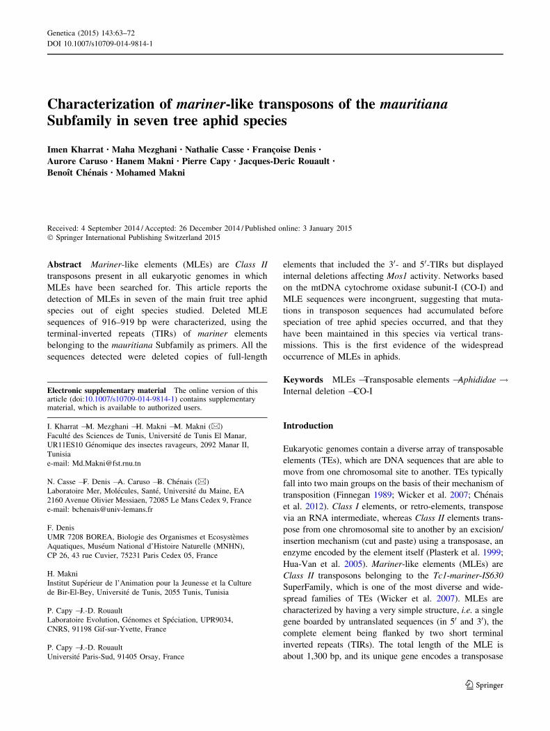
The mitochondrial CO-I barcoding marker has proved to bean effective tool for characterizing aphid species (Park
et al. 2011); therefore the genetic distance between species
were calculated in order to evaluate the divergences
between the molecular markers of studied species (Sup-plementary Data S3). This result showed low interspecific
distances that allowed performing a molecular network on
the CO-I sequences in order to compare the mtDNA rela-tionships with the MLE relationships. So, the CO-I net-
work, based on one sequence per species obtained from the
same individuals as MLEs and one GenBank entry for eachspecies, revealed seven haplotypes corresponding to the
seven aphids tested, which are encoded according to a
single change per base position (Fig. 4a). This CO-I-basednetwork supports the existence of two tribes according to
Blackman and Eastop (2000) and Remaudiere and Rem-
audiere (1997) i.e. the Macrosiphini, including P. persicaeand B. amygdalinus, and the Aphidini, which includes A.
gossypii, A. pomi, A. spiraecola, H. pruni, and T. aurantii.
Analysis of MLE relationships for the same seven treeaphid species was used to construct an MLE network
(Fig. 4b), indicating that species that do or do not belong to
the same tribe may share closely-related MLEs. Thus,phylogenic relationships constructed from MLE and CO-I
sequences were incongruent for the seven species tested inthis work. Aphid MLE sequences showed evidence of an
erratic distribution. The incongruence of the two networks
may suggest that MLEs have evolved independently fromthe speciation events during the evolution of aphid species.
Discussion
Elements of the mauritiana Subfamily were detected inseven tree aphid species (i.e. A. gossypii, A. pomi, A.
Fig. 2 Sequence alignment of the Mos-1 transposase (accessionnumber: X78906) and Aphidmarcons in silico translation. Black boxesindicate highly conserved amino acids residues. Green boxescorrespond to conserved motifs of the catalytic triad, each D of thecatalytic core is positioned below and indicated by an arrow. Red
boxes correspond to the predicted bipartite and monopartite NLS, theblue dashed box corresponds to the Helix-Turn-Helix motif of theTIR binding domain, and the yellow box to a motif conserved inmariner elements (Robertson 1993; Bui et al. 2007)
Genetica (2015) 143:63–72 67
123

spiraecola, B. amygdalinus, H. pruni, P. persicae, and T.
aurantii) genomes using the inverted terminal repeat of theMos1 element as primers, whereas the eighth species (A.
punicae) was negative. This finding is not surprising, and is
in agreement with previous reports showing that MLEs arewidespread and diverse in insects (Robertson 1993; Rou-
leux-Bonnin et al. 2005; Wang et al. 2011). Other mariner
elements belonging to other Subfamilies may exist in theseaphid species as reported by Mittapalli et al. (2011), which
have shown the presence of irritans (Agmar1) and melli-
fera (Agmar2) elements in the genome of the soybeanaphid A. glycine. By contrast the in silico genome mining
of the model aphid Acyrthosiphon pisum retrieved no sig-
nificant hit for MLEs belonging to the mauritiana andmellifera subfamilies, whereas some irritans elements have
been found (data not shown). The absence of mauritiana
and mellifera MLEs in the sequenced genome of A. pisummay be related to the fact that only 50 % of the genome is
currently annotated. This may also indicate that all aphid
genomes do not contain the same MLEs.
Using an absolute quantification protocol as well as acomparative estimation with the single copy gene RPL7
(Mittapalli et al. 2011), quantitative PCR results indicate
that MLEs are present in aphids in a low copy number (i.e.1 or 2 copies), below the detection limit of quantitative
PCR (data not shown). This is in agreement with the results
of Mittapalli et al. (2011) obtained for MLEs in A. glycine.Low copy MLEs have also been reported in Drosophila
ananassae (Robertson and Lampe 1995) and D. sechellia
(Capy et al. 1992).In our study, a total of 28 full-length aphid elements
were obtained displaying 68 % homology with D. simulans
and D. teissieri mariner elements. Sequences analysisrevealed defective elements containing several kinds of
mutations generating stop codons, frameshifts, and non-
functional transposase, suggesting that transposase genesmay now be evolving as pseudogenes, accumulating
mutations neutrally by vertical inactivation as are several
elements belonging to the Tc1-mariner-IS630 SuperFamily(Hartl et al. 1997; Brunet et al. 2002). Pairwise comparison
of the MLEs isolated showed a high degree of similarity,
indicating that they shared a common origin. It is possiblethat the deletion occurred in an ancestral population before
the Tribes diverged in the Eocene (Kim et al. 2011). If so,
all these deletions can be expected to have resulted fromthe same ancestral event.
Alignment of aphid MLEs with Mos1 showed that the
internal deletion is flanked by short direct repeats of
b Fig. 3 Classification of the Aphid MLEs in the mauritiana Subfamilyof the mariner Family. a A set of 313 sequences was classified by theUPGM-VM method on the basis of the available whole nucleotidesequences. The main Families of the Tc1-mariner-IS630 Superfamilyof transposable elements are located at the lower right part of therosette: Chl (Chlorophyllis = Plant mariner = DD39D in plants),Gam (Gambol = DD34E in animals), Pog (Pogo in animals andLemis in plants), Tco (Tc1 = DD34E in animals), Lud (Luden-sis = maT pp. = Rosa in animals), Tct (Tc3 in animals), Msq(Mosquitis = DD37E in animals), Fot (Fotis in fungi), Mel (Melilotisin bacteria with ISRm10), Jap (Japonis in bacteria with IS870), Son(Sonneis in bacteria with ID630), Mat (Matelotis = maT pp. = Mor-i = DD37D in animals). The mariner Family (DD34D in animals)splits into two subgroups: Atlantis (Atl including the Irritans) andMareNostrum (Mar). In this second group, the main SubFamilies arerepresented: Mellifera (Mel), Elegans (Ele), Cecropia (Cem and Cec),Chitwoodis (Chi), Indianus (Ind), Briggsae (Bri) and Cemar2 (Ce2).The mauritiana Subfamily appears to be structured into a largenumber of Tribes. Hymenopteris (Hym), Dipteris (Dip, with Mos1)are well characterized. The other Tribes, e.g. Nikananis (Nik), are stillspeculative, because of the small number of sequences (most of whichwere only partial sequences) and the small number of host species.The last Tribe is Puceronis (Puc), which includes all the newsequences of fruit tree aphid elements presented in this paper. Thisclassification clearly shows that the Puceronis Tribe belongs to theMauritiana Subfamily. The complete list of the 313 sequences, withaccession numbers, lengths, and host species names is available inSupplementary Data S4. b Zoom on the Mauritiana Subfamilyshowing the puceronis Tribe
68 Genetica (2015) 143:63–72
123
3–5 bp. Such repeats were found at the extremities of
internal deletions of some MLEs (Brunet et al. 2002).Indeed, it seems that the part of MLE element likely to be
deleted is similar at the 50 and 30 ends. Analogous results
were obtained for Lemi elements, in which deletions werescattered throughout the sequences in the Ogris and Po-
ucetis Subfamilies, and three categories of microhomolo-
gies were detected (Negoua et al. 2013). However for themauritiana Subfamily within Drosophilidae species,
internal deletions occur mainly in the 50 part of elements
(Brunet et al. 2002). MLE Aphid sequences also revealedmicrohomologies, implying that homologous recombina-
tion between small direct repeats may be the cause of
deleted elements among MLEs. Various mechanisms couldexplain the origin of deletions in conjunction with the
presence of SDRs, such as ectopic recombination and
abortive gap repair (Negoua et al. 2013). It should thereforebe possible to find independent deletions with similar
breakpoints.
The predicted consensus transposase Aphidmarconsdisplays the MLE signature sequence DD(34)D, as well as
important deletions in some key regions. These deletions
span the NLS motif in the N terminal part of the protein,and also the third ‘‘D’’ of the catalytic domain in the
C-terminal region (Doak et al. 1994). The main conserved
motif of the mariner transposase WVPHEL was slightlymodified by WV(N)(L)EL, however the second conserved
motif, YSPDLAP, was deleted in all species. This deletion
is specific to aphids and may be a selective mutation sharedby all aphid species.
The comparison of nucleotide sequences has shown high
degree of similarity among tree aphid MLE copies indi-cating either (1) an earlier invasion of Mos1 copies into the
host genome dating from the Eocene followed by a phase
of senescence in the common aphid ancestor and by somesubstitutions or (2) an horizontal transfer of deleted copies
through transposase from endogenous active elements.
Like all genes, TEs are transmitted vertically from acommon ancestor in the process of speciation. In this case,
the relationships between sequences of elements must
reflect the phylogenetic relationships between their hosts.Therefore, a comparative CO-I based network showing the
relationships between the seven aphid species studied was
constructed, and was consistent with classical phylogeneticanalyses based on morphological and molecular charac-
teristics (Foottit et al. 2008; Cœur D’Acier et al. 2008; Kim
et al. 2011; Blackman and Eastop 2007; Leclant 2000). Themitochondrial CO-I barcoding marker has proved an
effective tool for characterising aphid species (Park et al.
2011). Rigorous statistical proof of horizontal transmissionrequires the demonstration of a statistically-significant
inconsistency in the molecular phylogeny (or haplotype
network) of the transposable element as compared to thoseof single-copy, non-transposable sequences from the same
A. gossypii
A. pomi
A. spiraecola
H. pruni
B. amygdalinus
P. persicae
T. aurantii
10 mutations 1 mutation
Ap
Ap Ap
Ap Ap
Pp
Pp
Pp
Pp
Pp
Ta
Hp
Hp
Hp
Hp Hp
As
As
As As
Ag
Ag
Ag
Ag Ag
Ba
Ba
Ba
As
(A) (B)
Fig. 4 Parsimonious haplotype network generated from CO-Isequences (a) and MLE sequences (b) within seven aphids species.a The ellipse size corresponds to the haplotype frequency. Red circlesindicate mutation steps corresponding to hypothetical haplotypes,which were not detected among the specimens analyzed. For eachspecies, one sequence was sequenced from the same individual usedfor TIR detection, the other one being based on the followingGenBank entries: Aphis gossypii (EU701419.1), Aphis pomi(EU701479.1), Aphis spiraecola (EU701501.1), Toxoptera aurantii
(EU701931.1), Hyalopterus pruni (GU457791.1), Pterochloroidespersicae (JN644646.1), Myzus persicae (EU701803.1). b Red circlesrepresent forms inferred from, but not detected amongst the haplotypesamples; Ag, Agosmar 1–5 from Aphis gossypii; Ap, Apommar 1–5from Aphis pomi, As, Aspimar 1–5 from Aphis spiraecola; Ba,Bamymar 1–3 from Brachycaudus amygdalinus; Hp, Hprumar 1–5from Hyalopterus pruni, Pp, Ppermar 1–4; from Pterochloroidespersicae, and Ta, Taurmar1 from Toxoptera aurantii
Genetica (2015) 143:63–72 69
123
genomes (Lawrence and Hartl 1992; Clark et al. 1994;
Robertson and Lampe 1995; Capy et al. 1994a, b). Here,the topologies of haplotype networks are highly incon-
gruent, showing that MLEs have evolved independently of
the speciation events. A similar observation was also beenreported for the Bambusoideae Subfamily (Zhou et al.
2011). These results imply that horizontal transfer events
might occur between species originating within a relativelysmall geographic range. This is reflected in particular by
the presence of nearly identical MLEs in distantly-relatedspecies, and the presence of very diverse MLEs within the
same species. This may be due to the occurrence of hori-
zontal transfer events between phylogenetically-distantspecies during aphid evolution or the existence of an
ancestral MLEs polymorphism followed by divergent
evolution and stochastic loss (Hartl et al. 1997). In thiswork, we found that all copies of MLEs were deleted, and
that the deletions were similar for both the Macrosiphini
tribe, including Pterochloroides persicae and Brachycau-dus amygdalinus, and the Aphidini tribe, which includes
Aphis gossypii, Aphis pomi, Aphis spiraecola, Hyalopterus
pruni, and Toxoptera aurantii.In conclusion, there are two possible explanations for
such a topology. Either, amplification must have occurred
within tree aphid tribes after the inactivation of MLEcopies and before ancestral divergence occurred between
the groups. Or, alternatively, MLEs copies are long-term
inactivated copies, but they were recently amplified byanother copy encoding an active transposase. However, no
active copy has so far been detected in these species. These
results highlight the difficulties of explaining the evolutionof the Mos1-like elements in tree aphid species. While
mariner elements may have been present in the ancestor of
tree aphid species, this does not exclude the possibility ofseveral losses and horizontal transfer (Brunet et al. 1999).
This observation is unexpected, because it is highly
improbable that long forms (916–919 bp) of a single MLEelement could have been transferred between species sev-
eral times during evolution. Concerning the way the
putative horizontal transfer occurs, several scenarios can beproposed, but as mentioned by Loreto et al. (2008) none of
them have been demonstrated. The aphids species studied
here infect each a different plant from two plant Families(i.e. Rutacae and Rosacae) but some aphids are polypha-
gous (e.g. Aphis gossypii or A. spiraecola) and virus
present in the aphid (e.g. Tristeza virus in A. spiraecola)may be transmitted to the host plant and then represent a
potential vector of horizontal transfer. Moreover, whatever
aphids are fed on the same plants, many vectors can be alsoconsidered like common virus, bacteria or other parasites.
Apart from horizontal transfer, another hypothesis could
explain the incongruence between networks based on CO-Iand TEs. Ancestral duplication and/or polymorphism could
have created paralogous copies of TEs that have evolved
distinctly in different genomes (Capy et al. 1994a, b).A better understanding of insect TEs will enable us to
make better-informed use of the currently available TE-
based tools. This means that fundamental studies havesignificant implications for the applications of TE-based
molecular tools. As in so many areas of biology, the
availability of genome sequences from related species aswell as individuals within populations will greatly facilitate
the investigation and application of insect TEs.
Acknowledgments This work received funding for the UR11 ES10by the Tunisian Ministry of Higher Education and ScientificResearch. The authors are grateful to Monika Ghosh for revising theEnglish text.
References
Altschul SF, Gish W, Miller W, Myers EW, Lipman DJ (1990) Basiclocal alignment search tool. J Mol Biol 215:403–410
Auge-Gouillou C, Bigot Y, Pollet N, Hamelin MH, Meunier-RotivalM, Periquet G (1995) Human and other mammalian genomescontain transposons of the mariner family. FEBS Lett368:541–546
Auge-Gouillou C, Hamelin MH, Demattei MV, Periquet G, Bigot Y(2001) The ITR binding domain of the mariner Mos-1transposase. Mol Genet Genomics 265:58–65
Barry EG, Witherspoon DJ, Lampe DJ (2004) A bacterial geneticscreen identifies functional coding sequences of the insectmariner transposable element Famar1 amplified from thegenome of the earwig, Forficula auricularia. Genetics166:823–833
Bigot Y, Brillet B, Auge-Gouillou C (2005) Conservation ofpalindromic and mirror motifs within inverted terminal repeatsof mariner-like elements. J Mol Biol 351:108–116
Blackman RL, Eastop VF (2000) Aphids on the world’s crops: anidentification and information guide, 2nd edn. Wiley Ltd.,Chichester
Blackman RL, Eastop VF (2007) Taxonomic issues. In: van EmdenHF, Harrington R (eds) Aphids as crop pests. CABI, Walingford,UK, pp 1–29
Brunet F, Godin F, Bazin C, Capy P (1999) Phylogenetic analysis ofMos1-like transposable elements in the Drosophilidae. J MolEvol 49:760–768
Brunet F, Giraud T, Godin F, Capy P (2002) Do deletions of Mos1-like elements occur randomly in the Drosophilidae family? J MolEvol 54:227–234
Bui QT, Delauriere L, Casse N, Nicolas V, Laulier M, Chenais B(2007) Molecular characterization and phylogenetic position of anew mariner-like element in the coastal crab, Pachygrapsusmarmoratus. Gene 396:248–256
Bui QT, Casse N, Leignel V, Nicolas V, Chenais B (2008)Widespread occurence of mariner transposon in coastal crabs.Mol Phylogenet Evol 47:1181–1189
Capy P, David JR, Hartl DL (1992) Evolution of the transposableelement mariner in the Drososphila melanogaster species group.Genetica 86:37–46
Capy P, Anxolabehere D, Langin T (1994a) The strange phylogeniesof transposable elements: are horizontal transfers the onlyexplanation? Trends Genet 10:7–12
70 Genetica (2015) 143:63–72
123
Capy P, Langin T, Bigot Y, Brunet F, Daboussi MJ, Periquet G,David JR, Hartl DL (1994b) Horizontal transmission versusancient origin: mariner in the witness box. Genetica 93:161–170
Casacuberta E, Casacuberta JM, Puigdomenech P, Monfort A (1998)Presence of miniature inverted-repeat transposable elements(MITEs) in the genome of Arabidopsis thaliana: characterisationof the Emigrant family of elements. Plant J 16:79–85
Casse N, Bui QT, Nicolas V, Renault S, Bigot Y, Laulier M (2006)Species sympatry and horizontal transfers of mariner transpo-sons in marine crustacean genomes. Mol Phylogenet Evol40:609–619
Chenais B, Caruso A, Hiard S, Casse N (2012) The impact oftransposable elements on eukaryotic genomes: from genome sizeincrease to genetic adaptation to stressful environments. Gene509:7–15
Clark JB, Maddison WP, Kidwell MG (1994) Phylogenetic analysissupports horizontal transfer of P transposable elements. Mol BiolEvol 11:40–50
Claudianos C, Brownlie J, Russell R, Oakeshott J, Whyard S (2002)maT-A clade of transposons intermediate between mariner andTc1. Mol Biol Evol 19:2101–2109
Cœur D’Acier A, Cocuzza G, Jousselin E, Cavalieri V, Barbagallo S(2008) Molecular phylogeny and systematic in the genusBrachycaudus (Homoptera: Aphididae): insights from a com-bined analysis of nuclear and mitochondrial genes. Zool Scr37:175–193
Doak TG, Doerder FP, Jahn CL, Herrick G (1994) A proposedsuperfamily of transposase genes: transposon-like elements inciliated protozoa and a common ‘‘D35E’’ motif. Proc Natl AcadSci USA 91:942–946
Doyle JJ, Doyle JL (1987) A rapid DNA isolation procedure for smallquantities of fresh leaf tissue. Photochem Bull 19:11–15
Feschotte C, Wessler SR (2002) Mariner-like tranposases arewidespread and diverse in flowering plants. Proc Natl AcadSci USA 99:280–285
Feschotte C, Osterlund MT, Peeler R, Wessler SR (2005) DNA-bindingspecificity of rice mariner-like transposases and interactions withStowaway MITEs. Nucleic Acids Res 33:2153–2165
Finnegan DJ (1989) Eukaryotic transposable elements and genomeevolution. Trends Genet 5:103–107
Folmer O, Black M, Hoeh W, Lutz R, Vrijenhoek R (1994) DNAprimers for amplification of mitochondrial cytochrome c oxidasesubunit I from diverse metazoan invertebrates. Mol Mar BiolBiotechnol 3:294–299
Foottit RG, Maw HE, Von Dohlen CD, Hebert PD (2008) Speciesidentification of aphids (Insecta: Hemiptera: Aphididae) throughDNA barcodes. Mol Ecol Resour 8:1189–1201
Hartl DL, Lozovskaya ER, Nurminsky DI, Lohe AR (1997) Whatrestricts the activity of mariner-like transposable elements?Trends Genet 13:197–201
Hua-Van A, Le Rouzic A, Maisonhaute C, Capy P (2005) Abundance,distribution and dynamics of retrotransposable elements andtransposons: similarities and differences. Cytogenet Genome Res110:426–440
Ivics Z, Hackett PB, Plasterk RH, Izsvak Z (1997) Molecularreconstruction of sleeping beauty, a Tc1-like transposon fromfish, and its transposition in human cells. Cell 91:501–510
Jacobson JW, Medhora MM, Hartl DL (1986) Molecular structure ofa somatically unstable transposable element in Drosophila. ProcNatl Acad Sci USA 83:8684–8688
Kim H, Lee S, Jang Y (2011) Macroevolutionary patterns in theaphidini aphids (Hemiptera: Aphididae): diversification, hostassociation, and biogeographic origins. PLoS One 6:e24749
Kimura M (1980) A simple method for estimating evolutionary rateof base substitutions through comparative studies of nucleotidesequences. J Mol Evol 16:111–120
Kosugi S, Hasebe M, Tomita M, Yanagawa H (2009) Systematicidentification of yeast cell cycle-dependent nucleocytoplasmicshuttling proteins by prediction of composite motifs. Proc NatlAcad Sci USA 106:10171–10176
Lawrence JG, Hartl DL (1992) Inference of horizontal genetictransfer from molecular data: an approach using the bootstrap.Genetics 131:753–760
Leclant F (2000) Les pucerons des plantes cultivees: clefs d’identi-fication. Quae, Versailles, France
Leroy H, Castagnone-Sereno P, Renault S, Auge-Gouillou C, BigotY, Abad P (2003) Characterization of Mcmar1, a mariner-likeelement with large ITR from the phytoparasitic nematodeMeloidogyne chitwoodi. Gene 304:35–41
Loreto EL, Carareto CM, Capy P (2008) Revisiting horizontal transferof transposable elements in Drosophila. Heredity 100:545–554
Medhora M, Maruyama K, Hartl DL (1991) Molecular and functionalanalysis of the mariner element Mos1 in Drosophila. Genetics128:311–318
Mittapalli OL, Rivera-Vega L, Bhandary B, Bautista MA, MamidalaP, Michel AP, Shukle RH, Mian MA (2011) Cloning andcharacterization of mariner-like elements in the soybean aphid,Aphis glycines Matsumura. Bull Entomol Res 101:697–704
Munoz-Lopez M, Siddique A, Bischerour J, Lorite P, Chalmers R,Palomeque T (2008) Transposition of Mboumar-9: identificationof a new naturally active mariner-family transposon. J Mol Biol382:567–572
Narasimhan G, Bu C, Gao Y, Wang X, Xu N, Mathee K (2002)Mining for motifs in protein sequences. J Comput Biol9:707–720
Negoua A, Rouault JD, Chakir M, Capy P (2013) Internal deletions oftransposable elements: the case of Lemi elements. Genetica141:369–379
Park DS, Foottit R, Maw E, Hebert PD (2011) Barcoding bugs: DNA-based identification of the true bugs (Insecta: Hemiptera:Heteroptera). PLoS One 6:e18749
Plasterk RH, Izsvak Z, Ivics Z (1999) Resident aliens: the Tc1/mariner superfamily of transposable elements. Trends Genet15:326–332
Polzin T, Daneschmand SV (2003) On Steiner trees and minimumspanning trees in hypergraphs. Oper Res Lett 31:12–20
Remaudiere G, Remaudiere M (1997) Catalogue des Aphididae dumonde Pucerons (Homoptera : Aphididae). Editions INRA
Rezende-Teixeira P, do Amaral JB, Siviero F, Machado-Santelli GM(2012) Molecular characterization of a mariner-like element in theAtta sexdens rubropilosa genome. Genet Mol Res 11:1475–1485
Rice P, Longden I, Bleasby A (2000) EMBOSS: the Europeanmolecular biology open software suite. Trends Genet16:276–277
Robertson HM (1993) The mariner transposable element is wide-spread in insects. Nature 362:241–245
Robertson HM (2002) Evolution of DNA transposons in eukaryotes.In: Gellert M, Lambowitz A, Craig NL, Robert Craigie R (eds)Mobile DNA II. ASM Press, Washington, pp 1093–1110
Robertson HM, Lampe DJ (1995) Distribution of transposableelements in arthropods. Ann Rev Entomol 40:333–357
Robertson HM, Martos R (1997) Molecular evolution of the secondancient human mariner transposon, Hsmar2, illustrates patternsof neutral evolution in the human genome lineage. Gene205:219–228
Robertson HM, McLeod EG (1993) Five major subfamilies ofmariner transposable elements in insects, including the Medi-terranean fruit fly, and related arthropods. Insect Mol Biol2:125–139
Robertson HM, Zumpano KL (1997) Molecular evolution of anancient mariner transposon, Hsmar1, in the human genome.Gene 205:203–217
Genetica (2015) 143:63–72 71
123

Rouault JD, Casse N, Chenais B, Hua-Van A, Filee J, Capy P (2009)Automatic classification within families of transposable ele-ments: application to the mariner Family. Gene 448:227–232
Rouleux-Bonnin F, Petit A, Demattei MV, Bigot Y (2005) Evolutionof full-length and deleted forms of the mariner-like element,Botmar1, in the genome of the bumble bee, Bombus terrestris(Hymenoptera: Apidae). J Mol Evol 60:736–747
Silva JC, Bastida F, Bidwell SL, Johnson PJ, Carlton JM (2005) Apotentially functional mariner transposable element in the protistTrichonomas vaginalis. Mol Biol Evol 22:126–134
Tamura K, Peterson D, Peterson N, Stecher G, Nei M, Kumar S(2011) MEGA5: molecular evolutionary genetics analysis usingmaximum likelihood, evolutionary distance, and maximumparsimony methods. Mol Biol Evol 28:2731–2739
Thompson JD, Higgins DG, Gibson TJ (1994) CLUSTAL W:improving the sensitivity of progressive multiple sequence
alignment through sequence weighting, position specific gappenalties and weight matrix choice. Nucleic Acids Res22:4673–4680
Wang J, Miller TA, Park Y (2011) Identification of mariner-likeelements belonging to the cecropia subfamily in two closelyrelated Helicoverpa species. Insect Sci 18:619–628
Wicker T, Sabot F, Hua-Van A, Bennetzen JL, Capy P, Chalhoub B,Flavell A, Leroy P, Morgante M, Panaud O, Paux E, SanMiguelP, Schulman AH (2007) A unified classification system foreukaryotic transposable elements. Nat Rev Genet 8:973–982
Zhou MB, Lu JJ, Zhong H, Tang KX, Tang DQ (2010) Distributionand polymorphism of mariner-like elements in the Bambusoi-deae subfamily. Plant Syst Evol 289:1–11
Zhou MB, Zhong H, Tang DQ (2011) Isolation and characterizationof seventy-nine full length mariner-like transposase genes in theBambusoideae subfamily. J Plant Res 124:607–617
72 Genetica (2015) 143:63–72
123
![Repeated horizontal transfers of four DNA transposons in ......the P element of Drosophila [9]. More than 330 cases (188 cases for DNA transposons and 142 cases for RNA transposons)](https://img.pdfslide.us/doc/110x75/60b5e3afdf2f26263048a93b/repeated-horizontal-transfers-of-four-dna-transposons-in-the-p-element-of.jpg)