Embed Size (px)
Citation preview

1
Characterization of glucose oxidation by gold nanoparticles using
nanoceria
Nathan J. Lang, Biwu Liu and Juewen Liu*
Department of Chemistry and Waterloo Institute for Nanotechnology, University of Waterloo,
Waterloo, Ontario, Canada, N2L 3G1
E-mail: [email protected]
The final publication is available at Elsevier via http://dx.doi.org/10.1016/j.jcis.2014.04.025." © 2014. This manuscript version is made available under the CC-BY-NC-ND 4.0 license http://creativecommons.org/licenses/by-nc-nd/4.0/
2
Abstract.
Gold nanoparticles (AuNPs) can oxidize glucose, producing hydrogen peroxide and gluconic acid,
which are the same products as those generated by glucose oxidase (GOx). In this regard, AuNPs are a
nanozyme. Herein, a new colorimetric method is developed to understand the surface chemistry of gold
nanoparticles for this oxidation reaction. The color of nanoceria is changed to yellow by the hydrogen
peroxide generated glucose oxidation. Using this assay, we find that adsorption of small molecules
such as citrate does not deactivate AuNPs, while adsorption of polymers including serum proteins and
high molecular weight polyethylene glycol inhibits glucose oxidation. In addition to glucose, AuNPs
can also oxidize galactose. Therefore, this reaction is unlikely to be directly useful for glucose
detection for biomedical applications. On the other hand, AuNPs might serve as a general oxidase for a
broad range of substrates. The glucose oxidation reaction is slower at lower pH. Since the reaction
generates an acid product, glucose oxidation becomes slower as the reaction proceeds. The effects of
temperature, AuNP size, and reaction kinetics have been systematically studied. This work provides
new insights regarding the surface chemistry of AuNPs as a nanozyme.

3
Introduction
Nanozymes are nanoparticles with catalytic activity [1-3]. In the past decade, gold nanoparticles
(AuNPs) [4-7], magnetic iron oxide NPs [8], and cerium oxide NPs (nanoceria) have been reported to
mimic various enzymes [9-16]. Oxidation of glucose by AuNPs was first reported by Comotti et al in
2004 [4]. In this reaction, oxygen and glucose are consumed to produce gluconic acid and hydrogen
peroxide (H2O2) [4-6, 17, 18], which are the same products as those generated by glucose oxidase
(GOx). Therefore, AuNPs are a mimic of GOx. The enzyme properties of these two have been
systematically compared and the kcat and Km values are reported to be quite similar [6].
Recently, analytical chemists picked up this reaction for biosensor development and many
interesting observations were made. For example, the H2O2 generated during glucose oxidation was
used as a reducing agent to react with HAuCl4 [19]. The newly reduced gold is deposited on the
original particle surface to produce enlarged AuNPs. The growth of AuNP size is monitored by the
shift of the surface plasmon peak or color change [5, 6]. With a short single-stranded DNA, the glucose
oxidation reaction by AuNPs is impeded, which is attributed to the adsorption of DNA onto the gold
surface. In addition, it was noticed that AuNP nanozymes seem to be deactivated in the reaction
process (so called self-limiting reaction). In other words, the glucose conversion becomes progressively
slower as the reaction proceeds. This gives a relatively small turnover number, which may compromise
its application. This self-limiting behavior was attributed to the capping of AuNP surface by the
gluconate product [4-6]. Both DNA adsorption and gluconate adsorption indicate the importance of the
surface chemistry of AuNPs for catalysis.
Despite these progresses, a lot remains to be learned to fully understand AuNP nanozymes. First,
we are intrigued by the surface chemistry aspect of this reaction. In particular, we aim to compare the
activity of AuNPs as a function of the binding affinity and size of surface ligands. Second, we study the
buffer conditions, from which we suggest an alternative explanation for the self-limiting reaction.
4
Finally, protein enzymes have excellent substrate specificity. It is unclear whether AuNPs is specific
for glucose; a few other sugar molecules have been tested in this work as well.
Materials and Methods
Chemicals. AuNPs (5, 10, 20, 30, and 50 and 100 nm) were purchased from Ted Pella Inc. AuNPs (5,
13, and 50 nm) were prepared by NaBH4 or citrate reduction in our own lab. HAuCl4, nanoceria,
glucose, sodium gluconate, galactose, fructose, bovine serum, and hydrogen peroxide were purchased
from Sigma-Aldrich. Trisodium citrate was from Mandel Scientific Inc (Guelph, Ontario, Canada).
Sucrose and all the PEG samples were from VWR. Milli-Q water was used for preparing all the
solutions. The original 20% stock solution of nanoceria has a particle concentration of 860 M. We
typically dilute it first 47.3 times and then 32.26 times to reach a final particle concentration of ~564
nM in the final assay tube.
Preparing AuNPs. 13 nm AuNPs were prepared by the standard citrate reduction method (particle
concentration 10 nM) [20]. 5 nm AuNPs were prepared by mixing 125 µM HAuCl4 and 1 mM
NaHCO3 with a freshly prepared 100 mM solution of NaBH4, added in 10 µL drops. The initial volume
was 40 mL. The solution was stirred and cooled with ice throughout the synthesis. The addition was
stopped by adding ~240 µL of NaBH4. Note that the color stops changing after adding roughly 40 µL.
The molar concentration of the as-prepared 5 nm AuNP is ~4.4 nM.
UV-vis spectroscopy. In a typical assay, ~2.2 nM 5 nm AuNPs were mixed with 10 mM phosphate
buffer (pH 8) and 5 mM glucose. After 45 min, the samples were treated with 10 mM KCN to dissolve
the AuNPs, and then mixed with 564 nM (nanoparticle concentration) CeO2. After CeO2 addition,
exposure to light was minimized, as this bleaches the yellow color produced by Ce reacting with H2O2.
The samples were scanned using a UV-vis spectrometer (Agilent 8453A). The extinction ratio of 400
nm/290 nm was used to quantify the amount of H2O2 produced by the AuNP and glucose reaction. It is
important to note that fresh nanoceria may need to be prepared occasionally as we found that the
5
spectrum of the nanoceria by itself changed after a few days. It is also important to keep the pH
buffered at roughly 8 for the AuNPs to work properly. To test the effect of AuNP size, 5, 10, 20, 30, 50,
and 100 nm diameter AuNPs were mixed with 5 mM glucose and 10 mM phosphate buffer (pH 8).
Varying reaction conditions. To study the effect of PEG adsorption, AuNP samples were mixed with
1 mM of either PEG 200, 400, 2k, 20k or 35k before the glucose addition. To test sensor specificity,
AuNPs were mixed with 5 mM of either sucrose, fructose, galactose, ethylene glycol, or glycerol
before glucose was added. To test the effect of citrate, 3 mM trisodium citrate was mixed with the
NaBH4 reduced 5 nm AuNPs before adding glucose. To test the sensor at different pH levels, a 10 mM
citrate buffer of either pH 4, 6, or 8 was substituted for the 10 mM phosphate buffer used otherwise. pH
10 and 12 were achieved by adding NaOH to pH 8 phosphate buffer. To observe the effect of
temperature, samples with 5 mM glucose and 5 nm AuNPs with phosphate buffer were incubated for
45 min at the appropriate temperature. For these experiments, the sample lids were shut to avoid
evaporation of H2O2 or water.
Monitoring pH change. A 5 mL sample of 5 nm AuNPs were prepared without buffer. pH was
monitored using a pH meter (UltraBasic, Denver Instrument) for 4 h, mixing with a final concentration
of 1.5 mM NaOH approximately at the end of each hour.

6
Results and Discussion
Visual detection. One method to monitor the glucose oxidation reaction is to add HAuCl4 to produce
new gold surfaces [6, 21]. However, this will change the original gold surface chemistry, introducing
artifacts for our mechanistic studies. To solve this problem, we developed a new method using
nanoceria. Nanoceria normally has a light yellow color at high concentration. At low concentration, it
is almost colorless. In the presence of H2O2, an intense yellow/orange color is generated [22]. The
detailed reaction mechanism between nanoceria and H2O2 is quite complex, since H2O2 can act as an
oxidizing agent, a reducing agent and a ligand [23]. This color change and the related redox reactions
have been applied to design various sensors [9, 22, 24-26].
Our reaction scheme is presented in Figure 1A. AuNPs convert glucose into gluconic acid,
which dissociates into gluconate and proton. Hydrogen peroxide is also produced in the same process.
We employ nanoceria as the colorimetric reporter for H2O2. Our AuNPs were prepared by reducing
HAuCl4 with NaBH4, yielding an average particle diameter of ~5 nm as characterized by dynamic light
scattering (DLS, Figure 1B, red trace). Transmission electron microscopy (TEM) shows that these
AuNPs are spherical (Figure S1, Electronic Supplementary Information, ESI). Our nanoceria has a
diameter of ~5 nm as characterized by TEM (Figure 1D), and DLS showed a similar average size of
but a broader size distribution (Figure 1B, blue trace). With a concentration of 0.02% nanoceria (860
nM particle concentration), almost no color is initially perceived by the naked human eye; however
adding H2O2 produces a bright yellow color (Figure 1A). The UV-vis spectrum of fresh nanoceria has a
peak at 290 nm. In the presence of H2O2, this peak disappeared while the absorption at ~400 nm
increases. By subtracting the original nanoceria spectrum (black trace, Figure 1A) from the one after
adding H2O2 (normalizing at 290 nm), a difference spectrum with a peak at 400 nm was obtained (red
dashed spectrum). This new species explains the yellow color. We estimated the extinction coefficient
of 5 nm nanoceria (after H2O2 treatment) to be 10106 M-1cm-1 at 400 nm, which is comparable with

7
AuNPs of similar size (e.g. 9 106 M-1cm-1 for 4.6 nm AuNPs) [27]. Such a high extinction coefficient
makes it possible to achieve sensitive visual detection. For subsequent quantitative studies, the ratio of
absorbance at 400 nm over 290 nm was used to quantify the color change and thus of the amount of
hydrogen peroxide produced in a sample. Such ratiometric methods are convenient for quantification
and have been used to quantify the color change of AuNPs [28]. Using nanoceria to detect H2O2 is
highly sensitive [22], and in our system we can easily detect 3 parts-per-million H2O2 (Figure S2).
Figure 1. (A) Schematic presentation of AuNP reacting with glucose and oxygen to produce gluconic
acid and H2O2. The pH decreases during the reaction. A photograph of visible colour change after H2O2
addition to nanoceria is also shown. (B) DLS spectra of 5 nm AuNPs and nanoceria used in this work.
(C) UV-vis spectra of nanoceria with and without H2O2 addition. The red dashed spectrum is obtained
by subtracting the black spectrum from the red one (normalized at 290 nm). (D) TEM image of
nanoceria.
Effect of buffer conditions. To under the kinetics of the reaction described in Figure 1A, we mixed
glucose with AuNPs and then added nanoceria at designated time points. To minimize the background
8
color interference from AuNPs, KCN was added to dissolve the AuNPs before adding nanoceria.
Control experiments showed that KCN does not interfere with the reaction between nanoceria and
H2O2 (Figure S3). Without KCN, the surface plasmon peak of AuNPs can be observed at 520 nm
(Figure S4). If As expected, higher ratio of absorption at 400 nm over 290 nm is observed by using
longer incubation time and the reaction reaches a plateau in 30 min (Figure 2A), where the absorbance
ratio is ~0.08. There is still a lot of room for nanoceria to further change its color, since the ratio could
reach ~0.4 based on the UV-vis spectra in Figure 1C. Since the amount of glucose should be in excess,
we reason that the reaction rate is decreased as the reaction proceeds. This reaction kinetics is
comparable with that monitored by the increase of AuNP size using HAuCl4 [6]. The similar oxidation
rates suggest that both sensors are governed by the same reaction mechanism, where the signal
generation step is not the rate limiting step. Increasing temperature decreased the amount of color
change in our system (Figure 2B). Glucose oxidation by AuNPs was reported to be faster at higher
temperature [18]. Using HAuCl4 for signaling, higher temperature indeed produced faster AuNP
growth [6]. Since we added nanoceria after reacting AuNPs with glucose, it is likely that H2O2 might
escape from water at higher temperature. Overall, our reaction was quite stable from room temperature
to ~50 C, where the signal decreased by just ~10%. More drastic change was observed at even higher
temperatures. Since the reaction product contains an acid, the pH may change during the reaction
process. Next we studied the effect of pH, where the glucose oxidation reaction was significantly less
efficient at lower pH (Figure 2C). The optimal pH was ~8, and the rate did not increase further at pH
10 or 12. We noted that the AuNPs aggregated at pH 12 since the color turned purple.

9
Figure 2. Optimization of glucose detection conditions. (A) Glucose conversion after various time
intervals. Samples contained 10 mM glucose, 6 nM CeO2 and 1.25 nM of 5 nm AuNPs. (B) Effect of
temperature on glucose conversion. (C) Effect of pH on glucose conversion.
pH-limited reaction. Since nanoceria provides a convenient assay to study glucose oxidation by
AuNPs, we further used this reaction to understand the surface chemistry of AuNPs during the reaction
process. Previous reports showed that the reaction is self-limited. For example, the enlargement of
AuNPs in the presence of HAuCl4 became gradually inhibited as the reaction proceeded [6]. This self-
limiting behavior was also observed in our nanoceria signaling method, since the absorbance ratio
barely reached 0.1, while this ratio could reach 0.5 with sufficient amount of H2O2. Therefore, a
challenge in improving this sensor is to increase catalytic turnover.
Figure 2A shows that the reaction is basically stopped at 30 min. It is unlikely that all the
glucose has been consumed at this point since even higher glucose concentration produced the same
response (see below). Therefore, product inhibition should be the reason for the lack of more glucose
conversion after 30 min. Since gluconic acid is the only other product in addition to H2O2, it was
proposed that gluconate was adsorbed by AuNPs to inhibit the reaction, which has been confirmed by
XPS spectroscopy [6]. This conclusion was made based on the AuNP enlargement reaction, which
involves the deposition of new gold and is quite different from the direct measurement of H2O2 in our
system. To test this in our system, we directly added sodium gluconate to the AuNPs before adding
10
glucose. Interestingly, the inhibition effect of gluconate was minimal (Figure 3A), since all the samples
with up to 5 mM gluconate showed similar ratios. Figure 3C shows the color of the samples with and
without 5 mM gluconate. Therefore, the inhibition is unlikely to be related to the direct adsorption of
gluconate in our system. Since gluconic acid also produces protons and we know that the reaction is
significantly slower at lower pH (Figure 2C), we further tested pH change. In a tube containing a 5 mL
sample, we started with a pH value of 8.05 which was the initial pH of the 5 nm gold. Within 10 min of
adding 25 mM glucose, the pH dropped to ~7.0 and further incubation resulted in the pH dropping to
~6.7 in 1 h (Figure 3B). The pH was easily brought back up by adding NaOH and pH drop was again
observed. This process can be cycled many times. Each time, a final concentration of 1.5 mM NaOH
was added, suggesting that 1.5 mM glucose was converted. Since our AuNP concentration was ~0.1
M, each AuNP was able to turnover approximately 1,500 glucose molecules in 1 h. To explain the
kinetics in Figure 2A, it is likely that as the rate of H2O2 production is reduced as the pH drops, there is
more time for the H2O2 to evaporate. Therefore, despite there being a continuous increase in the
quantity of H2O2 in the system, waiting for longer time does not help to improve the signal. We need to
point out that while pH plays a major role in our system with nanoceria-based detection, the effect of
gluconate is likely to be more important in other systems such as the catalyzed growth of AuNPs,
where pH was found to have minimal effects on the AuNP enlargement reaction [6].
Glucose sensing. With these understandings, we next tested the use of this system to detect glucose.
Since both the sensing component (AuNPs) and the signaling component (nanoceria) are nanoparticles,
this system could be a robust sensor without any biomolecules. As shown in Figure 3D, we observed a
yellow color with just 1 mM glucose in 30 min detection time and saturated color was observed with
glucose concentration higher than 5 mM. This was again attributed to pH change, retarding further
reactions. The quantification is shown in Figure 3E and the detection limit is determined to be 0.3 mM
glucose. To confirm specificity, we mixed the AuNPs with sucrose, fructose, galactose, glycerol or

11
ethylene glycol, where strong signal was observed only with galactose besides glucose (Figure 3F).
Galactose is the C-4 epimer of glucose and AuNPs do not have the ability to distinguish between these
two. In this regard, GOx has better selectivity since galactose is not a good substrate for it [29].
Therefore, while AuNPs mimics GOx, they cannot be used as a sensor for glucose at its current form
due to the lack of specificity. On the other hand, it might be a general oxidase with a broad substrate
range.
12
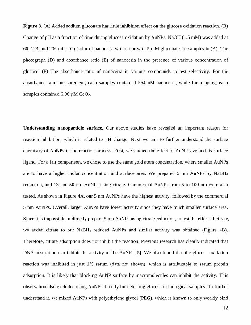
Figure 3. (A) Added sodium gluconate has little inhibition effect on the glucose oxidation reaction. (B)
Change of pH as a function of time during glucose oxidation by AuNPs. NaOH (1.5 mM) was added at
60, 123, and 206 min. (C) Color of nanoceria without or with 5 mM gluconate for samples in (A). The
photograph (D) and absorbance ratio (E) of nanoceria in the presence of various concentration of
glucose. (F) The absorbance ratio of nanoceria in various compounds to test selectivity. For the
absorbance ratio measurement, each samples contained 564 nM nanoceria, while for imaging, each
samples contained 6.06 µM CeO2.
Understanding nanoparticle surface. Our above studies have revealed an important reason for
reaction inhibition, which is related to pH change. Next we aim to further understand the surface
chemistry of AuNPs in the reaction process. First, we studied the effect of AuNP size and its surface
ligand. For a fair comparison, we chose to use the same gold atom concentration, where smaller AuNPs
are to have a higher molar concentration and surface area. We prepared 5 nm AuNPs by NaBH4
reduction, and 13 and 50 nm AuNPs using citrate. Commercial AuNPs from 5 to 100 nm were also
tested. As shown in Figure 4A, our 5 nm AuNPs have the highest activity, followed by the commercial
5 nm AuNPs. Overall, larger AuNPs have lower activity since they have much smaller surface area.
Since it is impossible to directly prepare 5 nm AuNPs using citrate reduction, to test the effect of citrate,
we added citrate to our NaBH4 reduced AuNPs and similar activity was obtained (Figure 4B).
Therefore, citrate adsorption does not inhibit the reaction. Previous research has clearly indicated that
DNA adsorption can inhibit the activity of the AuNPs [5]. We also found that the glucose oxidation
reaction was inhibited in just 1% serum (data not shown), which is attributable to serum protein
adsorption. It is likely that blocking AuNP surface by macromolecules can inhibit the activity. This
observation also excluded using AuNPs directly for detecting glucose in biological samples. To further
understand it, we mixed AuNPs with polyethylene glycol (PEG), which is known to only weakly bind
13
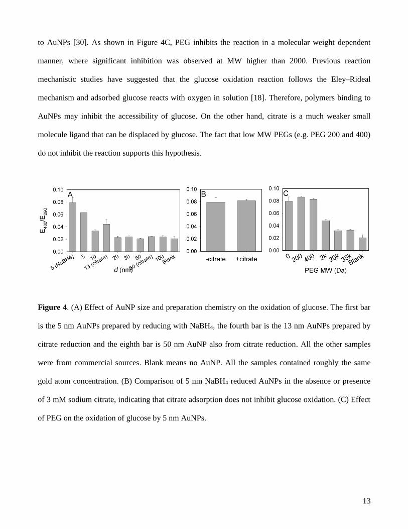
to AuNPs [30]. As shown in Figure 4C, PEG inhibits the reaction in a molecular weight dependent
manner, where significant inhibition was observed at MW higher than 2000. Previous reaction
mechanistic studies have suggested that the glucose oxidation reaction follows the Eley–Rideal
mechanism and adsorbed glucose reacts with oxygen in solution [18]. Therefore, polymers binding to
AuNPs may inhibit the accessibility of glucose. On the other hand, citrate is a much weaker small
molecule ligand that can be displaced by glucose. The fact that low MW PEGs (e.g. PEG 200 and 400)
do not inhibit the reaction supports this hypothesis.
Figure 4. (A) Effect of AuNP size and preparation chemistry on the oxidation of glucose. The first bar
is the 5 nm AuNPs prepared by reducing with NaBH4, the fourth bar is the 13 nm AuNPs prepared by
citrate reduction and the eighth bar is 50 nm AuNP also from citrate reduction. All the other samples
were from commercial sources. Blank means no AuNP. All the samples contained roughly the same
gold atom concentration. (B) Comparison of 5 nm NaBH4 reduced AuNPs in the absence or presence
of 3 mM sodium citrate, indicating that citrate adsorption does not inhibit glucose oxidation. (C) Effect
of PEG on the oxidation of glucose by 5 nm AuNPs.
14
Conclusions
In summary, we have revealed a number of important properties of AuNPs relating to their use as a
GOx mimic. First, the reaction is slower at lower pH and glucose oxidation produces acidic products,
thus forming a self-limiting system. The reaction can proceed for thousands of turnovers on each AuNP
and the pH drop can be compensated by adding base. Second, by coupling the reaction of nanoceria
with hydrogen peroxide and the glucose oxidation by AuNPs, we presented an all-nanoparticle-based
assay for studying this reaction. We demonstrated that AuNPs can oxide not only glucose but also
galactose, thus serving as a general oxidase. For this reason and for the inhibited activity by polymer
adsorption, AuNPs are unlikely to be used for glucose detection. Third, we studied the surface
chemistry of AuNPs and found that small molecule ligands containing multiple hydroxide or oxygen
groups do not bind to the AuNPs surface strong enough to inhibit the reaction. Even with a relatively
common ligand citrate, the AuNPs can still catalyze the reaction at the same rate. On the other hand,
even weakly adsorbed polymers such as PEG can effectively inhibit glucose oxidation.
Supplementary Information: TEM of AuNPs and nanoceria reacting with H2O2.
Acknowledgements. Funding for this work is from the University of Waterloo, the Canadian
Foundation for Innovation, and Natural Sciences and Engineering Research Council of Canada
(NSERC). J. Liu receives Early Researcher Award from the Ontario Ministry of Research and
Innovation.

15
References
1. N. A. Kotov, Science 330 (2010) 188.
2. H. Wei; E. Wang, Chem. Soc. Rev. 42 (2013) 6060.
3. C. J. Brown; R. G. Bergman; K. N. Raymond, J. Am. Chem. Soc. 131 (2009) 17530.
4. M. Comotti; C. Della Pina; R. Matarrese; M. Rossi, Angew. Chem., Int. Ed. 43 (2004) 5812.
5. X. Zheng; Q. Liu; C. Jing; Y. Li; D. Li; W. Luo; Y. Wen; Y. He; Q. Huang; Y.-T. Long; C. Fan,
Angew. Chem., Int. Ed. 50 (2011) 11994.
6. W. Luo; C. Zhu; S. Su; D. Li; Y. He; Q. Huang; C. Fan, ACS Nano 4 (2010) 7451.
7. X. Li; Z. Qi; K. Liang; X. Bai; J. Xu; J. Liu; J. Shen, Catal. Lett. 124 (2008) 413.
8. X. Li; F. Wen; B. Creran; Y. Jeong; X. Zhang; V. M. Rotello, Small 8 (2012) 3589.
9. A. Asati; S. Santra; C. Kaittanis; S. Nath; J. M. Perez, Angew. Chem., Int. Ed. 48 (2009) 2308.
10. J. Chen; S. Patil; S. Seal; J. F. McGinnis, Nat Nano 1 (2006) 142.
11. T. Pirmohamed; J. M. Dowding; S. Singh; B. Wasserman; E. Heckert; A. S. Karakoti; J. E. S.
King; S. Seal; W. T. Self, Chem. Comm. 46 (2010) 2736.
12. C. Korsvik; S. Patil; S. Seal; W. T. Self, Chem. Comm. (2007) 1056.
13. Y. Peng; X. Chen; G. Yi; Z. Gao, Chem. Comm. 47 (2011) 2916.
14. R. Pautler; E. Y. Kelly; P.-J. J. Huang; J. Cao; B. Liu; J. Liu, ACS Appl. Mater. Inter. 5 (2013)
6820.
15. C. Xu; Z. Liu; L. Wu; J. Ren; X. Qu, Adv. Funct. Mater. 24 (2014) 1624.
16. C. Xu; X. Qu, NPG Asia Mater. 6 (2014) e90.
17. D. Zeng; W. Luo; J. Li; H. Liu; H. Ma; Q. Huang; C. Fan, Analyst 137 (2012) 4435.
18. P. Beltrame; M. Comotti; C. Della Pina; M. Rossi, Applied Catalysis A: General 297 (2006) 1.
19. R. de la Rica; M. M. Stevens, Nat Nano 7 (2012) 821.
20. J. Liu; Y. Lu, Nat. Protoc. 1 (2006) 246.
21. Q. Jiang; Z.-G. Wang; B. Ding, Small 9 (2013) 1016.
16
22. M. Ornatska; E. Sharpe; D. Andreescu; S. Andreescu, Anal. Chem. 83 (2011) 4273.
23. P. Yu; S. A. Hayes; T. J. O'Keefe; M. J. O'Keefe; J. O. Stoffer, J. Electrochem. Soc. 153 (2006)
C74.
24. E. Sharpe; T. Frasco; D. Andreescu; S. Andreescu, Analyst 138 (2013) 249.
25. J. Njagi; C. Ispas; S. Andreescu, Anal. Chem. 80 (2008) 7266.
26. J. Njagi; M. M. Chernov; J. C. Leiter; S. Andreescu, Anal. Chem. 82 (2010) 989.
27. X. O. Liu; M. Atwater; J. H. Wang; Q. Huo, Colloid. Surface. B. 58 (2007) 3.
28. J. W. Liu; Y. Lu, J. Am. Chem. Soc. 125 (2003) 6642.
29. R. Bentley, Nature 176 (1955) 870.
30. X. Zhang; P.-J. J. Huang; M. R. Servos; J. Liu, Langmuir 28 (2012) 14330.


![The Mechanism of the Oxidation of Glucose by Bromine[1]](https://img.pdfslide.us/doc/110x75/56d6bf1e1a28ab301694ef8a/the-mechanism-of-the-oxidation-of-glucose-by-bromine1.jpg)