Embed Size (px)
Citation preview

Peng et al. BMC Microbiol (2021) 21:283 https://doi.org/10.1186/s12866-021-02338-4
RESEARCH
Characterization of fungal communities on shared bicycles in Southwest ChinaLu Peng1,2, Bi Qin2,3, Zhu Shen1,2 and Siyu Wang1*
Abstract
Background: The widespread use of shared bicycles has increased the demand and sanitary requirements for shared bicycles. Previous studies have identified potentially pathogenic bacteria on the surfaces of shared bicycles, but fun-gal communities have not been investigated.
Methods: We sampled shared-bicycle handles and saddles from five selected locations in a metropolis (Chengdu, China, n = 98) and used surrounding air deposition samples as controls (n = 12). Full-length ITS sequencing and multi-ple bioinformatic analyses were utilized to reveal fungal community structures and differences.
Results: Aspergillus was dominant on both the handles and saddles of shared bicycles, and Alternaria and Clad-osporium were the most abundant families in the air samples. Significant differences in fungal community structures were found among the three groups. The handle samples contained higher abundances of Aureobasidium melanoge-num and Filobasidium magnum than the saddle and air samples. The saddle samples had a higher abundance of Clad-osporium tenuissimum than the other two sample types (P < 0·05). A higher abundance of fungal animal pathogens on shared-bicycle surfaces than in air by FUNGuild (P < 0·05). Moreover, the co-occurrence network of fungi on handles was more stable than that on saddles.
Conclusion: There were more potential pathogens, including Aspergillus pseudoglaucus, Aureobasidium melanoge-num, Kazachstania pintolopesii, Filobasidium magnum, Candida tropicalis, and Malassezia globose were found on shared bicycles than in air, suggesting that hands should not contact mucous membrane after cycling, especially in suscep-tible individuals, and hygiene management of shared bicycles should be given more attention by relevant organiza-tions worldwide.
Keywords: Shared-bicycle, Fungal communities, Potential pathogens
© The Author(s) 2021. Open Access This article is licensed under a Creative Commons Attribution 4.0 International License, which permits use, sharing, adaptation, distribution and reproduction in any medium or format, as long as you give appropriate credit to the original author(s) and the source, provide a link to the Creative Commons licence, and indicate if changes were made. The images or other third party material in this article are included in the article’s Creative Commons licence, unless indicated otherwise in a credit line to the material. If material is not included in the article’s Creative Commons licence and your intended use is not permitted by statutory regulation or exceeds the permitted use, you will need to obtain permission directly from the copyright holder. To view a copy of this licence, visit http:// creat iveco mmons. org/ licen ses/ by/4. 0/. The Creative Commons Public Domain Dedication waiver (http:// creat iveco mmons. org/ publi cdoma in/ zero/1. 0/) applies to the data made available in this article, unless otherwise stated in a credit line to the data.
IntroductionShared bicycles, as a widely implemented public health project, have been made available in more than 1000 cit-ies in the past decade. Shared bicycles has changed the travel mode of the urban population to some extent and have gradually become indispensable [1]. In recent years,
especially during the coronavirus disease 2019 (COVID-19) pandemic, the demand for shared bicycles increased due to their convenience and environmental friendliness in cities with large populations. As a public transporta-tion tool, shared bicycles are stored in outdoor public areas for long periods of time and repeatedly contacted by different people and their clothes, which harbor differ-ent microbes, including bacteria and fungi. The microbes on a shared bicycle will change and further spread during consecutive use. Potentially pathogenic bacteria [2] and antimicrobial-resistant Enterobacteriaceae [3] have been sequenced from samples from shared bicycles. How-ever, to date, no study has yet focused on fungi on shared
Open Access
*Correspondence: [email protected] Department of Dermatology, Institute of Dermatology and Venereology, Sichuan Academy of Medical Sciences & Sichuan Provincial People’s Hospital, No.32, Western 2nd Section, 1st Ring Rd, Qingyang District, Chengdu 610072, Sichuan, ChinaFull list of author information is available at the end of the article

Page 2 of 11Peng et al. BMC Microbiol (2021) 21:283
bicycles. Fungi are the most common microorganisms in the environment; types of fungi can vary widely and be potentially pathogenic in humans under certain cir-cumstances. Thus, our study is the first to analyze of the structure and function of fungi on the shared bicycles.
MethodSample collectionSwab samples of saddle and handle surfaces were col-lected from a number of shared bicycles in five locations in a metropolitan area (Chengdu, China) in July 2020. Samples from shared bicycles that had gone unused for a long time or were obviously damaged were excluded. Random samples from nearby air sampling sites were col-lected on the same day in each selected area. The sam-ples were collected from the handles and saddles of the shared bicycles (approximately 10 cm [2]) with DNA-free swabs (Puritan, Me, USA), which were applied for 15 s. Each surface was sampled with a new cotton swabs and rotated three times in a nonoverlapping area; sampling was repeated three times. Air samples were collected by a sterile filter paper in a petri dish, which was placed at a high location in the selected area for 6 h. After sampling, each sample was stored in a unique labeled sterile cen-trifuge tube and transported to the laboratory on dry ice before being immediately stored in a refrigerator at − 80 ° Celsius for further analysis.
DNA extraction and internal transcribed spacer (ITS) amplification sequencingGenomic DNA from samples collected from the dif-ferent sampling sites and surfaces was extracted by the CTAB/SDS method. The concentration and purity of the extracted DNA was measured with a 1% agarose gel and then diluted with 1 ng/μl sterile water.
The ITS region was amplified by specific primers with barcodes and Phusion® high-fidelity PCR master mix with GC buffer (New England Biolabs). The PCR prod-ucts were purified with a QIAquick@ Gel Extraction Kit (QIAGEN) after mixing with the same volume of 1X loading buffer and detection by electrophoresis in a 2% agarose gel.
Following the manufacturer’s instructions, sequencing libraries were generated using the SMRTbellTM Tem-plate Prep Kit (PacBio). After assessment using a Qubit® 2.0 fluorometer (Thermo Scientific) and a FEMTO Pulse system, the library was sequenced on the PacBio Sequel platform.
We corrected raw sequence reads using circular con-sensus sequencing (CCS, SMRT Link version 7.0), with 3 passes and a minimum predicted accuracy of 99% [4]. After obtaining denoised FASTQ sequences of expected amplicon size (minLength 500 bp, maxLength 1000 bp)
and removing chimeras by simple sequence repeat (SSR) detection, we used ITSx software (http:// micro biolo gy. se/ softw are/ itsx) to extract the full-length sequences of filtered reads as clean reads.
Operational taxonomic unit (OTU) clustering and species annotationWe used Uparse software (Uparse version 7.0.1001, http:// drive5. com/ uparse/) to assign sequences with ≥97% similarity to the same OTU. Then, we selected the sequence with the highest frequency in each OTU as a representative sequence, which was further anno-tated using the BLAST method (http:// qiime. org/ scrip ts/ assign_ taxon omy. html) based on QIIME software (version 1.9.1) and the Unite database (https:// unite. ut. ee/) [5]. After data analysis, we acquired the OTU abun-dance at the phylum, class, order, family, genus, and spe-cies levels for each sample. The phylogenetic relationship of all the representative OTUs was assembled by MUS-CLE software (version 3.8.31, http:// www. drive5. com/ muscle/).
Fungal community structure analysisBefore subsequent analysis, OTU abundance data were normalized to the fewest sequences. Alpha diversity indices, including the Chao 1, abundance-based cover-age estimator (ACE), and Shannon indices, were calcu-lated using QIIME (version 1.9.1) and R software (version 2.15.3). Differences in alpha diversity between groups were analyzed by the Wilcox test, and the significance threshold was set at 0·05.
Beta diversity, including weighted UniFrac and unweighted pair-group methods with arithmetic mean (UPGMA) clustering, was calculated by QIIME software (version 1.9.1). Principal coordinate analysis (PCoA) was performed to obtain principal coordinates and visual-ize complex, multidimensional data by R software (Fac-toMineR package, WGCNA package, stat packages and ggplot2 package, version 2.15.3).
Linear discriminant analysis effect size (LefSe) and MetaStat were used to identify the taxa that were dif-ferentially abundant among the shared-bicycle surfaces and the air. A linear discriminant analysis score cutoff of 4·0 and other parameters were set as defaults. For the MetaStat analysis, species with significant differences between groups were analyzed with R software (version 2.15.3), and a p value less than 0·05 was set as the signifi-cance threshold.
For functional analysis of fungal communities, we taxonomically parsed fungal OTUs into trophic modes and guilds using FUNGuild [6]. To ensure validity, we retained only the guilds with confidence ratings of “prob-able” and “highly probable.”
Page 3 of 11Peng et al. BMC Microbiol (2021) 21:283
Co-occurrence networks between genera from the samples of bicycles were constructed and visualized in GraphViz software (version 2.38.0). Spearman’s rank cor-relations between selected genera were calculated using an R package. A valid co-occurrence was considered to have a strong correlation if Spearman’s correlation coef-ficient (ρ) was greater than 0·6, with a corrected signifi-cance level less than 0·01.
ResultsSampling of shared‑bicycles and surrounding airWe collected samples (n = 130) from several shared bicy-cles and air in five selected locations (central, eastern, western, southern and northern areas) of a metropolis (Chengdu, China) for fungal community analysis. The central location in Chengdu was near general hospitals, and the eastern location was in a residential area; in con-trast, the western location was near busy commercial streets. The southern and northern locations were close to a senior high school and metro station, respectively. A total of 110 qualified DNA samples were obtained, including samples from shared bicycles (n = 98) and the air around them (A group, n = 12). Shared-bicycle
samples were collected from the handles (H group, n = 50) and saddles (S group, n = 48).
Eligible DNA extracted from these samples was sub-jected to ITS sequencing analysis. After stringent-quality sequencing and filtering, we obtained a total of 1,321,604 clean reads (82% of the total 1,608,556 raw reads) from the 110 samples. Then, 3724 unique OTUs were clus-tered, and 717 genera and 49 classes from 14 fungal phyla were identified by the UNITE database; 1·1% of the OTUs could not be matched to taxa in the database.
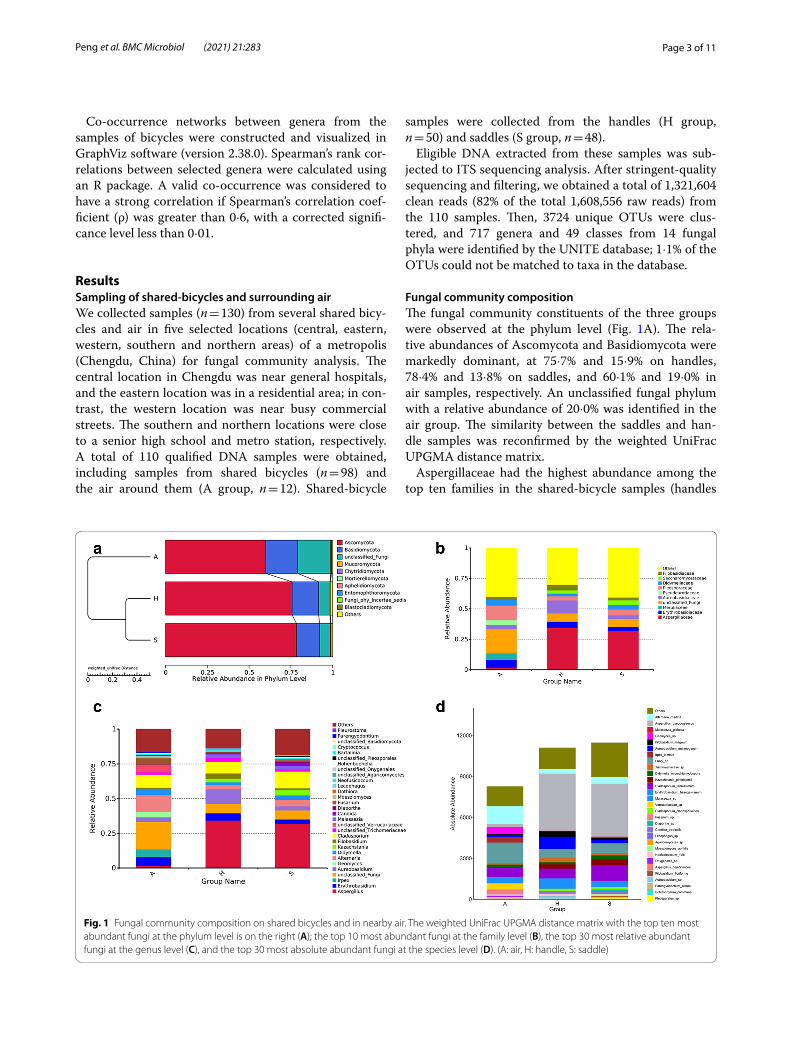
Fungal community compositionThe fungal community constituents of the three groups were observed at the phylum level (Fig. 1A). The rela-tive abundances of Ascomycota and Basidiomycota were markedly dominant, at 75·7% and 15·9% on handles, 78·4% and 13·8% on saddles, and 60·1% and 19·0% in air samples, respectively. An unclassified fungal phylum with a relative abundance of 20·0% was identified in the air group. The similarity between the saddles and han-dle samples was reconfirmed by the weighted UniFrac UPGMA distance matrix.
Aspergillaceae had the highest abundance among the top ten families in the shared-bicycle samples (handles
Fig. 1 Fungal community composition on shared bicycles and in nearby air. The weighted UniFrac UPGMA distance matrix with the top ten most abundant fungi at the phylum level is on the right (A); the top 10 most abundant fungi at the family level (B), the top 30 most relative abundant fungi at the genus level (C), and the top 30 most absolute abundant fungi at the species level (D). (A: air, H: handle, S: saddle)
Page 4 of 11Peng et al. BMC Microbiol (2021) 21:283
and saddles) (Fig. 1B), followed by Aureobasidiaceae in handle samples and unclassified fungi in saddle sam-ples. These results differed from those of the air samples, which were characterized by a relatively high abundance of unclassified fungi and Pleosporaceae, which accounted for approximately 19·7% and 12·2%, respectively.
The relative abundances of the top 30 fungal con-stituents at the genus level in the three groups also varied (Fig. 1C, see Additional Fig. 1). Excluding unclas-sified fungi, five genera, namely, Aspergillus, Candida, Alternaria, Cladosporium and Erythrobasidium, were enriched in the handle samples. Aspergillus and Clad-osporium also dominated the mycobiota in the saddle samples. The fungal communities in air samples were more homogeneous; Alternaria and Cladosporium had the highest abundances, followed by Irpex and Erythrobasidium.
At the species level, the absolute abundance of Asper-gillus pseudoglaucus was overrepresented in the handle and saddle samples. Alternaria rhadina was the most abundant species in air, followed by Cladosporium ten-uissimum and Erythrobasidium hasegawianum, which were highly abundant in all three groups. In addition, Aureobasidium melanogenum and Filobasidium magnum were the main species on handles. Cladosporium tenuis-simum, Alternaria rhadina, and Kazachstania pintol-opesii were the main species on saddles. Malassezia sp., Candida tropicalis, Malassezia globose and Trichomer-iaceae sp. were also included in the top 30 most abundant species (Fig. 1D).
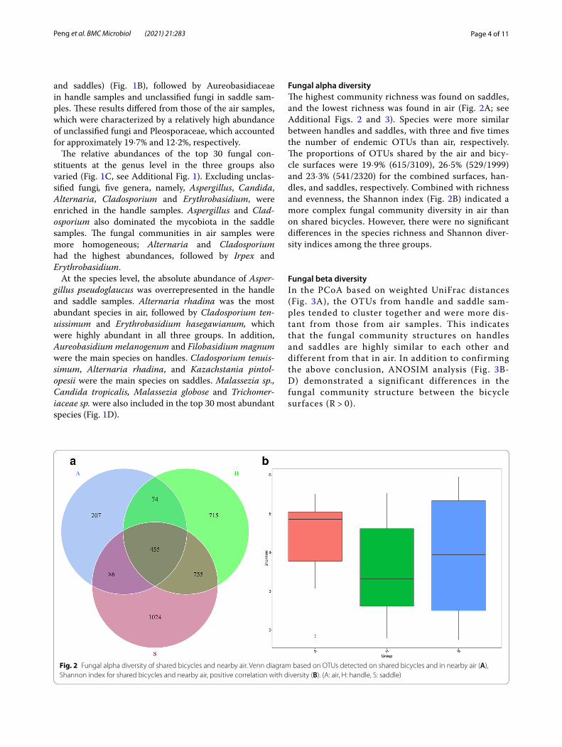
Fungal alpha diversityThe highest community richness was found on saddles, and the lowest richness was found in air (Fig. 2A; see Additional Figs. 2 and 3). Species were more similar between handles and saddles, with three and five times the number of endemic OTUs than air, respectively. The proportions of OTUs shared by the air and bicy-cle surfaces were 19·9% (615/3109), 26·5% (529/1999) and 23·3% (541/2320) for the combined surfaces, han-dles, and saddles, respectively. Combined with richness and evenness, the Shannon index (Fig. 2B) indicated a more complex fungal community diversity in air than on shared bicycles. However, there were no significant differences in the species richness and Shannon diver-sity indices among the three groups.
Fungal beta diversityIn the PCoA based on weighted UniFrac distances (Fig. 3A), the OTUs from handle and saddle sam-ples tended to cluster together and were more dis-tant from those from air samples. This indicates that the fungal community structures on handles and saddles are highly similar to each other and different from that in air. In addition to confirming the above conclusion, ANOSIM analysis (Fig. 3B-D) demonstrated a significant differences in the fungal community structure between the bicycle surfaces (R > 0).
Fig. 2 Fungal alpha diversity of shared bicycles and nearby air. Venn diagram based on OTUs detected on shared bicycles and in nearby air (A), Shannon index for shared bicycles and nearby air, positive correlation with diversity (B). (A: air, H: handle, S: saddle)

Page 5 of 11Peng et al. BMC Microbiol (2021) 21:283
Fig. 3 Fungal beta diversity of shared bicycle and nearby air samples. Weighted UniFrac distance-based PCoA. The percentage represents the contribution of the principal component to the sample difference. Each diamond in the diagram represents a sample, and H and S samples tend to cluster together and away from group A, indicating that the fungal community structure on Hs and Ss is highly similar and greatly different from that of A. ANOSIM intergroup difference analysis. The ordinate represents the rank of the distance between samples, “Between” in the abscissa represents the results between two groups, and the other two represent the results within each group (B-D). (A: air, H: handle, S: saddle)
Page 6 of 11Peng et al. BMC Microbiol (2021) 21:283
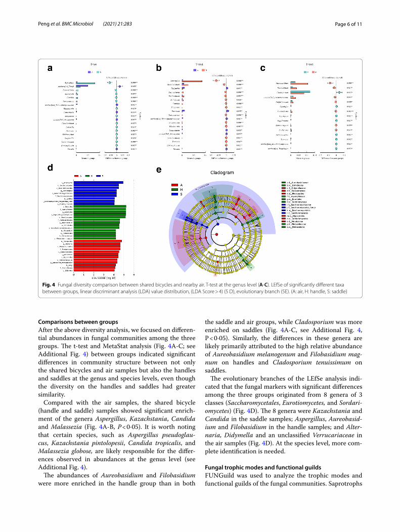
Comparisons between groupsAfter the above diversity analysis, we focused on differen-tial abundances in fungal communities among the three groups. The t-test and MetaStat analysis (Fig. 4A-C; see Additional Fig. 4) between groups indicated significant differences in community structure between not only the shared bicycles and air samples but also the handles and saddles at the genus and species levels, even though the diversity on the handles and saddles had greater similarity.
Compared with the air samples, the shared bicycle (handle and saddle) samples showed significant enrich-ment of the genera Aspergillus, Kazachstania, Candida and Malassezia (Fig. 4A-B, P < 0·05). It is worth noting that certain species, such as Aspergillus pseudoglau-cus, Kazachstania pintolopesii, Candida tropicalis, and Malassezia globose, are likely responsible for the differ-ences observed in abundances at the genus level (see Additional Fig. 4).
The abundances of Aureobasidium and Filobasidium were more enriched in the handle group than in both
the saddle and air groups, while Cladosporium was more enriched on saddles (Fig. 4A-C, see Additional Fig. 4, P < 0·05). Similarly, the differences in these genera are likely primarily attributed to the high relative abundance of Aureobasidium melanogenum and Filobasidium mag-num on handles and Cladosporium tenuissimum on saddles.
The evolutionary branches of the LEfSe analysis indi-cated that the fungal markers with significant differences among the three groups originated from 8 genera of 3 classes (Saccharomycetales, Eurotiomycetes, and Sordari-omycetes) (Fig. 4D). The 8 genera were Kazachstania and Candida in the saddle samples; Aspergillus, Aureobasid-ium and Filobasidium in the handle samples; and Alter-naria, Didymella and an unclassified Verrucariaceae in the air samples (Fig. 4D). At the species level, more com-plete identification is needed.
Fungal trophic modes and functional guildsFUNGuild was used to analyze the trophic modes and functional guilds of the fungal communities. Saprotrophs
Fig. 4 Fungal diversity comparison between shared bicycles and nearby air. T-test at the genus level (A-C). LEfSe of significantly different taxa between groups, linear discriminant analysis (LDA) value distribution, (LDA Score > 4) (5 D), evolutionary branch (5E). (A: air, H: handle, S: saddle)

Page 7 of 11Peng et al. BMC Microbiol (2021) 21:283
had an influential relative abundance of approximately 50% in bicycle surface specimens and approximately 1/8 in air specimens (see Additional Fig. 5). The heatmap representing functional taxonomy shows a high abun-dance in air, which is mostly nonoverlapping with that on shared bicycles (Fig. 5A). In the handle and saddle sam-ples, the abundance of animal pathogens was obviously higher than that in the air samples (Fig. 5A). Compared with those in the saddle samples, more animal pathogens and fewer endophyte-plant pathogens were found in the handle samples (P < 0·05, Fig. 5B-D).
Fungal co‑occurrence networksCorrelation (ρ = 0·6) and P values (P = 0·01) were established. The fungal co-occurrence networks were
obviously distinct between the shared-bicycle surfaces (Fig. 6A and B). Compared with the saddle specimens, the handle specimens had a larger clustering coefficient (CC: 50·5% vs. 48·8%) and a smaller average path length (APL: 2·18 vs. 3·03), indicating that the fungal commu-nities on the handles were more closely connected than those on the saddles; the lower species richness was reconfirmed by the APL combined with a 0·2% smaller graph density.
Considering the statistical parameters, seven genera were enriched on handles. Two of the core genera, Asper-gillus and Kazachstania, were mutually positively related, and both were associated with two subdominant genera (Candida and Thermomyces). Aspergillus was negatively correlated with four other enriched genera (unclassified
Fig. 5 Fungal trophic modes and functional guilds in the air, handle, and saddle samples. Relative abundance of functional guilds (A). T-test of functional guilds (B-D). (A: air, H: handle, S: saddle)

Page 8 of 11Peng et al. BMC Microbiol (2021) 21:283
fungi, Alternaria, Aureobasidium, and unclassified Tri-chomeriaceae) and two subdominant genera (Periconia and Didymella). Kazachstania had an inverse correlation with the dominant genera Alternaria and Cladosporium and unclassified fungi. However, lower relative abun-dances and greater mutual distances were present among genera of phylum Basidiomycota.
Eight genera were enriched on saddles. Among them, Aspergillus showed a negative relationship with Didy-mella, Cladosporium, and an unclassified fungi and a positive relationship with Kazachstania and the subdom-inant genus Thermomyces. Cladosporium was negatively correlated with Alternaria and an unclassified fungi. Erythrobasidium, as a highlighted genus of the phylum Basidiomycota, was positively correlated with Aureoba-sidium and negatively correlated with Thermomyces.
DiscussionOur study identified differences in the compositions of fungal communities between the surfaces of shared bicycles and the surrounding air. The OTUs shared by the air deposition and bike surface specimens accounted for only 19·9% of the bike surface OTUs (Fig. 2A). Sig-nificant enrichment of the genera Aspergillus, Kazach-stania, Candida and Malassezia was found on bicycles,
probably because Candida tropicalis and Malassezia glo-bose are resident fungi of human skin [7, 8]. Aspergillus pseudoglaucus, a common indoor fungus, has of osmo- and xero-characteristics and is widespread [9]. Skin and clothes contact may be the reason for their enrichment on shared-bicycle surfaces. Kazachstania pintolopesii has been isolated from forest soil and has a high mini-mum growth temperature of 20–43 °C [10]. Therefore, dust exposure under high-temperature conditions dur-ing summer may explain the high abundance of Kazach-stania pintolopesii. Thus, we hypothesize that the major factor affecting the composition of the fungal community on the surface of shared bicycles is not the ambient air but the flora from the surface of the palm or clothing or road dust. Ambient temperature and humidity also have an effect on the microflora. However, the exact reasons for these results need to be further studied.
Regarding the fungal community structure, we found a significant difference between the saddle and handle samples. Aureobasidium melanogenum requires a rela-tive humidity greater than 90% for proliferation, though it can tolerate extremely harsh conditions [11]. Filoba-sidium magnum requires a slightly acidic environment [12]. Their specific requirements for humidity and pH, respectively, may explain their higher abundance on
Fig. 6 Fungal co-occurrence networks in the handle and saddle samples. Handle (A). Saddle (B). A strong and significant correlation (Spearman ρ < 0·6, P < 0·01) was detected. A node represents a fungal genus, and the size of the node is representative of the average relative abundance of the genus. The node colors represents phyla. Regarding the lines between two nodes, red represents a positive correlation, while blue represents a negative correlation
Page 9 of 11Peng et al. BMC Microbiol (2021) 21:283
handles, which are exposed to moist, sweaty hands, than on saddles. Cladosporium tenuissimum is an endo-phytic plant pathogen. It was more abundant on saddles than on handles, which was likely due to the shady trees in the parking lot.
According to the fungal co-occurrence networks, the fungal biota in the air samples were more com-plex and closely related than those in the shared-bicycle surface samples. This also indicated that fungi in the air had little influence on the fungal community of the bicycle surface. Among the bicy-cle samples, the fungal communities on the handles were more closely related, and those on the saddles were more intricate. These difference may be related to the ability of fungi to colonize different materi-als, the effects of human sweat and clothing, or the frequency at which the different parts of the shared bicycles were disinfected.
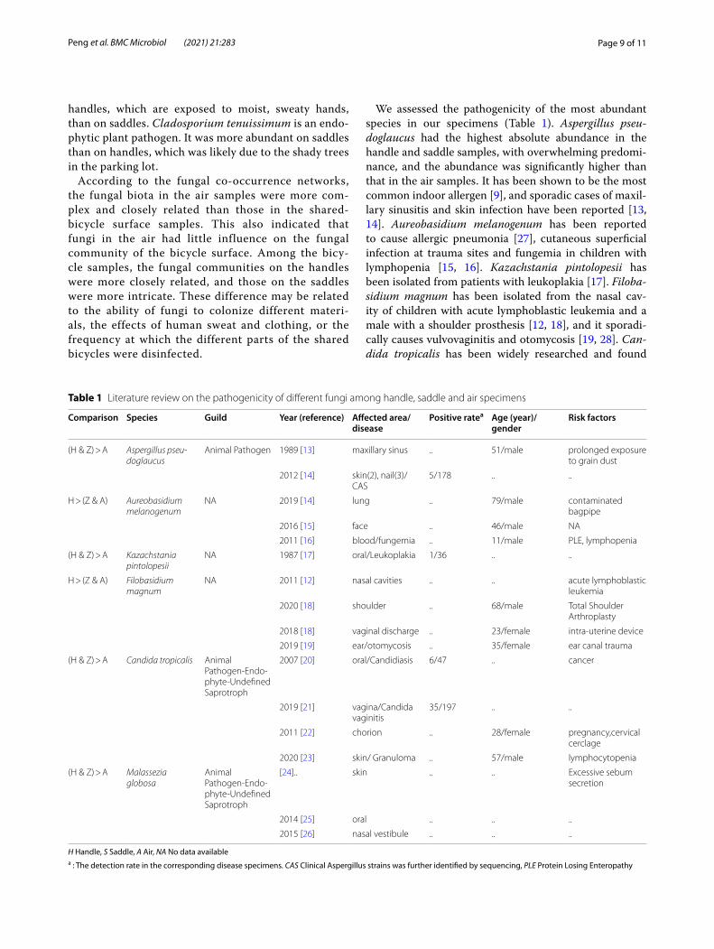
We assessed the pathogenicity of the most abundant species in our specimens (Table 1). Aspergillus pseu-doglaucus had the highest absolute abundance in the handle and saddle samples, with overwhelming predomi-nance, and the abundance was significantly higher than that in the air samples. It has been shown to be the most common indoor allergen [9], and sporadic cases of maxil-lary sinusitis and skin infection have been reported [13, 14]. Aureobasidium melanogenum has been reported to cause allergic pneumonia [27], cutaneous superficial infection at trauma sites and fungemia in children with lymphopenia [15, 16]. Kazachstania pintolopesii has been isolated from patients with leukoplakia [17]. Filoba-sidium magnum has been isolated from the nasal cav-ity of children with acute lymphoblastic leukemia and a male with a shoulder prosthesis [12, 18], and it sporadi-cally causes vulvovaginitis and otomycosis [19, 28]. Can-dida tropicalis has been widely researched and found
Table 1 Literature review on the pathogenicity of different fungi among handle, saddle and air specimens
H Handle, S Saddle, A Air, NA No data availablea : The detection rate in the corresponding disease specimens. CAS Clinical Aspergillus strains was further identified by sequencing, PLE Protein Losing Enteropathy
Comparison Species Guild Year (reference) Affected area/disease
Positive ratea Age (year)/ gender
Risk factors
(H & Z) > A Aspergillus pseu-doglaucus
Animal Pathogen 1989 [13] maxillary sinus .. 51/male prolonged exposure to grain dust
2012 [14] skin(2), nail(3)/CAS
5/178 .. ..
H > (Z & A) Aureobasidium melanogenum
NA 2019 [14] lung .. 79/male contaminated bagpipe
2016 [15] face .. 46/male NA
2011 [16] blood/fungemia .. 11/male PLE, lymphopenia
(H & Z) > A Kazachstania pintolopesii
NA 1987 [17] oral/Leukoplakia 1/36 .. ..
H > (Z & A) Filobasidium magnum
NA 2011 [12] nasal cavities .. .. acute lymphoblastic leukemia
2020 [18] shoulder .. 68/male Total Shoulder Arthroplasty
2018 [18] vaginal discharge .. 23/female intra-uterine device
2019 [19] ear/otomycosis .. 35/female ear canal trauma
(H & Z) > A Candida tropicalis Animal Pathogen-Endo-phyte-Undefined Saprotroph
2007 [20] oral/Candidiasis 6/47 .. cancer
2019 [21] vagina/Candida vaginitis
35/197 .. ..
2011 [22] chorion .. 28/female pregnancy,cervical cerclage
2020 [23] skin/ Granuloma .. 57/male lymphocytopenia
(H & Z) > A Malassezia globosa
Animal Pathogen-Endo-phyte-Undefined Saprotroph
[24].. skin .. .. Excessive sebum secretion
2014 [25] oral .. .. ..
2015 [26] nasal vestibule .. .. ..

Page 10 of 11Peng et al. BMC Microbiol (2021) 21:283
to reside on mucosal membranes rather than skin, with strong opportunistic pathogenicity and a high proportion of fluconazole-resistant properties [7, 20, 21]. The endo-metrium is also at risk of Candida tropicalis infection during pregnancy, and the skin is susceptible to hypoim-munity [22, 23]. Although M. globose is the most com-mon fungus on human skin, it normally concentrates on the face and upper trunk due to its lipophilicity, and the hands play a core role in the process of fungal metasta-sis [8]. Their ectopic or excessive aggregation can lead to allergies or fungal infections [24–26], at the same time. FunGuild shows that animal pathogen has an obvious preponderance on the surface of shared bicycles, espe-cially on the handles. Therefore, it is recommended that users (especially those with trauma, allergy, chronic dis-ease, immunosuppression or immunodeficiency) first sterilize the handles and seat before using a shared bicy-cle. The timely cleaning and disinfection of the hands and clothes should not be neglected. It also suggests that governments should carry out regular and effective disin-fection of shared bicycles to avoid the potential spread of diseases and allergens, especially during hot and humid summers.
We compared our fungal species of the handles with the top ten most abundant eukaryotes of palm from a review of human skin microorganisms [ 29]. Malassezia globose, Candida parapsilosis and Malassezia sympo-dialis coexisted in the handle group and human palms. Malassezia globose is the second most abundant fungus on the palm but ranked eighteenth in relative abundance on shared-bicycle handles. The other two coexistent species were detected in relatively small quantities in the handle samples. This suggests that the main source of these three fungi may be direct contact with human palms. However, such a large difference in the fungal community between palms and handles could be due to research bias or the interaction of more complex factors, such as handle temperature, humidity, hand disinfection and afforest environments.
Healthy people are generally immune to most patho-genic fungi. Moreover, Trichophyton, Microsporum, and Epidermophyton, which commonly cause cutaneous mycoses, were not discovered. This was probably due to (i) the small sample size; (ii) specimen collection during the rainy season, causing some of the fungi to be washed off from the bicycle by heavy rain, with the few remaining reads removed in the data quality control stage; and (iii) the need for strict environmental conditions (tempera-ture or pH) or culture media with specific nutrition.
The limitations and future directions are as follows. (i) The UNITs database at the species level is still incomplete for fungal identification. (ii) ITS sequencing is based on genes and does not indicate fungal survival status. (iii)
Specimens were collected in July, representing only the characteristics of shared-bicycle flora in summer. Addi-tional studies with larger sample sizes are needed to fur-ther support our conclusion. The detection of viruses and bacteria in addition to fungi can be used to analyze microbial community structures in more dimensions. The correlations of environmental factors (season and weather, temperature, humidity, etc.) with microorgan-isms should be assessed.
ConclusionOur study analyzed fungal community structures in shared-bicycle surface (handles and saddles) samples; these communities were obviously different from those in the air deposition specimens, and more animal patho-gens were present on bicycles than in air. In addition, we analyzed the possible factors leading to these differences and reviewed the pathogenicity of some significantly dif-ferent species. Thus, governments around the world with shared-bicycle programs, bicycle program managers, and the public are reminded to pay attention to shared-bicy-cle hygiene.
Supplementary InformationThe online version contains supplementary material available at https:// doi. org/ 10. 1186/ s12866- 021- 02338-4.
Additional file 1: Figure 1. Ternary plot of fungi among the three groups. (A: air, H: handle, S: saddle).
Additional file 2: Figure 2. OTU rarefaction curve for the three groups.
Additional file 3: Figure 3. Fungal alpha diversity of shared bicycles and nearby air. Chao 1 analysis (A), ACE analysis (B) (Chao 1 and ACE indices are positively correlated with the richness of the fungal community).
Additional file 4: Figure 4. Fungal diversity comparison between shared bicycles and nearby air by t-tests at the species level (A-C). The top 12 significantly different species as confirmed by the MetaStat analysis (D).
Additional file 5: Figure 5. Relative abundances of fungal trophic modes among the three groups.
AcknowledgmentsThis work was supported by National Natural Science Foundation of China (No. 81771783) and Sichuan Science and Technology Program (No. 2019JDTD0027).
Authors’ contributionsLu Peng contributed to sample collection, literature search, data analysis, statistical analysis, and writing – original draft of the manuscript; Bi Qin contributed to sample collection, data curation, literature search; Zhu Shen contributed to literature search, statistical analysis, data interpretation, and writing – review of the manuscript; Siyu Wang contributed to conception and design of the study, project administration, sample collection, data analysis, literature search, and writing – review of the manuscript. All authors read and approved the final manuscript.
FundingThis work was supported by the National Natural Science Foundation of China (No. 81771783) and Sichuan Science and Technology Program (No. 2019JDTD0027).

Page 11 of 11Peng et al. BMC Microbiol (2021) 21:283
Availability of data and materialsThe datasets used and/or analysed during the current study are available from the corresponding author on reasonable request.
Declarations
Ethics approval and consent to participateNot applicable.
Consent for publicationNot applicable.
Competing interestsThe authors have no conflict of interest to declare.
Author details1 Department of Dermatology, Institute of Dermatology and Venereology, Sichuan Academy of Medical Sciences & Sichuan Provincial People’s Hospital, No.32, Western 2nd Section, 1st Ring Rd, Qingyang District, Chengdu 610072, Sichuan, China. 2 School of Medicine, University of Electronic Science and Tech-nology of China, Chengdu 610054, China. 3 Department of Dermatology, Acu-puncture & Moxibustion Research Institute, Sichuan Academy of Traditional Chinese Medicine, Sichuan Second Hospital of Traditional Chinese Medicine, Chengdu 610031, Sichuan, China.
Received: 9 July 2021 Accepted: 27 September 2021
References 1. https:// en. wikip edia. org/ wiki/ List of bicycle-sharing systems (Accessed
January 13, 2021) 2. Sun C, Yuan T, Chen L, Xie Z, Shen Z. Occurrence of potentially
pathogenic bacteria on shared bicycles. Int J Hyg Environ Health. 2020;224:113442.
3. Zou ZY, Lei L, Chen QY, et al. Prevalence and dissemination risk of antimicrobial-resistant Enterobacteriaceae from shared bikes in Beijing, China. Environ Int. 2019;132:105119.
4. Altschul SF, Gish W, Miller W, Myers EW, Lipman DJ. Basic local alignment search tool. J Mol Biol. 1990;215:403–10.
5. Nilsson RH, Ryberg M, Kristiansson E, Abarenkov K, Larsson KH, Kõljalg U. Taxonomic reliability of DNA sequences in public sequence databases: a fungal perspective. PLoS One. 2006;1:e59.
6. Nguyen NH, Song Z, Bates ST, Branco S, Tedersoo L, Menke J, et al. FUN-Guild: an open annotation tool for parsing fungal community datasets by ecological guild. Fungal Ecol. 2016;20:241–8.
7. Horváth E, Sipiczki M, Csoma H, Miklós I. Assaying the effect of yeasts on growth of fungi associated with disease. BMC Microbiol. 2020;20:320.
8. Grice EA, Segre JA. The skin microbiome [published correction appears in Nat Rev. Microbiol. 2011 Aug; 9: 626]. Nat Rev Microbiol. 2011;9:244–53.
9. Chen AJ, Hubka V, Frisvad JC, et al. Polyphasic taxonomy of Aspergillus section Aspergillus (formerly Eurotium), and its occurrence in indoor environments and food. Stud Mycol. 2017;88:37–135.
10. Watson K, Arthur H, Blakey M. Biochemical and morphological correla-tions in the thermophilic enteric yeasts, Saccharomyces telluris, Candida slooffii, Torulopsis bovina and T. pintolopesii. In: Current Developments in Yeast Research; 1981. p. 499–503.
11. Moreno LF, Vicente VA, de Hoog S. Black yeasts in the omics era: Achieve-ments and challenges. Med Mycol. 2018;56:32–41.
12. Khan Z, Mokaddas E, Ahmad S, Burhamah MH. Isolation of Cryptococ-cus magnus and Cryptococcus chernovii from nasal cavities of pediatric patients with acute lymphoblastic leukemia. Med Mycol. 2011;49:439–43.
13. Aznar C, de Bievre C, Guiguen C. Maxillary sinusitis from Microascus cinereus and Aspergillus repens. Mycopathologia. 1989;105(2):93–7.
14. Hubka V, Kubatova A, Mallatova N, et al. Rare and new etiological agents revealed among 178 clinical Aspergillus strains obtained from Czech patients and characterized by molecular sequencing. Med Mycol. 2012;50(6):601–10.
15. Chen WT, Tu ME, Sun PL. Superficial Phaeohyphomycosis Caused by Aureobasidium melanogenum Mimicking Tinea Nigra in an Immuno-competent Patient and Review of Published Reports. Mycopathologia. 2016;181:555–60.
16. Mershon-Shier KL, Deville JG, Delair S, et al. Aureobasidium pullulans var. melanigenum fungemia in a pediatric patient. Med Mycol. 2011;49:80–3.
17. Krogh P, Holmstrup P, Thorn JJ, Vedtofte P, Pindborg JJ. Yeast species and biotypes associated with oral leukoplakia and lichen planus. Oral Surg Oral Med Oral Pathol. 1987;63(1):48–54.
18. Baptista M, Sevivas N, Ferreira NV, Fardilha L, Varanda P, Mateus C. Cryp-tococcus magnus Periprosthetic Shoulder Infection: A Case Report. JBJS Case Connect. 2020;10:e20.00507.
19. Ghajari A, Lotfali E, Norouzi M, Arab-Mazar Z. First report of Vulvovaginitis due to Cryptococcus magnus in Iran. Curr Med Mycol. 2018;4:30–3.
20. González Gravina H, González de Morán E, Zambrano O, et al. Oral Can-didiasis in children and adolescents with cancer. Identification of Candida spp. Med Oral Patol Oral Cir Bucal. 2007;12(6):E419–23.
21. Paulo A, de Medeiros M, Vieira de Melo AP, Gonçalves SS, Milan EP, Chaves GM. Genetic relatedness among vaginal and anal isolates of Candida albicans from women with vulvovaginal candidiasis in north-east Brazil. J Med Microbiol. 2014;63(Pt 11):1436–45.
22. Canpolat FE, Çekmez F, Tezer H. Chorioamnionitis and neonatal sepsis due to Candida tropicalis. Arch Gynecol Obstet. 2011;283(4):919–20.
23. Yang H, Xu X, Ran X, Ran Y. Successful Treatment of Refractory Candidal Granuloma by Itraconazole and Terbinafine in Combination with Hyper-thermia and Cryotherapy. Dermatol Ther (Heidelb). 2020;10(4):847–53.
24. Theelen B, Cafarchia C, Gaitanis G, Bassukas ID, Boekhout T, Dawson TL Jr. Malassezia ecology, pathophysiology, and treatment [published correction appears in Med Mycol. 2019 Apr 1;57(3):e2]. Med Mycol. 2018;56(suppl_1):S10–25.
25. Dupuy AK, David MS, Li L, et al. Redefining the human oral mycobiome with improved practices in amplicon-based taxonomy: discovery of Malassezia as a prominent commensal. PLoS One. 2014;9(3):e90899.
26. Hee JW, Daniel C, Hoon CJ, et al. Analysis of the nasal vestibule mycobi-ome in patients with allergic rhinitis. Mycoses. 2015;58:167–72.
27. Ziegler K, Joest M, Turan N, Schmidt D, Rath PM, Steinmann J. Hypersen-sitivity pneumonitis of a bagpipe player: Fungal antigens as trigger? Med Mycol Case Rep. 2019;24:44–7.
28. Aboutalebian S, Mahmoudi S, Okhovat A, Khodavaisy S, Mirhendi H. Oto-mycosis Due to the Rare Fungi Talaromyces purpurogenus, Naganishia albida and Filobasidium magnum. Mycopathologia. 2020;185:569–75.
29. Byrd AL, Belkaid Y, Segre JA. The human skin microbiome. Nat Rev Micro-biol. 2018;16(3):143–55.
Publisher’s NoteSpringer Nature remains neutral with regard to jurisdictional claims in pub-lished maps and institutional affiliations.