Embed Size (px)
Citation preview

CHARACTERIZATION OF FBXO9 IN DROSOPHILA MELANOGASTER
by Poly Talukdar, B.Sc.
A Thesis submitted
to the School of Graduate Studies in partial fulfillment of the
requirements for the degree of
Master of Science
Department of Biology
Memorial University of Newfoundland
April, 2016
St. John’s, Newfoundland and Labrador

ii
ABSTRACT
The study of disease in model organisms is a fundamental and important stepping-stone in
understanding and uncovering the mechanisms behind disease pathology in humans. The
purpose of this work was to identify potential targets for the treatment and prevention of
Parkinson disease using Drosophila melanogaster. Commonly known as the fruit fly, D.
melanogaster is one of the important model organisms used extensively in biological
research. Moreover, it has conserved developmental processes and mechanisms shared
with human neurodegenerative disorders. Parkinson disease (PD) is a progressive
neurodegenerative disorder characterized by death of dopamine producing cells of the
substantia nigra affects about 1% of people over 60 years old worldwide. In mammals,
Fbxo9 is a substrate recognition component of the SCF (SKP1-cullin-Fbox)-type E3
ubiquitin ligase complex. Some targets of Fbxo9, including an extensive array of proteins,
are degraded via the ubiquitin-proteasome system. In this study, a potential D.
melanogaster homologue of Fbxo9, CG5961, was identified. The Fbxo9 homologue in D.
melanogaster has been conserved through evolution and retains many of the functional
domains. The main goal of this project was to determine if Fbxo9 can be implicated in
modeling PD in D. melanogaster. To investigate its role in neuronal survival, I over-
expressed and down-regulated Fbxo9 in neuron-rich eye and dopaminergic neurons.
Through assessments of eye morphology, climbing ability and ageing analysis, it was
found that loss-of-function of Fbxo9 causes a PD like symptom. I expect that the
knowledge obtained by determining the pathways involved in PD in D. melanogaster will
help uncover potential new therapeutic approaches for research in human as well as other
genes in both humans and flies.

iii
ACKNOWLEDGEMENTS
I would like to express sincere thanks to my supervisor, Dr. Brian E. Staveley for giving
me the opportunity to undertake an M.Sc. in Biology at Memorial University of
Newfoundland. He has supported me academically and financially. He has been
supportive, patient, encouraging and above all, available throughout, given that he has the
busy schedule to maintain.
A special thanks goes to Dr. Dawn Marshall and Dr. Dawn Bignell who have guided and
suggested academically. I am extremely grateful to the School of Graduate Studies,
Memorial University of Newfoundland for funding two years full time M.Sc. program and
the National Sciences and Engineering Council of Canada for the Discovery Grant to Dr.
Brian E. Staveley.
I would like to give my thanks to the members of the Staveley research group: Peter,
Jennifer, Eric, Colleen, and Mahin for their kind help and friendly support. All of them
have been very good friends. Specially, great thanks to Peter for being so helpful, listening
and advising me academically.
Finally I wish to thank my dearest husband Dr. Bijon K. Sarker for allowing me to study
for two year M.Sc. alone and standing by me when I was in need. I would have not been
able to complete my research without his support. I am also grateful to my great father and
mother for enduring and encouraging me throughout my study. Their unconditional love
for me remained the same throughout my academic journey for two years.

iv
TABLE OF CONTENTS
ABSTRACT…………………………………………………………………………. ii
ACKNOWLEDGEMENTS………………………………………………………….. iii
TABLE OF CONTENTS……………………………………………………………. iv
LIST OF TABLES…………………………………………………………………… vi
LIST OF FIGURES………………………………………………………………….. vii
LIST OF ABBREVIATIONS………………………………………………………... viii
INTRODUCTION……………………………………………………………………. 1
Parkinson Disease……………………………………………………………… 1
PD Gene Loci………………………………………………………………….. 2
Identification of Fbxo7 as PARK15……………………………………………. 4
Drosophila melanogaster as a model organism………………………………... 4
Drosophila in PD modeling……………………………………………………. 6
Drosophila models of PD………………………………………………………. 7
The α-synuclein model…………………………………………………… 7
The LRRK2/Lrrk model…………………………………………………. 9
The parkin/PINK1 model………………………………………………... 10
Skp-Cullin-Fbox containing complex (SCF) Ubiquitin Ligases……………… 12
F-box proteins……………………………………………………………. 14
F-box only protein 7……………………………………………………… 16
F-box only protein 9……………………………………………………… 18
F-box only protein 32…………………………………………………….. 18
Thesis objectives……………………………………………………................... 20
MATERIALS AND METHODS……………………………………………………... 21
Bioinformatics analysis…………………………………………………………. 21
Drosophila melanogaster………………………………………………………. 22
Media…………………………………………………………………….. 22
Stocks…………………………………………………………………….. 22
Ageing assay…………………………………………………………………..... 23

v
Locomotion assay………………………………………………………………. 25
Scanning electron microscopy of the compound eye………………………….. 25
RESULTS……………………………………………………………………………... 27
Bioinformatics analysis………………………………………………………… 27
Eye analysis……………………………………………………………………. 35
Effects of directed over-expression and RNA interference of Fbxo9…… 35
Investigation of Fbxo9 and Foxo interactions in the compound eye……. 39
Ageing analysis………………………………………………………………… 42
The effects of loss-of-function and gain-of-function of Fbxo9…………. 42
Investigating the effects of Fbxo9 on the α-synuclein model of PD……. 47
Climbing analysis……………………………………………………………… 50
Effects of gain-of-function and loss-of-function of Fbxo9……………… 50
Investigating the influence of altering Fbxo9 expression on the α-
synuclein model………………………………………………………….
54
DISCUSSION…………………………………………………………………………. 56
CONCLUSION………………………………………………………………………. 64
REFERENCES……………………………………………………………………….. 66

vi
LIST OF TABLES
Table 1. Gene loci implicated in Parkinson disease……………………………………...
3
Table 2. Genotypes of fly stocks used in this study………………………………………
24
Table 3. Similarity scores of Fbxo9 protein homologous from vertebrate and
invertebrate species…………………………………………………………………..
31
Table 4. Protein names and GenBank accession numbers of Vertebrate and invertebrate
versions of Fbxo7, Fbxo9 and Fbxo32………………………………………….
34
Table 5. A summary of the biometric analysis of the directed overexpression and RNA
interference of Fbxo9 in the Drosophila compound eye………………………...
37
Table 6. A summary of the ommatidia number, bristle number and ommatidia area
when Foxo is co-overexpressed with lacZ, Fbxo9RNAi
and Fbxo9EP in the
Drosophila compound eye……………………………………………………...
40
Table 7. The Log-Rank comparison of the survival curves for the directed
overexpression and RNA interference of Fbxo9 in the dopaminergic
neurons…………………………………………………………………………..
44
Table 8. The Log-Rank survival curve comparison for the directed co-overexpression
of α-synuclein and Fbxo9 or Fbxo9RNAi in the dopaminergic
neurons…………………………..........................................................................
48
Table 9. Statistical analysis of climbing index curves for the directed overexpression
and RNA interference of Fbxo9 in the dopaminergic neurons………………….
51
Table 10. Statistical analysis of climbing index curves for the co-overexpression of α-
synuclein and Fbxo9 or Fbxo9RNAi in the dopaminergic neurons………………
55

vii
LIST OF FIGURES
Figure 1: Alignment of human Fbxo9 (NP_258441.1) protein with Drosophila CG5961
(NP_650206.1) ……….......................................................................................
29
Figure 2: Fbxo9 is well conserved in vertebrates and invertebrates……………………...
31
Figure 3: Human Fbxo9 and Drosophila Fbxo7 share some similarity…………………..
32
Figure 4: Similarity between Human Fbxo9 and Drosophila Fbxo32……………………
33
Figure 5: Fbxo7, Fbxo9 and Fbxo32 are conserved between Drosophila and Human…...
34
Figure 6: Biometric analysis of gain-of-function and loss-of-function of Fbxo9 in the
Drosophila compound eye……………………………………………………...
38
Figure 7: Biometric analysis of the Drosophila compound eye in flies overexpressing
Foxo together with overexpression or loss-of-function of Fbxo9……………..
41
Figure 8: Effect of loss-of-function and overexpression of Fbxo9 on lifespan in
Drosophila……………………………………………………………………...
46
Figure 9: Effect of α-synuclein overexpression together with loss-of-function or
overexpression of Fbxo9 in dopaminergic neurons on lifespan in
Drosophila……………………………………………………………………...
49
Figure 10: Effect of gain-of-function and loss-of-function of Fbxo9 on climbing ability
in Drosophila…………………………………………………………………...
53
Figure 11: Analysis of climbing ability of Drosophila flies overexpressing α-synuclein
together with Fbxo9 or Fbxo9RNAi in the dopaminergic neurons. ……………...
55

viii
LIST OF ABBREVIATIONS
AD
ANOVA
ATP
ATP13A2
AR
BLAST
BLASTp
C
CI
cm
CNS
COPD
CO2
DA
Ddc
ddH2O
Dilp
dLRRK
DNA
dNTP
E1
E2
E3
eIF3-f
EOPD
ERK
FBP
FBXO9
FBXO7
FBXO32
FBXL
autosomal dominant
analysis of variance
adenosine triphosphate
ATPase type 13A2
autosomal recessive
Basic Local Alignment Search Tool
Protein Basic Local Alignment Search Tool
carboxyl terminus
confidence interval
centimeter
central nervous system
chronic obstructive pulmonary disease
carbon dioxide
dopaminergic
Dopa decarboxylase
double distilled H20
Drosophila Insulin-like peptide
Drosophila leucine-rich repeat kinase
deoxynucleotide
deoxynucleotide triphosphate
ubiquitin-activating enzyme
ubiquitin-conjugating enzyme
ubiquitin-ligase
eukaryotic initiation factor 3 - subunit 5
early-onset parkinson disease
extra cellular signal-regulated kinase
F-box containing protein
F box-only protein 9
F box-only protein 7
F box-only protein 32
F-box leucine rich repeat

ix
FBXO
FBXW
Foxo
FPD
g
GAK
GAL4
GBA
GIGYF2
GMR
GOF
HECT
HLA-DRA
HNHc
HSP70
HTRA2
H2O
IGF-1
k
kDa
L
LBs
LN
LOPD
LRR
LRRK2
MAPKK
mg
MIT
mL
mRNA
N/A
NCBI
F-box other
F-box WD40
forkhead box other
familial forms of Parkinson disease
gram
cyclin G associated
yeast transcriptional activator for galactase-inducible genes
glucocerebrosidase
GRB10 interacting GYF protein 2
Glass Multiple Reporter
gain-of-function kinase
Homologous to the E6-AP Carboxyl Terminus
major histocompatibility complex, class II, DR alpha
HNH nuclease family
heat shock protein 70
HtrA serine peptidase 2
water
Insulin-like Growth Factor 1
slope
kilo Daltons
litre
lewy bodies
lewy neurite
late-onset Parkinson disease
leucine rich repeat
leucine-rich repeat kinase 2
mitogen activated protein kinase kinase kinase
milligram
microtubule interacting and trafficking domain
mililitre
messenger RNA
not applicable
National Center for Biotechnology Information

x
NES
NLS
ntc
ORF
PARK
PD
PI31
P13K
PIP2
PIP3
PINK1
PLA2G6
PRR
PTEN
RING
RNAi
RNA
ROS
SCF
SEMs
SNc
SNCA
SOD
SEM
TPR
TH
UAS
UbI
UCH-L1
UPS
WT
° C
µm
nuclear export sequence
nuclear localization sequence
nutcracker
open reading frame
Parkinson disease-associated gene locus
Parkinson Disease
31 kDa proteasome inhibitor
phosphatidylinositol 3-kinase
phosphatidylinositol-4,5-biphosphate
phosphatidylinositol (3,4,5)-triphosphate
PTEN-induced putative kinase 1
phospholipase A2, group VI
Proline rich region
phosphatase and tension homologue
really interesting new gene
RNA-interference
Ribonucleic acid
reactive oxygen species
Skp1-Cullin1-Fbox
scanning electron micrographs
substntia nigra pars compacta
subthalamic nucleus
superoxide dismutase
standard error of the mean
tetracycline peptide repeat
Tyrosine hydroxylase
upstream activator sequences
ubiquitin like-domain
ubiquitin carboxyl-terminal hydrolase L1
ubiquitin proteasome system
wild type
degrees Celsius
micrometre

xi
α-synuclein
alpha-synuclein

1
INTRODUCTION
Parkinson Disease
Parkinson disease (PD) is the second most common progressive neurodegenerative
disorder: affecting about 1% of people over 60 years old worldwide (Lew, 2007). It is
associated with a movement disorder, which is characterized by tremor, rigidity, postural
instability and bradykinesia. Although most symptoms are associated with motor disorder,
non-motor symptoms such as cognitive, psychiatric and emotional problems are also seen
in this disease (Tadaiesky et al., 2008). The neuropathological distinctive characteristics
showed by PD patients are Lewy Bodies (LB) and Lewy Neurites (LN) in surviving
neurons. The ultimate dysfunction of these neurons is responsible for the symptoms and
pathology of PD (Bekris et al., 2010). PD is associated with the progressive degeneration
of dopaminergic neurons in the substantia nigra of the midbrain region and the subsequent
loss of dopamine (Dauer and Przedborski, 2003). The study of this disease in well-known
genetically versatile model organism such as Drosophila melanogaster is a fundamental
and important stepping stone in understanding and uncovering the mechanisms behind
disease pathology in a human host.
Many genetic and environmental factors have been identified for the progression of
PD. Most forms of PD are known to be sporadic with no known causes (Cauchi and
Heuvel, 2006; Lu and Vogel, 2009). Several environmental factors have been well
documented correlating with the onset of PD such as chemical exposure, brain trauma,
obesity, age and diabetes (Vanitallie, 2008). Alternatively, some familial forms of PD have
been found in association with different types of alteration in genes (Bereznai and Molnar,
2009). The discovery of the familial forms of PD-associated genes inherently provides us
the opportunity to study both sporadic and familial PD in model organisms.

2
PD Gene Loci
There are 18 Parkinson-associated (PARK) gene loci identified to date through a
combination of linkage, segregation and sequence analysis; though several of these gene
loci require validation by independent studies (Table 1). The first of the genes found in
association with the rare familial forms of PD (FPD) is α-synuclein (Polymeropoulos et al.,
1997; Kruger et al., 1998). Among these gene loci, several have been cloned and include α-
synuclein/PARK1 (Polymeropoulos et al., 1997), Parkin/PARK2 (Kitada et al., 1998),
Ubiquitin C-terminal hydrolase1 (Uchl-1)/PARK5 (Leroy et al., 1998), Phosphatase and
tensin homologue [PTEN] induced kinase (Pink1)/PARK6 (Valente et al., 2004), DJ-
1/PARK7 (Bonifati et al., 2003) and leucine rich repeat kinase 2 (LRRK2)/PARK8
(Zimprich et al., 2004). Among the genes identified, α-synuclein/ PARK1 and Leucine-rich
repeat kinase 2 or LRRK2 are known as autosomal dominant alleles whereas the rest are
autosomal recessive alleles (Staveley, 2012). The identification of the genes has helped to
better understand the underlying pathological mechanism of FPD. This pathological
mechanism also supports us in understanding sporadic causes of PD. A considerable
number of studies prior to modeling PD in animal have been done and thus these studies
proposed some pathogenic mechanisms like protein mis-folding, protein abnormal
accumulation oxidative stress, mitochondrial dysfunction and caspase activation. The
endeavors to identify the central reasons for PD significantly led us to determine the
necessary tools through the use of animals in the experiments. The promising indications
are found from the modeling of PD in animals including rats, monkeys, flies, mice and
worms (Kuwahara et al., 2006). Among all model organism studies, the genetic studies in
Drosophila melanogaster have advantages over other animal models.

3
Table 1: Gene loci implicated in Parkinson disease.
Locus Gene Chromosome Inheritance Clinical phenotype
PARK1/
PARK4
SNCA 4q21 AD EOPD
PARK2 Parkin 6q25.2-q27 AR Juvenile and EOPD
PARK3 Unknown 2p13 AD LOPD
PARK5 UCH-L1 4p14 AD LOPD
PARK6 PINK1 1p35-p36 AR EOPD
PARK7 DJ-1 1p36 AR EOPD
PARK8 LRRK2 12q12 AD LOPD
PARK9 ATPA13A2 1p36 AR Kufor-Rakeb
syndrome
PARK10 Unknown 1p32 AD Unclear
PARK11 GIGYF2 2q36-q37 AD LOPD
PARK12 Unknown Xq X-linked Unclear
PARK13 HTRA2 2p13 AD Unclear
PARK14 PLA2G6 22q13.1 AR Parkinsonian with
additional features
PARK15 FBXO7 22q12-q13 AR EOPD
PARK16 Unknown 1q32 Susceptibility
locus
LOPD
PARK17 GAK 4p16 Susceptibility
locus
LOPD
PARK18 HLA-DRA 6p21.3 Susceptibility
locus
LOPD
Gaucher's locus GBA 1q21 Information
not available
Information
not available
AD is autosomal dominant, AR is autosomal recessive, EOPD is early-onset Parkinson
disease and LOPD is late-onset Parkinson disease (adapted from Kumar, 2012).

4
Identification of Fbxo7 as PARK15
The Drosophila melanogaster homologue of the human Fbxo7 gene has been
identified as PARK15 (Dolomount & Staveley, unpublished). The D. melanogaster
homologue of Fbxo7 was named as nutcracker for its involvement in terminal
differentiation of male germ cells in (Bader et al., 2010). Fbxo7 in human and D.
melanogaster has the highest amino acid similarity in their F-box domains.
Fbxo7 (nutcracker, PARK15) was originally studied for its involvement in the
caspase activation during sperm differentiation, but later it was found that it is associated
with the early onset of parkinsonian-pyramidal syndrome such as Babinski sign, hyper-
reflexia, and spasticity with equinovarus deformity (Di Fonzo et al., 2009). Mutation in
Fbxo7/ PARK15 gene has been found in association with early-onset parkinsonian-
syndrome, which was described several decades ago, but the gene locus has been recently
mapped (Davison, 1954; Di Fonzo et al., 2009). The nutcracker protein has been found to
act as an E3-ligase through interaction with the SCF-ubiquitin ligase complex. The F-box
domain found in nutcracker binds and activates caspases required for differentiation of
sperm. Loss-of-function in nutcracker has been found to decrease proteosomal activity
while the number and distribution of proteasomes remain the same (Dolomount
unpublished). This suggests that when nutcracker is dysfunctional, the proteolysis process
is disrupted at the protein ubiquitination stage. This also suggests that nutcracker's role as
an E3-ligase in the ubiquitin proteolysis method that parallels to mutation process in
Fbxo7.
Drosophila melanogaster as a model organism
In order to investigate the different functions of human disease genes over one

5
hundred year, a wealthy number of experimental approaches have been applied. One
approach that has drawn much attention for modeling neurodegenerative diseases involves
human disease gene expression in the “common fruit fly” Drosophila melanogaster. D.
melanogaster has been widely used as a model organism due to its small size, rapid
generation time, and development of many tool-boxes of genetic techniques (Merzetti et
al., 2013). Meta-analysis of the genome of D. melanogaster showed that more than 75% of
human disease genes are conserved between flies and mammals (Reiter et al., 2001; Lloyd
and Taylor, 2010). D. melanogaster was the first complex organism whose genome was
sequenced (Adams et al., 2000). The genome of D. melanogaster is simple compared to
mammalian counterparts because it has lower genetic redundancies (Bier, 2005).
Additionally, Drosophila is also used as a model organism for some other reasons (Celotto
and Palladino, 2005). The high degree of amino acid conservation was discovered in
Drosophila through different proteomic analyses. Moreover, the presence of a complex
nervous system and the short life-time (approximately fifty days) has made D.
melanogaster an effective model organism for studying different neurological diseases.
Drosophila is specifically advantageous in the area of neuroscience, where it has
been used to study neural development, neural circuitry and neural disease (Venken et al.,
2011). The developing D. melanogaster eye is a suitable system for studying cellular
mechanisms including cell fate specification, cell-cell communication and signalling
methods (Thomas and Wassarman, 1999). The D. melanogaster brain has more than
300,000 neurons and is organized into different specialized areas that are used for learning,
olfaction, vision and memory (Wolf and Herbelein, 2003; Cauchi and Heuvel, 2006;
Hardaway, 2010). Flies like humans show complex behaviors including learning, memory
and motor ability that decline with age (Mockett et al., 2003; Simon et al., 2006). The
presence of homologous PD genes and a high degree of functional conservation contribute

6
to the use of D. melanogaster as a model organism for PD research. Previous work from
different laboratories (Botella et al., 2009) including in our lab, has shown that D.
melanogaster is a useful model organism for PD research.
Drosophila in PD modeling
To produce loss-of-function and gain-of-function phenotypes that may recapitulate
symptoms of a given disease, reverse genetics can be applied. Gain-of-function is a
condition that confers new or enhanced activity upon a gene and the RNA-i dependent
loss-of-function phenotypes, result in reduced or abolished gene function. To produce gain-
of-function or the loss-of-function phenotypes, that results in reduced or abolished gene
function, the bi-partite UAS/GAL4 (upstream activating sequence/ yeast transcriptional
activator for galactose inducible genes) system has been extensively used for the ectopic
expression of specific genes in Drosophila (Brand and Perrimon, 1993). This method
allows the directed expression of target genes in different tissues including eyes, muscles,
neurons or the whole body. To generate transgenic fly lines, Drosophila carrying the
transgene under UAS control are crossed to flies expressing the yeast transcription factor
GAL4 under the control of a specific cell- and tissue-specific promoter. The target gene is
silent in the absence of GAL4. The UAS has binding sites for GAL4 proteins and is fused
to the target genes. When these two lines are combined, GAL4 binds to the UAS and
triggers the transcription of the gene of interest. This target gene expressed in the offspring
of the controlled breeding experiments is subject to control of expression with regard to
level, timing and tissue specificity. The GAL4 driver lines used in the PD disease models
are dopaminergic neuron promoter Ddc (DOPA decarboxylase) and eye-specific promoters
GMR (Glass Multiple Reporter) including many others (Gong and Golic, 2003).

7
Analyses of these progeny give the opportunity to approach an extensive range of
basic biological investigations including the thorough modelling of human disease
(Staveley, 2012). It should be noted that the use of the UAS/Gal4 system in Drosophila
requires caution since there may be a Gal4 effect in some experiments including cell death
in the neuron-rich compound eyes (Kramer and Staveley, 2003). This UAS/Gal4 system
gives the study of PD in Drosophila an extensive and powerful method in identifying the
mechanisms behind this disease in human subjects.
Another genetic method (that) has been developed in Drosophila couples RNA
interference (RNAi) with the UAS/GAL4 system, which allows the study of transcriptional
''knock down'' effects (Dietzl et al., 2007) or loss-of function phenotypes. Furthermore,
UAS/GAL4 system is another genetic method (Feany and Bender, 2000) and this method
was used for overexpression of both wild type and mutant human α-synuclein in
Drosophila neurons.
Drosophila models of PD
The α-synuclein model
α-synuclein was the first gene in Drosophila that was found to have a link with the
inheritable form of PD (Whitworth, 2011). The α-synuclein gene, the prime component of
Lewy bodies (LB) in both sporadic and familial PD and its aggregation is believed to be
the main cause of PD (Feany and Bender, 2000; Michno et al., 2005). It has been reported
that mutations that result in amino acid substitution in human α-synuclein (PARK1/PARK4)
protein, including A30P, A53T and E46K produce autosomally dominant versions of PD
(Polymeropoulos et al., 1997; Kruger et al., 1998; Zarranz et al., 2004). An early onset of
familial versions of PD results when a triplication occurs in the α-synuclein gene locus

8
(Singleton et al., 2003). In the brains of PD patients, it has been found that phosphorylation
occurs at Ser129 of α-synuclein (Fujiwara et al., 2002). These results indicate the
importance of phosphorylation of α-synuclein in the pathogenesis of PD.
Since Drosophila has been found to lack orthologues of α-synuclein, including other
members of the synuclein family, the GAL4/UAS system was used to drive directed
expression of both wild and mutant forms of the protein (A30P and A53T) to model PD in
Drosophila (Botella et al., 2009). It has been reported that when A30P, A53T and wild type
α-synuclein are expressed, flies show the key features of PD such as dopaminergic (DA)
neurons loss, retinal degeneration and locomotor dysfunction (Feany and Bender, 2000).
These Drosophila phenotypes exhibiting PD provide the opportunity to study α-synuclein
aggregation and toxicity and information about genetic interactions and pharmacological
approaches. Using a Drosophila model of polyglutamine disease, it has been found that
directed expression of the molecular chaperone HSP70 suppresses polyglutamine-induced
neurodegeneration in vivo (Warrick et al., 1999). When the α-synuclein expressing flies
were fed with geldanamycin, a chaperone inductor, DA neurons were protected (Auluck et
al., 2005). This indicates that compounds that regulate the stress response are a promising
approach to treating PD.
It has been found that when α-synuclein and parkin are co-expressed in the
Drosophila developing eye, this decreases the retinal degeneration and improves the
climbing ability and when they are co-expressed in the DA neurons, this slightly increases
their lifespan (Haywood and Staveley, 2006). When Rab1, a guanosine triphosphate is co-
expressed with α-synuclein, DA neuronal loss was rescued (Cooper et al., 2006). When
PTEN induced putative kinase 1 (Pink1) is overexpressed, premature loss of climbing
ability, ommatidial array degeneration and eye development defects, induced by α-

9
synuclein, were found to be rescued (Todd and Staveley, 2008). When α-synuclein
expressing flies are exposed to hyperoxia, neurotoxicity and DA degeneration result
(Botella et al., 2008). Pharmacological agents such as L-DOPA have been found to restore
the PD phenotype in α-synuclein flies (M'Angale unpublished). Keep with same
perspective, there is another study (Pendleton et al., 2002) in which it has been found that
atropine works to a lesser extent than other anti-Parkinson compounds.
To manipulate gene expression in this Drosophila model of PD, the ectopic
expression method has been widely used. In a cross between Ddc-Gal4 and UAS-α-
synuclein transgenic flies, α-synuclein was expressed in the dopaminergic neurons
(Haywood and Staveley, 2004). The resulting offspring showed loss of climbing ability
and, impaired mobility control, which is characteristic of PD. This result supports the use
of Drosophila as suitable model for PD. To determine the effect of over-expression and
reduced expression of Fbxo9 and α-synuclein in Drosophila, we have used dopaminergic
neuron driver Ddc-Gal4 to direct the expression of these genes in dopaminergic neurons.
We hypothesized that Fbxo9RNAi in α-synuclein flies would result in the decreased loss of
climbing ability and lifespan.
The LRRK2/Lrrk model
Leucine rich repeat kinase 2 (LRRK2) encodes a complex 2567 amino acid and
contains a leucine rich repeat (LRR), Ras of complexes (ROC), a protein kinase domain of
the MAPKKK family, putative serine/threonine kinase, GTPase domains and many WD40
protein–protein interactions domains (Zimprich et al., 2004; West et al., 2005; Botella et
al., 2009). The physiological activity of this protein is unclear but its multiple domains
indicate its involvement in different types of cellular processes (Paisan-Ruiz et al., 2004;

10
Zimprich et al., 2004; Banerjee et al., 2009). It has been found that the LRRK2/ MAPKKK
domain is involved in PD, which indicates enzymatic phosphorylation alteration (Taylor et
al., 2006; Liu et al., 2008). It has been found that there are 29 different mutations in the
LRRK2 gene (Paisan-Ruiz et al., 2004; Zimprich et al., 2004) that is associated with the
autosomal dominant or gain of function forms of PD.
Drosophila has a single orthologue (dLRRK) that is highly expressed in heads (DA
neurons) and it is essential for DA neuron protection in flies (Lee et al., 2007; Wang et al.,
2008; Imai et al., 2008). The pleomorphic structure of LRRK2-linked PD suggests that
LRRK2 is involved in the pathway of synthesis of other proteins implicated not only in PD,
but also in other neurodegenerative diseases (Ross et al., 2006; Taylor et al., 2006). It has
been found in one study that Lrrk mutant flies show locomotor dysfunction and a decrease
in immune-staining of tyrosine hydroxylase/TH in DA neurons (Lee et al., 2007). While in
another study, it has been found that Lrrk mutant flies were relatively normal indicating
that Lrrk is not necessary for DA neuron survival (Wang et al., 2008). Co-expression of
mutant forms of human (G2019S) and Drosophila (12020T) of LRRK2 resulted in DA
neurons loss (Imai et al., 2008; Liu et al., 2008). The few studies of Lrrk/LRRK2 in flies
have provided information that is more likely to significantly help our understanding of
PD.
The parkin/PINK1 model
It has been found that the parkin gene acts as an E3 ubiquitin ligase that targets the
mis-folded proteins for degradation via the ubiquitin proteasome pathway (Kitada et al.,
1998). The autosomal recessive form of early onset PD occurs due to loss of function of
the parkin gene. This parkin protein has been found to be present in the mitochondria
(Darios et al., 2003) indicating its role for maintaining of this organelle.

11
The usefulness of Drosophila as a PD model has been recently found by reduction
of neuronal-specific staining (either GFP or TH staining) or the dopaminergic neurons
degeneration or cell death in parkin mutants (Greene et al., 2003; Cha et al., 2005;
Whitworth et al., 2005; Wang et al., 2007). The similar findings have been found when
parkin mutants are overexpressed, suggesting a harmful effect in Drosophila DA neurons
(Sang et al., 2007). Mutant parkin flies have been found to show reduced lifespan,
developmental delay, male sterility, and mobility dysfunction due to muscle degeneration
(Green et al., 2003). When the human parkin mutant (R375W) is overexpressed in flies, it
results in age-dependent DA neuron degeneration, locomotor dysfunction that increases
with age and mitochondrial dysfunction in flight muscles (Wang et al., 2007). These results
indicate that parkin mutant (R375W) expression causes adverse outcomes. They also
suggest the interesting possibility of selecting parkin mutations that may directly exert
neurotoxicity.
The Human Phosphatase and Tension homologue (PTEN) induced kinase (Pink1) is
a 581 amino acid protein with a highly conserved serine or threonine kinase domain of
Ca2+ and mitochondrial targeting signal sequences (Thomas and Beal, 2007). Pink1, as
observed for parkin, has been found to be expressed in heads and tastes of adult male flies
and it also contains a targeting signal for mitochondria (Clark et al., 2006; Park et al.,
2006). It has been found that mutations in PTEN-induced putative kinase 1 (Pink1) are the
main cause of autosomal recessive forms of PD (Kitada et al., 1998; Valente et al., 2004).
In order to maintain the mitochondrial fission/fusion pathway, it has been identified that
there is a genetic interaction between parkin and gene PTEN-induced putative kinase
1(PINK1) (Botella et al., 2009). This involvement implicates Pink1 as an important
regulator of fission/fusion, acting upstream of the E3 ubiquitin ligase, and parkin, to
maintain proper mitochondrial integrity and function (Clark et al., 2006 and Park et al.,

12
2006). This indicates that the Pink1 and parkin act to regulate the mitochondrial
fission/fusion pathway.
The human Pink1 is homologous to D. melanogaster Pink1 and they are
functionally conserved (Clark et al., 2006; Park et al., 2006). Transgenic RNAi and
transposon-mediated mutagenesis approaches were used to produce the Drosophila Pink1
model. The resulting flies showed the same characteristics as parkin mutant, flies such as
reduced lifespan, developmental delay, abnormal position of wings, ommatidial and DA
neuron degeneration, male sterility, mobility dysfunction, and mitochondrial dysfunction in
their flight muscle (Petit et al., 2005; Clark et al., 2006; Park et al., 2006; Yang et al.,
2006; Wang et al., 2006). It has been found that when human Pink1, Drosophila Pink1 and
parkin are expressed together, muscle integrity is restored (Clark et al., 2006; Park et al.,
2006; Yang et al., 2006). It can be concluded that suppression of Drosophila Pink1 may
drive the age dependent muscle degeneration.
Skp-Cullin-F-box containing complex (SCF) Ubiquitin Ligases
Ubiquitin consists of 76 amino acids that were originally found to bind the
chromosomal protein histone (Goldknopf and Busch, 1977). The ubiquitin-proteasome
system (UPS) is a process by which intracellular proteins are degraded in a highly
complex, temporally controlled and tightly regulated manner and plays an important role in
different basic cellular processes (Ciechanover and Brundin, 2003). The UPS is essential
for cell growth by controlling the mitotic cycle through degradation of cyclins, cyclin-
dependent kinases, and cyclin dependent kinase inhibitors (King et al., 1996; Hershko A,
1997). The ubiquitin-proteasome pathway drives cell cycle progression not only by
regulating cyclin-dependent kinase activity but by directly influencing chromosome and
spindle dynamics. Protein degradation via the ubiquitin-proteasome pathway is described
13
by two general steps: 1) tagging of the substrate via covalent bond formation with ubiquitin
molecules and 2) degradation of the tagged protein by the 26S proteasome and release of
free, reusable ubiquitin (Glickman and Ciechanover, 2002). In spite of intensive research,
there are still unknown in areas in intracellular protein degradation and major questions
have remained unexplored.
Ubiquitin ligases act as part of the UPS and consist of three major components.
Initially, the ubiquitin-activating enzyme E1 activates ubiquitin in an ATP-dependent
manner to form a high-energy thiol ester intermediate (Ciechanover and Brundin, 2003).
The E2 ubiquitin-conjugating enzyme then forms a thioester linkage between ubiquitin and
E2 (Scheffner, 1995). The ubiquitin protein ligase E3 acts to bind to the target protein and
interacts with E2 to covalently bind to target protein (Adams, 2003). This process is
repeated many times, creating a polyubiquitin chain, which gives a signal to the target
protein for degradation by the 26S proteasome (Wilkinson, 1999; Pickart, 2001). There are
two main classes of E3 enzymes. The first one is the Homologous to the E6-AP carboxyl
terminus (HECT) domain E3s, which directly bind ubiquitin molecules via thio-ester
linkages and function as intermediate proteins (Huibregtse et al., 1995). The second class
is the Really Interesting New Gene (RING) domain E3s, which use a Zn binding motifs to
drive E2 towards the protein for ubiquitination instead of directly binding to ubiquitin
molecules (Lorick et al., 1999). F-box proteins are one essential component of RING
domain E3 ubiquitin ligases and function as substrate specific targeting proteins for
ubiquitination. The 26S proteasome recognizes lysine-48 linked polyubiquitin chains for
destruction whereas lysine-63 linkages are involved in the modification of enzymatic
activity (Li et al., 2007a).
One of the common groups of E3 ligases is SCF (Skp-Cul; F- box) complex and
consists of four proteins: SKP1, Cul1, ROC1 and an F-box protein (Cardozo and Pagano,

14
2004). The cullin subunit Cul1 interacts with SKP1 that is bound to the F-box protein in
the complex and ROC1 acts as a RING-finger protein that transfers the ubiquitin molecule
from E2 to E3 via an interaction with Cul1 (Teixeira and Reed, 2013). The F-box proteins
give the substrate specificity to the SCF complexes by binding to the target substrate
through different domains when F-box binds to SKP-1 (Bai et al., 1996). The link between
these proteins and a number of important biological processes has cemented F-box proteins
as essential members of cellular machinery.
In PD, loss-of-function mutations in enzymes essential for the ubiquitin proteasome
system appear to play a major role in the build-up of proteinaceous inclusions and Lewy
Bodies development (Layfield et al., 2003). Investigating the relationship between the
ubiquitin system and PD may provide us the opportunity to study the mechanisms of PD at
the molecular and cellular level.
F-box proteins
The F-box proteins are characterized by approximately 50 amino acids conserved
domains which function in mediating protein-protein interactions (Kipreos and Pagano,
2000). F-box proteins are named after Cyclin F, which has been characterized as the first
member of the family and is involved in different processes in many organisms (Merzetti
et al., 2013). F-box proteins are divided into three classes: FBXW, FBXL and FBXO
based on their protein interaction domains: WD40 repeat domain, leucine rich repeat
(LRR) and 'other ', respectively (Jin et al., 2004). The WD40 domain has been found to
function as a scaffold for protein complex assembly and is essential in several cellular
processes such as proteasome function (Neer et al., 1994). LRRK2 consists of a series of
amino acids and functions as a staging ground for other types of protein-protein
interactions (Kobe and Deisenhofer, 1994). F-box proteins FBXO-, has other domains such

15
as PDZ (Post synaptic density protein, Drosophila disc large tumour suppressor, and
Zonula occludens-1 protein), zinc-finger, CASH and proline-rich domains (Cardozo and
Pagano, 2004). The diversity of these F-box proteins indicates that F-box proteins act as
specific targeting adapters for SCF E3 ubiquitin ligases.
F-box proteins have been found to interact with the adaptor protein SKP1 (SkpA)
in Drosophila (Bai et al., 1996; Dui et al., 2012). F-boxes function as the substrate
recognition component in Skp1-Cullin-F-box (SCF) E3 ubiquitin ligases, where they are
essential to target proteins for ubiquitylation and degradation by the 26S proteasome (Durr
et al., 2006). Substrate specificity of SCF complexes is the function of F-box proteins. F-
box proteins also connect the ubiquitination machinery and other cellular processes by
exerting controls over the stability of different substrate proteins (Ho et al., 2006). The F-
box proteins of the SCF ubiquitin ligase complex are responsible for recognizing different
target substrates for ubiquitination. F-box proteins have also been found in an association
with cellular functions such as signal transduction and cell cycle regulation (Craig and
Tyers, 1999). Some F-box proteins may also function independently of the SCF complex
and the 26S proteasome. Many F-box proteins have been found in gene networks widely
regulated by microRNA-mediated gene silencing via RNA interference in plants (Jones-
Rhoades et al., 2006) and in many flowering plants the s-locus F-box has been found to
inhibit self-fertilization (Qiao et al., 2004). Two F-box proteins have been found to control
mitochondrial fusion and tubule formation in Saccharomyces cerevisiae (Durr et al., 2006).
In both mammals and flies, F-box proteins have been found to be an essential factor in
circadian rhythm (Merzetti et al., 2013). They can also function in regulation of specific
substrate such as cullins (Cope and Deshaies, 2003). Substrates for ubiquitination via F-
box E3 activity act in regulating organ formation and hormone response in plants (Gray et
al., 1999), spermatogenesis in Caenorhabditis elegans (Clifford et al., 2000), and as

16
regulators of cell cycle and cell progression in mammals (Merzetti et al., 2013).
In flies and mammals, many conserved members of the F-box protein family have
been identified that can be studied in flies to provide the potential relationship between
various disorders and processes and their mammalian counterparts (Merzetti et al., 2013).
In mammals, 75 F-box proteins have been found (Jin et al., 2004) compared with 45 in
flies (Dui et al., 2012). Among these identified proteins, 21 have been found to be present
in both lineages (Merzetti et al., 2013). In Drosophila, 12 F-box proteins have known
substrates (Skaar et al., 2009a, 2009b). The functions of these proteins are varied but
mutations in their genes have been shown to lead to phenotypes of disease or defects in cell
growth regulation in hosts (Merzetti et al., 2013).
F-box only protein 7
F-box only protein 7 is a member of the F-box-containing protein (FBP) family
containing 40 amino acid domains (F-boxes) and is encoded by the Fbxo7 gene (Ho et al.,
2008). The F-box motif (329-375 residues) directs its interaction with E2-ubiquitin
conjugating enzyme that contains many ubiquitin-binding domains (Kirk et al., 2008). In
the C-terminus region, Fbxo7 contains a proline rich region (PRR). Substrates of Fbxo7
destined for SCF ubiquitin proteolysis bind to the PRR and are then ubiquitinated and
targeted for degradation. Fbxo7 has been found to be expressed especially in cerebral
cortex, globus pallidum and substantia nigra regions of the brain and less expressed in the
hippocampus and cerebellum (Zhao et al., 2011).
Fbxo7 has been found to function as the targeting component of the ubiquitin
proteasome system and mutation of Fbxo7 could lead to aggregation of protein build up
and failure of protein recycling and repair (Merzetti et al., 2013). Aberrant proteins can
17
cause impaired mitochondrial function as well as impede other important intracellular
processes such as cell death. The Fbxo7 gene has been found associated with the early-
onset parkinsonian pyramidal syndrome, which was described a few decades ago but for
which a gene locus only recently has been mapped (Davison, 1954; Di Fonzo et al., 2009).
The distinguishing features of this disease include progressive degeneration of pyramidal,
and extrapyramidal regions in combination with the substantia nigra pars compacta in the
brain (Di Fonzo et al., 2009). Fibres expanding from the putamen to globus pallidus and
ansa lenticularis are degenerated and motor neurons are reduced in the globus pallidus
(Ross, 1955). This early-onset PD, which has been studied in European families, showed
juvenile Parkinsonian traits with other symptoms such as increased tendon flexes spasticity
and Babinski signs (Di Fonzo et al., 2009). In a Dutch family, two affected siblings were
found with two novel Fbxo7 mutations: one is a splice-site mutation (IVS7 + IG/T) and
another one is a single base substitution (p.Thr22Met). Mutations in F-box only protein 7
have been found to cause juvenile-onset Parkinsonism with many other characteristics,
such as pyramidal signs, dementia and dystonia (Lai et al., 2012). This most severe
phenotype has been found in association with a homozygous truncation mutation (p.
Arg498Stop) of Fbxo7 in an Italian family. The severe phenotype was also associated with
a homozygous similar mutation (p. Arg378Gly). The Fbxo7 gene has recently been
identified in an Iranian kindred who has shown the characteristics of young-onset PD with
pyramidal signs, such as spastic weakness and Babinski signs (Shojaee et al., 2008). So,
loss-of-function mutation in may cause PD. The Fbxo7 gene was hence termed PARK15.
Fbxo7 in both mammals and flies interacts with a 31 kDa proteasome inhibitor
(PI31) to start the process of proteasome degradation of target substrates (Bader et al.,
2010). This proteasome inhibitor was named as it inhibits the 20S proteasome macropain
(Chu-Ping et al., 1992). The Fbxo7 protein binds with PI31 via a shared N-terminal

18
domain named Fbxo7/PI31 (FP) (Kirk et al., 2008). Evidently, PI31 acts as an essential
mediator in the ubiquitin-proteasome system.
F-box only protein 9
F-box only protein 9 is, encoded in humans by the Fbxo9 gene (Cenciarelli et al.,
1999; Winston et al., 1999). The protein encoded by Fbxo9 gene belongs to the Fbxo class.
Alternative splicing of this gene generates at least 3 transcript variants diverging at the 5'
terminus. The substrate specificity of Fbxo9 is unknown.
F-box only protein 32
The Fbxo32/atrogin, a gene that is up-regulated in skeletal muscles during muscle
wasting, is conserved between mammals and flies (Merzetti et al., 2013). The Fbxo32 has
been found to be overexpressed in a significant amount of human diseases and through its
ubiquitin ligase function as a part of the SCF ubiquitin complex it functions in muscle
wasting (Russell, 2010). Fbxo32 is a member of the highly conserved F-box protein family
consisting of a PDZ domain instead of WD40 and LRR domains (Colleen Furlong [nee
Connors], unpublished). It has been found that atrogin is overexpressed in amyotrophic
lateral sclerosis (ALS) (Leger et al., 2006). Also, Fbxo32 has been found to be
overexpressed in paraplegia, chronic obstructive pulmonary disease (COPD), injury of
spinal cord and immobilization of limb (Doucet et al., 2007; Ju and Chen, 2007; Russell,
2010; Urso et al., 2007). The D. melanogaster atrogin homologue, CG11658, has been
found to be over-expressed by 30% in the muscle-wasting mutant (Bulchand et al., 2010).
Conversely, when atrogin is under-expressed, it has been found that muscle loss decreases
in Drosophila models (Colleen Furlong [nee Connors], unpublished). When atrogin is
knocked out, there is a reduction in muscle wasting by 50 % in mice (Latres et al., 2005). It

19
has been found that in mammals, atrogin targets proteins required for muscle synthesis
including MyoD and eukaryotic initiation factor 3- subunit 5 (eIF3-f), degrades them and
initiates cell death of muscle (Tintignac et al., 2005; Lagirand-Cantaloube et al., 2008). It
can be concluded that Fbxo32/atrogin is essential for muscle degradation.
The forkhead box, subgroup "O" (Foxo) transcription factors were first discovered
as proto-oncogenes, which were disrupted as a result of chromosomal translocations
leading to acute myeloid leukemia and rabdomyosarcoma (Sublett and Shapiro, 1995 and
Borkhardt et al., 1997). Studies in mammalian cell culture have shown that in the absence
of Akt signaling, Foxo is able to activate gene transcription and cause cell death, cell cycle
arrest, or cell senescence (Arden and Biggs, 2002 and Burgering and Kops, 2002). The
atrogin/ Fbxo32 is the target for forkhead box, sub-group "O" (Foxo) transcription factors
and can trigger skeletal muscle atrophy when insulin or insulin-like growth factor-1 (IGF-
1) is absent. In mammals, it has been found that when insulin receptor signalling pathway
has decreased activity, muscle atrophy is increased (Bodine et al., 2001b) and atrogin
expression is also increased (Sacheck et al., 2004). Conversely, it has been found that when
the insulin signalling pathway is activated, muscle atrophy is suppressed (Rommel et al.,
2001). The expression of this gene in various human conditions makes it a potential
candidate for further studies, in particular potential organismal modeling of this range of
human conditions.

20
Thesis objectives
In my study, I have performed a bioinformatics analysis of Fbxo9 and
Fbxo7/PARK15. I have analyzed the speculated homologous relationship between these
two F-box proteins to assess the possibility of using Drosophila as model for PD. Our
research group has determined that Fbxo9 is very similar to Fbxo7/PARK15 (Colleen
Furlong [nee Connors], unpublished). We hypothesized that Fbxo9 loss-of-function would
also cause a PD-like phenotype in D. melanogaster. The objectives of this thesis are to
determine the effects of loss-of-function and gain-of-function of Fbxo9 in Drosophila
compound eye, climbing ability and lifespan. It will further determine the interaction
between Fbxo9 and Foxo in Drosophila compound eye. The effects of Fbxo9 on the α-
synuclein model will be further examined.
21
MATERIALS AND METHODS
Bioinformatics analysis
Identification of Drosophila melanogaster homologue of human Fbxo9
The amino acid sequence of human Fbxo9 (accession number NP_258441.1) was
obtained from the National Center for Biotechnology Information (NCBI) database
(http://www.ncbi.nlm.nih.gov/). A translated nucleotide data base using protein query
search (tBLASTn) was performed using the Basic Local Alignment Search Tool (BLAST)
(www.ncbi.blast.com) to find the D. melanogaster homologue (accession number
NP_650206.1) of human Fbxo9. The D. melanogaster homologue was identified as gene
CG5961.
Identification of other homologues of Drosophila melanogaster CG5961 and conserved
domains
NCBI Homologene (www.ncbi.nlm.nih.com) and a nucleotide query search of the
nucleotide database (BLASTn) were used to find homologues of D. melanogaster
CG5961, by interrogating with the D. melanogaster CG5961 was used to search the
BLAST database. To find the similarity between sequences, ClustalW2
(http://www.ebi.ac.uk/Tools/msa/clustalw2) was used (Larkin et al., 2007). The NCBI
Conserved Domain Search Tool (http://www.ncbi.nlm.nih.gov/Structure/cdd/wrpsb.cgi)
was used to find different conserved domains. To align (1) vertebrate and invertebrate
versions of Fbxo9 (2) nutcracker and CG5961 and (3) atrogin and Fbxo9, ClustalW2 was
used. The F-box domain, Tetracycline peptide repeat (TPR), HNH nuclease family
(HNHc), (Jin et al., 2004) and Microtubule Interacting and Trafficking molecule Domain
(MIT) were identified using Pfam (pfam.xpfam.org) (Finn et al., 2014). Nuclear

22
localization Sequence (NLS) was identified using NLSmapper (http://nls-
mapper.iab.keio.ac.jp/cgi-bin/NLS_Mapper_form.cgi) (Kosugi et al., 2009). Cladograms
were created using ClustalW2. The similarity scores correspond to the evolutionary
relations between species.
Drosophila melanogaster
Media
D. melanogaster stocks were maintained on a standard cornmeal-yeast-molasses-
agar medium (65 g/L cornmeal, 50 ml/L molasses, 10 g/L yeast, 5.5 g/L agar and 950 ml/L
water) and those stocks were stored at room temperature. To prevent the growth of mold,
the medium used was treated with 2.5 ml/L propionic acid and 5 mL of 10% methyl
paraben in ethanol. Seven mL aliquots of the medium were poured into vials of plastic
vials and allowed to solidify. The vials were then stored at 4° C to 6° C until they were
used. Drosophila stocks were maintained on this medium for 2 to 3 weeks and were then
transferred to new media. The medium was prepared by Dr. Brian E. Staveley
approximately twice a month.
Stocks
Recombinant line GMR-Gal4; UAS-Foxo, prepared by Dr. Brian E. Staveley, was
used in this study. UAS-lacZ, GMR-Gal4 and TH-Gal4 fly lines that were also used were
obtained from Bloomington Drosophila Stock Centre at Indiana University. Moreover,
Ddc-Gal4 fly lines were provided by Dr. J. Hirsh (University of Virginia) and UAS-α-
synuclein fly lines were provided by Dr. M Feany (Harvard Medical School) for this study.
Fly lines of Fbox9 EP stock number 30076 and Fbox9RNAi stock number 31368 were
obtained from Bloomington Drosophila Stock Centre. Additionally, UAS-PI31 provided by

23
Dr. H. Steller of Rockefeller University was also used in this current study.
Ageing assay
To avoid crowding during development, crosses were made in several vials, each
containing 2 to 4 females and 2 to 4 males of each genotype. Approximately 300 male flies
were collected under gaseous carbon dioxide (CO2) every 24 h-ours upon eclosion. These
flies were then transferred to plastic vials containing fresh standard medium with 20 flies
maximum in a single vial so that overcrowding could be avoided. Flies were observed,
media replenished and scored for the presence of dead flies every second day starting two
days after collection. Flies were considered dead when there was no movement during
agitation (Staveley et al., 1990). The ageing assay was continued until all of the flies were
dead. Data were analyzed using GraphPad Prism 5.03 software (Slade and Staveley, 2015).
Survival curves were compared using a log-rank tests where a p-value less than or equal to
0.05 was considered significant.

24
Table 2. Genotypes of fly stocks used in this study.
Genotype Abbreviation Expression Balancer Reference
Control Lines
w ; UAS-lacZ 4-1-2
w; UAS-PI31
UAS-lacZ
UAS-PI31
Brand et al.,
1994
Bader et al.,
2010
H. Steller
Driver Lines
w ; GMR-Gal412
w ; Ddc-Gal4HL83D
w ; Ddc-Gal4HL836
w ; pale-Gal43
GMR-Gal4
Ddc-Gal4 3D
Ddc-Gal4 36
TH-Gal4
Eye
Dopaminergic
neurons plus
Dopaminergic
neurons plus
Dopaminergic
neurons
Freeman,1996
Li et al., 2000
Li et al., 2000
Experimental Lines
w; Fbxo9 RNAiJFO1332
w; Fbxo9 EPCG5961
Fbxo9 RNAi
Fbxo9 EP
Derivative Lines
w; GMR-Gal4/CyO ;
UAS-Foxo /TM3
w; GMR-Gal4/CyO;
UAS-PI31 /TM3
w; UAS-a-synuclein/CyO;
Ddc-Gal4 /TM3
GMR-Gal4;
UAS-Foxo
GMR-Gal4;
UAS-PI31
a-synuclein;Ddc-
Gal4
Dopaminergic
neurons
CyO, Curly;
TM3, Stubble
Kramer et
al.,2003
Staveley
construct
Staveley
construct
Compound Lines
Ddc-Gal4HL83D/CyO;
Ddc-Gal4HL836/TM3
Ddc-Gal4 3D;Ddc-
Gal4 36
Staveley
construct

25
Locomotion assay
Approximately 70 male flies were collected under gaseous CO2 upon eclosion and
scored for climbing ability as described by Todd and Staveley (2004). Flies were
maintained at 25°C on standard cornmeal-yeast-molasses-agar medium. Flies were
maintained in vials of fresh medium with a maximum of 10 flies per vial and transferred to
new food twice in a week throughout the experiment. Flies were assayed for first climbing
ability when they were 2 days old. Flies were then assayed every 7 days until all of the
flies were dead. Climbing ability was determined using a climbing apparatus that of a 30
cm long glass tube with a 1.5 cm diameter. The tube was divided into five 2 cm sections
along with a buffer zone. Transferred without anesthesia, each vial was assayed ten times
and flies were given 10 seconds to see which sections they had reached. Flies were scored
10 times per trial. A climbing ability was determined using the climbing index Σ(nm)/N
where n is the number of flies at a given level, m is the level (1-5) and N is the total
number of flies scored for the given trial (Todd and Staveley, 2004). Locomotion data
analysis was done using GraphPad Prism 6.0 software. To compare locomotion ability, the
climbing index was subtracted from 5 and climbing curves were fitted using non-linear
regressions. Slopes of the climbing curve were compared using a 95% confidence interval,
and a p-value less than or equal to 0.05 was considered significant.
Scanning electron microscopy of the compound eye
Several male flies were collected upon eclosion and were aged for 3 to 5 days on
standard cornmeal-yeast-molasses-agar medium at 25° C. Flies were then stored at -80°C
before being mounted on aluminum studs with the left eye facing upwards and desiccated
overnight. Prepared flies were gold coated before photographs were taken using Hitachi S-
570 Scanning Electron Microscope, located at Bruneau Centre for Innovation and Research

26
(IIC) at 150X magnification. At least 15 eye images per genotype were analyzed using
NIH ImageJ software (Abromoff et al., 2004) in order to determine the total number of
ommatidia, total number of bristles and ommatidium area. Data were analyzed using
GraphPad 6.0 Prism software. To compare the measured parameters, unpaired t-tests were
carried out and p values less than or equal to 0.05 were considered significant.

27
RESULTS
Bioinformatics analysis
Identification of Fbxo9 in Drosophila melanogaster
The amino acid sequence of the Fbxo9 protein from Homo sapiens was obtained
from Genbank (NP_258441.1). A tBLASTn search of the D. melanogaster genome was
conducted and the D. melanogaster gene CG5961 product was identified as the most
similar protein sequence. The proteins share 150 identical, 99 highly conserved and 49 less
conserved amino acids, and the overall similarity and identity between the proteins is 34%
and 67%, respectively. The alignment shows that these proteins share the Tetracycline
peptide repeat (TPR), Microtubule interacting and trafficking molecule (MIT), F box-only
protein (F-box), Nuclear localization sequence (NLS), and HNH nuclease family (HNHc)
(Figure 1).
Fbxo9 protein is conserved between vertebrates and invertebrates
A BLASTn search identified potentially homologous versions of vertebrate and
invertebrate Fbxo9, including Homo sapiens (NP_258441.1), Pan troglodytes
(NP_001153767.1), Mus musculus (NP_076094.2), Drosophila melanogaster
(NP_650206.1), Culex quinquefasciatus (XP_001863207.1) and Anopheles gambiae
(XP_308962). The alignment of vertebrate and invertebrate Fbxo9 proteins show that the
TPR domain, MIT domain, F-box domain, NLS domain, HNHc domain and Microtubule
interacting and trafficking molecule domain are all highly conserved among the different
proteins (Figure 2). The proteins share 112 identical, 91 highly conserved and 27 less
conserved amino acids being 26% identical and 27% similar. The scores of similarity are

28
summarized in Table 3. This relative degree of similarity corresponds inversely to the
evolutionary distances between vertebrate and invertebrate species of Fbxo9.
Relationship between Fbxo9, Fbxo7 and Fbxo32
The Drosophila F-box protein nutcracker has been found to interact with
Drosophila PI31 (Bader et al., 2010). Human Fbxo9 and Drosophila Fbxo7 show some
similarity in the PI31 and F-box region. They share 78 highly conserved, 51 conserved and
47 less conserved amino acids being 34% similar and 14% identical (Figure 3). Human
Fbxo9 and Drosophila Fbxo32 show some similarity at the amino terminus and within the
potential LZ, LCD and PDZ domains. They share 65 conserved, 65 highly conserved and
54 less conserved amino acids being 17% identical and 28 % similar (Figure 4).
Bioinformatics analysis of the F-box proteins Fbxo9 (CG5961), Fbxo7 (nutcracker) and
Fbxo32 (atrogin) reveals that they are evolutionarily conserved between vertebrates and
invertebrates (Figure 5). Within the sub-group, Fbxo9 and Fbxo7 are most similar, with
Fbxo32 more distant. GenBank accession numbers for vertebrate and invertebrate versions
of Fbxo7/nutcracker, Fbxo9/CG5961 and Fbxo32/atrogin are shown in Table 4.

29
TPR
Homo MAEAEEDCHSDTVRADDDEENESPAETDLQAQLQMFRAQWMFELAPGVSSSNLENRPCRA 60
Drosophila -------------MSDVDSDGEEPTRKTGTNALDEFRENWQRELQEHTTNTGSRSHSEAG 47
:* *.:.*.*:.. *: ** :* ** .:.:. ..:. .
MIT
Homo ARGSLQKTSADTKGKQEQAKEEKARELFLKAVEEEQNGALYEAIKFYRRAMQLVPDIEFK 120
Drosophila DR----LTAANSNLSEADLLQAKAESLYRTAVQLEQRGKVYDALPFYRKATQIVPDIEFR 103
* *:*::: .: : : **..*: .**: **.* :*:*: ***:* *:******:
Homo ITYTRSP-------------DGDGVGNSYIEDNDDDSKMADLLSYFQQQLTFQ-----ES 162
Drosophila FYEQQKQKLSNDVSKKYLNLANDLAKQLDLGQSDGEEVVDNLYEKFQHDLRQKNIYNGKM 163
: :. .* . : : :.*.:. : :* . **::* : :
F-box
Homo VLKLCQPELESSQIHISVLPMEVLMYIFRWVVSSDLDLRSLEQLSLVCRGFYICARDPEI 222
Drosophila IASSRDANVLTTGLHFADLPPEIVMRILRWVVSAQLDMRSLEQCAAVCKGFYVYARDEEL 223
: . :.:: :: :*:: ** *::* *:*****::**:***** : **:***: *** *:
Homo WRLACLKVWGRSCIKLVP--------YTSWREMFLERPRVRFDGVYISKTTYIRQGEQSL 274
Drosophila WRLACVKVWGHNVGTLEAQDSDVSNVFHSWRDMFIRRDRVLFNGCYISKTTYLRMGENSF 283
*****:****:. .* . : ***:**:.* ** *:* *******:* **:*:
Homo DG-FYRAWHQVEYYRYIRFFPDGHVMMLTTPEEPQSIVPRLRTRNTRTDAILLGHYRLSQ 333
Drosophila QDQFYRPVQLVEYYRYIRFLPDGKVLMMTTADEPAQGVSKLKHVNNVRAEMLRGRYRLFG 343
:. ***. : *********:***:*:*:**.:** . *.:*: *. :* *:***
NLS
Homo DTDNQTKVFAVITKKKEEKPLDYKYRYFRRVPVQEADQSFHVGLQLCSSGHQRFNKLIWI 393
Drosophila -----STVTLVLQKSQQRGPANVRQRRGSIMPVDEDSSQFLIELRIAGTTKRRCAQLVWS 398
:.* *: *.::. * : : * :**:* ...* : *::..: ::* :*:*
HNHc
Homo HHSCHITYKSTGETAVSAFEIDKMYTPLFFARVRSYTAFSERPL- 437
Drosophila HY-TLVQKRNKVDISSEFDLTEAKYPALRFSTVKSYHLDADAPLA 442
*: : :.. : : . : *..* *: *:** :: **
Figure 1: Alignment of human Fbxo9 (NP_258441.1) protein with Drosophila CG5961
(NP_650206.1). The highlighted turquoise region is the TPR domain, the yellow region is
the MIT domain, the red region is the F-box domain, the green region is the NLS domain
and the pink region is the HNHc domain. TPR is the Tetracycline peptide repeat, MIT is
the Microtubule interacting and trafficking molecule domain, NLS is the Nuclear
localization sequence and HNHc is the HNH nuclease family. ‘‘*’’ indicates fully
conserved amino acid, ‘‘:’’indicates conserved amino acid with less similar properties and
‘‘.’’ indicates conserved amino acid with less similar properties.

30
Homo MAEAEEDCHSDTVRADDDEENESPAETD-------LQAQLQMFRAQWMFELAPGVSSSNL 53
Pan MAEAEEDCHSDTVRADDDEENESPAETD-------LQAQLQMFRAQWMFELAPGVSSSNL 53
Mus MAEAEEDCHSDADRVGD-EGNESPAERD-------LQAQLQMFRAQWMFELTPGVGSSHG 52
Culex MDASAGGGGDSTGKGEDEDESSSSSVDGGVQTT------LDEFRERWQQELKKEPGTAQN 54
Anopheles MDSTSSDAGK-----EDDDESSSSSTTSGSEATSPKRSELDDFREQWQKELKKEQHVASA 55
Drosophila MSDVD----------SDGEEPTRKTGTN----------ALDEFRENWQRELQEHTTNTGS 40
* * : : . *: ** .* ** :
MIT
Homo ENRPCRAARGSLQKTSADTKGKQEQAKEEKARELFLKAVEEEQNGALYEAIKFYRRAMQL 113
Pan ENRPCRAARGSLQKTSADTKGKQEQAKEEKARELFLKAVEEEQNGALYEAIKFYRRAMQL 113
Mus ETRPCRAGRSSMLKAAADTKGRQELAKEEKARELFLQAVEEEQNGALYEAIKFYRRAMQL 112
Culex VA----VAQQDQNQ---------NLSVEQRARALFLEGSEMERVGKVFEAMRLYRRAVQL 101
Anopheles AT----VAPAGGNVGDG------KDSIEQQARLLFQQGSELERSGKVFEAMRLYRRATQL 105
Drosophila RS----HSEAGDRLTAANSNLSEADLLQAKAESLYRTAVQLEQRGKVYDALPFYRKATQI 96
. . : :*. *: . : *: * :::*: :**:* *:
Homo VPDIEFKITYTRS------------PDGDGVGNSYIE----DNDDDSKMADLLSYFQQQL 157
Pan VPDIEFKITYTRS------------PDGDGVGNSYIE----DNDDDSKMADLLSYFQQQL 157
Mus VPDIEFKITYTRS------------PDGDGVGSGYIE----ENEDASKMADLLSYFQQQL 156
Culex VPDIEFRVYEKRTPAKQASGDVSASSEIDALSNELLEVTLDEDDENLENVDLVLRFQNLL 161
Anopheles VPDIEFRVYDKKH-AKATT----AAAEVDGLMERMLEANIDEDEENLEGVDLGLRFQTLM 160
Drosophila VPDIEFRFYEQQK-QKLSNDVSKKYLNLANDLAKQLDLGQSDGEEVVD--NLYEKFQHDL 153
******:. : : :: :.:: . :* ** :
F-box
Homo TFQ-----ESVLKLCQPELESSQIHISVLPMEVLMYIFRWVVSSDLDLRSLEQLSLVCRG 212
Pan TFQ-----ESVLKLCQPELESSQTHISVLPMEVLMYIFRWVVSSDLDLRSLEQLSLVCRG 212
Mus TLQ-----ESVLKLCQPELETSQTHISVLPMEVLMYIFRWVVSSDLDLRSLEQLSLVCRG 211
Culex AKS---RKLFERASGDRGLIVTSAHFSDLPMEVILYILRWVVSSDLDLRSMERFGRVCRG 218
Anopheles ARS---GKLFERASGDRKLIVTSAHFSDLPMEVILYILRWVVSNDLDLKSLERFASVCRG 217
Drosophila RQKNIYNGKMIASSRDANVLTTGLHFADLPPEIVMRILRWVVSAQLDMRSLEQCAAVCKG 213
. : : : *:: ** *::: *:***** :**::*:*: . **:*
Homo FYICARDPEIWRLACLKVWGRSCIKLVP--------YTSWREMFLERPRVRFDGVYISKT 264
Pan FYICARDPEIWRLACLKVWGRSCIKLVP--------YTSWREMFLERPRVRFDGVYISKT 264
Mus FYICARDPEIWRLACLKVWGRSCMKLVP--------YASWREMFLERPRVRFDGVYISKT 263
Culex FYLLARDPEIWRRACVRLWGVNVGNLKG------SPFASWREMYINRPRVHFHGCYISRT 272
Anopheles FYLLARDPEIWRHACMRIWGVNLGVLKG------TPFSSWREMYINRPRILFHGCYISRT 271
Drosophila FYVYARDEELWRLACVKVWGHNVGTLEAQDSDVSNVFHSWRDMFIRRDRVLFNGCYISKT 273
**: *** *:** **:::** . * : ***:*::.* *: *.* ***:*
Homo TYIRQGEQS-LDGFYRAWHQVEYYRYIRFFPDGHVMMLTTPEEPQSIVPRLRTRNTRTDA 323
Pan TYIRQGEQS-LDGFYRAWHQVEYYRYIRFFPDGHVMMLTTPEEPQSIVPRLRTRNTRTDA 323
Mus TYIRQGEQS-LDGFYRAWHQVEYYRYMRFFPDGHVMMLTTPEEPPSIVPRLRTRNTRTDA 322
Culex SYLRYGENSFQDQFYRPVQLVEYYRYFRFFADGSVLMLTSAEEPQSCVGKLKPRSPVQNE 332
Anopheles SYLRSGENSFQDQFYRPIQLVEYYRYFRFFADGKVLMMTTADEPQQCVVRLKQRVPTQNE 331
Drosophila TYLRMGENSFQDQFYRPVQLVEYYRYIRFLPDGKVLMMTTADEPAQGVSKLKHVNNVRAE 333
:*:* **:* * ***. : ******:**:.** *:*:*:.:** . * :*:
NLS
Homo ILLGHYRLSQDTDNQTKVFAVITKKK--EEKPLDYKYRYFRRVPVQEADQSFHVGLQLCS 381
Pan ILLGHYRLSQDTDNQTKVFAVITKKK--EEKPLDYKYRYFRRVPVQEADQSFHVGLQLCS 381
Mus ILLGHYRLSQDADNQTKVFAVITKKK--EEKPLDHKYRYFRRVPVQEADHSFHVGLQLCS 380
Culex ILKGHYRLRNDE-----LIIAVQRKR--SNVQSQRPGRKK-EIEAEFGQQTLYLELGIVS 384
Anopheles ILRGHYRLHDDI-----VIVVIQRNRPSAAGQMQRPGRKARDIEPEYGQQTFLMELQIVS 386
Drosophila MLRGRYRLFGST-----VTLVLQKSQ--QRGPANVRQRRGSIMPVDEDSSQFLIELRIAG 386
:* *:*** . : .: :.: : * : : . : : * : .

31
HNHc
Homo SGHQRFNKLIWIHHSCHITYKSTGETAVSAFEIDK-MYTPLFFARVRSYTAFSERPL- 437
Pan SGHQRFNKLIWIHHSCHITYKSTGETAVSAFEIDK-MYTPLFFARVRSYTAFSERPL- 437
Mus SGHQRFNKLIWIHHSCHITYKATGETAVSAFEIDK-MYTPLLFARVRSYTAFSERPL- 436
Culex TAKRAFSQLHWRQYS--MVQLRNNQETTTTFELNSSKYPTLFFSRVKSYHQESEGPLK 440
Anopheles TGKRPFSQLHWKQYT--MVQQRNNQEKTTQFELTTTKYPPLYFSRVKSYHQESEGPLK 442
Drosophila TTKRRCAQLVWSHYT--LVQKRNKVDISSEFDLTEAKYPALRFSTVKSYHLDADAPLA 442
: :: :* * ::: :. . : *:: *..* *: *:** :: **
Figure 2: Fbxo9 is well conserved in vertebrates and invertebrates. The highlighted
turquoise region is the TPR domain, the yellow region is the MIT domain, the red region is
the F-box domain, the green is the NLS domain and the pink region is the HNHc domain.
TPR is the Tetracycline peptide repeat, MIT is the Microtubule interacting and trafficking
molecule domain, NLS is the Nuclear localization sequence and HNHc is the HNH
nuclease family. ‘‘*’’ indicates fully conserved amino acid, ‘‘:’’indicates conserved amino
acid with less similar properties and ‘‘.’’ indicates conserved amino acid with less similar
properties. The Genbank accession number for Homo sapiens is NP_258441.1, Pan
troglodytes NP_001153767.1, Mus musculus NP_076094.2, Drosophila melanogaster
NP_650206.1, Culex quinquefasciatus XP_001863207.1 and Anopheles gambiae
XP_308962.
Table 3. Similarity scores of Fbxo9 protein homologues from vertebrate and
invertebrate species.
Species % Similarity to Fbxo9
(Homo sapiens)
% Similarity to CG5961
(Drosophila melanogaster)
Pan troglodytes 99.77 34.22
Mus musculus 91.51 34.4
Anopheles gambiae 36.61 40.95
Culex quinquefasciatus 38.44 38.86
Drosophila melanogaster 34.22 …….
Homo sapiens ……… 34.22

32
Fbxo9 MAEAEEDCHSDTVRADDDEENESPAETDLQAQLQMFRAQWMFELAPGVSSSNLENRPCRA 60
Fbxo7 -------------MSDTKSEIEG------------------FIAIPTTSGEQQQQQPQQQ 29
:* ..* *. * * .*..: :::* :
Fbxo9 ARGSLQKTSADTKGKQEQAKEEKARELFLKAVEEEQNGALYEAIKFYRRAMQLVPDIEFK 120
Fbxo7 QNEQQVVGTKDIKAPDQVGKKQRPRLIQEKSTQETN------PLILEHATLEWVP----- 78
. . : * *. :: .*:::.* : *:.:* : .: : : ::: **
Fbxo9 ITYTRSPDGDGVGNSYIEDNDDDSKMADLLSYFQQQLTFQESVLKLCQPELESSQIHISV 180
Fbxo7 -----------------------QHMDKLLNQYQECR----------------------K 93
.:* .**. :*:
Fbxo9 LPMEVLMYIFRWVVSSDLDLRSLEQLSLVCRGFYICARDPEIWRLACLKVWGRSCIKLVP 240
Fbxo7 MPAAEWLHLLTYLVALECGFVEEETFAQKR---HLIQPVPSFSSFHAQNVR-----ILSE 145
:* :::: ::*: : .: . * :: :: *.: : . :* *
PI31
Fbxo9 YTSWREMFLERPRVRFDGVYISKTTYIRQGEQSLDGFYRAWHQVEYYRYIRFFPDGHVMM 300
Fbxo7 QPARYEVCFN------DTVYIMRLRTLLDKHAPEETSLVAALQCR----LMAVSLGDQLM 195
.: *: :: * *** : : : . . : * * . : .. *. :*
Fbxo9 LTTPEEPQSIVPRLRTRNTRTDAILLGHYRLSQDTDNQTKVFAVITKKKEEKPLDYKYRY 360
Fbxo7 ITLSPAPPSKEPGYSVS------LSIGRYVLNIQAKNK----PIYHRFRKLDELSYQLKQ 245
:* . * * * . : :*:* *. ::.*: .: : :: . *.*: :
F-box
Fbxo9 FRRVPVQEADQSFHVGLQLCSSGHQRFNKLIWIHHSCHITYKSTGETAVSAFEIDKMYTP 420
Fbxo7 HLFQPMRSQ-QLMQMEMKLQPS------LLGLPDELYFEIFRYLDKSQLNVVARVNRHLH 298
. *::. * ::: ::* .* * .. . :: .:: :... : :
Fbxo9 LFFARVRSYTAFSERPL 437
Fbxo7 FYSKEVERKRLKGGRS- 314
:: .*. . *.
Figure 3: Human Fbxo9 and Drosophila Fbxo7 share some similarity. ClustalW2
multiple alignment of Human Fbxo9 and Drosophila Fbxo7. The highlighted red region is
the PI31 binding region and the turquoise region is the F-box domain. ‘‘*’’ indicates fully
conserved amino acid, ‘‘:’’indicates conserved amino acid with less similar properties and
‘‘.’’ indicates conserved amino acid with less similar properties.

33
Amino terminus Fbxo9 MAEAEEDCHSDTVRADDDEENESPAETDLQAQLQMFRAQWMFELAPGVSSSNLENRPCRA 60
Fbxo32 MAFISKDFR-----------------------------------SPGETWIKTDG----- 20
** .:* : :** : : :.
Fbxo9 ARGSLQKTSADTKGKQEQAKEEKARELFLKAVEEEQNGALYEAIKFYRRAMQLVPDIEFK 120
Fbxo32 --GWERSKVLECGGKRKRHHSE--------------GSSSYQDSDSSEEEAVMPPHYHIT 64
* :.. : **::: :.* ..: *: . .. : *. .:.
Potential LZ
Fbxo9 ITYTRSPDGDGVGNSYIEDNDDDSKMADLLSYFQQQLTFQESVLKLCQPELESSQIHISV 180
Fbxo32 IRCTREIAG-------FNGLSEAVKRLDFRRSVRDRKRFHY----ICAFLLLVSNKGIAS 113
* **. * ::. .: * *: .::: *: :* * *: *:
LCD
Fbxo9 LPMEVLMYIFRWVVSSDLDLRSLEQLSLVCRGFYICARDPEIWRLACLKVWGRSCIKLVP 240
Fbxo32 LPGSAQRQLLQMVEEVASHVNDSQQHPNVLRG--LALKLEHIVSQENQKCWGK---PLGS 168
** .. ::: * . .:.. :* . * ** :. : .* * **: * .
Fbxo9 YTSWREMFLERPRVRFDGVYISKTTYIRQGEQSLDGFYRAWHQVEYYRYIRFFPDGHVMM 300
Fbxo32 TYLWKEHMATIKRIQRVASQIEIREPDPEAKPKLHELP-----EECVREIILCIADHRDL 223
*:* : *:: . *. :.: .*. : * * * : .* :
Fbxo9 LTTPEEPQSIVPRLRTRNTRTDAILLGHYRLSQDTDNQTKVFAVITKKKEEKPLDYKYRY 360
Fbxo32 ESAAEAWETMAKLVSEQRIWR---ELTRFHFNQRQIHTILDLDKFKQMGEIKDWKQIYHQ 280
::.* :::. : :. * ::::.* : : :.: * * . *:
Fbxo9 FRRVPVQEADQSFHVGLQLCSSGHQRFNKLIWIHHSCHIT--YKSTGETAVSAFEIDKMY 418
Fbxo32 LRRTYGVNDDYQFAEVLALCRSCCCLFWPSDGHPCIVDQSPDYKQRLEEAGGQLALAQPV 340
:**. : * .* * ** * * . : **. * * . : : :
PDZ
Fbxo9 TPLFFARVRSYTAFSERPL 437
Fbxo32 PPAQFLKYFSL-------- 351
.* * : *
Figure 4: Similarity between Human Fbxo9 and Drosophila Fbxo32. ClustalW2
multiple alignment of Human Fbxo9 and Drosophila Fbxo32. The highlighted green region
is the amino terminus, the yellow region is the potential LZ domain, the turquoise region is
the LCD domain and the red region is the PDZ domain. ‘‘*’’ indicates fully conserved
amino acid, ‘‘:’’ indicates conserved amino acid with less similar properties and ‘‘.’’
indicates conserved amino acid with less similar properties.

34
Table 4. Protein names and GenBank accession numbers of vertebrate and
invertebrate versions of Fbxo7, Fbxo9 and Fbxo32.
Species Accession number Protein name
Homo sapiens NP_258441.1 F-box only protein 9
isoform 2
Drosophila melanogaster NP_650206.1 CG5961, isoform A
Homo sapiens NP_036311.3 F-box only protein 7
isoform 1
Drosophila melanogaster AAF47792.2 AAF47792.2
Homo sapiens NP_478136.1 F-box only protein 32
isoform 1
Drosophila melanogaster NP_648498.1 CG11658, isoform A
Figure 5: Fbxo7, Fbxo9 and Fbxo32 are conserved between Drosophila and Human. The GenBank accession number for Homo sapiens Fbxo9 is NP_258441.1, Fbxo7
NP_036311.3, Fbxo32 NP_478136.1; for Drosophila melanogaster Fbxo9 is
NP_650206.1, Fbxo7 is AAF47792.2 and Fbxo32 NP_648498.1.

35
Eye analysis
Effects of directed overexpression and RNA interference of Fbxo9
The D. melanogaster compound eye development is very precise and regular. The
development and organization of each ommatidium and its array is tightly controlled
(Thomas and Wassarman, 1999). Each eye is composed of approximately 700-800
ommatidia under normal conditions. If any disruption occurs it may result changes in
ommatidia number, bristle number and/ or ommatidia size. The eye is a photoreceptor
organ and has within it neurons and other neuronally derived tissues such as glia, bristles
and sockets. Under this precept, we investigated using biometric analysis whether
overexpression or RNA-i dependent under-expression of Fbxo9 has any effect on the
development of specialized neurons in the eye. To determine the effects of gain-of-function
and loss-of-function of Fbxo9 in the Drosophila eye, the eye specific transgenic driver
GMR-Gal4 was used to express these transgenes. A summary of ommatidia number, bristle
number and ommatidia area is shown in Table 5. Analysis of the scanning electron
micrographs (Figure 6) showed that RNA-i of the Fbxo9 RNAi gene significantly
decreased ommatidia number, bristle number and size of each ommatidia . With expression
of Fbxo9 RNAi under the control of GMR-Gal4 driver, the average number of ommatidia,
bristle and ommatidium area per eye were 578.3 ± 1.745, 410.1 ± 3.952 and 166.3 ±
0.8489 µm2, respectively. This is compared to the UAS-lacZ control where the average
number of ommatidia, bristle and ommatidium area per eye were 678.3 ± 2.393, 575.4 ±
3.144 and 186.5 ± 1.410 µm2, respectively (Table 5). Overexpression of Fbxo9
significantly increased ommatidia number and bristle number but there was no significant
difference in the ommatidium area. When Fbxo9 was overexpressed using GMR-Gal4
driver, the average number of ommatidia per eye was 699.4 ± 1.756, bristle number was
611.6 ± 1.693 compared to UAS-lacZ -expressing control where the average number of

36
ommatidia per eye was 678.3 ± 2.393 and bristle number was 575.4 ± 3.144. It can be
concluded that loss-of-function of Fbxo9 in the fly eyes through eye-specific expression of
Fbxo9RNAi leads to a significant reduction in the number of ommatidia, bristles as well as in
ommatidia area, whereas overexpression of Fbxo9 leads to significantly increases.

37
Table 5. A summary of the biometric analysis of the directed overexpression and RNA
interference of Fbxo9 in the Drosophila compound eye.
A. Ommatidia number
Genotype Number of eyes
analyzed (N)
Mean ± SEM p-value compared
to control
Significant
GMR-Gal4/ UAS-lacZ 15 678.3 ± 2.393 N/A N/A
GMR-Gal4/ Fbxo9RNAi 15 578.3 ± 1.745 < 0.0001 Yes
GMR-Gal4/ Fbxo9EP 15 699.4 ± 1.756 < 0.0001 Yes
B. Bristle number
Genotype Number of eyes
analyzed (N)
Mean ± SEM p-value compared
to control
Significant
GMR-Gal4/ UAS-lacZ 14 575.4 ± 3.144 N/A N/A
GMR-Gal4/ Fbxo9RNAi 14 410.1 ± 3.952 < 0.0001 Yes
GMR-Gal4/ Fbxo9EP 14 611.6 ± 1.693 < 0.0001 Yes
C. Ommatidium area
Genotype Number of eyes
analyzed (N)
Mean ± SEM p-value compared
to control
Significant
GMR-Gal4/ UAS-lacZ 15 186.5 ± 1.410
µm2
N/A N/A
GMR-Gal4/ Fbxo9RNAi 15 166.3 ± 0.8489
µm2
< 0.0001 Yes
GMR-Gal4/ Fbxo9EP 15 187.3 ± 1.236
µm2
0.6988 No
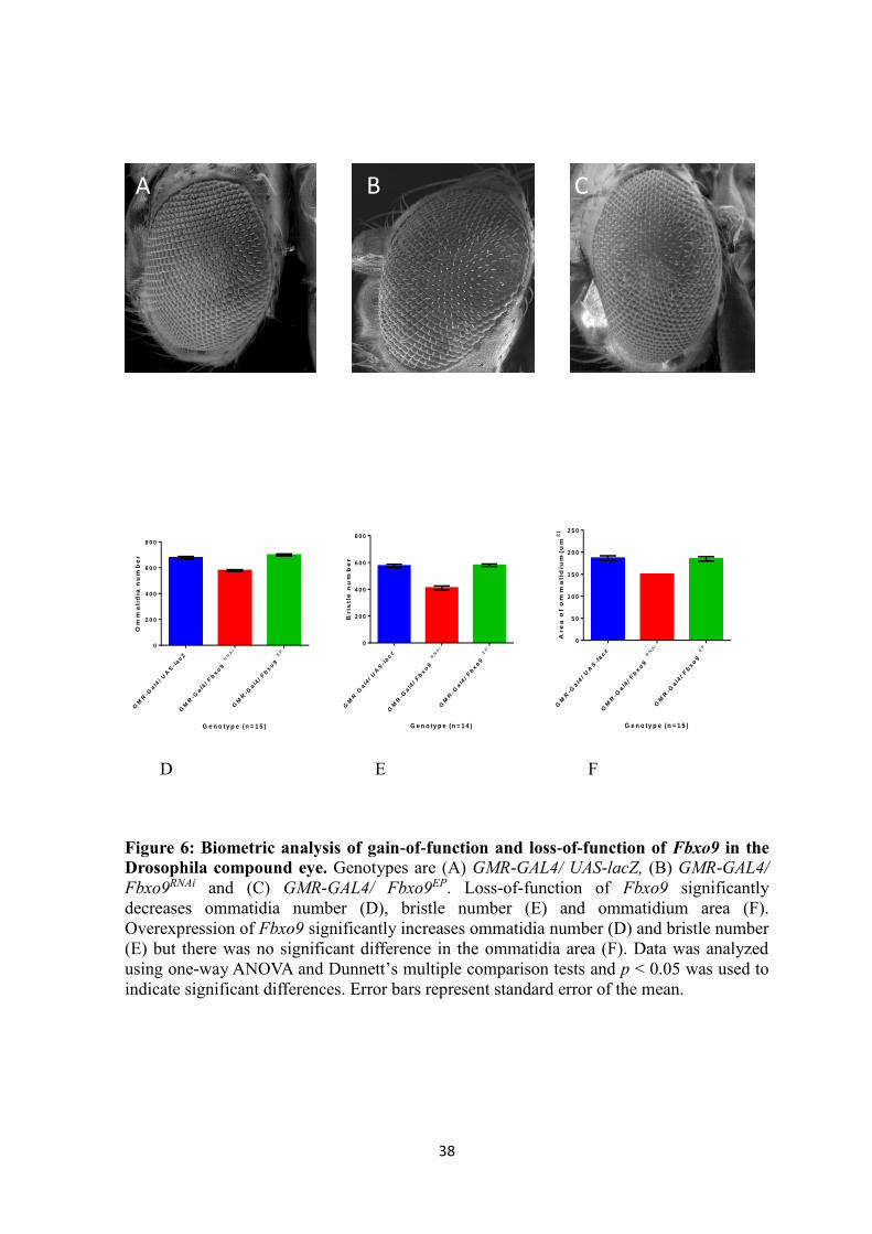
38
GM
R-G
al4
/ U
AS
-lacZ
GM
R-G
al4
/ F
bxo
9 R
NA
i
GM
R-G
al4
/ F
bxo
9 E
P
0
2 0 0
4 0 0
6 0 0
8 0 0
G e n o ty p e (n = 1 5 )
Om
ma
tid
ia n
um
be
r
GM
R-G
al4
/ U
AS
-lacz
GM
R-G
al4
/ Fbxo9 R
NA
i
GM
R-G
al4
/ Fbxo9 E
P
0
2 0 0
4 0 0
6 0 0
8 0 0
G e n o ty p e (n = 1 4 )
Bris
tle
nu
mb
er
GM
R-G
al4
/ U
AS
-lacz
GM
R-G
al4
/ Fbxo9 R
NA
i
GM
R-G
al4
/ Fbxo9 E
P
0
5 0
1 0 0
1 5 0
2 0 0
2 5 0
G e n o ty p e (n = 1 5 )
Are
a o
f o
mm
atid
ium
(u
m2
)
D E F
Figure 6: Biometric analysis of gain-of-function and loss-of-function of Fbxo9 in the
Drosophila compound eye. Genotypes are (A) GMR-GAL4/ UAS-lacZ, (B) GMR-GAL4/
Fbxo9RNAi and (C) GMR-GAL4/ Fbxo9EP. Loss-of-function of Fbxo9 significantly
decreases ommatidia number (D), bristle number (E) and ommatidium area (F).
Overexpression of Fbxo9 significantly increases ommatidia number (D) and bristle number
(E) but there was no significant difference in the ommatidia area (F). Data was analyzed
using one-way ANOVA and Dunnett’s multiple comparison tests and p < 0.05 was used to
indicate significant differences. Error bars represent standard error of the mean.
A
B C
C

39
Investigation of Fbxo9 and Foxo interactions in the compound eye
Next, the effects on the compound eye of co-overexpression of Fbxo9 and Foxo, as
well as overexpression of Foxo together with RNAi-dependent reduced expression of
Fbxo9, were determined. Fbxo9 was co-expressed with both a Drosophila and a mouse
version (Foxo1) of Foxo, which was previously generated by another research group using
GMR-Gal4; UAS-Foxo driver (Kramer et al., 2003). The elevated expression of Foxo can
create an eye phenotype where a greater number of ommatidia and bristles are decreased
(Kramer et al., 2003). A summary of ommatidia number, bristle number and ommatidia
area is shown in Table 6. Analysis of scanning electron micrographs of eyes (Figure 7)
show that RNAi-dependent reduced expression of Fbxo9 together with Foxo
overexpression significantly decreased the ommatidia number, bristle number and
ommatidium area compared to the Foxo overexpressing control fly GMR-Gal4; UAS-
Foxo/UAS-lacZ. The average number of ommatidia decreased from 382.9 ± 3.344 to 183.1
± 2, bristle number decreased from 7.467 ± 0.1333 to 3.600 ± 0.1309 and ommatidia area
decreased from 197.9 ± 0.2794 µm2 to 123.8 ± 0.3114 µm2 (Table 6).
Over-expression of Fbxo9 and Foxo together significantly increased the ommatidia
number (Figure 7D) and, bristle number (Figure 7E) and ommatidium area (Figure 7F).
The average number of ommatidia, bristles and ommatidium area in the GMR-Gal4; UAS-
Foxo/Fbxo9EP expressing flies was 432.7 ± 3.138, 11.60 ± 0.1309, and 205.9 ± 0.6252 µm2
respectively, compared to the Foxo overexpressing control flies (GMR-Gal4; UAS-Foxo/
UAS-lacZ) where the average number of ommatidia, bristles and ommatidium area per eye
was 382.9 ± 3.344, 7.467 ± 0.1333, 197.9 ± 0.2794 µm2, respectively. It can be concluded
that co-overexpression of Fbxo9 and Foxo significantly increases ommatidia number and,
bristle number and ommatidium area.

40
Table 6. A summary of the ommatidia number, bristle number and ommatidia area
when Foxo is co-overexpressed with lacZ, Fbxo9RNAi and Fbxo9EP in the Drosophila
compound eye.
A. Ommatidia number
Genotype Number of eyes
analyzed (N)
Mean ± SEM p-value compared
to control
Significant
GMR-Gal4;UAS-
Foxo/ UAS-lacZ
15 382.9 ± 3.344,
n=15
N/A N/A
GMR-Gal4;UAS-
Foxo/ Fbxo9RNAi
15 183.1 ± 2.918,
n=15
< 0.0001 Yes
GMR-GAL4;UAS-
Foxo/ Fbxo9EP
15 432.7 ± 3.138,
n=15
< 0.0001 Yes
B. Bristle number
Genotype Number of eyes
analyzed (N)
Mean± SEM p-value compared
to control
Significant
GMR-Gal4;UAS-
Foxo/ UAS-lacZ
15 7.467 ± 0.1333 N/A N/A
GMR-Gal4;UAS-
Foxo/ Fbxo9RNAi
15 3.600 ± 0.1309 < 0.0001 Yes
GMR-GAL4;UAS-
Foxo/ Fbxo9EP
15 11.60 ± 0.1309 < 0.0001 Yes
C. Ommatidium area
Genotype Number of eyes
analyzed (N)
Mean± SEM p-value compared
to control
Significant
GMR-Gal4;UAS-
Foxo/ UAS-lacZ
15 197.9 ± 0.2794
µm2
N/A N/A
GMR-Gal4;UAS-
Foxo/ Fbxo9 RNAi
15 123.8 ± 0.3114
µm2
< 0.0001 Yes
GMR-GAL4;UAS-
Foxo/ Fbxo9 EP
15 205.9 ± 0.6252
µm2
< 0.0001 Yes
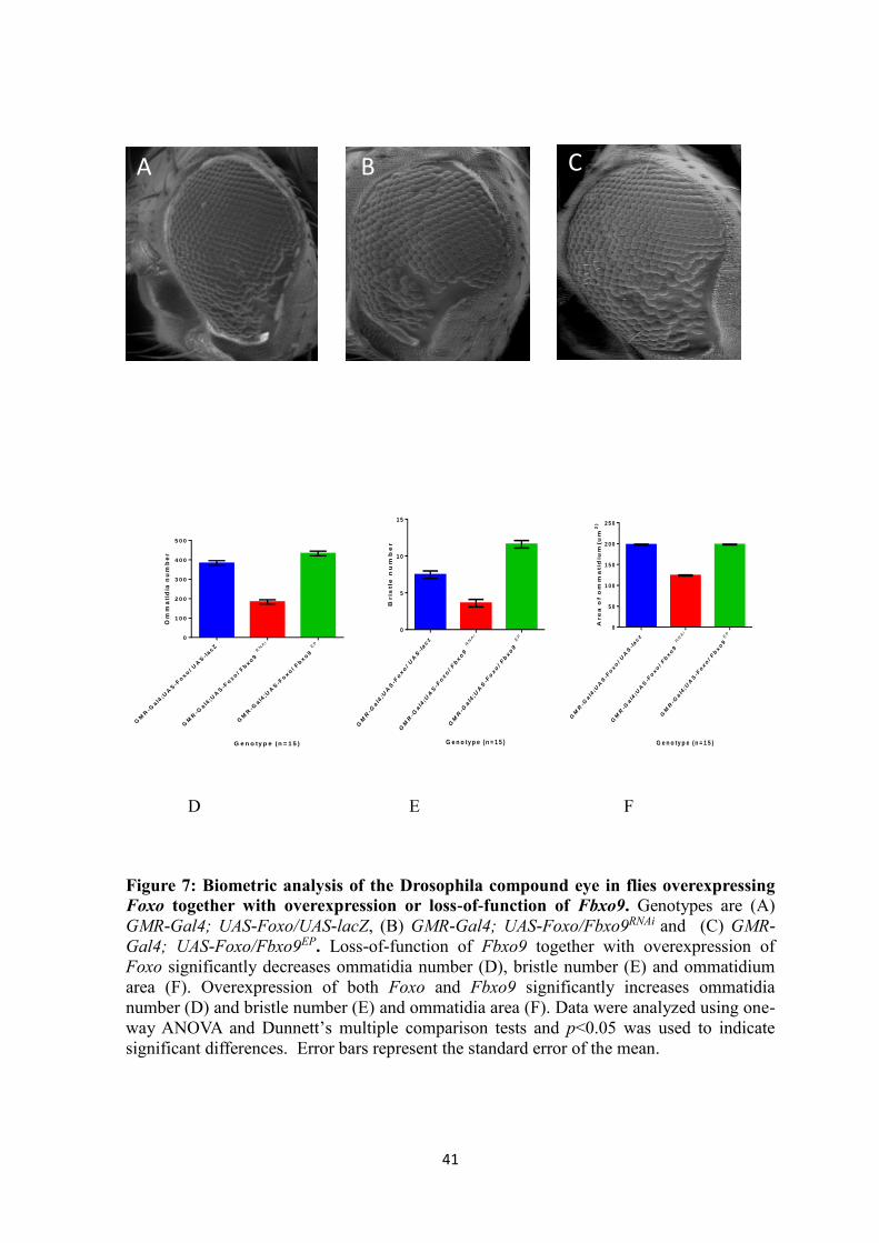
41
GM
R-G
al4
;UA
S-F
oxo
/ U
AS
-lacZ
GM
R-G
al4
;UA
S-F
oxo
/ F
bxo
9 R
NA
i
GM
R-G
al4
;UA
S-F
oxo
/ F
bxo
9 E
P
0
1 0 0
2 0 0
3 0 0
4 0 0
5 0 0
G e n o ty p e (n = 1 5 )
Om
ma
tid
ia n
um
be
r
GM
R-G
al4
;UA
S-F
oxo/ U
AS
-lacz
GM
R-G
al4
;UA
S-F
oxo/ Fbxo9 R
NA
i
GM
R-G
al4
;UA
S-F
oxo/ Fbxo9 E
P
0
5
1 0
1 5
G e n o ty p e (n = 1 5 )
Bris
tle
nu
mb
er
GM
R-G
al4
;UA
S-F
oxo/ U
AS
-lacz
GM
R-G
al4
;UA
S-F
oxo/ Fbxo9 R
NA
i
GM
R-G
al4
;UA
S-F
oxo/ Fbxo9
EP
0
5 0
1 0 0
1 5 0
2 0 0
2 5 0
G e n o ty p e (n = 1 5 )
Are
a o
f o
mm
atid
ium
(u
m2
)
D E F
Figure 7: Biometric analysis of the Drosophila compound eye in flies overexpressing
Foxo together with overexpression or loss-of-function of Fbxo9. Genotypes are (A)
GMR-Gal4; UAS-Foxo/UAS-lacZ, (B) GMR-Gal4; UAS-Foxo/Fbxo9RNAi and (C) GMR-
Gal4; UAS-Foxo/Fbxo9EP. Loss-of-function of Fbxo9 together with overexpression of
Foxo significantly decreases ommatidia number (D), bristle number (E) and ommatidium
area (F). Overexpression of both Foxo and Fbxo9 significantly increases ommatidia
number (D) and bristle number (E) and ommatidia area (F). Data were analyzed using one-
way ANOVA and Dunnett’s multiple comparison tests and p<0.05 was used to indicate
significant differences. Error bars represent the standard error of the mean.
C A B

42
Ageing analysis
The effects of loss-of-function and gain-of-function of Fbxo9
The main feature of PD is the age dependent degeneration of dopaminergic
neurons. The selective death and degeneration of these dopaminergic neurons lead us to
investigate the effects of Fbxo9 on these neurons. To determine whether Fbxo9 has any
effects on the DA neurons, the gene was both overexpressed and silenced via RNA
interference in the DA neurons. The ageing analysis was carried out in parallel with the
climbing assay in order to determine the changes in the climbing ability as a result of
premature senescence.
To investigate the effects of gain-of-function and loss-of-function of Fbxo9 on
ageing and lifespan, driver lines Ddc-Gal436 (II), Ddc-Gal43D (III), Ddc-Gal43D; DDC-
Gal436 and TH-Gal4 were used to express these transgenes in the fly dopaminergic
neurons. Survival curves are illustrated in Figure 8. The results indicate that loss-of-
function of Fbxo9 using the Ddc-Gal436 (II), Ddc-Gal43D (III), Ddc-Gal43D; Ddc-Gal436
and TH-Gal4 drivers significantly decreased longevity compared to lacZ-expressing
controls (Figure 8 A-D). The median lifespan for Fbxo9RNAi flies using these four drivers
was 74, 76, 72 and 70 days, respectively. This is compared to the control flies whose
median lifespan was 80, 82, 78 and 75 days, respectively. The log-rank test showed that the
Fbxo9RNAi longevity curves are significantly different (p<0.0001) from the control curves.
Overexpression of Fbxo9 using the Ddc-Gal436 (II), Ddc-Gal43D (III), Ddc-Gal43D;
DDC-Gal436 and TH-Gal4 drivers significantly increased longevity compared to lacZ-
expressing controls (Figure 8 A-D). The median lifespan for flies overexpressing Fbxo9
using these four drivers was 84, 86, 84 and 82, respectively. This is compared to the
control flies whose median lifespan was 80, 82, 78 and 75, respectively. The log-rank test

43
showed that the longevity curves for the Fbxo9 overexpression flies are significantly
different (p<0.0001) from the control curves. Statistical analysis of longevity is
summarized in Table 7. Loss-of-function of Fbxo9 in fly dopaminergic neurons leads to a
reduction in lifespan, which is characteristic of PD-like phenotype, whereas overexpression
of Fbxo9 significantly increases lifespan.

44
Table 7. The Log-Rank comparison of the survival curves for the directed
overexpression and RNA interference of Fbxo9 in the dopaminergic neurons. Chi-
square values and p-values were calculated using UAS-lacZ controls.
Genotype Number of
Flies
(Deaths)
Median
Survival
(Days)
Chi Square p-value Significant
Ddc-Gal43D;
Ddc-Gal436/
UAS-lacZ
300 78 125.6 N/A N/A
Ddc-Gal43D;
Ddc-Gal436/
Fbxo9EP
300 84 9.644 0.0019 Yes
Ddc-Gal43D;
Ddc-Gal436/
Fbxo9RNAi
263 72 76.11 <0.0001 Yes
Ddc-Gal436/
UAS-lacZ
306 80 87.73 N/A N/A
Ddc-Gal436 /
Fbxo9EP
300 84 14.85 0.0001 Yes
Ddc-Gal436 /
Fbxo9RNAi
277 74 59.55 <0.0001 Yes
Ddc-Gal43D/
UAS-lacZ
334 82 62.30 N/A N/A
Ddc-Gal43D/
Fbxo9EP
290 86 14.34 0.0002 Yes
Ddc-Gal43D/
Fbxo9RNAi
300 76 47.59 <0.0001 Yes
TH-Gal4/
UAS-lacZ
274 75 82.27 N/A N/A
TH-Gal4/
Fbxo9EP
280 82 8.124 0.0042 Yes
TH-Gal4/
Fbxo9RNAi
250 70 47.89 <0.0001 Yes

45
0 2 0 4 0 6 0 8 0 1 0 0
0
5 0
1 0 0
D a y s
Pe
rc
en
t s
urv
iv
al
D d c -G a l43 6
/ U A S - la c Z
D d c -G a l43 6
/ F b x o 9E P
D d c -G a l43 6
/ F b x o 9R N A i
A
0 2 0 4 0 6 0 8 0 1 0 0
0
5 0
1 0 0
D a y s
Pe
rc
en
t s
urv
iva
l
D d c -G a l43 D
/ U A S - la c Z
D d c -G a l43 D
/ F b x o 9E P
D d c -G a l43 D
/ F b x o 9R N A i
B

46
0 2 0 4 0 6 0 8 0 1 0 0
0
5 0
1 0 0
D a y s
Pe
rc
en
t s
urv
iva
lD d c -G a l4
3 D ; D d c -G a l4
3 6 / U A S - la c Z
D d c -G a l43 D
; D d c -G a l43 6
/ F b x o 9E P
D d c -G a l43 D
; D d c -G a l43 6
/ F b x o 9R N A i
C
D
Figure 8: Effect of loss-of-function and overexpression of Fbxo9 on lifespan in
Drosophila. Expression of Fbxo9RNAi using the Ddc-Gal436 (A), Ddc-Gal43D (B), Ddc-
Gal43D; Ddc-Gal436 (C), and TH-Gal4 (D) drivers significantly decreases longevity.
Overexpression of Fbxo9 using the same four drivers significantly increases longevity.
Longevity is shown as the percent survival as a function of time. A p-value less than 0.01 is
considered as significant by the log-rank test. Error bars represent the standard error of the
mean.
0 2 0 4 0 6 0 8 0 1 0 0
0
5 0
1 0 0
D a y s
Pe
rc
en
t s
urv
iva
l
T H -G a l4 / U A S -la cZ
T H -G a l4 / F b xo 9E P
T H -G a l4 / F b xo 9R N A i
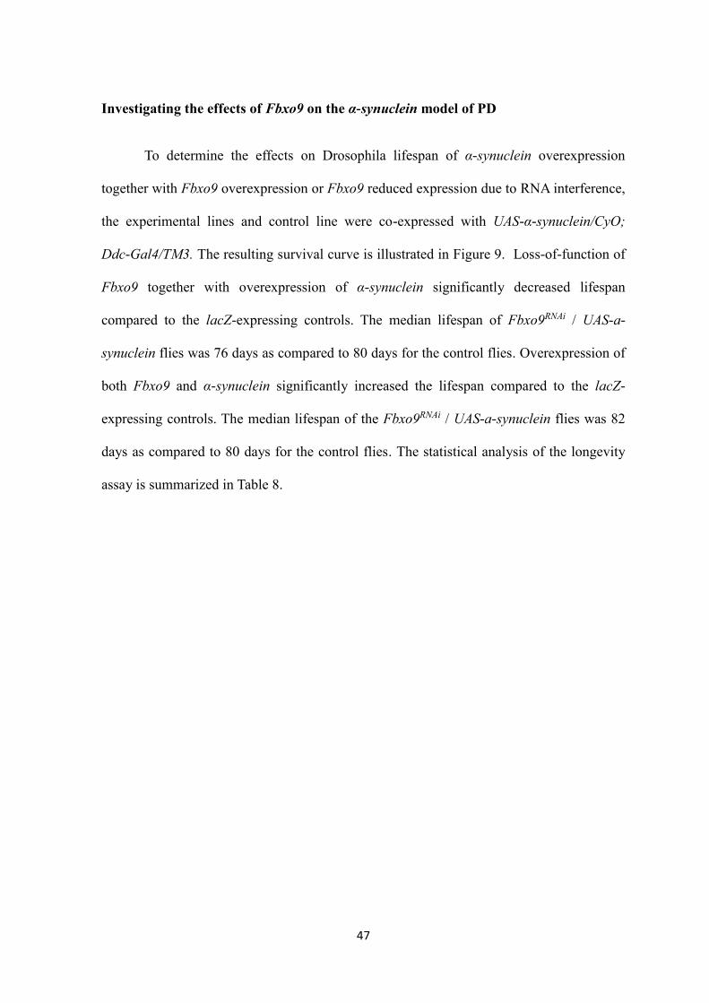
47
Investigating the effects of Fbxo9 on the α-synuclein model of PD
To determine the effects on Drosophila lifespan of α-synuclein overexpression
together with Fbxo9 overexpression or Fbxo9 reduced expression due to RNA interference,
the experimental lines and control line were co-expressed with UAS-α-synuclein/CyO;
Ddc-Gal4/TM3. The resulting survival curve is illustrated in Figure 9. Loss-of-function of
Fbxo9 together with overexpression of α-synuclein significantly decreased lifespan
compared to the lacZ-expressing controls. The median lifespan of Fbxo9RNAi / UAS-a-
synuclein flies was 76 days as compared to 80 days for the control flies. Overexpression of
both Fbxo9 and α-synuclein significantly increased the lifespan compared to the lacZ-
expressing controls. The median lifespan of the Fbxo9RNAi / UAS-a-synuclein flies was 82
days as compared to 80 days for the control flies. The statistical analysis of the longevity
assay is summarized in Table 8.

48
Table 8. The Log-Rank survival curve comparison for the directed co-overexpression
of α-synuclein and Fbxo9 and or Fbxo9RNAi in the dopaminergic neurons. Chi-square
values and p-values were calculated using UAS-lacZ controls.
Genotype Number of
Flies (Deaths)
Median
Survival
(Days)
Chi Square P value Significant
UAS-αsyn/
UAS-lacZ
367 80 68.65 N/A N/A
UAS-asyn/
Fbxo9 EP
347 82 45.52 <0.0001 Yes
UAS-asyn/
Fbxo9RNAi
302 76 41.43 <0.0001 Yes

49
0 2 0 4 0 6 0 8 0 1 0 0
0
5 0
1 0 0
D a y s
Pe
rc
en
t s
urv
iva
l
U A S - s y n / U A S - la c Z
U A S - s y n / F b x o 9E P
U A S - s y n / F b x o 9R N A i
Figure 9: Effect of α-synuclein overexpression together with loss-of-function or
overexpression of Fbxo9 in dopaminergic neurons on lifespan in Drosophila. Loss-of-
function of Fbxo9 together with α-synuclein overexpression significantly decreases
longevity while overexpression of both Fbxo9 and α-synuclein significantly increases
longevity. Ageing is shown as percent survival as a function of time. A p-value less than
0.01 was considered significant by the log-rank test. Error bars represent the standard error
of the mean.
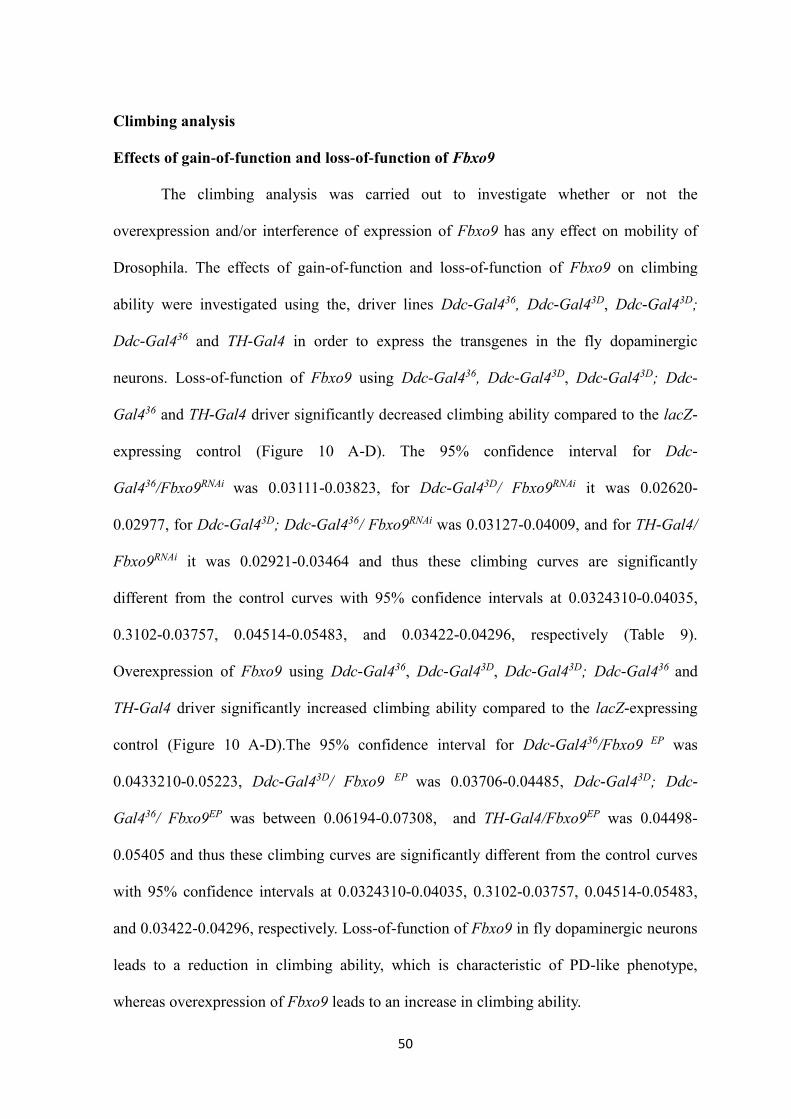
50
Climbing analysis
Effects of gain-of-function and loss-of-function of Fbxo9
The climbing analysis was carried out to investigate whether or not the
overexpression and/or interference of expression of Fbxo9 has any effect on mobility of
Drosophila. The effects of gain-of-function and loss-of-function of Fbxo9 on climbing
ability were investigated using the, driver lines Ddc-Gal436, Ddc-Gal43D, Ddc-Gal43D;
Ddc-Gal436 and TH-Gal4 in order to express the transgenes in the fly dopaminergic
neurons. Loss-of-function of Fbxo9 using Ddc-Gal436, Ddc-Gal43D, Ddc-Gal43D; Ddc-
Gal436 and TH-Gal4 driver significantly decreased climbing ability compared to the lacZ-
expressing control (Figure 10 A-D). The 95% confidence interval for Ddc-
Gal436/Fbxo9RNAi was 0.03111-0.03823, for Ddc-Gal43D/ Fbxo9RNAi it was 0.02620-
0.02977, for Ddc-Gal43D; Ddc-Gal436/ Fbxo9RNAi was 0.03127-0.04009, and for TH-Gal4/
Fbxo9RNAi it was 0.02921-0.03464 and thus these climbing curves are significantly
different from the control curves with 95% confidence intervals at 0.0324310-0.04035,
0.3102-0.03757, 0.04514-0.05483, and 0.03422-0.04296, respectively (Table 9).
Overexpression of Fbxo9 using Ddc-Gal436, Ddc-Gal43D, Ddc-Gal43D; Ddc-Gal436 and
TH-Gal4 driver significantly increased climbing ability compared to the lacZ-expressing
control (Figure 10 A-D).The 95% confidence interval for Ddc-Gal436/Fbxo9 EP was
0.0433210-0.05223, Ddc-Gal43D/ Fbxo9 EP was 0.03706-0.04485, Ddc-Gal43D; Ddc-
Gal436/ Fbxo9EP was between 0.06194-0.07308, and TH-Gal4/Fbxo9EP was 0.04498-
0.05405 and thus these climbing curves are significantly different from the control curves
with 95% confidence intervals at 0.0324310-0.04035, 0.3102-0.03757, 0.04514-0.05483,
and 0.03422-0.04296, respectively. Loss-of-function of Fbxo9 in fly dopaminergic neurons
leads to a reduction in climbing ability, which is characteristic of PD-like phenotype,
whereas overexpression of Fbxo9 leads to an increase in climbing ability.

51
Table 9. Statistical analysis of climbing index curves for the directed overexpression
and RNA interference of Fbxo9 in dopaminergic neurons. The 95% confidence interval
was compared to the UAS-lacZ control flies.
Genotype Slope Standard
Error
95%
Confidence
Interval
R 2 Significant
Ddc-Gal43D;
Ddc-Gal436/
UAS-lacZ
0.04999 0.002415 0.04514-
0.05483
0.9503 N/A
Ddc-Gal43D;
Ddc-Gal436/
Fbxo9EP
0.06751 0.002777 0.06194-
0.07308
0.9691 Yes
Ddc-Gal43D;
Ddc-Gal436/
Fbxo9RNAi
0.03568 0.002193 0.03127-
0.04009
0.9032 Yes
Ddc-Gal436/
UAS-lacZ
0.03639 0.001974 0.0324310-
0.04035
0.9238 N/A
Ddc-Gal436 /
Fbxo9EP
0.04778 0.001771 0.0433210-
0.05223
0.9509 Yes
Ddc-Gal436 /
Fbxo9RNAi
0.03467 0.001771 0.03111-
0.03823
0.9334 Yes
Ddc-Gal43D/
UAS-lacZ
0.03430 0.001632 0.3102-
0.03757
0.9389 N/A
Ddc-Gal43D/
Fbxo9EP
0.04096 0.001943 0.03706-
0.04485
0.9448 Yes
Ddc-Gal43D/
Fbxo9RNAi
0.02799 0.0008877 0.02620-
0.02977
0.9673 Yes
TH-Gal4/
UAS-lacZ
0.03859 0.002174 0.03422-
0.04296
0.9257 N/A
TH-Gal4/
Fbxo9EP
0.04951 0.002255 0.04498-
0.05405
0.9564 Yes
TH-Gal4/
Fbxo9RNAi
0.03193 0.001344 0.02921-
0.03464
0.9518 Yes

52
0 2 0 4 0 6 0 8 0
0
1
2
3
4
5
D a y s
5-c
lim
bin
g i
nd
ex
D d c -G a l43 6
/ U A S - la c Z
D d c -G a l43 6
/ F b x o 9E P
D d c -G a l43 6
/ F b x o 9R N A i
A
0 2 0 4 0 6 0 8 0
0
1
2
3
4
5
D a y s
5-c
lim
bin
g i
nd
ex
D d c -G a l43 D
/ U A S - la c Z
D d c -G a l43 D
/ F b x o 9E P
D d c -G a l43 D
/ F b x o 9R N A i
B

53
C
0 2 0 4 0 6 0 8 0
0
1
2
3
4
5
Da y s
5-c
lim
bin
g i
nd
ex
T H -G A L 4 / U A S -la cZ
T H -G A L 4 / F b x o 9E P
T H -G a l4 / F b xo 9R N A i
D
Figure 10: Effect of gain-of-function and loss-of-function of Fbxo9 on climbing ability
in Drosophila. Expression of Fbxo9RNAi using Ddc-Gal436 (A), Ddc-Gal43D (B), Ddc-
Gal43D; Ddc-Gal436 (C) and TH-Gal4 (D) drivers significantly decreases climbing ability.
Overexpression of Fbxo9 using the same four drivers significantly increases climbing
ability. The data were analyzed by non-linear fitting of the climbing curves with 95%
confidence intervals to determine significance. Error bars represent the standard error of
the mean.
0 2 0 4 0 6 0 8 0
0
1
2
3
4
5
D a y s
5-c
lim
bin
g i
nd
ex
D d c -G a l43 D
; D d c -G a l43 6
/
U A S -la cZ
D d c -G a l43 D
; D d c -G a l43 6
/
F b xo9E P
D d c -G a l43 D
; D d c -G a l43 6
/
F b xo9R N A i
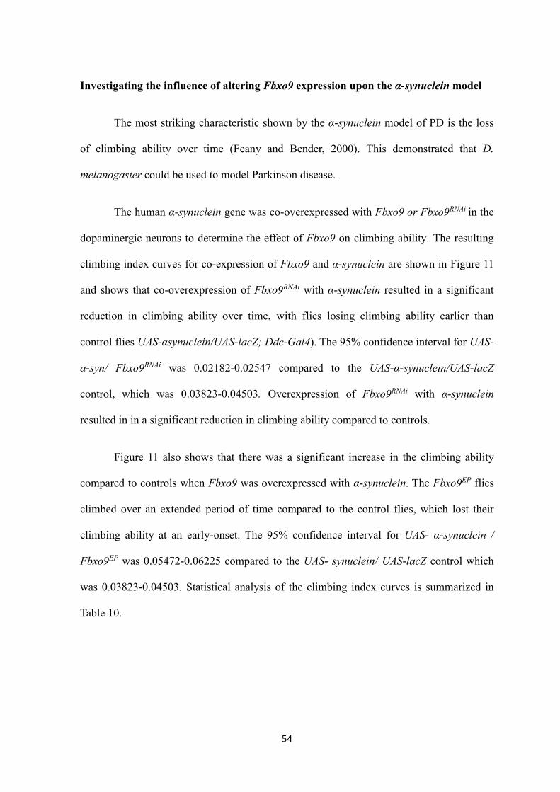
54
Investigating the influence of altering Fbxo9 expression upon the α-synuclein model
The most striking characteristic shown by the α-synuclein model of PD is the loss
of climbing ability over time (Feany and Bender, 2000). This demonstrated that D.
melanogaster could be used to model Parkinson disease.
The human α-synuclein gene was co-overexpressed with Fbxo9 or Fbxo9RNAi in the
dopaminergic neurons to determine the effect of Fbxo9 on climbing ability. The resulting
climbing index curves for co-expression of Fbxo9 and α-synuclein are shown in Figure 11
and shows that co-overexpression of Fbxo9RNAi with α-synuclein resulted in a significant
reduction in climbing ability over time, with flies losing climbing ability earlier than
control flies UAS-αsynuclein/UAS-lacZ; Ddc-Gal4). The 95% confidence interval for UAS-
a-syn/ Fbxo9RNAi was 0.02182-0.02547 compared to the UAS-α-synuclein/UAS-lacZ
control, which was 0.03823-0.04503. Overexpression of Fbxo9RNAi with α-synuclein
resulted in in a significant reduction in climbing ability compared to controls.
Figure 11 also shows that there was a significant increase in the climbing ability
compared to controls when Fbxo9 was overexpressed with α-synuclein. The Fbxo9EP flies
climbed over an extended period of time compared to the control flies, which lost their
climbing ability at an early-onset. The 95% confidence interval for UAS- α-synuclein /
Fbxo9EP was 0.05472-0.06225 compared to the UAS- synuclein/ UAS-lacZ control which
was 0.03823-0.04503. Statistical analysis of the climbing index curves is summarized in
Table 10.

55
Table 10. Statistical analysis of climbing index curves for the co-overexpression of α-
synuclein and Fbxo9 or Fbxo9RNAi in the dopaminergic neurons. The 95% confidence
intervals were compared to the UAS-lacZ control flies.
Genotype Slope Standard Error 95% Confidence
Intervals
R 2 Significant
UAS-asyn/
UAS-lacZ
0.04163 0.001690 0.03823-0.04503 0.9600 N/A
UAS-asyn/
Fbxo9EP
0.05848 0.001872 0.05472-0.06225 0.9798 Yes
UAS-asyn/
Fbxo9RNAi
0.02364 0.0009094 0.02182-0.02547 0.9504 Yes
0 2 0 4 0 6 0 8 0
0
1
2
3
4
5
D a y s
5-c
lim
bin
g i
nd
ex
U A S - s y n / U A S - la c Z
U A S - s y n / F b x o 9E P
U A S - s y n / F b x o 9R N A i
Figure 11: Analysis of climbing ability of Drosophila flies overexpressing α-synuclein
together with Fbxo9 or Fbxo9RNAi in the dopaminergic neurons. RNAi dependent
interference of Fbxo9 expression together with α-synuclein overexpression significantly
decreases climbing ability. Overexpression of both Fbxo9 and α-synuclein significantly
increases climbing ability. Data were analyzed by non-linear fitting of the climbing curves
with 95% confidence intervals to determine significance. Error bars represent the standard
error of the mean.

56
DISCUSSION
Drosophila Fbxo9 is homologous to mammalian Fbxo9
Bioinformatics analysis was first performed on the D. melanogaster homologue in
order to identify the similarity between CG5961 and Fbxo9 ,along with several other
homologues from vertebrates and invertebrates. Furthermore, bioinformatics analysis was
carried out to find out the relationship of Fbxo9 with other FBXO proteins such as Fbxo7
and Fbxo32. When a cladogram was created with these three FBXO proteins, CG5961
clustered together with Fbxo9. This indicates that CG5961 is most closely related to
Fbxo9. As all of the genes from the different species clustered together, it may be
concluded that Fbxo genes may exist in the common ancestor of the species. Alignment of
human Fbxo9 and Drosophila CG5961 shows that the evidence of the conservation of the
F-box proteins is 33% identical and 67% similar. The proteins are highly conserved in the
F-box region that is essential for the SCF-ubiquitin ligase complexes. Alignment of human
Fbxo9 and Drosophila CG5961 with other vertebrate and invertebrate species further
indicates the conservation of the F-box proteins being 26% identical and 27% similar and
they further share the conserved domains such as TPR domain, MIT domain, F-box
domain, SNHc and NLS. Due to the high level of conservation, it can be inferred that there
are tight evolutionary constraints placed among the genes. Within the sub-group, it has
been found that Fbxo7 and Fbxo9 are more similar than Fbxo32. Alignment of the Human
Fbxo9 and Drosophila Fbxo7 showed that they are analogous, being 34% similar and 14%
identical and they share structural similarities such as the PI31-binding region and the F-
box region. Alignment of the human Fbxo9 and Drosophila Fbxo32 showed that they are
analogous being 17% identical and 28 % similar and they additionally share structural
similarities such as Foxo-binding region, amino terminus, LCD and PDZ domain.

57
In order to identify the similarity of Fbxo9 with Fbxo7 and Fbxo32, bioinformatics
analysis was performed. Both the mammalian and fly form of Fbxo7 bind PI31 to initiate
successful proteasome-mediated degradation of a target substrate (Bader et al., 2010).
FBXO7 was originally studied as a gene involved in caspase activation in sperm
differentiation. However, it has been linked to early-onset parkinsonian-pyramidal
syndrome. Since alignment of the Human Fbxo9 and Drosophila Fbxo7 showed that they
are analogous being 14% identical and 34% similar and they share structural similarities
such as the PI31-binding region and the F-box region, so it can be speculated that Fbxo9
may also lead to PD-like symptom.
It has been found that when Fbxo32, also known as atrogin, is up-regulated in
skeletal muscles, it results in muscle wasting. In mammals, atrogin targets proteins
essential for muscle synthesis such as MyoD and eIF3-f (eukaryotic initiation factor 3 -
subunit 5) and, consequently, it degrades them and induces muscle cell death (Tintignac et
al., 2005 and Lagirand-Cantaloube et al., 2008). The atrogin/ Fbxo32 is the target for Foxo
and can trigger skeletal muscle atrophy when insulin or insulin-like growth factor-1 (IGF-
1) is absent (Connors and Staveley, unpublished). Since alignment of the Human Fbxo9
and Drosophila Fbxo32 showed that they are analogous, being 17% identical and 28 %
similar and they share structural similarities such as Foxo-binding region, amino terminus,
LCD and PDZ domain, it can be speculated that expression of Fbxo9 gene in various
human conditions makes it a potential candidate for studies, in particular potential
organismal modeling of human conditions.
Effects of loss-of-function of Fbxo9 in the Drosophila melanogaster compound eye
Genetic expression studies using the Drosophila eye have been extensively used
in the study of neurodegeneration because of the conservation of key signaling pathways

58
between humans and Drosophila and the ease of quantifying degeneration of photoreceptor
neurons associated with each Drosophila ommatidium. In D. melanogaster, eye
development is tightly controlled during the organization of the ommatidial array (Thomas
and Wassarman, 1999). The eye also consists of specialized structures called sensory
bristles that provide the opportunity for neurogenesis examination and for detection of
even slight abnormalities (Baker, 2000). We expressed reduced levels of Fbxo9 in the
Drosophila eye using the transgenic driver GMR-Gal4. The characteristics can be studied
through bioinformetric analysis to show the effects of altered gene expression, such as
loss-of-function of Fbxo9.
In Drosophila, Fbxo9 and Fbxo7 are quite similar at the amino acid level.
Therefore, Fbxo9 was examined to investigate the possible redundancy in the function
between Fbxo9 and Fbxo7. Since Fbxo7 is closely related to Fbxo9, so we hypothesized
that suppressing of Fbxo9 activity through RNA-interference would also lead to a PD-like
phenotype in flies. We have shown that suppression of Fbxo9 activity has a deleterious
effect on the Drosophila eye morphology. As expected, loss-of-function of Fbxo9 in the fly
eyes through eye-specific expression of Fbxo9RNAi leads to a significant reduction in the
number of ommatidia and, bristles as well as a reduction in ommatidia area, whereas
overexpression of Fbxo9 leads to a significant increase in the number of ommatidia and,
bristles as well as a reduction in ommatidia area. No previous has been conducted on the
effects of Fbxo9 in Drosophila eyes. Therefore, the reason for this reduction in the number
of ommatidia, bristle and ommatidia area is unclear. However, reduction in the number of
ommatidia, bristle and ommatidia area suggests a reduction in cell number during eye
development. Reduction of cell number can occur through either increased cell death, or
decreased cell proliferation (Kramer et al., 2003). This may be inferred that loss-of-
function of Fbxo9 causes inhibition of cell growth required for the normal eye

59
development.
Overexpression of Foxo under the control of the GMR-Gal4 transgene results in a
significant decrease in the number of ommatidia and bristles (Kramer et al., 2003).
Previous work in our lab showed that overexpression of the forkhead box, subgroup "O"
(Foxo) transcription factors and F-box only protein 32/ atrogin decreases ommatidia
number, bristle number and ommatidia area, that reflect cell number and cell size, which
indicates its role in the control of body size through alterations in cell size and cell number.
(Colleen Furlong [nee Connors], unpublished). The atrogin/ Fbxo32 is the target for Foxo
and can trigger skeletal muscle atrophy in the form of PD model when insulin or insulin-
like growth factor-1 (IGF-1) is absent. Overexprssion of Foxo has been linked to
neurotoxicity (Kanao et al., 2010). In our study, we have performed a bioinformatic
analysis of Fbxo9 and atrogin/Fbxo32. We have analyzed the speculated homologous
relationship between these two F-box proteins to assess the possibility of using Drosophila
as a disease model for PD. To determine the role of Foxo and Fbxo9 in PD etiology, we
have used the eye specific driver GMR-Gal4 to drive the expression of these genes in the
eyes. To determine the effect of loss-of-function of Fbxo9, we used RNA interference to
decrease the expression of the genes to very low levels. In this study, we have found that
the co-overexpression of Fbxo9RNAi and Foxo causes a reduction in the ommatidia number,
bristle number and ommatidia area whereas the co-overexpression of Fbxo9 and Foxo is
causes an increase in the ommatidia number, bristle number and ommatidia area. Since no
previous studies have been conducted in flies eyes associated with Fbxo9 and Foxo, the
reason for this reduction in the number of ommatidia, bristle and ommatidia area is unclear.
However, reduction in the number of ommatidia, bristle and ommatidia area suggests a
reduction in cell number during eye development. Reduction of cell number can occur
through either increased cell death, or decreased of cell proliferation (Kramer et al., 2003).

60
This may be presumed that loss-of-function of Fbxo9 produces inhibition of cell growth
that is necessary for the normal eye development.
Effects of loss-of-function of Fbxo9 on climbing ability and lifespan
We have shown that suppression of Fbxo9 activity has a deleterious effect on
Drosophila dopaminergic neurons. As loss-of-function mutations in human Fbxo7 lead to
parkinsonian-pyramidal syndrome, and Fbxo7 is closely related to Fbxo9, we hypothesized
that suppression of Fbxo9 activity through RNA-interference would also lead to a PD-like
phenotype in flies. A decrease in nutcracker/Fbxo7 expression causes a decrease in
lifespan (Merzetti and Staveley, unpublished). As expected, loss-of-function of Fbxo9 in
fly dopaminergic neurons leads to a reduction in climbing ability and lifespan, which is
characteristic of PD-like phenotype, whereas overexpression of Fbxo9 is favorable,
compared to control flies. No previous studies have been conducted in climbing ability and
lifespan associated with Fbxo9, therefore the reason for this reduction in lifespan and
climbing ability is unclear. This may be due to an increase in apoptosis or a decrease in cell
growth during development. This may be also due to selective apoptotic death of these DA
neurons and decreased cellular protection and survival. Dopaminergic neurons may die as a
result of apoptosis in PD (Lev et al., 2003). This process occurs may be due to the
accumulation of endogenous toxic proteins or environmental toxins. Exploration of the role
of cell survival signaling in the selective loss of dopaminergic neurons in Drosophila may
provide further insight into the basis of PD. However, the Fbxo7 gene has been found
associated with the early-onset parkinsonian pyramidal syndrome and the distinguishing
features of this disease include progressive degeneration of the pyramidal, and
extrapyramidal regions in combination with the substantia nigra pars compacta in the
brain (Davison, 1954; Di Fonzo et al., 2009). Since Fbxo9 and Fbxo7 are closely related

61
and since Fbxo7 leads to PD-like symptoms, it is not a surprise that Fbxo9 also leads to a
PD like phenotype.
The recapitulation of PD-like symptoms due to over-expression of a gene,
especially the age-dependent loss of climbing ability in D. melanogaster, provides the
potential model in the study of biological basis of the disease (Feany and Bender, 2000;
Haywood and Staveley, 2004). Locomotion abnormality is one of the neurological
characteristics of PD. In our study, loss-of-function of Fbxo9 through RNA-interference, in
the fly dopaminergic neurons, leads to a reduction in climbing ability compared to control
flies (Figure 10). It is possible that this occurs due to inhibition of cell growth. Similar
results have been reported before where the α-synuclein model resulted in an age-
dependent loss of climbing ability where the mutant flies were unable to climb above the
first section of the climbing apparatus during the last days of their lives (Feany and Bender,
2000). Additionally, it also has been reported that mutations in the TH-encoding gene pale
cause an age dependent loss of climbing ability (Tempel et al., 1984; Pendletont et al.,
2002). In another experiment it has been found that loss-of-function of dLRRK leads to a
slight locomotor abnormality but has no effect on survival of DA neurons (Lee et al., 2007;
Tain et al., 2009b). In one experiment it was found that when (Pink1) overexpression
rescued the premature loss of climbing abilities induced by α-synuclein (Todd and
Staveley, 2008). The similar result has been found in this experiment when Fbxo9 is
overexpressed within dopaminergic neurons, the flies climb longer and lose their climbing
ability later than the control flies.
No previous ageing or mobility studies have been performed associated with the
directed alteration of Fbxo9 expression, so the reason for the observed results is not clear.
This may be due to an increase in apoptosis or a decrease in cell growth during

62
development. This may be also due to selective apoptotic death of these DA neurons and
decreased cellular protection and survival. It is possible that a reduction in Fbxo9
expression increases the rate of apoptosis or inhibits cell growth. Similar results have been
reported in a previous experiment where flies with loss-of-function of the parkin gene
showed the characteristics of PD such as shortened lifespan, reduced climbing ability and
degeneration of DA neurons (Greene et al., 2003; Whitworth et al., 2005). The mutant
parkin flies were also found to have developmental delays and mobility dysfunction due to
muscle degeneration. Additionally, the Drosophila Pink1 fly model involving the gene
shows the same characteristics such as reduced lifespan, developmental delay and DA
neuron degeneration as the parkin mutant flies (Petit et al., 2005; Clark et al., 2006). It has
been found that a PINK1-RNAi knockdown in flies resulted in loss of DA neurons even at
10 days (Wang et al., 2006). In our study, when Fbxo9 is overexpressed in the
dopaminergic neurons, the flies live longer than the control flies, indicating that Fbxo9
may play a protective role by decreasing the rate of apoptosis and thus possibly
maintaining healthy neurons.
Effect of RNA-interference of Fbxo9 in the α-synuclein model of PD
The α-synuclein model of PD in Drosophila did not result in any difference in
lifespan between the control and wild type, A13T and A30P α-synuclein flies (Feany and
Bender, 2000). Co-expression of Fbxo7RNAi with α-synuclein results in increased lifespan
compared to control (Merzetti and Staveley, unpublished). In this study, overexpression of
Fbxo9 with α-synuclein also resulted in an increase in lifespan compared to controls. A
similar result was found in an experiment where co-expression of α-synuclein and parkin
slightly increased the lifespan of the flies (Haywood and Staveley, 2006). In our study, co-
overexpression of α-synuclein and Fbxo9RNAi resulted in a decreased lifespan compared to
63
the control flies, and this may be due to inhibition of cell survival in the dopaminergic
neurons.
Flies that express A30P, A53T and wild type versions of α-synuclein experience
loss of DA neurons and age dependent loss of locomotor ability when GAL4 drivers,
including Ddc-Gal4, are used (Feany and Bender, 2000). In our study, co-overexpression
of α-synuclein with Fbxo9RNAi under the control of the Ddc-Gal4 driver, resulted in a
significant difference in climbing ability compared to the control flies. Fbxo9RNAi/ UAS-α-
syn expressing flies lost their climbing ability earlier than the control flies (Figure 15). This
may be because loss-of-function of Fbxo9 causes inhibition of the survival of the
dopaminergic neurons. Additionally in another experiment, it has been reported that α-
synuclein flies show a less pronounced DA neuron degeneration as only a partial (50%)
decrease was observed (Auluck et al., 2002; Bayersdorfer et al., 2010). In our study, an
opposite effect was found when α-synuclein was co-overexpressed with Fbxo9.
Overexpression of Fbxo9 with α-synuclein resulted in an increase in climbing ability
compared to controls. A similar result was found when α-synuclein and parkin were co-
overexpressed in Drosophila DA neurons, and this resulted in a slight increase in climbing
ability (Haywood and Staveley, 2006). So it can be concluded that when α-synuclein and
parkin are co-overexpressed, they play a vital role in maintaining healthy dopaminergic
neurons.

64
CONCLUSION
In this study, we have confirmed that gene CG5961 of Drosophila melanogaster is
the homologue of human Fbxo9. We have shown that Fbxo9, Fbxo7 and Fbxo32 are
closely related but Fbxo9 and Fbxo7 are found more similar than Fbxo9 and Fbxo32. We
have shown suppression of Fbxo9 through RNA interference in the dopaminergic neurons
of Drosophila leading to parkinsonian-pyramidal syndrome. In this study, we have found
that loss-of-function of Fbxo9 through the eye specific driver GMR-Gal4 leads to a
significant reduction in the number of ommatidia, bristles as well as in ommatidia area,
whereas overexpression of Fbxo9 results in a significant increase in the number of
ommatidia, bristles as well as in ommatidia area. We also have found that RNAi-dependent
reduced expression of Fbxo9 together with Foxo overexpression significantly decreased
the ommatidia number, bristle number and ommatidium area compared to the Foxo
overexpressing control fly GMR-Gal4; UAS-Foxo/UAS-lacZ whereas co-overexpression of
Fbxo9 and Foxo significantly increases the ommatidia number, bristle number and
ommatidium area. We also found that, expression of Fbxo9RNAi using the Ddc-Gal43D
(III), Ddc-Gal436 (II), Ddc-Gal43D; Ddc-Gal436, and TH-Gal4 drivers significantly
decreases longevity and climbing ability whereas overexpression of Fbxo9 using the same
four drivers remarkably increases longevity and climbing ability.
In addition, co-overexpression of α-synuclein and Fbxo9RNAi results in a decreased
longevity and climbing ability compared to control whereas overexpression of both Fbxo9
and α-synuclein is favorable compared to control. This demonstrates that the directed
inhibition of expression of Fbxo9 lead to the enhancement of PD-like symptoms in the α-
synuclein-induced Drosophila model of PD. Further studies looking at the effect of Fbxo9
on Foxo activity and α-synuclein activity may uncover underlying mechanisms that
mediate a shift towards apoptosis. We have developed a new model of human Parkinson

65
disease that will provide further information in the disease etiology. We expect that the
knowledge obtained by determining the pathways involved in Parkinson disease in
Drosophila will help uncover potential new therapeutic approaches for human subjects.

66
REFERENCES
Abramoff, M.D., Magalhães, P.J., and Ram, S.J. (2004). Image processing with ImageJ.
Biophotonics international, 11: 36-42.
Adams, J. (2003). The proteasome: structure, function, and role in the cell. Cancer
Treatment Reviews, Supplement 1: 3-9.
Adams, M.D., Celniker, S.E., Holt, R.A., Evans, C.A, Gocayne, J.D., Amanatides, P.G.,
Scherer, S.E., Li, P.W., Hoskins, R.A., and Galle, R.F. (2000). The genome sequence of
Drosophila melanogaster. Science, 287: 2185–2195.
Arden, K.C., Biggs, W.H. (2002). Regulation of the FoxO family of transcription factors
by phosphatidylinositol-3 kinase-activated signaling Archives of Biochemistry and
Biophysics, 403: 292-298.
Auluck, P.K., Chan, H.Y., Trojanowski, J.Q., Lee, V.M. and Bonini, N.M. (2002).
Chaperone suppression of alpha-synuclein toxicity in a Drosophila model for Parkinson's
disease. Science, 295: 865-868.
Auluck, P.K., Meulener, M.C. and Bonini, N.M. (2005). Mechanisms of suppression of
{alpha}-synuclein neurotoxicity by geldanamycin in Drosophila. The Journal of Biological
Chemistry, 280: 2873-2878.
Bader, M., Arama, E., and Steller, H. (2010). A novel Fbox protein is required for caspase
activation during cellular remodeling in Drosophila. Development, 137: 1679-1688.
Baker, N.E. (2000). Cell proliferation, survival and death in the Drosophila eye. Seminars
in Cell and Developmental Biology, 12: 499-507.
Bai, C., Sen, P., Hofmann, K., Ma, L., Goebl, M., Harper, J.W., and Elledge, S.J. (1996).
SKP1 connects cell cycle regulators to the ubiquitin proteolysis machinery through a novel
motif, the F-box. Cell, 86: 263-274.
Banerjee, R., Starkov, A.A., Beal, M.F., and Thomas, B. (2009). Mitochondrial dysfunction
in the limelight of Parkinson's disease pathogenesis. Biochimica et Biophysica Acta, 1792:
651-663.
Bayersdorfer, F., Voigt, A., Schneuwly, S., and Botella, JA. (2010). Dopamine-dependent
neurodegeneration in Drosophila models of familial and sporadic Parkinson's disease.
Neurobiology of Dissease, 40: 113-119.
Bekris, L.M., Mata, I.F., and Zabetian, C.P. (2010). The Genetics of Parkinson Disease.
Journal of Geriatric Psychiatry and Neurology, 23: 228-242.
Bereznai, B., and Molnar, M.J. (2009). Genetics and present therapy options in Parkinson's
disease: a review. Ideggyogy Sz, 62: 155-163.

67
Bier, E. (2005). Drosophila, the golden bug, emerges as a tool for human genetics. Nature
Reviews Genetics, 6: 9-23.
Bodine, S.C., Stitt, T.N., Gonzalez, M., Kline, W.O., Stover, G.L., Bauerlein, R.,
Zlotchenko, E., Scrimgeour, A., Lawrence, J.C., Glass, D.J., and Yancopoulos, G.D.
(2001b). Akt/mTOR pathway is a crucial regulator of skeletal muscle hypertrophy and can
prevent muscle atrophy in vivo. Nature Cell Biology, 3: 1014-1019.
Bonifati, V., Rizzu, P., Squitieri, F., Krieger, E., Vanacore, N., van Swieten, J.C., Brice, A.,
van Duijn, C.M., Oostra, B., Meco, G., and Heutink, P. (2003). DJ-1(PARK7), a novel
gene for autosomal recessive, early onset parkinsonism. Neurological Sciences, 24: 159-
160.
Borkhardt, A., Repp, R., Haas, O.A., Leis, T., Harbott, J., Kreuder, J., Hammermann, J.,
Henn, T., and Lampert, F. (1997). Cloning and characterization of AFX, the gene that fuses
to MLL in acute leukemias with a t(X;11)(q13;q23) Oncogene, 14: 195-202.
Botella, J.A., Bayersdorfer, F., and Schneuwly, S. (2008). Superoxide dismutase
overexpression protects dopaminergic neurons in a Drosophila model of Parkinson's
disease. Neurobiology of Disease, 30: 65-73.
Botella, J.A., Bayersdorfer, F., Gmeiner, F., and Schneuwly, S. (2009). Modelling
Parkinson's disease in Drosophila. Neuromolecular Medicine, 11: 268-280.
Brand, A.H., and Perrimon, N. (1993). Targeted gene expression as a means of altering cell
fates and generating dominant phenotypes. Development, 118: 401-415.
Brand, A.H., Manoukian, A.S., and Perrimon, N. Ectopic expression in Drosophila.
(1994). Methods in Cell Biology, 44: 635-654.
Burgering, B.M., and Kops, G.J. Cell cycle and death control: long live Forkheads. (2002).
Trends in Biochemical Sciences, 27: 352-360.
Bulchand, S., Menon, S.D, George, S.E., and Chia, W. (2010). Muscle wasted: a novel
component of the Drosophila histone locus body required for muscle integrity. Journal of
Cell Science, 123: 2697-2707.
Cardozo, T., and Pagano, M. (2004). The SCF ubiquitin ligase: insights into a molecular
machine. Nature Reviews Molecular Cell Biology, 5: 739-751.
Cauchi, R.J., Heuvel, M. (2006). The fly as a model for neurodegenerative diseases: is it
worth the jump? Neurodegenerative Diseases, 3: 338-356.
Celotto, A.M., and Palladino, M.J. (2005). Drosophila: a "model" model system to study
neurodegeneration. Molecular Interventions, 5: 292-303.
Cenciarelli, C., Chiaur, D.S., Guardavaccaro, D., Parks, W., Vidal, M., and Pagano, M.
(1999). Identification of a family of human F-box protein. Current Biology, 9: 1177–1179.
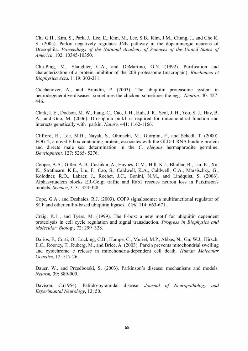
68
Cha G.H., Kim, S., Park, J., Lee, E., Kim, M., Lee, S.B., Kim, J.M., Chung, J., and Cho K.
S. (2005). Parkin negatively regulates JNK pathway in the dopaminergic neurons of
Drosophila. Proceedings of the National Academy of Sciences of the United States of
America, 102: 10345-10350.
Chu-Ping, M., Slaughter, C.A., and DeMartino, G.N. (1992). Purification and
characterization of a protein inhibitor of the 20S proteasome (macropain). Biochimica et
Biophysica Acta, 1119: 303-311.
Ciechanover, A., and Brundin, P. (2003). The ubiquitin proteasome system in
neurodegenerative diseases: sometimes the chicken, sometimes the egg. Neuron, 40: 427-
446.
Clark, I. E., Dodson, M. W., Jiang, C., Cao, J. H., Huh, J. R., Seol, J. H., Yoo, S. J., Hay, B.
A., and Guo, M. (2006). Drosophila pink1 is required for mitochondrial function and
interacts genetically with parkin. Nature, 441: 1162-1166.
Clifford, R., Lee, M.H., Nayak, S., Ohmachi, M., Giorgini, F., and Schedl, T. (2000).
FOG-2, a novel F-box containing protein, associates with the GLD-1 RNA binding protein
and directs male sex determination in the C. elegans hermaphrodite germline.
Development, 127: 5265- 5276.
Cooper, A.A., Gitler, A.D., Cashikar, A., Haynes, C.M., Hill, K.J., Bhullar, B., Liu, K., Xu,
K., Strathearn, K.E., Liu, F., Cao, S., Caldwell, K.A., Caldwell, G.A., Marsischky, G.,
Kolodner, R.D., Labaer, J., Rochet, J.C., Bonini, N.M., and Lindquist, S. (2006).
Alphasynuclein blocks ER-Golgi traffic and Rab1 rescues neuron loss in Parkinson's
models. Science, 313: 324-328.
Cope, G.A., and Deshaies, R.J. (2003). COP9 signalosome: a multifunctional regulator of
SCF and other cullin-based ubiquitin ligases. Cell, 114: 663-671.
Craig, K.L., and Tyers, M. (1999). The F-box: a new motif for ubiquitin dependent
proteolysis in cell cycle regulation and signal transduction. Progress in Biophysics and
Molecular Biology, 72: 299–328.
Darios, F., Corti, O., Lücking, C.B., Hampe, C., Muriel, M.P., Abbas, N., Gu, W.J., Hirsch,
E.C., Rooney, T., Ruberg, M., and Brice, A. (2003). Parkin prevents mitochondrial swelling
and cytochrome c release in mitochondria-dependent cell death. Human Molecular
Genetics, 12: 517-26.
Dauer, W., and Przedborski, S. (2003). Parkinson’s disease: mechanisms and models.
Neuron, 39: 889-909.
Davison, C.(1954). Paliido-pyramidal disease. Journal of Neuropathology and
Experimantal Neurology, 13: 50.

69
Dietzl, G., Chen, D., Schnorrer, F., Su, K.C., Barinova, Y., Fellner, M., Gasser, B., Kinsey,
K., Oppel, S., Scheiblauer, S., Couto, A., Marra, V., Keleman, K., Dickson, B.J. (2007). A
genome-wide transgenic RNAi library for conditional gene inactivation in Drosophila.
Nature, 448: 151-156.
Di Fonzo, A., Dekker, M.C., Montagna, P., Baruzzi, A., Yonova, E.H., Correia Guedes, L.,
Szczerbinska, A., Zhao, T., Dubbel-Hulsman, L.O., Wouters, C.H., de Graaff, E., Oyen,
W.J., Simons, E.J., Breedveld, G.J., Oostra, B.A., Horstink, M.W., Bonifati, V. (2009).
FBXO7 mutations cause autosomal recessive, early-onset parkinsonian-pyramidal
syndrome. Neurology, 72: 240-245.
Doucet, M., Russell, A.P., Leger, B., Debigare, R., Joanisse, D.R., Caron, M.A., Leblanc,
P., and Maltais, F. (2007). Muscle atrophy and hypertrophy signaling in patients with
chronic obstructive pulmonary disease. American Journal of Respiratory and Critical Care
Medicine, 176: 261-269.
Dui, W., Lu, W., Ma, J., and Jiao, R. (2012). A Systematic Phenotypic Screen of F-box
Genes Through a Tissue specific RNAi-based Approach in Drosophila. Journal of Genetics
and Genomics, 39: 397-413.
Dürr, M., Escobar-Henriques, M., Merz, S., Geimer, S., Langer, T., and Westermann, B.
(2006). Nonredundant roles of mitochondria-associated F-box proteins Mfb1 and Mdm30
in maintenance of mitochondrial morphology in yeast. Molecular Biology of the Cell, 17:
3745-3755.
Feany, M.B., and Bender, W.W. (2000). A Drosophila model of Parkinson's disease.
Nature, 404: 394-398.
Finn, R.D., Bateman. A., Clements, J., Coggill, P., Eberhardt, R.Y. ., Eddy, S.R., Heger,
A., Hetherington, K., Holm, L., Mistry, J., Sonnhammer, E.L.L., Tate, J., and Punta, M.
(2014). The Pfam protein families database. Nucleic Acids Research Database Issue, 42:
D222-D230.
Freeman, M. (1996). Reiterative use of the EGF receptor triggers differentiation of all cell
types in the Drosophila eye. Cell, 87: 651-60.
Fujiwara, H., Hasegawa, M., Dohmae, N., Kawashima, A., Masliah, E., Goldberg, M.S.,
Shen,J., Takio, K. and Iwatsubo, T. ( 2002). Alpha-Synuclein is phosphorylated in
Synucleinopathy lesions. Nature Cell Biology, 4: 160-164.
Glickman, M.H., and Ciechanover, A. (2002). The ubiquitin-proteasome proteolytic
pathway: destruction for the sake of construction. Physiological Reviews, 82: 373-428.
Goldknopf, I.L., and Busch, H. (1977). Isopeptide linkage between nonhistone and histone
2A polypeptides of chromosomal conjugate-protein A24. Proceedings of the National
Academy of Sciences of the United States of America, 74: 864-868.

70
Gong, W.J., and Golic, K.G. (2003). Ends-out, or replacement, gene targeting in
Drosophila. Proceedings of the National Academy of Sciences of the United States of
America, 100: 2556-2561.
Gray, W.M., Del Pozo, J.C., Walker, L., Hobbie, L., Risseeuw, E., Banks, T., Crosby, W.L.,
Yang, M., Ma, H., and Estelle, M. (1999). Identification of an SCF ubiquitinligase complex
required for auxin response in Arabidopsis thaliana. Genes and Development, 13: 1678-
1691.
Greene, J.C., Whitworth, A.J., Kuo, I., Andrews, L.A., Feany, M.B., and Pallanck, L.J.
(2003). Mitochondrial pathology and apoptotic muscle degeneration in Drosophila parkin
mutants. Proceedings of the National Academy of Sciences of the United States of
America, 100: 4078-4083.
Hardaway, A.J. (2010). Genetics in Invertebrates: Modeling dopaminergic signaling and
neurodegeneration. Vanderbilt Reviews Neuroscience, 2: 55-61.
Haywood, A.F., and Staveley, B.E. (2004). Parkin counteracts symptoms in a Drosophila
model of Parkinson's disease. BMC Neuroscience, 5: 14.
Haywood, A.F., and Staveley, B.E. (2006). Mutant alpha-synuclein-induced degeneration is
reduced by parkin in a fly model of Parkinson's disease. Genome, 49: 505-510.
Hershko, A. (1997). Roles of ubiquitin-mediated proteolysis in cell cycle control. Current
Opinion in Cell Biology, 9: 788-799.
Ho, M.S., Tsai, P.I., and Chien, C.T. (2006). F-box proteins: the key to protein degradation.
Journal of Biomedical Science, 13: 181–191.
Ho, M.S., Ou, C., Chan, Y-r., Chien, C-T., and Pi, H. (2008). The utility F-box for protein
destruction. Cellular and Molecular Life Sciences, 65: 1977-2000.
Huibregtse, J. M., Scheffner, M., Beaudenon, S., and Howley, P. M. (1995). A family of
proteins structurally and functionally related to the E6-AP ubiquitin-protein ligase.
Proceedings of the National Academy of Sciences of the United States of America, 92:
2563–2567.
Imai, Y., Gehrke, S., Wang, H. Q., Takahashi, R., Hasegawa, K., Oota, E., and Lu, B.
(2008). Phosphorylation of 4E-BP by LRRK2 affects the maintenance of dopaminergic
neurons in Drosophila. EMBO Journal, 27: 2432–2443.
Jin, J., Cardozo, T., Lovering, R.C., Elledge, S.J., Pagano, M., and Harper, J.W. (2004).
Systematic analysis and nomenclature of mammalian F-box proteins. Genes Development,
18: 2573-2580.
Jones-Rhoades, M.W., Bartel, D.P., and Bartel, B. (2006). MicroRNAS and their regulatory
roles in plant. Annual Review of Plant Biology, 57: 19–53.

71
Ju, C.R., and Chen, R.C. (2007). Molecular and biological research on respiratory muscle
wasting in chronic obstructive pulmonary disease. Zhonghua Jie He He Hu Xi Za Zhi, 30:
613-615.
Kanao, T., Venderova, K., Park, D.S., Unterman, T., Lu, B. and Imai, Y. (2010). Activation
of Foxo by LRRK2 induces expression of proapoptotic proteins and alters survival of
postmitotic dopaminergic neuron in Drosophila. Human Molecular Genetics, 19: 3747-
3758.
King, R.W., Deshaies, R.J., Peters, J-M., and Kirschner, M.W. (1996). How proteolysis
drives the cell cycle. Science, 274: 1652-1659.
Kipreos, E.T., and Pagano, M. (2000). The F-box protein family. Genome Biology, 1:
3002.1-3002.7.
Kirk, R., Laman, H., Knowles, P.P., Murray-Rust, J., Lomonosov, M., Meziane, E.K., and
McDonald, N.Q. (2008). Structure of a conserved dimerization domain within the F-box
protein Fbxo7 and the PI31 proteasome inhibitor. The Journal of Biologica Chemistry, 283:
22325-22335.
Kitada, T., Asakawa, S., Hattori, N., Matsumine, H., Yamamura, Y., Minoshima, S.,
Yokochi, M., Mizuno, Y., and Shimizu, N. (1998). Mutations in the parkin gene cause
autosomal recessive juvenile parkinsonism. Nature, 392: 605-608.
Kobe, B., and Deisenhofer, J. (1994). The leucine-rich repeat: a versatile binding motif.
Trends in Biochemical Sciences, 19: 415-421.
Kosugi S., Hasebe M., Matsumura N., Takashima H., Miyamoto-Sato E., Tomita M., and
Yanagawa H. (2009). Six classes of nuclear localization signals specific to different
binding grooves of importin α. The Journal of Biological Chemistry. 284: 478-485.
Kramer, J.M., Davidge, J.T., Lockyer, J.M., and Staveley, B.E. (2003). Expression of
Drosophila FOXO regulates growth and can phenocopy starvation. BMC Developmental
Biology, 3: 5.
Kramer, J.M., and Staveley, B.E. (2003). GAL4 causes developmental defects and
apoptosis when expressed in the developing eye of Drosophila melanogaster. Genetics and
Molecular Research, 2: 43-47.
Krüger, R., Kuhn, W., Müller, T., Woitalla, D., Graeber, M., Kösel, S., Przuntek, H.,
Epplen, J.T., Schöls, L., and Riess, O. (1998). Ala30Pro mutation in the gene encoding
alpha-synuclein in Parkinson's disease. Nature Genetics, 18: 106-108.
Kuwahara, T., Koyama, A., Gengyo-Ando, K., Masuda, M., Kowa, H., Tsunoda, M.,
Mitani, S., and Iwatsubo, T. (2006). Familial Parkinson mutant alpha-synuclein causes
dopamine neuron dysfunction in transgenic Caenorhabditis elegans. The Journal Of
Biological Chemistry. 281: 334–340.
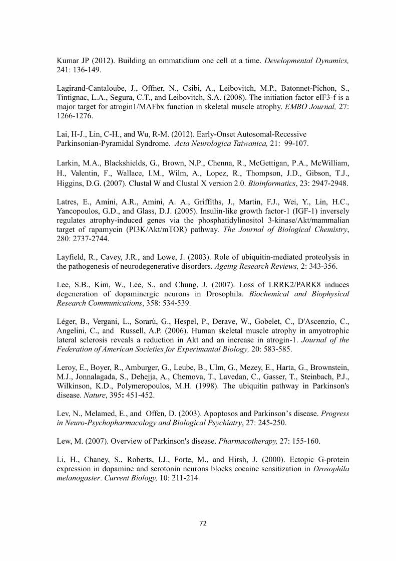
72
Kumar JP (2012). Building an ommatidium one cell at a time. Developmental Dynamics,
241: 136-149.
Lagirand-Cantaloube, J., Offner, N., Csibi, A., Leibovitch, M.P., Batonnet-Pichon, S.,
Tintignac, L.A., Segura, C.T., and Leibovitch, S.A. (2008). The initiation factor eIF3-f is a
major target for atrogin1/MAFbx function in skeletal muscle atrophy. EMBO Journal, 27:
1266-1276.
Lai, H-J., Lin, C-H., and Wu, R-M. (2012). Early-Onset Autosomal-Recessive
Parkinsonian-Pyramidal Syndrome. Acta Neurologica Taiwanica, 21: 99-107.
Larkin, M.A., Blackshields, G., Brown, N.P., Chenna, R., McGettigan, P.A., McWilliam,
H., Valentin, F., Wallace, I.M., Wilm, A., Lopez, R., Thompson, J.D., Gibson, T.J.,
Higgins, D.G. (2007). Clustal W and Clustal X version 2.0. Bioinformatics, 23: 2947-2948.
Latres, E., Amini, A.R., Amini, A. A., Griffiths, J., Martin, F.J., Wei, Y., Lin, H.C.,
Yancopoulos, G.D., and Glass, D.J. (2005). Insulin-like growth factor-1 (IGF-1) inversely
regulates atrophy-induced genes via the phosphatidylinositol 3-kinase/Akt/mammalian
target of rapamycin (PI3K/Akt/mTOR) pathway. The Journal of Biological Chemistry,
280: 2737-2744.
Layfield, R., Cavey, J.R., and Lowe, J. (2003). Role of ubiquitin-mediated proteolysis in
the pathogenesis of neurodegenerative disorders. Ageing Research Reviews, 2: 343-356.
Lee, S.B., Kim, W., Lee, S., and Chung, J. (2007). Loss of LRRK2/PARK8 induces
degeneration of dopaminergic neurons in Drosophila. Biochemical and Biophysical
Research Communications, 358: 534-539.
Léger, B., Vergani, L., Sorarù, G., Hespel, P., Derave, W., Gobelet, C., D'Ascenzio, C.,
Angelini, C., and Russell, A.P. (2006). Human skeletal muscle atrophy in amyotrophic
lateral sclerosis reveals a reduction in Akt and an increase in atrogin-1. Journal of the
Federation of American Societies for Experimantal Biology, 20: 583-585.
Leroy, E., Boyer, R., Amburger, G., Leube, B., Ulm, G., Mezey, E., Harta, G., Brownstein,
M.J., Jonnalagada, S., Dehejja, A., Chemova, T., Lavedan, C., Gasser, T., Steinbach, P.J.,
Wilkinson, K.D., Polymeropoulos, M.H. (1998). The ubiquitin pathway in Parkinson's
disease. Nature, 395: 451-452.
Lev, N., Melamed, E., and Offen, D. (2003). Apoptosos and Parkinson’s disease. Progress
in Neuro-Psychopharmacology and Biological Psychiatry, 27: 245-250.
Lew, M. (2007). Overview of Parkinson's disease. Pharmacotherapy, 27: 155-160.
Li, H., Chaney, S., Roberts, I.J., Forte, M., and Hirsh, J. (2000). Ectopic G-protein
expression in dopamine and serotonin neurons blocks cocaine sensitization in Drosophila
melanogaster. Current Biology, 10: 211-214.
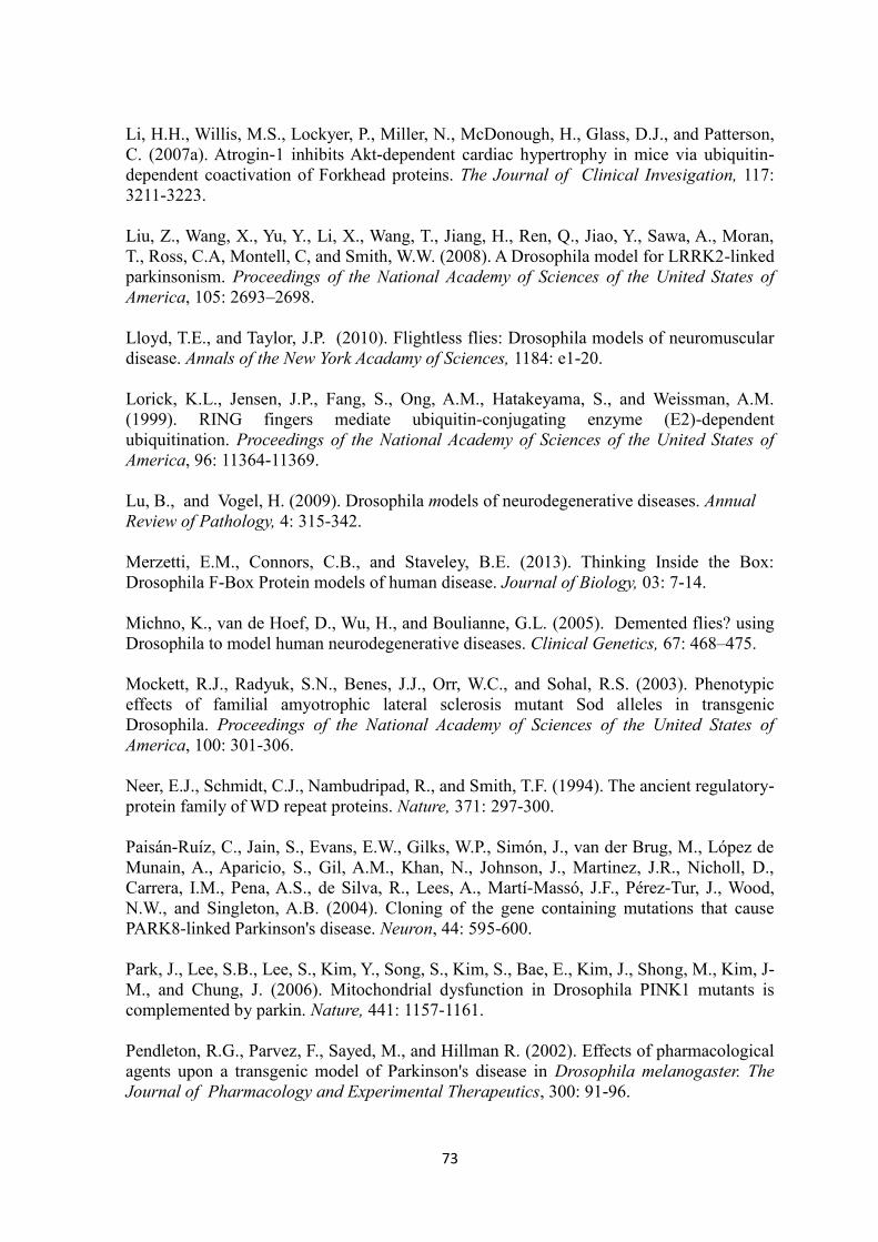
73
Li, H.H., Willis, M.S., Lockyer, P., Miller, N., McDonough, H., Glass, D.J., and Patterson,
C. (2007a). Atrogin-1 inhibits Akt-dependent cardiac hypertrophy in mice via ubiquitin-
dependent coactivation of Forkhead proteins. The Journal of Clinical Invesigation, 117:
3211-3223.
Liu, Z., Wang, X., Yu, Y., Li, X., Wang, T., Jiang, H., Ren, Q., Jiao, Y., Sawa, A., Moran,
T., Ross, C.A, Montell, C, and Smith, W.W. (2008). A Drosophila model for LRRK2-linked
parkinsonism. Proceedings of the National Academy of Sciences of the United States of
America, 105: 2693–2698.
Lloyd, T.E., and Taylor, J.P. (2010). Flightless flies: Drosophila models of neuromuscular
disease. Annals of the New York Acadamy of Sciences, 1184: e1-20.
Lorick, K.L., Jensen, J.P., Fang, S., Ong, A.M., Hatakeyama, S., and Weissman, A.M.
(1999). RING fingers mediate ubiquitin-conjugating enzyme (E2)-dependent
ubiquitination. Proceedings of the National Academy of Sciences of the United States of
America, 96: 11364-11369.
Lu, B., and Vogel, H. (2009). Drosophila models of neurodegenerative diseases. Annual
Review of Pathology, 4: 315-342.
Merzetti, E.M., Connors, C.B., and Staveley, B.E. (2013). Thinking Inside the Box:
Drosophila F-Box Protein models of human disease. Journal of Biology, 03: 7-14.
Michno, K., van de Hoef, D., Wu, H., and Boulianne, G.L. (2005). Demented flies? using
Drosophila to model human neurodegenerative diseases. Clinical Genetics, 67: 468–475.
Mockett, R.J., Radyuk, S.N., Benes, J.J., Orr, W.C., and Sohal, R.S. (2003). Phenotypic
effects of familial amyotrophic lateral sclerosis mutant Sod alleles in transgenic
Drosophila. Proceedings of the National Academy of Sciences of the United States of
America, 100: 301-306.
Neer, E.J., Schmidt, C.J., Nambudripad, R., and Smith, T.F. (1994). The ancient regulatory-
protein family of WD repeat proteins. Nature, 371: 297-300.
Paisán-Ruíz, C., Jain, S., Evans, E.W., Gilks, W.P., Simón, J., van der Brug, M., López de
Munain, A., Aparicio, S., Gil, A.M., Khan, N., Johnson, J., Martinez, J.R., Nicholl, D.,
Carrera, I.M., Pena, A.S., de Silva, R., Lees, A., Martí-Massó, J.F., Pérez-Tur, J., Wood,
N.W., and Singleton, A.B. (2004). Cloning of the gene containing mutations that cause
PARK8-linked Parkinson's disease. Neuron, 44: 595-600.
Park, J., Lee, S.B., Lee, S., Kim, Y., Song, S., Kim, S., Bae, E., Kim, J., Shong, M., Kim, J-
M., and Chung, J. (2006). Mitochondrial dysfunction in Drosophila PINK1 mutants is
complemented by parkin. Nature, 441: 1157-1161.
Pendleton, R.G., Parvez, F., Sayed, M., and Hillman R. (2002). Effects of pharmacological
agents upon a transgenic model of Parkinson's disease in Drosophila melanogaster. The
Journal of Pharmacology and Experimental Therapeutics, 300: 91-96.

74
Petit, A., Kawarai, T., Paitel, E., Sanjo, N., Maj, M., Scheid, M., Chen, F., Gu,Y.,
Hasegawa, H., Salehi-Rad, S., Wang, L., Rogaeva, E., Fraser, P., Robinson, B., St. George-
Hyslop, P., and Tandon, A. (2005). Wild-type PINK1 prevents basal and induced neuronal
apoptosis, a protective effect abrogated by Parkinson disease-related mutations. The
Journal of Biological Chemistry, 280: 34025-34032.
Pickart, C.M. (2001). Mechanisms underlying ubiquitination. Annual Review of
Biochemistry, 70: 503–533.
Polymeropoulos, M.H., Lavedan, C., Leroy, E., Ide, S.E., Dehejia, A., Dutra, A., Pike, B.,
Root, H., Rubenstein, J., Boyer, R., Stenroos, E.S., Chandrasekharappa, S., Athanassiadou,
A., Papapetropoulos, T., Johnson, W.G., Lazzarini, A.M., Duvoisin, R.C., Iorio, G.D.,
Golbe, L.I., Nussbaum, R.L. (1997). Mutation in the α-Synuclein gene identified in
families with Parkinson’s disease. Science, 276: 2045-2047.
Qiao, H., Wang, F., Zhao, L., Zhou, J., Zhao, L., Zhang, y., Robbins, T.P., and Xuea, Y.
(2004). The F-Box Protein AhSLF-S2 Controls the Pollen Function of S-RNase–Based
Self-Incompatibility. The Plant Cell, 16: 2306-2322.
Reiter, L.T., Potocki, L., Chien, S., Gribskov, M., and Bier, E. (2001). A systematic
analysis of human disease-associated gene sequences in Drosophila melanogaster.
Genome Research, 11: 1114-1125.
Rommel, C., Bodine, S.C., Clarke, B.A., Rossman, R., Nunez, L., Stitt, T.N., Yancopoulos,
G.D., and Glass, D.J. (2001). Mediation of IGF-1-induced skeletal myotube hypertrophy
by PI(3)K/Akt/mTOR and PI(3)K/Akt/GSK3 pathways. Nature Cell Biology, 3: 1009-
1013.
Ross, R.T. (1955). Pallido-Pyramidal Disease. Canadian Medical Association Journal, 73:
17–22.
Ross, O.A., Toft, M., Whittle, A.J., Johnson, J.L., Papapetropoulos, S., Mash, D.C., Litvan,
I., Gordon, M.F., Wszolek, Z.K., Farrer, M.J., and Dickson, D.W. (2006). Lrrk2 and Lewy
body disease. Annals of Neurology, 59: 388-393.
Russell, A.P. (2010). Molecular regulation of skeletal muscle mass. Clinical and
Experimental Pharmacology and Physiology, 37: 378–384.
Sacheck, J.M., Ohtsuka, A., McLary, S.C., and Goldberg, A.L. (2004). IGF-I stimulates
muscle growth by suppressing protein breakdown and expression of atrophy-related
ubiquitin ligases, atrogin-1 and MuRF1. American Journal of Physiology, Endocrinology
and Metabolism, 287: E591-601.
Sang, T.K., Chang, H.Y., Lawless, G.M., Ratnaparkhi, A., Mee, L., Ackerson, L.C.,
Maidment, N.T., Krantz, D.E., and Jackson, G.R. (2007). A Drosophila model of mutant
human parkin-induced toxicity demonstrates selective loss of dopaminergic neurons and
dependence on cellular dopamine. The Journal of Neuroscience, 27: 981-992.

75
Scheffner, M., Nuber, U., and Huibregtse, J.M. (1995). Protein ubiquitination involving an
E1-E2-E3 enzyme ubiquitin thioester cascade. Nature, 373: 81-83.
Shojaee, S., Sina, F., Banihosseini, S.S., Kazemi, M.H., Kalhor, R., Shahidi, G.A.,
Fakhrai-Rad, H., Ronaghi, M., and Elahi, E. (2008). Genomewide linkage analysis of a
Parkinsonian-pyramidal syndrome pedigree by 500 K SNP arrays. The American Journal
of Human Genetics, 82: 1375-1384.
Simon, A.F., Liang, D.T., and Krantz, D.E. (2006). Differential decline in behavioral
performance of Drosophila melanogaster with age. Mechanisms of Ageing and
Development, 127: 647-651.
Singleton, A.B., Farrer, M., Johnson, J., Singleton, A., Hague, S., Kachergus, J., Hulihan,
M., Peuralinna, T., Dutra, A., Nussbaum, R., Lincoln, S., Crawley, A., Hanson, M.,
Maraganore, D., Adler, C., Cookson, M.R., Muenter, M., Baptista, M., Miller, D.,
Blancato, J., Hardy, J., and Gwinn-Hardy, K. (2003). alpha-Synuclein locus triplication
causes Parkinson's disease. Science, 302: 841.
Skaar, J.R., D'Angiolella, V., Pagan, J.K., and Pagano, M. (2009a). SnapShot: F Box
Proteins II. Cell, 137: 1358.e1–1358.e2.
Skaar, J.R., Pagan, J.K., and Pagano, M. (2009b). SnapShot: F box proteins I. Cell, 137:
1160-1160.e1.
Slade, J.D., and Staveley, B.E. (2015). Compensatory growth in Drosophila Akt1 mutants.
BioMed Central Research Notes 8:77
Staveley, B.E., Phillips, J.P, Hilliker, A.J. (1990). Phenotypic consequences of copper-zinc
superoxide dismutase overexpression in Drosophila melanogaster. Genome, 33: 867-872.
Staveley, B.E. (2012). Successes of Modelling Parkinson Disease in Drosophila.
Mechanisms in Parkinson's Disease- Models and Treatments, ISBN: 978-953-307-876-2.
Sublett, J.E., Jeon, I.S., and Shapiro, D.N. (1995). The alveolar rhabdomyosarcoma
PAX3/FKHR fusion protein is a transcriptional activator. Oncogene, 11: 545-552.
Tadaiesky, M.T., Dombrowski, P.A., Figueiredo, C.P., Cargnin-Ferreria, E., Da Cunha, C.,
and Takahashi, R.N. (2008). Emotional, cognitive and neurochemical alterations in a
premotor stage model of Parkinson's disease. Neuroscience, 156: 830-40.
Tain, L. S., Mortiboys, H., Tao, R. N., Ziviani, E., Bandmann, O., and Whitworth, A. J.
(2009b). Rapamycin activation of 4E-BP prevents parkinsonian dopaminergic neuron loss.
Nature Neuroscience, 12: 1129-1135.
Taylor, J.P., Mata, I.F., and Farrer, M.J. (2006). LRRK2: a common pathway for
parkinsonism, pathogenesis and prevention? Trends in Molecular Medicine, 12: 76-82.
Teixeira L.K., and Reed, S.I. (2013). Ubiquitin Ligases and Cell Cycle Control. Annual
Review of Biochemistry, 82: 387–414.

76
Tempel, B.L., Livingstone, M.S., and Quinn, W.G. (1984). Mutations in the dopa
decarboxylase gene affect learning in Drosophila. Proceedings of the National Academy of
Sciences of the United States of America, 81: 3577–3581.
Thomas, B.J., and Wassarman, D.A. (1999). A fly's eye view of biology. Trends in
Genetics, 15: 184-90.
Thomas, B., and Beal, M.F. (2007). Parkinson’s disease. Human Molecular Genetics, 16:
183-194.
Tintignac, L.A., Lagirand, J., Batonnet, S., Sirri, V., Leibovitch, M.P., and Leibovitch, S.A.
(2005). Degradation of MyoD mediated by the SCF (MAFbx) ubiquitin ligase. The Journal
of Biological Chemistry, 280: 2847-2856.
Todd, A.M., and Staveley, B.E. (2004). Novel assay and analysis for measuring climbing
ability in Drosophila. Drosophila Information Services, 87: 101-107.
Todd, A.M., and Staveley, B.E. (2008). Pink1 suppresses alpha-synuclein-induced
phenotypes in a Drosophila model of Parkinson's disease. Genome, 51: 1040-1046.
Urso, M.L., Chen, Y.W., Scrimgeour, A.G., Lee, P.C., Lee, K.F., and Clarkson, P.M.
(2007). Alterations in mRNA expression and protein products following spinal cord injury
in humans. The Journal of Physiology, 579(Pt): 877-892.
Valente, E.M., Abou-Sleiman, P.M., Caputo, V., Muqit, M.M., Harvey, K., Gispert, S., Ali,
Z., Del Turco, D., Bentivoglio, A.R., Healy, D.G., Albanese, A., Nussbaum, R., González-
Maldonado, R., Deller, T., Salvi, S., Cortelli, P., Gilks, W.P., Latchman, D.S., Harvey,
R.J., Dallapiccola, B., Auburger, G., and Wood, N.W. (2004). Hereditary early-onset
Parkinson's disease caused by mutations in PINK1. Science, 304: 1158-60.
Vanitallie, T.H. (2008). Parkinson disease: primacy of age as a risk factor for mitochondrial
dysfunction. Metabolism, Suppl 2: S50–S55.
Venken, K.J.T., Simpson, J.H., and Bellen, H.J. (2011). Genetic manipulation of genes and
cells in the nervous system of the fruit fly. Neuron, 72: 202-231.
Wang, D., Qian, L., Xiong, H., Liu, J., Neckameyer, W.S., Oldham, S., Xia, K., Wang, J.,
Bodmer, R.,and Zhang, Z. (2006). Antioxidants protect PINK1-dependent dopaminergic
neurons in Drosophila. Proceedings of the National Academy of Sciences of the United
States of America, 103: 13520-13525.
Wang, C., Lu, R., Ouyang, X., Ho, M.W., Chia, W., Yu, F., and Lim, K.L. (2007).
Drosophila overexpressing parkin R275W mutant exhibits dopaminergic neuron
degeneration and mitochondrial abnormalities. The Journal of Neuroscience, 27: 8563-
8570.
Wang, D., Tang, B., Zhao, G., Pan, Q., Xia, K., Bodmer, R., and Zhang, Z. (2008).
Dispensable role of Drosophila ortholog of LRRK2 kinase activity in survival of
dopaminergic neurons. Molecular Neurodegeneration, 3: 3.

77
Warrick, J. M., Chan, H. Y., Gray-Board, G. L., Chai, Y., Paulson H.L., and Bonini, N.M.
(1999). Suppression of polyglutamine-mediated neurodegeneration in Drosophila by the
molecular chaperone HSP70. Nature Genetics, 23: 425–428.
West, A. B., Moore, D. J., Biskup, S., Bugayenko, A., Smith, W.W., Ross, C.A., Dawson,
V.L., and Dawson, T.M. (2005). Parkinson’s disease-associated mutations in leucine-rich
repeat kinase 2 augment kinase activity. Proceedings of the National Academy of Sciences
of the United States of America, 102: 16842-16847.
Whitworth, A.J., Theodore, D.A., Greene, J.C., Benes, H., Wes, P.D., and Pallanck, L.J.
(2005). Increased glutathione S-transferase activity rescues dopaminergic neuron loss in a
Drosophila model of Parkinson's disease. Proceedings of the National Academy of Sciences
of the United States of America, 102: 16842-16847.
Whitworth, A.J. (2011). Drosophila models of Parkinson's disease. Advances in Genetics,
73: 1-50.
Wilkinson, K.D. (1999). Ubiquitin-dependent signaling: the role of ubiquitination in the
response of cells to their environment. The Journal of Nutrition, 129: 1933–1936.
Winston, J.T., Koepp, D.M., Zhu, C., Elledge, S.J., and Harper, J.W. (1999). A family of
mammalian F-box protein. Current Biology, 9: 1180–1182.
Wolf, F.W., and Herbelein, U. (2003). Invertebrate modles of drug abuse. Journal of
Neurobiology, 54: 161-178.
Yang, Y., Gehrke, S., Imai, Y., Huang, Z., Ouyang, Y., Wang, J.W., Yang, L., Beal, M.F.,
Vogel, H., and Lu, B. (2006). Mitochondrial pathology and muscle and dopaminergic
neuron degeneration caused by inactivation of Drosophila Pink1 is rescued by Parkin.
Proceedings of the National Academy of Sciences of the United States of America, 103:
10793-10798.
Zarranz, J.J., Alegre, J., Gómez-Esteban, J.C., Lezcano, E., Ros, R., Ampuero, I., Vidal,
L., Hoenicka, J., Rodriguez, O., Atarés, B., Llorens, V., Gomez Tortosa, E., del Ser, T.,
Muñoz, D.G., de Yebenes, J.G. (2004). The new mutation, E46K, of alpha-synuclein
causes Parkinson and Lewy body dementia. Annals of Neurology, 55: 164-173.
Zhao, T., De Graaff, E., Breedveld, G.J., Loda, A., Severijnen, L.A., Wouters, C.H.,
Verheijen, F.W., Dekker, M.C.J., Montagna, P., Willemsen, R., Oostra, B.A., and Bonifati,
V. (2011). Loss of Nuclear Activity of the FBXO7 Protein in Patients with Parkinsonian-
Pyramidal Syndrome (PARK15). PLoS ONE, 6: 1-13.
Zimprich, A., Biskup, S., Leitner, P., Lichtner, P., Farrer, M., Lincoln, S., Kachergus, J.,
Hulihan, M., Uitti, R.J., Calne, D.B., Stoessl, A.J., Pfeiffer, R.F., Patenge, N., Carbajal,
I.C., Vieregge, P., Asmus, F., Müller-Myhsok, B., Dickson D.W., Meitinger, T., Strom,
T.M., Wszolek, ZK., and Gasser, T. (2004). Mutations in LRRK2 cause autosomal-
dominant parkinsonism with pleomorphic pathology. Neuron, 44: 601-607.

78