Embed Size (px)
Citation preview

Hamline UniversityDigitalCommons@Hamline
Departmental Honors Projects College of Liberal Arts
Spring 2015
Characterization of CP43 and PIF1 GWASSelected Candidate Genes in Medicago truncatulaand their Contribution to Rhizobia Symbiosis andPhenotypes of Interest Using an hpRNA BasedRNAi PlatformNathaniel Henning
Follow this and additional works at: https://digitalcommons.hamline.edu/dhp
Part of the Biology Commons
This Honors Project is brought to you for free and open access by the College of Liberal Arts at DigitalCommons@Hamline. It has been accepted forinclusion in Departmental Honors Projects by an authorized administrator of DigitalCommons@Hamline. For more information, please [email protected], [email protected].
Recommended CitationHenning, Nathaniel, "Characterization of CP43 and PIF1 GWAS Selected Candidate Genes in Medicago truncatula and theirContribution to Rhizobia Symbiosis and Phenotypes of Interest Using an hpRNA Based RNAi Platform" (2015). DepartmentalHonors Projects. 30.https://digitalcommons.hamline.edu/dhp/30

Characterization of CP43 and PIF1 GWAS Selected Candidate Genes in Medicago truncatula and their
Contribution to Rhizobia Symbiosis and Phenotypes of Interest Using an hpRNA Based RNAi Platform
Nathaniel Henning
An Honors thesis submitted for partial fulfillment of the requirements for graduation with honors in
Biology from Hamline University.
April 23rd
, 2015

2
Table of Contents
Abstract 3
Introduction 4
Methods 14
Results and Discussion 17
Conclusion 24
Future Directions 24
References 26
Acknowledgements 29
Appendix 30

3
Abstract
Medicago truncatula is a model organism used to study the symbiotic relationship between
plants and nitrogen fixing soil bacteria of the genus rhizobia. By studying the genes that are
involved in this symbiosis we can accomplish the goal of transplanting the symbiotic relationship
into agriculture staples (e.g. corn, wheat, etc.) that do not utilize similar symbiotic relationships
with nitrogen fixers. This would help to reduce our reliance on nitrogen-based fertilizers. In order to
identify plant genes that might be important for establishing and regulating optimal nitrogen-fixing
symbioses, a large-scale Genome Wide Association Study (GWAS) was performed. This study
identified the top 50 candidate genes that were hypothesized to be relevant to Medicago-
Sinorhizobium symbiosis. The purpose of the work presented as part of this thesis was to
characterize two candidate genes, PIF1 and CP43 and investigate their role in symbiosis. These
genes were identified by GWAS and encode a DNA helicase and a chlorophyll apoprotein,
respectively. Hairpin RNA constructs (hpRNA) were created and inserted into Medicago truncatula
to reduce expression of the PIF1 and CP43. These constructs reduced the expression of the two
candidate genes by 80-85%. However, reduced gene expression of the candidate genes did not lead
to significant phenotypic changes in the height of the plants or number of nodules on the plant
roots in comparison to unmodified control plants. Interestingly, preliminary results indicate that
reduction of CP43 gene expression alters the strain of rhizobia that nodulates the Medicago
truncatula roots. This observation suggests that the CP43 gene may play a role in host-partner
specificity in the symbiotic relationship between Medicago truncatula and rhizobia.

4
Introduction
Medicago truncatula
Medicago truncatula is a small annual galegoid, or cool season, legume that originated in the
Mediterranean basin and is a member of the Fabaceae, or Leguminosae, family. In the wild it grows to
be approximately 10-60 cm tall, has trifoliate leaves, and produces seeds that are contained in small
spine-covered pods. It is closely related to several genera of staple agricultural crops, such as both red
and white clover, pea, chickpea, and broad bean. Research into the evolution of the species had
indicated that Medicago truncatula separated from these legume species approximately 25 Mya and
from commercially important soybeans and common beans approximately 60 Mya (Young and Uvardi,
2009). M. truncatula has a relatively small genome of about 500-550 million base pairs compared to the
1.1-1.15 billion base pairs soybean genome or 738 million base pairs chickpea genome. Aside from its
small genome size, Medicago has several other features that make it an excellent model including being
diploid, having relatively quick seed-to-seed time of about 3 months, availability of a diverse set of
ecotypes (over 228 different accessions) ideal for plant research, and its ability to both self and cross
pollinate.
M. truncatula is commercially used in Australia as an annual forage crop to feed livestock.
However, because of its relatively small genome, quick seed-to-seed generation time, small size, and
amenability to varying levels of genetic transformation, it is primarily used as a model organism for
genetics research. The initial interest in M. truncatula as a model organism stems from its symbiotic
relationship with nitrogen-fixing bacteria of the genus rhizobia. The plant is also used as a model
organism for the development of biological markers, for example M. truncatula sequence based simple
sequence repeat markers (SSR) that have provided markers for linkage mapping in other crop legumes
(Young and Uvardi, 2009) or the intron-targeted markers based on the M.truncatula that have been
used in mapping L. culinaris (lentils). Plant biotechnology is another area in which discoveries in M.
truncatula have been applied to other legume species, specifically alfalfa. Among these applications are
the RCT1 anthracnose resistance gene, affecting resistance to a fungal disease, and lignin biosynthesis
genes discovered in M. truncatula, which when knocked out in alfalfa reduce its lignin content and
increase its digestibility by livestock (Young and Uvardi, 2009). Other studies have shown the use of gene
discovery in M. truncatula and subsequent translation into crop legume species (Deavours and Dixon
2005, Chen and Dixon 2007, and Pennycooke et al. 2008). Therefore, M. truncatula is a functional and
useful model organism for studying various aspects of plant biology research beyond looking at the
genes regulating symbiosis with rhizobia.
Rhizobia and Nitrogen fixation
Nitrogen, N, is an essential element of plant growth and is involved in many important processes
including, but not limited to, the synthesis of proteins and production of chlorophyll. Plant species are
unable to generate their own nitrogen or fix atmospheric nitrogen on their own. Because of their
inability to fix nitrogen plants depend on atmospheric nitrogen being fixed into a biologically available
form, such as ammonia, through various environmental processes that are part of the Nitrogen cycle
(see figure 1). The nitrogen cycle accounts for an estimated 3x10⁹ tons of atmospheric nitrogen being
converted into ammonia through various means (Zahn, 1999). Nitrogen is fixed through non-biological
means with lightning strikes accounting for approximately 10% of the world’s supply of fixed nitrogen
(Zahn, 1999). Biological means of nitrogen fixation involve many species of prokaryotes (diazotrophs).

5
Nitrogen sources for these processes include animal waste, decaying animal and plants, and nitrogen in
the atmosphere. Nitrogen is also put into the system through human activities such as nitrogen based
fertilizers and industrial emissions.
Figure 1. Diagram of the nitrogen cycle. Diagram shows the major processes by which molecular
nitrogen is converted into reactive nitrogen and vice versa. Inputs into the system are also listed with
orange indicating anthropogenic (human) inputs into the nitrogen cycle and blue indicating natural
inputs into the system (Gruber and Galloway, 2008).
To fulfill their need for nitrogen, legumes have developed a biological method of nitrogen
fixation through a mutualistic symbiotic relationship with a species of prokaryotes, soil bacteria of the
genus rhizobia. These bacteria are capable of fixing atmospheric nitrogen, N2, into a biologically
available form, ammonia, NH3. In return for providing the plant with fixed nitrogen the bacteria are
provided with the nutrients such as carbohydrates and proteins, required for growth and proliferation.
Through this mutualistic relationship legumes contribute 150 million metric tons of nitrogen to the
ecosystem in both managed and natural ecosystems (Smil, 2005; Cleveland et al, 1999).
In order to establish the mutualistic relationship with legume plants, the bacteria invade the
roots of legumes and form outgrowths, also known as nodules, through the process known as
nodulation (See Figure 2 for generalized process). This process was reviewed by Jones et. al., 2007 in
which they described the step-by-step process by which rhizobia invade plant roots to form nodules.
Their review summarized previous research that had found that the first step is an initial signal exchange
between the eventual host plant and the soil bacteria. This signal comes in the form of flavonoids, 2-
phenyl-1,4-benzopyrone derivatives, being secreted by the host plants roots. The flavonoids are taken
up by the soil bacteria where they bind to and activate nodulation proteins (NodD), called nodulation
factors. These nodulation proteins are part of the LysR family of transcription factors, regulating the
transcription of specific parts of DNA by binding to it and acting as an activator, repressor, or an
enhancer. The NodD proteins are activator proteins, and they activate the genes required to initiate

6
nodulation of plant roots, specifically Nod factors (NFs), which are secreted by the bacteria. These NFs
can vary in structure between bacterial species and some species secrete more than one type of NFs.
After being secreted, NF is bound by the M. truncatula Nod Factor Perception (MtNFP) receptor, a LysM
family receptor kinase, in the membrane of the host plant’s root hair cells. The activation of this
receptor leads to an increase in the intracellular concentration of calcium present in the root hairs. This
concentration fluctuation alters the structure of the root hair cytoskeleton. The alterations to the
cytoskeleton lead to the curling of the root hairs, which traps the bacteria within what is now known as
a tight colonized curled root hair (CCRH). Simultaneously, NF causes cortex cells to undergo mitosis,
which creates the cells that are infected by the invading bacteria known as the Nodule Primordium. The
initial steps required for nodulation are followed by the development of the infection thread.
Figure 2. General step-by-step process for nodulation of plant roots. Figure shows a simplified process
by which rhizobia (or other symbiotic Nitrogen fixing bacteria) nodulate plant roots (Deakin and
Boughton, 2009).
The formation of the infection thread is the next critical step for the successful nodulation of
plant roots and internalizes the rhizobia within the host plants roots (See Figure 2). After being trapped
by the CCRH, the rhizobia begin secreting symbiotically active exopolysaccharides that stimulate the
growth of the root hair cell membrane, which grows into the interior plant, specifically the cortex of the
plant roots. This ingrowth of the infection thread allows for the internalization of the bacteria into the
cortical cells of the roots. The invading bacteria are taken up by the cortical cells, the Nodule
Primordium, via endocytosis and are subsequently referred to as a symbiosome (Jones et al., 2007). The
bacteria enclosed in the symbiosome differentiate into bacteroids, a form that is capable of nitrogen
fixation. The nodules continue to develop as the cortex cells divide, which leads to visible outgrowths on
the outside of roots. The nodules also develop vascular tissue which transports the Nitrogen compounds
secreted by the bacteroids (ammonia) to the shoot. The vascular tissue also transports organic
compounds required for proliferation to the bacteroids from the plant. The rhizobia are also provided
with the proteins including the oxygen binding leghemoglobin (which gives nodules their distinct pink
color) and sucrose synthase (allows for the synthesis of malate through the breakdown of sucrose).
Once established the symbiosis is maintained by the inability of the plants immune system to identify,
bind to, and eliminate rhizobia. The relationship is also maintained by the plants ability to control and
support bacteroid survival and differentiation. However, little is known about the exact mechanism that

7
maintains the symbiotic relationship and research into possible mechanisms is ongoing (Vlassek and
Vanderleyden, 2007).
Relevance
Most agriculturally important plants lack the ability to convert N2 to ammonia, the main form of
nitrogen incorporated in proteins and macromolecules essential for plant growth. Therefore, many
agriculturalists spray synthetic nitrogen -based fertilizers on their fields to promote plant growth. In
2012, 22.5 million tons of fertilizers were used in the United States and approximately 185.1 million tons
used worldwide with most of this fertilizer being used to promote the growth of corn (The Fertilizer
Institute). The use of fertilizers has lead, alongside other factors, to steadily increasing crop yields and
now up to 40% of protein consumed globally comes from fields that use nitrogen based fertilizers
(Peoples et al, 2004). The heavy use of these fertilizers by the United States has contributed to
deleterious effects on the environment as 50-60% of the nitrogen based fertilizers are lost from fields
through soil erosion, runoff, leaching of nitrate or through gaseous emissions (Peoples et al, 2004). This
leaching has serious health and environmental implications including: water pollution through hypoxia
(oxygen deprivation), hypertrophication (uncontrolled growth or “blooms” caused by high nutrient
levels), and methemoglobinemia (high blood concentrations of Fe3+
), greenhouse warming, and a
reduction in diversity of vegetation.
A more traditional method of reintroducing biologically available nitrogen into the soil is crop
rotation using a plant such as soybeans, which would fix atmospheric nitrogen into ammonia. The ability
of soybeans and other legumes to fix nitrogen is linked to their symbiotic relationship with the nitrogen
fixing bacteria of the genus rhizobia. Other genera of nitrogen fixing soil bacteria such as Frankia,
Anabaena, Azotobacter, and Clostridium also contribute to the availability of nitrogen through the
conversion of atmospheric Nitrogen into ammonia. Legumes and other pasture species are capable of
returning the equivalent of 200-300kg ha-1
of nitrogen fertilizer to the soil through biologically mediated
nitrogen fixation (Zahran 1999). Through the analysis of genomic SNPs (single nucleotide
polymorphisms), INDEls (insertions and deletions), and other techniques we can identify genetic
determinants that are important to making nitrogen-fixing symbiotic relationships possible. The ultimate
goal of these research studies is to transplant important genetic determinants that contribute to
effective symbioses into crop species that typically deplete rather than replenish soil nitrogen such as
the agricultural staples, corn and wheat. By transplanting this system it would be possible to reduce
dependence of modern industrial agriculture on synthetic nitrogen based fertilizers and through this
reduce the impact of nitrogen runoff on the environment.
Genome Wide Association Studies
Genome Wide Association Studies, or GWAS, use a genotyped DNA marker to look at the
association between the marker and a phenotype of interest such as a disease state like Cystic Fibrosis
in humans or the flowering date for a plant. The biological markers used by most GWAS are the Single
Nucleotide Polymorphism or SNP (pronounced Snip). SNPs are single base-pair changes in the DNA
sequence of an organism. SNPs occur relatively frequently, there are over 6 million SNPs present across
the 225 accessions of M. truncatula, and generally have minimal impact on biological systems. However,
some SNPs such as those of Cystic Fibrosis show significant impact on biological systems. Another
property of SNPs that is important to GWAS is the concept of linkage disequilibrium (LD), which
describes the degree to which an allele of one SNP is inherited or correlated with an allele of another

SNP within a population (Bush and Moor, 2012). It is a concept that is directly linked to the idea that two
markers (such as a SNP) on a chromosome remain
LD allows for association studies, like GWAS,
phenotype of interest (direct association) or that a SNP is indirectly linked to a phenotype through
another SNP (indirect association). Because of the possibility of both direct and indirect associatio
between a significant SNP and phenotype GWAS does not by itself determine causal relationships, but
instead significant SNPs must be followed
In order to perform a GWAS two primary technologies are used to sequence and assay fo
These are the Illumina and Affymetrix platforms, and both are able to measure SNP variation using DNA
microarray technology. After using one of these platforms to identify SNPs researchers perform
case/control studies to score the phenotypes, eithe
the Illumina/Affymetrix genotyped populations. After genotyping the population and gathering
phenotype data from individuals, statistical analysis is performed to look at the association of SNPs and
the phenotype of interest. Statistical analysis is performed using a wide variety of techniques
generalized linear model approaches for quantitative traits
often performed using suitable software packages
Geddes et al., 2011). The data from this analysis is generally arranged into Manhattan Plots, which plot
the significance of a SNPs association with a trait to the location on a chromosome (see Figure
the genome locations that show a significant contribution to
can be chosen for further study.
Figure 3. Example of GWAS 3.5 Manhattan Plot
candidate genes for specific phenotypes.
represents significance. The light grey band of noise indicates locations without a significant impact on
the particular. Colored dots indicate potential significant contrib
figure for both the fifth and sixth chromosomes we see significant contri
(see red boxed regions).
For M. truncatula a GWAS was performed by Stanton
providing 1 SNP per 43 bases in the genome
SNP within a population (Bush and Moor, 2012). It is a concept that is directly linked to the idea that two
chromosome remain physically joined through generations of a population.
LD allows for association studies, like GWAS, to find that a SNP is statistically associated with a
phenotype of interest (direct association) or that a SNP is indirectly linked to a phenotype through
(indirect association). Because of the possibility of both direct and indirect associatio
between a significant SNP and phenotype GWAS does not by itself determine causal relationships, but
instead significant SNPs must be followed by other research methods.
In order to perform a GWAS two primary technologies are used to sequence and assay fo
These are the Illumina and Affymetrix platforms, and both are able to measure SNP variation using DNA
microarray technology. After using one of these platforms to identify SNPs researchers perform
to score the phenotypes, either categorically or quantitatively, of individuals within
the Illumina/Affymetrix genotyped populations. After genotyping the population and gathering
phenotype data from individuals, statistical analysis is performed to look at the association of SNPs and
the phenotype of interest. Statistical analysis is performed using a wide variety of techniques
generalized linear model approaches for quantitative traits or mixed model ANOVAs. This analysis is
often performed using suitable software packages such as TASSEL 3.0 (Bush and Moor, 2012; Stanton
The data from this analysis is generally arranged into Manhattan Plots, which plot
the significance of a SNPs association with a trait to the location on a chromosome (see Figure
genome locations that show a significant contribution to the phenotype of interest
. Example of GWAS 3.5 Manhattan Plot from Stanton-Geddes et al (2013) used
r specific phenotypes. The x-axis represents location on the chromosome and y
The light grey band of noise indicates locations without a significant impact on
the particular. Colored dots indicate potential significant contributions to a particular phenotype. In this
figure for both the fifth and sixth chromosomes we see significant contributions by candidates 14 and 17
a GWAS was performed by Stanton-Geddes et al, 2013 using 6,344,25
in the genome. These SNPs were identified by sequencing the genomes of
8
SNP within a population (Bush and Moor, 2012). It is a concept that is directly linked to the idea that two
physically joined through generations of a population.
to find that a SNP is statistically associated with a
phenotype of interest (direct association) or that a SNP is indirectly linked to a phenotype through
(indirect association). Because of the possibility of both direct and indirect association
between a significant SNP and phenotype GWAS does not by itself determine causal relationships, but
In order to perform a GWAS two primary technologies are used to sequence and assay for SNPs.
These are the Illumina and Affymetrix platforms, and both are able to measure SNP variation using DNA
microarray technology. After using one of these platforms to identify SNPs researchers perform
, of individuals within
the Illumina/Affymetrix genotyped populations. After genotyping the population and gathering
phenotype data from individuals, statistical analysis is performed to look at the association of SNPs and
the phenotype of interest. Statistical analysis is performed using a wide variety of techniques such as the
This analysis is
such as TASSEL 3.0 (Bush and Moor, 2012; Stanton-
The data from this analysis is generally arranged into Manhattan Plots, which plot
the significance of a SNPs association with a trait to the location on a chromosome (see Figure 3). From
the phenotype of interest candidate genes
used to determine
axis represents location on the chromosome and y-axis
The light grey band of noise indicates locations without a significant impact on
utions to a particular phenotype. In this
butions by candidates 14 and 17
Geddes et al, 2013 using 6,344,256 SNPs,
identified by sequencing the genomes of

9
226 accessions of M. truncatula using an in silico SNP platform. After genotyping, eight seeds per
accession, for a total of 2000 seeds, were planted and harvested 30 days after growth. During harvest
plants were assessed for phenotypes of interest. These phenotypes were: height, total nodulation,
nodulation of the top 5 cm of the root, nodulation of the bottom 5 cm of the root, strain occupancy of
the top 5cm, strain occupancy of the bottom 5 cm, and flowering date. The phenotypic data was then
linked to the accessions genotype.
The variation in the phenotype data between accessions was then compared to the data
gathered through genotyping and was organized into Manhattan plots, as seen in figure 3, significant
SNPs were given one of three colors, which represent different minor allele frequency (MAF) values,
while SNPs that were not significant contributors were made gray. These MAF values give us the
frequency at which the less common, or minor, allele of a SNP is present in the population of an
organism. These plots were created using a mixed model ANOVA with kinship as a covariate and were
analyzed using TASSEL 3.0. By looking at the loci strongly associated with the phenotype and comparing
with the reference genome Stanton-Geddes et al (2013) were able to develop a list of candidate genes
for each of the phenotypes of interest. Many of these identified candidate genes have functions that
correspond directly with the phenotype of interest, but this is not always the case as will be seen with
the two candidates chosen for the purpose of this study, candidate #17 and candidate #14.
Strain Occupancy
Strain occupancy is another phenotype of interest for the mutualistic relationship between
legumes and rhizobia based on the notion that only one strain of rhizobia will be present in a nodule and
that strains of rhizobia compete to nodulate plant roots. It is of interest to researchers because the
nitrogen fixing efficiency varies among strains of rhizobia (reviewed by Vlassak, et al., 2001). Multiple
studies have indicated that strains of rhizobia competitively nodulate legume roots and that generally
only one strain of rhizobia will occupy a given nodule (Duodu et al., 2009). Many factors can influence
nodule occupancy by a given strain of rhizobia including responses to abiotic and biotic environmental
variables, ratio of the various strains, and the genetics of both the strains and the host plant. An
example of this was seen in studies of the competitiveness of R. leguminosarum and R. tropici done by
Streit et al., 1992 during which it was shown that these two strains’ ability to use aromatic compounds
was a key to their ability to nodulate competitively with other strains. Strains also show a preference for
nod gene inducers, growth stimulators, and chemoattractants secreted by plants (reviewed in Vlassak et
al., 2010). Other determinants include nod genes, cell surface determinants, environmental, and other
host and bacteria specific factors. A study by Duodu et al., 2008, showed that this competitiveness
manifested during the initial stages of infection. Studies have also shown that the nitrogen fixation
efficiency varies for different strains of rhizobia, so by finding the genes involved in regulating selection
of the bacteria species by the plant one could select for the most efficient strain of rhizobia, which in
part leads to strain occupancy being a phenotype of interest.
RNA interference via hpRNA
There are several methods used to investigate the relationship between a gene and an observed
phenotype, inlcuding chemical mutagenesis, recombination, transposon mutagenesis and RNA
interference (RNAi) technologies used to create knockdowns or knockouts in the genes of interest. The
role of candidate genes 14 and 17 in the Medicago-sinorhizobium symbiosis was investigated using RNA

interference (RNAi). This process consists of introducing a gene that codes for a hairpin RNA construct to
induce post-transcriptional gene silencing (PTGS)
Figure 4. Simplified Translation of RNA into protein
mechanism used by cells during normal
circumstances a ribosome binds to mRNA, which then translates the mRNA into a functional
the right is the mechanism for RNA interference (RNAi)
mechanism, the enzyme DICER cuts up the
the enzyme Argonaut these siRNA bind to mRNA,
The mechanism used by hpRNAs
natural viral defense pathway. RNAi technology has been shown to
including Arabidopsis thaliana and Medicago
Stoutjesdijk et al, 2002; Wesley et al, 2001).
produce small RNAs between 21-25 nucleotides in length
stranded RNAs made of a promoter, sense arm, intron, anti
binds to the hpRNA and cuts it into
RNA fragments can produce gene-silencing events when
be called short interfering RNAs (siRNAs). The gene silencing caused by these siRNAs is mediated
through the action of the protein Argonaut, which binds the siRNA to their complementary strand of
mRNA. The binding of the siRNA to the mRNA causes the degradation of mRNA in a post
fashion by the RNA-induced silencing complex (RISC) (Limpens et al, 2004).
In order to get the hpRNA construct int
hairpin into a plasmid. This plasmid is then introduced into
electroporation or heat shock. Transformed
or leaves of the plants using classic Ti transformati
gene silencing is created by designing the sense and anti
homologous to the gene of interest
been highly efficient in studies using different plant species such as Arabidopsis and
truncatula. In these studies, up to 91% of subjects display
2004).
Goal and Hypothesis for this Study
This process consists of introducing a gene that codes for a hairpin RNA construct to
transcriptional gene silencing (PTGS) or mRNA degradation in plants.
Simplified Translation of RNA into protein. On the left is a simplified version of the
normal gene expression and translation of a protein. Under normal
some binds to mRNA, which then translates the mRNA into a functional
sm for RNA interference (RNAi) via hairpin RNA (hpRNA). In this simplified
the enzyme DICER cuts up the double stranded hpRNA into siRNA. With the assistance of
the enzyme Argonaut these siRNA bind to mRNA, causing its degradation by normal cell
hpRNAs to introduce gene silencing takes advantage of
. RNAi technology has been shown to be effective in several
Medicago truncatula (Fusaro et al, 2006, Limpens et al, 2004;
Stoutjesdijk et al, 2002; Wesley et al, 2001). The pathway is mediated by Dicer-like (DCL) proteins, which
25 nucleotides in length by dicing up dsRNA. HpRNAs
made of a promoter, sense arm, intron, anti-sense arm, and a terminator
it into small RNA fragments including the sense and anti-sense arms.
silencing events when they are homologous to mRNA causing them to
be called short interfering RNAs (siRNAs). The gene silencing caused by these siRNAs is mediated
through the action of the protein Argonaut, which binds the siRNA to their complementary strand of
ng of the siRNA to the mRNA causes the degradation of mRNA in a post
induced silencing complex (RISC) (Limpens et al, 2004).
In order to get the hpRNA construct into the plant researchers clone the DNA encoding the
rpin into a plasmid. This plasmid is then introduced into Agrobacterium strains by transformation via
Transformed Agrobacterium cells are then used to transform
of the plants using classic Ti transformation as described by (Chabaud et al., 2006
designing the sense and anti-sense arms of the hpRNA construct
homologous to the gene of interest (Limpens et al, 2004). The gene targeting and transformation
in studies using different plant species such as Arabidopsis and Medicago
up to 91% of subjects display the desired altered phenotypes (Limpens et al,
10
This process consists of introducing a gene that codes for a hairpin RNA construct to
version of the
of a protein. Under normal
some binds to mRNA, which then translates the mRNA into a functional protein. On
In this simplified
hpRNA into siRNA. With the assistance of
by normal cellular processes.
of the plants
effective in several plant species
(Fusaro et al, 2006, Limpens et al, 2004;
like (DCL) proteins, which
are double
sense arm, and a terminator (figure 4). Dicer
sense arms. These
they are homologous to mRNA causing them to
be called short interfering RNAs (siRNAs). The gene silencing caused by these siRNAs is mediated
through the action of the protein Argonaut, which binds the siRNA to their complementary strand of
ng of the siRNA to the mRNA causes the degradation of mRNA in a post-transcriptional
clone the DNA encoding the
by transformation via
are then used to transform the roots
Chabaud et al., 2006). Targeted
sense arms of the hpRNA construct to be
The gene targeting and transformations have
Medicago
desired altered phenotypes (Limpens et al,

11
The goal of this study was to determine whether candidate genes 14 and 17 contribute
significantly to the establishment of optimal symbiosis between Medicago truncatula and rhizobia.
Optimal symbiosis was assessed by analyzing the following phenotypes: height, total nodulation,
nodulation of top 5 cm, nodulation of bottom 5 cm, and strain occupancy of the nodules found in the
top and bottom 5 cm of the plant roots. The plants targeted against these specific candidate genes were
compared to the wild type seed line M. truncatula HM340.
The Manhattan plots derived from the Genome Wide Association Study performed by Stanton-
Geddes et al (2013) suggest that regions on chromosome 5 and 6 show significant contributions to the
strain occupancy phenotype. Based on this observation, I hypothesized that knocking down the
expression of candidate genes 14 and 17 will lead to significant changes in the strain occupancy of the
top and bottom 5cm in the mutant lines when compared to wild type plants (See figure 3 for position).
Candidate 14
Candidate 14 is a gene located on chromosome 5 and codes for CP43, a Photosystem II
Chlorophyll Apoprotein. Phylogenetic trees constructed using BLAST indicate that Candidate 14 is highly
conserved across all 226 accessions of M. truncatula (see Appendix for phylogenetic tree). Photosystem
II is one of the protein complexes that make up the photosynthetic pathway, which is what allows plants
and cyanobacteria to produce energy by harvesting sunlight, water, and carbon dioxide. The pathway is
made up of two complexes, Photosystem I and Photosystem II. Photosystem II is the first of the two
complexes involved in the light-dependent reactions of photosynthesis. Bricker and Frankel (2002)
investigated the structure and function of the CP47 and CP43 proteins that make up Photosystem II
(PSII), but of particular interest to this study is their discoveries regarding CP43 structure and function.
Figure 6. A structural schematic of CP43. Visible are the 6 transmembrane regions including N- and C-
termini that are both present in the cytoplasm (Manna and Vermaas, 1997).

12
Figure 7. A schematic of the Photosystem II complex. It shows the position of CP43 and CP47 relative to
other components of photosystem II and the oxygen evolving complex (Yamamoto, 2001).
Structurally CP43 and CP47 are similar and are both made up of six transmembrane α-helices
with both the N- and C-termini exposed to the stroma. They contain multiple Histidine residues which
are important to binding chlorophyll and stabilizing the PSII complex. Mutation studies have indicated
that the removal of these residues leads to impaired light energy transfer to the reaction center and
destabilization of the PSII complex (Mann and Vermaas, 1997). These two proteins are part of the
oxygen-evolving PSII core, which contains CP43, CP47, D1, D2, and the α and β subunits of cytochrome
b. CP43 and CP47 are also part of the main antennae system and are responsible for the transfer of
excitation from the exterior antennae to the reaction center core of PSII. Secondarily, CP43 and CP47
function as part of the water oxidation process (Bricker and Frankel, 2002). This part of their function
was confirmed through further mutational studies in which mutation of CP43 Arginine 357 lead to
severe impairment of the H20 oxidation complex with up a ~20% reduction of activity seen in mutant
plant lines compared to the wild type (Hwang et al, 2007). Together this indicates that CP43 is essential
for homeostasis in plants. Mutation, through the replacement of important amino acid residues, in this
protein caused diminished function of PSII and the photosynthetic pathway by destabilizing the PSII
complex. However, the connections between CP43 function and rhizobial nodulation of M. truncatula
are unknown and have yet to be investigated.
Candidate 17
Candidate 17 is a gene located on the sixth chromosome and codes for a PIF1-like Helicases.
Examination of phylogenic trees constructed using BLAST indicates that the gene is not well conserved
across all 226 accession of M. truncatula indicating some variation between accessions (see appendix for
phylogenic tree). Helicases are a ubiquitous group of enzymes that are found in all organisms. Helicases
are essential to the occurrence of nucleic acid metabolism and thus essential to all organisms including
humans, plants, parasites, and yeast. The PIF-1 or Phytochrome Interacting Factor 1 is a super family of

13
ATP-dependent helicases that are found in most if not all eukaryotes. They have an extremely wide
variety of functions and are involved in DNA replication, DNA repair and DNA replication. They have
important roles in the replication of telomeric, chromosomal, ribosomal, and mitochondrial DNA. They
are involved in the repair of DNA, meiotic recombination, genome stability of mitochondria, and the
disruption of DNA complexes. Helicases have also been implicated in the development of cancer and
also debilitating hereditary diseases. Essentially PIF1-like Helicases are ubiquitous enzymes with an
extremely wide variety of functions (Bochman et al, 2010).
Research on this family of proteins in Arabidopsis has indicated that PIF1 helicases may be
involved in the regulation of the chlorophyll biosynthesis pathway (Moon et al., 2008). Other research
has been done that indicates that PIF1 helicases are important to double strand break repair (DSB) and
homologous recombination (HR), which allows plants to avoid using the more error prone non-
homologous end-joining which often introduces deletions or insertions of foreign nucleotides at the
break site (Knolls and Puchta, 2010). Other research has indicated that a quartet of PIFs (PIF1, 3, 4, 5)
will interact with one another to mediate growth of Arabidopsis thaliana (Soy et al, 2013). However,
similar to with candidate 14 little research has been done into the possible interaction between PIF1-like
helicases and the nodulation of plant roots by rhizobia or their role in other types of bacterial infection
and symbiosis. Some limited amount of research has indicated that PIF1 and PIF2 deficiency will lead to
increased infections by at least one strain of nucleopolyhedrovirus that infects insects, but no
comparable research can be found for bacteria (Clavijo, et al 2009).

14
Methods
Plant Growth and Phenotypic Analysis
Seeds were scarified using sulfuric acid, sterilized in 10% bleach, rinsed, and cold stratified on
sterile filter paper for 4 days in the dark. Seeds were then planted in bleach-sterilized 650ml
conetainers, which were filled with a 4:1 mixture of sand:perlite. Plants were grown for 30 days in a
growth chamber with a 16:8 hour light:day cycle at 22C and the plants were watered 3 days per week
with a sterile Fahraeus plant growth solution (Barker et al., 2006; Stanton-Geddes et al., 2013).
The plants were inoculated once per week with 8-10mL of Sinorhizobium meliloti strains M249
and KH46C. M249 and KH46C were grown in liquid TY medium containing calcium at 30C for 72 hours.
Strains were then diluted 1:200 (KH46C) and 1:400 (M249) using sterile Fahraeus solution. Inoculum was
delivered using a 10mL pipette or poured using a 50mL Falcon Tube.
Plants were harvested at 30 days of growth and phenotypes were assessed using standard
methods for M. truncatula as described by Stanton-Geddes et al (2013). Roots were washed using
deionized water and placed on wetted paper towels prior to being assessed. Nodules were harvested
using a scalpel randomly from the top and bottom 5cm and placed in 96 well plate with 100ul of 1xPBS
solution. Nodules were then covered and stored at 4C for 24 hours prior to being plated.
To harvest rhizobia strains from plant nodules, the nodules were pierced using a wooden
spatula and suspended and mixed in 1XPBS. TY plates were divided into 6 equal sections and the
nodule-PBS solutions were then streaked onto these plates. Plates were then incubated for 72 hours at
30C.
Genomic DNA was extracted from the bacteria isolated from nodules after 72 hours of
incubation using protocol from Wilson, 1997. This bacterial DNA was then used as a template for PCR
targeting the genes encoding type IV secretion systems in strains M249 and KH36c. PCR products were
resolved by agarose gel electrophoresis. Genomic DNA extracted from liquid cultures of M249 or KH46C
was used as a positive control for the PCR reactions.
Confirming and Characterizing Transgene
DNA was extracted from leaf explants using a Qiagen DNeasy Plant miniprep kit. The plant DNA
extracted was used as a template for PCR reactions using 3 different sets of primers targeting specific
parts of the hpRNA construct (see figure 8). Gel electrophoresis was performed to observe the PCR
reaction products and evaluate the presence of the transgene in the plants. A candidate specific hpRNA
plasmid construct was used as a positive control for the PCR reactions.

Figure 8. Gene map of three primer sets
and determine orientation of the transgene.
Quantifying Knockdown
RT-qPCR was used to quantify knockdown of gene expression
order to perform RT-qPCR, RNA extractions and
synthesize cDNA, leaves were harvested from plants and then immediately flash frozen in liquid
nitrogen. A modified Trizol RNA extraction method
modification to the Trizol protocol involved leaves being flash frozen using liquid nitrogen immediately
after harvesting using. The flash frozen samples were then pulverized using sterile m
with additional liquid nitrogen added to prevent thawing of the samples
Extracted RNA was purified using RNeasy column
products retrieved from this extraction we
strand cDNA synthesis protocol as described by
strand synthesis system for RT-PCR.
qPCR reactions were carried out using ROCHE480 qPCR machine.
2000 ng of cDNA, 10 nM of candidate
mix. Samples were all done in triplicate and primers for two house
gene expression. These two housekeeping genes were elongation factor 1
retrieved was then analyzed using a
modified ΔΔCt method for calculating gene expression that allowed for the use of two housekeeping
genes. The ΔΔCt method involves using the threshold cycle (C
amplification curve and a threshold line, normalized to the housekeeping g
modified to account for the use of multiple housekeeping genes
housekeeping genes were being expressed stably by the organism
et al., 2007).
. Gene map of three primer sets and the associated targets of the three primers used to confirm
and determine orientation of the transgene.
to quantify knockdown of gene expression of candidate genes 14 and 17.
, RNA extractions and cDNA synthesis were performed. To extract RNA and
eaves were harvested from plants and then immediately flash frozen in liquid
extraction method was used to extract RNA from leaf samples
modification to the Trizol protocol involved leaves being flash frozen using liquid nitrogen immediately
after harvesting using. The flash frozen samples were then pulverized using sterile mortars and pestles
with additional liquid nitrogen added to prevent thawing of the samples (Curtin, unpublished data)
Extracted RNA was purified using RNeasy columns manufactured by Qiagen (Valencia,
retrieved from this extraction were used for cDNA synthesis using a Superscript III RT
as described by Life Technologies (Carlsbad, CA), SuperScript III f
.
qPCR reactions were carried out using ROCHE480 qPCR machine. Each PCR reaction contained
of candidate gene specific primer and 1.96 uM concentration of the SYBR green
Samples were all done in triplicate and primers for two housekeeping genes were used
. These two housekeeping genes were elongation factor 1-α (EF1α) and Actin II
a Microsoft excel based qPCR calculator. The qPCR calculator used a
od for calculating gene expression that allowed for the use of two housekeeping
method involves using the threshold cycle (Ct), which is the intersection between the
amplification curve and a threshold line, normalized to the housekeeping gene. This method was then
modified to account for the use of multiple housekeeping genes to allow confirmation that the
housekeeping genes were being expressed stably by the organism (See figure 9 for equation)
15
and the associated targets of the three primers used to confirm
of candidate genes 14 and 17. In
extract RNA and
eaves were harvested from plants and then immediately flash frozen in liquid
samples. The
modification to the Trizol protocol involved leaves being flash frozen using liquid nitrogen immediately
ortars and pestles
(Curtin, unpublished data).
, CA). The RNA
using a Superscript III RT-PCR first
, SuperScript III first-
Each PCR reaction contained
concentration of the SYBR green
keeping genes were used to normalize
α (EF1α) and Actin II. Data
The qPCR calculator used a
od for calculating gene expression that allowed for the use of two housekeeping
the intersection between the
. This method was then
to allow confirmation that the
for equation) (Hellemans

16
1. ∆∆Ct = ∆Ct (Gene of Interest – Reference gene) - ∆Ct (Control - Reference gene)
2. Normalized Expression = 2∆∆Ct
3.
Figure 9. Equations used for the calculation of gene expression from qPCR data using the ∆∆Ct method.
Second equation assumes 100% primer efficiency. Third Equation was developed by Hellemans, et al
(2007) and was used to calculate normalized expression (NRQ) for the purpose of this research using
multiple housekeeping genes where E is the efficiency of the primer, and f is the housekeeping genes
used.

Results and Discussion
PCR techniques were first used to confirm that the plant lines used contained the
vector construct using three different sets of primers. The three primers tes
marker, the BAR gene, and also looked for the promoter (GMUBI) and terminator (OCSR) of the Hairpin.
Several plants, 25%, were confirmed transgenic by the presence of
(see figure 10). As expected, bands of the same
the hairpin plasmid construct (see figure 1
the plants were successfully transformed with the plasmid construct containing
RNAi experiments. These results were expected due to the high efficiency of transformation observed in
M. truncatula plants when using the
Figure 10. Confirming presence of transgene
from plant leaves. Three different sets of
hairpin and the marker gene (BAR gene). The dark bands indicate the presence of the fragment being
amplified using PCR. Above you can se
WRKY, and OCSR. This indicates that plant lines used were successfully transformed using the hpRNA
vector.
Analysis of Transgenic Plant Phenotypes
After 30 days of growth, the plants were harvested using
and methods section. The following phenotypes were analyzed: height, total nodulation, nodulation in
the top 5 cm, and nodulation in the bottom 5 cm of the plant root
differences between the control and the transgenic plants. As shown in figure
see an average height of 6.09± 0.96cm and for candidate 14 we see a height of 5.37±1.35cm for the
transgenic plants compared to a height of 6.24±
see mean values of 4.33±0.471 (candidate 14), 4.67±1.86 (candidate 17), and 4.65±1.62 (control). For
nodulation of the top 5cm we scored values of 3.67±0.471 (candidate 14),
PCR techniques were first used to confirm that the plant lines used contained the
using three different sets of primers. The three primers tested for the biological
marker, the BAR gene, and also looked for the promoter (GMUBI) and terminator (OCSR) of the Hairpin.
were confirmed transgenic by the presence of PCR products bands of
bands of the same size were observed in the positive controls
plasmid construct (see figure 10, lanes labeled PC and lanes 1-5). These data
were successfully transformed with the plasmid construct containing the hairpin needed for
RNAi experiments. These results were expected due to the high efficiency of transformation observed in
plants when using the Agrobacterium transformation system. .
Confirming presence of transgene was done using Hot Start PCR methods on DNA extracted
from plant leaves. Three different sets of preexisting primers were used to confirm each half of the
hairpin and the marker gene (BAR gene). The dark bands indicate the presence of the fragment being
plified using PCR. Above you can see confirmation of the transgene in several WPT lines for the Bar,
WRKY, and OCSR. This indicates that plant lines used were successfully transformed using the hpRNA
Phenotypes
the plants were harvested using the protocol outlined in the materials
The following phenotypes were analyzed: height, total nodulation, nodulation in
the top 5 cm, and nodulation in the bottom 5 cm of the plant roots. The data showed no significant
differences between the control and the transgenic plants. As shown in figure #11, for candidate 17 we
6.09± 0.96cm and for candidate 14 we see a height of 5.37±1.35cm for the
pared to a height of 6.24± 0.798cm for the control plants. For total nodulation we
see mean values of 4.33±0.471 (candidate 14), 4.67±1.86 (candidate 17), and 4.65±1.62 (control). For
nodulation of the top 5cm we scored values of 3.67±0.471 (candidate 14), 4.33±1.51 (candidate 17), and
17
PCR techniques were first used to confirm that the plant lines used contained the hairpin RNA
ted for the biological
marker, the BAR gene, and also looked for the promoter (GMUBI) and terminator (OCSR) of the Hairpin.
bands of 615bp in size
positive controls containing
ese data indicated that
the hairpin needed for
RNAi experiments. These results were expected due to the high efficiency of transformation observed in
on DNA extracted
primers were used to confirm each half of the
hairpin and the marker gene (BAR gene). The dark bands indicate the presence of the fragment being
e confirmation of the transgene in several WPT lines for the Bar,
WRKY, and OCSR. This indicates that plant lines used were successfully transformed using the hpRNA
protocol outlined in the materials
The following phenotypes were analyzed: height, total nodulation, nodulation in
The data showed no significant
for candidate 17 we
6.09± 0.96cm and for candidate 14 we see a height of 5.37±1.35cm for the
0.798cm for the control plants. For total nodulation we
see mean values of 4.33±0.471 (candidate 14), 4.67±1.86 (candidate 17), and 4.65±1.62 (control). For
4.33±1.51 (candidate 17), and

4.15±1.39 (control). When compared using a T
compared to the wild type controls had P
between wild type and transgenic plants. This can also be seen in the error bars
bars of the wild type and controls overlap considerably further supporting a lack of statistical
significance. These results are in agreement
Stanton-Geddes et al., 2013. This GWAS analysis
contribute significantly to the phenotypes
Figure 11. Phenotype data gathered
scarification. Plants were harvested using standard protocol found in Stanton
significant difference between wild type control and transgenic plants is indicated
that was gathered (see appendix). Th
plots indicating these genes show no significant contribution to height or nodulation (total, top 5cm,
bottom 5cm).
Additionally, we confirmed that the hpRNA construct was successful
of the candidate genes using qPCR (see figure
delta Ct method described by Hellemans et al (2007)
normalized expression of candidate genes 14 and 17 in comparison
genes, actinII and EF1α. Using the normalized expression data the control was set to 100% expression
and the whole plant transformations were
the hairpin RNAs reduced the expression of the candidate 14 gene by up to 85% and that candidate 17
gene was reduced by upwards to 95%. These results were significantly different from the expressi
seen in the control plants with P-Values<0.05
calculated using a T-Test as the only difference between the two plant lines was the presence of the
hpRNA construct. This indicates that the hpRNA c
the candidate genes in the transgenic plants
4.3% of the control for candidate 17 and 14.7% and 19.3% of the control for candidate 14 plant lines
Interestingly, the different plant lines were also significantly different from one another in the
reduced expression of the candidate genes
4.15±1.39 (control). When compared using a T-test all values indicated that the transgenic plants when
compared to the wild type controls had P-Values>0.05, which indicated no significant difference
sgenic plants. This can also be seen in the error bars in figure #11, the error
bars of the wild type and controls overlap considerably further supporting a lack of statistical
e results are in agreement with the data shown by the Manhattan plots produced by
. This GWAS analysis indicated that neither candidate 14 or 17 should
phenotypes related to plant height and different types of nodulation.
gathered after plants were harvested at ~3 weeks post germination via
Plants were harvested using standard protocol found in Stanton-Geddes et al (2010).
significant difference between wild type control and transgenic plants is indicated by phenotype data
). These results match the data shown by the GWAS 3.5
plots indicating these genes show no significant contribution to height or nodulation (total, top 5cm,
we confirmed that the hpRNA construct was successful at reducing the expression
(see figure 12). The expression data was analyzed using
Hellemans et al (2007). This method consisted of calculat
candidate genes 14 and 17 in comparison to the following housekeeping
Using the normalized expression data the control was set to 100% expression
and the whole plant transformations were set to a percentage of the control. From this we found that
the hairpin RNAs reduced the expression of the candidate 14 gene by up to 85% and that candidate 17
gene was reduced by upwards to 95%. These results were significantly different from the expressi
Values<0.05 (see appendix for P-Values). Statistical significance was
Test as the only difference between the two plant lines was the presence of the
. This indicates that the hpRNA construct was successfully reducing the expression of
the candidate genes in the transgenic plants, which can be seen in mean normalized values of 18.5% and
4.3% of the control for candidate 17 and 14.7% and 19.3% of the control for candidate 14 plant lines
the different plant lines were also significantly different from one another in the
reduced expression of the candidate genes produced by the hairpin construct with P-Values<0.05 when
18
test all values indicated that the transgenic plants when
Values>0.05, which indicated no significant difference
in figure #11, the error
bars of the wild type and controls overlap considerably further supporting a lack of statistical
an plots produced by
indicated that neither candidate 14 or 17 should
related to plant height and different types of nodulation.
post germination via
Geddes et al (2010). No
by phenotype data
GWAS 3.5 Manhattan
plots indicating these genes show no significant contribution to height or nodulation (total, top 5cm,
reducing the expression
. The expression data was analyzed using the delta
calculating the
housekeeping
Using the normalized expression data the control was set to 100% expression
set to a percentage of the control. From this we found that
the hairpin RNAs reduced the expression of the candidate 14 gene by up to 85% and that candidate 17
gene was reduced by upwards to 95%. These results were significantly different from the expression
Statistical significance was
Test as the only difference between the two plant lines was the presence of the
onstruct was successfully reducing the expression of
, which can be seen in mean normalized values of 18.5% and
4.3% of the control for candidate 17 and 14.7% and 19.3% of the control for candidate 14 plant lines.
the different plant lines were also significantly different from one another in the level of
Values<0.05 when
expression data between candidates was compare
This indicates the possibility of some variability in the effectiveness of hpRNA in reducing expression of
targets.
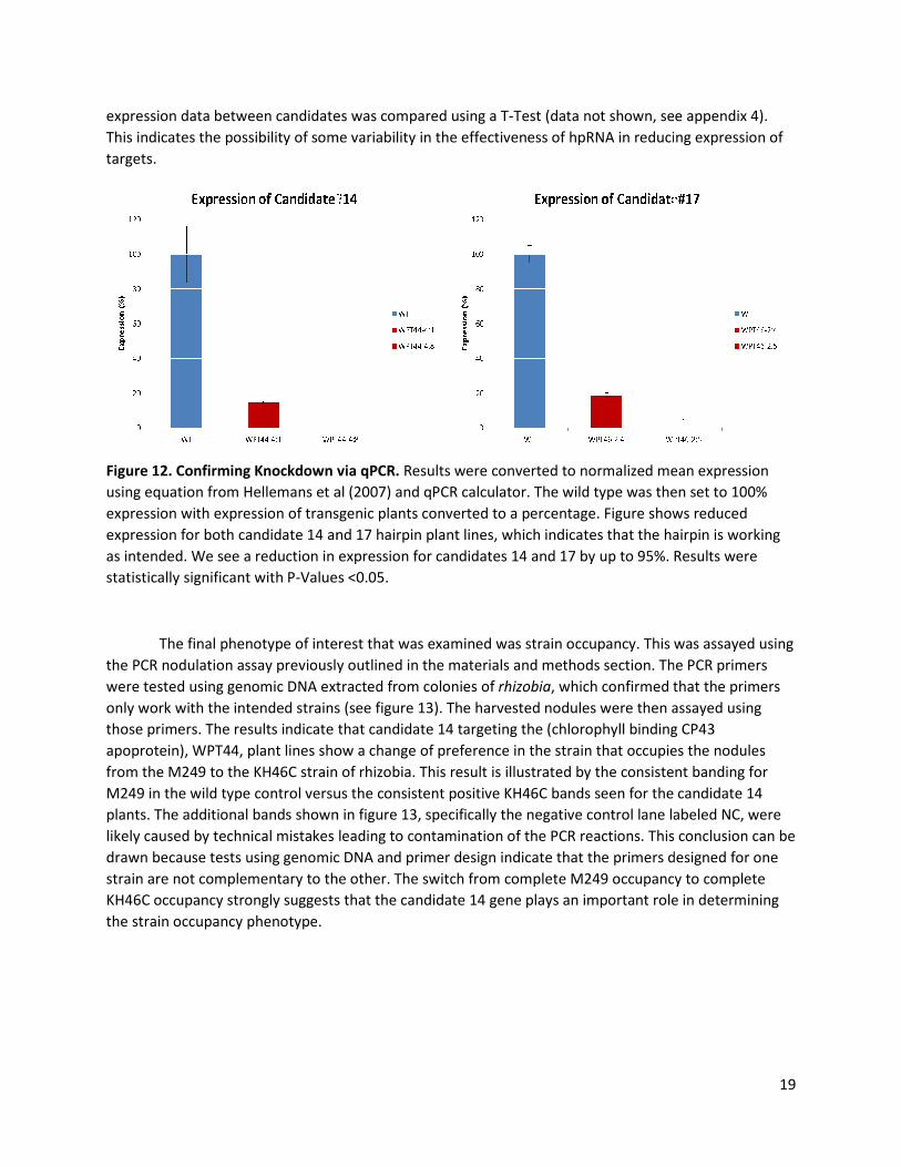
Figure 12. Confirming Knockdown via qPCR.
using equation from Hellemans et al (2007) and qPCR calculator. T
expression with expression of transgenic plants converted to a percentage
expression for both candidate 14 and 17 hairpin pl
as intended. We see a reduction in
statistically significant with P-Values <0.05.
The final phenotype of interest that was examined was
the PCR nodulation assay previously outline
were tested using genomic DNA extracted from colonies of
only work with the intended strains
those primers. The results indicate that
apoprotein), WPT44, plant lines show a change of preference
from the M249 to the KH46C strain of rhizobia
M249 in the wild type control versus the consistent positive KH46C bands seen for the candidate 14
plants. The additional bands shown
likely caused by technical mistakes leading to contamination of the PCR reactions.
drawn because tests using genomic DNA and primer design indicate that the
strain are not complementary to the other.
KH46C occupancy strongly suggests
the strain occupancy phenotype.
expression data between candidates was compared using a T-Test (data not shown, see appendix 4)
This indicates the possibility of some variability in the effectiveness of hpRNA in reducing expression of
via qPCR. Results were converted to normalized mean
using equation from Hellemans et al (2007) and qPCR calculator. The wild type was then
with expression of transgenic plants converted to a percentage. Figure shows reduced
expression for both candidate 14 and 17 hairpin plant lines, which indicates that the hairpin is working
expression for candidates 14 and 17 by up to 95%. Results were
Values <0.05.
The final phenotype of interest that was examined was strain occupancy. This was assayed using
the PCR nodulation assay previously outlined in the materials and methods section. The PCR primers
were tested using genomic DNA extracted from colonies of rhizobia, which confirmed that the primers
he intended strains (see figure 13). The harvested nodules were then assayed using
those primers. The results indicate that candidate 14 targeting the (chlorophyll binding CP43
lines show a change of preference in the strain that occupies the nodules
from the M249 to the KH46C strain of rhizobia. This result is illustrated by the consistent banding for
versus the consistent positive KH46C bands seen for the candidate 14
shown in figure 13, specifically the negative control lane labeled NC,
technical mistakes leading to contamination of the PCR reactions. This conclusion can be
drawn because tests using genomic DNA and primer design indicate that the primers designed for one
complementary to the other. The switch from complete M249 occupancy to complete
strongly suggests that the candidate 14 gene plays an important role in determining
19
(data not shown, see appendix 4).
This indicates the possibility of some variability in the effectiveness of hpRNA in reducing expression of
Results were converted to normalized mean expression
then set to 100%
. Figure shows reduced
hairpin is working
Results were
strain occupancy. This was assayed using
in the materials and methods section. The PCR primers
, which confirmed that the primers
The harvested nodules were then assayed using
candidate 14 targeting the (chlorophyll binding CP43
occupies the nodules
consistent banding for
versus the consistent positive KH46C bands seen for the candidate 14
, specifically the negative control lane labeled NC, were
This conclusion can be
primers designed for one
9 occupancy to complete
that the candidate 14 gene plays an important role in determining

Figure 13. Preliminary Nodulation A
system genes of the rhizobia. The targeted genes were unique for each of the two
used (M249 and KH46C) each of which
Top image shows confirmation that PCR targets only one strain and validity of multiplex usage of primer
sets. Middle shows testing of M249 primer with wild type nodules
KH46C primers with candidate 14 nodules (labeled WPT 44).
When comparing the results of both the
previously published research, there is
study. Previous mutational studies performed on Candidate 14 have shown that CP43 is an integral part
of the photosynthetic pathway. Replacement of the conserved histidine (in particular His40, His105, and
His 119) residues with Glutamine and Tyrosine residues was perform
They then measured the activity of Photosystem II by measuring electron transport and looking at the
fluorescence spectra of chlorophyll. Their study showed that replacing these residues destabilized the
photosystem II complex, partially inhibited the oxidation of Q
utilization of absorbed light for the electron transfer performed by photosystem II (Manna and Vermass,
1997). Another study done by Hwang, et al (2007) showed that Mutation
impaired function of photosystem II. The replacement of this residue impaired the catalytic s
the H20 oxidation complex and reduced its
approximately 85%. However, the photosystem ii complex was st
more than 80% of the concentration of reaction centers when compared to the wild type (Hwang et al,
2007). Other studies have also shown
of trypsin greatly affected the oxygen
All of these studies indicate that CP43 deficiency affects the stability of photosystem II and also
the ability for photosystem II to perform its part i
our study do not indicate any significant change to physical phenotypes being associated to the over
80% reduction in gene expression caused by the hpRNA construct. No statistically significant changes
were detected height, total nodulation, nodulation of the top 5 cm or nodulation of the bottom 5cm.
Potentially explaining this lack of
top/bottom 5cm is a recent study done by Boehm, et al (2012)
research into a new light. The study by Boehm, et al (2012) indicates that
complexes in Synechocystis, a strain of unicellular cyanobacteria that performs photosynthesis, show
some ability for self-repair and is able
Preliminary Nodulation Assay via PCR. Primers were designed that targeted the
system genes of the rhizobia. The targeted genes were unique for each of the two strains of rhizobia
each of which use a different secretion system (Type IV and III respectively)
Top image shows confirmation that PCR targets only one strain and validity of multiplex usage of primer
Middle shows testing of M249 primer with wild type nodules (Labeled WT). Bottom image shows
mers with candidate 14 nodules (labeled WPT 44).
results of both the strain occupancy assay and the phenotyping
there is substantial disagreement with the results found during this
mutational studies performed on Candidate 14 have shown that CP43 is an integral part
of the photosynthetic pathway. Replacement of the conserved histidine (in particular His40, His105, and
His 119) residues with Glutamine and Tyrosine residues was performed by Manna and Vermass (1997).
They then measured the activity of Photosystem II by measuring electron transport and looking at the
fluorescence spectra of chlorophyll. Their study showed that replacing these residues destabilized the
ex, partially inhibited the oxidation of QA, and also decreased the efficiency of
utilization of absorbed light for the electron transfer performed by photosystem II (Manna and Vermass,
1997). Another study done by Hwang, et al (2007) showed that Mutation of Arginine 357 of CP43 also
impaired function of photosystem II. The replacement of this residue impaired the catalytic s
0 oxidation complex and reduced its ability to evolve O2 compared to the wild type by
photosystem ii complex was still able to assemble and contained
more than 80% of the concentration of reaction centers when compared to the wild type (Hwang et al,
also shown that the removal of CP43 using chemical methods such a
of trypsin greatly affected the oxygen-evolving complex of PSII (Bricker and Frankel, 2002).
of these studies indicate that CP43 deficiency affects the stability of photosystem II and also
the ability for photosystem II to perform its part in the photosynthetic pathway. However, the results of
our study do not indicate any significant change to physical phenotypes being associated to the over
80% reduction in gene expression caused by the hpRNA construct. No statistically significant changes
were detected height, total nodulation, nodulation of the top 5 cm or nodulation of the bottom 5cm.
Potentially explaining this lack of change to height, total nodulation, and nodulation of the
recent study done by Boehm, et al (2012). This work places the results of my
into a new light. The study by Boehm, et al (2012) indicates that the CP43-less photosystem II
, a strain of unicellular cyanobacteria that performs photosynthesis, show
is able to retain functionality. In Boehm’s study, CP43 was inactivated
20
. Primers were designed that targeted the secretion
strains of rhizobia
(Type IV and III respectively).
Top image shows confirmation that PCR targets only one strain and validity of multiplex usage of primer
. Bottom image shows
strain occupancy assay and the phenotyping with
with the results found during this
mutational studies performed on Candidate 14 have shown that CP43 is an integral part
of the photosynthetic pathway. Replacement of the conserved histidine (in particular His40, His105, and
ed by Manna and Vermass (1997).
They then measured the activity of Photosystem II by measuring electron transport and looking at the
fluorescence spectra of chlorophyll. Their study showed that replacing these residues destabilized the
, and also decreased the efficiency of
utilization of absorbed light for the electron transfer performed by photosystem II (Manna and Vermass,
of Arginine 357 of CP43 also
impaired function of photosystem II. The replacement of this residue impaired the catalytic s-state of
compared to the wild type by
able to assemble and contained
more than 80% of the concentration of reaction centers when compared to the wild type (Hwang et al,
that the removal of CP43 using chemical methods such as the use
evolving complex of PSII (Bricker and Frankel, 2002).
of these studies indicate that CP43 deficiency affects the stability of photosystem II and also
n the photosynthetic pathway. However, the results of
our study do not indicate any significant change to physical phenotypes being associated to the over
80% reduction in gene expression caused by the hpRNA construct. No statistically significant changes
were detected height, total nodulation, nodulation of the top 5 cm or nodulation of the bottom 5cm.
change to height, total nodulation, and nodulation of the
the results of my
less photosystem II
, a strain of unicellular cyanobacteria that performs photosynthesis, show
was inactivated by

21
transforming bacteria using the psbC gene. The psbC gene codes for CP43 in Synechocystis. This allowed
for the in vivo assembly of the CP43-less RC47 assembly complex whose activity was measured using
protein analysis, low temperature absorption spectroscopy, transient absorbance spectroscopy, and
mass spectrometry. The data shows that RC47 was still able to photoreduce QA , and photosystem II
retained its structural stability. However, similar to what was seen in previous studies PSII was still
incapable of oxidizing water leading to decreased photosynthetic activity (Hwang et al, 2012).
A new study by Hwang, et al (2012) indicates the possibility for organisms to repair their CP43-
less photosystem II complexes (PSII). However, little research has been done in plants to determine if
plants can self-repair PSII when it is deficient in CP43. Most research has instead focused on the
dysfunction of PSII when point mutations are introduced or key amino acids residues are replaced.
Hwang, et al (2012) also knocked out expression of the CP43 gene, whereas our study only reduced the
expression of the gene. This means that the transformed plants used for this phenotyping experiment
likely retained some, though greatly reduced, amounts of CP43 which may have allowed it to retain
enough function to avoid the impairment seen in other mutational experiments. It is also possible that
multiple copies of the CP43 gene exist in the M. truncatula genome allowing retained function in the
case one gene is damaged.
Another possibility for lack of agreement of our data with previous studies is that most studies
are done with knockouts of the CP43 complex consisting of replacement of amino acids and deletions. In
contrast, the research presented in this thesis used an RNAi knockdown platform, which would allow for
some level or limited expression of the candidate gene. This could prevent significant phenotypic
effects by allowing some CP43 to be produced in the transgenic plants; this amount may be enough for
the plant to operate normally. Another possibility is that a different gene may serve a similar purpose,
which means that the hpRNAs would only affect one of multiple copies of a gene. Alternatively, the gene
encoding CP43 might be indirectly associated with the strain occupancy phenotype and it could be a
false positive found by the GWAS performed by Stanton-Geddes et al (2013).
The results obtained by candidate gene 17 showed a similar trend to data of candidate gene 14.
Candidate 17 is a PIF1-Helicase that is ubiquitous among eukaryotes due to the exceptionally wide
variety of functions in DNA repair, recombination, and replication. Because of Helicases and PIF1s are
present in most if not all organisms and have a multitude of functions there is a large body of research
concerning their functions. Research by Moon, et al (2007) indicated that PIF1 is involved in the
regulation of chlorophyll biosynthesis in Arabidopsis. Their research indicated that PIF1 negatively
regulated chlorophyll biosynthesis and seed germination under low light or dark conditions, but that
PIF1 was degraded when light was prevalent. In particular their research looked at the binding of G-box
(CACGTG) sequences by PIF1 and the subsequent activation of transcription of the promoters Porc and
FeCHII both of which are promoters involved with regulating the biosynthesis of chlorophyll in
Arabidopsis (Moon et al, 2007). Further research by Knoll and Puchta (2010) indicated that PIF1-
helicases were involved in maintaining the genome stability and the process of meiotic recombination in
Arabidopsis. They found that the inactivation of PIF1 led to an increase in the rate of DNA
rearrangements, issues with DNA replication were encountered, and that Okazaki fragments were
unable to mature (Knoll and Puchta, 2010). Even more research by Bochman, et al (2010) displayed the
even larger variety of functions that PIF1-like helicases have including affecting the replication of
telomeric, ribosomal, and mitochondrial DNA and also play in important role in maintaining genome
stability in eukaryotes (Bochman et al, 2010). However, there is little research into the role that PIF-1

22
helicases play in infection by organisms outside of work currently being done in viruses where work on
insect nucleopolyhedrovirus indicated that infection of PIF1 and PIF2 deficient genotypes showed
increased viral potency (Clavijo et al, 2009).
Based on the fact that research indicates PIF1-Helicases are involved in chlorophyll biosynthesis, which
is essential for plant growth there should be some phenotypic effect. However, based on the wide
variety of helicase functions it is possible that candidate 17 is not a PIF1 helicase that is essential to the
regulation of this pathway. Instead it may be a gene that is expressed primarily in the roots of M.
truncatula. The lack of statistically significant phenotypic effects found in this study appears to agree
with this conclusion along with the limited body of research that has been done on PIF1 specifically in
plants. However, new Genome Wide Association Study (GWAS) data shows that this candidate still has a
statistically significant correlation to strain occupancy, which indicates that there is perhaps some role
for PIF1 helicase nodulation. However, further research into exact structure and function of candidate
17 would be required to discern its role in nodulation through the phenotype of strain occupancy. This
could be done using NMR and protein isolation methods to look at the structure of the protein.
Genome Wide Association Studies
A new Genome Wide Association Study (GWAS) was recently done using new genome
assemblies, which altered the list of candidate genes that would significantly contribute to the strain
occupancy phenotype. For the occupancy B phenotype (Top 5 cm) candidate genes 14 and 17 are no
longer indicated to be significant contributors, which differs from the previous GWAS performed by
Stanton-Geddes et al. (2013) (see Figure 4). In this Manhattan plot both candidate genes are below the
significance threshold. However, the Manhattan plot for the occupancy A phenotype (bottom 5 cm)
indicates that candidate 14 but not 17 should have a significant contribution towards this phenotype
(see figure 3). The data from the preliminary assay indicates that candidate 14 does have a significant
contribution to the strain occupancy phenotype, which agrees with the preliminary assay performed for
this thesis. This preliminary assay showed a complete switch from M249 to the KH46C strain. Candidate
17 no longer shows a significant contribution to either strain occupancy phenotype, which means that it
is no longer a candidate gene for determining the strain occupancy phenotype. The nodulation assay
was not able to successfully determine whether or not there was a change in strain occupancy for this
experiment, so a nodulation assay should be done to confirm that the new Manhattan plots are not
false negatives which can be seen in GWAS (Wray et al., 2013).

23
Figure 14. GWAS 4.0 Manhattan Plot. New GWAS performed in 2014 indicates that Candidate 14 shows
a significant contribution while candidate 17 does not to the occupancy A phenotype (highlighted in
green).
Figure 15. GWAS 4.0 Manhattan Plaot. New GWAS performed in 2014 indicates that both genes
(highlighted in green) do not significantly contribute to the Occupancy A phenotype, which differs from
previous data found in GWAS performed by Stanton-Geddes et al, 2013\

24
Conclusions
The goal of this study was to determine whether candidate genes 14 and 17 contributed
significantly to the establishment of optimal symbiosis between Medicago truncatula and rhizobia.
Another goal of the study was to determine the effectiveness of the hpRNA construct in reducing gene
expression of candidates 14 and 17. Based on the research of Stanton-Geddes et al (2013), we
hypothesized that knocking down the expression of candidate genes 14 and 17 would lead to significant
changes in the strain occupancy of the top and bottom 5cm in the mutant lines when compared to wild
type plants, but wouldn’t contribute significantly to other phenotypes of interest (height, total
nodulation, nodulation of top/bottom 5cm). During the course of my research we were able to
determine the following:
• hpRNA constructs were able to effectively reduce gene expression of candidates 14 and 17 by
up to 95%. Construct was also confirmed to be present in 25% of transgenic plants using PCR
methods.
• That knockdown of candidates 14 and 17 did not significantly affect the following phenotypes:
height, total nodulation or nodulation of the top/bottom 5 cm.
• That a preliminary assay indicates a significant effect to strain occupancy with a change of strain
preference from the M249 rhizobia strain to the KH46C strain.
The above supported my hypothesis that the hpRNA construct would be effective at knocking
down expression of candidate genes in M. truncatula. Furthermore, these results also supported that
the knockdown of these candidates would not significantly affect any phenotypes of interest aside from
the strain occupancy. This agrees with the data provided by Stanton-Geddes et al (2013).
Future Directions
First and foremost, another nodulation assay needs to be performed based on the preliminary
results of the strain occupancy assay done for this study. This would allow determination of whether the
effect of knockdown causes a significant effect. There are several possible methods that have been used
by other scientists. Stanton-Geddes et al (2013) used an antibody dot blot immunoassay to determine
strain occupancy. Another method would be ERIC-polymerase chain reaction (ERIC-PCR), which was
used effectively by Duodu et al (2008) to determine strain occupancy of nodules. Another possibility of
confounding factors is contamination of the plates used to grow colonies of the strain occupying
nodules or the PBS solution used to suspend the nodules after harvesting. Using more stringent sterile
technique may allow for the standard PCR technique to be effective in determining strain occupancy.
Performing all of these together should allow for the elimination of variables and to successfully assay
the association of candidates 14 and 17 for determining the strain occupancy phenotype. A final
modification for the study would be to use plants that underwent the transformation protocol, but
tested negative for presence of the transgene. This would be done in place of the HM340 plants that
were used in this thesis. This would allow for the introduction of fewer variables by testing plants that
underwent all of the same experimental procedures.
Another option for further study would be to knockout (eliminating gene expression) rather
than knockdown (reducing gene expression) the gene using a different technology. A current prominent
technology used to knockout genes in many different organisms including plants is the clustered
regularly interspaced short palindromic repeat or CRISPR system and the associated Cas9 protein

25
(CRISPR/Cas9) (Ran et al., 2013). The CRISPR/Cas9 system is based on the innate immune system found
in both bacteria and archaea. This system uses a guide RNA, referred to as CRISPR RNAs (crRNAs)
created by integrating short pieces of DNA homologous to the virus or plasmid sequence to eliminate
outside DNA and RNA. To use this system in plants and other organisms to knockout genes the guide
RNA is designed to be complementary to a 20 nucleotide segment of the target DNA (Ran et al., 2013).
Once the complementary segment is bound the Cas9 induces a double strand break leading to non-
homologous end joining (NHEJ) in which endogenous DNA repair machinery joins the broken ends of the
DNA. This can introduce an indel mutation, which causes gene knockout. This technology is currently
used to knockout genes in M. truncatula, mice, monkeys, and several other organisms (Niu et al., 2014,
Platt et al., 2014). To apply this system, we have begun designing and implementing a two gRNA
CRISPR/Cas9 that targets candidates 14 and 17, which would allow us to test a knockout of the two
candidate genes and compare it to results derived from using an RNAi knockdown platform. We have
successfully constructed a CRISPR/Cas9 system targeting the two candidates and are now preparing to
test it using a Hairy Root experiment, which will allow us to assess the systems function prior to
implementing in a whole plant transformation. However, it would be important to keep in mind the
potential of off-target effects that could take place using the CRISPR/Cas9 system and an analysis of
possible off-target effects contributing to any phenotypes seen as a result of an experiment in M.
truncatula would need to be considered (Hsu, et al., 2013).

26
References
Amerongen, Herbert and Roberta Croce. “Light Harvesting in Photosystem II” Photosynth Res 116
(2013): 251-263.
Barker, et al. “Growing M. truncatula: Choice of Substrates and Growth Conditions” Medicago
truncatula handbook. Noble Foundation, 2006.
Bochman, Matthew L., Nasim Sabouri, and Virginia A. Zakian. "Unwinding the Functions of the Pif1
Family Helicases." DNA Repair 9.3 (2010): 237-49.
Boehm et al. "Subunit Composition of CP43-less Photosystem II Complexes of Synechocystis Sp. PCC
6803: Implications for the Assembly and Repair of Photosystem II." Philosophical Transactions of
the Royal Society B: Biological Sciences 367.1608 (2012): 3444-454.
Bricker, Terry M., and Laurie K. Frankel. "The Structure and Function of CP47 and CP43 in Photosystem
II." Photosynthesis Research 72 (2002): 131-46.
Bush, William and Jason Moore. “Genome-Wide Association Studies” Plos Computational Biology 8(12).
Clavijo, et al. “Mixtures of Complete and pif1- and pif2-Deficiient Genotypes Are Required for Increased
Potency of an Insect Nucleopolyhedrovirus.” Journal of Virology 83 (2009): 5127-5136.
Deakin, Willliam and William Broughton. “Symbiotic use of pathogenic strategies: rhizobial protein
secretion systems” Nature Reviews Microbiology 7 (2009): 312-320.
Duodu, Samuel and Caroline Brophy. “Competitiveness of a native Rhizobium Leguminosarum biovar
trifolii strain for nodule occupancy is manifested during infection” Plant Soil 318 (2009): 117-
126.
Fire, Andrew. "RNA-triggered Gene Silencing." Trends in Genetics 15.9 (1999): 358-63.
Flint, Jonathan. "Gwas." Current Biology 23.7 (2013): R265-266. Web.
Fusaro, et al. “RNA interference-inducing hairpin RNAs in plants act through the viral defense pathway”
EMBO reports 7 (2006): 1168-1175.
Gruber, Nicolas and James Galloway. “An Earth-system perspective of the global nitrogen cycle” Nature
451 (2008): 293-296.
Helliwell, Chris A., and Peter M. Waterhouse. "Constructs and Methods for Hairpin RNA-Mediated Gene
Silencing in Plants." Methods in Enzymology 392 (2005): 24-35.
Hwang, et al. “Mutation of Aginine 357 of the CP43 Protein of Photosystem II Severely Impairs the
Catalytic S-State Cycle of the H20 Oxidation Complex” American Chemical Society 46 (2007).
Jones, et al. “How rhizobial symbionts invade plants: the Sinorhizobium-Medicago model” Nature
Reviews 5 (2007): 619-633.

27
Keunhwa, et al. “Phytocromes inhibit hypocotyl negative gravitropism by regulating the development of
endodermal amyloplasts through phytocrhome-interacting factors” PNAS 4 (2011): 1729-1734.
Knoll, Alexander, and Holger Puchta. “The role of DNA helicases and their interaction partners in
genome stability and meiotic recombination in plants.” Journal of Experimental Botany 62
(2011): 1565-1579.
Limpens, et al. “RNA interference in Agrobacterium rhizogenes transformed roots of Arabidopsis and
Medicago truncatula” Journal of Experimental Botany 55 (2004): 983-992.
Manna, Pradip, and Wim Vermaas. "Mutational Studies on Conserved Histidine Residues in the
Chlorophyll-Binding Protein CP43 of Photosystem II." European Journal of Biochemistry 247.2
(1997): 666-72.
Moon, Jennifer, et al. "PIF1 Directly and Indirectly Regulates Chlorophyll Biosynthesis to Optimize the
Greening Process in Arabidopsis." National Center for Biotechnology Information. U.S. National
Library of Medicine, 30 June 2008.
Peoples et al. “Pathways of Nitrogen loss and Their Impacts on Human Health and the Environment”
Agriculture and the Nitrogen Cycle. (2004): 53-70.
Preuss, Sasha and Craig Pikaard. “Targeted Gene Silencing in Plants Using RNA Interference” RNA
Interference (RNAi) Nuts & Bolts of siRNA technology. Washington University, 2003.
Simms, et al. “An empirical test of partner choice mechanisms in a wild legume-rhizobium interaction”
Proceedings of the Royal Society 273 (2006): 77-81.
Stanton-Geddes, et al. "Candidate Genes and Genetic Architecture of Symbiotic and Agronomic Traits
Revealed by Whole-Genome, Sequence-Based Association Genetics in Medicago Truncatula."
Ed. Lewis Lukens. PLOS ONE 8.5 (2013): E65688.
Soy, et al. “PIF1 promotes phytochrome-regulated growth under photoperiodic conditions in
Arabidopsis together with PIF3, PIF4, and PIF5” Journal of Experimental Botany (2013).
Stoutjesdijk, et al. “hpRNA-mediated Targeting of the Arabidposis FAD2 Gene Gives Highly Efficient and
Stable Silencing.” Plant Physiology 129 (2002): 1723-1731.
Tadege, et al. “Mutagenesis and Beyond! Tools for Understanding Legume Biology” Current Opinion in
Plant Biology 12 (2009): 193-201.
Vlassak, et al. “Factors Influencing Nodule Occupancy by Inoculant Rhizobia” Critical Reviews in Plant
Sciences 16 (2010): 163-229.
Wang, Kan. Agrobacterium Protocols. Totowa, NJ: Humana, 2006.
Waterhouse, Peter and Christopher Helliwell. “Exploring Plant Genomes by RNA-Induced Gene
Silencing” Nature Reviews 4 (2003): 29-38.
Wesley, et al. “Construct design for efficient, effective and high-throughput gene silencing in plants” The
Plant Journal 27 (2001): 581-590.

28
Yamamoto, Yasusi. “Quality Control of Photosystem II” Plant Cell Physiology. 42 (2001):121-128.
Young, Nevin and Michael Uvardi. Translating Medicago truncatula genomics to crop legumes. “Current
Opinions in Plant Biology” 12 (2009): 193-201.
Young, et al. “The Medicago Genome Provides Insight into the Evolution of Rhizobial Symbioses” Nature
480 (2012): 520-524.
Zahran, Hussein Hamdi. “Rhizobium-Legume Symbiosis and Nitrogen Fixation under Severe Conditions
and in an Arid Climate. Microbiology and Molecular Biology reviews 63 (1999): 969-989.

29
Acknowledgements
I would like to thank the following people for their assistance with this project. Nevin Young,
Shaun Curtin and Roxanne Denny for not only allowing me to work in the lab, but also for welcoming me
and providing incredible guidance through the heat of summer, cold of winter, and showers of spring.
For teaching me how to deal with failure, the ins and outs of more protocols than I care to count, and
listening to my endless questions. I would like to thank Betsy Martinez-Vaz for being my research
advisor, senior seminar advisor, and for sponsoring this honors thesis. For her endless patience with
multiple drafts including a rather rough first draft and a only somewhat smoother second draft, power
points, posters, emails, and presentations. I would also like to thank her for giving me the opportunity to
succeed and pushing me forward towards success despite my stubborn refusal to walk towards it at
times. I would like to thank Mariya Khan for her continued support and listening to my nattering about
plant genetics, correcting my wordy sentences, and listening to my many complaints about failing
experiments. I would also like to thank the other members of my defense committee, Irina Makarevitch
and Susie Steinbach, for their participation in this process. I would like to thank Zoe Lautz for listening to
my ramble endlessly about ΔΔCt and other qPCR data analysis equations and Charlene for providing me
with motivational messages for me to wake up to every day. Finally, I would like to thank the NSF for
funding this project and for keeping me fed and watered during the summer.

Appendix
Appendix 1. Phylogenic trees for Candidates 14 (ic trees for Candidates 14 (left) and 17 (right)
30

31
Appendix 3. Raw and Treated Phenotype data from harvest.
Plant Height (cm) Nod Top 5cm Nod Btm 5cm Total nod
HM340 5.5 5 1 8
HM340 5.0 5 0 9
HM340 6.5 5 1 6
HM340 5.6 8 1 9
HM340 5.0 4 1 5
HM340 5.5 6 0 6
HM340 4.0 1 0 3
HM340 6.1 5 0 5
HM340 4.8 4 1 6
Average 5.33 4.78 0.556 6.33
Std. Dev. 0.696 1.75 0.497 1.89
46-2-1 4.1 2 0 2
46-2-2 6 4 0 4
46-2-3 5.5 2 0 2
46-2-4 5.8 4 0 4
46-2-5 6.8 4 0 4
46-2-6 5.4 3 0 5
46-2-7 5.6 3 0 3
46-2-8 5.7 3 0 3
46-2-9 6.4 4 0 4
46-2-10 6.5 7 0 7
46-2-11 5.8 5 0 5
46-2a-1 7 5 0 5
46-2a-2 6.9 5 0 5
46-2a-3 7.1 4 2 6
46-2a-4 6.8 3 3 6
46-2a-5 5.8 3 0 4
46-2a-6 5.7 7 0 7
46-2a-7 7.3 5 0 5
46-2a-8 7.4 6 0 6
46-2a-9 7.3 4 4 8
46-2a-10 5.7 3 0 3
46-2a-11 6.3 4 0 4
46-2a-12 5.9 4 0 4
Average 6.21 4.09 0.391 4.61
Std. Dev. 0.778 1.32 1.05 1.52
44-1-1 5.8 4 0 4
44-1-2 6.3 5 0 5
44-1-3 5.5 5 0 5
44-1-4 4.6 3 0 3
44-1-5 5.9 5 0 5
44-1-6 6.6 5 1 6
44-1-7 5.5 3 0 3
44-1-8 6 4 0 4
44-1-9 6.3 6 0 6
44-1-10 7 6 0 6
44-1-11 5.6 6 1 7
44-1-12 5.7 4 0 4
44-1-13 6.4 6 0 6
44-1-14 5.8 4 0 4
44-1-15 6.6 5 0 5
44-1-16 6.3 5 0 5
44-1-17 6.5 4 1 5
44-1-18 7.1 6 0 6
Average 6.08 4.78 0.167 4.94
Std. Dev. 0.591 0.975 0.373 1.08
44-4-1 3.3 3 0 3
44-4-2 6.7 3 0 3
44-4-3 6.5 4 0 4
44-4-4 5.3 2 0 2
44-4-5 5.2 3 0 3
44-4-6 5.9 3 1 4
44-4-7 5.8 2 2 4
44-4-8 5.1 3 0 3
44-4-9 6.1 3 0 3
44-4-10 6.5 6 0 6
44-4-11 7.7 5 0 5
44-4-12 6.9 6 0 6
44-4-13 5.7 3 0 3
44-4-14 6.4 4 0 4
44-4-15 7.6 4 1 5
44-4-16 5.5 3 0 3
44-4-17 5.7 2 0 2
44-4-18 6.8 4 0 4
44-4-19 6.9 5 0 5
44-4-20 5.8 3 0 3
Average 6.07 3.55 0.200 3.75
Std. Dev. 0.96 1.16 0.510 1.13

32
Appendix 4. Normalized qPCR data
Appendix 5. Normalized qPCR data and transformation to percentage expression
Appendix 6. Normalized qPCR data T-Test and corresponding P-Values derived from T-Test Tables.
Name Mean NormalizedSE of Mean SE %
14_WPT44_4_1 0.000149 7.39E-06 4.960706
14_WPT44_4_8 0.000196 1.72E-05 8.781246
14_WPT46_2_4 0.000272 1.27E-05 4.654147
14_WPT46_2_5 0.001016 0.000164 16.17918
17_WPT44_4_1 0.042466 0.002128 5.010148
17_WPT44_4_8 0.025041 0.003319 13.25257
17_WPT46_2_4 0.007855 0.000742 9.448466
17_WPT46_2_5 0.001842 0.000318 17.28922
Name Mean Normalized SE of Mean
WT 0.001016 0.000164
WPT44-4:1 0.000149 7.39E-06
WPT44-4:8 0.000196 1.72E-05
WT 100 16.1417323
WPT44-4:1 14.66535433 0.7273622
WPT44-4:8 19.29133858 1.69291339
Name Mean Normalized SE of Mean
WT 0.042466 0.002128
17_WPT46_2_4 0.007855 0.000742
17_WPT46_2_5 0.001842 0.000318
WT 100 5.01106768
WPT46-2:4 18.49715066 1.74728018
WPT46-2:5 4.337587717 0.74883436
Comparison T-Test Values P-value Significant?
WPT44:4-1 vs WT 5.287 0.00617 Significant
WPT44:4-8 vs WT 5.000 0.00749 Significant
WPT 44:4-1 vs 8 6.360 0.00313 Significant
WPT46:2-5 vs WT 19.089 4.40E-05 Significant
WPT46:2-4 vs WT 16.260 8.4E-05 Significant
WPT46:2-4 vs 5 8.103 0.001261 Significant


![Nuclear Pif1 is Post Translationally Modified and ... · 7/6/2020 · 18]. Given Pif1’s involvement in a plethora of cellular activities, it still remains a mystery how its numerous](https://img.pdfslide.us/doc/110x75/5f3af30a4137a17a927c8342/nuclear-pif1-is-post-translationally-modified-and-762020-18-given-pif1as.jpg)