Embed Size (px)
Citation preview
Characterization of CopperNanoparticles Synthesized by aNovel Microbiological MethodRatnika Varshney, Seema Bhadauria, M.S. Gaur, and Renu Pasricha
The exploitation ofvarious biomaterials for the biosynthesis ofnanoparticlesis considered as green technology as itdoes not involve any harmful chemicals.The present study reports the synthesisof copper nanoparticles which involvesnon-pathogenic bacterial strain Pseudomonas stutzeri, isolated from soil.These copper nanoparticles are furthercharacterized for size and shape distributions by ultraviolet-visible spectroscopy, x-ray diffraction, and high resolution transmission electron microscopytechniques. The results showed that theparticles are spherical and quite stablein nature and shows surface plasmonresonance clearly featured in the optical spectra in visible region.
INTRODUCTION
Low-dimensional nanoscale materials are currently of great interest due totheir unique electronic, optical, and mechanical properties. '-4 Among the coinage metals, Ag and Au have been studied the most because of their stabilityand their intense absorption band in thevisible region, often called surface plasmon absorption. Cu, in turn, is the leaststudied out of the coinage metals dueto its high instability for oxidation, andmost of the time it forms complexes withwater molecules in aqueous media.5,6
Particularly in transition metal oxides,Cu-based nanoparticles and nanowireshave been synthesized by a variety ofmethods, and because of a reductionin dimensionality, their ferromagneticpolarizations are quite different fromthose observed in transition metals.7
-9
Cu nanoparticles were widely used asalternative catalysts,IO such as selectivehydrogenation and methanol synthesisreactions, which make them suitablefor application in the field of catalysis.Nano-sized Cu particles are widely used
in fields of lubricants, polymers/plastic,metallic coating and ink. Cu sulfide haspotential applications in solar cells, IRdetectors and lubrication. II
There are several reports availableof physical and chemical synthesisof Cu and Cu based nanomaterials.Chuncheng Hao et al. have reported thepreparation of Cu nanoparticles encapsulated in graphitic carbon shells usingmodified arc plasma method. 12 Surfactant-assisted electrochemical proce-
How would you......describe the overall significanceof this paper?
This paper reports a bacterialstrain for the synthesis ofcoppernanoparticles. This is a novelmethod as it doesn't involve anyharmful and environmentallytoxic chemicals used previously inconventional chemical reductionmethods for the preparation ofcopper nanoparticles.
...describe this work to amaterials science and engineeringprofessional with no experience inyour technical specialty?
Copper nanoparticles synthesizedwith this method are spherical andwell dispersed. They have organiccoating on the suiface which isreducing and not only acts ascapping agent but also prevents themfrom oxidation, which seems to bea major problem with the previousmethods involved in the coppernanoparticles synthesis.
...describe this work to alayperson?
This method reports use ofa nonpathogenic bacterial strain andspherical nanoparticles ofcopperare synthesized which are very usefulin the field ofcatalysis. It is a simple,rapid, cost efficient and greenmethod which is free from usingany toxic reducing chemicals. This
t.:: provides a platform for new metallicnanoparticles synthesis.
dures have been shown to be a powerfultool for preparing stable nanoparticlescomposed of a wide range of metalsand compounds. 13-
18 The most variedand versatile preparation route appearsto be the so-called "sacrificial anode"electrolysis that is usually carried outin the presence of cationic surfactantssuch as tetra-alkyl-ammonium salts. Ina seminal paper based on the combineduse of transmission electron and scanning tunneling microscopies, the groupof M. Reetz demonstrated that thesemetal nanoparticles possess a core-shellstructure in which the metallic core isstabilized by a monolayer of tetra alkylammonium species, and the thicknessof this shell is linearly correlated withthe length of the alkyl chains. 19 In thelast decade, the electrolytic productionof nanoparticles and nanocompositescontaining platinum group elementshas been extensively studied, due tothe strong catalytic properties of theseproducts. 13,zQ-25 The electro-synthesis ofCu and Ag colloids, on the other hand,has been the subject of only a few reports. 14,26-28 But no reports are available on the biological synthesis of Cunanomaterials. It is, therefore, important to develop synthetic strategies thatare simple, cost-effective, environmentfriendly, easily scalable and at the sametime with parameters to control size andshape of the materials. Hence the development of a novel method for the preparation of Cu nanoparticles is inevitable.With these ideas in mind, an attempt hasbeen made in this investigation to synthesize Cu nanoparticles by a novel biological method using non-pathogenicbacterial strain Pseudomonas stutzeriisolated from soil and characterize themfor their properties by methods of absorption optical spectroscopy and transmission electron microscopy (HRTEM).
EXPERIMENTAL PROCEDURESBacterial strains were enumerated from soil around the sewage outfall of the small
scale electroplating industry from Dhakran, Agra by Serial dilution-agar plating method(Nigam 1965). 1 x 1O-3 dm3 inoculum was transferred into 50 x 1O-3 dm3 medium containing (gil) Peptone, 5; Beef extract, 3; Sodium chloride, 5; Agar, 15; pH 7.2 in Erlenmeyerflasks. Cells were grown at 37°C for 24 h and then harvested by centrifugation (8,000 rmin.-', 10 min. at room temperature). The cell pellet was resuspended and centrifugedthree times in deionized water. Copper sulfate was purchased from Hi-media, and wasused as received. In a typical synthesis for nanoparticles using Pseudomonas stutzeri, thecarefully weighted 0.1 g biomass was added to 100 ml of 1 mM aqueous CuS0
4solution,
in conical flasks of 250 ml content. The flasks were thereafter incubated in incubatorshaker at 150 rpm at room temperature.
To confirm the synthesis of nanoparticles, characterization was done by UV-Vis spectroscopy, x-ray diffraction, high-resolution transmission electron microscopy and selected area electron diffraction techniques. The bioreduction of Copper ions in aqueoussolution was monitored by periodic sampling of aliquots (0.2 ml) of the suspension, thendiluting the samples with 2 ml deionized water and subsequently measuring ultra violet visible (UV-vis) spectra of the resulting diluents. UV-vis spectroscopy analyses ofcopper nanoparticles produced were carried out on ELICO UV spectrophotometers ata resolution of 1 nm. X-ray diffraction measurements of the bioreduced solution, dropcoated onto glass substrate, were done by an X'Pert Pro x-ray diffractometer instrumentoperating at a voltage of 45 kV and a current of 40 rnA with Cu Ka radiation. Samples forHR-TEM analysis were prepared on carbon- coated copper grids. The films on the coppergrids were allowed to stand for 2 min following which the extra solution was removedusing a blotting paper and the grid was allowed to dry prior to measurement and analysiswas carried out on Tecnai G2 F30 S-Twin (FEI; Super Twin lens with Cs =1.2 mm) instrument operated at an accelerating voltage at 300 kV (field emission gun), having a pointresolution of 0.2 nm and lattice resolution of 0.14 nm.
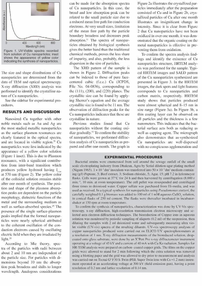
o 400 600 800Wavelength (nm)
Figure 1. UV-Visible spectra recordedfrom solution of eu nanoparticles; insetshows the appearance of yellow colorindicating the synthesis of nanoparticles.
The size and shape distributions of Cunanoparticles are determined from thedata of TEM and optical spectroscopy.X-ray diffraction (XRD) analysis wasperformed to identify the crystalline nature of Cu nanoparticles.
See the sidebar for experimental procedures.
RESULTS AND DISCUSSION
Nanosized Cu together with othernoble metals such as Au and Ag arethe most studied metallic nanoparticlesas the surface plasmon resonances areclearly featured in the optical spectra,and are located in visible region.29 Cunanoparticles were first indicated by theappearance of a yellow color solution(Figure 1 inset). This is due to Plasmonresonance, with a significant contribution from inter-band transition whichproduces yellow hydrosol having I
max
at 570 nm (Figure 2). The yellow colorof the colloidal solution remains stableafter one month of synthesis. The position and shape of the plasmon absorption peaks are dependent on the particlemorphology, dielectric functions of themetal and the surrounding medium aswell as surface-absorbed species.29 Thepresence of the single surface-plasmonpeaks implied that the formed nanoparticles were nearly spherical attributedto the coherent oscillation of the conduction electrons caused by oscillatingelectric field when they are irradiated bylight.30
According to Mie theory, spectra of the particles with radii betweenabout 2 and 10 nm are independent ofthe particle size. For particles with dimensions beyond 10 nm the absorption peak broadens and shifts to longerwavelength. Analogous considerations
can be made for the absorption spectraof Cu nanoparticles. In this case, thebroad and low absorption peak can berelated to the small particle size due toa reduced mean free path for conductionelectrons. At very small sizes, limitationof the mean free path by the particleboundary broadens and decreases peakabsorption.3' The spectra of nanoparticles obtained by biological synthesisgives the better band than the traditionalchemical methods, proves the less shareof impurity, and also, probably, the lessdispersion in the size of particles.
The XRD pattern of the sample isshown in Figure 2. Diffraction peakscan be indexed to those of pure facecentered cubic (f.c.c.) Cu (JCPDS,File No. 04-0836), corresponding tothe (111), (200), and (220) planes. Thecrystallite size can be found by applying Sherrer's equation and the averagecrystallite size is found to be 11 nm. Theobservation of diffraction peaks for theCu nanoparticles indicates that these arecrystalline in nature.
Previous studies found that Cunanoparticles without the coating oxidize gradually.J2 To confirm the stabilityof nanoparticles, we performed diffraction analysis of Cu nanoparticles as prepared and after one month. The graph in
Figure 2a illustrates the crystallized particles immediately after the preparationconsisted of Cu and in Figure 2b, crystallized particles of Cu after one monthillustrates an insignificant change inintensity. Since it is clear from Figure2 that Cu nanoparticles have not beenoxidized in over one month, it was demonstrated that the organic coating on themetal nanoparticles is effective in preventing them from oxidation.
To confirm the spectra analysis findings and identify the existence of Cunanoparticles structure, HRTEM analysis was performed for the sample. Typical HRTEM images and SAED patternof the Cu nanoparticles synthesized arepresented in Figure 3. In the HRTEMimages, the dark spots and light featurescorresponds to Cu nanoparticles andcarbon matrix, respectively. HRTEMstudy shows that particles producedwere almost spherical and 8-15 nm insize range (Figure 3a). In Figure 3b, athin coating layer can be observed onall particles and the thickness is a fewnanometers. This indicates that the bacterial surface acts both as reducing aswell as capping agent. The micrographalso demonstrates that as-synthesizedCu nanoparticles are well-dispersedwith no conspicuous agglomeration and
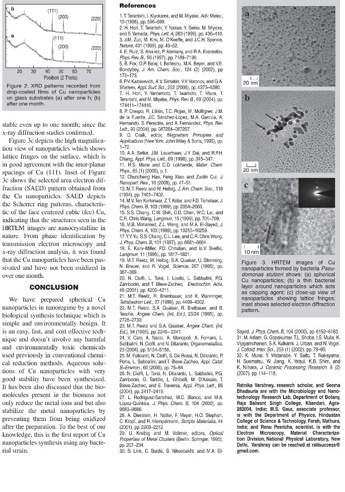
(111)J (200) (220)
-L--J(111)
(200) (220)_L J20 30 40 50 60 70
Position (2 Theta)
Figure 2. XRD patterns recorded fromdrop-coated films of Cu nanoparticleson glass substrates (a) after one h; (b)after one month.
stable even up to one month; since thex-ray diffraction studies confirmed.
Figure 3c depicts the high magnification view of nanoparticles which showslattice fringes on the surface, which isin good agreement with the inter-planarspacings of Cu (lll). Inset of Figure3c shows the selected area electron diffraction (SAED) pattern obtained fromthe Cu nanoparticles. SAED depictsthe Scherrer ring patterns, characteristic of the face centered cubic (fcc) Cu,indicating that the structures seen in theHRTEM images are nanocrystalline innature. From phase identification bytransmission electron mjcroscopy andx-ray diffraction analysis, it was foundthat the Cu nanoparticles have been passivated and have not been oxidized inover one month.
CONCLUSION
We have prepared spherical Cunanoparticles in nanoregime by a novelbiological synthesis technique which issimple and environmentally benign. Itis an easy, fast, and cost effective technique and doesn't involve any harmfuland environmentally toxic chemicalsused previously in conventional chemical reduction methods. Aqueous solutions of Cu nanoparticles with verygood stability have been synthesized.It has been also discussed that the biomolecules present in the biomass notonly reduce the metal ions and but alsostabilize the metal nanoparticles bypreventing them from being oxidizedafter the preparation. To the best of ourknowledge, this is the first report of Cunanoparticles synthesis using any bacterial strain.
References
1. TTeranishi, I. Kiyokawa, and M. Miyake, Adv. Mater.,10 (1998), pp. 596-599.2. H. Hori, T Teranishi, Y. Nakae, Y. Seino, M. Miyake,and S. Yamada, Phys. Lett. A, 263 (1999), pp. 406-41 O.3. J.M. Zuo, M. Kim, M. O'Keeffe, and J.C.H. Spence,Nature,401 (1999), pp.49-52.4. E. Ruiz, S. Alvarez, PAlemany, and R.A. Evarestov,Phys. Rev. B., 56 (1997), pp. 7189-7196.5. B. Fox, O.P. Balaj, I. Balteanu, M.K. Beyer, and VE.Bondybey, J. Am. Chem. Soc., 124 (2) (2002), pp172-173.6. PV Kazakevich, AV Simakin, VV Voronov, and G.A.Shafeev, Appl. Surf. Sci., 252 (2006), pp.4373-4380.7. H. Hori, Y. Yamamoto, T Iwamoto, T Miura, TTeranishi, and M. Miyake, Phys. Rev. B., 69 (2004), pp.174411-174416.8. P Crespo, R. Litran, TC. Rojas, M. Multigner, J.M.de la Fuente, J.C. Sanchez-Lopez, MA Garctia, A.Hernando, S. Penades, and A. Fernandez, Phys. Rev.Lett., 93 (2004), pp.087204-087207.9. D. Craik, editor, Magnetism Principles andApplications (New York: John Wiley & Sons, 1995), pp.1-72.10. A.A. Setlur, J.M. Lauerhaas, J.Y. Dai, and R.PH.Chang, Appl. Phys. Lett., 69 (1996), pp. 345-347.11. R.S. Mane and C.D. Lokhande, Mater. Chem.Phys., 65 (1) (2000), p. 1.12. Chuncheng Hao, Feng Xiao, and Zuolin Cui, J.Nanopart. Res., 10 (2008), pp. 47-51.13. M.T Reetz and W Helbig, J. Am. Chem. Soc., 116(1994), pp. 7401-7402.14. M.V. Ten Kortenaar, Z.T Kolar, and F.D. Tichelaar, J.Phys. Chem. B, 103 (1999), pp.2054-2060.15. S.S. Chang, C.W Shih, C.D. Chen, WC. Lai, andC.R. Chris Wang, Langmuir, 15 (1999), pp.701-709.16. M.B. Mohamed, Z.L. Wang, and MA EI-Sayed, J.Phys. Chem. A, 103 (1999), pp. 10255-10259.17. Y.Y. Yu, S.S. Chang, C.L. Lee, and C.R. Chris Wang,J. Phys. Chem. B, 101 (1997), pp.6661-6664.18. E. Kariv-Miller, PD. Christian, and IcV Svetlic,Langmuir, 11 (1995), pp. 1817-1821.19. M.T Reetz, W Helbig, SA Quaiser, U. Stimming,N. Breuer, and R. Vogel, Science, 267 (1995), pp.367-369.20. N. Cioffi, L. Torsi, I. Losito, L. Sabbatini, PG.Zambonin, and T Bleve-Zacheo, Electrochim. Acta,46 (2001), pp. 4205-4211.21. M.T Reetz, R. Breinbauer, and K. Wanninger,Tetrahedron Lett., 37 (1996), pp. 4499-4502.22. M.T Reetz, SA Quaiser, R. Breibauer, and B.Tesche, Angew Chem. (Int. Ed.), 23/24 (1995), pp.2728-2730.23. M.T Reetz and SA Quaiser, Angew Chern. (Int.Ed.), 34 (1995), pp. 2240- 2241.24. V Calo, A. Nacci, A. Monopoli, A. Fornaro, L.Sabbatini, N. Cioffi, and N. Ditaranto, Organometaffics,23 (2004), pp. 5154-515825. M. Faticanti, N. Cioffi, S. De Rossi, N. Ditaranto, PPorta, L. Sabbatini, and T Bleve-Zacheo, Appl. Catal.B-Environ., 60 (2005), pp.75-84.26. N. Cioffi, L. Torsi, N. Ditaranto, L. Sabbatini, PG.Zambonin, G. Tantillo, L. Ghibelli, M. D'Alessio, TBleve-Zacheo, and E. Traversa, Appl. Phys. Lett., 85(2004), pp. 2417-241927. L. Rodriguez-Sanchez, M.C. Blanco, and M.A.Lopez-Quintela, J. Phys. Chem. B, 104 (2000), pp.9683-9688.28. A. Dierstein, H. Natter, F. Meyer, H.O. Stephan,C. Kropf, and R. Hempelmann, Scripta Materialia, 44(2001), pp. 2209-2212.29. U. Kreibig and M. Vollmer, editors, OpticalProperties of Metal Clusters (Berlin: Springer, 1995),pp.207-234.30. S. Link, C. Burda, B. Nikoobakht, and M.A. EI-
Figure 3. HRTEM images of Cunanoparticles formed by bacteria Pseudomonas stutzeri shows: (a) sphericalCu nanoparticles; (b) a thin bacteriallayer around nanoparticles which actsas capping agent; (c) close-up view ofnanoparticles showing lattice fringes;inset shows selected electron diffractionpattern.
Sayed, J. Phys. Chem. B, 104 (2000), pp. 6152-6163.31. M. Aslam, G. Gopakumar, TL. Shoba, I.S. Mulla, K.Vijayamohanan, S.K. Kulkarni, J. Urban, and WVogel,J. Colloid. Inter. Sci., 255 (1) (2002), pp.79-90.32. K. Murai, Y. Watanabe, Y. Saito, T Nakayama,H. Suematsu, W Jiang, K. Yatsui, K.B. Shim, andK. Niihara, J. Ceramic Processing Research, 8 (2)(2007), pp. 114-118.
Ratnika Varshney, research scholar, and SeemaBhadauria are with the Microbiology and Nanotechnology Research Lab, Department of Botany,Raja Balwant Singh College, Khandari, Agra282004, India; M.S. Gaur, associate professor,is with the Department of Physics, HindustanCollege of Science & Technology, Farah, Mathura,India; and Renu Pasricha, scientist, is with theElectron Microscopy, Material Characterization Division, National Physical Laboratory, NewDelhi. Varshney can be reached at [email protected].