Embed Size (px)
Citation preview
DOI: http://dx.doi.org/10.1590/1516-1439.336714Materials Research. 2015; 18(2): 382-389 © 2015
*e-mail: [email protected]
1. IntroductionAn ideal endodontic repairing biomaterial would adhere
to the tooth structure, maintain suitable sealing, be insoluble in tissue fluids, be dimensionally stable, non-resorbable, radiopaque and show biocompatibility1.
Calcium aluminate-based cement (CAC) has already been studied for biomedical applications as endodontics2-6. This material presented good physical and mechanical properties7, biocompatibility8,9 and acted as a chemical and mechanical barrier against bacterial microleakage4. This sort of cement has also shown an absence of inflammatory reaction and it was biocompatible when tested on rat subcutaneous tissue2. In the presence of radiopacifier (bismuth oxide), it has also presented suitable radiopacity for all studied thicknesses, as recommended by ISO 687610.
Another important desired property is bioactivity of endodontic materials, which has been defined as “the bond ability with the host bone tissue”. In order to avoid costly experiments, various in vitro tests have been used to predict the in vivo bone bioactivity of bioceramics. One common method that has been used for testing in vitro bioactivity is to evaluate the apatite-formation ability of bioceramics in the simulated body fluids (SBF)11,12. The examination of apatite formation on the surface of a material in SBF is useful
for predicting the in vivo bone bioactivity of the material. Besides that it can reduce both, the number of animals used and the time length of the experiments, speeding up the development of new types of bioactive materials11. For a full evaluation of the in vitro bioactivity it is recommend the combination of SBF and cell experiment methods12,13. The ability to stimulate a cell response is very important and complements the in vitro apatite deposition data.
Recent studies involving the use of CAC are based on commercial products comprising a mixture of different crystalline phases2-7. In vitro characterization of commercial CAC has been carried out in simulated body fluid solutions (SBF) prepared according to Kokubo’s (KSBF) or Rigo’s (RSBF) techniques. CAC presented ability of stimulating hydroxyapatite deposition in SBF, although the solution composition defines the sort of phases precipitated. The optimal condition for the calcium phosphate phase precipitation was attained in RSBF and a surface layer of stoichiometric hydroxyapatite could be detected by XRD14.
Improvements can be achieved by investigating the CAC production routes aiming at the proper balance between the phases and the control of impurities that may affect its performance for biomedical applications. The production of CAC phases was previously studied by the authors using Al2O3-CaCO3 or Al2O3-CaO as initial raw materials. The
Characterization of Calcium Aluminate Cement Phases when in Contact with Simulated Body Fluid
Ivone Regina de Oliveiraa*, Talita Luana de Andradea, Renata Martins Parreiraa,
Marcos Jacobovitzb, Victor Carlos Pandolfellic
aInstituto de Pesquisa e Desenvolvimento – IP&D, Universidade do Vale do Paraíba – Univap, Av. Shishima Hifumi, 2911, São José dos Campos, SP, Brazil
bConsultório Dr. Marcos Jacobovitz, Rua Maestro João Sepe, 900, sala 164, Edifício Medical Center- Jardim Paraíso, São Carlos, SP, Brazil
cDepartamento de Engenharia de Materiais – Dema, Universidade Federal de São Carlos – UFSCar, Rod. Washington Luiz, Km 235, São Carlos, SP, Brazil
Received: October 6, 2014; Revised: March 25, 2015
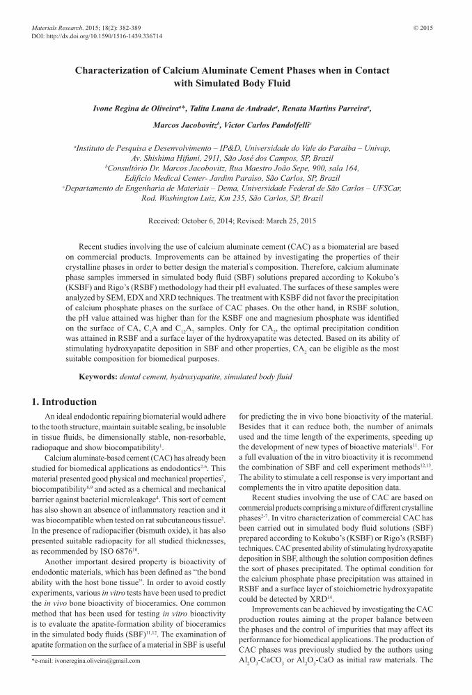
Recent studies involving the use of calcium aluminate cement (CAC) as a biomaterial are based on commercial products. Improvements can be attained by investigating the properties of their crystalline phases in order to better design the material’s composition. Therefore, calcium aluminate phase samples immersed in simulated body fluid (SBF) solutions prepared according to Kokubo’s (KSBF) and Rigo’s (RSBF) methodology had their pH evaluated. The surfaces of these samples were analyzed by SEM, EDX and XRD techniques. The treatment with KSBF did not favor the precipitation of calcium phosphate phases on the surface of CAC phases. On the other hand, in RSBF solution, the pH value attained was higher than for the KSBF one and magnesium phosphate was identified on the surface of CA, C3A and C12A7 samples. Only for CA2, the optimal precipitation condition was attained in RSBF and a surface layer of the hydroxyapatite was detected. Based on its ability of stimulating hydroxyapatite deposition in SBF and other properties, CA2 can be eligible as the most suitable composition for biomedical purposes.
Keywords: dental cement, hydroxyapatite, simulated body fluid

Characterization of Calcium Aluminate Cement Phases when in Contact with Simulated Body Fluid2015; 18(2) 383
Al2O3-CaO route enabled the production of the target phases (CA, CA2, C3A and C12A7) with higher purity compared to the Al2O3-CaCO3 one in which C stands for CaO and A for Al2O3
15.Therefore, in this work in vitro characterization of
calcium aluminate cement phases (CA, CA2, C3A and C12A7) was evaluated when in contact with simulated body fluid solutions. This can highlight the importance of each particular crystalline phase comprising the CAC containing product and provide better tools and insights in terms of designing its composition, application and performance.
2. Experimental ProceduresCalcium aluminate cement phases were previously
synthesized by the CaO and Al2O3 calcination using electrical furnace at different temperatures and dwell time of one hour15.
The reactions involved to produce CA, CA2, C3A and C12A7 phases as listed below:
CaO + Al2O3 → CaAl2O4 (CA) (1)
CaO + 2Al2O3 → CaAl4O7 (CA2) (2)
3CaO + Al2O3 → Ca3Al2O6 (C3A) (3)
12CaO + 7Al2O3 → Ca12Al14O33 (C12A7) (4)
The raw material amounts used and the firing conditions were adjusted in order to produce different CAC phases as presented in Table 1. Following that, the resulting phases
were dry ball milled for 17 hours using high alumina spheres with a 5:1 ratio (ball: powder)15.
Aqueous suspensions of CAC phases were prepared using a standard laboratory mixer under a 2000 rpm. Deionized water was added to these powders to obtain a homogeneous paste. The solid amount in the suspension is presented in Table 1. C3A and C12A7 suspensions were prepared with higher water content. The affinity of CaO with water and the high CaO/Al2O3 ratio in these phases led to heat evolution when in contact with water. Thus, due to water evaporation, it was necessary to increase the content of water added.
The suspensions were used to prepare cylindrical samples (10mm diameter x 5mm height). After shaping, the samples were kept at 37±1 °C in a climatic chamber under a saturate environment (100% R. H.) for 24 hours. After that, the samples were withdrawn out of the molds and kept for 7 days at 37 °C under the same environmental conditions described above. After finishing this step, they were placed into containers with 50 ml of water or SBF solutions and kept at 37 °C (100% R. H.). The pH of these solutions was measured at certain time intervals, using pH electrode connected to an automatic data recorder system.
The SBF solutions named KSBF and RSBF were prepared according to the procedure described in the literature11,16, as shown in Table 2.
The 7-day-set samples were also placed into containers with 47 mL of SBF solutions in order to keep a 0.1cm–1 surface area-to-volume ratio and under stirring at 37 °C with the help of a shaker. Afterwards the samples were gently rinsed with deionized water, followed by drying at room temperature according to the literature17. The surface of the samples was
Table 1. Processing conditions for the production of calcium aluminate cement phases based on Al2O3-CaO mixture and the respective solid content in the suspension.
CAC phase Materials (wt%) Temperature (oC) Solids in the suspension (wt%)
CA Al2O3:CaO (65:35) 1300 63CA2 Al2O3:CaO (78:22) 1450 67C3A Al2O3:CaO (38:62) 1300 59
C12A7 Al2O3:CaO (51:49) 1300 50
Table 2. Reagent content for preparing the simulated body fluid (SBF) solutions (1L) according to the literature11,16.
Reagent Order SBF 1.5 Kokubo (KSBF) Order SBF 1.5 Rigo (RSBF)Water #0 750 ml #0 400 mlNaCl #1 11.994 g #1 11.992 g
NaHCO3 #2 0.525 g #4 0.529 gKCl #3 0.336 g #2 0.335 g
K2HPO4 #4 0.288 g #3 0.261 gMgCl2.6H2O #5 0.458 g #8 0.458 g
HCl 1M #6 60 mlHCl 0.1M #6 15 ml
CaCl2.2H2O #7 0.507 g #7 0.551 gNa2SO4 #8 0.107 g #5 0.107 g
(CH2OH)3CNH2 #9 9.086 g(CH2OH)3CNH2
0.05M#9 Suitable amount for adjusting pH 7.25
HCl 1M #10 Suitable amount for adjusting pH 7.25HCl 0.1M #10 Suitable amount for adjusting pH 7.25
Oliveira et al. 384 Materials Research
analyzed by scanning electron microscopy (SEM) and energy dispersive X-ray analysis (EDX) before and after treatment in SBF solutions. The samples before and after immersion in SBF were also analyzed by X-ray diffraction (XRD) using CuKα radiation (1.54439 Å) in order to identify the phases formed on the sample surface.
3. Results and Discussion3.1. pH tests
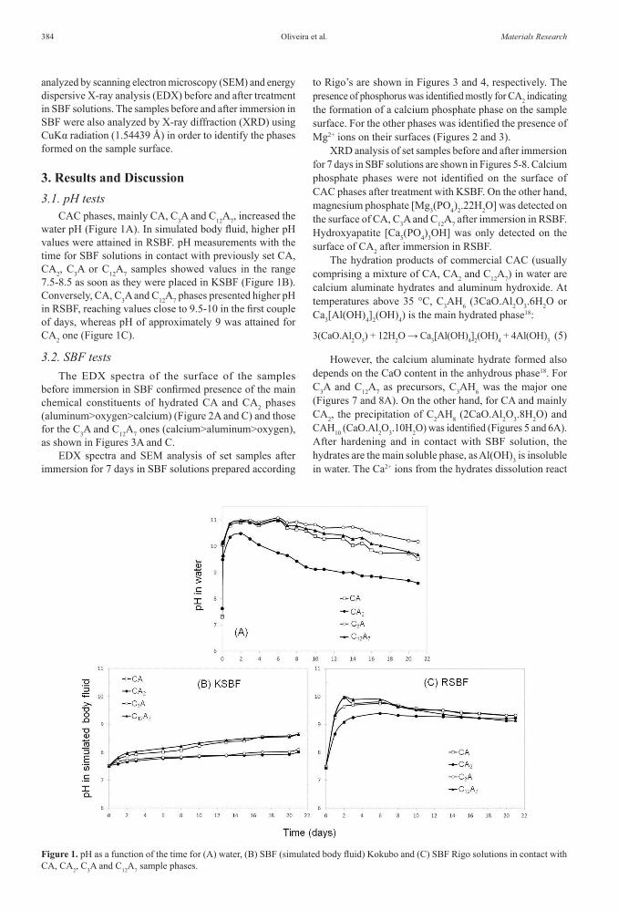
CAC phases, mainly CA, C3A and C12A7, increased the water pH (Figure 1A). In simulated body fluid, higher pH values were attained in RSBF. pH measurements with the time for SBF solutions in contact with previously set CA, CA2, C3A or C12A7 samples showed values in the range 7.5-8.5 as soon as they were placed in KSBF (Figure 1B). Conversely, CA, C3A and C12A7 phases presented higher pH in RSBF, reaching values close to 9.5-10 in the first couple of days, whereas pH of approximately 9 was attained for CA2 one (Figure 1C).
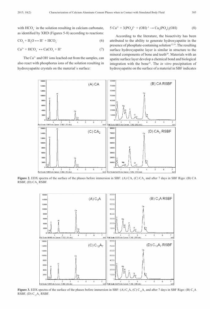
3.2. SBF testsThe EDX spectra of the surface of the samples
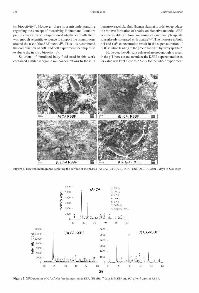
before immersion in SBF confirmed presence of the main chemical constituents of hydrated CA and CA2 phases (aluminum>oxygen>calcium) (Figure 2A and C) and those for the C3A and C12A7 ones (calcium>aluminum>oxygen), as shown in Figures 3A and C.
EDX spectra and SEM analysis of set samples after immersion for 7 days in SBF solutions prepared according
to Rigo’s are shown in Figures 3 and 4, respectively. The presence of phosphorus was identified mostly for CA2 indicating the formation of a calcium phosphate phase on the sample surface. For the other phases was identified the presence of Mg2+ ions on their surfaces (Figures 2 and 3).
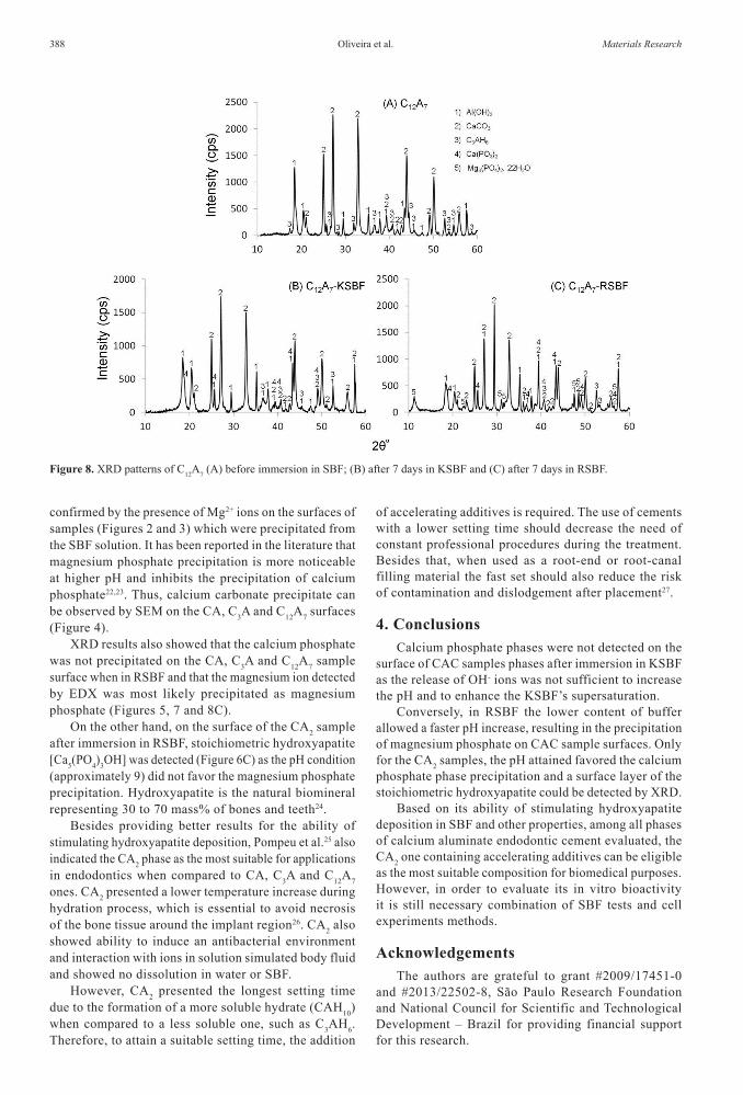
XRD analysis of set samples before and after immersion for 7 days in SBF solutions are shown in Figures 5-8. Calcium phosphate phases were not identified on the surface of CAC phases after treatment with KSBF. On the other hand, magnesium phosphate [Mg3(PO4)2.22H2O] was detected on the surface of CA, C3A and C12A7 after immersion in RSBF. Hydroxyapatite [Ca5(PO4)3OH] was only detected on the surface of CA2 after immersion in RSBF.
The hydration products of commercial CAC (usually comprising a mixture of CA, CA2 and C12A7) in water are calcium aluminate hydrates and aluminum hydroxide. At temperatures above 35 °C, C3AH6 (3CaO.Al2O3.6H2O or Ca3[Al(OH)4]2(OH)4) is the main hydrated phase18:
3(CaO.Al2O3) + 12H2O → Ca3[Al(OH)4]2(OH)4 + 4Al(OH)3 (5)
However, the calcium aluminate hydrate formed also depends on the CaO content in the anhydrous phase18. For C3A and C12A7 as precursors, C3AH6 was the major one (Figures 7 and 8A). On the other hand, for CA and mainly CA2, the precipitation of C2AH8 (2CaO.Al2O3.8H2O) and CAH10 (CaO.Al2O3.10H2O) was identified (Figures 5 and 6A). After hardening and in contact with SBF solution, the hydrates are the main soluble phase, as Al(OH)3 is insoluble in water. The Ca2+ ions from the hydrates dissolution react
Figure 1. pH as a function of the time for (A) water, (B) SBF (simulated body fluid) Kokubo and (C) SBF Rigo solutions in contact with CA, CA2, C3A and C12A7 sample phases.
Characterization of Calcium Aluminate Cement Phases when in Contact with Simulated Body Fluid2015; 18(2) 385
Figure 2. EDX spectra of the surface of the phases before immersion in SBF: (A) CA, (C) CA2 and after 7 days in SBF Rigo: (B) CA RSBF, (D) CA2 RSBF.
Figure 3. EDX spectra of the surface of the phases before immersion in SBF: (A) C3A, (C) C12A7 and after 7 days in SBF Rigo: (B) C3A RSBF, (D) C12A7 RSBF.
with HCO3– in the solution resulting in calcium carbonate,
as identified by XRD (Figures 5-8) according to reactions:
CO2 + H2O ↔ H+ + HCO3– (6)
Ca2+ + HCO3– ↔ CaCO3 + H+ (7)
The Ca2+ and OH- ions leached out from the samples, can also react with phosphorus ions of the solution resulting in hydroxyapatite crystals on the material´s surface:
5 Ca2+ + 3(PO4)3– + (OH)–1 → Ca5(PO4)3(OH) (8)
According to the literature, the bioactivity has been attributed to the ability to generate hydroxyapatite in the presence of phosphate-containing solution12,19. The resulting surface hydroxyapatite layer is similar in structure to the mineral components of bone and teeth20. Materials with an apatite surface layer develop a chemical bond and biological integration with the bone21. The in vitro precipitation of hydroxyapatite on the surface of a material in SBF indicates
Oliveira et al. 386 Materials Research
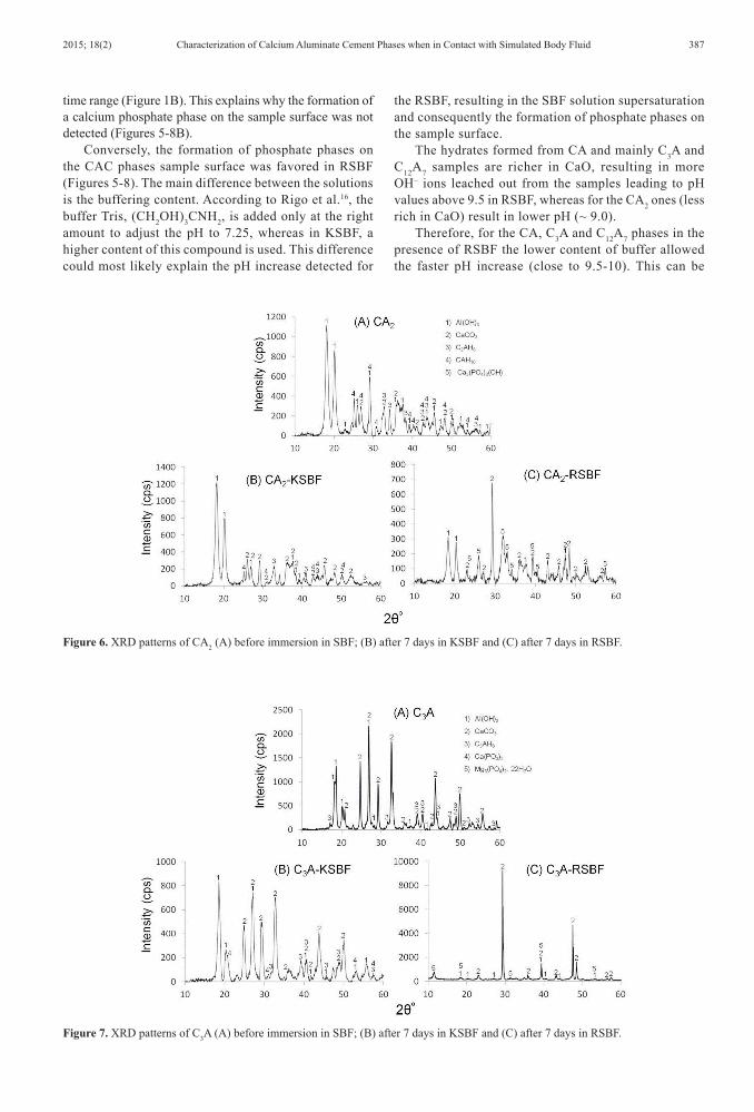
Figure 4. Electron micrographs depicting the surface of the phases (A) CA, (C) C3A, (B) CA2, and (D) C12A7 after 7 days in SBF Rigo
Figure 5. XRD patterns of CA (A) before immersion in SBF; (B) after 7 days in KSBF and (C) after 7 days in RSBF.
its bioactivity17. However, there is a misunderstanding regarding the concept of bioactivity. Bohner and Lemaitre published a review which questioned whether currently there was enough scientific evidence to support the assumptions around the use of the SBF method13. Thus it is recommend the combination of SBF and cell experiment techniques to evaluate the in vitro bioactivity12.
Solutions of simulated body fluid used in this work contained similar inorganic ion concentrations to those in
human extracellular fluid (human plasma) in order to reproduce the in vitro formation of apatite on bioactive material. SBF is a metastable solution containing calcium and phosphate ions already saturated with apatite11,16. The increase in both pH and Ca2+ concentration result in the supersaturation of SBF solution leading to the precipitation of hydroxyapatite20.
However, the OH- ions released are not enough to result in the pH increase and to induce the KSBF supersaturation as its value was kept close to 7.5-8.5 for the whole experiment
Characterization of Calcium Aluminate Cement Phases when in Contact with Simulated Body Fluid2015; 18(2) 387
Figure 6. XRD patterns of CA2 (A) before immersion in SBF; (B) after 7 days in KSBF and (C) after 7 days in RSBF.
Figure 7. XRD patterns of C3A (A) before immersion in SBF; (B) after 7 days in KSBF and (C) after 7 days in RSBF.
time range (Figure 1B). This explains why the formation of a calcium phosphate phase on the sample surface was not detected (Figures 5-8B).
Conversely, the formation of phosphate phases on the CAC phases sample surface was favored in RSBF (Figures 5-8). The main difference between the solutions is the buffering content. According to Rigo et al.16, the buffer Tris, (CH2OH)3CNH2, is added only at the right amount to adjust the pH to 7.25, whereas in KSBF, a higher content of this compound is used. This difference could most likely explain the pH increase detected for
the RSBF, resulting in the SBF solution supersaturation and consequently the formation of phosphate phases on the sample surface.
The hydrates formed from CA and mainly C3A and C12A7 samples are richer in CaO, resulting in more OH– ions leached out from the samples leading to pH values above 9.5 in RSBF, whereas for the CA2 ones (less rich in CaO) result in lower pH (~ 9.0).
Therefore, for the CA, C3A and C12A7 phases in the presence of RSBF the lower content of buffer allowed the faster pH increase (close to 9.5-10). This can be
Oliveira et al. 388 Materials Research
confirmed by the presence of Mg2+ ions on the surfaces of samples (Figures 2 and 3) which were precipitated from the SBF solution. It has been reported in the literature that magnesium phosphate precipitation is more noticeable at higher pH and inhibits the precipitation of calcium phosphate22,23. Thus, calcium carbonate precipitate can be observed by SEM on the CA, C3A and C12A7 surfaces (Figure 4).
XRD results also showed that the calcium phosphate was not precipitated on the CA, C3A and C12A7 sample surface when in RSBF and that the magnesium ion detected by EDX was most likely precipitated as magnesium phosphate (Figures 5, 7 and 8C).
On the other hand, on the surface of the CA2 sample after immersion in RSBF, stoichiometric hydroxyapatite [Ca5(PO4)3OH] was detected (Figure 6C) as the pH condition (approximately 9) did not favor the magnesium phosphate precipitation. Hydroxyapatite is the natural biomineral representing 30 to 70 mass% of bones and teeth24.
Besides providing better results for the ability of stimulating hydroxyapatite deposition, Pompeu et al.25 also indicated the CA2 phase as the most suitable for applications in endodontics when compared to CA, C3A and C12A7 ones. CA2 presented a lower temperature increase during hydration process, which is essential to avoid necrosis of the bone tissue around the implant region26. CA2 also showed ability to induce an antibacterial environment and interaction with ions in solution simulated body fluid and showed no dissolution in water or SBF.
However, CA2 presented the longest setting time due to the formation of a more soluble hydrate (CAH10) when compared to a less soluble one, such as C3AH6. Therefore, to attain a suitable setting time, the addition
of accelerating additives is required. The use of cements with a lower setting time should decrease the need of constant professional procedures during the treatment. Besides that, when used as a root-end or root-canal filling material the fast set should also reduce the risk of contamination and dislodgement after placement27.
4. ConclusionsCalcium phosphate phases were not detected on the
surface of CAC samples phases after immersion in KSBF as the release of OH- ions was not sufficient to increase the pH and to enhance the KSBF’s supersaturation.
Conversely, in RSBF the lower content of buffer allowed a faster pH increase, resulting in the precipitation of magnesium phosphate on CAC sample surfaces. Only for the CA2 samples, the pH attained favored the calcium phosphate phase precipitation and a surface layer of the stoichiometric hydroxyapatite could be detected by XRD.
Based on its ability of stimulating hydroxyapatite deposition in SBF and other properties, among all phases of calcium aluminate endodontic cement evaluated, the CA2 one containing accelerating additives can be eligible as the most suitable composition for biomedical purposes. However, in order to evaluate its in vitro bioactivity it is still necessary combination of SBF tests and cell experiments methods.
AcknowledgementsThe authors are grateful to grant #2009/17451-0
and #2013/22502-8, São Paulo Research Foundation and National Council for Scientific and Technological Development – Brazil for providing financial support for this research.
Figure 8. XRD patterns of C12A7 (A) before immersion in SBF; (B) after 7 days in KSBF and (C) after 7 days in RSBF.
Characterization of Calcium Aluminate Cement Phases when in Contact with Simulated Body Fluid2015; 18(2) 389
References1. Roberts HW, Toth JM, Berzins DW and Charlton DG. Mineral
trioxide aggregate material use in endodontic treatment: a review of the literature. Dental Materials. 2008; 24(2):149-164. http://dx.doi.org/10.1016/j.dental.2007.04.007. PMid:17586038
2. Aguilar FG, Roberti Garcia LF and Panzeri Pires-de-Souza FC. Biocompatibility of new calcium aluminate cement (EndoBinder). Journal of Endodontics. 2012; 38(3):367-371. http://dx.doi.org/10.1016/j.joen.2011.11.002. PMid:22341076
3. Engqvist H, Schultz-Walz JE, Loof J, Botton GA, Mayer D, Phaneuf MW, et al. Chemical and biological integration of a mouldable bioactive ceramic material capable of forming apatite in vivo in teeth. Biomaterials. 2004; 25(14):2781-2787. http://dx.doi.org/10.1016/j.biomaterials.2003.09.053. PMid:14962556
4. Jacobovitz M, Vianna ME, Pandolfelli VC, Oliveira IR, Rossetto HL and Gomes BPFA. Root canal filling with cements based on mineral aggregates: an in vitro analysis of bacterial microleakage. Oral Surgery, Oral Medicine, Oral Pathology, Oral Radiology, and Endodontics. 2009; 108(1):140-144. http://dx.doi.org/10.1016/j.tripleo.2009.03.013. PMid:19540451
5. Loof J, Engqvist H, Ahnfelt NO, Lindqvist K and Hermansson L. Mechanical properties of a permanent dental restorative material based on calcium aluminate. Journal of Materials Science. Materials in Medicine. 2003; 14(12):1033-1037. http://dx.doi.org/10.1023/B:JMSM.0000003999.52349.0d. PMid:15348495
6. Pandolfelli VC, Oliveira IR, Rossetto HL and Jacobovitz M. Aluminous cement-based composition for application in endodontics and cementitious product obtained thereof 2009. WO/2009/067774. 2009.
7. Oliveira IR, Pandolfelli VC and Jacobovitz M. Chemical, physical and mechanical properties of a novel calcium aluminate endodontic cement. International Endodontic Journal. 2010; 43(12):1069-1076. http://dx.doi.org/10.1111/j.1365-2591.2010.01770.x. PMid:20726916
8. Castro-Raucci LM, Oliveira IR, Teixeira LN, Rosa AL, Oliveira PT and Jacobovitz M. Effects of a novel calcium aluminate cement on the early events of the progression of osteogenic cell cultures. Brazilian Dental Journal. 2011; 22(2):99-104. http://dx.doi.org/10.1590/S0103-64402011000200002. PMid:21537581
9. Silva EJNL, Herrera DR, Almeida JFA, Ferraz CCR, Gomes BPFA and Zaia AA. Evaluation of cytotoxicity and up-regulation of gelatinases in fibroblast cells by three root repair materials. International Endodontic Journal. 2012; 45(9):815-820. http://dx.doi.org/10.1111/j.1365-2591.2012.02038.x. PMid:22452531
10. Aguilar FG, Garcia LF, Rossetto HL, Pardini LC and Pires-de-Souza FC. Radiopacity evaluation of calcium aluminate cement containing different radiopacifying agents. Journal of Endodontics. 2011; 37(1):67-71. http://dx.doi.org/10.1016/j.joen.2010.10.001. PMid:21146080
11. Kokubo T and Takadama H. How useful is SBF in predicting in vivo bone bioactivity? Biomaterials. 2006; 27(15):2907-2915. http://dx.doi.org/10.1016/j.biomaterials.2006.01.017. PMid:16448693
12. Wu C and Xiao Y. Evaluation of the in vitro bioactivity of bioceramics. Bone and Tissue Regeneration Insights. 2009; 2:25-29.
13. Bohner M and Lemaitre J. Can bioactivity be tested in vitro with SBF solution? Biomaterials. 2009; 30(12):2175-2179. http://dx.doi.org/10.1016/j.biomaterials.2009.01.008. PMid:19176246
14. Oliveira IR, Andrade TL, Jacobovitz M and Pandolfelli VC. Bioactivity of calcium aluminate endodontic cement. Journal of
Endodontics. 2013; 39(6):774-778. http://dx.doi.org/10.1016/j.joen.2013.01.013. PMid:23683278
15. Andrade TL, Santos GL, Pandolfelli VC and Oliveira IR. Synthesis optimization of calcium aluminate cement phases for biomedical applications [in Portuguese]. Cerâmica. 2014; 60(353):88-95. http://dx.doi.org/10.1590/S0366-69132014000100013.
16. Rigo ECS, Boschi AO, Yoshimoto M, Allegrini S Jr, Konig B Jr and Carbonari MJ. Evaluation in vitro and in vivo of biomimetic hydroxyapatite coated on titanium dental implants. Materials Science and Engineering C. 2004; 24(5):647-651. http://dx.doi.org/10.1016/j.msec.2004.08.044.
17. Wang X, Sun H and Chang J. Characterization of Ca3SiO5/CaCl2 composite cement for dental application. Dental Materials. 2008; 24(1):74-82. http://dx.doi.org/10.1016/j.dental.2007.02.006. PMid:17391748
18. Garcia JR, Oliveira IR and Pandolfelli VC. Hydration process and the mechanisms of retarding and accelerating the setting time of calcium aluminate cement. [in Portuguese]. Cerâmica. 2007; 53:42-56.
19. Tay FR, Pashley DH, Rueggeberg FA, Loushine RJ and Weller RN. Calcium phosphate phase transformation produced by the interaction of the portland cement component of white mineral trioxide aggregate with a phosphate-containing fluid. Journal of Endodontics. 2007; 33(11):1347-1351. http://dx.doi.org/10.1016/j.joen.2007.07.008. PMid:17963961
20. Coleman NJ, Nicholson JW and Awosanya K. A preliminary investigation of the in vitro bioactivity of white portland cement. Cement and Concrete Research. 2007; 37(11):1518-1523. http://dx.doi.org/10.1016/j.cemconres.2007.08.008.
21. Sarkar NK, Caicedo R, Ritwik P, Moiseyeva R and Kawashima I. Physicochemical basis of the biologic properties of mineral trioxide aggregate. Journal of Endodontics. 2005; 31(2):97-100. http://dx.doi.org/10.1097/01.DON.0000133155.04468.41. PMid:15671817
22. Barrére F, Layrolle P, van Blitterswijk CA and de Groot K. Biomimetic calcium phosphate coatings on Ti6AI4V: a crystal growth study of octacalcium phosphate and inhibition by Mg2+ and HCO3-. Bone. 1999;25(2, Suppl):107S-111S. http://dx.doi.org/10.1016/S8756-3282(99)00145-3. PMid:10458288
23. Martens CS and Harriss RC. Inhibition of apatite precipitation in the marine environment by magnesium ions. Geochimica et Cosmochimica Acta. 1970; 34(5):621-625. http://dx.doi.org/10.1016/0016-7037(70)90020-7.
24. Loof J. Calcium-aluminate as biomaterial: synthesis, design and evaluation. [Thesis]. Uppsala: Faculty of Science and Technology; 2008.
25. Pompeu LLMF, Santos GL, Pandolfelli VC and Oliveira IR. Calcium aluminates potential for endodontics and orthopedics application [in Portuguese]. Cerâmica. 2013; 59:216-224. http://dx.doi.org/10.1590/S0366-69132013000200004.
26. Oréfice RL, Pereira MM and Mansur HS. Biomaterials: fundamentals and applications. Rio de Janeiro: Medical Culture; 2006. 538 p.
27. Camilleri J. The physical properties of accelerated Portland cement for endodontic use. International Endodontic Journal. 2008; 41(2):151-157. PMid:17931386.