Embed Size (px)
Citation preview
JOURNAL OF BACTERIOLOGY, Aug. 2009, p. 4786–4797 Vol. 191, No. 150021-9193/09/$08.00�0 doi:10.1128/JB.00437-09Copyright © 2009, American Society for Microbiology. All Rights Reserved.
Characterization of �-Butyrolactone Autoregulatory Signaling GeneHomologs in the Angucyclinone Polyketide WS5995B Producer
Streptomyces acidiscabies�
Frank G. Healy,1* Kevin P. Eaton,1 Prajit Limsirichai,1 Joel F. Aldrich,1Alaina K. Plowman,1 and Russell R. King2
Department of Biology, Trinity University, San Antonio, Texas 78212,1 Agriculture and Agri-Food Canada, Research Branch,Fredericton Research Centre, Fredericton, New Brunswick, Canada E3B4Z72
Received 31 March 2009/Accepted 18 May 2009
Organisms belonging to the genus Streptomyces produce numerous important secondary metabolites andundergo a sophisticated morphological differentiation program. In many instances these processes are underthe control of �-butyrolactone (GBL) autoregulatory systems. Streptomyces acidiscabies strain 84.104 producesthe secondary metabolite aromatic angucyclinone polyketide WS5995B. In order to explore the role of GBLregulatory circuitry in WS5995B production and morphogenesis in S. acidiscabies, a gene cluster encoding GBLautoregulatory signaling homologs was identified and characterized. Two GBL receptor homologs, sabR andsabS, were found flanking a GBL synthase homolog sabA. Strains carrying mutations in sabS produced elevatedlevels of WS5995B and displayed conditional morphological defects reminiscent of defects seen in StreptomycesbldA mutants. Notably, sabS possesses a TTA codon predicted to be recognized by tRNAleu. sabA mutantsproduced higher levels of WS5995B than the wild-type strain but to a lesser extent than the levels of WS5995Bseen in sabS mutants. Purified recombinant SabR and SabS were tested for their abilities to bind predictedAT-rich autoregulatory element (ARE) boxes within the sabRAS region. SabS did not bind any DNA sequencesin this region, while SabR bound an ARE box in the region upstream of sabS. Quantitative reverse transcrip-tion-PCR analysis revealed higher levels of sabS transcript in sabR mutants than in the wild-type strain,suggesting that sabS expression is repressed by SabR. Based on these data, we propose that the S. acidiscabiessabRAS genes encode components of a signaling pathway which participates in the regulation of WS5995Bproduction and morphogenesis.
Members of the genus Streptomyces are renowned for theirmorphological complexity as well as their capacity to producea wide variety of important secondary metabolites, includingpolyketides and nonribosomal peptides. In many instances, theexpression of structural genes encoding secondary metabolitepathway enzymes is regulated by the interplay of low-molecu-lar-weight hydrophobic, membrane-diffusible �-butyrolactone(GBL) autoregulator compounds with specific cognate GBLreceptors. GBL autoregulator synthesis requires the autoregu-lator GBL synthase, and as GBL is produced, intracellularGBL levels increase. GBL receptors are transcriptional regu-lators belonging to the TetR superfamily of transcription fac-tors (34, 37). These proteins possess helix-turn-helix (HTH)DNA binding domains which recognize AT-rich autoregula-tory element (ARE) DNA sequence targets (34).
Typical GBL signaling relies on changes in ARE-receptorinteractions in response to increasing intracellular levels ofGBL. The best-studied GBL signaling system is that of Strep-tomyces griseus (35). The GBL receptor ArpA binds targetDNA sequence in the upstream region of adpA, resulting inrepression of adpA expression by obstructing RNA polymeraseaccess to adpA promoter elements. Biosynthesis of the S. gri-seus GBL A factor (2-isocapryloyl-3-R-hydroxymethyl-�-butyro-
lactone) requires the product of the autoregulator gene afsA.As A factor reaches sufficient levels within the cell, the binaryArpA/A-factor complex dissociates from the target nucleotidesequence, thereby allowing adpA expression. AdpA is requiredfor expression of a set of genes which constitute the AdpAregulon. This includes genes required for morphological dif-ferentiation, streptomycin biosynthesis, grixazone production,and the production of other metabolites (16).
Other examples of GBL signaling systems in streptomyceteswhich have been described include the SCB1 system of Strep-tomyces coelicolor, which regulates production of both actino-rhodin and undecylprodigiosin (42); the control of blue pig-ment and nucleoside antibiotic biosynthesis by the IM-2-C6system of Streptomyces lavendulae (23); and the virginiae bu-tanolide control of virginiamycin production by Streptomycesvirginiae (31). Additionally, in at least some cases, secretedcompounds other than those produced by cognate GBL syn-thases are capable of activating GBL signaling pathways inStreptomyces. For example, Birko et al. (3) have reportedthat a 324-amino-acid peptide known as factor C producedby certain streptomycetes is capable of restoring phenotypicdefects of A-factor-deficient mutants of S. griseus.
Streptomyces acidiscabies strain 84.104 represents an inter-esting bacterium to explore GBL signaling phenomena since itproduces both nonribosomal peptide and polyketide secondarymetabolite compounds. These include the nonribosomal cyclicdipeptide thaxtomin A (25) and the angucyclinone polyketideWS5995B (17, 21). WS5995B (Fig. 1) exhibits antimicrobial
* Corresponding author. Mailing address: Department of Biology,One Trinity Place, Trinity University, San Antonio, TX 78212. Phone:(210) 999-7242. Fax: (210) 999-7229. E-mail: [email protected].
� Published ahead of print on 22 May 2009.
4786
on June 6, 2019 by guesthttp://jb.asm
.org/D
ownloaded from
activity (17; also our unpublished results), whereas thaxtominA displays phytotoxic activity against susceptible plant hosts(13, 29). Other phytopathogenic streptomycetes are known inaddition to S. acidiscabies which produce thaxtomin toxins.Among these, it was recently shown that Streptomyces scabiesstrain 87.22 possesses an AraC/XylS-type transcriptional reg-ulator to control expression of thaxtomin synthetase structuralgene expression in response to cellobiose (18). The regulationof thaxtomin biosynthesis has not been explored in S. acidis-cabies, and, interestingly, S. acidiscabies is the only currentlyavailable organism known to produce WS5995B.
The widespread utilization of GBL signaling systems inthe regulation of morphogenesis and secondary metabolismamong streptomycetes along with the poorly understood reg-ulatory mechanisms governing production of the nonribosomalpeptide thaxtomin and the type II polyketide WS5995B in S.acidiscabies prompted us to explore and characterize the pos-sible roles played by GBL signaling in the production of thesetwo distinct types of secondary metabolites in S. acidiscabies.
Our results reported here describe the identification and char-acterization of a GBL signaling system consisting of the GBLsynthase homolog sabA and two adjacent flanking genes, sabRand sabS, encoding GBL receptors. The results of genetic andbiochemical studies described here reveal a role for this GBLsystem in regulating production of WS5995B. In our analysis ofGBL mutants and metabolite production profiles, we find nochanges in thaxtomin production relative to the wild-typestrain, suggesting that the GBL system described here does notplay a role in the biosynthesis of thaxtomin or its regulation. Inaddition to GBL’s effect on WS5995B production, we alsoreport a conditional morphological defect in strains carryingmutations in the GBL receptor sabS, suggesting that SabSplays a role in morphogenesis.
MATERIALS AND METHODS
Bacterial strains, plasmids, growth, and culture conditions. Bacterial strainsand plasmids used in this study are given in Table 1. Escherichia coli strains weregrown at 37°C in Luria-Bertani medium. Antibiotics ampicillin, kanamycin, apra-mycin, and chloramphenicol, when used for selection in E. coli, were added tomedium at final concentrations of 100, 50, 100, and 30 �g/ml, respectively.Growth and transformation procedures for E. coli strains were carried out usingstandard methods (40). S. acidiscabies cultures were routinely grown at 30°C inliquid tryptic soy broth medium in 250-ml baffled flasks containing 20 to 30 ml ofmedium or on ISP2 solid medium (41). For analysis of WS5995B production,Streptomyces cultures were grown at 30°C in liquid SGM (Streptomyces growthmedium) or on solid SGM (21) without added calcium carbonate. Growth ofStreptomyces strains in SGM was measured by determining wet weights of cellsthat had been collected by vacuum filtration. Following conjugal transfer ofmutant alleles or complementing plasmids from E. coli donors, Streptomycestransconjugants were selected on modified ISP4 medium. Modified ISP4 me-dium was prepared as previously described (7, 41), except that calcium carbonatewas omitted, and the medium was amended with 0.5 g/liter yeast extract and 1g/liter tryptone. Additionally, after an autoclaving step, 0.1 volume of sterile 400mM MgCl2 was added to medium to give a final MgCl2 concentration of 40 mM
FIG. 1. Structure of WS5995B.
TABLE 1. Bacterial strains and plasmids used in this study
Strain or plasmid Relevant properties and/or function Reference orsource
StrainsS. acidiscabies 84.104 Wild-type WS5995B producer 21
SabR4–9 S. acidiscabies 84.104 �sabR GBL receptor mutant This studySabA5 S. acidiscabies 84.104 �sabA GBL synthase mutant This studyAKP938 S. acidiscabies 84.104 �sabS GBL receptor mutant This study
E. coli DH5�MCR Cloning host; F� mcrA �(mrr-hsdRMS-mcrBC) �80dlacZ�M15 �(lacZYA-argF)U196 endA1recA1 deoR thi-1 phoA supE44 �� gyrA96 relA1
13
E. coli BW25113 Strain used for PCR targeted mutagenesis; �araBAD hsdR514 8E. coli S17-1 Donor strain for conjugal transfer; thi pro hsdR hsdM� recA RP4 tra 13E. coli BL21�DE3 Strain for recombinant protein expression Novagen
PlasmidspUC19 General cloning vector 13pGBLBP1 pUC19 carrying sabRAS genes on a 7-kb KpnI fragment This studypOJ260 Suicide vector for integration in Streptomyces; aac(3)IV oriColE1 RP4 oriT lacZ� 13pALE21 pOJ260 carrying sabRAS genes on 7-kb KpnI fragment This studypKD46 Vector carrying arabinose-inducible � Red recombinase; bla araC � exo repA101ts oriR101 8pKD3 Template plasmid carrying Flp recognition target-flanked cat gene for creating PCR
mutagenesis cassette8
pCP20 bla cat thermal induction of FLP synthesis 8pIJ86 Streptomyces complementation plasmid; oriColE1 SCP2* aac(3)IV ermE*pa Mervyn BibbpIJ86::sabS pIJ86 carrying wild-type sabS allele under transcriptional control of ermE*p This studypET26b T7 RNA polymerase-dependent recombinant protein expression vector NovagenpET26b::sabR pET26b carrying sabR encoding GBL receptor SabR This studypET26b::sabS pET26b carrying sabS encoding GBL receptor SabS This study
a ermE*p, ermE* promoter.
VOL. 191, 2009 GBL SIGNALING GENES IN S. ACIDISCABIES 4787
on June 6, 2019 by guesthttp://jb.asm
.org/D
ownloaded from
in agar medium. Apramycin sulfate and nalidixic acid were each added to ISP4medium at 25 �g/ml to select for Streptomyces transconjugants and to counter-select E. coli donors, respectively.
Cloning of GBL signaling genes sabRAS in S. acidiscabies 84.104. All oligonu-cleotide primers used in this study are presented in Table 2. Consensus degen-erate hybrid oligonucleotide primers (39) were selected using protein alignmentsof previously characterized GBL receptors. The oligonucleotide primer pairGBL-F/GBL-R (Table 2) was used to amplify GBL receptor gene fragmentsfrom S. acidiscabies 84.104 genomic DNA using “touchdown PCR” as previouslydescribed (13). Cloned amplification products were sequenced and used asprobes for the identification of complete genes and flanking regions from genelibraries using colony hybridization methods (13). The nucleotide sequence ofthe sabRAS region was determined on both strands of a 7-kb KpnI insert inplasmid vector pUC19 (pGBLBP1) using a primer walking approach. DeducedGBL receptor and synthase protein sequences were aligned using MUSCLE,version 3.7. Alignments were refined using Gblocks, version 0.91b, and phylog-enies were constructed with the maximum-likelihood method using PhyML,
version 3.0. Phylograms were visualized and edited using TreeDyn, version198.3 (9).
Mutagenesis and complementation of sabRAS genes. The 7-kb KpnI fragmentfrom pGBLBP1 was cloned into the KpnI site of plasmid vector pOJ260, and theresulting plasmid (pALE21) was introduced into E. coli strain BW25113 carryingplasmid pKD46, encoding bacteriophage lambda Red recombinase. Culturescarrying both of these plasmids were grown in LB medium containing apramycinand ampicillin. BW25113(pALE21/pKD46) was grown in SOB medium contain-ing 1 mM L-arabinose to induce expression of Red recombinase, as previouslydescribed (8). Oligonucleotide primer pairs sabRdelF/sabRdelR, sabAdelF/sabAdelR, and sabSdelF/sabSdelR (Table 2) were selected to generate in-framedeletions of sabR, sabA, and sabS on plasmid pALE21, respectively. Theseprimers were used to amplify the chloramphenicol acetyltransferase gene fromtemplate plasmid pKD3, with resulting products carrying 5 ends with homologyto sabR, sabA, or sabS in pALE21 (8, 11). Gel-purified PCR products wereelectroporated into washed suspensions of BW25113(pALE21/pKD46) as de-scribed previously (8), and transformed cells carrying mutagenized pALE21 wereselected on LB agar containing chloramphenicol. Overnight cultures of resultingchloramphenicol-resistant colonies were grown in LB medium containing chlor-amphenicol, and plasmids were extracted from these cultures. Plasmids thusobtained were used to transform suspensions of competent E. coli DH5� cells tochloramphenicol resistance. Plasmids extracted from these transformants wereused to transform BW25113(pCP20) to apramycin resistance in order to excisethe chloramphenicol resistance gene as described previously (8).
pALE21 derivatives carrying deletions in sabR, sabA, and sabS were used totransform either E. coli strain S17-1 or the ET12567(pUZ8002) strain to apra-mycin resistance. Resulting transformants were used for intergeneric conjugalmatings with S. acidiscabies 84.104 recipient strains as previously described (13),with the exception that mating mixtures were plated on 25 ml of modified ISP4medium instead of AS-1 medium. Following overnight growth of mating mixturesat 30°C, transconjugants were selected by overlaying plates with 5 ml of softnutrient agar containing nalidixic acid and apramycin sulfate (150 �g/ml each) togive final concentrations of 25 �g/ml in agar medium. Incubation of plates wascontinued at 30°C for 3 to 5 days until transconjugants were visible on agarsurface. Transconjugant colonies were then transferred to ISP2 medium con-taining nalidixic acid and apramycin. Liquid cultures of transconjugants weregrown nonselectively in tryptic soy broth for two or three serial transfers to allowfor loss of integrated plasmid. PCR assays were used to screen apramycin-sensitive derivatives for loss of plasmid and retention of mutant alleles. Thesemutants were used to study the roles of GBL signaling genes in secondarymetabolite biosynthesis and morphological differentiation on solid medium.
In order to verify linkage of mutant phenotypes with deleted GBL alleles,the primer pairs sabRcomplF/sabRcomplR, sabAcomplF/sabAcomplR, andsabScomplF/sabScomplR were used to amplify the wild-type sabR, sabA, and sabSopen reading frames (ORFs), respectively, from S. acidiscabies 84.104 genomicDNA using high-fidelity Pfu polymerase (New England Biolabs). Amplificationproducts were purified and digested with restriction enzymes HindIII and BglII.Plasmid pIJ86 was also digested with HindIII and BglII, and the ORFs encodingthese GBL signaling genes were ligated into pIJ86 using T4 DNA ligase(New England Biolabs). The resulting plasmids, pIJ86::sabR, pIJ86::sabA, andpIJ86::sabS, harboring wild-type genes under the transcriptional control of theermE* promoter were used to transform E. coli S17-1 or ET12567(pUZ8002),and conjugations were performed using these strains as donor and the �sabR,�sabA, or �sabS strain as recipient. Resulting transconjugants were selected onISP4 medium amended with apramycin and nalidixic acid.
Analysis of WS5995B production. S. acidiscabies 84.104 wild type and GBLdeletion mutants were grown on ISP2 medium, and mycelial fragments wereused to inoculate tryptic soy broth liquid medium (20 ml in 250-ml baffledErlenmeyer flasks). Cultures were grown at 30°C and 180 rpm for approximately20 h. Two milliliters of these cultures was used to inoculate 280 ml of SGM in2.8-liter baffled Fernbach flasks. Culture medium was collected at various times,and cells were removed by vacuum filtration. Wet cell weight determinations ofcultures were made, and filtrates were extracted using Strata-X solid-phasecartridges as described by the manufacturer (Phenomenex).
Extracted metabolites were dissolved in methanol and investigated by thin-layer chromatography (TLC) analysis on silica gel plates (250 �m; Whatman).UV and visible absorbance spectra were collected using a Beckman DU640Bspectrophotometer. Quantitative high-performance liquid chromatography anal-ysis was done using a Hewlett Packard 1090 liquid chromatograph equipped withdiode-array detector and a Phenomenex C18 Luna (5 �m; 150 by 4.6 mm)column. Detection wavelength range was from 200 to 600 nm. Solvent A wasH2O � H3PO4 at 99.9:0.1; solvent B was 100% CH3CN. Samples of extracts wereinjected and chromatographed at a 1 ml/min flow rate. Extract components were
TABLE 2. Oligonucleotide primers used in this study
Primer Sequence (5–3)
GBL-F .................CAAGCAGGAGCGCGCSRTCCGSACGBL-R.................CCTTGGACTGCAAGTGGAAGTASAsabRex-F .............AAGGATCATATGGCTAAACAGGAGCGCsabRex-R.............AAGGATCTCGAGCGAAGCCACCCTTTCGGGsabSex-F ..............TTCCTTCATATGGCGAGGCAGTTACGCsabSex-R..............TTCCTTCTCGAGGGACACGCACGCCGCCGTARE-F.................AGTCGGCAGTCTTCGAGTGTARE-R ................GCTCGTCGATTCGTAACCATAREdupF............TGAGAAACAAACGGCATGACCCGTTCTTTT
ACCGAREdupR ...........CGGTAAAAGAACGGGTCATGCCGTTTGTTT
CTCAsabRdelF .............CCAAGCCATCGTGGAAGCGGCCGGTGAGG
TTTTCGACGAGCACGGTTACATGTGTAGGCTGGAGCTGCTTC
sabRdelR.............CGAACGGCACCTGCTCGCCCAGCACCGCCTGCGCCAGCGCCTCCTTGGACCATATGAATATCCTCCTTAG
sabAdelF .............GTTCGTCCACCGCGCAGCCCTCGCCGAAACGTTTCTCACCGGCTGGGAGCTGTGTAGGCTGGAGCTGCTTC
sabAdelR.............CGGCACCGGCCCGACCTCCAACCGGTGCGGATCCGTGGTGACTTCGAAGGCATATGAATATCCTCCTTAG
sabSdelF ..............ATGGCGAGGCAGTTACGCGCCGAACAGACCCGCGCGACGATCATCACGGCGTGTGTAGGCTGGAGCTGCTTC
sabSdelR..............TCAGGACACGCACGCCGCCGTGATCTCCCGCTCCAGCCTCGACGCCAGGCTCAGCATATGAATATCCTCCTTAG
sabRcomplF ........ACCTAAGCTTATCGGCCAACTCGGTCCsabRcomplR .......AACCAGATCTACGTTTCTTTCTCCGCsabAcomplF ........ACCTAAGCTTTGTTCGGCCCGATCAsabAcomplR .......AACCAGATCTCGGGTCATGCCGTTTGTsabScomplF.........ACCTAAGCTTTGTGTACAGGGGGTGAGAsabScomplR ........AACCAGATCTCGATCCGAACAGCGCCGhrdBF...................GAAGACCGCCGCCAARAARACNRChrdBR..................GGGTGGCGCAGCTTGSWCATNGTYTThrdBFrt................CTGGATGAGGTCCAGGAAGAhrdBRrt ...............CCGGTCAAGGACTACCTCAAsabRFrt................GTACTTCCACTTCCCGTCCAsabRRrt ...............CAACAAGGAACTGGTCAGCAsabAFrt................ACACCCCGTTTTCTTCGACsabARrt ...............GACCCCACACTCGAAGACTGsabSFrt.................ATCTGTTCGACCGACATGGTsabSRrt ................CGCTTCGAGGGAGGAGTAGsabSQRTF ..........GCGCCCTGTACTTCCACTTsabSQRTR..........TGGGACTGGAGTTCCATGAThrdBQRTF..........CTCTTCCTGGACCTCATCCAhrdBQRTR .........GGCGTACGTGGAGAACTTGT
4788 HEALY ET AL. J. BACTERIOL.
on June 6, 2019 by guesthttp://jb.asm
.org/D
ownloaded from
eluted with a linear gradient of 25% B to 60% B over 21 min. Identification andquantification of WS5995B in culture filtrate extracts were accomplished usingauthentic WS5995B standard preparations (21).
Cloning, expression, and purification of recombinant SabR and SabS. ThesabR and sabS ORFs were amplified from S. acidiscabies 84.104 genomic DNAusing high-fidelity Pfu polymerase and the oligonucleotide primer pairs sabRex-F/sabRex-R (for SabR expression) and sabSex-F/sabSex-R (for SabS expression)(Table 2). Amplification reaction products were purified and digested with re-striction enzymes NdeI and XhoI. The expression vector pET26b (Novagen) wasalso digested with NdeI and XhoI, and sabR and sabS ORF restriction digestionproducts were ligated into the vector to produce recombinant protein expressionconstructs pET26b::sabR and pET26b::sabS. Competent cells of E. coli strainBL21(�DE3) were transformed with these plasmids, and transformants wereselected on LB agar containing kanamycin. Transformants were grown in 500 mlof LB medium containing kanamycin at 30°C. When the culture reached anoptical density at 600 nm of approximately 0.6, protein expression was inducedwith the addition of IPTG (isopropyl--D-thiogalactopyranoside) to a final con-centration of 80 �M. Cultures were grown for an additional 6 h and thenharvested by centrifugation. Cell pellets were washed once in buffer containing20 mM sodium phosphate, 500 mM NaCl, and 20 mM imidazole, pH 7.4, andthen resuspended in 1 to 2 ml of the same buffer containing 1 mM dithiothreitol(DTT) and 1 mM phenylmethylsulfonyl fluoride. Cell suspensions were dis-rupted by passage through a French pressure minicell at 16,000 lb/in2. The celllysate was centrifuged at 20,000 � g for 30 min, and the resulting soluble proteinfraction was loaded onto a 1-ml HisGraviTrap column (GE Healthcare). Proteinfractions were eluted with 20 mM sodium phosphate–500 mM NaCl, pH 7.4,containing increasing amounts of imidazole. Fractions were analyzed using so-dium dodecyl sulfate-polyacrylamide gel electrophoresis (PAGE), and the frac-tions containing pure SabR or SabS (as judged by electrophoretic homogeneity)were pooled and desalted on PD-10 desalting columns (GE Healthcare). Pro-teins obtained in this manner were stored at �20°C or �70°C in 50 mM Tris-Cl,pH 7.6, 200 mM NaCl, 1 mM DTT, and 50% glycerol. Protein determinationswere made using a Bradford assay with bovine serum albumin as the standard.
Electrophoretic mobility shift assays. DNA fragments encompassing AREsequences within the sabRAS region were amplified from template plasmidpGBLBP1. The oligonucleotide primer pair ARE-F/ARE-R was used to amplifya 208-bp DNA fragment encompassing the upstream region of sabS. ARE-F (40pmol) was first end labeled in a separate reaction using [�-32P]ATP (6,000Ci/mmol; PerkinElmer) and polynucleotide kinase (Roche) according to themanufacturer’s recommendations. Labeled primer products were separated fromunincorporated nucleotide using Micro Bio-Spin columns (Bio-Rad) and used inPCR along with oligonucleotide primer ARE-R, plasmid template pGBLBP1,and high-fidelity Pfu polymerase. Amplified labeled PCR product was separatedfrom excess primer using QiaQuick PCR spin columns (Qiagen). DNA bindingassays contained 2.5 � 103 cpm of labeled DNA, 50 ng of calf thymus DNA, 50mM NaCl, 10 mM MgCl2, 10 mM Tris-Cl, 1 mM DTT, and 50 �g/ml bovineserum albumin, pH 7.4. Various amounts of recombinant SabR or SabS wereadded to the reaction tubes. For competition experiments, unlabeled competitorduplex DNA corresponding to ARE sequence was prepared by briefly heatingand annealing stoichiometric amounts of complementary oligonucleotidesAREdupF/AREdupR and adding these to DNA binding reaction mixtures.Binding reactions (10 �l) were carried out for 20 min at 30°C, after whichreaction products were electrophoresed through nondenaturing 5% polyacryl-amide gels in 0.5� Tris-borate-EDTA buffer (40). Gels were dried under vacuumand exposed to a phosphorimager screen (Amersham Biosciences). Exposedscreens were scanned using a Typhoon Trio� Variable Mode Imager (Amer-sham Biosciences); band intensities of scanned screens were quantified usingImageQuant TL software (Amersham Biosciences). Experiments were repeatedat least four times for each protein and ARE substrate combination.
RNA extraction and reverse transcriptase PCR assays. Cultures of S. acidis-cabies wild-type 84.104 and of sabR, sabA, and sabS mutants were grown intryptic soy broth or SGM to mid-log phase, and cells were collected by centrif-ugation. Cell pellets were briefly homogenized using a tissue grinder and treatedwith lysozyme. Total RNA was extracted from cells using an RNeasy minikitfollowing the manufacturer’s guidelines (Qiagen). Following extraction, RNAsamples were treated with RNase-free DNase I (New England Biolabs). Reversetranscription reactions were carried out using Superscript II reverse transcriptase(Invitrogen) and reverse transcriptase primers sabRRrt, sabARrt, and sabSRrt(Table 2). PCR amplification of DNase-treated samples prior to cDNA synthesisyielded no amplification products, indicating that nucleic acid PCR amplificationproducts observed following reverse transcription were produced solely fromcDNA templates derived from RNA. Following cDNA synthesis, reverse tran-scription reaction products were used for PCR amplification using the primer
pairs sabRFrt/sabRRrt, sabAFrt/sabARrt, and sabSFrt/sabSRrt. PCR was car-ried out for 30 cycles, and amplification products were electrophoresed through1% agarose gels in Tris-acetate-EDTA buffer. PCR amplification product bandintensities on gels were imaged using a VersaDoc system (Bio-Rad) and quan-tified using Quantity One software. Normalization of amplification productsfrom cDNA templates using GBL primers was done using the hrdB gene encod-ing the principal sigma factor of S. acidiscabies 84.104. hrdB was cloned from S.acidiscabies 84.104 genomic DNA using degenerate oligonucleotide primers de-signed from alignments of conserved regions of principal sigma factors from S.coelicolor (SCO5820), Streptomyces avermitilis (SAV_2444), and S. griseus(EMBL accession X75952) (Table 2). The deduced amino acid sequence of thecloned S. acidiscabies 84.104 hrdB gene displayed 95% identity with S. coelicolorHrdB (not shown). The oligonucleotide primer pair hrdBFrt/hrdBRrt was usedfor amplification of hrdB cDNA from RNA samples.
Quantitative reverse transcription-PCR (qRT-PCR) was performed using anApplied Biosystems 7500 real-time PCR system and SYBR green QuantitativeRT-PCR Kit with Moloney murine leukemia virus reverse transcriptase, RNaseinhibitor, and JumpStart Taq DNA polymerase (Sigma). Specific primers usedfor amplification of sabS (sabSQRT) and hrdB (hrdBQRT) are given in Table 2.Amplicon specificity was checked using qRT-PCR dissociation curve analysis.The relative increase in sabS expression in wild-type and sabR mutant strains wasdetermined using the Relative Quantification Method (Applied Biosystems 7500System). Briefly, threshold cycle (CT) values were normalized to hrdB mRNAlevels for RNA samples from each strain, and mean relative expression ratioswere calculated using the ��CT method. Values given for the relative increase inexpression represent the means of three independent experiments.
Nucleotide sequence accession number. The nucleotide sequence of thesabRAS region described here has been deposited in the NCBI databaseunder accession number FJ821515.
RESULTS AND DISCUSSION
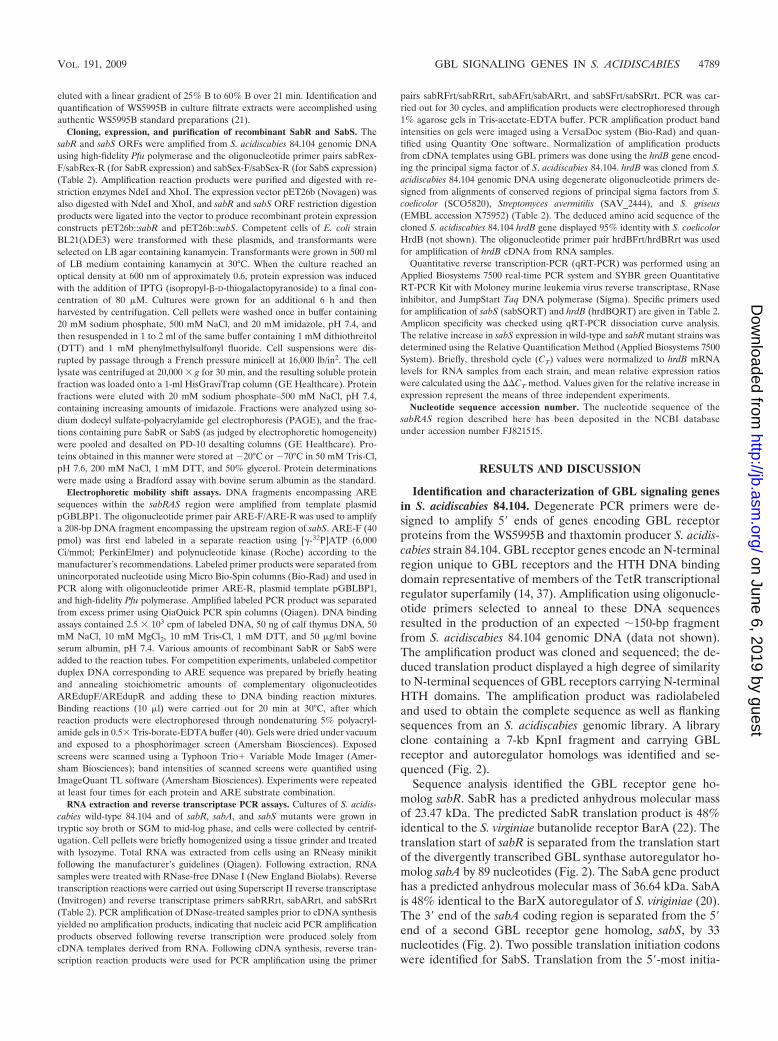
Identification and characterization of GBL signaling genesin S. acidiscabies 84.104. Degenerate PCR primers were de-signed to amplify 5 ends of genes encoding GBL receptorproteins from the WS5995B and thaxtomin producer S. acidis-cabies strain 84.104. GBL receptor genes encode an N-terminalregion unique to GBL receptors and the HTH DNA bindingdomain representative of members of the TetR transcriptionalregulator superfamily (14, 37). Amplification using oligonucle-otide primers selected to anneal to these DNA sequencesresulted in the production of an expected �150-bp fragmentfrom S. acidiscabies 84.104 genomic DNA (data not shown).The amplification product was cloned and sequenced; the de-duced translation product displayed a high degree of similarityto N-terminal sequences of GBL receptors carrying N-terminalHTH domains. The amplification product was radiolabeledand used to obtain the complete sequence as well as flankingsequences from an S. acidiscabies genomic library. A libraryclone containing a 7-kb KpnI fragment and carrying GBLreceptor and autoregulator homologs was identified and se-quenced (Fig. 2).
Sequence analysis identified the GBL receptor gene ho-molog sabR. SabR has a predicted anhydrous molecular massof 23.47 kDa. The predicted SabR translation product is 48%identical to the S. virginiae butanolide receptor BarA (22). Thetranslation start of sabR is separated from the translation startof the divergently transcribed GBL synthase autoregulator ho-molog sabA by 89 nucleotides (Fig. 2). The SabA gene producthas a predicted anhydrous molecular mass of 36.64 kDa. SabAis 48% identical to the BarX autoregulator of S. viriginiae (20).The 3 end of the sabA coding region is separated from the 5end of a second GBL receptor gene homolog, sabS, by 33nucleotides (Fig. 2). Two possible translation initiation codonswere identified for SabS. Translation from the 5-most initia-
VOL. 191, 2009 GBL SIGNALING GENES IN S. ACIDISCABIES 4789
on June 6, 2019 by guesthttp://jb.asm
.org/D
ownloaded from
tion codon would result in a protein with a predicted anhy-drous molecular mass of 25.44 kDa and is 70% identical toCprB of S. coelicolor (32). A second in-frame translation ini-tiation codon is also observed (see Fig. 6), and translation fromthis codon would result in the production of a protein withanhydrous molecular mass of 24.12 kDa. Both initiationcodons are preceded by appropriately spaced Shine-Dalgarnosequences complementary to the 3 end of S. acidiscabies 16SrRNA (45).
Both GBL receptor homologs SabR and SabS have distinctN-terminal HTH motifs as well as conserved residues thoughtto constitute a hydrophobic GBL binding pocket, based onstructural studies of the GBL receptor CprB (32). Proteinsequence alignments using SabA and other GBL synthasesrequired for GBL biosynthesis, most notably AfsA, showed
that SabA contains conserved residues involved in GBL bio-synthesis (19).
In order to evaluate the evolutionary relationships of SabR,SabS, and SabA to other GBL signaling gene components,phylogenetic trees were constructed using these proteins andhomologous proteins, and these results are presented in Fig. 3.Inspection of the tree in Fig. 3A shows SabR to be somewhatrelated to the GBL receptors TylP and TylQ while SabR isdistantly related to SabS. SabS, on the other hand, is mostclosely related to CprA. Results presented in Fig. 3B revealSabA to be closely related to a possible GBL synthase,AAM78023.1, found in Streptomyces carzinostaticus (W. Liu etal., unpublished results). The phylogenetic data presented herelargely support results of previous phylogenetic analyses sug-gesting that GBL receptor genes and their cognate synthasesare evolving independently with respect to one another (34).
Characterization of sabR, sabA, and sabS mutants. In orderto study the roles of sabRAS genes in S. acidiscabies, in-framedeletion mutants of each gene were constructed using a mod-ification of the bacteriophage lambda Red recombinasemethod developed by Datsenko and Wanner (8) and lateradapted to Streptomyces (11). Our approach involved PCRamplification of the template plasmid pKD3 cat antibiotic re-sistance gene and electroporation mutagenesis of a derivativeof the suicide plasmid vector pOJ260 (pALE21) which carriesthe cloned mutagenesis target genes and the origin of conjugalDNA transfer oriT (2). Previous conjugation experiments usingS. acidiscabies utilized AS-1 medium for plating of conjugationmixtures (12, 13). More recent conjugation studies with S.acidiscabies in our lab have shown that improved results couldbe obtained using ISP4 medium amended with yeast extractand 40 mM MgCl2. Experiments using this medium resulted in10- to 30-fold increases in observed transconjugant frequency.
FIG. 2. Genetic organization of the sabRAS region in S. acidisca-bies 84.104. GBL receptor gene homologs sabR and sabS and GBLsynthase homolog sabA are indicated by arrows below scale, given inkilobases. ARE on the diagram represents a 26-bp ARE DNA se-quence (not drawn to scale) bound by SabR (Fig. 7). For comparativepurposes, a partial list of characterized receptor/synthase partnersfrom other streptomycetes is given below the sabRAS gene map al-though these gene pairs are not necessarily organized in the samemanner as shown in figure.
FIG. 3. Phylogenetic analysis of sabRAS gene products. (A) Phylogram showing relationships between Streptomyces GBL synthases and SabA.(B) Phylogram showing relationships between Streptomyces GBL receptors and S. acidiscabies SabS and SabR proteins. Proteins are representedby gene identification (gi) numbers and names in the NCBI/GenBank protein sequence database; SabR_S.ansochromogenes represents GBLreceptor SabR found in Streptomyces ansochromogenes. Bootstrap values are indicated at branches.
4790 HEALY ET AL. J. BACTERIOL.
on June 6, 2019 by guesthttp://jb.asm
.org/D
ownloaded from
Lower increases were seen with the addition of 10, 20, and 30mM MgCl2, and no difference in transconjugant numbers wasobserved between 40 mM and 50 mM MgCl2 amendments(data not shown). Similar transconjugant frequencies were ob-tained using either E. coli conjugal donor strain S17-1 orET12567(pUZ8002).
The wild-type strain and the �sabR, �sabA, and �sabS mu-tants were grown on ISP2 medium, SGM, and oatmeal agarmedium to identify differences in colony morphology and/or inthe appearance of pigmented secondary metabolites. SGMsupports production of the yellow pigmented angucyclinonepolyketide WS5995B (21), whereas oatmeal-based mediumsupports production of high levels of thaxtomins in S. acidis-cabies (28). After growth in oatmeal broth medium, no differ-ences in growth yields between the wild-type and mutantstrains were observed (data not shown). Similarly, followingextraction and silica gel and reverse-phase TLC analysis ofoatmeal broth culture filtrates, no differences in thaxtominproduction were seen between the wild-type and mutantstrains (data not shown).
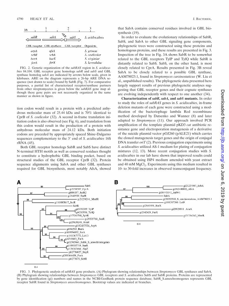
While no changes in thaxtomin production were seen be-tween the wild type and the mutants, a comparison of the wildtype and the �sabA and �sabS strains grown on solid SGMshowed that these two mutants produced higher levels of ayellow diffusible pigment than the parent strain (Fig. 4A). No
significant differences in pigment production relative to theparent strain were observed in �sabR mutants on SGM oroatmeal agar medium (data not shown). To verify that thepigment overproduction phenotypes observed in sabA andsabS mutants were due to the mutant sabA and sabS alleles, thewild-type alleles of these genes were cloned into plasmid pIJ86under the transcriptional control of the constitutive ermE*promoter (1). Normal wild-type levels of pigment productionwere seen in mutant transconjugants carrying the plasmids,demonstrating that pigment production phenotypes were dueto mutations in sabA and sabS (Fig. 4B and C).
In addition to differences in pigment production by sabS andsabA mutants, we also observed morphological differences insabS mutants when the organism was cultured on ISP2 agarmedium, a rich medium in comparison to mannitol-basedSGM or oatmeal-based medium. Mutants grown on ISP2 me-dium largely failed to produce aerial hyphae from substratemycelium, even after prolonged growth (Fig. 4D). Partial res-toration of normal morphological development was observedin sabS mutants on ISP2 when they carried wild-type sabS onthe multicopy plasmid pIJ86 (Fig. 4D). This effect was notobserved when the sabS mutant was cultured on mannitol- orstarch-based medium, such as SGM (Fig. 4B). While aerialhyphal development of sabS mutants was not impaired onSGM, we did observe a more compact, tighter colony morphol-
FIG. 4. Phenotypic properties of sabR, sabA, and sabS mutants. (A) Pigment production characteristics of sabR, sabA, and sabS deletionmutants on SGM. wt, wild-type strain S. acidiscabies 84.104. (B) Complementation of sabS deletion mutant on SGM with plasmid pIJ86::sabS.(C) Complementation of sabA deletion mutant on SGM with plasmid pIJ86::sabA. (D) Complementation of conditional morphological defect ofsabS mutant on ISP2 medium with plasmid pIJ86::sabS.
VOL. 191, 2009 GBL SIGNALING GENES IN S. ACIDISCABIES 4791
on June 6, 2019 by guesthttp://jb.asm
.org/D
ownloaded from
ogy of sabS mutants on SGM plates. Partial restoration ofnormal colony morphology was observed in sabS mutants onSGM when they carried wild-type sabS on the multicopy plas-mid pIJ86 (Fig. 4B).
The conditional morphological defect observed on ISP2 me-dium is reminiscent of effects seen in, e.g., S. coelicolor bldAmutants or other strains (e.g., adpA mutants) with defects ingenes containing UUA codons when cultured on rich, high-osmolarity medium such as R2YE (24, 27, 33). Morphologicaldefects can be suppressed in such mutants when cells arecultured on mannitol-based medium. The bldA gene encodestRNAleu and recognizes rare UUA codons in high-GC contentStreptomyces mRNA transcripts. Numerous genes containingTTA codons have been identified, and many of these are genesinvolved in morphogenesis or secondary metabolism (5). No-tably, the sabS gene sequence possesses a TTA leucine codonin the 5 end of the gene. The codon sequence occurs at eithernucleotides 13 to 15 (the 5th triplet codon) or at nucleotides 52to 54 (the 18th triplet codon), depending on which translationstart codon is used (see also Fig. 6A). Based on alignmentswith other GBL receptor homologs, the amino acid positionoccupied by leucine in SabS resides in the N terminus of theprotein in a region preceding the DNA binding HTH domain.Further, this amino acid position is usually occupied by glu-tamic acid (12 out of 18 proteins examined); there are no otheroccurrences of leucine at this position in accessible GBL re-ceptor sequences we examined. Although specific target genessubject to regulation by SabS have not yet been identified,these results suggest that SabS could function as a transcrip-tional regulator of genes related to morphological develop-ment. These studies are currently under way.
To more thoroughly investigate the nature of the diffusibleyellow pigment compound produced in excess in sabS and sabAmutants, the mutants and wild-type strains were grown in liq-uid SGM, and culture filtrates were extracted using solid-phasemethods. Extracts were analyzed using silica gel TLC and com-pared with preparations of pure WS5995B. Extracts of the�sabS and �sabA culture filtrates contained higher levels of ayellow pigment that was also present in the wild-type culturefiltrate extracts, indicating overproduction of a metabolite inthe mutant strains. The compound also displayed chromato-graphic properties on TLC plates similar to pure WS5995B.The yellow compound was recovered from preparative silicagel medium, and absorbance scans revealed that the materialexhibited spectroscopic properties identical to those of pureWS5995B (data not shown). The properties of silica gel-puri-fied compound as determined by analytical high-performanceliquid chromatography were also identical to those of pureWS5995B (data not shown). While higher levels of WS5995Bwere found in both �sabS and �sabA mutants, the amounts ofWS5995B produced by the �sabS mutant were substantiallyhigher than in the �sabA mutant.
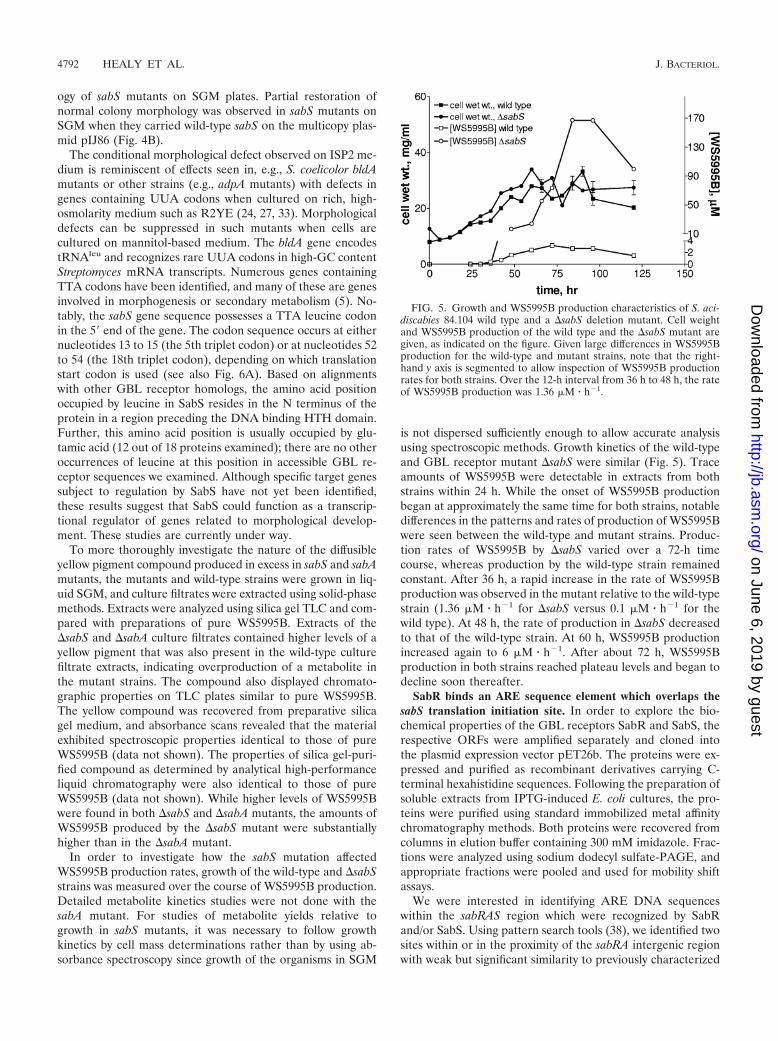
In order to investigate how the sabS mutation affectedWS5995B production rates, growth of the wild-type and �sabSstrains was measured over the course of WS5995B production.Detailed metabolite kinetics studies were not done with thesabA mutant. For studies of metabolite yields relative togrowth in sabS mutants, it was necessary to follow growthkinetics by cell mass determinations rather than by using ab-sorbance spectroscopy since growth of the organisms in SGM
is not dispersed sufficiently enough to allow accurate analysisusing spectroscopic methods. Growth kinetics of the wild-typeand GBL receptor mutant �sabS were similar (Fig. 5). Traceamounts of WS5995B were detectable in extracts from bothstrains within 24 h. While the onset of WS5995B productionbegan at approximately the same time for both strains, notabledifferences in the patterns and rates of production of WS5995Bwere seen between the wild-type and mutant strains. Produc-tion rates of WS5995B by �sabS varied over a 72-h timecourse, whereas production by the wild-type strain remainedconstant. After 36 h, a rapid increase in the rate of WS5995Bproduction was observed in the mutant relative to the wild-typestrain (1.36 �M � h�1 for �sabS versus 0.1 �M � h�1 for thewild type). At 48 h, the rate of production in �sabS decreasedto that of the wild-type strain. At 60 h, WS5995B productionincreased again to 6 �M � h�1. After about 72 h, WS5995Bproduction in both strains reached plateau levels and began todecline soon thereafter.
SabR binds an ARE sequence element which overlaps thesabS translation initiation site. In order to explore the bio-chemical properties of the GBL receptors SabR and SabS, therespective ORFs were amplified separately and cloned intothe plasmid expression vector pET26b. The proteins were ex-pressed and purified as recombinant derivatives carrying C-terminal hexahistidine sequences. Following the preparation ofsoluble extracts from IPTG-induced E. coli cultures, the pro-teins were purified using standard immobilized metal affinitychromatography methods. Both proteins were recovered fromcolumns in elution buffer containing 300 mM imidazole. Frac-tions were analyzed using sodium dodecyl sulfate-PAGE, andappropriate fractions were pooled and used for mobility shiftassays.
We were interested in identifying ARE DNA sequenceswithin the sabRAS region which were recognized by SabRand/or SabS. Using pattern search tools (38), we identified twosites within or in the proximity of the sabRA intergenic regionwith weak but significant similarity to previously characterized
FIG. 5. Growth and WS5995B production characteristics of S. aci-discabies 84.104 wild type and a �sabS deletion mutant. Cell weightand WS5995B production of the wild type and the �sabS mutant aregiven, as indicated on the figure. Given large differences in WS5995Bproduction for the wild-type and mutant strains, note that the right-hand y axis is segmented to allow inspection of WS5995B productionrates for both strains. Over the 12-h interval from 36 h to 48 h, the rateof WS5995B production was 1.36 �M � h�1.
4792 HEALY ET AL. J. BACTERIOL.
on June 6, 2019 by guesthttp://jb.asm
.org/D
ownloaded from
ARE sequences in other streptomycetes. These sites werefound centered at �12 with respect to the sabR translationstart and at �14 with respect to the sabA translation start (datanot shown). A radiolabeled 294-bp PCR product was gener-ated which encompassed the sabRA intergenic region and 5ends of both genes, including predicted ARE sites. This la-beled fragment was used in gel mobility shift assays to detectbinding by SabR and/or SabS. Numerous reaction conditionswere tested, and no specific binding activity was observed foreither SabR or SabS, indicating that no ARE sequences arepresent in the sabRA intergenic region (not shown).
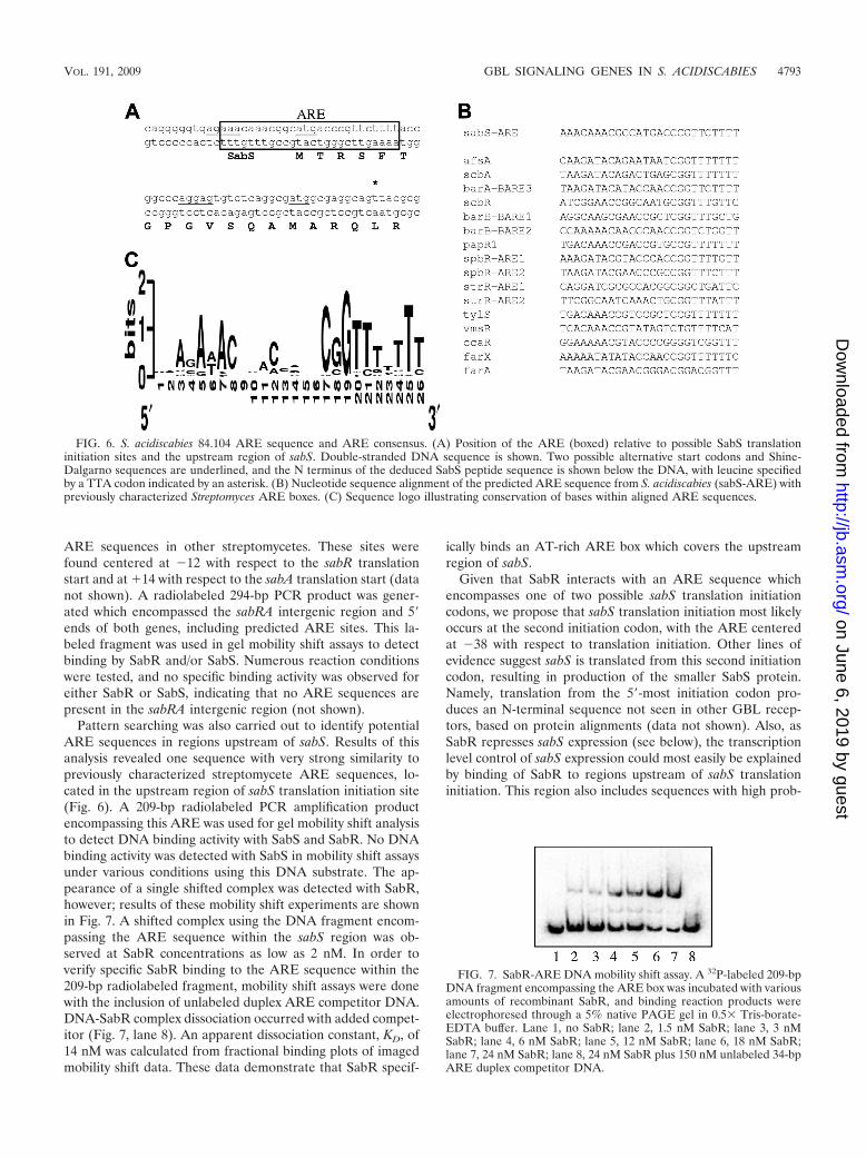
Pattern searching was also carried out to identify potentialARE sequences in regions upstream of sabS. Results of thisanalysis revealed one sequence with very strong similarity topreviously characterized streptomycete ARE sequences, lo-cated in the upstream region of sabS translation initiation site(Fig. 6). A 209-bp radiolabeled PCR amplification productencompassing this ARE was used for gel mobility shift analysisto detect DNA binding activity with SabS and SabR. No DNAbinding activity was detected with SabS in mobility shift assaysunder various conditions using this DNA substrate. The ap-pearance of a single shifted complex was detected with SabR,however; results of these mobility shift experiments are shownin Fig. 7. A shifted complex using the DNA fragment encom-passing the ARE sequence within the sabS region was ob-served at SabR concentrations as low as 2 nM. In order toverify specific SabR binding to the ARE sequence within the209-bp radiolabeled fragment, mobility shift assays were donewith the inclusion of unlabeled duplex ARE competitor DNA.DNA-SabR complex dissociation occurred with added compet-itor (Fig. 7, lane 8). An apparent dissociation constant, KD, of14 nM was calculated from fractional binding plots of imagedmobility shift data. These data demonstrate that SabR specif-
ically binds an AT-rich ARE box which covers the upstreamregion of sabS.
Given that SabR interacts with an ARE sequence whichencompasses one of two possible sabS translation initiationcodons, we propose that sabS translation initiation most likelyoccurs at the second initiation codon, with the ARE centeredat �38 with respect to translation initiation. Other lines ofevidence suggest sabS is translated from this second initiationcodon, resulting in production of the smaller SabS protein.Namely, translation from the 5-most initiation codon pro-duces an N-terminal sequence not seen in other GBL recep-tors, based on protein alignments (data not shown). Also, asSabR represses sabS expression (see below), the transcriptionlevel control of sabS expression could most easily be explainedby binding of SabR to regions upstream of sabS translationinitiation. This region also includes sequences with high prob-
FIG. 6. S. acidiscabies 84.104 ARE sequence and ARE consensus. (A) Position of the ARE (boxed) relative to possible SabS translationinitiation sites and the upstream region of sabS. Double-stranded DNA sequence is shown. Two possible alternative start codons and Shine-Dalgarno sequences are underlined, and the N terminus of the deduced SabS peptide sequence is shown below the DNA, with leucine specifiedby a TTA codon indicated by an asterisk. (B) Nucleotide sequence alignment of the predicted ARE sequence from S. acidiscabies (sabS-ARE) withpreviously characterized Streptomyces ARE boxes. (C) Sequence logo illustrating conservation of bases within aligned ARE sequences.
FIG. 7. SabR-ARE DNA mobility shift assay. A 32P-labeled 209-bpDNA fragment encompassing the ARE box was incubated with variousamounts of recombinant SabR, and binding reaction products wereelectrophoresed through a 5% native PAGE gel in 0.5� Tris-borate-EDTA buffer. Lane 1, no SabR; lane 2, 1.5 nM SabR; lane 3, 3 nMSabR; lane 4, 6 nM SabR; lane 5, 12 nM SabR; lane 6, 18 nM SabR;lane 7, 24 nM SabR; lane 8, 24 nM SabR plus 150 nM unlabeled 34-bpARE duplex competitor DNA.
VOL. 191, 2009 GBL SIGNALING GENES IN S. ACIDISCABIES 4793
on June 6, 2019 by guesthttp://jb.asm
.org/D
ownloaded from
abilities of promoter elements (data not shown). These areasof inquiry are currently being investigated.
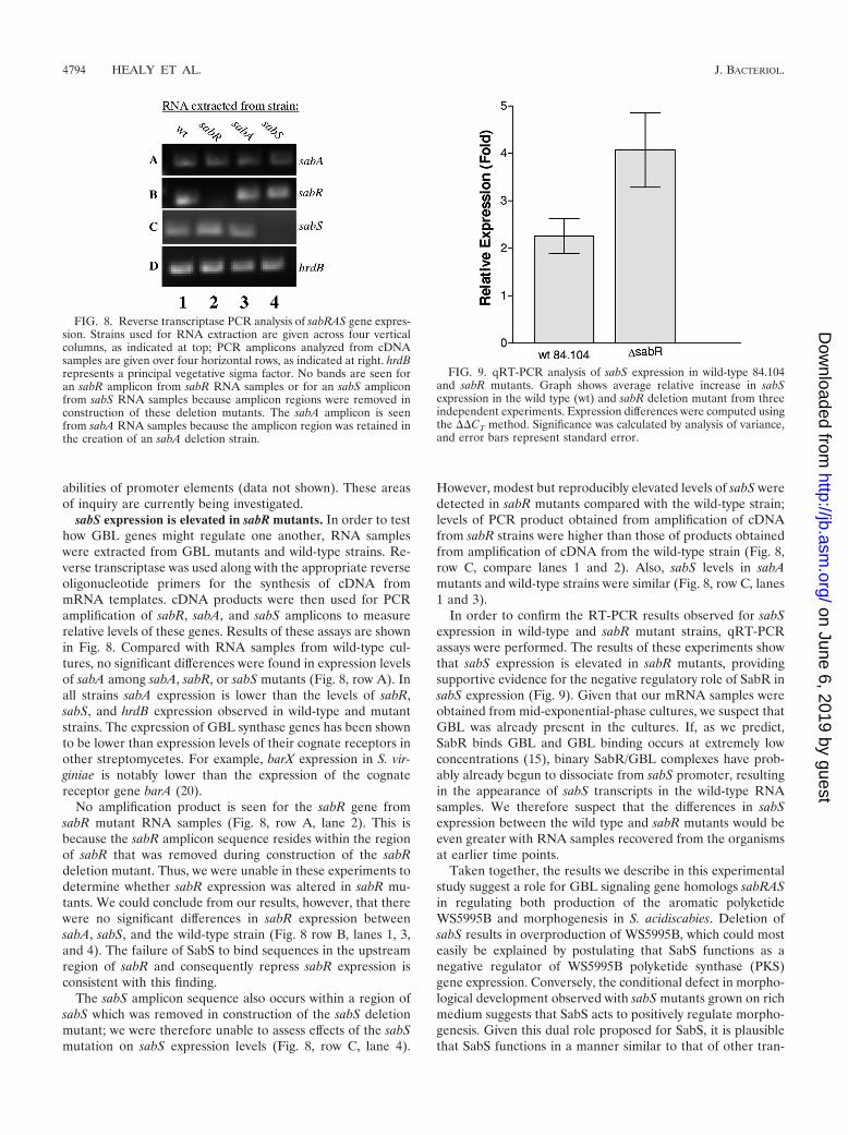
sabS expression is elevated in sabR mutants. In order to testhow GBL genes might regulate one another, RNA sampleswere extracted from GBL mutants and wild-type strains. Re-verse transcriptase was used along with the appropriate reverseoligonucleotide primers for the synthesis of cDNA frommRNA templates. cDNA products were then used for PCRamplification of sabR, sabA, and sabS amplicons to measurerelative levels of these genes. Results of these assays are shownin Fig. 8. Compared with RNA samples from wild-type cul-tures, no significant differences were found in expression levelsof sabA among sabA, sabR, or sabS mutants (Fig. 8, row A). Inall strains sabA expression is lower than the levels of sabR,sabS, and hrdB expression observed in wild-type and mutantstrains. The expression of GBL synthase genes has been shownto be lower than expression levels of their cognate receptors inother streptomycetes. For example, barX expression in S. vir-giniae is notably lower than the expression of the cognatereceptor gene barA (20).
No amplification product is seen for the sabR gene fromsabR mutant RNA samples (Fig. 8, row A, lane 2). This isbecause the sabR amplicon sequence resides within the regionof sabR that was removed during construction of the sabRdeletion mutant. Thus, we were unable in these experiments todetermine whether sabR expression was altered in sabR mu-tants. We could conclude from our results, however, that therewere no significant differences in sabR expression betweensabA, sabS, and the wild-type strain (Fig. 8 row B, lanes 1, 3,and 4). The failure of SabS to bind sequences in the upstreamregion of sabR and consequently repress sabR expression isconsistent with this finding.
The sabS amplicon sequence also occurs within a region ofsabS which was removed in construction of the sabS deletionmutant; we were therefore unable to assess effects of the sabSmutation on sabS expression levels (Fig. 8, row C, lane 4).
However, modest but reproducibly elevated levels of sabS weredetected in sabR mutants compared with the wild-type strain;levels of PCR product obtained from amplification of cDNAfrom sabR strains were higher than those of products obtainedfrom amplification of cDNA from the wild-type strain (Fig. 8,row C, compare lanes 1 and 2). Also, sabS levels in sabAmutants and wild-type strains were similar (Fig. 8, row C, lanes1 and 3).
In order to confirm the RT-PCR results observed for sabSexpression in wild-type and sabR mutant strains, qRT-PCRassays were performed. The results of these experiments showthat sabS expression is elevated in sabR mutants, providingsupportive evidence for the negative regulatory role of SabR insabS expression (Fig. 9). Given that our mRNA samples wereobtained from mid-exponential-phase cultures, we suspect thatGBL was already present in the cultures. If, as we predict,SabR binds GBL and GBL binding occurs at extremely lowconcentrations (15), binary SabR/GBL complexes have prob-ably already begun to dissociate from sabS promoter, resultingin the appearance of sabS transcripts in the wild-type RNAsamples. We therefore suspect that the differences in sabSexpression between the wild type and sabR mutants would beeven greater with RNA samples recovered from the organismsat earlier time points.
Taken together, the results we describe in this experimentalstudy suggest a role for GBL signaling gene homologs sabRASin regulating both production of the aromatic polyketideWS5995B and morphogenesis in S. acidiscabies. Deletion ofsabS results in overproduction of WS5995B, which could mosteasily be explained by postulating that SabS functions as anegative regulator of WS5995B polyketide synthase (PKS)gene expression. Conversely, the conditional defect in morpho-logical development observed with sabS mutants grown on richmedium suggests that SabS acts to positively regulate morpho-genesis. Given this dual role proposed for SabS, it is plausiblethat SabS functions in a manner similar to that of other tran-
FIG. 8. Reverse transcriptase PCR analysis of sabRAS gene expres-sion. Strains used for RNA extraction are given across four verticalcolumns, as indicated at top; PCR amplicons analyzed from cDNAsamples are given over four horizontal rows, as indicated at right. hrdBrepresents a principal vegetative sigma factor. No bands are seen foran sabR amplicon from sabR RNA samples or for an sabS ampliconfrom sabS RNA samples because amplicon regions were removed inconstruction of these deletion mutants. The sabA amplicon is seenfrom sabA RNA samples because the amplicon region was retained inthe creation of an sabA deletion strain.
FIG. 9. qRT-PCR analysis of sabS expression in wild-type 84.104and sabR mutants. Graph shows average relative increase in sabSexpression in the wild type (wt) and sabR deletion mutant from threeindependent experiments. Expression differences were computed usingthe ��CT method. Significance was calculated by analysis of variance,and error bars represent standard error.
4794 HEALY ET AL. J. BACTERIOL.
on June 6, 2019 by guesthttp://jb.asm
.org/D
ownloaded from
scription factors which function as intermediates in the pro-cessing of GBL signaling inputs to multiple outputs. For ex-ample, in Streptomyces pristinaespiralis the GBL receptor SpbRcontrols pristinamycin production through binding at the pro-moter region of the pristinamycin pathway-specific activatorpapR1. Additionally, SpbR plays a role in morphological de-velopment (10). It is likely that SabS responds to a ligand andthat target gene expression is mediated through this interac-tion. While SabS does not bind ARE sequences in the sabRASregion, it is plausible that, given its similarity to other GBLreceptors, SabS binds ARE sequences elsewhere in the ge-nome in a manner which would result in negative regulation ofexpression of a type II PKS gene cluster encoding enzymesrequired for WS5995B biosynthesis and in positive regulationof genes involved in morphogenesis. Whether such controlphenomena would be exerted directly or indirectly, throughpathway-specific promoter interactions such as occurs at, e.g.,papR1 is unknown. We are currently developing methods toidentify DNA binding sites for SabS to elucidate the position ofSabS within a regulatory network which would govern second-ary metabolism and morphological development. Using a strat-egy similar to the one we used for the identification of GBLreceptor genes (48), we have also identified type II PKS genesequences from S. acidiscabies genomic DNA which may en-code WS5995B PKS and pathway activators regulated by SabS-DNA interactions.
While it is tempting to speculate that SabS and SabR bindGBL compounds produced by S. acidiscabies since they areclearly homologous with other characterized GBL receptorproteins and since they lie immediately adjacent to the GBLsynthase homolog sabA, we have no direct evidence at this timethat either protein binds GBL compounds. It is for this reasonthat we refer to SabS and SabR as GBL receptor homologs,pending biochemical verification of GBL binding properties ofthese proteins. There are instances of GBL receptor homologswhich do not appear to bind GBLs. For example, regulation ofalpomycin biosynthesis in Streptomyces ambofaciens occursthrough interactions between the GBL receptor homologAlpZ and a ligand exhibiting chemical properties inconsistentwith those of GBL compounds (4). Also, it has been reportedthat the S. virginiae GBL receptor BarB does not exhibit GBLbinding activity (30). Moreover, based on phylogenetic argu-ments, it has been proposed that “GBL receptors” predateGBL synthases and initially functioned as DNA binding pro-teins prior to acquiring GBL binding properties (34). Thus,SabR and/or SabS may bind ligands other than GBL com-pounds.
As GBL compounds are typically produced in minute quan-tities and are recovered through extraction into organic sol-vent, large volumes of culture (�400 liter) and solvent arerequired for extraction to obtain quantities of material suffi-cient for absolute structural characterization of GBLs fromStreptomyces (see, for example, reference 43). For these tech-nical and economic reasons, we have not approached the ques-tion of a GBL structure from S. acidiscabies. However, usingelectrospray ionization mass spectrometry methods with cul-ture extracts as described by Yang et al. (49), we have detectedfragment ions bound by our purified GBL receptors withmasses consistent with molecules possessing lactone functionalgroups such as those found in GBL compounds (data not
shown). The proximity of sabA to both sabR and sabS alongwith the conservation of residues in SabA predicted to berequired for GBL synthase activity (19) invites the specula-tion that sabA encodes a GBL synthase. Technical chal-lenges exist in the biochemical characterization of SabA.For example, since we do not have a GBL structure from ourorganism, we lack information regarding SabA enzymaticsubstrates.
Nonetheless, we find changes in WS5995B production and inmorphological properties in S. acidiscabies GBL signalingpathway homolog mutants consistent with disruption of anautoregulatory GBL-like pathway. Let us assume, for examplethat sabA encodes a GBL synthase which produces a GBLligand that, in turn, binds the divergently expressed GBL re-ceptor SabR. Then, elevated production of WS5995B observedin sabA mutants could be attributed to SabR-mediated repres-sion of sabS, resulting in derepression of WS5995B PKS ex-pression. The observation that sabR mutants appear phenotyp-ically similar to the wild-type strain can be explained byassuming that if SabR represses sabS expression and if SabSalso binds GBL produced by SabA, the normal course ofthreshold accumulation of GBL produced by SabA would stillresult in dissociation of SabS from target regulatory sequences.Thus, sabS repression of PKS gene expression is still ultimatelysubject to control by GBL binding so that SabS can only re-press PKS expression until GBL accumulates, and at that pointone would observe nearly wild-type levels of WS5995B, even ina sabR mutant, since it would still synthesize GBL.
The significance of the TTA leucine codon in sabS is notknown. Recent bioinformatics analyses of four sequencedStreptomyces genomes shows that TTA-containing genes areoften associated with secondary metabolite biosynthetic geneclusters, particularly in those genes encoding likely regulatoryfunctions (5). While the genome of S. acidiscabies has not beensequenced, we assume that UUA codons are recognized by abldA-type tRNA, as seen in other streptomycetes (24, 26, 46,47). Our RT-PCR data suggest that sabS expression is elevatedin sabR mutants. If SabR represses sabS expression in theabsence of a ligand (e.g., a GBL) and if the intracellular ac-cumulation of ligand results in derepression of sabS expres-sion, we suspect that translation of the GBL receptor SabSwould be dependent on bldA tRNA.
The AdpA transcriptional regulator is encoded by a UUA-containing gene; it is present in all Streptomyces genomes se-quenced to date and has been characterized in both S. griseusand S. coelicolor. AdpA plays a central role in morphogenesisand secondary metabolism in both organisms, and the genepossesses UUA codons in both organisms. Yet only the S.griseus adpA gene appears to function in a GBL-dependentregulatory cascade (6, 36, 44). AdpA-dependent expression oftarget genes in S. griseus occurs following derepression of adpAexpression as a result of dissociation of A factor-ArpA com-plexes from adpA promoter. Given the central regulatory roleof AdpA, it is possible that SabS could function downstream ofAdpA in the bldA-dependent regulation of morphogenesis andWS5995B biosynthesis. Future research will be directed towardfurther characterization of the network governing secondarymetabolism and morphogenesis in this organism.
VOL. 191, 2009 GBL SIGNALING GENES IN S. ACIDISCABIES 4795
on June 6, 2019 by guesthttp://jb.asm
.org/D
ownloaded from
ACKNOWLEDGMENTS
We thank the following people: Takuya Nihira and Shigeru Kitanifor generously providing VB-C6 and IM-2-C6 compounds; Alaina Ed-munds for constructing plasmid pALE21; Merv Bibb, Joanne Willey,and Ron Parry for providing strains and plasmids; and Justin Nodwelland Jurgen Rohr for thoughtful discussions. We also thank the anon-ymous reviewers of the manuscript for valuable and critical comments.
This work was supported through funding from NSF (MCB 0442509),HHMI, and Merck-AAAS.
REFERENCES
1. Bibb, M. J., J. White, J. M. Ward, and G. R. Janssen. 1994. The mRNA forthe 23S rRNA methylase encoded by the ermE gene of Saccharopolysporaerythraea is translated in the absence of a conventional ribosome-binding site.Mol. Microbiol. 14:533–545.
2. Bierman, M., R. Logan, K. O’Brien, E. T. Seno, R. N. Rao, and B. E.Schoner. 1992. Plasmid cloning vectors for the conjugal transfer of DNAfrom Escherichia coli to Streptomyces spp. Gene 116:43–49.
3. Birko, Z., S. Bialek, K. Buzas, E. Szajli, B. A. Traag, K. F. Medzihradszky,S. Rigali, E. Vijgenboom, A. Penyige, Z. Kele, G. P. van Wezel, and S. Biro.2007. The secreted signaling protein factor C triggers the A-factor responseregulon in Streptomyces griseus: overlapping signalling routes. Mol. Cell Pro-teomics 6:1248–1256.
4. Bunet, R., M. V. Mendes, N. Rouhier, X. Pang, L. Hotel, P. Leblond, and B.Aigle. 2008. Regulation of the synthesis of the angucyclinone antibioticalpomycin in Streptomyces ambofaciens by the autoregulator receptor AlpZand its specific ligand. J. Bacteriol. 190:3293–3305.
5. Chater, K. F., and G. Chandra. 2008. The use of the rare UUA codon todefine “expression space” for genes involved in secondary metabolism, de-velopment and environmental adaptation in Streptomyces. J. Microbiol. 46:1–11.
6. Chater, K. F., and S. Horinouchi. 2003. Signalling early developmentalevents in two highly diverged Streptomyces species. Mol. Microbiol. 48:9–15.
7. Cheng, Y.-Q., G.-L. Tang, and B. Shen. 2002. Identification and localizationof the gene cluster encoding biosynthesis of the antitumor macrolactamleinamycin in Streptomyces atroolivaceous S-140. J. Bacteriol. 184:7013–7024.
8. Datsenko, K. A., and B. L. Wanner. 2000. One-step inactivation of chromo-somal genes in Escherichia coli K-12 using PCR products. Proc. Natl. Acad.Sci. USA 97:6640–6645.
9. Dereeper, A., V. Guignon, G. Blanc, S. Audic, S. Buffet, F. Chevenet, J. F.Dufayard, S. Guindon, V. Lefort, M. Lescot, J. M. Claverie, and O. Gascuel.2008. Phylogeny.fr: robust phylogenetic analysis for the non-specialist. Nu-cleic Acids Res. 36:W465–W469.
10. Folcher, M., H. Gaillard, L. T. Nguyen, K. T. Nguyen, P. Lacroix, N. Bamas-Jacques, M. Rinkel, and C. J. Thompson. 2001. Pleiotropic functions of aStreptomyces pristinaespiralis autoregulator receptor in development, antibi-otic biosynthesis, and expression of a superoxide dismutase. J. Biol. Chem.276:44297–44306.
11. Gust, B., G. L. Challis, K. Fowler, T. Kieser, and K. F. Chater. 2003.PCR-targeted Streptomyces gene replacement identifies a protein domainneeded for biosynthesis of the sesquiterpene soil odor geosmin. Proc. Natl.Acad. Sci. USA 100:1541–1546.
12. Healy, F. G., S. B. Krasnoff, M. Wach, D. M. Gibson, and R. Loria. 2002.Involvement of a cytochrome P450 monooxygenase in thaxtomin A biosyn-thesis by Streptomyces acidiscabies. J. Bacteriol. 184:2019–2029.
13. Healy, F. G., M. Wach, S. B. Krasnoff, D. M. Gibson, and R. Loria. 2000. ThetxtAB genes of the plant pathogen Streptomyces acidiscabies encode a peptidesynthetase required for phytotoxin thaxtomin A production and pathogenic-ity. Mol. Microbiol. 38:794–804.
14. Hinrichs, W., C. Kisker, M. Duvel, A. Muller, K. Tovar, W. Hillen, and W.Saenger. 1994. Structure of the TetR repressor-tetracycline complex andregulation of antibiotic resistance. Science 264:418–420.
15. Horinouchi, S. 2002. A microbial hormone, A-factor, as a master switch formorphological differentiation and secondary metabolism in Streptomyces gri-seus. Front. Biosci. 7:2045–2057.
16. Horinouchi, S. 2007. Mining and Polishing of the Treasure Trove in theBacterial Genus Streptomyces. Biosci. Biotechnol. Biochem. 71:283–299.
17. Ikushima, H., M. Okamoto, H. Tanaka, O. Ohe, M. Kohsaka, H. Aoki, andH. Imanaka. 1980. New anticoccidial antibiotics, WS-5995 A and B I. Iso-lation and characterization. J. Antibiot. 33:1107–1113.
18. Joshi, M. V., D. R. Bignell, E. G. Johnson, J. P. Sparks, D. M. Gibson, andR. Loria. 2007. The AraC/XylS regulator TxtR modulates thaxtomin biosyn-thesis and virulence in Streptomyces scabies. Mol. Microbiol. 66:633–642.
19. Kato, J. Y., N. Funa, H. Watanabe, Y. Ohnishi, and S. Horinouchi. 2007.Biosynthesis of gamma-butyrolactone autoregulators that switch on second-ary metabolism and morphological development in Streptomyces. Proc. Natl.Acad. Sci. USA 104:2378–2383.
20. Kawachi, R., T. Akashi, Y. Kamitani, A. Sy, U. Wangchaisoonthorn, T.Nihira, and Y. Yamada. 2000. Identification of an AfsA homologue (BarX)from Streptomyces virginiae as a pleiotropic regulator controlling autoregu-
lator biosynthesis, virginiamycin biosynthesis and virginiamycin M1 resis-tance. Mol. Microbiol. 36:302–313.
21. King, R. R., and C. H. Lawrence. 1996. Isolation and identification of pig-ments generated in vitro by Streptomyces acidiscabies. J. Agric. Food Chem.44:2849–2851.
22. Kinoshita, H., H. Ipposhi, S. Okamoto, H. Nakano, T. Nihira, and Y.Yamada. 1997. Butyrolactone autoregulator receptor protein (BarA) as atranscriptional regulator in Streptomyces virginiae. J. Bacteriol. 179:6986–6993.
23. Kitani, S., Y. Yamada, and T. Nihira. 2001. Gene replacement analysis of thebutyrolactone autoregulator receptor (FarA) reveals that FarA acts as anovel regulator in secondary metabolism of Streptomyces lavendulae FRI-5. J.Bacteriol. 183:4357–4363.
24. Kwak, J., L. A. McCue, and K. E. Kendrick. 1996. Identification of bldAmutants of Streptomyces griseus. Gene 171:75–78.
25. Lawrence, C. H., M. C. Clark, and R. R. King. 1990. Induction of commonscab symptoms in aseptically cultured potato tubers by the vivotoxin, thax-tomin. Phytopathology 80:606–608.
26. Leskiw, B. K., M. J. Bibb, and K. F. Chater. 1991. The use of a rare codonspecifically during development? Mol. Microbiol. 5:2861–2867.
27. Li, W., J. Wu, W. Tao, C. Zhao, Y. Wang, X. He, G. Chandra, Z. Zhou,K. F. Chater, and M. Tao. 2007. A genetic and bioinformatic analysis ofStreptomyces coelicolor genes containing TTA codons, possible targets forregulation by a developmentally significant tRNA. FEMS Microbiol. Lett.266:20–28.
28. Loria, R., R. A. Bukhalid, R. A. Creath, R. H. Leiner, M. Olivier, and J. C.Steffens. 1995. Differential production of thaxtomins by pathogenic Strepto-myces species in vitro. Phytopathology 85:537–541.
29. Loria, R., R. A. Bukhalid, B. A. Fry, and R. R. King. 1997. Plant pathoge-nicity in the genus Streptomyces. Plant Dis. 81:836–846.
30. Matsuno, K., Y. Yamada, C. K. Lee, and T. Nihira. 2004. Identification bygene deletion analysis of BarB as a negative regulator controlling an earlyprocess of virginiamycin biosynthesis in Streptomyces virginiae. Arch. Micro-biol. 181:52–59.
31. Nakano, H., E. Takehara, T. Nihira, and Y. Yamada. 1998. Gene replace-ment analysis of the Streptomyces virginiae barA gene encoding the butyro-lactone autoregulator receptor reveals that BarA acts as a repressor invirginiamycin biosynthesis. J. Bacteriol. 180:3317–3322.
32. Natsume, R., Y. Ohnishi, T. Senda, and S. Horinouchi. 2004. Crystal struc-ture of a gamma-butyrolactone autoregulator receptor protein in Streptomy-ces coelicolor A3(2). J. Mol. Biol. 336:409–419.
33. Nguyen, K. T., J. Tenor, H. Stettler, L. T. Nguyen, L. D. Nguyen, and C. J.Thompson. 2003. Colonial differentiation in Streptomyces coelicolor dependson translation of a specific codon within the adpA gene. J. Bacteriol. 185:7291–7296.
34. Nishida, H., Y. Ohnishi, T. Beppu, and S. Horinouchi. 2007. Evolution ofgamma-butyrolactone synthases and receptors in Streptomyces. Environ. Mi-crobiol. 9:1986–1994.
35. Ohnishi, Y., J. Ishikawa, H. Hara, H. Suzuki, M. Ikenoya, H. Ikeda, A.Yamashita, M. Hattori, and S. Horinouchi. 2008. Genome sequence of thestreptomycin-producing microorganism Streptomyces griseus IFO 13350. J.Bacteriol. 190:4050–4060.
36. Ohnishi, Y., H. Yamazaki, J. Kato, A. Tomono, and S. Horinouchi. 2005.AdpA, a central transcriptional regulator in the A-factor regulatory cascadethat leads to morphological development and secondary metabolism in Strep-tomyces griseus. Biosci. Biotechnol. Biochem. 69:431–439.
37. Ramos, J. L., M. Martinez-Bueno, A. J. Molina-Henares, W. Teran, K.Watanabe, X. Zhang, M. T. Gallegos, R. Brennan, and R. Tobes. 2005. TheTetR family of transcriptional repressors. Microbiol. Mol. Biol. Rev. 69:326–356.
38. Rigoutsos, I., and A. Floratos. 1998. Combinatorial pattern discovery inbiological sequences: the Teiresias algorithm. Bioinformatics 14:55–67.
39. Rose, T. M., E. R. Schultz, J. G. Henikoff, S. Pietrokovski, C. M. McCallum,and S. Henikoff. 1998. Consensus-degenerate hybrid oligonucleotide primersfor amplification of distantly related sequences. Nucleic Acids Res. 26:1628–1635.
40. Sambrook, J., E. F. Fritsch, and T. Maniatis. 1989. Molecular cloning: alaboratory manual, 2nd ed. Cold Spring Harbor Laboratory Press, ColdSpring Harbor, NY.
41. Shirling, E. B., and D. Gottlieb. 1966. Methods for characterization ofStreptomyces species. Int. J. Syst. Bacteriol. 16:313–340.
42. Takano, E., R. Chakraburtty, T. Nihira, Y. Yamada, and M. J. Bibb. 2001.A complex role for the gamma-butyrolactone SCB1 in regulating antibi-otic production in Streptomyces coelicolor A3(2). Mol. Microbiol. 41:1015–1028.
43. Takano, E., T. Nihira, Y. Hara, J. J. Jones, C. J. Gershater, Y. Yamada, andM. Bibb. 2000. Purification and structural determination of SCB1, a gamma-butyrolactone that elicits antibiotic production in Streptomyces coelicolorA3(2). J. Biol. Chem. 275:11010–11016.
44. Takano, E., M. Tao, F. Long, M. J. Bibb, L. Wang, W. Li, M. J. Buttner, M. J.Bibb, Z. X. Deng, and K. F. Chater. 2003. A rare leucine codon in adpA is
4796 HEALY ET AL. J. BACTERIOL.
on June 6, 2019 by guesthttp://jb.asm
.org/D
ownloaded from
implicated in the morphological defect of bldA mutants of Streptomycescoelicolor. Mol. Microbiol. 50:475–486.
45. Takeuchi, T., H. Sawada, F. Tanaka, and I. Matsuda. 1996. Phylogeneticanalysis of Streptomyces spp. causing potato scab based on 16S rRNA se-quences. Int. J. Syst. Bacteriol. 46:476–479.
46. Tao, W. F., J. Wu, Z. X. Deng, and M. F. Tao. 2007. Cloning of bldA and theeffect on morphological differentiation and avermectins production in Strep-tomyces avermitilis NRRL8165. Wei Sheng Wu Xue Bao 47:34–38.
47. Trepanier, N. K., S. Jensen, D. C. Alexander, and B. K. Leskiw. 2002. The
positive activator of cephamycin C and clavulinic acid production in Streptomy-ces clavuligerus is mistranslated in a bldA mutant. Microbiol. 148:643–656.
48. Wawrik, B., L. Kerkhof, G. J. Zylstra, and J. J. Kukor. 2005. Identificationof unique type II polyketide synthase genes in soil. Appl. Environ. Microbiol.71:2232–2238.
49. Yang, Y. H., H. S. Joo, K. Lee, K. K. Liou, H. C. Lee, J. K. Sohng, and B. G.Kim. 2005. Novel method for detection of butanolides in Streptomyces coeli-color culture broth, using a His-tagged receptor (ScbR) and mass spectrom-etry. Appl. Environ. Microbiol. 71:5050–5055.
VOL. 191, 2009 GBL SIGNALING GENES IN S. ACIDISCABIES 4797
on June 6, 2019 by guesthttp://jb.asm
.org/D
ownloaded from


![INDEX [link.springer.com]978-1-4684-4619... · 2017. 8. 29. · triplet state, 114 Acridone, potassium salt r-butyrolactone reaction, 114, 115 Acridone, N-substituted fluorescence](https://img.pdfslide.us/doc/110x75/60fa2d05f21a9b206b77c60d/index-link-978-1-4684-4619-2017-8-29-triplet-state-114-acridone.jpg)