Embed Size (px)
Citation preview

Characterization and DiagnosticApplication of Trypanosoma cruziTrypomastigote Excreted-SecretedAntigens Shed in Extracellular VesiclesReleased from Infected Mammalian Cells
Norma L. Bautista-López,a,b Momar Ndao,b,c Fabio Vasquez Camargo,b,c
Takeshi Nara,d Takeshi Annoura,e Darryl B. Hardie,f Christoph H. Borchers,f
Armando Jardima,b
Institute of Parasitology, McGill University, Montreal, Quebec, Canadaa; Centre for Host Parasite-Interactions,McGill University, Montreal, Quebec, Canadab; National Reference Centre for Parasitology, Research Institute ofthe McGill University Health Centre, Montreal, Quebec, Canadac; Department of Molecular and CellularParasitology, Juntendo University Graduate School of Medicine, Tokyo, Japand; Department of Parasitology,National Institute of Infectious Diseases, Tokyo, Japane; University of Victoria Genome British ColumbiaProteomics Centre, Victoria, Canadaf
ABSTRACT Chagas disease, caused by Trypanosoma cruzi, although endemic inmany parts of Central and South America, is emerging as a global health threatthrough the potential contamination of blood supplies. Consequently, in the ab-sence of a gold standard assay for the diagnosis of Chagas disease, additionalantigens or strategies are needed. A proteomic analysis of the trypomastigoteexcreted-secreted antigens (TESA) associated with exosomal vesicles shed by T.cruzi identified �80 parasite proteins, with the majority being trans-sialidases.Mass spectrometry analysis of immunoprecipitation products performed usingChagas immune sera showed a marked enrichment in a subset of TESA proteins.Of particular relevance for diagnostic applications were the retrotransposon hotspot (RHS) proteins, which are absent in Leishmania spp., parasites that oftenconfound diagnosis of Chagas disease. Interestingly, serological screens using re-combinant RHS showed a robust immunoreactivity with sera from patients withclinical stages of Chagas ranging from asymptomatic to advance cardiomyopathyand this immunoreactivity was comparable to that of crude TESA. More impor-tantly, no cross-reactivity with RHS was detected with sera from patients withmalaria, leishmaniasis, toxoplasmosis, or African sleeping sickness, making thisprotein an attractive reagent for diagnosis of Chagas disease.
KEYWORDS parasite, proteomics, TESA, Trypanosoma cruzi, trypomastigote
Chagas disease is a neglected tropical disease caused by the protozoan parasiteTrypanosoma cruzi. Chagas disease is a major cause of morbidity and mortality in
South and Latin America, where currently �9 million people are infected with T. cruzi.However, with increased globalization and immigration, Chagas disease has emergedas a health threat in Europe, Asia, and North America, due to transmission of T. cruzithrough blood transfusions, organ transplants, or congenital infections associated withtransplacental transfer of this parasite from mother to newborn (1–5). It is estimated that�300,000 infected individuals currently live in the United States, while in Canada theinfection rate among �130, 000 Latino immigrants was �0.09% (6).
Chagas disease is characterized by an acute and chronic phase of infection. Theacute stage of the disease develops after a short period (1 to 2 weeks) following
Received 2 August 2016 Returned formodification 26 August 2016 Accepted 11November 2016
Accepted manuscript posted online 14December 2016
Citation Bautista-López NL, Ndao M, CamargoFV, Nara T, Annoura T, Hardie DB, BorchersCH, Jardim A. 2017. Characterization anddiagnostic application of Trypanosoma cruzitrypomastigote excreted-secreted antigensshed in extracellular vesicles released frominfected mammalian cells. J Clin Microbiol55:744 –758. https://doi.org/10.1128/JCM.01649-16.
Editor Yi-Wei Tang, Memorial Sloan-KetteringCancer Center
Copyright © 2017 American Society forMicrobiology. All Rights Reserved.
Address correspondence to Armando Jardim,[email protected].
For a commentary on this article, see https://doi.org/10.1128/JCM.02353-16.
IMMUNOASSAYS
crossm
March 2017 Volume 55 Issue 3 jcm.asm.org 744Journal of Clinical Microbiology
on May 21, 2020 by guest
http://jcm.asm
.org/D
ownloaded from

transmission of the T. cruzi parasites, where it typically presents with the clinical signsof elevated fever, aches, and an acute inflammatory response that reduces the parasiteburden (7–9). In some individuals, low levels of intracellular parasites continue toproliferate and persist in tissues for decades, remaining asymptomatic, which results inthe establishment of a chronic infection (8, 10). In �30% of chronic disease cases,patients develop significant complications, which may include megacolon, neurologicalcomplications, and cardiomyopathy that is characterized by an enlargement of theheart, ventricular arrhythmias, and eventual death due to general heart failure (11, 12).Infants and newborns are the demographic group with the highest risk of developinga chronic infection (8, 13).
Approaches currently used for Chagas diagnosis include microscopy, whichdetects parasites in tissues, quantitative PCR (qPCR), which measures levels ofparasite DNA in host tissues, and serological methods, such as enzyme-linkedimmunosorbent assays (ELISA) and immunoblotting, which detect circulating T.cruzi-specific antibodies. Microscopy and PCR-based methods are more effective fordiagnosing acute or congenital forms of Chagas disease (14, 15), while serologicaltests using either parasite-derived antigens, recombinant proteins, or syntheticpeptides are preferred for diagnosis of chronic infections (16). Despite the sensi-tivity of serological tests, current Chagas disease diagnostic tests may lack speci-ficity due to cross-reactivity with the related parasites Leishmania spp. and Trypano-soma rangeli (17). Consequently, the Pan American Health Organization hasrecommended (18) the use of two different assays for a confirmatory diagnosis ofChagas infection (19–21). A typical serological method recommended for confirm-ing Chagas disease uses the trypomastigote excretory-secretory antigens (TESA)either in an ELISA or immunoblotting format to detect antibodies that cross-reactwith proteins or glycoconjugates released by T. cruzi (22–25).
It is known that T. cruzi parasites, like many other cells, release extracellular vesiclesthat are postulated to be involved in cell-cell communication or in the modulation ofthe host immune responses to promote the establishment of an infection (26–29).These vesicles typically consist of a lipid bilayer membrane containing integral mem-brane proteins and a luminal cavity that is loaded with a variety of soluble proteins andnucleic acids (RNA and DNA). In T. cruzi parasites, two classes of vesicles, based on size,have been characterized. These include exovesicles (EVs; also referred to as ectosomes;100 to 1,000 nm), which bud directly from the plasma membrane, and exosomes (30 to100 nm), which are vesicles that are secreted into the extracellular environmentfollowing the fusion of multivesicular endosomes with the plasma membrane, typicallyoccurring at the flagellar pocket membrane (28, 30–33). A proteomic analysis ofextracellular vesicles released by metacyclic trypomastigotes and epimastigotes inculture demonstrated the presence of two populations of EVs containing plasmamembrane and intracellular proteins, and also nucleic acids (26, 29, 32–34). Interest-ingly, treatment of mice with EVs shed by axenic trypomastigotes caused a down-modulation of the host immune response that was associated with higher parasitemiaand an exacerbated inflammatory response that resulted in increased mortality follow-ing infection (26, 35). The T. cruzi small membrane proteins (TcSMP) family of proteinsor phosphatases detected on T. cruzi EVs has been shown to trigger Ca2� signaling andlysosome mobilization/exocytosis, events that promote formation of parasitophorousvacuoles and parasite invasion (36, 37). A similar modulation of macrophage responseswas observed following exposure to purified Leishmania exosomes, a strategy thatenhances intracellular parasite survival (38, 39). Mechanistic studies suggest that in theearly stages of infection by T. cruzi, parasites promote the release of plasma membranevesicles from the host cell, which may contribute to parasite survival in the circulatorysystem, an event thought to help mediate host cell invasion (40).
Although ELISAs and immunoblot serological assays using TESA are highly sensitive,there are some concerns due to the cross-reactivity for patients infected with Leish-mania, which may lead to misdiagnosis. However, the identification of antigens that areonly expressed by T. cruzi parasites would significantly increase the specificity of
T. cruzi Trypomastigote Exosome Antigens Journal of Clinical Microbiology
March 2017 Volume 55 Issue 3 jcm.asm.org 745
on May 21, 2020 by guest
http://jcm.asm
.org/D
ownloaded from

serological assays. In addition, the availability of diagnostic testing that would quanti-tatively detect the levels of T. cruzi antigens in body fluids, such as plasma or urine,could potentially be used to measure parasite burdens during acute and chronic phasesof Chagas disease.
Metacyclic trypomastigotes released by the insect vector invade phagocytic andnonphagocytic cells and, once internalized, transform to the amastigote stage, whichreplicates in the cytosolic compartment. These amastigotes then convert back to thetrypomastigote stage prior to rupturing the host cell. It is conjectured that the intra-cellular stages of T. cruzi, like those of the related parasite Leishmania spp. (38, 41), shedexosomes into the circulatory system by exocytosis (31) or when the host cell bursts.Indeed, EVs may account for T. cruzi antigens observed in the circulation and in urineof Chagas patients or infected animals with acute or chronic infections (42–44).However, no systematic analysis of T. cruzi antigens released from infected host cellshas been reported, despite the use of TESA as a reagent in disease diagnosis (22, 25, 32,45–47).
In this study, we selectively employed a purification strategy designed to isolateTESA EVs released by T. cruzi trypomastigotes and amastigotes in infected Vero cellsand then conducted a proteomic analysis of these vesicles. Moreover, affinity columnsgenerated from the immune sera of Chagas patients allowed the immunoprecipitationand proteomic characterization of numerous T. cruzi proteins that are likely releasedinto the circulatory system during the chronic phase of infection
RESULTSTrypomastigote excreted-secreted antigen preparations contain EVs. Vero cell
cultures infected with T. cruzi trypomastigotes are known to spontaneously shedparasite antigens into the culture supernatant. To perform an in-depth characterizationof parasite proteins associated with EVs in TESA preparations, these proteins wereconcentrated from the culture supernatant by ultracentrifugation sedimentation andthen purified by sucrose density flotation centrifugation to eliminate proteins that werenot encapsulated or associated with EV membranes. Analysis of high-speed centrifu-gation supernatants and the sucrose density-purified EVs by using silver-stained SDS-PAGE showed that the supernatant fraction contained predominantly proteins withmasses of �100 kDa. In contrast, proteins partitioning with the EVs had masses rangingfrom �30 to 245 kDa (Fig. 1A). Moreover, the proteins cofractionating with EVs isolatedfrom different batches of TESA exhibited a highly reproducible pattern, as shown by thesimilar SDS-PAGE profiles (Fig. 1A, lanes 2 and 3). The presence of EVs in the top fractionfrom the sucrose density gradient was validated by negative-stain electron microscopy
FIG 1 Isolation and characterization of TESA exovesicles. (A) Crude TESA isolated from infected Vero cellculture supernatant was concentrated by ultrafiltration, and a supernatant (lane 1) and EV pellets fromtwo different batches (lanes 2 and 3) were isolated by ultracentrifugation as described in Materials andMethods. (B) The presence of EV in the sucrose density flotation fraction was determined by negativestaining electron microscopy.
Bautista-López et al. Journal of Clinical Microbiology
March 2017 Volume 55 Issue 3 jcm.asm.org 746
on May 21, 2020 by guest
http://jcm.asm
.org/D
ownloaded from
analysis. The electron-dense vesicles had diameters of �60 to 100 nm (Fig. 1B), whichis consistent with the size ranges previously reported for EVs (48).
Proteomic analysis of TESA EVs. Liquid chromatography-tandem mass spectrom-etry (LC-MS/MS) spectra obtained for purified TESA EV proteins were used to search T.cruzi and Macaque spp. genome databases. The latter genome database was includedsince it was speculated that the TESA EV preparations would contain a mixture ofparasite and Vero cell-derived proteins. LC-MS/MS analysis generated 11,016 spectracorresponding to 766 proteins containing at least two high-confidence unique peptidesat a protein identification confidence level of 95% (see Table S1 in the supplementalmaterial). The bulk of these proteins were of Vero cell origin (Table S1), many of whichhave been previously detected in EVs isolated from mammalian cells (49). The remain-ing proteins were derived from either T. cruzi trypomastigotes or amastigotes (TablesS1 and S2). Quantification analysis performed using spectral counts indicated that only�10% of the total proteins detected in the EV preparations were of T. cruzi origin.
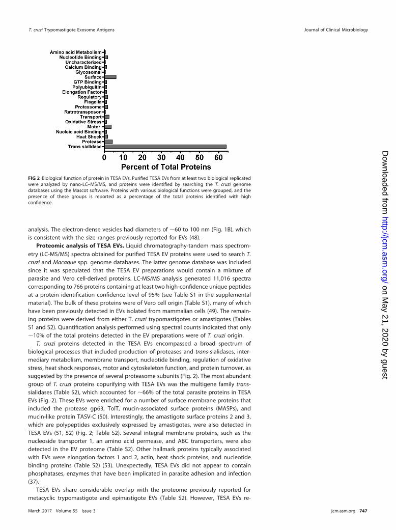
T. cruzi proteins detected in the TESA EVs encompassed a broad spectrum ofbiological processes that included production of proteases and trans-sialidases, inter-mediary metabolism, membrane transport, nucleotide binding, regulation of oxidativestress, heat shock responses, motor and cytoskeleton function, and protein turnover, assuggested by the presence of several proteasome subunits (Fig. 2). The most abundantgroup of T. cruzi proteins copurifying with TESA EVs was the multigene family trans-sialidases (Table S2), which accounted for �66% of the total parasite proteins in TESAEVs (Fig. 2). These EVs were enriched for a number of surface membrane proteins thatincluded the protease gp63, TolT, mucin-associated surface proteins (MASPs), andmucin-like protein TASV-C (50). Interestingly, the amastigote surface proteins 2 and 3,which are polypeptides exclusively expressed by amastigotes, were also detected inTESA EVs (51, 52) (Fig. 2; Table S2). Several integral membrane proteins, such as thenucleoside transporter 1, an amino acid permease, and ABC transporters, were alsodetected in the EV proteome (Table S2). Other hallmark proteins typically associatedwith EVs were elongation factors 1 and 2, actin, heat shock proteins, and nucleotidebinding proteins (Table S2) (53). Unexpectedly, TESA EVs did not appear to containphosphatases, enzymes that have been implicated in parasite adhesion and infection(37).
TESA EVs share considerable overlap with the proteome previously reported formetacyclic trypomastigote and epimastigote EVs (Table S2). However, TESA EVs re-
FIG 2 Biological function of protein in TESA EVs. Purified TESA EVs from at least two biological replicatedwere analyzed by nano-LC–MS/MS, and proteins were identified by searching the T. cruzi genomedatabases using the Mascot software. Proteins with various biological functions were grouped, and thepresence of these groups is reported as a percentage of the total proteins identified with highconfidence.
T. cruzi Trypomastigote Exosome Antigens Journal of Clinical Microbiology
March 2017 Volume 55 Issue 3 jcm.asm.org 747
on May 21, 2020 by guest
http://jcm.asm
.org/D
ownloaded from

leased by trypomastigotes/amastigotes in Vero cell cultures contained a number ofunique protein markers previously used, including the amastigote surface proteins 2and 3, proteasome subunits, transporter proteins, a polyubiquitin protein, and com-plement regulatory proteins (Table S2).
Immunoreactivity of Chagas patient sera with TESA EV proteins. TESA is rou-tinely used for serodiagnosis of Chagas disease (24, 25). However, a comprehensiveanalysis of antigens that cross-react with sera from Chagas patients with variousdegrees of cardiomyopathy (54) has not been performed. Western blot analysis usingsera from uninfected control subjects exhibited no significant immunoreactivity withproteins from Leishmania donovani promastigotes, Trypanosoma brucei procyclics, or T.cruzi trypomastigotes. The sera from asymptomatic Chagas patients showed very weakimmunoreactivity with high-molecular-weight proteins in the TESA EVs (Fig. 3), aspreviously reported with epimastigote antigens (55). In contrast, a robust cross-reactivity was detected with T. cruzi trypomastigotes and TESA EVs when blots wereprobed with sera from patients with electrocardiograph abnormalities or ventriculararrhythmia (Fig. 3A). While immunoreactive proteins �20 to 300 kDa in mass wereobserved in T. cruzi trypomastigote lysates, only proteins of �80 kDa were detected inthe TESA EVs (Fig. 3A). This is consistent with the proteomic analysis, which showed thatthe most abundant proteins were trans-sialidases (Fig. 2). The sera from Chagas diseasepatients with cardiopathologies also exhibited cross-reactivity with whole-cell lysatesfrom Leishmania spp. and T. brucei cultures (Fig. 3A).
To identify the immunoreactive antigens in the TESA EVs, extracts were incubatedwith immunoaffinity resins containing covalently coupled IgG antibodies from unin-fected donors or Chagas patients with clinical symptoms of ventricular arrhythmia (54).Western blot analysis using biotinylated Chagasic IgG antibodies showed strong im-munostaining of high-molecular-weight species in the total TESA EVs, Triton X-100extracts of EVs, and proteins isolated on the immunoaffinity columns; however, only an�50-kDa protein was detected for the immunoaffinity resin IgG antibodies fromuninfected controls (Fig. 3B).
LC-MS/MS analysis of TESA EV immunoprecipitations performed with Chagasic IgGantibodies identified 111 T. cruzi proteins containing at least two unique high-confidence peptides with masses ranging from 11 to 519 kDa (Table 1; Table S3).
FIG 3 Immunoreactivity of TESA with Chagasic sera. (A) Western blots containing whole-cell extracts of L. donovani promas-tigotes (lane 1), T. brucei procyclics (lane 2), T. cruzi trypomastigotes (lane 3), or TESA EVs (lane 4) were probed with pooledsera from Colombian uninfected controls or Chagasic patients with various stages of cardiopathologies as previously detailed(54). (B) IgG antibodies isolated from uninfected controls or Chagas patients with ventricular arrhythmia were coupled to aprotein G column, and reactive proteins were immunoprecipitated using these affinity matrices for Western blotting. Proteinsin the total TESA EV fraction (lane 1), Triton X-100 extracts of TESA EVs (lane 2), precipitated by uninfected control IgG (lane3), or IgG from Chagas patients (lane 4) were resolved by SDS-PAGE, and the membranes were probed with biotinylated IgGantibodies from uninfected subjects or Chagas-infected subjects. Primary antibodies were detected using streptavidin-horseradish peroxidase (Western blot membranes were exposed on film for 60 s).
Bautista-López et al. Journal of Clinical Microbiology
March 2017 Volume 55 Issue 3 jcm.asm.org 748
on May 21, 2020 by guest
http://jcm.asm
.org/D
ownloaded from

TABLE 1 Most abundant immunoaffinity-purified proteins from TESA extracellular vesiclesa
TESA protein Accession no. Mass (kDa)
No. of peptides in:
Nonimmuneserum
Chagas immuneserum
RetrotransposonRHS protein K4E7X9 77 0 29
Paraflagellar rodParaflagellar rod protein 3 K4EBQ5 70 0 18Paraflagellar rod protein 3 K2MCT5 69 0 17
MitochondrialATP synthase subunit alpha K4E934 63 2 17ATP synthase subunit beta K2M741 56 0 9Enoyl coenzyme A hydratase, mitochondrial K4DTD3 29 0 8Malic enzyme K4E9P4 63 0 6
trans-SialidasePutative uncharacterized protein Q7YZX6 95 0 16trans-Sialidase, group VIII, putative TcCLB.506537.200 120 0 15trans-Sialidase, putative (fragment) K4DRD9 35 0 13trans-Sialidase Q9BHJ5 71 0 12trans-Sialidase, putative K4E110 81 0 12trans-Sialidase, putative (fragment) K4DRD9 35 0 11trans-Sialidase P23253 120 0 10trans-Sialidase, putative (fragment) Q4D3D0 50 0 10
GlycosomalGlycosomal membrane protein (PEX11) K4DY43 27 0 8Fructose-bisphosphate aldolase K4DLE5 41 0 7
Tubulin�-Tubulin (pseudogene) TcCLB.509003.70 33 0 6�-Tubulin Q26973 47 0 6
GlycotransferaseUDP-Gal/DP-GlcNAc-dependent glycosyltransferase Q4CS30 43 0 9
ProteasesCytoskeleton-associated protein CAP5.5,cysteine peptidase K4E5Y1 88 0 13Serine carboxypeptidase S28, putative K4DZV5 54 0 6
Heat shock proteinHeat shock protein 70 B5U6T4 71 0 3
UncharacterizedUncharacterized protein K4EDD2 34 0 8Uncharacterized protein K4EBS5 88 0 7
Cytoskeleton proteinClathrin heavy chain K4E050 193 0 12
Sterol synthesisLanosterol synthase, putative K4EA17 103 0 9
Endoplasmic rectiulumPretranslocation protein, alpha subunit, SEC61-like TcCLB.506297.240 54 0 8Glucose-regulated protein 78 K4DT97 71 0 6
LysosomalLysosomal �-mannosidase, putative K4E2E6 111 0 7
AcidocalcisomeVacuolar-type proton translocating pyrophosphatase 1 K2NKA3 85 0 7
Miscellaneous proteinsSurface protein TolT, putative Q4CPM8 32 0 5
aAmino acid sequences for the proteins may be found in the NCBI Protein, Tritryp, and Uniprot/TrEMBL databases using the accession numbers listed in the table.
T. cruzi Trypomastigote Exosome Antigens Journal of Clinical Microbiology
March 2017 Volume 55 Issue 3 jcm.asm.org 749
on May 21, 2020 by guest
http://jcm.asm
.org/D
ownloaded from

Although trans-sialidases remained the most abundant group of proteins in the im-munoprecipitation assay, Chagasic IgG affinity resin showed a significant enrichment ofmitochondrial proteins, retrotransposon hot spot (RHS) proteins, proteases, and mul-tiple uncharacterized proteins (Fig. 4A). A notable enrichment was also observed forparaflagellar rod proteins, which are preferentially expressed in trypomastigotes (56)and glycosomal proteins (Fig. 4B; Table 1).
LC-MS/MS analysis also detected a number of T. cruzi proteins that nonspecificallycopurified with uninfected control and Chagas patient IgG immunoaffinity columnsthat included histones H2 and H4, calpains, mitochondrial proteins, and uncharacter-ized proteins (Table 1). Similarly, a number of Vero cell proteins also nonspecificallybound to the uninfected control and Chagas IgG affinity columns (Table S3).
Reactivity of Chagas sera with TESA EV proteins. We next exploited the avail-ability of the recombinant T. cruzi paraflagellar rod 3 (57) and RHS protein (K4E7X9) toassess the use of these proteins for the serological diagnosis of Chagas disease. Westernblot assays with recombinant PFR1 and total TESA EV extracts probed with pooled serafrom uninfected control subjects showed no detectable cross-reactivity with theseantigens. In contrast, sera from Chagas patients who were clinically asymptomatic,exhibited electrocardiogram (ECG) abnormalities, or ventricular arrhythmia showedrobust cross-reactivity with PFR1 and TESA EV antigens (Fig. 5A). Similarly, Chagasicsera, but not the sera from uninfected controls, showed a strong immunoreactivity withrecombinant RHS and TESA EVs (Fig. 5B). Interestingly, Chagas immune sera, in additionto reacting with the full-length recombinant RHS protein (75 kDa), also detectedmultiple fragments arising from either aborted translation or proteolysis in the Esche-richia coli expression system. These shorter fragments cross-reacted with anti-hexahistidine antibodies, which detected the N-terminal hexahistidine affinity tag (seeFig. S1 in the supplemental material). This result suggests that recombinant RHS is arelatively unstable protein that is subject to degradation, particularly within theC-terminal region of the protein.
ELISAs performed on serum samples from individual Columbian and Venezuelanpatients with various clinical manifestations of Chagas disease showed strong immunereactivity with TESA, with responses ranging from optical densities at 450 nm (OD450)from 0.04 to 2.4 (Fig. 6A), while serum from noninfected control subjects showed nonotable signal development. With TESA, �31% of patients with Chagas disease whowere classified as clinically asymptomatic or as exhibiting ECG abnormalities hadresponses on the ELISA that were below the cutoff value. This value decreased to�2.5% for patients exhibiting ventricular arrhythmia (Fig. 6A). A similar robust response
FIG 4 Enrichment of TESA proteins with immune sera. Proteins in TESA EVs were immunoprecipitated using affinity matricescontaining IgG antibodies from uninfected control subjects or IgG from Chagas patients with ventricular arrhythmia. Bound proteinswere analyzed by LC-MS/MS. Proteins were grouped on the basis of biological function (A), and the abundance levels of proteins ineach group were determined for the total number of spectra obtained for each group (B).
Bautista-López et al. Journal of Clinical Microbiology
March 2017 Volume 55 Issue 3 jcm.asm.org 750
on May 21, 2020 by guest
http://jcm.asm
.org/D
ownloaded from

was observed for RHS with sera from Chagas patients, with responses (OD450 values)ranging from 0.02 to 2.3. Although the mean values obtained for TESA and RHS werecomparable, the responses with RHS alone were more variable, with a greater numberof patients (24 to 40%) having responses below the cutoff value (Fig. 6A). This result isplausible, as not all patients may mount similar immune responses to RHS. A recentstudy showed that sera from Chagas patients exhibited a more robust humoralresponse to total parasite lysates, compared to the cross-reactivity with syntheticpeptides corresponding to the conserved C-terminal region found in the family ofMASPs, particularly in patients with cardiopathy (58). Surprisingly, although PFR1exhibited notable immunoreactivity on Western blots with Chagasic pooled sera (Fig.5), the responses in ELISAs were significantly dampened (OD450, 0.1 to 1.3) (Fig. 6A).
FIG 5 Immunoreactivity of Chagas patient sera with recombinant T. cruzi proteins. Western blots containing recombinantparaflagellar rod protein (lane 1) or total TESA EVs (lane 2) (A) or recombinant retrotransposon hot spot protein (lanes 1) ortotal TESA EVs (lane 2) (B) were probed with antisera from uninfected control subjects or with pooled sera from Chagaspatients with different clinical stages of the infections (Western blot exposure to film was 2 min).
FIG 6 ELISA analysis of Chagas patients. (A) Microtiter plates were coated with crude TESA, recombinant RHS, or PFR1 protein, and ELISAswere performed with individual sera collected from Columbian and Venezuelan subjects, including noninfected controls or patients withvarious clinical degrees of Chagas disease. Positive-control sera were generated by pooling sera from Chagas patients with ventriculararrhythmia, and negative-control sera were obtained from a Quebec donor subject that had never traveled to South America. Sera werediluted 1:400. (B) The cross-reactivity of sera from patients infected with Leishmania, malaria parasites, Toxoplasma, or T. brucei with theT. cruzi RHS recombinant protein was examined by ELISA. All assays were performed in triplicate using a 1:400 dilution of sera. The thickgray line in the scatter plots represents the minimum cutoff value required for a positive response, and the black lines correspond to themean OD450 values for each group of samples.
T. cruzi Trypomastigote Exosome Antigens Journal of Clinical Microbiology
March 2017 Volume 55 Issue 3 jcm.asm.org 751
on May 21, 2020 by guest
http://jcm.asm
.org/D
ownloaded from

Despite the reduced ELISA signal response of sera from Chagas patients, the responsefrom Chagas patients with ventricular arrhythmia were significant (P � 0.005) (Fig. 6A).The difference in immunoreactivity may be attributed to the fact that PFR1 is moreextensively denatured on Western blots than is antigen bound to the surfaces ofmicrotiter plates. Previous studies have shown modest reactivity of Chagas immunesera with paraflagellar rod proteins (59).
To examine the specificity of RHS, ELISAs were performed with sera from patientsinfected with Leishmania, malaria parasites, Toxoplasma gondii, Trypanosoma brucei, ornoninfected control sera obtained from Canadian subjects who had never traveled toSouth America. A robust signal was observed for TESA and RHS when probed with serafrom Chagas patients with ventricular arrhythmia (Fig. 6B). Analysis of the sera frommalaria patients or Toxoplasma-infected or T. brucei-infected patients with TESA andRHS were below the cutoff value of the assay and indicated no significant cross-reactivity. In contrast, with the TESA and the RHS antigen, 2 out of 10 and 3 out of 10,respectively, serum samples from patients with leishmaniasis exhibited strong reactivitythese antigens (Fig. 6B). The fact that the Leishmania genome does not encode RHSproteins suggests that the patients with a positive immune response were infected withT. cruzi parasites.
DISCUSSION
In this study, we performed for the first time a proteomic analysis of T. cruzi proteinsthat were associated with vesicles shed by trypomastigote and amastigote intracellularstages of this parasite. Proteomic analysis showed that the TESA used in a number ofdiagnostic assays (9, 22–24) contains a mixture of Vero cell and T. cruzi proteins (seeTable S1 in the supplemental material). Purified TESA EVs, which by transmissionelectron microscopy had a diameter of �80 to 100 nm, were found to contain apreponderance of trans-sialidases, mucins, and MASPs, which may be anchored to theparasite cell surface via glycosyl phosphatidylinositol (GPI) lipid or inserted into the EVmembrane via a conserved C-terminal region (58, 60, 61). This finding was in agreementwith those of previous studies showing that trans-sialidases constitute the dominantgroup of proteins released by trypomastigotes in infected animals or cell cultures (32,45, 62). The gp63 proteases, which are expressed by trypomastigotes and amastigotes(63), as well as several integral membrane proteins with transporter activity (Table S2),were also detected, which is consistent with the hypothesis that T. cruzi vesicles in theTESA preparation are derived primarily from the trypomastigote or amastigote plasmamembrane (33, 37, 64). In addition, TESA EVs also contained a variety of proteins knownto localize to the glycosome, flagella, mitochondria, nuclei, or cytosol (Table 1; TableS2), many of which have been previously detected in purified EVs from Leishmania andTrypanosoma brucei (38, 41, 65). Of note was the absence of glycolytic enzymes, whichwere previously observed in T. cruzi metacyclic trypomastigotes and epimastigotes andLeishmania EVs (33, 38, 41, 53).
Although TESA is a typical antigen used for serological diagnosis of Chagas disease,concerns regarding the specificity of this assay have been raised. TESA polypeptideswith masses of 60 to 150 kDa have been implicated as a source of potentiallycross-reactivity with leishmaniasis immune serum (24, 38). This is not surprising, sincemultiple sequence analyses showed that many of the T. cruzi proteins shed by trypo-mastigotes share a high degree of sequence homology with proteins from Leishmaniaand T. brucei.
More recently, a proteomic analysis of EVs isolated from axenic cultures of T. cruzimetacyclic trypomastigotes and epimastigotes (33) showed significant overlap with theTESA EV proteome, as reflected by the presence of membrane-bound proteins such asthe flagellar calcium binding protein (FCaBP), trans-sialidases, the surface proteasegp63, and elongation factor proteins (Table S2). Two of these proteins, FCaBP and gp63,have been previously suggested as potential candidates for diagnosis of T. rangeliinfections (66). However, the latter EVs contain a number of additional proteins thatwere preferentially enriched in immunoprecipitates of the TESA EVs. Examples include
Bautista-López et al. Journal of Clinical Microbiology
March 2017 Volume 55 Issue 3 jcm.asm.org 752
on May 21, 2020 by guest
http://jcm.asm
.org/D
ownloaded from

the amastigote surface proteins ASP2 and ASP3 (51, 52), paraflagellar rod proteins,several mitochondrial membrane proteins (ATP synthase, ADP/ATP translocase, andcytochrome c1), the glycosome membrane protein Gim5A, and the Golgi apparatusprotein UDP-galactose glycosyltransferase (Tables S2 and S3). Although our studiesfocused on EVs released into the culture supernatant following trypomastigote-mediated rupture of Vero cells, proteomic analysis revealed that EVs contained bothVero and T. cruzi proteins. However, it is unclear if host and parasite proteins segregateto separate vesicles or colocalize to the same EV. The latter possibility is supported byrecent experiments showing that host cells infected with Leishmania spp. or T. cruzireleased EVs contained both host and parasite proteins (38, 40, 41). Indeed, immuno-electron microscopy studies confirmed that EVs released by T. cruzi do not have ahomogeneous composition, since some vesicles were found to contain the membrane-anchored MASPs while other vesicle populations contained clathrin (58). Both of thelatter proteins were also detected in this study (Tables S2 and S3).
Previous attempts to characterize antigenic components in TESA preparations byusing immunoaffinity purification (24) identified a pattern of �10 immunoreactiveproteins, ranging in size from 25 to 220 kDa; however, the sequences of these antigenicproteins were not determined. Employing a combination of immunoprecipitation withaffinity-purified IgG antibodies from Chagas patients and LC-MS/MS analysis, we de-tected a set of soluble, integral membrane, and peripheral membrane proteins thatwere enriched compared to the content of the total TESA EV proteome, indicating thatthese proteins induce a robust immune response. Notable among these were a familyof retrotransposon hot spot and paraflagellar rod proteins (Table S3), the latter of whichis more abundantly expressed in the motile trypomastigote stage of the parasite (67).It is interesting that posttranslational sumoylation modification has been detected withthe latter two proteins (57, 68). RHS proteins have been localized to the nucleus and areencoded by a multigene family present in T. cruzi and T. brucei but not in theLeishmania genome, presumably because Leishmania spp. lack mobile retrotransposonelements (69). A recent proteomic analysis identified �39 RHS isoforms that wereexpressed by T. cruzi bloodstream trypomastigotes (70); however, the diversity of RHSproteins detected in EV preparations was much more restricted, as only 8 full-lengthRHS variants were observed (Table S3). In ELISAs, only the recombinant RHS showed astrong response with immune sera from patients with various clinical degrees ofChagas disease. It is noteworthy that proteomic analysis of T. brucei EVs also detectedan RHS1 protein (71). However, multiple sequence alignments of RHS protein revealedthat T. cruzi and T. brucei proteins share �33% sequence identity (data not shown).More importantly, no cross-reactivity was observed between T. cruzi RHS and immunesera from patients with African sleeping sickness or leishmaniasis, indicating that RHSmay be used as an antigen to increase the specificity of Chagas disease diagnosis.Interestingly, recent reports have shown that MASPs trigger a rapid humoral IgMresponse but limited IgG class switching during infection (58); consequently, it is notsurprising that our immunoaffinity LC-MS/MS strategy required a pool of IgG antibodiesfrom Chagasic patients.
It is possible that EVs released from infected host cells may in part account for T.cruzi antigens previously detected in the circulatory system or urine of infected patients(72–75), making EVs an attractive and tractable biological tool for facilitating directantigen detection in fluids of Chagas disease patients. Indeed, EVs are emerging as aunique mechanism for enriching low-level antigens for cancer diagnosis (76), andrecent studies using T. cruzi-specific RNA aptamers detected antigens in TESA prepa-rations or serum from mice with acute or chronic T. cruzi infection (77).
MATERIALS AND METHODSSerum samples. A total of 188 participants with various stages of Chagas disease or noninfected
control patients were recruited in a cross-sectional study conducted in Bucaramanga, Colombia. Thecriteria for grouping the participants was based on a combination of the New York Heart Association(NYHA) classifications and other clinical information (serology, ECGs, echocardiograms, and chest X-rays),
T. cruzi Trypomastigote Exosome Antigens Journal of Clinical Microbiology
March 2017 Volume 55 Issue 3 jcm.asm.org 753
on May 21, 2020 by guest
http://jcm.asm
.org/D
ownloaded from
findings commonly used to classify the stage of chronic Chagas infection. These participants have beendescribed previously (54).
Positive control sera represented a pool of sera from patients with clinically confirmed Chagasdisease with ventricular arrhythmia. The negative-control serum was from a Quebec donor who hadnever traveled to South America. TESA and RHS antigen were screened for reactivity with sera collectedfrom donors with confirmed Leishmania (n � 4), malaria parasite (n � 5), Toxoplasma gondii (n � 5), andT. brucei (n � 5) infections. The latter sera were obtained from the National Reference Centre forParasitology/J. D. Maclean Centre for Tropical Diseases at McGill University Health Centre. All samplesused in this study were anonymized.
Trypomastigote excreted-secreted antigen and EV isolation. TESA proteins from T. cruzi strainTulahuen were prepared as described previously (22, 24). Briefly, Vero cell monolayers at 75% confluencewere infected for 4 days with trypomastigotes (1 � 109/ml) in Eagle’s minimal essential mediumsupplemented with fetal bovine serum (FBS) at 37°C. Monolayers were stringently washed with Eagle’sminimal essential medium without FBS and then incubated for an additional 18 to 20 h at 37°C inmedium lacking both FBS and phenol red. The supernatant was collected, centrifuged at 2,800 � g toremove trypomastigotes or Vero cell debris, and then passed through a 0.22-�m filter to remove largemembrane fragments (Millipore, Bradford, MA). The filtrates was centrifuged at 20,000 � g for 30 min at4°C to remove membrane fragments, and the supernatant was centrifuged at 100,000 � g for 16 h at 4°Cin a Beckman-Coulter type 70 Ti rotor to sediment EVs. The EV pellet was washed four times with 1.0 mlof phosphate-buffered saline (PBS) at 49,000 rpm for 1 h in a Beckman-Coulter tabletop ultracentrifugeequipped with a TLA 100.3 rotor to remove residual extravesicular proteins. TESA EVs were furtherpurified by sucrose density flotation centrifugation. Briefly, EV pellets (100 �g of total protein) resus-pended in 2.0 ml PBS containing 40% sucrose, 400 mM NaCl were overlaid with 2.5 ml of PBS containing35% sucrose, 400 mM NaCl, and then 0.5 ml of PBS and the samples were centrifuged at 28,000 rpm for18 h at 4°C in an SW55 Ti rotor. Fractions (1 ml) were collected from the top of the gradient, diluted 4-foldwith PBS, and subjected to centrifugation at 200,000 � g for 1 h at 4°C in an SW55 Ti rotor to pellet EVs.Pellets were resuspended in 100 �l of PBS, aliquoted, and stored at �80°C. For crude TESA preparationsused in the ELISAs, the filtered lysates were concentrated 32-fold using a 30,000-molecular-weight-cutoff(MWCO) ultrafiltration unit (EMD Millipore, Etobicoke, ON, Canada) as previously described (24).
Purification of IgG antibodies and immunoprecipitation. Ten-milliliter samples of pooled serafrom uninfected control subjects or Chagas patients with severe ventricular arrhythmia were clarified bycentrifugation (3,000 � g, 10 min, 4°C) and then filtered through a 0.22-�m filter. The serum was diluted2-fold with PBS and then passed through a protein G-Sepharose column (0.5 by 10 cm; GE Healthcare LifeSciences, Montreal, QC, Canada). The column was washed with 20 column volumes of 20 mM sodiumphosphate (pH 7.0) to remove unbound proteins, and IgG antibodies were eluted with 100 mM glycineat pH 2.6. Column fractions were neutralized with 1.0 M Tris-HCl (pH 9.0), and proteins were quantifiedspectrophotometrically at 280 nm. Fractions containing antibodies were analyzed by SDS-PAGE, pooledand dialyzed against 100 mM NaHCO3, 500 mM NaCl (pH 8.5) for 24 h at 4°C, and then concentrated to�1.0 mg/ml using an Amicon ultracentrifugation device with a 30,000 MWCO.
An immunoaffinity resin was generated by loading purified human IgG antibodies (100 �g) onto 200�l of packed protein G-Sepharose resin, and IgG antibodies were cross-linked to protein G by usingdisuccinimidyl suberate (2 mg/ml in PBS; ThermoFisher Scientific, Rockford, IL) for 60 min at 25°C on anend-on-end rotator. The resin was washed with 20 column volumes of PBS to remove the cross-linkingreagent. Immunoreactive proteins were isolated by mixing purified TESA EVs (50 �g in 100 �l of PBS)treated with 0.1% Triton X-100 to release entrapped proteins, with 100 �l of packed affinity resincontaining covalently coupled control or Chagas IgG antibodies. The mixture was incubated for 16 h at4°C with end-on-end mixing, and unbound proteins were removed by washing resin 4 times with 1.0 mlof PBS containing 0.05% Triton X-100 and 4 times with 1.0 ml of PBS. Bound proteins were analyzed bymass spectrometry following an on-bead tryptic digestion.
Western blotting. Western blot analysis of trypomastigote lysates (1 � 105 parasites/lane) or EVs (2�g/lane) was performed by resolving proteins on a 6% or 8% SDS-PAGE gel, transferring proteins topolyvinylidene difluoride (PVDF) membranes (Bio-Rad, Mississauga, ON, Canada), and blocking with 2%milk powder in PBS containing 0.05% Tween 20 (PBST). Membranes were probed with primary antibodiesdiluted 1:2,000 in PBST containing 2% skimmed milk powder. Anti-human and anti-mouse antibodiesconjugated to horseradish peroxidase were used as secondary antibodies. Membranes were developedwith the ECL Western Lightening Plus detection system (PerkinElmer, Waltham, MA). Alternatively,Western blots were probed with either IgG antibodies isolated from uninfected control or Chagaspatients with severe cardiomyopathy; samples were biotinylated with biotin–N-hydroxysuccinimide esterfor 2 h at 20°C as described by the manufacturer (Invitrogen, Burlington, ON, Canada). Bound antibodieswere detected using a streptavidin-horseradish peroxidase conjugate (Invitrogen).
For Western blot analysis using recombinant protein, �1.0 �g of PFR1 or RHS, or 10 �g of TESA EVs,was resolved on an 8% SDS-PAGE gel and transferred to a PVDF membrane, and membranes wereblocked with 2% skimmed milk powder prior to probing with antisera (1:2,000 dilution) from uninfectedcontrols or Chagas patients with asymptomatic clinical signs, ECG abnormalities, or ventricular arrhyth-mia. Bound antibodies were detected with anti-human antibodies conjugated to horseradish peroxidase.
ELISAs. Immulon 2HB 96-well microtiter plates (ThermoFisher Scientific) were coated (100 �l/well)with 1 �g/ml of crude TESA preparation (24), recombinant paraflagellar rod, or retrotransposon hot spotproteins in 100 mM sodium carbonate solution (pH 9.6) at 4°C for 16 h. Plates were washed with PBSTand blocked for 1 h at 37°C with 5% bovine serum albumin in PBST. Human control and immune serawere diluted 1:400 in PBST, and 100-�l aliquots were added to each well and incubated at 37°C for 1 h.
Bautista-López et al. Journal of Clinical Microbiology
March 2017 Volume 55 Issue 3 jcm.asm.org 754
on May 21, 2020 by guest
http://jcm.asm
.org/D
ownloaded from

Each serum sample was tested in duplicate against the three antigens, and assays were repeated at leasttwice. Plates were extensively washed with PBST and then probed with a 1:16,000 dilution of horseradishperoxidase-conjugated goat anti-human IgG (Perkin-Elmer Life Sciences, Boston, MA) in PBST for 30 minat 37°C. Plates were washed with PBST and developed using the substrate 3,3=,5,5=-tetramethylbenzidine(100 �l/well; Serologicals Corporation, MA) for 10 min at 20°C for 10 min. Reactions were terminated bythe addition of 50 �l/well of 0.5 M H2SO4, and the optical density was measured at 450 nm using a TecanELISA reader. For this study, ELISAs were performed on individual serum samples from 35 noninfectedcontrol subjects, 29 patients with clinically asymptomatic Chagas disease, 29 patients with Chagas-associated ECG abnormalities, and 44 patients with ventricular arrhythmia. The cross-reactivity of serafrom individuals infected with malaria parasites (5 patients), Toxoplasma gondii (5 patients), Trypanosomabrucei (5 patients), or Leishmania sp. (5 patients [collected in Venezuela]) were screened by ELISA usingTESA and RHS antigen. The latter sera were obtained from the National Reference Centre for Parasitology,Research Institute of the McGill University Health Centre. These samples were collected as part of aprevious study that examined the utility of matrix metalloproteases in the diagnosis of Chagas disease(54). The cutoff values for the ELISAs were calculated as previously described (25), and a one-way analysisof variance (ANOVA) was performed using the Origin 2016 graphing package to calculate the P values.
TEM. TESA EVs isolated by sucrose density flotation centrifugation were absorbed onto Formvar/carbon-coated copper grids, washed in deionized water, and stained with 1% uranyl acetate for 1 min.Grids were allowed to air dry and viewed on a microscope. All samples were viewed on a JEOL 1200EXtransmission electron microscope at an accelerating voltage of 80 kV. All transmission electron micros-copy (TEM) experiments were performed at the Facility for Electron Microscopy Research, McGillUniversity.
Proteomic analysis. Sucrose density-purified EVs (20 �g) or proteins bound to immunoaffinitybeads were diluted in 50 mM ammonium acetate (pH 7.5) with 6 M guanidinium hydrochloridebuffer containing 0.5% octylglucoside, and proteins were reduced with 5 mM dithiothreitol andincubated at 80°C for 15 min. Samples were alkylated with 10 mM iodoacetamide at 20°C for 20 minand then quenched with 5 mM dithiothreitol. Modifying reagents, lipids, and detergent wereremoved with five 250-�l exchanges with 50 mM ammonium acetate (pH 7.5) buffer using a10,000-MWCO ultrafiltration unit. Proteins concentrated to 25 �l were digested by adding 1 �gPromega sequencing-grade trypsin and incubating the sample for 16 h at 37°C. Peptides werepurified from supernatant on stage tips (C18), vacuum dried, and then solubilized in 10 �l of 0.1%formic acid prior to mass spectrometry analysis.
Peptides were separated on a reversed-phase PicoFrit column (New Objective, Woburn, MA) packedwith Michrom Magic C18 (100 Å, 5 �m; nanoLC) and coupled to a Velos Pro LTQ-Orbitrap massspectrometer (ThermoFisher Scientific, Pittsburgh, PA). Peptide separation was performed with 2 to 50%solvent B (90% acetonitrile–10% water– 0.1% formic acid) in a 120-min linear gradient at 300 nl/min. Massspectra were acquired using a data-dependent acquisition mode and the Excalibur software version 1.6.2.Each full-scan mass spectrum (400 to 2,000 m/z) was followed by collision-induced dissociation of the 20most intense ions. Dynamic exclusion was set for a period of 3 s and a tolerance of 100 ppm and thenanalyzed using the Protein Discover version 1.4.1 software (ThermoFisher Scientific). MGF format samplefiles were then analyzed using Mascot (version 2.4.0; Matrix Science, London, United Kingdom), andspectra were used to search the Trypanosoma cruzi version 13-03 and Brener databases, as well as theMacaque database to identify proteins derived from the Vero cells. All MS/MS samples were analyzedusing Mascot with a fragment ion mass tolerance of 0.80 Da and a parent ion tolerance of 10 ppm. Massspectrometry analysis was performed at the UVic-Genome BC Proteomic Centre, Victoria, BC, Canada, andThe Proteomics Platform, Quebec Genomics Centre.
Production of T. cruzi PFR1 and retrotransposon hot spot proteins. E. coli strain ER2566 cellstransformed with the plasmid pET151/D-TOPO-TcPFR1 (57) were grown to an OD600 of 0.7 at 37°C in LBsupplemented with 50 �g/ml ampicillin, and protein expression was induced with 0.5 mM isopropyl-�-D-isothiogalactopyranoside at 25°C for 5 h. Cultures (1.0-liter volumes) were harvested, and the cellpellets were resuspended in 22.5 ml of 40 mM phosphate buffer (pH 7.4) containing an EDTA-freeprotease inhibitor cocktail (Roche Molecular Biochemicals). Cells were lysed with three passes through aFrench press, and clarified lysates were made up to 500 mM NaCl prior to loading onto a 4-mlNi2�-nitrilotriacetic acid–agarose column (Qiagen). The column was washed with 150 ml of 40 mMphosphate buffer (pH 7.4), 500 mM NaCl (buffer I), and then 100 ml buffer I containing 20 mM imidazole.Bound proteins were eluted with a 10-ml step gradient of buffer I containing 80 to 400 mM imidazole.Fractions containing an �70-kDa protein were pooled, concentrated to �0.5 mg/ml using a 3,000-MWCO Amicon ultrafiltration unit, flash-frozen in liquid nitrogen, and stored at �80°C.
For expression of the RHS protein (accession number EKG06703.1), the open reading frame was PCRamplified using the forward primer 5=-gtcacatATGTCTGGACGGCCCGAG-3= and the reverse primer 5=-ttcgaggatccTTAGAGAACCACAGGAGTTTCTCG-3= containing the restriction endonuclease sites NdeI andBamHI (lowercase text in the sequences). PCR mixtures containing 50 ng of genomic DNA isolated fromT. cruzi strain Tulahuen trypomastigotes, 200 nM each primer, 200 �M deoxynucleoside triphosphates(dNTP), and 1 U of Q5 DNA polymerase (New England BioLabs) were run with an amplification programthat included an initial denaturation at 98°C for 120 s followed by 25 cycles of denaturation at 98°C for30 s, annealing at 60°C for 30 s, and elongation at 72°C for 2 min. The PCR product was cloned into theNdeI/BamHI sites of the pET15b vector to generate the pET-15b-RHS expression vector. The recombinantRHS protein was expressed using the protocol described for the production of PFR1.
T. cruzi Trypomastigote Exosome Antigens Journal of Clinical Microbiology
March 2017 Volume 55 Issue 3 jcm.asm.org 755
on May 21, 2020 by guest
http://jcm.asm
.org/D
ownloaded from

SUPPLEMENTAL MATERIAL
Supplemental material for this article may be found at https://doi.org/10.1128/JCM.01649-16.
DATA SET S1, XLSX file, 0.06 MB.DATA SET S2, XLSX file, 0.01 MB.DATA SET S3, XLSX file, 0.02 MB.TEXT S1, PDF file, 0.04 MB.
ACKNOWLEDGMENTSWe thank Jeannie Mui (Facility for Electron Microscopy Research, McGill University,
Montreal, Canada) for help with the TEM analysis and Abhishek Chatterjee, AshleyTamming, and Anwer Kottarampatel (Institute of Parasitology, McGill University) forassistance in the cloning and production of recombinant proteins.
This project was funded by an operating grant from the Ministère de l’Économie, del’Innovation et des Exportations of Quebec and an NSERC Discovery Grant (238249).
REFERENCES1. Bern C, Montgomery SP. 2009. An estimate of the burden of Chagas
disease in the United States. Clin Infect Dis 49:e52– e54. https://doi.org/10.1086/605091.
2. Martin DL, Goodhew B, Czaicki N, Foster K, Rajbhandary S, Hunter S,Brubaker SA. 2014. Trypanosoma cruzi survival following cold storage:possible implications for tissue banking. PLoS One 9:e95398. https://doi.org/10.1371/journal.pone.0095398.
3. Huprikar S, Bosserman E, Patel G, Moore A, Pinney S, Anyanwu A,Neofytos D, Ketterer D, Striker R, Silveira F, Qvarnstrom Y, Steurer F,Herwaldt B, Montgomery S. 2013. Donor-derived Trypanosoma cruziinfection in solid organ recipients in the United States, 2001-2011. Am JTransplant 13:2418 –2425. https://doi.org/10.1111/ajt.12340.
4. Gascon J, Bern C, Pinazo M-J. 2010. Chagas disease in Spain, the UnitedStates and other non-endemic countries. Acta Trop 115:22–27. https://doi.org/10.1016/j.actatropica.2009.07.019.
5. Montgomery SP, Starr MC, Cantey PT, Edwards MS, Meymandi SK. 2014.Neglected parasitic infections in the United States: Chagas disease. AmJ Trop Med Hyg 90:814 – 818. https://doi.org/10.4269/ajtmh.13-0726.
6. Schmunis GA, Yadon ZE. 2010. Chagas disease: a Latin American healthproblem becoming a world health problem. Acta Trop 115:14 –21.https://doi.org/10.1016/j.actatropica.2009.11.003.
7. Gutierrez FRS, Guedes PMM, Gazzinelli RT, Silva JS. 2009. The role ofparasite persistence in pathogenesis of Chagas heart disease. ParasiteImmunol 31:673– 685. https://doi.org/10.1111/j.1365-3024.2009.01108.x.
8. Rassi A, Jr, Rassi A, Marin-Neto JA. 2010. Chagas disease. Lancet 375:1388 –1402. https://doi.org/10.1016/S0140-6736(10)60061-X.
9. Ramirez JD, Guhl F, Umezawa ES, Morillo CA, Rosas F, Marin-Neto JA,Restrepo S. 2009. Evaluation of adult chronic Chagas’ heart diseasediagnosis by molecular and serological methods. J Clin Microbiol 47:3945–3951. https://doi.org/10.1128/JCM.01601-09.
10. Apt W, Aguilera X, Arribada A, Perez C, Miranda C, Zulantay I, Apt P,Cortes P, Rodriguez J. 1994. Treatment of chronic human Chagas diseasewith itraconazole and allopurinol. Rev Med Chil 122:420 – 427.
11. Marin-Neto JA, Cunha-Neto EC, Maciel BC, Simoes MV. 2007. Pathogen-esis of chronic Chagas heart disease. Circulation 115:1109 –1123. https://doi.org/10.1161/CIRCULATIONAHA.106.624296.
12. Berkowitz AL, Raibagkar P, Pritt BS, Mateen FJ. 2015. Neurologic mani-festations of the neglected tropical diseases. J Neurol Sci 349:20 –32.https://doi.org/10.1016/j.jns.2015.01.001.
13. Rendell VR, Gilman RH, Valencia E, Galdos-Cardenas G, Verastegui M,Sanchez L, Acosta J, Sanchez G, Ferrufino L, LaFuente C, Abastoflor MdCColanzi R, Bern C. 2015. Trypanosoma cruzi-infected pregnant womenwithout vector exposure have higher parasitemia levels: implications forcongenital transmission risk. PLoS One 10:e0119527. https://doi.org/10.1371/journal.pone.0119527.
14. Qvarnstrom Y, Schijman AG, Veron V, Aznar C, Steurer F, da Silva AJ.2012. Sensitive and specific detection of Trypanosoma cruzi DNA inclinical specimens using a multi-target real-time PCR approach. PLoSNegl Trop Dis 6:e1689. https://doi.org/10.1371/journal.pntd.0001689.
15. Brasil P, De Castro L, Hasslocher-Moreno A, Sangenis L, Braga J. 2010.
ELISA versus PCR for diagnosis of chronic Chagas disease: systematicreview and meta-analysis. BMC Infect Dis 10:337. https://doi.org/10.1186/1471-2334-10-337.
16. Bern C, Kjos S, Yabsley MJ, Montgomery SP. 2011. Trypanosoma cruzi andChagas’ disease in the United States. Clin Microbiol Rev 24:655– 681.https://doi.org/10.1128/CMR.00005-11.
17. Gomes YM, Lorena VM, Luquetti AO. 2009. Diagnosis of Chagas disease:what has been achieved? What remains to be done with regard todiagnosis and follow up studies? Mem Inst Oswaldo Cruz 104(Suppl1):115–121. https://doi.org/10.1590/S0074-0276200900090017.
18. Jannin J, Salvatella R. 2006. Quantitative estimates of Chagas disease inthe Americas. PAHO report OPS/HDM/CD/425-06. Pan American HealthOrganization, Washington, DC.
19. Hernandez P, Heimann M, Riera C, Solano M, Santalla J, Luquetti AO,Beck E. 2010. Highly effective serodiagnosis for Chagas’ disease. ClinVaccine Immunol 17:1598 –1604. https://doi.org/10.1128/CVI.00489-08.
20. Pereira Gde A, Louzada-Neto F, Barbosa Vde F, Ferreira-Silva MM, deMoraes-Souza H. 2012. Performance of six diagnostic tests to screen forChagas disease in blood banks and prevalence of Trypanosoma cruziinfection among donors with inconclusive serology screening based onthe analysis of epidemiological variables. Rev Bras Hematol Hemoter34:292–297. https://doi.org/10.5581/1516-8484.20120074.
21. Sanchez-Camargo CL, Albajar-Vinas P, Wilkins PP, Nieto J, Leiby DA, ParisL, Scollo K, Florez C, Guzmin-Bracho C, Luquetti AO, Calvo N, TadokoroK, Saez-Alquezar A, Palma PP, Martin M, Flevaud L. 2014. Comparativeevaluation of 11 commercialized rapid diagnostic tests for detectingTrypanosoma cruzi antibodies in serum banks in areas of endemicity andnonendemicity. J Clin Microbiol 52:2506 –2512. https://doi.org/10.1128/JCM.00144-14.
22. Umezawa ES, Nascimento MS, Kesper N, Coura JR, Borges-Pereira J,Junqueira AC, Camargo ME. 1996. Immunoblot assay using excreted-secreted antigens of Trypanosoma cruzi in serodiagnosis of congenital,acute, and chronic Chagas’ disease. J Clin Microbiol 34:2143–2147.
23. Umezawa ES, Nascimento M, Stolf AM. 2001. Enzyme-linked immunosor-bent assay with Trypanosoma cruzi excreted-secreted antigens (TESA-ELISA) for serodiagnosis of acute and chronic Chagas’ disease. DiagnMicrobiol Infect Dis 39:169 –176. https://doi.org/10.1016/S0732-8893(01)00216-4.
24. Berrizbeitia M, Ndao M, Bubis J, Gottschalk M, Ache A, Lacouture S,Medina M, Ward BJ. 2006. Purified excreted-secreted antigens fromTrypanosoma cruzi trypomastigotes as tools for diagnosis of Chagas’disease. J Clin Microbiol 44:291–296. https://doi.org/10.1128/JCM.44.2.291-296.2006.
25. Nakazawa M, Rosa D, Pereira V, Moura M, Furtado V, Souza W, Barros M,Abath F, Gomes Y. 2001. Excretory-secretory antigens of Trypanosomacruzi are potentially useful for serodiagnosis of chronic Chagas’ disease.Clin Diagn Lab Immunol 8:1024 –1027.
26. Trocoli Torrecilhas AC, Tonelli RR, Pavanelli WR, da Silva JS, SchumacherRI, de Souza W, E Silva NC, de Almeida Abrahamsohn I, Colli W, MansoAlves MJ. 2009. Trypanosoma cruzi: parasite shed vesicles increase heart
Bautista-López et al. Journal of Clinical Microbiology
March 2017 Volume 55 Issue 3 jcm.asm.org 756
on May 21, 2020 by guest
http://jcm.asm
.org/D
ownloaded from

parasitism and generate an intense inflammatory response. MicrobesInfect 11:29 –39. https://doi.org/10.1016/j.micinf.2008.10.003.
27. Bang C, Thum T. 2012. Exosomes: new players in cell-cell communica-tion. Int J Biochem Cell Biol 44:2060 –2064. https://doi.org/10.1016/j.biocel.2012.08.007.
28. Colombo M, Raposo G, Théry C. 2014. Biogenesis, secretion, and inter-cellular interactions of exosomes and other extracellular vesicles. AnnRev Cell Develop Biol 30:255–289. https://doi.org/10.1146/annurev-cellbio-101512-122326.
29. Garcia-Silva MR, das Neves RF, Cabrera-Cabrera F, Sanguinetti J, Medei-ros LC, Robello C, Naya H, Fernandez-Calero T, Souto-Padron T, de SouzaW, Cayota A. 2014. Extracellular vesicles shed by Trypanosoma cruzi arelinked to small RNA pathways, life cycle regulation, and susceptibility toinfection of mammalian cells. Parasitol Res 113:285–304. https://doi.org/10.1007/s00436-013-3655-1.
30. Schorey JS, Cheng Y, Singh PP, Smith VL. 2015. Exosomes and otherextracellular vesicles in host-pathogen interactions. EMBO Rep 16:24 – 43. https://doi.org/10.15252/embr.201439363.
31. Raposo G, Stoorvogel W. 2013. Extracellular vesicles: exosomes, mi-crovesicles, and friends. J Cell Biol 200:373–383. https://doi.org/10.1083/jcb.201211138.
32. Goncalves MF, Umezawa ES, Katzin AM, De Sousa W, Alves MJM, Zin-gales B, Colli W. 1991. Trypanosoma cruzi: shedding of surface antigensas membrane vesicles. Exp Parasitol 72:43–53. https://doi.org/10.1016/0014-4894(91)90119-H.
33. Bayer-Santos E, Aguilar-Bonavides C, Rodrigues SP, Cordero EM, MarquesAF, Varela-Ramirez A, Choi H, Yoshida N, da Silveira JF, Almeida IC. 2013.Proteomic analysis of Trypanosoma cruzi secretome: characterization oftwo populations of extracellular vesicles and soluble proteins. J Pro-teome Res 12:883– 897. https://doi.org/10.1021/pr300947g.
34. Bayer-Santos E, Lima FM, Ruiz JC, Almeida IC, da Silveira JF. 2014.Characterization of the small RNA content of Trypanosoma cruzi extra-cellular vesicles. Mol Biochem Parasitol 193:71–74. https://doi.org/10.1016/j.molbiopara.2014.02.004.
35. Montaner S, Galiano A, Trelis M, Martin-Jaular L, del Portillo HA, Bernal D,Marcilla A. 2014. The role of extracellular vesicles in modulating the hostimmune response during parasitic infections. Front Immunol 5:433.https://doi.org/10.3389/fimmu.2014.00433.
36. Martins NO, Souza RT, Cordero EM, Maldonado DC, Cortez C, Marini MM,Ferreira ER, Bayer-Santos E, Almeida IC, Yoshida N, Silveira JF. 2015.Molecular characterization of a novel family of Trypanosoma cruzi sur-face membrane proteins (TcSMP) involved in mammalian host cell in-vasion. PLoS Negl Trop Dis 9:e0004216. https://doi.org/10.1371/journal.pntd.0004216.
37. Neves RFC, Fernandes ACS, Meyer-Fernandes JR, Souto-Padrón T. 2014.Trypanosoma cruzi-secreted vesicles have acid and alkaline phosphataseactivities capable of increasing parasite adhesion and infection. ParasitolRes 113:2961–2972. https://doi.org/10.1007/s00436-014-3958-x.
38. Silverman JM, Clos J, de’Oliveira CC, Shirvani O, Fang Y, Wang C, FosterLJ, Reiner NE. 2010. An exosome-based secretion pathway is responsiblefor protein export from Leishmania and communication with macro-phages. J Cell Sci 123:842– 852. https://doi.org/10.1242/jcs.056465.
39. Lambertz U, Silverman JM, Nandan D, McMaster WR, Clos J, Foster LJ,Reiner NE. 2012. Secreted virulence factors and immune evasion invisceral leishmaniasis. J Leukoc Biol 91:887– 899. https://doi.org/10.1189/jlb.0611326.
40. Cestari I, Ansa-Addo E, Deolindo P, Inal JM, Ramirez MI. 2012. Trypano-soma cruzi immune evasion mediated by host cell-derived microvesicles.J Immunol 188:1942–1952. https://doi.org/10.4049/jimmunol.1102053.
41. Hassani K, Olivier M. 2013. Immunomodulatory impact of Leishmania-induced macrophage exosomes: a comparative proteomic and func-tional analysis. PLoS Negl Trop Dis 7:e2185. https://doi.org/10.1371/journal.pntd.0002185.
42. Araujo FG, Chiari E, Dias JCP. 1981. Demonstration of Trypanosoma cruziantigen in serum from patients with Chagas disease. Lancet 317:246 –249. https://doi.org/10.1016/S0140-6736(81)92088-2.
43. Araujo FG, Sharma SD, Tsai V, Cox P, Remington JS. 1982. Monoclonalantibodies to stages of Trypanosoma cruzi: characterization and use forantigen detection. Infect Immun 37:344 –349.
44. Freilij HL, Corral RS, Katzin AM, Grinstein S. 1987. Antigenuria in infantswith acute and congenital Chagas’ disease. J Clin Microbiol 25:133–137.
45. Affranchino JL, Ibanez CF, Luquetti AO, Rassi A, Reyes MB, Macina RA,Aslund L, Pettersson U, Frasch ACC. 1989. Identification of a Trypano-soma cruzi antigen that is shed during the acute phase of Chagas’
disease. Mol Biochem Parasitol 34:221–228. https://doi.org/10.1016/0166-6851(89)90050-9.
46. Umezawa ES, Shikanai-Yasuda MA, Stolf AM. 1996. Changes in isotypecomposition and antigen recognition of anti-Trypanosoma cruzi antibod-ies from acute to chronic Chagas disease. J Clin Lab Anal 10:407– 413.
47. Yokoyama-Yasunaka JK, Pral EM, Oliveira Júnior OC, Alfieri SC, Stolf AM.1994. Trypanosoma cruzi: identification of proteinases in shed compo-nents of trypomastigote forms. Acta Trop 57:307–315. https://doi.org/10.1016/0001-706X(94)90076-0.
48. Kowal J, Tkach M, Thery C. 2014. Biogenesis and secretion of exosomes.Curr Opin Cel l B iol 29:116 –125. https : //doi .org/10.1016/j.ceb.2014.05.004.
49. Peterson DB, Sander T, Kaul S, Wakim BT, Halligan B, Twigger S, PritchardKA, Oldham KT, Ou J-S. 2008. Comparative proteomic analysis of PAI-1and TNF-alpha-derived endothelial microparticles. Proteomics8:2430 –2446. https://doi.org/10.1002/pmic.200701029.
50. Bernabó G, Levy G, Ziliani M, Caeiro LD, Sánchez DO, Tekiel V. 2013.TcTASV-C, a protein family in Trypanosoma cruzi that is predominantlytrypomastigote-stage specific and secreted to the medium. PLoS One8:e71192. https://doi.org/10.1371/journal.pone.0071192.
51. Pan AA, McMahon-Pratt D. 1989. Amastigote and epimastigote stage-specific components of Trypanosoma cruzi characterized by using mono-clonal antibodies: purification and molecular characterization of an 83-kilodalton amastigote protein. J Immunol 143:1001–1008.
52. Boscardin SB, Kinoshita SS, Fujimura AE, Rodrigues MM. 2003. Immuni-zation with cDNA expressed by amastigotes of Trypanosoma cruzi elicitsprotective immune response against experimental Infection. Infect Im-mun 71:2744 –2757. https://doi.org/10.1128/IAI.71.5.2744-2757.2003.
53. Kim D-K, Lee J, Simpson RJ, Lotvall J, Gho YS. 2015. EVpedia: a commu-nity Web resource for prokaryotic and eukaryotic extracellular vesiclesresearch. Semin Cell Develop Biol 40:4 –7. https://doi.org/10.1016/j.semcdb.2015.02.005.
54. Bautista-Lopez NL, Morillo CA, Lopez-Jaramillo P, Quiroz R, Luengas C,Silva SY, Galipeau J, Lalu MM, Schulz R. 2013. Matrix metalloproteinases2 and 9 as diagnostic markers in the progression to Chagas cardiomy-opathy. Am Heart J 165:558 –566. https://doi.org/10.1016/j.ahj.2013.01.001.
55. Cervantes-Landin AY, Martinez I, Schabib M, Espinoza B. 2014. Highmolecular weight proteins of Trypanosoma cruzi reduce cross-reactionwith Leishmania spp. in serological diagnosis tests. Biomed Res Int2014:365403. https://doi.org/10.1155/2014/365403.
56. Quanquin NM, Galaviz C, Fouts DL, Wrightsman RA, Manning JE. 1999.Immunization of mice with a TolA-like surface protein of Trypanosomacruzi generates CD4� T-cell-dependent parasiticidal activity. Infect Im-mun 67:4603– 4612.
57. Annoura T, Makiuchi T, Sariego I, Aoki T, Nara T. 2012. SUMOylation ofparaflagellar rod protein, PFR1, and its stage-specific localization inTrypanosoma cruzi PLoS One 7:e37183. https://doi.org/10.1371/journal.pone.0037183.
58. De Pablos LM, Díaz Lozano IM, Jercic MI, Quinzada M, Giménez MJ,Calabuig E, Espino AM, Schijman AG, Zulantay I, Apt W, Osuna A. 2016.The C-terminal region of Trypanosoma cruzi MASPs is antigenic andsecreted via exovesicles. Sci Rep 6:27293. https://doi.org/10.1038/srep27293.
59. Cooley G, Etheridge RD, Boehlke C, Bundy B, Weatherly DB, MinningT, Haney M, Postan M, Laucella S, Tarleton RL. 2008. High throughputselection of effective serodiagnostics for Trypanosoma cruzi infection.PLoS Negl Trop Dis 2:e316. https://doi.org/10.1371/journal.pntd.0000316.
60. Agusti R, Couto AS, Campetella OE, Frasch AC, de Lederkremer RM. 1997.The trans-sialidase of Trypanosoma cruzi is anchored by two differentlipids. Glycobiology 7:731–735. https://doi.org/10.1093/glycob/7.6.731.
61. Queiroz RML, Charneau S, Bastos IMD, Santana JM, Sousa MV, RoepstorffP, Ricart CAO. 2014. Cell surface proteome analysis of human-hostedTrypanosoma cruzi life stages. J Proteome Res 13:3530 –3541. https://doi.org/10.1021/pr401120y.
62. Ouaissi MA, Taibi A, Cornette J, Velge P, Marty B, Loyens M, Esteva M,Rizvi FS, Capron A. 1990 Characterization of major surface and excretory-secretory immunogens of Trypanosoma cruzi trypomastigotes and iden-tification of potential protective antigen. Parasitol Res 100 115–124.
63. Grandgenett PM, Coughlin BC, Kirchhoff LV, Donelson JE. 2000. Differ-ential expression of GP63 genes in Trypanosoma cruzi Mol BiochemParasitol 110:409 – 415. https://doi.org/10.1016/S0166-6851(00)00275-9.
64. Da Silveira JF, Abrahamsohn PA, Colli W. 1979. Plasma membrane
T. cruzi Trypomastigote Exosome Antigens Journal of Clinical Microbiology
March 2017 Volume 55 Issue 3 jcm.asm.org 757
on May 21, 2020 by guest
http://jcm.asm
.org/D
ownloaded from
vesicles isolated from epimastigote forms of Trypanosoma cruziBiochim Biophys Acta 550:222–232. https://doi.org/10.1016/0005-2736(79)90209-8.
65. Szempruch AJ, Sykes SE, Kieft R, Dennison L, Becker AC, Gartrell A, MartinWJ, Nakayasu ES, Almeida IC, Hajduk SL. 2016. Extracellular vesicles fromTrypanosoma brucei mediate virulence factor transfer and cause hostanemia. Cell 164:246 –257. https://doi.org/10.1016/j.cell.2015.11.051.
66. Wagner G, Yamanaka LE, Moura H, Lückemeyer DD, Schlindwein AD,Stoco PH, Ferreira HB, Barr JR, Steindel M, Grisard EC. 2013. The Trypano-soma rangeli trypomastigote surfaceome reveals novel proteins andtargets for specific diagnosis. J Proteomics 82:52– 63. https://doi.org/10.1016/j.jprot.2013.02.011.
67. Paba J, Santana JM, Teixeira ARL, Fontes W, Sousa MV, Ricart CAO. 2004.Proteomic analysis of the human pathogen Trypanosoma cruzi Proteom-ics 4:1052–1059. https://doi.org/10.1002/pmic.200300637.
68. Bayona JC, Nakayasu ES, Laverrière M, Aguilar C, Sobreira TJP, Choi H,Nesvizhskii AI, Almeida IC, Cazzulo JJ, Alvarez VE. 2011. SUMOylationpathway in Trypanosoma cruzi: functional characterization and pro-teomic analysis of target proteins. Mol Cell Proteomics 10:M110.007369.https://doi.org/10.1074/mcp.M110.007369.
69. Bringaud F, Biteau N, Melville SE, Hez S, El-Sayed NM, Leech V, BerrimanM, Hall N, Donelson JE, Baltz T. 2002. A new, expressed multigene familycontaining a hot spot for insertion of retroelements is associated withpolymorphic subtelomeric regions of Trypanosoma brucei Eukarot Cell1:137–151. https://doi.org/10.1128/EC.1.1.137-151.2002.
70. Brunoro GV, Caminha MA, Ferreira AT, Leprevost FDV, Carvalho PC,Perales J, Valente RH, Menna-Barreto RF. 2015. Reevaluating theTrypanosoma cruzi proteomic map: the shotgun description of blood-
stream trypomastigotes. J Proteomics 115:58 – 65. https://doi.org/10.1016/j.jprot.2014.12.003.
71. Geiger A, Hirtz C, Becue T, Bellard E, Centeno D, Gargani D, Rossignol M,Cuny G, Peltier J-B. 2010. Exocytosis and protein secretion in Trypano-soma BMC Microbiol 10:20. https://doi.org/10.1186/1471-2180-10-20.
72. Bongertz V, Hungerer KD, Galvao-Castro B. 1981. Trypanosoma cruzi:circulating antigens. Mem Inst Oswaldo Cruz 76:71– 82. https://doi.org/10.1590/S0074-02761981000100008.
73. Corral RS, Orn A, Grinstein S. 1992. Detection of soluble exoantigens ofTrypanosoma cruzi by a dot-immunobinding assay. Am J Trop Med Hyg46:31–38.
74. Umezawa ES, Shikanaiyasuda MA, Dasilveira JF, Cotrim PC, Paranhos G,Katzin AM. 1993. Trypanosoma cruzi: detection of a circulating antigen inurine of chagasic patients sharing common epitopes with an immuno-dominant repetitive antigen. Exp Parasitol 76:352–357. https://doi.org/10.1006/expr.1993.1043.
75. Petray P, Bonardello N, Clark R, Agranatti M, Corral R, Grinstein S. 1992.Evaluación del método de ELISA para detección de antígenos y comple-jos inmunes circulantes de Trypanosoma cruzi a través de un estudio decampo en una zona endémica de Argentina. Rev Instit Med Trop SaoPaulo 34:141–147. https://doi.org/10.1590/S0036-46651992000200010.
76. Boukouris S, Mathivanan S. 2015. Exosomes in bodily fluids are a highlystable resource of disease biomarkers. Proteomics Clin Appl 9:358 –367.https://doi.org/10.1002/prca.201400114.
77. Nagarkatti R, de Araujo FF, Gupta C, Debrabant A. 2014. Aptamer based,non-PCR, non-serological detection of Chagas disease biomarkers inTrypanosoma cruzi infected mice. PLoS Negl Trop Dis 8:e2650. https://doi.org/10.1371/journal.pntd.0002650.
Bautista-López et al. Journal of Clinical Microbiology
March 2017 Volume 55 Issue 3 jcm.asm.org 758
on May 21, 2020 by guest
http://jcm.asm
.org/D
ownloaded from