Embed Size (px)
Citation preview

Characteristics of submucosal gastric carcinoma with lymph node metastatic disease
H J Son, S Y Song,1 S Kim,3 J H Noh,2 T S Sohn,2 D S Kim1 & J C Rhee
Presented by intern 張家維
Histopathology 2005, 46, 158–165.

Introduction
2nd most common cause of cancer deaths East Asia and south America Korea and Japan early detection Early Gastric Carcinoma
Mucosa or Submucosa 5-year survival rate Lymph node metastasis
prognosis, submucosal invasion

Introduction
Depth of submucosal layer viaries The depth of submucosal gastric carcinoma
(SMGC) Lymph node metastasis
macroscopic appearance, location, size, tumor area, differentiation, invasion depth, submucosal vascularity, fibrosis near the tumor area
Age- and Sex- matched
Materials and methods
Sampling: 248 patients SMGC Surgical resection Samsung Medical Centre (Seoul, Korea) 1995/Jan.~ 2002/Oct. Total 917 patients SMGC
124 LN metastasis (13.5%) Anticancer therapy X Evidence of metastatic disease X

Materials and methods
124 SMGC with LN metastasis 124 SMGC without LN metastasis Age- and Sex- matched Specimens routinely examined 10% formalin Embedded in paraffin H & E stained
Materials and methods
Macroscopic:
Japanese Endoscopic Society Classification
Elevated, Depressed, Flat Microscopic:
Lauren classification

Materials and methods
Depth of submucosal invasion
1.Ocular lens scale, distance between the
lower edge of the muscularis mucosa and
the deepest invading front of the tumor
cells
2.sm3 method: sm1, sm2, sm3
3.sm2 method: smi , sme


Materials and methods
Tumor size
Longest dimension of the tumor area Tumor area
Longest dimension X its prependicular counterpart
Tumor vessels
vessels with a smooth muscle coat in the submucosa

Materials and methods
Statistical analysis:
Log linear model, McNemar’s test, A paired t-test, Wilcoxon’s signed rank test
P-value < 0.05 statistically significant SAS, version 6.12

Results
Pathological parameters and lymphnode metastatic disease
124 SMGC with lymph node metastasis 69 males (55.6%), 55 females (44.4%) 31~83 y/o, the mean age 56.3 y/o The main locations of the tumors 60 lower, 61 middle, 3 upper 1/3 Tumor size 5~125 mm, the mean = 45 mm Tumor area 0.8~105.0 cm2, the mean = 17.1 cm2
Results
Pathological parameters and lymphnode metastatic disease
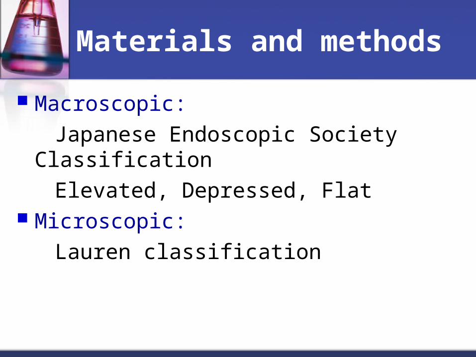
The gross types of the tumors
35 elevated (28.2%), 85 depressed (68.5%), 4 flat (3.2%)
The histological differentiations (Lauren’s)
53 intestinal (42.7%), 65 diffuse (52.8), 6 mixed (4.8%)
111 N1 (89.5%), 13 N2 (10.5%)


Results
Pathological parameters and lymphnode metastatic disease
Significantly associated with node-positive SMGC 1. presence of lymphatic tumor emboli 2. a larger tumor area 3. a larger tumor size 4. a non-flat gross type 5. an increased vascularity No significant relationship 1. location 2. Lauren classification 3. tumor related fibrosis


Results
Depth-related parameters and lymph node metastatic disease
Ocular scale-measured depth
1. proved to have a significant correlation
with node-positive SMGC
2. superficial invasive, deeply invasive (2mm) The sm3 method
not well correlated The sm2 method
not well correlated

Results
Multivariate analyses for possible indicators of LN metastatic disease
Multivariate logistic regression analysis location, gross type, Lauren’s classification,
lymphatic tumor emboli, increased vascularity, tumor-related fibrosis, tumor size, depth (sm2 method)
The incidence of lymph node metastatic disease increased in the presence of lymphatic tumor emboli and in the tumors that invaded more than half of the submucosal layer


Discussion
EGC, the “early” horrible disaster curable disease early diagnosis and treatment programs The term of EGC has 2 innate defect 1. lymph node metastatic diseases 2. discriminate submucosal tumor call invasion 5-year survival rate 93~99% for node-negative EGC 73~90% for node-positive EGC 90~100% for intramucosal confinement 73~90% for submucosal invasion

Discussion
The treatment now for EGC
conducting minimally invasive surgical procedures
endoscopic mucosal resection, laparoscopic partial resection
need careful and intensively subclassification Remove all metastatic lymph nodes ?
chance of a cure↓ Factors related to lymph node metastatic disease

Discussion
SMGC lymph node metastasis rate 10~25% 917 SMGC in this tiral 13.5% LN metastasis The parameters related to LN metastasis lymphatic tumor emboli (uni- or multi- variate
analysis) depth-related (accurate invasion depth, sm2
method)

Discussion
The best way to represent a submucosal tumor invasion Tsuchiya et al. = sm3 not appropriate for classifying tumor from endoscopic biopsy specimen Yasuda et al. = accurately the depth submucosal tumor invasion of locally resected tumor > 300μm gastrectomy + LN dissection Japanese Classification of Gastric Cancer criteria (0.5mm) depth of submucosal tumor invasion < 0.5mm sm1 depth of submucosal tumor invasion > 0.5mm sm2
Discussion
Univariate analysis
accurate depth of tumor invasion Multivariate analysis
relative depth of tumor invasion Both accurate depth and relative depth of tumor
invasion are important in predicting LN metastasis of SMGC
A small group of superficial submucosal tumor invasions (even <1mm)
presented LN metasitasis

Discussion
In general, EGC with LN metastasis large, depressed growth (or ulcer), poorly
differentiated adenocarcinoma associated with peptic ulceration
Tumor size contact with submucosal lymphatics and venules Vascularity higher incidence in node-positive SMGCLN metastasis might be associated with tumor cells coming into contact with submucosal lymphatic and venules

Discussion
Lymphatic tumor invasion and deeper tumor invasion into the submucosa
simple and easy parameters for predicting LN metastasis from limited surgery specimens
Small group of superficial involvement of submucosa
LN metastasis Carefully selected patients for minimalizing operation Pathologist should carefully investigate the lymphatic
invasion and the depth of tmor invasion

Characteristics of intramucosal gastric carcinoma with lymph node metastatic disease
S Y Song, S Park,2 S Kim,3 H J Son1 & J C Rhee1
Presented by intern 張家維

Results
macroscopic appearance location size differentiation presence of ulceration vascularity presence of gastritis cystica profunda-like
glandular proliferation disruption of the muscularis mucosae and invasion
into the muscularis mucosae

Results
diffuse type histology (P < 0.001) and deep invasion into the muscularis mucosae (P < 0.05) were indicators of node-positive intramucosal EGCs

Conclusions
These histological indicators are easily accessible and seem to predict lymph node metastatic disease in limited surgical specimens.
Patients should be carefully selected despite the recent trend toward less invasive resection of EGCs, especially for those apparently confined to the mucosa.

Thank you for your attention