Embed Size (px)
Citation preview
CHARACTERISTICS OF SOMATIC PAIN SENSITIZATION IN IRRITABLE
BOWEL SYNDROME
By
ANTHONY CARL RODRIGUES
A DISSERTATION PRESENTED TO THE GRADUATE SCHOOL OF THE UNIVERSITY OF FLORIDA IN PARTIAL FULFILLMENT
OF THE REQUIREMENTS FOR THE DEGREE OF DOCTOR OF PHILOSOPHY
UNIVERSITY OF FLORIDA
2005

Copyright 2005
by
Anthony Carl Rodrigues

This document is dedicated to my family for their unending support.

iv
ACKNOWLEDGMENTS
I thank my mentor, Andre P. Mauderli, for patiently guiding me through this
journey.

v
TABLE OF CONTENTS page
ACKNOWLEDGMENTS ................................................................................................. iv
LIST OF TABLES........................................................................................................... viii
LIST OF FIGURES ........................................................................................................... ix
ABSTRACT....................................................................................................................... xi
CHAPTER
1 GENERAL INTRODUCTION AND BACKGROUND..............................................1
2 HYPERSENSITIVITY TO CUTANEOUS THERMAL NOCICEPTIVE STIMULI IN IRRITABLE BOWEL SYNDROME ....................................................8
Introduction...................................................................................................................8 Methods ........................................................................................................................9
Subject Recruitment ..............................................................................................9 Daily Protocol......................................................................................................10 Pain Measurement ...............................................................................................11 Mapping and Rating of Clinical Pain ..................................................................11 Thermal Stimuli...................................................................................................12 Data Analysis.......................................................................................................13
Results.........................................................................................................................13 Discussion...................................................................................................................15
3 CUTANEOUS PAIN SENSITIZATION CHARACTERISTICS IN IRRITABLE BOWEL SYNDROME...............................................................................................22
Introduction.................................................................................................................22 Methods ......................................................................................................................26
Subjects................................................................................................................26 Pain Measurement ...............................................................................................27 Response-Dependent Stimulation Method ..........................................................27 Testing Protocol...................................................................................................28
Measurement of spontaneous pain ...............................................................28 Measurement of skin temperature ................................................................29 Thermal stimulation .....................................................................................29
vi
Data Analysis.......................................................................................................32 Results.........................................................................................................................33
Skin Temperature ................................................................................................33 Clinical Pain ........................................................................................................34 Induction of Stimulus-Induced Sensitization ......................................................34 Maintenance of Stimulus-Induced Sensitization .................................................35
Discussion...................................................................................................................38
4 THE EFFECT OF TOPICAL LOCAL ANESTHETICS ON SOMATIC PAIN SENSITIZATION IN IRRITABLE BOWEL SYNDROME.....................................48
Introduction.................................................................................................................48 Methods ......................................................................................................................50
Subjects................................................................................................................50 Pain Measurement ...............................................................................................51
Testing Protocol..........................................................................................................52 Daily Protocol......................................................................................................52 Thermal Nociceptive Stimulation Experiments ..................................................54 Data Analysis.......................................................................................................58
Results.........................................................................................................................59 Clinical Characteristics of Subjects.....................................................................59 Psychometric Characteristics of Subjects............................................................59 Skin Temperature ................................................................................................60 No-treatment (Baseline); Sensitivity to Prolonged Nociceptive Stimuli ............61 No-treatment (Baseline); Sensitivity to Brief Nociceptive Stimuli.....................62 Treatment Effect on Sensitivity to Non-painful Warm Stimuli ..........................64 Treatment Effect on Sensitivity to Prolonged Nociceptive Stimuli ....................65 Treatment Effect on Sensitivity to Brief Nociceptive Stimuli ............................65 Relationship Between Disease-Related Pain and Treatment Effect....................66
Discussion...................................................................................................................67
5 NEONATAL VISCERAL INFLAMMATION INCREASES PAIN BEHAVIOR IN A RODENT MODEL OF IRRITABLE BOWEL SYNDROME .........................75
Introduction.................................................................................................................75 Methods ......................................................................................................................76
Induction of Colitis:.............................................................................................76 Operant Escape Testing.......................................................................................77 Place Preference Testing .....................................................................................79 Paw Temperature Measurement ..........................................................................79 Protocol................................................................................................................79 Statistical Analysis ..............................................................................................80
Results.........................................................................................................................80 Temperature Effect on Operant Escape...............................................................80 Treatment Effect on Operant Escape...................................................................81 Place Preference Test ..........................................................................................82

vii
Paw Temperature Regulation ..............................................................................82
Discussion...................................................................................................................83
6 FINAL DISCUSSION................................................................................................93
LIST OF REFERENCES...................................................................................................98
BIOGRAPHICAL SKETCH ...........................................................................................104
viii
LIST OF TABLES
Table page 2-1 Skin temperature (oC) of each stimulation site.........................................................19
3-1 Experimental parameters.......................................................................................40
3-2 Pre- and post-stimulation skin temperatures (oC) on the palm of the non-stimulated hand ........................................................................................................40
3-3 REDSTIM experiment: mean thermode temperature (in oC) needed to maintain a 35% pain intensity. ...................................................................................................41
3-4 Long term sensitization induced by prolonged stimulus exposure. The temperature change from the first to the second series of identical stimulus timing parameters is shown......................................................................................41
3-5 REDSTIM experiment; pain intensity setpoint 35%: temperature change needed to compensate for change in stimulus duration or ISI respectively. ........................41
4-1 Treatment and stimulation schedule.........................................................................72
4-2 REDSTIM experiment: thermode temperatures during induction phase.................72
4-3 Data of ascending and descending temperature series were pooled. .......................72
4-4 Temperatures necessary to induce 10% and 25% pain intensities during the REDSTIM induction phase for the various testing days..........................................72
5-1 Rodent testing schedule............................................................................................87
5-2 Effect of plate temperature (36 vs. 44oC) on escape duration (sec) and escape latency group means (sec). .......................................................................................87
5-3 Effect of treatment on escape duration (sec) and escape latency (sec) group means........................................................................................................................87
ix
LIST OF FIGURES
Figure page 2-1 Comparison of stimulus-intensity response curves of control and IBS groups,
obtained at three segmentally widely spaced stimulus locations. ............................20
2-2 Stimulus temperature group means for three respective pain intensity levels. ........21
3-1 The principle of response-dependent stimulus modulation (REDSTIM). ...............42
3-2 REDSTIM parameters: (A) in experiment 1 the stimulus duration switched (B) in experiment 2 the Inter-Stimulus Interval (ISI) switched......................................43
3-3 Contralateral palm skin temperature data before stimulation onset (Pre) and after the end of the stimulus series (Post) for experiments 1 and 2..................................44
3-4 Experiment 1: sensitivity to changes in stimulus duration.......................................45
3-5 Experiment 2: sensitivity to changes in Inter-Stimulus Interval.. ............................46
3-6 REDSTIM experiment: relationship between sensitivity to changes in stimulus pulse duration and pain sensitivity.. .........................................................................47
4-1 No-treatment pain tests.............................................................................................73
4-2 Comparison of no-treatment thermal pain responses of the Control and IBS group.........................................................................................................................74
4-3 Comparison of treatment effects on thermal pain responses....................................74
5-1 Diagram of rodent operant assay..............................................................................88
5-2 Effect of phase 1 36oC on phase 2 test temperatures (36oC and 44oC) on escape durations and latencies. ............................................................................................89
5-3 Effect of phase 1 44oC on phase 2 test temperatures (36oC and 44oC) for group escape durations and latencies..................................................................................89
5-4 Effect of treatment on escape duration and latency. ................................................90
5-5 Sequential representation of the first four escape events on the time axis...............90

x
5-6 Place preference test group means for time spent on the 10oC and 45oC sides.. .....91
5-7 Skin temperature regulation recorded at four sites during a 10 min thermal stimulus (44oC) to the left hindpaw and for 10 minutes after the end of the stimulus.. ..................................................................................................................91
5-8 Skin temperature regulation recorded at four sites during a 10 min thermal stimulus (44oC) to the right hindpaw and for 10 minutes after the end of the stimulus.. ..................................................................................................................92
xi
Abstract of Dissertation Presented to the Graduate School of the University of Florida in Partial Fulfillment of the Requirements for the Degree of Doctor of Philosophy
CHARACTERISTICS OF SOMATIC PAIN SENSITIZATION IN IRRITABLE BOWEL SYNDROME
By
Anthony Carl Rodrigues
August 2005
Chair: Andre P. Mauderli Major Department: Neuroscience
Irritable Bowel Syndrome is a common gastrointestinal disease which is often
associated with extra-intestinal abdominal pain. Abnormalities in visceral perception
have long been reported, but conflicting results have clouded similar findings on somatic
pain perception. Recent results with IBS patients, as well as results from a relevant
animal model, reveal an increase in pain sensitivity throughout the body, including in
areas segmentally distant from the gut. The results of the present study suggest that the
spatially diffuse somatic hyperalgesia is independent of the presence / absence / intensity
of current visceral pain in IBS subjects and is not diminished by topical rectal lidocaine
administration.
1
CHAPTER 1 GENERAL INTRODUCTION AND BACKGROUND
The clinical and socioeconomic importance of pain cannot be overestimated
because it is the primary motivator for the utilization of the health care system by
accounting for over 70 million physician visits annually (Turk 2002). Besides the large
number of acute pain problems, an estimated 50 million people in the United States
suffer from chronic pain (Joranson 1994). Medications used to treat the pain are the
second most frequently prescribed class of drugs, and over the counter analgesics are the
most popular non-prescription medicines (Isaacson 2002). The currently available pain
medications typically are effective against acute pain that is caused by peripheral tissue
injury, while many chronic pain conditions remain difficult to treat. In chronic pain the
perceived intensity often has no clear relationship with the severity of tissue pathology in
the region where the pain is perceived. In fact, it is possible that pain is present in an
anatomical area where obvious pathology cannot be found. The development of
pharmacotherapeutic agents of predictable efficacy against chronic pain has been stifled
by the fact that the underlying mechanisms are largely unknown. It is becoming
increasingly clear that models of pain where the causative input signal, the transmission
of the signal to the central processor, and the processing / perception stage are organized
in a linear hierarchical manner are inadequate for studying chronic pain. It is more likely
that chronic pain can be explained by a network concept where functions contributing to
the interpretation of a signal are widely distributed throughout the CNS, autonomic-, and
immune system and are interconnected through a multitude of neural and endocrine
2
channels. In this model not only peripheral nociceptive inputs but any event that changes
the operational steady state of the network has the potential to result in pain. A first step
toward a better understanding of chronic pain mechanisms is to identify factors that
change the way pain signals are processed and the time constants of these changes. Any
chronic pain condition where pain does not heavily depend on nociceptive inputs from
the periphery, e.g., Irritable Bowel Syndrome (IBS) and Myofascial Pain Syndrome
(MPS), may serve as a model for the investigation of factors that may lead to a persistent
and widespread increase in pain sensitivity.
IBS patients typically present with unpleasant sensations in the bowel, such as
spasms and bloating and recurrent episodes of abdominal pain. MPS, on the other hand,
is a regional pain syndrome where the pain is predominantly localized in muscles of the
upper body (masticatory muscles, shoulder girdle, neck region) or the lumbar region.
MPS may be characterized by the presence of focal points of pain hypersensitivity
(trigger points) in muscles and/or connective tissues. Both IBS and MPS are frequent
comorbidities in patients with fibromyalgia syndrome (Whorwell et al., 1986; Veale et
al., 1991; Whitehead et al., 2002). MPS, IBS as well as FMS are all known to be
associated with hyperalgesia and/or allodynia that extends to symptom-free parts of the
body. This raises the question whether the difference between regional and generalized
chronic pain disorders is more of a quantitative rather than qualitative (mechanistic)
nature and a transition is possible from a regional to a generalized chronic pain condition.
IBS was chosen as the model in this series of studies to investigate whether and
how a regional pain disorder can render a patient pain-prone anywhere in the body and
potentially set the stage for the development of a generalized pain problem such as
3
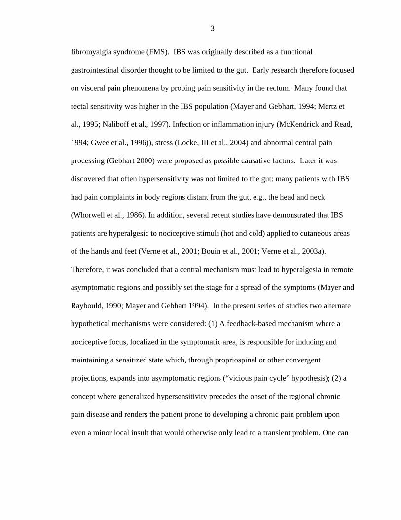
fibromyalgia syndrome (FMS). IBS was originally described as a functional
gastrointestinal disorder thought to be limited to the gut. Early research therefore focused
on visceral pain phenomena by probing pain sensitivity in the rectum. Many found that
rectal sensitivity was higher in the IBS population (Mayer and Gebhart, 1994; Mertz et
al., 1995; Naliboff et al., 1997). Infection or inflammation injury (McKendrick and Read,
1994; Gwee et al., 1996)), stress (Locke, III et al., 2004) and abnormal central pain
processing (Gebhart 2000) were proposed as possible causative factors. Later it was
discovered that often hypersensitivity was not limited to the gut: many patients with IBS
had pain complaints in body regions distant from the gut, e.g., the head and neck
(Whorwell et al., 1986). In addition, several recent studies have demonstrated that IBS
patients are hyperalgesic to nociceptive stimuli (hot and cold) applied to cutaneous areas
of the hands and feet (Verne et al., 2001; Bouin et al., 2001; Verne et al., 2003a).
Therefore, it was concluded that a central mechanism must lead to hyperalgesia in remote
asymptomatic regions and possibly set the stage for a spread of the symptoms (Mayer and
Raybould, 1990; Mayer and Gebhart 1994). In the present series of studies two alternate
hypothetical mechanisms were considered: (1) A feedback-based mechanism where a
nociceptive focus, localized in the symptomatic area, is responsible for inducing and
maintaining a sensitized state which, through propriospinal or other convergent
projections, expands into asymptomatic regions (“vicious pain cycle” hypothesis); (2) a
concept where generalized hypersensitivity precedes the onset of the regional chronic
pain disease and renders the patient prone to developing a chronic pain problem upon
even a minor local insult that would otherwise only lead to a transient problem. One can
4
speculate that a generalized hyperalgesic state could be the result of plasticity induced by
an insult early in life, and / or by genetic factors.
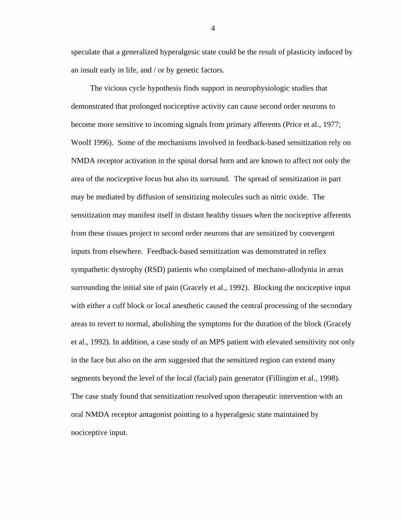
The vicious cycle hypothesis finds support in neurophysiologic studies that
demonstrated that prolonged nociceptive activity can cause second order neurons to
become more sensitive to incoming signals from primary afferents (Price et al., 1977;
Woolf 1996). Some of the mechanisms involved in feedback-based sensitization rely on
NMDA receptor activation in the spinal dorsal horn and are known to affect not only the
area of the nociceptive focus but also its surround. The spread of sensitization in part
may be mediated by diffusion of sensitizing molecules such as nitric oxide. The
sensitization may manifest itself in distant healthy tissues when the nociceptive afferents
from these tissues project to second order neurons that are sensitized by convergent
inputs from elsewhere. Feedback-based sensitization was demonstrated in reflex
sympathetic dystrophy (RSD) patients who complained of mechano-allodynia in areas
surrounding the initial site of pain (Gracely et al., 1992). Blocking the nociceptive input
with either a cuff block or local anesthetic caused the central processing of the secondary
areas to revert to normal, abolishing the symptoms for the duration of the block (Gracely
et al., 1992). In addition, a case study of an MPS patient with elevated sensitivity not only
in the face but also on the arm suggested that the sensitized region can extend many
segments beyond the level of the local (facial) pain generator (Fillingim et al., 1998).
The case study found that sensitization resolved upon therapeutic intervention with an
oral NMDA receptor antagonist pointing to a hyperalgesic state maintained by
nociceptive input.
5
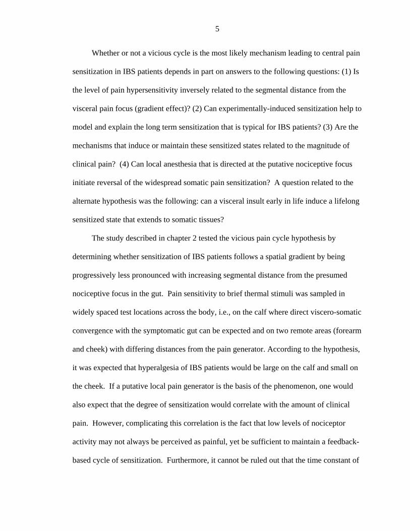
Whether or not a vicious cycle is the most likely mechanism leading to central pain
sensitization in IBS patients depends in part on answers to the following questions: (1) Is
the level of pain hypersensitivity inversely related to the segmental distance from the
visceral pain focus (gradient effect)? (2) Can experimentally-induced sensitization help to
model and explain the long term sensitization that is typical for IBS patients? (3) Are the
mechanisms that induce or maintain these sensitized states related to the magnitude of
clinical pain? (4) Can local anesthesia that is directed at the putative nociceptive focus
initiate reversal of the widespread somatic pain sensitization? A question related to the
alternate hypothesis was the following: can a visceral insult early in life induce a lifelong
sensitized state that extends to somatic tissues?
The study described in chapter 2 tested the vicious pain cycle hypothesis by
determining whether sensitization of IBS patients follows a spatial gradient by being
progressively less pronounced with increasing segmental distance from the presumed
nociceptive focus in the gut. Pain sensitivity to brief thermal stimuli was sampled in
widely spaced test locations across the body, i.e., on the calf where direct viscero-somatic
convergence with the symptomatic gut can be expected and on two remote areas (forearm
and cheek) with differing distances from the pain generator. According to the hypothesis,
it was expected that hyperalgesia of IBS patients would be large on the calf and small on
the cheek. If a putative local pain generator is the basis of the phenomenon, one would
also expect that the degree of sensitization would correlate with the amount of clinical
pain. However, complicating this correlation is the fact that low levels of nociceptor
activity may not always be perceived as painful, yet be sufficient to maintain a feedback-
based cycle of sensitization. Furthermore, it cannot be ruled out that the time constant of
6
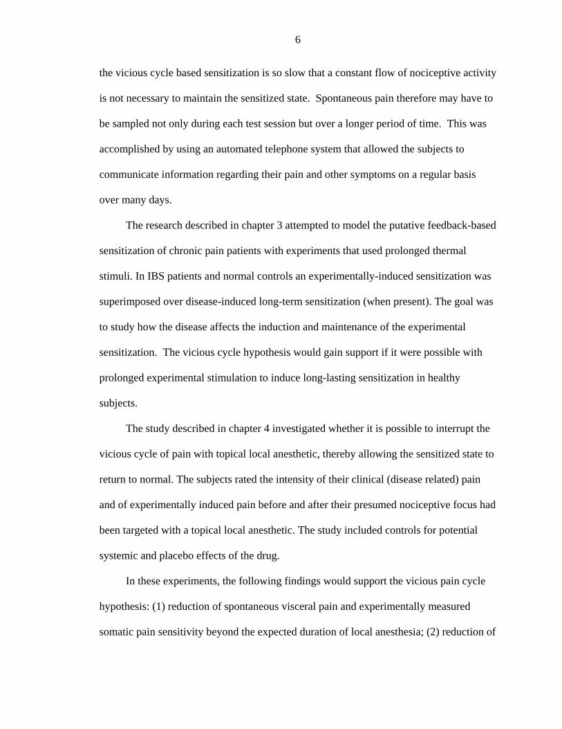
the vicious cycle based sensitization is so slow that a constant flow of nociceptive activity
is not necessary to maintain the sensitized state. Spontaneous pain therefore may have to
be sampled not only during each test session but over a longer period of time. This was
accomplished by using an automated telephone system that allowed the subjects to
communicate information regarding their pain and other symptoms on a regular basis
over many days.
The research described in chapter 3 attempted to model the putative feedback-based
sensitization of chronic pain patients with experiments that used prolonged thermal
stimuli. In IBS patients and normal controls an experimentally-induced sensitization was
superimposed over disease-induced long-term sensitization (when present). The goal was
to study how the disease affects the induction and maintenance of the experimental
sensitization. The vicious cycle hypothesis would gain support if it were possible with
prolonged experimental stimulation to induce long-lasting sensitization in healthy
subjects.
The study described in chapter 4 investigated whether it is possible to interrupt the
vicious cycle of pain with topical local anesthetic, thereby allowing the sensitized state to
return to normal. The subjects rated the intensity of their clinical (disease related) pain
and of experimentally induced pain before and after their presumed nociceptive focus had
been targeted with a topical local anesthetic. The study included controls for potential
systemic and placebo effects of the drug.
In these experiments, the following findings would support the vicious pain cycle
hypothesis: (1) reduction of spontaneous visceral pain and experimentally measured
somatic pain sensitivity beyond the expected duration of local anesthesia; (2) reduction of
7
experimentally measured somatic pain sensitivity following rectal lidocaine
administration is most pronounced in patients with abdominal pain and in segmental
proximity of this pain (i.e., the lumbo-sacral region). The “vicious pain cycle” hypothesis
would have to be questioned if (1) the level of somatic hypersensitivity does not correlate
with clinical pain; (2) baseline somatic hypersensitivity does not exhibit a spatial
gradient; (3) rectal lidocaine does not reduce sensitivity to experimental pain in the IBS
group; (4) anesthetic effect on somatic thermal pain sensitivity does not diminish in a
gradient fashion with increasing segmental distance from the area of abdominal pain.
The study described in chapter 5 was conducted in a rodent model and addressed
the alternate hypothesis that assumes that widespread sensitization predates the onset of
the chronic pain disease and makes the individual pain-prone and thus a likely candidate
for future pain problems. The study investigated whether a visceral chemical insult early
in life can mark the onset of lifelong pain hypersensitivity by inducing plastic changes in
neural or endocrine components of the pain processing system. An operant escape assay
was used to compare the responses of neonatally injured and naïve rodents to thermal
nociceptive paw stimuli.
8
CHAPTER 2 HYPERSENSITIVITY TO CUTANEOUS THERMAL NOCICEPTIVE STIMULI IN
IRRITABLE BOWEL SYNDROME
Introduction
Irritable bowel syndrome (IBS) is an intestinal ailment that may affect up to 20% of
the U.S. population (Verne and Cerda, 1997). It is one of the most frequent
gastrointestinal disorders seen by physician in the US, prompting up to 50% of referrals
to gastroenterologists and as many as 3.5 million clinical appointments annually (Sandler
1990). In spite of its common nature, the pathophysiological mechanisms of IBS are not
well understood. It is now established that most patients with IBS demonstrate
hypersensitivity in response to distension of the gut lumen (Mayer and Gebhart 1994;
Mertz et al., 1995; Naliboff et al., 1997). This hypersensitivity may account for typical
IBS symptoms of urgency, bloating, and abdominal pain. Several causative mechanisms
have been proposed for visceral hypersensitivity including inflammation injury
(McKendrick and Read 1994; Gwee et al., 1996)), stress (Locke, III et al., 2004), and
abnormal pain sensitization (Gebhart 2000). This study focuses on the contribution of
abnormal pain sensitization due to positive nociceptive feedback (vicious pain cycle) that
affects somatic tissues through viscero-somatic convergence. One could argue that the
effectiveness of a feedback-based sensitization mechanism would decrease with
increasing segmental distance from the nociceptive focus, and a recent report provides
support for this argument (Verne et al., 2001). The sensitization of IBS patients was
more pronounced on the feet (segmentally close to visceral pain focus) than the hands
9
when a hot water immersion stimulus was used. Shortcomings of the cited report are the
relatively poor spatial and temporal definition of the immersion stimulus and the fact that
a third segmentally more distant site was not tested to confirm the putative sensitization
gradient. This study revisits the issue with improved methodology. The specific
objectives are to probe cutaneous thermal pain sensitivity along the segmental axis,
including in dermatomes that are remote from the visceral pain focus. The hypotheses to
be tested are (1) that IBS subjects have elevated somatic pain sensitivity and (2) that
sensitization in IBS patients follows a gradient from lower to higher spinal segments, i.e.,
from the symptomatic region of the body toward the (asymptomatic) face. In other
words, the study attempts to determine whether sensitization is most pronounced in
symptomatic segments or alternatively, whether it is generalized without a segmental
gradient. Pain sensitivity was probed with cutaneous thermal stimulation to the lower
and upper extremities and the face. The stimuli were administered with a contact
thermode in order to assure that size of the stimulated area and stimulus duration were
clearly defined and identical in all locations.
Methods
Subject Recruitment
The recruitment and study procedures were approved by the University of Florida
Institutional Review Board and the Veterans SCI Committee. Written informed consent
was obtained from all participants. The criteria for members of the control group
required absence of (1) significant spontaneous pain anywhere in the body, (2) ongoing
pharmacotherapy with narcotics or antidepressants, (3) disease that might significantly
affect pain perception or unduly increase risks (e.g., neurological disorders, serious
psychiatric disorders, diabetes, hypertension, serious cardiovascular disorders, and
10
chronic pain diseases such as fibromyalgia syndrome). The criteria for the disease group
required a diagnosis of ongoing IBS based upon the Rome II criteria (Thompson et al.,
1999), supplemented by additional criteria: absence of other diseases (including other
chronic pain diseases ), risk factors, and ongoing drug treatments, as described for the
control group. Patients diagnosed with Fibromyalgia Syndrome were excluded from the
study. Initial screening consisted of blood pressure measurement, completion of a health
questionnaire and --for IBS patients-- a physical exam, administered by an experienced
gastroenterologist. Considering that all IBS patients (n=9, all diarrhea-predominant)
were females of childbearing age, only female individuals of the same age bracket were
recruited for the control group (n=12). Subject recruitment did not control for menstrual
phase and for whether or not contraceptives were taken. There is no reason to believe
that menstrual phase was not randomly distributed within groups and systematically
different between groups. Synchronization of menstrual phases is most likely among
females that live together, e.g. among college roommates (McClintock 1971).
Participants of our study were recruited from diverse social settings and had no close
contact with each other. Test sessions were scheduled independently of whether or not
IBS patients had an acute exacerbation of symptoms on the day of testing. Some but not
all of the patients reported spontaneous pain or discomfort during some of the sessions.
Daily Protocol
Participants began each daily session of the three-day study by mapping and rating
their spontaneous clinical pain. Subsequently, pain sensitivity was probed with thermal
contact stimuli at three locations on the right side of the body: on the lateral aspect of the
calf (same spinal segment as visceral symptoms, i.e., S1), the volar forearm (dermatomes
C6, T1) and the cheek (V2, V3). Stimulus locations were reproducibly defined relative to
11
anatomical land marks. Skin temperature at the stimulation site was measured with an
Exergen Dermatemp infrared temperature scanner model DT-1001 (Exergen
Corp.,Watertown, MA, USA) before and immediately after each stimulus series.
Pain Measurement
Spontaneous clinical pain and experimental pain (induced by thermode) were
measured with an electronic version of a visual analog scale (Price DD and Harkins SW,
1987). The electronic visual analog scale (eVAS) consisted of a low-friction sliding
potentiometer of 100 mm travel. The left endpoint of the scale was identified as “no
pain,” while the right endpoint was labeled as “intolerably intense pain.” There were no
divisions between these two anchors. The position of the slider was electronically
converted into a pain rating between 0 and 100%. The slider automatically returned to
the left (“no pain”) position after each rating. The eVAS was mounted into the surface
of a small inclined desk positioned to facilitate precise operation with minimal fatigue.
The custom-built testing system integrated all inputs (temperature process value, eVAS
signal) and outputs (stimulus temperature control, stimulus timing) and allowed
automated execution of test protocols with preprogrammed parameters, including limits
for temperature and pain intensity.
Mapping and Rating of Clinical Pain
At the beginning of all experimental sessions, subjects were asked to shade the
locations of spontaneous pain on an anatomical diagram and to rank these sites according
to pain intensity. Subsequently, the intensity of disease-related pain of the upper (head,
neck, shoulder, upper back, arms, hands) and lower (low back, bowel, legs, feet) parts of
the body were rated on the eVAS. The subjects were then asked to rate the
unpleasantness of the single most intense clinical pain. All these ratings were required to
12
be below 5% (on the 0-100% eVAS scale) for subjects to be admitted to the control
group.
Thermal Stimuli
Thermal stimuli were administered with a flat copper contact thermode of
23x23mm in size. The thermode was electronically held at the desired temperature by a
Peltier thermoelectric device. It was brought into light skin contact of reproducible force
by solenoid activation. A thermistor in the center of the thermode, very close to the
surface, sampled the temperature during each skin contact to record potential temperature
deviations resulting from the skin's heat sink effect. The stimulator assembly was
mounted on an adjustable arm (comparable to those that support dental x-ray machines)
for convenient and stable positioning for any desired stimulation site.
The relationship between stimulus temperature and pain intensity was assessed by
rating series of stimuli across a pain intensity range from threshold to at least 45%. The
thermode was preheated to the desired temperature before it contacted the skin. Contact
duration was short (3 seconds), and the intervals between each stimulus were relatively
long (30 seconds), to minimize sensitization. The temperature was set to 42.5 °C (i.e.,
below pain threshold of healthy individuals) for the first stimulus, and increased in 0.7 0C
increments from one pulse to the next until a pain rating of 45% was reached (ascending
temperature series). At that point, a descending temperature series of equal length was
initiated. The series ended with the starting temperature of the ascending series (i.e., 42.5
°C). The pain intensity was rated retrospectively within 5 seconds of the end of the
stimulus, at which time the eVAS slider automatically returned to the left endpoint. It
could be argued that randomized stimulus intensities should have been used to minimize
13
expectation-based response-bias. We chose to use the ascending and descending
intensity series instead in order (1) to avoid uncomfortably high pain intensities and
anxiety in subjects with unexpectedly high pain sensitivity and (2) to avoid unpredictable
distortion of the stimulus response function due to effects of stimulus history on pain
sensitivity (Lamotte and Campbell, 1978; Grill and Coghill, 2002).
Data Analysis
Analysis was performed with SPSS computer software. A two-parameter
regression function (pain intensity = exp [-A-B* temperature]) was used to estimate the
stimulus-intensity response relationship of each subject. Subsequent comparisons used
an ANOVA model and were limited to three representative points on the stimulus-
intensity response curve, i.e., 10% (near threshold), 20%, and 40% pain intensity. Pain
intensities up to 40% were used because the intensity of disease related pain of IBS
patients tends to be within this range. We argue that a test paradigm is most likely to
engage pathophysiologically relevant mechanisms (and thus to produce clinically
relevant data) when it represents disease-typical pain characteristics and takes into
account the possibility that some modulatory mechanisms might be active only within a
certain signal intensity range. Therefore, comparisons of pain sensitivity were made not
only near pain threshold but also at higher intensities.
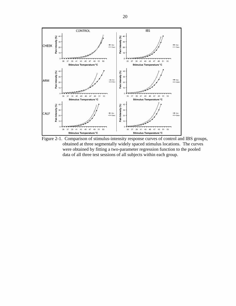
The two-parameter regression function was also fitted to the pooled data of each
group (all three test sessions combined) in order to generate stimulus-intensity response
curves representative of the entire groups (Figure 2-1).
Results
Nine diarrhea-predominant female IBS patients (age range 21-53, mean age: 36.3
years) and 12 healthy female controls (age range 19-45, mean age: 27 years) successfully
14
completed the study. There was no statistically significant age difference between the
groups (p=0.345). The two groups differed in their clinical pain ratings but not in their
pretest blood pressure or heart rate. None of the control subjects had any spontaneous
pain anywhere in the body. For the IBS group the mean spontaneous pain ratings for the
upper part of the body was 9.5% (standard deviation=5%), 20% for the lower part of the
body (standard deviation=14%), and the mean unpleasantness rating for the most intense
spontaneous pain site was 22% (standard deviation=17%).
In many cases the stimulus-intensity response curves of the ascending and
descending series did not overlap. Furthermore, the direction and magnitude of hysteresis
exhibited no discernible pattern. At the three representative points (10, 20 and 40% pain
intensity), the ascending and descending series did not differ to a statistically significant
degree. There were no significant differences in pain sensitivity between the three test
sessions. Therefore, for most analyses, the data of the ascending and descending series of
all three test sessions were pooled.
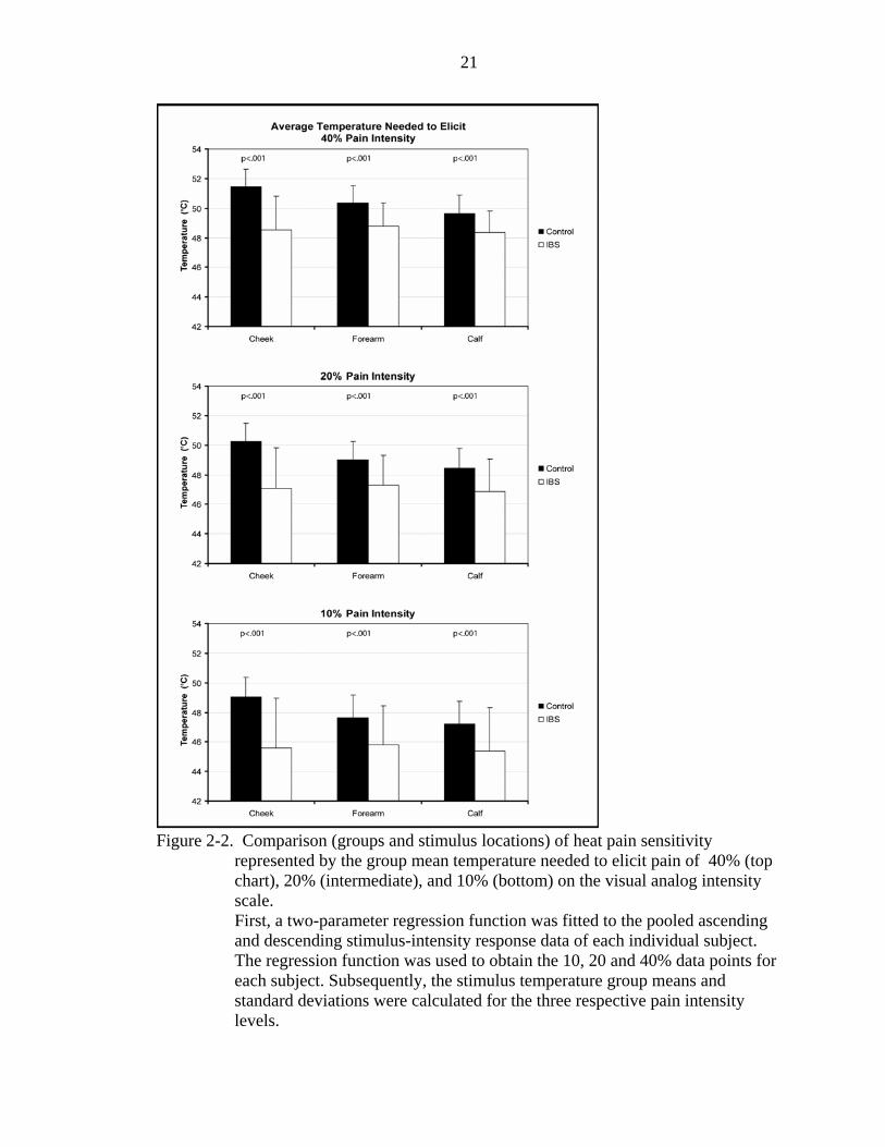
Thermal sensitivity of the IBS group was significantly higher in all three areas
tested (calf, forearm, and cheek): the temperatures necessary to reach pain levels of 10%,
20%, and 40% were significantly lower (p<.001) in IBS patients compared to the control
group. The data indicate that sensitization was not limited to symptomatic dermatomes
(L4-S2) but extended across the body, including the face (Figure 2-2). The results do not
suggest that the presence of disease (IBS) leads to a more pronounced sensitization in
lower segments (no sensitization gradient). Figure 2-2 shows that in both groups pain
sensitivities of the upper and lower extremities were similar. The face of control subjects
appears to be slightly less sensitive (higher stimulus temperature needed) than the other
15
locations; however, this trend is not statistically significant. Also, the difference between
IBS and control groups did not depend on the evoked pain intensity level, i.e., the degree
of sensitization of IBS patients was similar near threshold (10%) and at higher intensities.
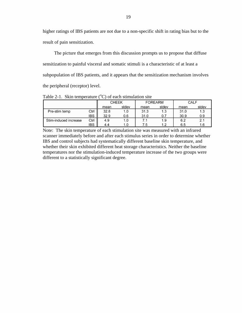
The idea that the higher pain ratings of IBS subjects could be due to abnormal skin
temperature regulation was considered. Pre- and post-stimulation skin temperature data
collected with an infrared scanner speak against this possibility. The pre-stimulation skin
temperatures and stimulation-induced temperature changes did not differ to a statistically
significant degree at any of the stimulation sites between the two groups (Table 2-1).
Lastly, no correlation was found between IBS subjects’ pain sensitivity of any of
the three test sites and their ratings (intensity and unpleasantness) of spontaneous pain.
Discussion
The results support hypothesis 1, which states that IBS patients have higher somatic
heat pain sensitivity compared to healthy pain free individuals, and this confirms what
others have found (Verne et al., 2001; Verne et al., 2003a). Pain sensitization of IBS
patients may not be limited to heat but extend to nociceptive cold: exaggerated sensitivity
to cold was reported in a study where the stimulus consisted of immersion of the
nondominant hand into 4 0C cold water, and where the diagnosis of IBS was based upon
the Rome II criteria (Bouin et al., 2001). In contrast, a group of investigators that used
modified Manning criteria (Manning et al., 1978) to define their IBS group failed to
confirm this finding (Whitehead et al., 1990). Their IBS group exhibited reduced
tolerance to visceral distension but not to a stimulus consisting of immersion of the hand
into ice water. The authors did not report the temperature of the water or whether or not
it was stirred to prevent thermal layering. Likewise, Zighelboim and coworkers failed to
find cold hypersensitivity in a hand immersion experiment using a water temperature of
16
12 0C (Zighelboim et al., 1995). They used a cohort of IBS patients (Rome criteria) with
minimal or no recent symptoms and considered the possibility that the absence of somatic
sensitization may have been due to insufficient severity of the disease. It could be argued
that the disagreement resulted from the fact that Zighelboim’s 12 0C water immersion
stimulus was not rated as painful but as of “moderate discomfort”, while Bouin’s 4 0C
immersion stimulus elicited pain. In summary, at least two possible factors that might
explain the different results of the cited studies must be considered: (1) the mechanism
that renders IBS patients more sensitive may be effective below pain threshold only for
visceral stimuli but be specific to the nociceptive range for somatic stimuli; (2) multiple
disease mechanisms may lead to the same diagnosis of IBS, and the diagnostic criteria
used may not adequately discriminate between them. Therefore, the results of similar
studies may differ due to different representation of subgroups in the samples used. In
this case, thermo-cutaneous nociceptive testing might be useful for discriminating
between subgroups of IBS patients and have the potential to evolve into a diagnostic tool.
Our data suggest that pain sensitization of IBS patients is similar along the entire
segmental axis and independent of the magnitude of disease-related spontaneous pain.
These results can be interpreted as a pain sensitization or disinhibition phenomenon that
is caused by a systemically or diffusely acting factor, and not by a localized vicious
cycle. These findings are in disagreement with an earlier IBS study that reported a higher
degree of sensitization on the foot than on the hands (Verne et al., 2001). The study used
the same selection criteria for IBS patients as the present study, however, the thermal
stimulus consisted of hot water immersion of hand and foot. This methodological
difference offers a possible explanation for the difference in results: immersion of the
17
foot leads to a larger stimulated area than immersion of the hand, and it cannot be ruled
out that IBS subjects are more sensitive to spatial summation. Furthermore, water
immersion leads to stimulation of hairy and glabrous skin, and the relative surface area of
the two skin types may be different for the immersed hand and foot. Thermal layering is
difficult to avoid with fluid immersion stimuli, and it may be different on hairy and
glabrous skin. The use of contact stimulation in the present study eliminated these
potentially confounding factors: the skin surface temperature was precisely maintained by
a feedback-controlled Peltier device; the uncertainty of the actual skin surface
temperature was reduced by sampling the temperature during each stimulus; the
stimulated skin was of the hairy type only, not a mix of hairy and glabrous skin as was
the case in the water immersion study, and the stimulated area was identical in size at all
locations. The probing of sensitivity, by including the face, extended over a larger
segmental area facilitating reliable detection of potential gradient effects.
Our findings of spatially diffuse, widespread pain sensitization of IBS patients are
incompatible with hypothesis 2, which states that sensitization of IBS patients is caused
by a vicious pain cycle that is most effective in close segmental proximity to the area of
spontaneous pain. However, the results are consistent with the clinical observation that
many IBS patients, in addition to intestinal symptoms, are prone to widespread pain
problems that can range from muscle pain to headaches (Whorwell et al., 1986). An
unanswered question is whether the mechanism of sensitization targets the central
nervous system or peripheral tissues. Our results combined with the data of others may
shed some light on the issue: studies that used electrical stimuli to the hands or gut (and
thus bypassed the peripheral receptor level) failed to find sensitization or --to the contrary
18
-- reported elevated detection (Accarino et al., 1995) and pain thresholds in IBS patients
(Cook et al., 1987). This raises the question of whether abnormal pain sensitivity of IBS
patients is the net effect of two antagonistic processes: a central inhibitory process
(possibly DNIC) and a diffuse (possibly endocrine) sensitization mechanism affecting
peripheral receptors (visceral and cutaneous). A testing method that bypasses receptors
(electrical stimulation) would be expected to reveal the central inhibitory effect, while a
receptors-mediated stimulation method would provide a measure of the net effect of both
mechanisms.
The finding of uniformly increased pain ratings, independent of stimulus location
or intensity requires us to examine the alternate hypothesis of a generalized shift in rating
bias of IBS patients, rather than a change in the sensory signal that reaches the conscious
level. This issue has not been specifically addressed by this and the cited studies,
however, preliminary insights may be possible based upon the literature. It appears that
the exaggerated sensitivity of IBS patients is specific to certain stimulus characteristics:
sensitization has been demonstrated with nociceptive hot (Verne et al., 2001) and
nociceptive cold (Bouin et al., 2001) but not by electrical (Cook et al., 1987; Accarino et
al., 1995) and not by uncomfortable yet non-painful cold (Zighelboim et al., 1995)
stimuli to the skin. This stimulus-specificity speaks against the notion of a mere rating
bias or hypervigilance as an explanation for higher intensity ratings of stimuli by IBS
patients, as compared to healthy individuals. The fact that ratings of visceral and
cutaneous stimuli return to normal when IBS patients are treated with rectal lidocaine
(Verne et al., 2003b) or systemic fentanyl (Lembo et al., 2000) further suggests that the
19
higher ratings of IBS patients are not due to a non-specific shift in rating bias but to the
result of pain sensitization.
The picture that emerges from this discussion prompts us to propose that diffuse
sensitization to painful visceral and somatic stimuli is a characteristic of at least a
subpopulation of IBS patients, and it appears that the sensitization mechanism involves
the peripheral (receptor) level.
Table 2-1. Skin temperature (oC) of each stimulation site
Note: The skin temperature of each stimulation site was measured with an infrared scanner immediately before and after each stimulus series in order to determine whether IBS and control subjects had systematically different baseline skin temperature, and whether their skin exhibited different heat storage characteristics. Neither the baseline temperatures nor the stimulation-induced temperature increase of the two groups were different to a statistically significant degree.
20
Figure 2-1. Comparison of stimulus-intensity response curves of control and IBS groups,
obtained at three segmentally widely spaced stimulus locations. The curves were obtained by fitting a two-parameter regression function to the pooled data of all three test sessions of all subjects within each group.
21
Figure 2-2. Comparison (groups and stimulus locations) of heat pain sensitivity
represented by the group mean temperature needed to elicit pain of 40% (top chart), 20% (intermediate), and 10% (bottom) on the visual analog intensity scale. First, a two-parameter regression function was fitted to the pooled ascending and descending stimulus-intensity response data of each individual subject. The regression function was used to obtain the 10, 20 and 40% data points for each subject. Subsequently, the stimulus temperature group means and standard deviations were calculated for the three respective pain intensity levels.
22
CHAPTER 3 CUTANEOUS PAIN SENSITIZATION CHARACTERISTICS IN IRRITABLE
BOWEL SYNDROME
Introduction
Irritable bowel syndrome (IBS) leads to episodic bowel dysfunction, intestinal
discomfort and occasionally pain. Furthermore, patients with IBS, when compared with
healthy individuals, appear to be more pain prone in non-visceral areas of the body
(Whorwell et al., 1986). This is consistent with experimental findings of exaggerated
sensitivity to painful stimuli in non-visceral tissues of IBS patients. Recent work
suggests that somatic hyperalgesia is not limited to dermatomes that have known
convergent projections with symptomatic visceral tissues: sensitization extends evenly
along the segmental axis and affects the face as much as the lower extremities (Rodrigues
et al., 2005). This pronounced and widespread somatic sensitization, which has been
found even during periods of symptom remission, may offer an explanation for the
frequent non-visceral pain conditions in areas of the body far removed from the bowel
(e.g., headaches) of IBS patients (Whorwell et al., 1986). Widespread somatic
sensitization is not unique to IBS: it has been reported for myofascial pain syndrome
(MPS) and fibromyalgia syndrome (FMS) as well (Maixner et al., 1998; Staud et al.,
2001). It may help explain why these patient groups tend to be pain prone across the
entire body. The factors that lead to this sensitization are not well understood. The
following questions to date have not been answered satisfactorily: (1) do the mechanisms
that induce, maintain, and reverse these sensitized states differ between subgroups of
23
patients? And consequently, (2) is the demonstration of somatic sensitization of
diagnostic relevance, i.e., is it a differentiating feature for certain subgroups of patients
that will require a special therapeutic approach to prevent or reverse widespread chronic
pain?
A first step toward answering these questions is to define and categorize the
sensitization phenomena seen in these patient groups. A second step, which promises to
reveal insights regarding the underlying mechanisms and thus point to therapeutic targets,
is to test how these phenomena respond to specific pharmacological probes. The present
study is limited to the first step and uses IBS as a model for studying persistent and
widespread somatic sensitization phenomena. The basic methodological approach of the
study is “signal tracking”. It is a commonly used method for the diagnosis of faulty
electronic circuitry, however, it may hold promise in a biological setting as a diagnostic
tool in diseases involving functional changes within the pain processing system. Signal
tracking injects a defined signal (“stimulus”) and measures the input-output relationship
of the processor. Even though the mechanisms of disease-related and stimulus-evoked
pain may not always completely overlap, signal tracking may reveal abnormalities that
also contribute to chronic pain. Diagnostic criteria derived from this approach are based
upon amplitude- and temporal aspects of the input-output function that is detected at
strategic points of the circuit. The first step is to create a library of input-output functions
found in normal and diseased populations. Interpretation of a sufficiently large data pool,
obtained with a battery of stimulus profiles and with outputs sampled at different
locations of the system, may allow dividing the patient pool into therapeutically relevant
subsets. Mechanistic insights can be expected from a second step where subgroup-
24
specific psychophysical phenomena will be studied in the presence of specific
pharmacological probes.
A number of investigators have focused on the first step by using a variety of
visceral and somatic stimuli and different types of psychophysical response measures on
IBS patients and matching controls. Studies that measured sensitivity took snapshots of
the amplitude aspect of the input-output relationship while others recorded how
sensitivity changes over time, i.e., they measured sensitization. Studies of visceral
sensitivity analyzed pain responses to rectal and jejunal mechanical or electrical stimuli.
Investigations of somatic sensitivity measured pain evoked by cutaneous electrical or
thermal stimuli. Visceral sensitivity was increased in some but not all patients that met
the diagnostic criteria for IBS (Zighelboim et al., 1995; Mertz et al., 1995). Somatic
sensitivity was abnormally high in most IBS patients when stimuli were receptor-
mediated and painful (heat, nociceptive cold) but not when they bypassed the receptor
level (electrical) or were non-painful (Cook et al., 1987; Accarino et al., 1995; Rodrigues
et al., 2005). The ability to discriminate mechanistically may be enhanced by tests that
include temporal aspects of the pain processor’s input-output relationship (rate of
temporal integration). A recent study (Munakata et al., 1997) used individual stimuli to
sample rectal sensitivity and found hyperalgesia only in a subset of IBS patients.
However, when repetitive sigmoid stimulation was used to compare the degree of
temporal integration of IBS and healthy subjects, all IBS- but no control subjects
developed hyperalgesia throughout the course of the prolonged stimulus series. Thus, it
appears that an abnormal induction process of transient visceral sensitization goes hand in
hand with the clinical diagnosis of IBS in most cases, while persistent changes in baseline
25
visceral sensitivity may be a defining feature of only a subset of IBS patients. The present
study continues this work by analyzing and comparing the characteristics of cutaneous
nociceptive temporal integration in samples of IBS patients and matched healthy control
subjects.
Temporal integration has been studied by others in MPS and FMS (Maixner et al.,
1998; Staud et al., 2001) using thermal wind-up paradigms. Sensitization was inferred
from the increase in pain ratings throughout series of brief thermal stimuli of equal
intensity. The duration of these experiments was short to avoid unacceptably high pain
intensities or tissue damage. It was therefore a method appropriate for analyzing
feedback-based sensitization phenomena that occur early during stimulus exposure.
Traditional wind-up experiments are likely to reveal phenomena related to the early
induction phase of persistent states of sensitization. They do not provide much
information about the mechanisms responsible for long-term maintenance of
sensitization. The present study focuses on the conditions needed in IBS patients and
healthy subjects to maintain an already established sensitized state and on
thermoregulatory consequences of prolonged thermal nociceptive stimulation. This will
include addressing specific questions like (1) how does a sensitized state, induced by
pulsed thermal stimulation, respond to changes in pulse duration or interval; and (2) are
the stimulus conditions required for maintaining a sensitized state different depending on
the presence or level of disease-related pain?
26
Methods
Subjects
The subjects were the same as those used for the experiments described in Chapter
2. Recruitment and study procedures were approved by the University of Florida
Institutional Review Board. Written informed consent was obtained from all participants.
The criteria for members of the control group required (1) no significant spontaneous
pain anywhere in the body, (2) no ongoing pharmacotherapy with narcotics or
antidepressants, (3) no disease that might significantly affect pain perception or unduly
increase risk of injury (e.g., neurological disorders, serious psychiatric disorders,
diabetes, hypertension, serious cardiovascular disorders, and chronic pain diseases such
as fibromyalgia syndrome). The criteria for the disease group required a diagnosis of
ongoing IBS based upon the Rome II criteria (Thompson et al., 1999), supplemented by
additional criteria: absence of other diseases (including other chronic pain diseases ), risk
factors, and ongoing drug treatments, as described for the control group. Patients with
any condition where spontaneous pain--according to the definition of the American
College of Rheumatology (Wolfe et al., 1990)--was widespread were excluded from the
study. This ruled out the participation of subjects that met the ACR diagnostic criteria for
fibromyalgia syndrome. Initial screening consisted of blood pressure measurement,
completion of a health questionnaire and--for IBS patients--a clinical diagnosis by a
physician. Considering that all IBS patients (n=8, all diarrhea-predominant) were
females of childbearing age, only female individuals of the same age bracket were
recruited for the control group (n=10). Subject recruitment did not control for menstrual
phase and for whether or not contraceptives were taken. There is no reason to believe
that menstrual phase was not randomly distributed within groups and systematically
27
different between groups. Synchronization of menstrual phases is most likely among
females that live together, e.g., among college roommates (McClintock 1971).
Participants of our study were recruited from diverse social settings and had no close
contact with each other. Test sessions were scheduled independently of whether or not
IBS patients had an acute exacerbation of symptoms on the day of testing. Some but not
all of the patients reported spontaneous pain or discomfort during some of the sessions.
All subjects were right-handed.
Pain Measurement
Spontaneous clinical pain and experimental pain (induced by thermode) were
measured with an electronic version of a visual analog scale (Price DD and Harkins SW
1987). The electronic visual analog scale (eVAS) consisted of a low-friction sliding
potentiometer of 100 mm travel. The left endpoint of the scale was identified as "no
pain", while the right endpoint was defined as "intolerably intense pain". There were no
divisions between these two anchors. The position of the slider was electronically
converted into a pain rating between 0 and 100%. The slider automatically returned to
the left (“no pain”) position when so required by the protocol. The eVAS was mounted
into the surface of a small inclined desk positioned to facilitate precise operation with
minimal fatigue. The custom-built testing system integrated all inputs (temperature
process value, eVAS signal) and outputs (stimulus temperature control, stimulus timing)
and allowed automated execution of test protocols with preprogrammed parameters,
including limits for temperature and pain intensity.
Response-Dependent Stimulation Method
Thermal stimuli were administered with a flat copper contact thermode of
23x23mm in size. The thermode was electronically held at the desired temperature by a
28
Peltier thermoelectric device. It was brought into light skin contact of reproducible force
by solenoid activation. Series of brief contacts were administered by periodically turning
the solenoid on and off. The duration of each contact, the interval between contacts
(Inter-Stimulus Interval, ISI) and the number of contacts of each series was
preprogrammed in the stimulator control software, allowing fully automated data
collection. Unlike most pain tests, the stimulation / data acquisition system used in this
study did not define the stimulus as the dependent variable and the subject’s rating of the
pain as the dependent variable. Similarly to a method described by Gracely et al., it
reversed this arrangement by linking the subject and the stimulator in a closed
proportional control loop (Figure 3-1) (Gracely et al., 1988). A pain intensity setpoint
was defined, and an algorithm in the stimulator control software calculated the deviation
of the patient’s actual pain rating from the setpoint as well as the derivative of this error.
These data were the basis for automatic adjustments of the stimulus temperature to
maintain an average pain rating that equaled the setpoint. The methodology has much in
common with an autopilot that keeps altitude or course of an aircraft near the desired
setpoint. Response-dependent stimulation (REDSTIM) allows prolonged stimulation
without the risk of an escalation of pain intensities to intolerable levels. Furthermore, it is
more likely to engage clinically relevant mechanisms, because –as is the case in chronic
pain—it maintains low to midrange signal intensities over prolonged periods of time.
Testing Protocol
Measurement of spontaneous pain
At the beginning of all experimental sessions, subjects were asked to shade the
locations of spontaneous pain on an anatomical diagram and to rank these sites according
to pain intensity. Subsequently, the intensity of disease-related pain of the upper (head,
29
neck, shoulder, upper back, arms, hands) and lower (low back, bowel, legs, feet) parts of
the body were rated on the eVAS. The subjects were then asked to rate the
unpleasantness of the single most intense clinical pain. All these ratings were required to
be below 5% (on the 0-100% eVAS scale) for subjects to be admitted to the control
group.
Measurement of skin temperature
Skin temperature at the stimulation site and the corresponding contralateral site was
measured with an Exergen Dermatemp infrared temperature scanner model DT-1001
(Exergen Corp.,Watertown, MA, USA) before and immediately after each stimulus
series.
Thermal stimulation
Thermal stimulation was conducted during three separate and identical daily
sessions. Each session included thermal tests designed to collect snapshots of pain
sensitivity. These protocols and data are presented in chapter 2. The present chapter
focuses on experiments that used prolonged series of stimuli and REDSTIM
methodology. During each session a separate REDSTIM experiment was conducted on
the thenar eminence of each hand.
Experiment 1. The thenar eminence of the left hand was the site for an experiment
that used four series of 25 brief thermal contact pulses each. The interval between stimuli
(ISI) was 3 sec throughout the experiment. The only difference between the series was
stimulus pulse duration. It was 1.0 sec for the pulses of series 1 and 3, 0.8 sec for series 2
and 4 (Figure 3-2 A, Table 3-1). As was discussed earlier, REDSTIM methodology
automatically adjusted stimulus temperature in order to maintain a constant average pain
intensity level throughout the experiment (35% on the visual analog scale). The goal was
30
to assess how much the stimulus temperature (series average) would have to change to
compensate for the change in pulse duration from one series to the next. The eVAS slider
automatically returned to the “no pain” position at the end of the fourth series.
Experiment 2. It began 3 minutes after the end of the first experiment and used
the thenar eminence of the right hand as the stimulation site. Like the first experiment it
consisted of four series of 25 thermal pulses each, without interruption between series.
Consistent with REDSTIM methodology, the temperature was automatically adjusted
from pulse to pulse in order to maintain a constant average pain intensity rating (35%)
throughout the experiment. Duration of all stimulus pulses for the entire experiment was
0.9 sec. ISI, however, was subject to change from series to series. It was 2.5 sec during
the first and third series, 3.5 sec during the second and fourth series (Figure 3-2 B, Table
3-1). The goal was to determine how much the stimulus temperature (series average)
would have to change to compensate for the change in ISI from one series to the next.
Induction phase. The temperature needed to maintain the pain intensity setpoint
under given circumstances varies from individual to individual and even more so between
the IBS and control groups. The stimulator software was designed to find the individually
appropriate temperature automatically during an induction phase that preceded series 1of
each experiment using the pulse duration and inter-stimulus interval parameters of the
first series. Since these parameters were different for the induction phases of the two
experiments, it is important to be aware that the cumulative stimulus duration per ten
seconds was larger for experiment 2 (3.6 seconds of stimulation compared to 3.3
seconds). Therefore, the induction phase of the second experiment was expected to be
shorter. However, past the induction phase, the cumulative stimulus duration was equal
31
for series 1 through 4 (90 seconds) The induction series began with a 430C pulse, which
was never perceived as painful by any subject. The temperature then increased from pulse
to pulse in 10C increments until the pain intensity rating reached 10% on the electronic
visual analog scale. Thereafter, the temperature continued to rise at a reduced rate (0.50C
/ pulse) if the pain intensity did not continue to increase over a period of 3 consecutive
pulses. The temperature remained unchanged from pulse to pulse if pain intensity
continued to increase across triplets of pulses. During the induction phase, the thermode
temperature could either remain the same or increase; it could not decrease. The
induction phase ended when the pain intensity rating first reached the setpoint of 35% on
the eVAS. At that point, temperature modulation became bidirectional, in proportion to
the deviation from setpoint, and the tests consisting of 4x25 pulses, as described above,
began.
Transition phase. Preliminary experiments had shown that an abrupt large change
of pulse duration or ISI from one series to the next could lead to a large deviation from
the pain intensity setpoint. To minimize such fluctuations a transition period of 4 pulses
was interposed between series. The total change of pulse duration or ISI between series
was divided up between the 4 transition pulses, resulting in small, almost unnoticeable
steps and preventing sudden changes in pain intensity.
Oscillations around setpoint. A well tuned proportional control system is
expected to approach a new setpoint with a damped oscillation which eventually
transitions into a new steady state. It soon became clear that in the case of REDSTIM,
oscillations around the setpoint are poorly damped and often continue indefinitely. The
most plausible explanation is the effect of “offset analgesia” (Grill and Coghill 2002):
32
when the stimulation control algorithm reduces the thermode temperature in order to
return a high pain rating to setpoint it triggers an “offset analgesia” event and pain
intensity drops disproportionately to near zero levels. This prompts the REDSTIM system
to incrementally increase temperature again. The subject, while in “offset analgesia”, will
not perceive a corresponding pain increase. When the “offset analgesia” ends (after
about 3 - 5 seconds) the pain rating will suddenly “catch up” and often overshoot the
setpoint. The above-setpoint error will trigger a new drop in stimulus temperature and
mark the beginning of a new cycle of the pain intensity oscillation. This phenomenon
could be minimized but not completely eliminated by proper choice of gain and time
constants within the control algorithm. The oscillations had the advantage that they
blinded the subject regarding the setpoint and possibly led to a more realistic model of
chronic pain, which often fluctuates in intensity. The REDSTIM method, unlike most
traditional pain tests, provided the opportunity to monitor the subject’s rating reliability
by comparing the mean eVAS ratings across a series with the setpoint. A mean rating
error >4% was grounds for exclusion of the series from data analysis.
Data Analysis
The following factors were included in the analysis:
1. Group (IBS patient, healthy control) 2. Day (each test was repeated on three different days) 3. Time (skin temperature was measured before and after stimulation) The following response variables were used:
Skin temperature: Pre-stimulus skin temperature (a) at stimulation site and (b) at
corresponding contralateral site; (c) Skin temperature recorded after end of stimulus
series at stimulation site and (d) at corresponding contralateral site.

33
Stimulus temperature needed to elicit a given pain intensity during induction
phase: (e) temperature when pain rating first reached 10% (“near pain threshold”); (f)
temperature at the end of the induction phase, i.e., when pain rating first reached set point
(35%). This was the point where the computer began modulating the thermode
temperature in order to maintain an eVAS score of 35% (“midrange pain intensity”,
presumed to be representative of intensities frequently experienced clinically).
Average stimulus temperature needed to maintain pain intensity setpoint:
(g) for series 1; (h) for series 2; (i) for series 3; (k) for series 4.
The data were analyzed using a repeated measures ANOVA model.
Results
Skin Temperature
Skin temperature was measured at the thenar eminence of both hands prior to
thermal stimulation on each of the three identical daily test sessions. The temperature did
not differ to a statistically significant degree between IBS and control groups and
between sessions. Repeated measures ANOVA revealed no significant group-by-day
interactions. Furthermore, no significant correlation was found between pre-stimulus
skin temperature at the stimulation site and the average thermode temperature needed to
maintain the pain intensity near the 35% setpoint during any of the four stimulus series
on any of the three daily test sessions (correlation coefficients ranged from 0.31 to -0.09).
Skin temperature was measured at the thenar eminence on the side opposite to
stimulation before and after stimulation (ANOVA variable “Time”) in order to assess
contralateral thermoregulatory responses that might be triggered by prolonged stimulus
exposure. Both IBS and control groups (ANOVA variable “Group”) underwent three
identical daily sessions (ANOVA variable “Day”). The data collected during experiment
34
1 (prolonged stimulation on left hand, skin temperature measurement on right hand) were
analyzed with repeated measures ANOVA. Significance was obtained for “Group” (F (1,
16) = 4.895, P = 0.042), “Time” (F (2, 16) = 14.325, P = .002) and the “Group x Time”
interaction (F (2, 16) = 6.690, P = .020). Similarly, for experiment 2 (prolonged
stimulation on right hand, skin temperature measurement on left hand) “Time” (F (2, 16)
= 9.076, P = .008) and the “Group x Time” interaction (F (2, 16) = 5.589, P = .031)
reached significance.
These findings suggest that the IBS and control group did not differ in their
baseline skin temperatures, i.e., the skin temperature before stimulation onset.
Furthermore, the data suggest that the skin temperature of the palm of the hand drops
when the corresponding location on the opposite side is subjected to prolonged thermal
stimulation. The temperature drop is smaller in the IBS group than in the control group.
In other words, IBS patients, when compared to healthy control subjects, appear to have a
reduced autonomic thermoregulatory response to a prolonged thermal stimulus on the
opposite side of the body (Table 3-2 and Figure 3-3).
Clinical Pain
There were no significant correlations between the ratings of disease-related pain
(intensity and unpleasantness) at the most painful site on the day of testing and the
average stimulus temperatures for any of the three daily sessions and for either of the two
experiments.
Induction of Stimulus-Induced Sensitization
In experiments 1 and 2 the thenar eminence of the left and right hand respectively
was exposed to a series of thermal contact stimuli of incrementally increasing
temperatures until a pain intensity of 35% was reached on the electronic visual analog
35
scale. The response variables derived from this portion of the experiment were thermode
temperature at the time when the pain intensity first reached 10% and when it reached
35%. A mixed model ANOVA included between-subjects factors (“Group”) and
within-subjects factors (“Day”; “Temperature @ 10%” pain intensity”; “Temperature @
35% pain intensity”). In both experiments a significant main effect of “Group” and
“Temperature” was found. In other words, for IBS subjects, when compared with healthy
controls, lower temperatures were needed to reach the 10% and 35% pain intensity levels
respectively (Figures 3-4A and 3-5A). Thus, IBS patients emerge as more sensitive to
thermal nociceptive stimuli in this experimental setting, as has been the case in other
types of tests (see chapter 2). No significant “Group” x “Temperature” interaction was
found in either of the two experiments, suggesting that the temperature increase needed to
boost pain intensity from 10% to 35% was not significantly different between groups.
This finding does not permit conclusions regarding sensitization rate because the
experiment did not control for the number of stimuli that were presented between the
10% and 35% pain intensity criterion. Under certain circumstances, as described in the
methods section, the stimulator maintained a constant temperature across consecutive
stimuli during the induction phase.
Maintenance of Stimulus-Induced Sensitization
It was argued that the average temperature required for maintaining the average
pain rating near the setpoint of 35% depends in part on the duration of the stimulus pulses
and the length of the intervals between pulses. The temperature changes needed to
compensate for modifications in pulse duration and ISI were measured in experiment 1
and 2 respectively. Each experiment was repeated 3 times on different days. A mixed
model ANOVA with “Group” as a between subjects factor and “Day” & “Average
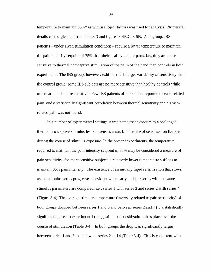
36
temperature to maintain 35%” as within subject factors was used for analysis. Numerical
details can be gleaned from table 3-3 and figures 3-4B,C, 3-5B. As a group, IBS
patients—under given stimulation conditions-- require a lower temperature to maintain
the pain intensity setpoint of 35% than their healthy counterparts, i.e., they are more
sensitive to thermal nociceptive stimulation of the palm of the hand than controls in both
experiments. The IBS group, however, exhibits much larger variability of sensitivity than
the control group: some IBS subjects are no more sensitive than healthy controls while
others are much more sensitive. Few IBS patients of our sample reported disease-related
pain, and a statistically significant correlation between thermal sensitivity and disease-
related pain was not found.
In a number of experimental settings it was noted that exposure to a prolonged
thermal nociceptive stimulus leads to sensitization, but the rate of sensitization flattens
during the course of stimulus exposure. In the present experiments, the temperature
required to maintain the pain intensity setpoint of 35% may be considered a measure of
pain sensitivity: for more sensitive subjects a relatively lower temperature suffices to
maintain 35% pain intensity. The existence of an initially rapid sensitization that slows
as the stimulus series progresses is evident when early and late series with the same
stimulus parameters are compared: i.e., series 1 with series 3 and series 2 with series 4
(Figure 3-4). The average stimulus temperature (inversely related to pain sensitivity) of
both groups dropped between series 1 and 3 and between series 2 and 4 (to a statistically
significant degree in experiment 1) suggesting that sensitization takes place over the
course of stimulation (Table 3-4). In both groups the drop was significantly larger
between series 1 and 3 than between series 2 and 4 (Table 3-4). This is consistent with
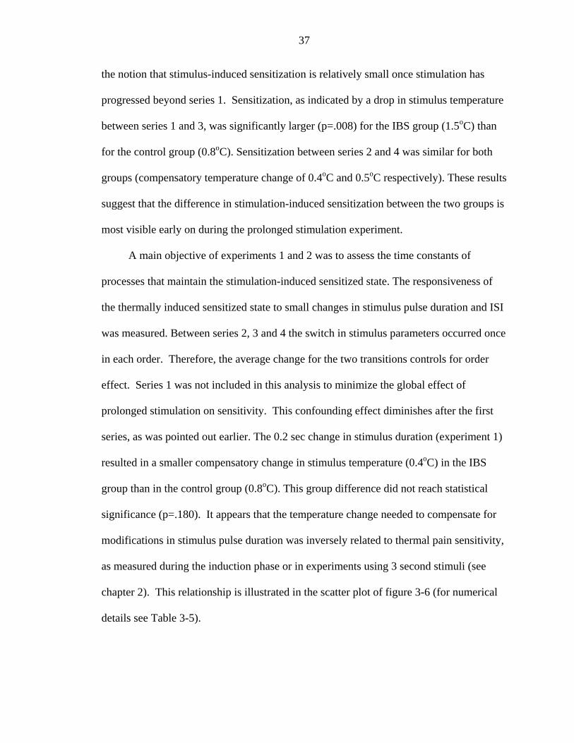
37
the notion that stimulus-induced sensitization is relatively small once stimulation has
progressed beyond series 1. Sensitization, as indicated by a drop in stimulus temperature
between series 1 and 3, was significantly larger (p=.008) for the IBS group (1.5oC) than
for the control group (0.8oC). Sensitization between series 2 and 4 was similar for both
groups (compensatory temperature change of 0.4oC and 0.5oC respectively). These results
suggest that the difference in stimulation-induced sensitization between the two groups is
most visible early on during the prolonged stimulation experiment.
A main objective of experiments 1 and 2 was to assess the time constants of
processes that maintain the stimulation-induced sensitized state. The responsiveness of
the thermally induced sensitized state to small changes in stimulus pulse duration and ISI
was measured. Between series 2, 3 and 4 the switch in stimulus parameters occurred once
in each order. Therefore, the average change for the two transitions controls for order
effect. Series 1 was not included in this analysis to minimize the global effect of
prolonged stimulation on sensitivity. This confounding effect diminishes after the first
series, as was pointed out earlier. The 0.2 sec change in stimulus duration (experiment 1)
resulted in a smaller compensatory change in stimulus temperature (0.4oC) in the IBS
group than in the control group (0.8oC). This group difference did not reach statistical
significance (p=.180). It appears that the temperature change needed to compensate for
modifications in stimulus pulse duration was inversely related to thermal pain sensitivity,
as measured during the induction phase or in experiments using 3 second stimuli (see
chapter 2). This relationship is illustrated in the scatter plot of figure 3-6 (for numerical
details see Table 3-5).
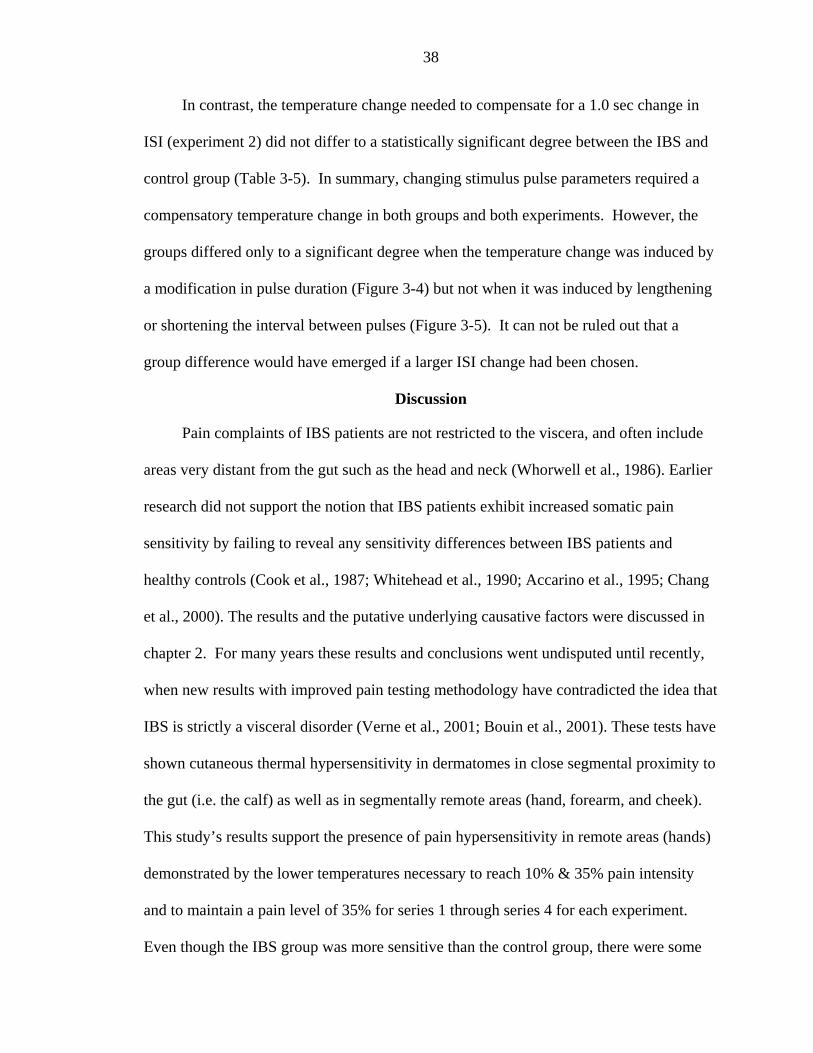
38
In contrast, the temperature change needed to compensate for a 1.0 sec change in
ISI (experiment 2) did not differ to a statistically significant degree between the IBS and
control group (Table 3-5). In summary, changing stimulus pulse parameters required a
compensatory temperature change in both groups and both experiments. However, the
groups differed only to a significant degree when the temperature change was induced by
a modification in pulse duration (Figure 3-4) but not when it was induced by lengthening
or shortening the interval between pulses (Figure 3-5). It can not be ruled out that a
group difference would have emerged if a larger ISI change had been chosen.
Discussion
Pain complaints of IBS patients are not restricted to the viscera, and often include
areas very distant from the gut such as the head and neck (Whorwell et al., 1986). Earlier
research did not support the notion that IBS patients exhibit increased somatic pain
sensitivity by failing to reveal any sensitivity differences between IBS patients and
healthy controls (Cook et al., 1987; Whitehead et al., 1990; Accarino et al., 1995; Chang
et al., 2000). The results and the putative underlying causative factors were discussed in
chapter 2. For many years these results and conclusions went undisputed until recently,
when new results with improved pain testing methodology have contradicted the idea that
IBS is strictly a visceral disorder (Verne et al., 2001; Bouin et al., 2001). These tests have
shown cutaneous thermal hypersensitivity in dermatomes in close segmental proximity to
the gut (i.e. the calf) as well as in segmentally remote areas (hand, forearm, and cheek).
This study’s results support the presence of pain hypersensitivity in remote areas (hands)
demonstrated by the lower temperatures necessary to reach 10% & 35% pain intensity
and to maintain a pain level of 35% for series 1 through series 4 for each experiment.
Even though the IBS group was more sensitive than the control group, there were some
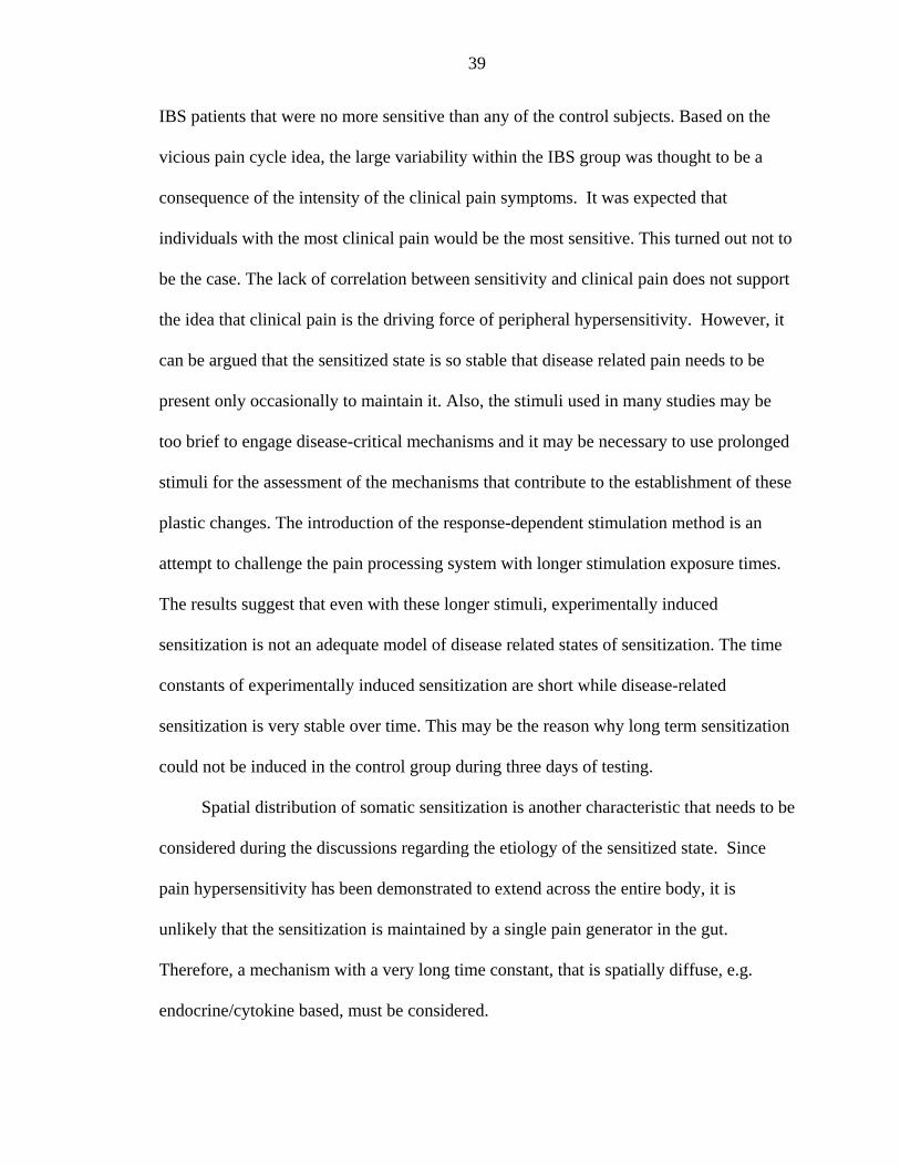
39
IBS patients that were no more sensitive than any of the control subjects. Based on the
vicious pain cycle idea, the large variability within the IBS group was thought to be a
consequence of the intensity of the clinical pain symptoms. It was expected that
individuals with the most clinical pain would be the most sensitive. This turned out not to
be the case. The lack of correlation between sensitivity and clinical pain does not support
the idea that clinical pain is the driving force of peripheral hypersensitivity. However, it
can be argued that the sensitized state is so stable that disease related pain needs to be
present only occasionally to maintain it. Also, the stimuli used in many studies may be
too brief to engage disease-critical mechanisms and it may be necessary to use prolonged
stimuli for the assessment of the mechanisms that contribute to the establishment of these
plastic changes. The introduction of the response-dependent stimulation method is an
attempt to challenge the pain processing system with longer stimulation exposure times.
The results suggest that even with these longer stimuli, experimentally induced
sensitization is not an adequate model of disease related states of sensitization. The time
constants of experimentally induced sensitization are short while disease-related
sensitization is very stable over time. This may be the reason why long term sensitization
could not be induced in the control group during three days of testing.
Spatial distribution of somatic sensitization is another characteristic that needs to be
considered during the discussions regarding the etiology of the sensitized state. Since
pain hypersensitivity has been demonstrated to extend across the entire body, it is
unlikely that the sensitization is maintained by a single pain generator in the gut.
Therefore, a mechanism with a very long time constant, that is spatially diffuse, e.g.
endocrine/cytokine based, must be considered.
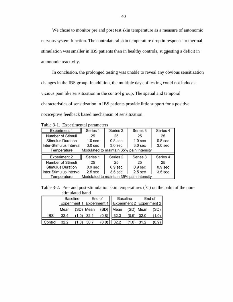
40
We chose to monitor pre and post test skin temperature as a measure of autonomic
nervous system function. The contralateral skin temperature drop in response to thermal
stimulation was smaller in IBS patients than in healthy controls, suggesting a deficit in
autonomic reactivity.
In conclusion, the prolonged testing was unable to reveal any obvious sensitization
changes in the IBS group. In addition, the multiple days of testing could not induce a
vicious pain like sensitization in the control group. The spatial and temporal
characteristics of sensitization in IBS patients provide little support for a positive
nociceptive feedback based mechanism of sensitization.
Table 3-1. Experimental parameters
Table 3-2. Pre- and post-stimulation skin temperatures (oC) on the palm of the non-
stimulated hand
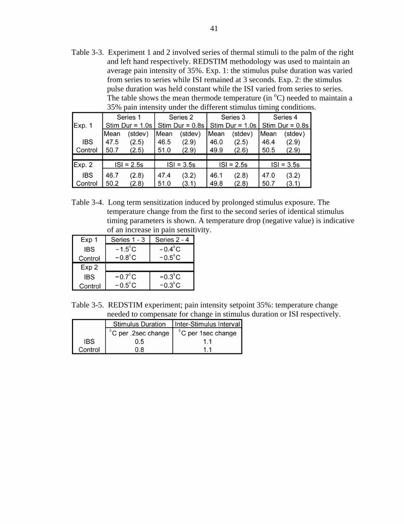
41
Table 3-3. Experiment 1 and 2 involved series of thermal stimuli to the palm of the right and left hand respectively. REDSTIM methodology was used to maintain an average pain intensity of 35%. Exp. 1: the stimulus pulse duration was varied from series to series while ISI remained at 3 seconds. Exp. 2: the stimulus pulse duration was held constant while the ISI varied from series to series. The table shows the mean thermode temperature (in oC) needed to maintain a 35% pain intensity under the different stimulus timing conditions.
Table 3-4. Long term sensitization induced by prolonged stimulus exposure. The
temperature change from the first to the second series of identical stimulus timing parameters is shown. A temperature drop (negative value) is indicative of an increase in pain sensitivity.
Table 3-5. REDSTIM experiment; pain intensity setpoint 35%: temperature change
needed to compensate for change in stimulus duration or ISI respectively.
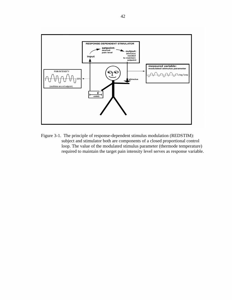
42
Figure 3-1. The principle of response-dependent stimulus modulation (REDSTIM):
subject and stimulator both are components of a closed proportional control loop. The value of the modulated stimulus parameter (thermode temperature) required to maintain the target pain intensity level serves as response variable.
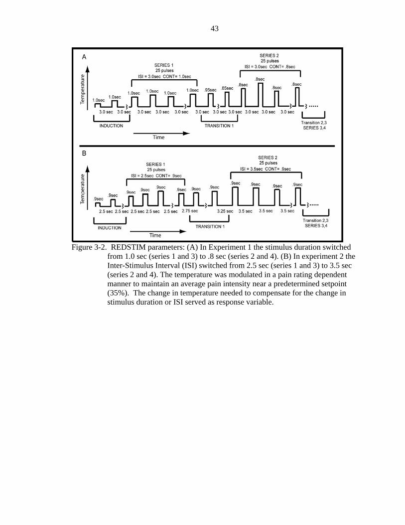
43
Figure 3-2. REDSTIM parameters: (A) In Experiment 1 the stimulus duration switched
from 1.0 sec (series 1 and 3) to .8 sec (series 2 and 4). (B) In experiment 2 the Inter-Stimulus Interval (ISI) switched from 2.5 sec (series 1 and 3) to 3.5 sec (series 2 and 4). The temperature was modulated in a pain rating dependent manner to maintain an average pain intensity near a predetermined setpoint (35%). The change in temperature needed to compensate for the change in stimulus duration or ISI served as response variable.
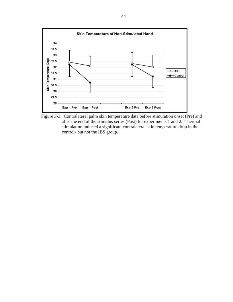
44
Figure 3-3. Contralateral palm skin temperature data before stimulation onset (Pre) and
after the end of the stimulus series (Post) for experiments 1 and 2. Thermal stimulation induced a significant contralateral skin temperature drop in the control- but not the IBS group.
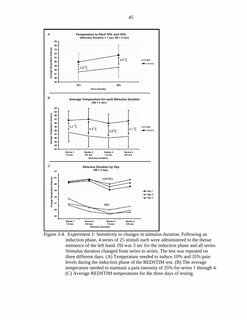
45
Figure 3-4. Experiment 1: Sensitivity to changes in stimulus duration. Following an
induction phase, 4 series of 25 stimuli each were administered to the thenar eminence of the left hand. ISI was 3 sec for the induction phase and all series. Stimulus duration changed from series to series. The test was repeated on three different days. (A) Temperature needed to induce 10% and 35% pain levels during the induction phase of the REDSTIM test. (B) The average temperature needed to maintain a pain intensity of 35% for series 1 through 4. (C) Average REDSTIM temperatures for the three days of testing.
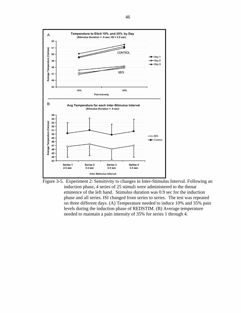
46
Figure 3-5. Experiment 2: Sensitivity to changes in Inter-Stimulus Interval. Following an
induction phase, 4 series of 25 stimuli were administered to the thenar eminence of the left hand. Stimulus duration was 0.9 sec for the induction phase and all series. ISI changed from series to series. The test was repeated on three different days. (A) Temperature needed to induce 10% and 35% pain levels during the induction phase of REDSTIM. (B) Average temperature needed to maintain a pain intensity of 35% for series 1 through 4.
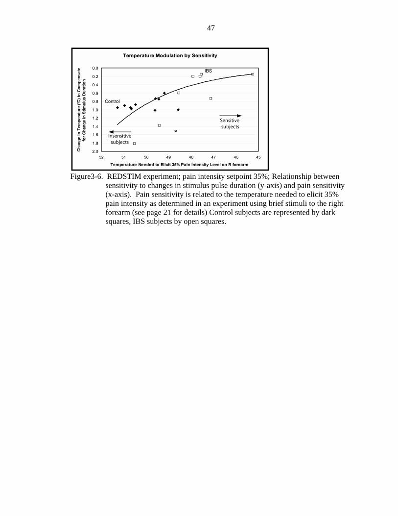
47
Figure3-6. REDSTIM experiment; pain intensity setpoint 35%; Relationship between
sensitivity to changes in stimulus pulse duration (y-axis) and pain sensitivity (x-axis). Pain sensitivity is related to the temperature needed to elicit 35% pain intensity as determined in an experiment using brief stimuli to the right forearm (see page 21 for details) Control subjects are represented by dark squares, IBS subjects by open squares.
48
CHAPTER 4 THE EFFECT OF TOPICAL LOCAL ANESTHETICS ON SOMATIC PAIN
SENSITIZATION IN IRRITABLE BOWEL SYNDROME
Introduction
Irritable bowel syndrome is a common gastrointestinal disorder that consists of
abnormal bowel movements that can be associated with abdominal pain. Often the pain
symptoms are not limited to the gut but expand to remote regions such as the neck and
head during the course of the disease. The question arises whether IBS is characterized
by pathophysiological mechanisms that are initially regional (in the gut), and nociceptive
signals emanating from the symptomatic viscera induce somatic sensitization across the
body. It is critical to ask whether a temporary interruption of nociceptive signals from the
gut allows somatic pain sensitivity of IBS patients to return to normal. In the event a
relationship between the presence of nociceptive signals from the gut and somatic pain
sensitivity cannot be demonstrated one needs to consider the alternate possibility that IBS
is a regional manifestation of a pathophysiological mechanism that affects the entire body
to begin with. According to this theory the primary problem would be a generalized state
of sensitization that affects visceral and somatic tissues alike and makes it more likely
that subsequent local insults develop into chronic pain problems. The present study
concentrates on the first hypothesis which states that widespread somatic sensitization of
IBS patients is maintained by a regional nociceptive focus, i.e., by nociceptive signals
from the symptomatic viscera. If the presence of visceral pain or at least of nociceptive
drive emanating from visceral tissues is a critical factor for somatic sensitization one
49
would expect that the degree of somatic sensitization correlates with the presence /
absence or frequency / intensity of spontaneous visceral pain. Furthermore, one would
expect that silencing the presumed visceral nociceptive focus with rectally administered
topical local anesthetics would lead to a reduction in somatic sensitization.
At least two studies (conducted on cohorts of IBS patients and patients with
myofascial pain syndrome respectively) have used local anesthetics in an attempt to
silence a nociceptive focus that was presumed to maintain a sensitized state (Fillingim et
al., 1998; Verne et al., 2003). Both have demonstrated a reduction in pain
hypersensitivity in remote areas following anesthetic intervention, therefore providing
support for the idea that a “vicious pain cycle” is an important factor in the maintenance
of hypersensitivity. However, other reports have put this view into question: topical
rectally administered lidocaine failed to reduce pain sensitivity in some studies (Lembo et
al., 1994) and “triggerpoint” injections with saline or simply an empty needle were no
less effective in reducing pain in myofascial pain patients than lidocaine injections
(Garvey et al., 1989). The disagreement between studies clearly suggests that questions
remain regarding the mechanism that maintains widespread somatic sensitization in
regional pain disorders.
The present study addresses a number of questions that have not been answered
conclusively by previous investigators. These questions are the following: (1) Is the
somatic hypersensitivity that is typical for many IBS patients nociceptive-specific or does
it extend to the perception of non-nociceptive stimuli? (2) Can the changes in somatic
sensitivity that follow rectal lidocaine administration conclusively be explained by the
topical anesthetic effect of the drug, or must systemic effects be considered as a possible
50
mechanism? A partially related question is: (3) Does the effect of lidocaine on somatic
pain sensitivity require that the subjects have IBS or can it be demonstrated also in
healthy pain-free individuals? (4) Is the effect of lidocaine more pronounced in the
segmental proximity of the presumed visceral pain focus or is it similar across the entire
body? (5) Is it possible that an unidentified placebo effect could explain the results of
previous studies, considering that it cannot be ruled out that some subjects might detect
the active drug by its numbing effect or its effect on the perception of the experimental
stimuli? The present study makes an attempt to address these questions with improved
methodology.
Methods
Subjects
Recruitment and study procedures were approved by the University of Florida
Institutional Review Board. Written informed consent was obtained from all participants.
The criteria for membership in the control group required (1) no significant spontaneous
pain anywhere in the body, (2) no ongoing pharmacotherapy with narcotics or
antidepressants, (3) no disease or condition that might significantly affect pain perception
or unduly increase risk of injury (e.g., neurological disorders, serious psychiatric
disorders, diabetes, hypertension, serious cardiovascular disorders, pregnancy, and
chronic pain diseases such as fibromyalgia syndrome, (4) no prior complications or
allergies with the local anesthetic lidocaine. The criteria for the disease group required a
diagnosis of ongoing IBS based upon the Rome II criteria (Thompson et al., 1999),
supplemented by additional criteria: absence of other diseases (including other chronic
pain diseases ), risk factors, and ongoing drug treatments, as described for the control
group. Patients with a history of widespread pain (e.g., individuals with fibromyalgia
51
syndrome) were excluded from the study. Initial screening consisted of blood pressure
measurement, completion of a health questionnaire and--for IBS patients-- a clinical
diagnosis by a physician. Subject recruitment did not control for menstrual phase and for
whether or not contraceptives were taken, but the respective information was recorded.
There is no reason to believe that menstrual phase was not randomly distributed within
groups and systematically different between groups. Synchronization of menstrual
phases is most likely among females that live together, e.g., among college roommates
(McClintock 1971). Participants of our study were recruited from diverse social settings
and had no close contact with each other. Test sessions were scheduled independently of
whether or not IBS patients had an acute exacerbation of symptoms on the day of testing.
Some but not all of the patients reported spontaneous pain or discomfort during some of
the sessions. All subjects were right-handed.
Pain Measurement
Spontaneous clinical pain and experimental pain (induced by thermode) were
measured with an electronic version of a visual analog scale (Price DD and Harkins SW,
1987). The electronic visual analog scale (eVAS) consisted of a low-friction sliding
potentiometer of 100 mm travel. The left endpoint of the scale was identified as "no
pain", while the right endpoint was labeled as "intolerably intense pain". There were no
divisions between these two anchors. The position of the slider was electronically
converted into a pain rating between 0 and 100%. The slider automatically returned to
the left (“no pain”) position when so required by the protocol. The eVAS was mounted
into the surface of a small inclined desk positioned to facilitate precise operation with
minimal fatigue. The custom-built testing system integrated all inputs (temperature
process value, eVAS signal) and outputs (stimulus temperature control, stimulus timing)
52
and allowed automated execution of test protocols with preprogrammed parameters,
including limits for temperature and pain intensity.
Testing Protocol
Daily Protocol
Participants began each daily session of the non-consecutive seven day study by
mapping and rating their spontaneous clinical pain. Pain testing began with a prolonged
thermal stimulus to the thenar eminence of the right hand. The stimulus was controlled as
a function of the pain intensity rating and maintained a 25% average pain intensity for
two minutes. Subsequently, pain sensitivity was probed with brief thermal contact
stimuli at three different locations on the right side of the body: on the lateral aspect of
the calf (dermatome S1; same spinal segment as visceral symptomatic area), the volar
forearm (dermatomes C6, T1) and the cheek (V2, V3). Testing concluded with a series
of non-painful warm stimuli on the hypothenar eminence of the left hand. Stimulus
locations were reproducibly defined relative to anatomical land marks. Anesthetic
intervention occurred on three days of the study prior to testing as explained in detail
below.
Measurement of spontaneous pain
At the beginning of all experimental sessions, subjects were asked to shade the
locations of spontaneous pain on an anatomical diagram and to rank these sites according
to pain intensity. Subsequently, the intensity of disease-related pain of the upper (head,
neck, shoulder, upper back, arms, hands) and lower (low back, bowel, legs, feet) parts of
the body were rated on the eVAS. The subjects were then asked to rate the
unpleasantness of the single most intense clinical pain. All these ratings were required to
be below 5% (on the 0-100% eVAS scale) for subjects to be admitted to the control
53
group. Furthermore, individuals with widespread pain (in more than 2 quadrants of the
body) were excluded from the study.
Phone survey and questionnaires
Participants were asked to call a phone survey system everyday throughout the
study to report sleeping habits, bowel movements, and the presence of clinical pain in
different areas of the body. In addition to completing a health questionnaire on the first
day of testing, the participants were given three psychological assessment forms (BECK
depression, State Trait Anxiety Inventory, and the Symptom Checklist for somatization)
which were filled out and returned on the third day of testing.
Drug Administration
A gastroenterologist digitally applied a jelly containing 2% lidocaine (AstraZeneca)
or a similar volume of placebo (KY Jelly) rectally and orally in all IBS and control
subjects prior to testing on three of the seven daily testing sessions (Table 4-1). The
lidocaine dose was between 150 and 300 mg per application. The application was the
responsibility of the same clinician that had performed the procedure in an earlier study
(Verne et al., 2003). Pain testing began within 15 minutes after administration, when the
lidocaine effect was expected to peak (Deboer et al., 1979). The order of anesthetic and
placebo were not random: the double placebo treatment (oral and rectal administration of
KY Jelly) was always first in order to allow within-subject control in the event that the
anesthetic had an irreversible therapeutic effect (Table 4-1). The subjects were not
informed about the order and expected effects of the treatments in order to minimize
rating bias. Sessions that included lidocaine administration were always followed by a
testing day without lidocaine application to facilitate detection of prolonged drug effects.
54
Measurement of skin temperature
Skin temperature at the stimulation site and the corresponding contralateral site was
measured with an Exergen Dermatemp infrared temperature scanner model DT-1001
(Exergen Corp.,Watertown, MA, USA) before and immediately after each stimulus
series.
Thermal Nociceptive Stimulation Experiments
Thermal stimuli were administered with a flat copper contact thermode of
23x23mm in size. The thermode was electronically held at the desired temperature by a
Peltier thermoelectric device. It was brought into light skin contact of reproducible force
by solenoid activation. A thermistor in the center of the thermode, very close to the
surface, sampled the temperature during each skin contact to record potential temperature
deviations resulting from the skin's heat sink effect. The stimulator assembly was
mounted on an adjustable arm (comparable to those that support dental x-ray machines)
for convenient and stable positioning for any desired stimulation site.
Sensitivity to prolonged stimuli
Traditional methods for studying central pain sensitization have used short series of
brief thermal stimuli of constant magnitude while measuring the change in pain intensity
rating throughout the series to define sensitization (Maixner et al., 1998; Staud et al.,
2001). These methods did not provide insight into pain modulation stages of late onset
because the stimulation series had to be short to prevent the pain ratings from reaching
unacceptably high levels. Furthermore, discrimination between mechanisms of different
activation thresholds was not possible because the pain intensity induced by the heat
pulses was not constant, but progressively increased throughout the series. The custom-
built thermal contact stimulator used in this study did not have the limitations of the
55
traditional methods that defined the stimulus as an independent variable and the subject's
rating of the pain as the dependent variable. This method, similar to a method described
by Gracely et al., reversed this arrangement and linked the subject and the stimulator in a
closed negative feedback loop (Gracely et al., 1988). The thermode temperature was
continuously adjusted in proportion to the deviation of the rating response on the eVAS
from a predetermined setpoint in order to maintain an average pain intensity rating near
the setpoint (Response-Dependent Stimulation method, REDSTIM).
The objective of the REDSTIM test was to measure how the presence of disease
affected the characteristics of sensitization and adaptation during prolonged thermal
stimulation, as well as how silencing the nociceptive focus with topical lidocaine altered
these stimulus-induced phenomena. The test induced and maintained an average pain
intensity level of 25% for two minutes by modulating the thermode temperature
(dependent variable). The temperature needed to maintain the desired pain intensity
under given test conditions varied from individual to individual and was very different
between the IBS and control groups. The stimulator software was designed to find the
individually appropriate temperature automatically during the induction phase of the
experiment. The induction series began with a thermode temperature of 34oC which was
never perceived as painful by any subject. The temperature then increased in 0.6oC
increments every second until the pain intensity rating reached 10% on the electronic
visual analog scale. Thereafter, the temperature continued to rise at a reduced rate (0.3oC
/ second) if the pain intensity did not continue to increase over a 3 second period. The
temperature remained unchanged when the pain intensity continued to increase after
passing the 10% level. During the induction phase, the thermode temperature could either
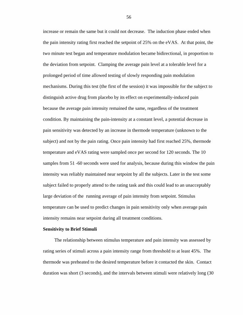
56
increase or remain the same but it could not decrease. The induction phase ended when
the pain intensity rating first reached the setpoint of 25% on the eVAS. At that point, the
two minute test began and temperature modulation became bidirectional, in proportion to
the deviation from setpoint. Clamping the average pain level at a tolerable level for a
prolonged period of time allowed testing of slowly responding pain modulation
mechanisms. During this test (the first of the session) it was impossible for the subject to
distinguish active drug from placebo by its effect on experimentally-induced pain
because the average pain intensity remained the same, regardless of the treatment
condition. By maintaining the pain-intensity at a constant level, a potential decrease in
pain sensitivity was detected by an increase in thermode temperature (unknown to the
subject) and not by the pain rating. Once pain intensity had first reached 25%, thermode
temperature and eVAS rating were sampled once per second for 120 seconds. The 10
samples from 51 -60 seconds were used for analysis, because during this window the pain
intensity was reliably maintained near setpoint by all the subjects. Later in the test some
subject failed to properly attend to the rating task and this could lead to an unacceptably
large deviation of the running average of pain intensity from setpoint. Stimulus
temperature can be used to predict changes in pain sensitivity only when average pain
intensity remains near setpoint during all treatment conditions.
Sensitivity to Brief Stimuli
The relationship between stimulus temperature and pain intensity was assessed by
rating series of stimuli across a pain intensity range from threshold to at least 45%. The
thermode was preheated to the desired temperature before it contacted the skin. Contact
duration was short (3 seconds), and the intervals between stimuli were relatively long (30
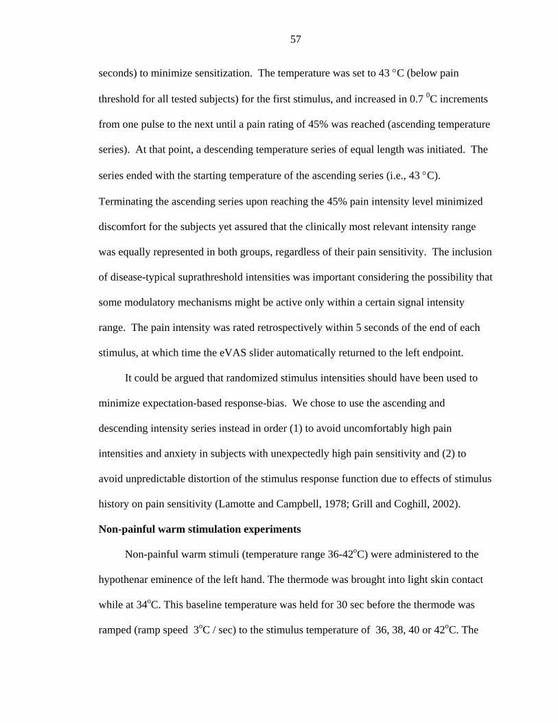
57
seconds) to minimize sensitization. The temperature was set to 43 °C (below pain
threshold for all tested subjects) for the first stimulus, and increased in 0.7 0C increments
from one pulse to the next until a pain rating of 45% was reached (ascending temperature
series). At that point, a descending temperature series of equal length was initiated. The
series ended with the starting temperature of the ascending series (i.e., 43 °C).
Terminating the ascending series upon reaching the 45% pain intensity level minimized
discomfort for the subjects yet assured that the clinically most relevant intensity range
was equally represented in both groups, regardless of their pain sensitivity. The inclusion
of disease-typical suprathreshold intensities was important considering the possibility that
some modulatory mechanisms might be active only within a certain signal intensity
range. The pain intensity was rated retrospectively within 5 seconds of the end of each
stimulus, at which time the eVAS slider automatically returned to the left endpoint.
It could be argued that randomized stimulus intensities should have been used to
minimize expectation-based response-bias. We chose to use the ascending and
descending intensity series instead in order (1) to avoid uncomfortably high pain
intensities and anxiety in subjects with unexpectedly high pain sensitivity and (2) to
avoid unpredictable distortion of the stimulus response function due to effects of stimulus
history on pain sensitivity (Lamotte and Campbell, 1978; Grill and Coghill, 2002).
Non-painful warm stimulation experiments
Non-painful warm stimuli (temperature range 36-42oC) were administered to the
hypothenar eminence of the left hand. The thermode was brought into light skin contact
while at 34oC. This baseline temperature was held for 30 sec before the thermode was
ramped (ramp speed 3oC / sec) to the stimulus temperature of 36, 38, 40 or 42oC. The
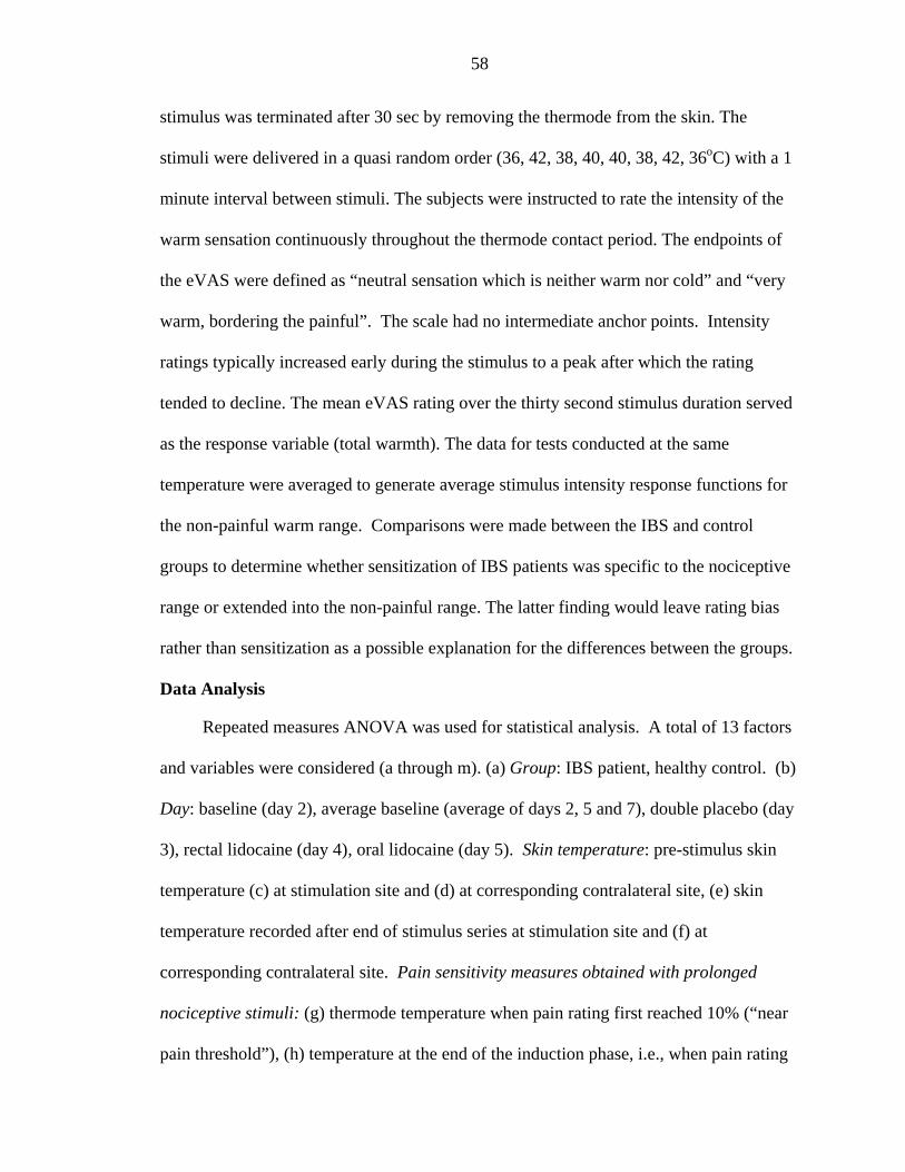
58
stimulus was terminated after 30 sec by removing the thermode from the skin. The
stimuli were delivered in a quasi random order (36, 42, 38, 40, 40, 38, 42, 36oC) with a 1
minute interval between stimuli. The subjects were instructed to rate the intensity of the
warm sensation continuously throughout the thermode contact period. The endpoints of
the eVAS were defined as “neutral sensation which is neither warm nor cold” and “very
warm, bordering the painful”. The scale had no intermediate anchor points. Intensity
ratings typically increased early during the stimulus to a peak after which the rating
tended to decline. The mean eVAS rating over the thirty second stimulus duration served
as the response variable (total warmth). The data for tests conducted at the same
temperature were averaged to generate average stimulus intensity response functions for
the non-painful warm range. Comparisons were made between the IBS and control
groups to determine whether sensitization of IBS patients was specific to the nociceptive
range or extended into the non-painful range. The latter finding would leave rating bias
rather than sensitization as a possible explanation for the differences between the groups.
Data Analysis
Repeated measures ANOVA was used for statistical analysis. A total of 13 factors
and variables were considered (a through m). (a) Group: IBS patient, healthy control. (b)
Day: baseline (day 2), average baseline (average of days 2, 5 and 7), double placebo (day
3), rectal lidocaine (day 4), oral lidocaine (day 5). Skin temperature: pre-stimulus skin
temperature (c) at stimulation site and (d) at corresponding contralateral site, (e) skin
temperature recorded after end of stimulus series at stimulation site and (f) at
corresponding contralateral site. Pain sensitivity measures obtained with prolonged
nociceptive stimuli: (g) thermode temperature when pain rating first reached 10% (“near
pain threshold”), (h) temperature at the end of the induction phase, i.e., when pain rating
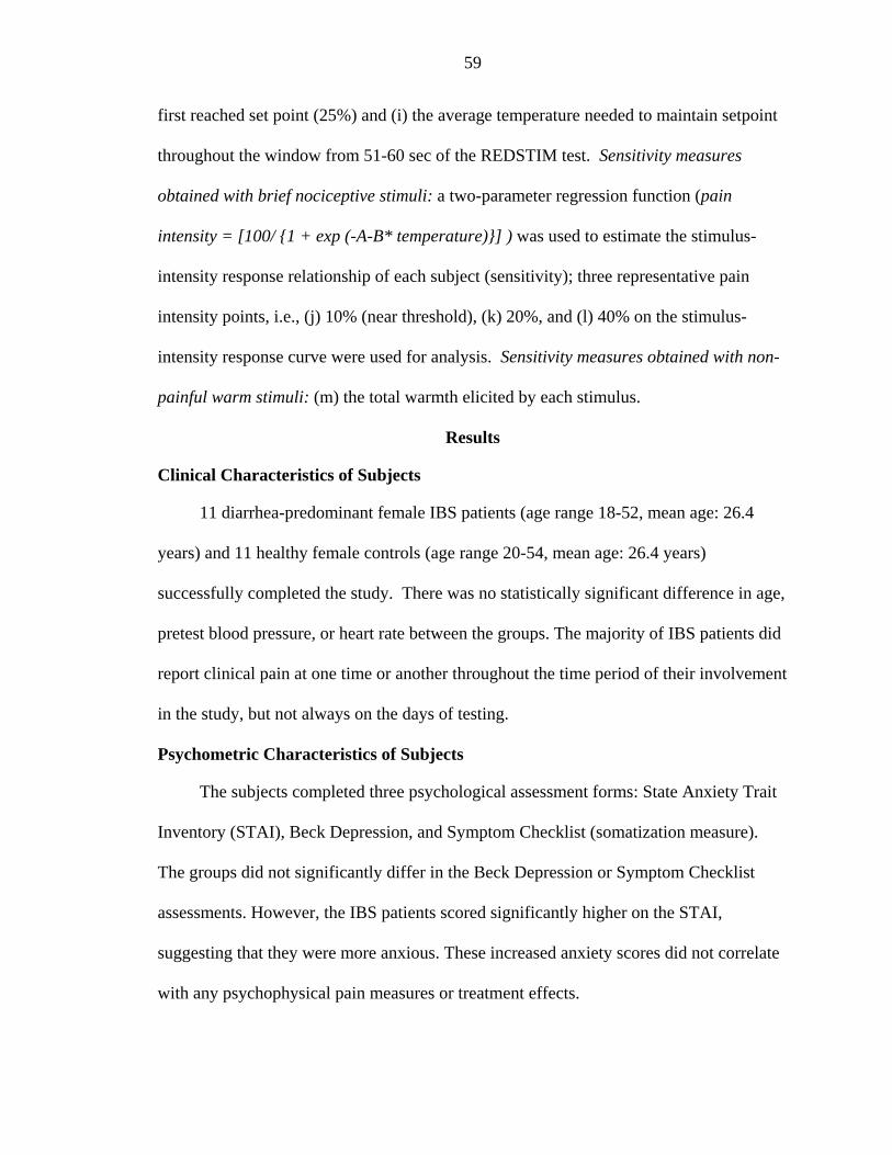
59
first reached set point (25%) and (i) the average temperature needed to maintain setpoint
throughout the window from 51-60 sec of the REDSTIM test. Sensitivity measures
obtained with brief nociceptive stimuli: a two-parameter regression function (pain
intensity = [100/ {1 + exp (-A-B* temperature)}] ) was used to estimate the stimulus-
intensity response relationship of each subject (sensitivity); three representative pain
intensity points, i.e., (j) 10% (near threshold), (k) 20%, and (l) 40% on the stimulus-
intensity response curve were used for analysis. Sensitivity measures obtained with non-
painful warm stimuli: (m) the total warmth elicited by each stimulus.
Results
Clinical Characteristics of Subjects
11 diarrhea-predominant female IBS patients (age range 18-52, mean age: 26.4
years) and 11 healthy female controls (age range 20-54, mean age: 26.4 years)
successfully completed the study. There was no statistically significant difference in age,
pretest blood pressure, or heart rate between the groups. The majority of IBS patients did
report clinical pain at one time or another throughout the time period of their involvement
in the study, but not always on the days of testing.
Psychometric Characteristics of Subjects
The subjects completed three psychological assessment forms: State Anxiety Trait
Inventory (STAI), Beck Depression, and Symptom Checklist (somatization measure).
The groups did not significantly differ in the Beck Depression or Symptom Checklist
assessments. However, the IBS patients scored significantly higher on the STAI,
suggesting that they were more anxious. These increased anxiety scores did not correlate
with any psychophysical pain measures or treatment effects.
60
Skin Temperature
Skin temperature was recorded prior to and at the conclusion of each test on both
the test site and corresponding contralateral site to indirectly determine whether
differences in spontaneous and stimulus-induced thermoregulation exist between groups.
The groups did not differ in baseline skin temperature on any site. Furthermore, the
contralateral skin temperature changes induced by series of brief (3 sec) heat stimuli were
similar in the IBS and control group. These findings are in agreement with results of a
study described in chapter 3. When the REDSTIM method was used to maintain a 25%
average pain intensity for 2 minutes the contralateral skin temperature of the control
group remained at 31.4oC while it drifted up slightly (by 0.8 oC) in the IBS group. The
contralateral skin temperature change did reach statistical significance and did not
correlate with any of the other variables (see below), including the type and location of
drug intervention.
In an earlier study (Chapter 3), where an average pain intensity level of 35% was
maintained with pulsed stimuli for over 7 minutes, the thermoregulatory difference
between the groups was statistically significant as the contralateral skin temperature
dropped by 1.5 degrees in the control group but only by 0.3 degrees in the IBS group.
Possible explanations for the much more pronounced thermoregulatory group difference
in the earlier study may be the longer stimulation period (approximately 100 seconds of
thermode contact accumulated over more than 7 minutes of stimulation) and the higher
average thermode temperature. The thermode temperature was higher because
stimulation used to maintain the pain intensity setpoint was pulsed rather than continuous
and the setpoint was higher (35%).
61
No-treatment (Baseline); Sensitivity to Prolonged Nociceptive Stimuli
During three of the seven daily test sessions pain sensitivity was assessed in the
absence of any lidocaine or vehicle treatment (Table 4-1). During these baseline sessions
the thenar eminence of the right hand was exposed to a prolonged thermal contact in
which the thermode temperature was incrementally increased until a pain intensity of
25% was reached on the electronic visual analog scale. An average intensity level of 25%
was then maintained for 2 minutes, using the REDSTIM feature of the stimulator. The
thermode temperatures at the time when the pain intensity first crossed the 10% and 25%
marks were significantly lower for the IBS group during all baseline sessions (Figure 4-
2), but the temperature difference between these marks was the same for both groups.
This suggests that IBS patients are more sensitive to thermal nociceptive stimuli than
healthy controls. It may be tempting to use the temperature difference between the 10%
and 25% mark to make inferences regarding the sensitization rate. However, this would
be incorrect because the time needed to increase pain intensity from 10% to 25%
depended on how the pain rating changed during the induction phase and thus was not the
same for all tests.
A close inspection of the data collected during the phase where the average
stimulus intensity was to be maintained at the 25% pain intensity level revealed that a
number of subjects were unable to attend to the rating task for much longer than
approximately 1 minute. Inattentiveness could lead to an unacceptably large deviation
from the pain intensity setpoint of 25% and thus invalidate the average thermode
temperature as an indicator of pain sensitivity. In all cases the pain rating remained
within 4 percentage points of setpoint during the first minute. In the interest of optimal
validity only data collected during the first minute were used for analysis and group
62
comparisons. Over this period, the running temperature average was calculated with a
window of 10 seconds (1 sample / sec). In both groups the running temperature average
increased progressively. The increase was steepest initially and flattened later (Figure 4-
1). This suggests that pain sensitivity decreased during this phase of prolonged
stimulation.
No-treatment (Baseline); Sensitivity to Brief Nociceptive Stimuli
The test was conducted on day 1 of the seven day protocol and was comprised of
an ascending and descending temperature series (stimulus contact duration 3 sec, ISI 30
sec). Pain intensity, in combination with the thermode temperature used to evoke the
pain, allowed inferences regarding pain sensitivity. The data of the ascending and
descending series were pooled for analysis because the direction and magnitude of the
series hysteresis exhibited no discernible pattern. The temperatures necessary to reach
pain levels of 10%, 20%, and 40% were significantly lower at all test sites in IBS patients
compared to the control group (Table 4-3). This suggests that the difference between IBS
and control groups did not depend on the evoked pain intensity level, and sensitization
was not limited to dermatomes in segmental proximity to the symptomatic area but
extended across the body, including to the face (Table 4-3, Figure 4-1B). These results
corroborate earlier work on a different cohort of subjects in which group differences in
sensitivity likewise was neither intensity- nor site-specific (chapter 2).
Control for Placebo- and Systemic Treatment Effects
The main goal was to determine whether the vicious pain cycle that presumably
started the expansion of sensitization in non-visceral areas of the body can be stopped.
Administered rectally, lidocaine was expected to silence the pain focus by its topical
anesthetic effect allowing neurons receiving convergent input from visceral and somatic
63
tissues to return to a normal sensitivity level. On a different day topical lidocaine was
given orally to control for potential systemic and placebo effects. Rectal and oral
administration were expected to lead to similar degrees of absorption into the systemic
circulation. In addition to the usual vehicle control tests, the oral lidocaine experiment
was useful as an enhanced placebo control because it was usually detectable as an active
compound by its numbing effect. Lidocaine administered rectally was identified by some
of the subjects by a numbing sensation in the anal region. The recognition as an active
drug is known to lead to an enhanced placebo effect. Every session involved
administration of cream in both locations simultaneously, i.e., rectally and orally. The
subjects were told that they would receive lidocaine and vehicle throughout the series of
tests. However, they were not informed about the location assignment of each compound
during any of the tests and where lidocaine was supposed to have an effect on pain
sensitivity. A prolonged or even irreversible desensitizing effect of rectal lidocaine could
not a priori be ruled out. This would have made it impossible to obtain within-subject
control data if the rectal lidocaine test was the first test of the series. Therefore,
unbeknownst to the subjects, the double oral and rectal placebo test always preceded the
rectal lidocaine test. This aspect of the protocol made blinding of the investigators more
difficult. Therefore, measures were taken to minimize the transmission of bias from
investigators to subjects: (1) the investigator administering the treatment was never
present during data collection. (2) a prerecorded standardized video sequence instead of
direct communication between investigator and subject was used for instruction regarding
the pain rating task; (3) non-verbal communication between investigator and subject was
minimized by an arrangement where the subject was always facing away from the
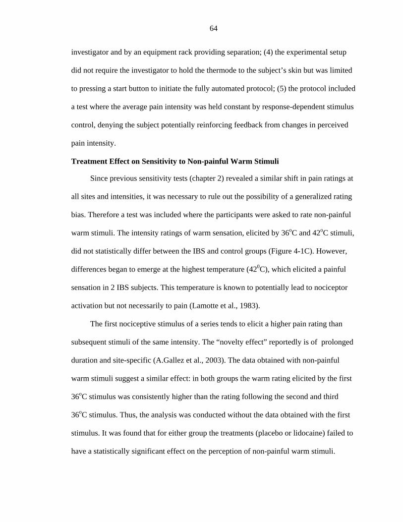
64
investigator and by an equipment rack providing separation; (4) the experimental setup
did not require the investigator to hold the thermode to the subject’s skin but was limited
to pressing a start button to initiate the fully automated protocol; (5) the protocol included
a test where the average pain intensity was held constant by response-dependent stimulus
control, denying the subject potentially reinforcing feedback from changes in perceived
pain intensity.
Treatment Effect on Sensitivity to Non-painful Warm Stimuli
Since previous sensitivity tests (chapter 2) revealed a similar shift in pain ratings at
all sites and intensities, it was necessary to rule out the possibility of a generalized rating
bias. Therefore a test was included where the participants were asked to rate non-painful
warm stimuli. The intensity ratings of warm sensation, elicited by 36oC and 42oC stimuli,
did not statistically differ between the IBS and control groups (Figure 4-1C). However,
differences began to emerge at the highest temperature (420C), which elicited a painful
sensation in 2 IBS subjects. This temperature is known to potentially lead to nociceptor
activation but not necessarily to pain (Lamotte et al., 1983).
The first nociceptive stimulus of a series tends to elicit a higher pain rating than
subsequent stimuli of the same intensity. The “novelty effect” reportedly is of prolonged
duration and site-specific (A.Gallez et al., 2003). The data obtained with non-painful
warm stimuli suggest a similar effect: in both groups the warm rating elicited by the first
36oC stimulus was consistently higher than the rating following the second and third
36oC stimulus. Thus, the analysis was conducted without the data obtained with the first
stimulus. It was found that for either group the treatments (placebo or lidocaine) failed to
have a statistically significant effect on the perception of non-painful warm stimuli.
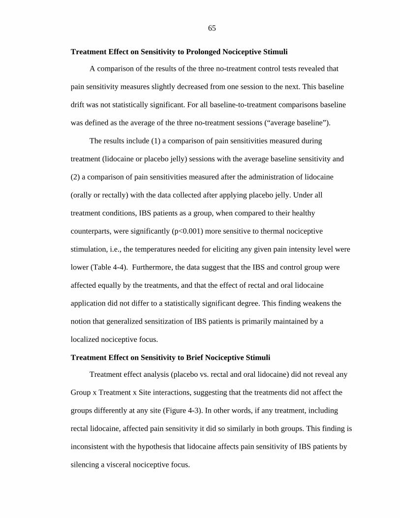
65
Treatment Effect on Sensitivity to Prolonged Nociceptive Stimuli
A comparison of the results of the three no-treatment control tests revealed that
pain sensitivity measures slightly decreased from one session to the next. This baseline
drift was not statistically significant. For all baseline-to-treatment comparisons baseline
was defined as the average of the three no-treatment sessions (“average baseline”).
The results include (1) a comparison of pain sensitivities measured during
treatment (lidocaine or placebo jelly) sessions with the average baseline sensitivity and
(2) a comparison of pain sensitivities measured after the administration of lidocaine
(orally or rectally) with the data collected after applying placebo jelly. Under all
treatment conditions, IBS patients as a group, when compared to their healthy
counterparts, were significantly (p<0.001) more sensitive to thermal nociceptive
stimulation, i.e., the temperatures needed for eliciting any given pain intensity level were
lower (Table 4-4). Furthermore, the data suggest that the IBS and control group were
affected equally by the treatments, and that the effect of rectal and oral lidocaine
application did not differ to a statistically significant degree. This finding weakens the
notion that generalized sensitization of IBS patients is primarily maintained by a
localized nociceptive focus.
Treatment Effect on Sensitivity to Brief Nociceptive Stimuli
Treatment effect analysis (placebo vs. rectal and oral lidocaine) did not reveal any
Group x Treatment x Site interactions, suggesting that the treatments did not affect the
groups differently at any site (Figure 4-3). In other words, if any treatment, including
rectal lidocaine, affected pain sensitivity it did so similarly in both groups. This finding is
inconsistent with the hypothesis that lidocaine affects pain sensitivity of IBS patients by
silencing a visceral nociceptive focus.
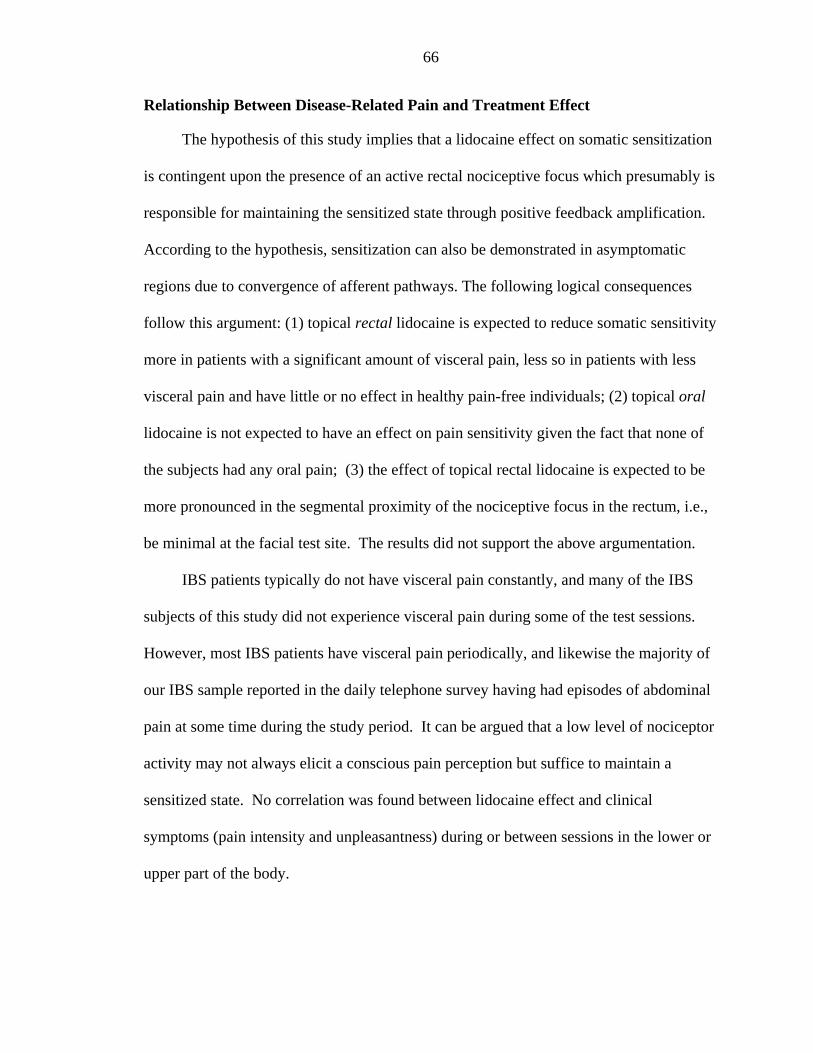
66
Relationship Between Disease-Related Pain and Treatment Effect
The hypothesis of this study implies that a lidocaine effect on somatic sensitization
is contingent upon the presence of an active rectal nociceptive focus which presumably is
responsible for maintaining the sensitized state through positive feedback amplification.
According to the hypothesis, sensitization can also be demonstrated in asymptomatic
regions due to convergence of afferent pathways. The following logical consequences
follow this argument: (1) topical rectal lidocaine is expected to reduce somatic sensitivity
more in patients with a significant amount of visceral pain, less so in patients with less
visceral pain and have little or no effect in healthy pain-free individuals; (2) topical oral
lidocaine is not expected to have an effect on pain sensitivity given the fact that none of
the subjects had any oral pain; (3) the effect of topical rectal lidocaine is expected to be
more pronounced in the segmental proximity of the nociceptive focus in the rectum, i.e.,
be minimal at the facial test site. The results did not support the above argumentation.
IBS patients typically do not have visceral pain constantly, and many of the IBS
subjects of this study did not experience visceral pain during some of the test sessions.
However, most IBS patients have visceral pain periodically, and likewise the majority of
our IBS sample reported in the daily telephone survey having had episodes of abdominal
pain at some time during the study period. It can be argued that a low level of nociceptor
activity may not always elicit a conscious pain perception but suffice to maintain a
sensitized state. No correlation was found between lidocaine effect and clinical
symptoms (pain intensity and unpleasantness) during or between sessions in the lower or
upper part of the body.
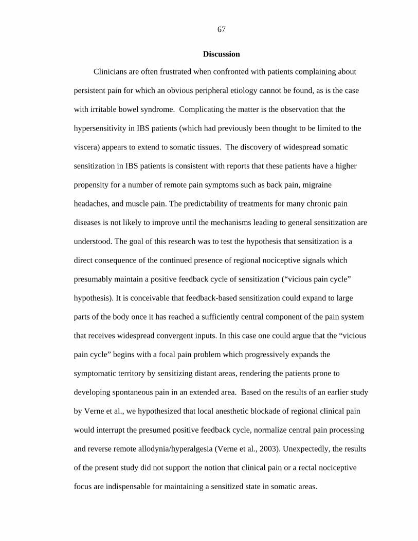
67
Discussion
Clinicians are often frustrated when confronted with patients complaining about
persistent pain for which an obvious peripheral etiology cannot be found, as is the case
with irritable bowel syndrome. Complicating the matter is the observation that the
hypersensitivity in IBS patients (which had previously been thought to be limited to the
viscera) appears to extend to somatic tissues. The discovery of widespread somatic
sensitization in IBS patients is consistent with reports that these patients have a higher
propensity for a number of remote pain symptoms such as back pain, migraine
headaches, and muscle pain. The predictability of treatments for many chronic pain
diseases is not likely to improve until the mechanisms leading to general sensitization are
understood. The goal of this research was to test the hypothesis that sensitization is a
direct consequence of the continued presence of regional nociceptive signals which
presumably maintain a positive feedback cycle of sensitization (“vicious pain cycle”
hypothesis). It is conceivable that feedback-based sensitization could expand to large
parts of the body once it has reached a sufficiently central component of the pain system
that receives widespread convergent inputs. In this case one could argue that the “vicious
pain cycle” begins with a focal pain problem which progressively expands the
symptomatic territory by sensitizing distant areas, rendering the patients prone to
developing spontaneous pain in an extended area. Based on the results of an earlier study
by Verne et al., we hypothesized that local anesthetic blockade of regional clinical pain
would interrupt the presumed positive feedback cycle, normalize central pain processing
and reverse remote allodynia/hyperalgesia (Verne et al., 2003). Unexpectedly, the results
of the present study did not support the notion that clinical pain or a rectal nociceptive
focus are indispensable for maintaining a sensitized state in somatic areas.
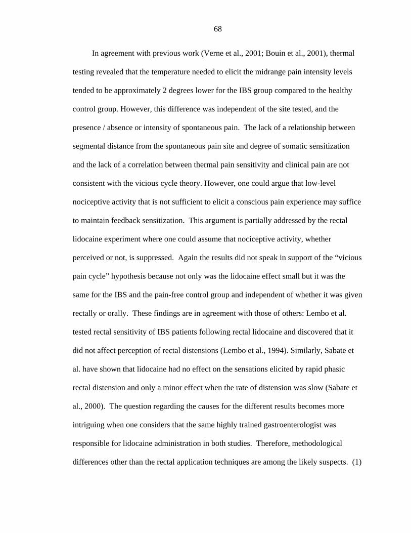
68
In agreement with previous work (Verne et al., 2001; Bouin et al., 2001), thermal
testing revealed that the temperature needed to elicit the midrange pain intensity levels
tended to be approximately 2 degrees lower for the IBS group compared to the healthy
control group. However, this difference was independent of the site tested, and the
presence / absence or intensity of spontaneous pain. The lack of a relationship between
segmental distance from the spontaneous pain site and degree of somatic sensitization
and the lack of a correlation between thermal pain sensitivity and clinical pain are not
consistent with the vicious cycle theory. However, one could argue that low-level
nociceptive activity that is not sufficient to elicit a conscious pain experience may suffice
to maintain feedback sensitization. This argument is partially addressed by the rectal
lidocaine experiment where one could assume that nociceptive activity, whether
perceived or not, is suppressed. Again the results did not speak in support of the “vicious
pain cycle” hypothesis because not only was the lidocaine effect small but it was the
same for the IBS and the pain-free control group and independent of whether it was given
rectally or orally. These findings are in agreement with those of others: Lembo et al.
tested rectal sensitivity of IBS patients following rectal lidocaine and discovered that it
did not affect perception of rectal distensions (Lembo et al., 1994). Similarly, Sabate et
al. have shown that lidocaine had no effect on the sensations elicited by rapid phasic
rectal distension and only a minor effect when the rate of distension was slow (Sabate et
al., 2000). The question regarding the causes for the different results becomes more
intriguing when one considers that the same highly trained gastroenterologist was
responsible for lidocaine administration in both studies. Therefore, methodological
differences other than the rectal application techniques are among the likely suspects. (1)

69
In Verne’s et al. earlier study the subjects may not have been adequately blinded
regarding the treatment because they may have identified lidocaine by its effect on
thermal pain early on during the simple pain tests. Memory-based comparisons of
perceived pain intensities are more likely to lead to recognition of the active drug when
the testing protocol is simple (only one stimulus was used for all cutaneous pain tests).
This recognition may have affected the ratings later in the test, resulting in a placebo
effect. (2) In the earlier study the pain intensity levels induced in the control group were
lower than those of the more sensitive IBS group. This may have left little room for a
lidocaine effect in the control group (the present study’s comparisons were made at the
same pain intensity level for both groups and similar lidocaine effects were found). (3)
The lidocaine effects reported in the earlier study may not have been limited to the rectal
area but may have been systemic. Verne’s et al. blood test to rule out systemic lidocaine
levels may not have been sensitive enough to detect the lowest biologically effective
levels. A number of studies have demonstrated significant reductions in pain with doses
below or slightly above the 1 microgram / milliliter concentration (Brose and Cousins,
1991; Groudine et al., 1998; Mallon and Thomas, 2000; Koppert et al., 2004). Systemic
lidocaine is known to have an attenuating effect on pain responses in neuropathic
conditions of human and animal models, as well as in human post-operative abdominal
pain (Bach et al., 1990; Mao et al., 1992; Koppert et al., 1998) . Interestingly,
intravenous lidocaine has been shown to reduce secondary mechanical hyperalgesia
without affecting sensitivity thresholds (Dirks et al., 2000), a result which parallels the
absence of a lidocaine effect found in the non-nociceptive tests of the present study. It,
therefore, cannot be ruled out that levels of lidocaine below the detection threshold of
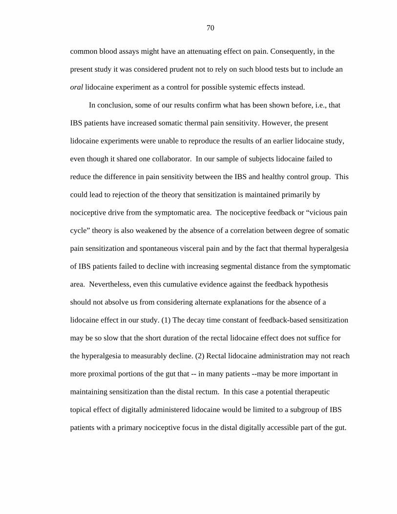
70
common blood assays might have an attenuating effect on pain. Consequently, in the
present study it was considered prudent not to rely on such blood tests but to include an
oral lidocaine experiment as a control for possible systemic effects instead.
In conclusion, some of our results confirm what has been shown before, i.e., that
IBS patients have increased somatic thermal pain sensitivity. However, the present
lidocaine experiments were unable to reproduce the results of an earlier lidocaine study,
even though it shared one collaborator. In our sample of subjects lidocaine failed to
reduce the difference in pain sensitivity between the IBS and healthy control group. This
could lead to rejection of the theory that sensitization is maintained primarily by
nociceptive drive from the symptomatic area. The nociceptive feedback or “vicious pain
cycle” theory is also weakened by the absence of a correlation between degree of somatic
pain sensitization and spontaneous visceral pain and by the fact that thermal hyperalgesia
of IBS patients failed to decline with increasing segmental distance from the symptomatic
area. Nevertheless, even this cumulative evidence against the feedback hypothesis
should not absolve us from considering alternate explanations for the absence of a
lidocaine effect in our study. (1) The decay time constant of feedback-based sensitization
may be so slow that the short duration of the rectal lidocaine effect does not suffice for
the hyperalgesia to measurably decline. (2) Rectal lidocaine administration may not reach
more proximal portions of the gut that -- in many patients --may be more important in
maintaining sensitization than the distal rectum. In this case a potential therapeutic
topical effect of digitally administered lidocaine would be limited to a subgroup of IBS
patients with a primary nociceptive focus in the distal digitally accessible part of the gut.
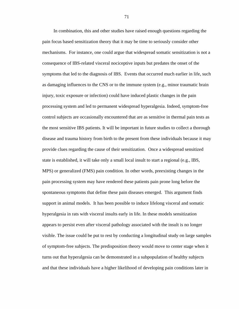
71
In combination, this and other studies have raised enough questions regarding the
pain focus based sensitization theory that it may be time to seriously consider other
mechanisms. For instance, one could argue that widespread somatic sensitization is not a
consequence of IBS-related visceral nociceptive inputs but predates the onset of the
symptoms that led to the diagnosis of IBS. Events that occurred much earlier in life, such
as damaging influences to the CNS or to the immune system (e.g., minor traumatic brain
injury, toxic exposure or infection) could have induced plastic changes in the pain
processing system and led to permanent widespread hyperalgesia. Indeed, symptom-free
control subjects are occasionally encountered that are as sensitive in thermal pain tests as
the most sensitive IBS patients. It will be important in future studies to collect a thorough
disease and trauma history from birth to the present from these individuals because it may
provide clues regarding the cause of their sensitization. Once a widespread sensitized
state is established, it will take only a small local insult to start a regional (e.g., IBS,
MPS) or generalized (FMS) pain condition. In other words, preexisting changes in the
pain processing system may have rendered these patients pain prone long before the
spontaneous symptoms that define these pain diseases emerged. This argument finds
support in animal models. It has been possible to induce lifelong visceral and somatic
hyperalgesia in rats with visceral insults early in life. In these models sensitization
appears to persist even after visceral pathology associated with the insult is no longer
visible. The issue could be put to rest by conducting a longitudinal study on large samples
of symptom-free subjects. The predisposition theory would move to center stage when it
turns out that hyperalgesia can be demonstrated in a subpopulation of healthy subjects
and that these individuals have a higher likelihood of developing pain conditions later in
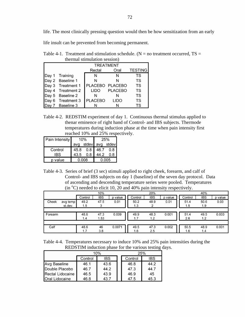
72
life. The most clinically pressing question would then be how sensitization from an early
life insult can be prevented from becoming permanent.
Table 4-1. Treatment and stimulation schedule. (N = no treatment occurred, TS = thermal stimulation session)
Table 4-2. REDSTIM experiment of day 1. Continuous thermal stimulus applied to
thenar eminence of right hand of Control- and IBS subjects. Thermode temperatures during induction phase at the time when pain intensity first reached 10% and 25% respectively.
Table 4-3. Series of brief (3 sec) stimuli applied to right cheek, forearm, and calf of
Control- and IBS subjects on day 1 (baseline) of the seven day protocol. Data of ascending and descending temperature series were pooled. Temperatures (in oC) needed to elicit 10, 20 and 40% pain intensity respectively.
Table 4-4. Temperatures necessary to induce 10% and 25% pain intensities during the
REDSTIM induction phase for the various testing days.
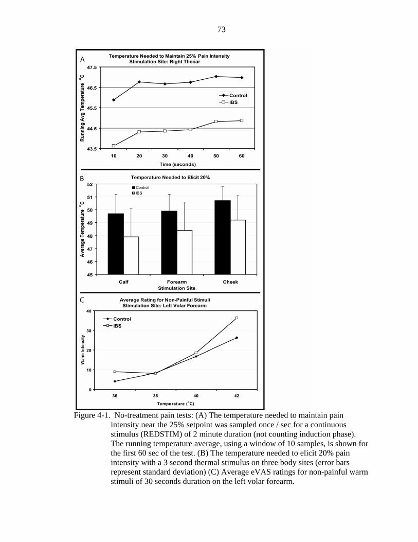
73
Figure 4-1. No-treatment pain tests: (A) The temperature needed to maintain pain
intensity near the 25% setpoint was sampled once / sec for a continuous stimulus (REDSTIM) of 2 minute duration (not counting induction phase). The running temperature average, using a window of 10 samples, is shown for the first 60 sec of the test. (B) The temperature needed to elicit 20% pain intensity with a 3 second thermal stimulus on three body sites (error bars represent standard deviation) (C) Average eVAS ratings for non-painful warm stimuli of 30 seconds duration on the left volar forearm.
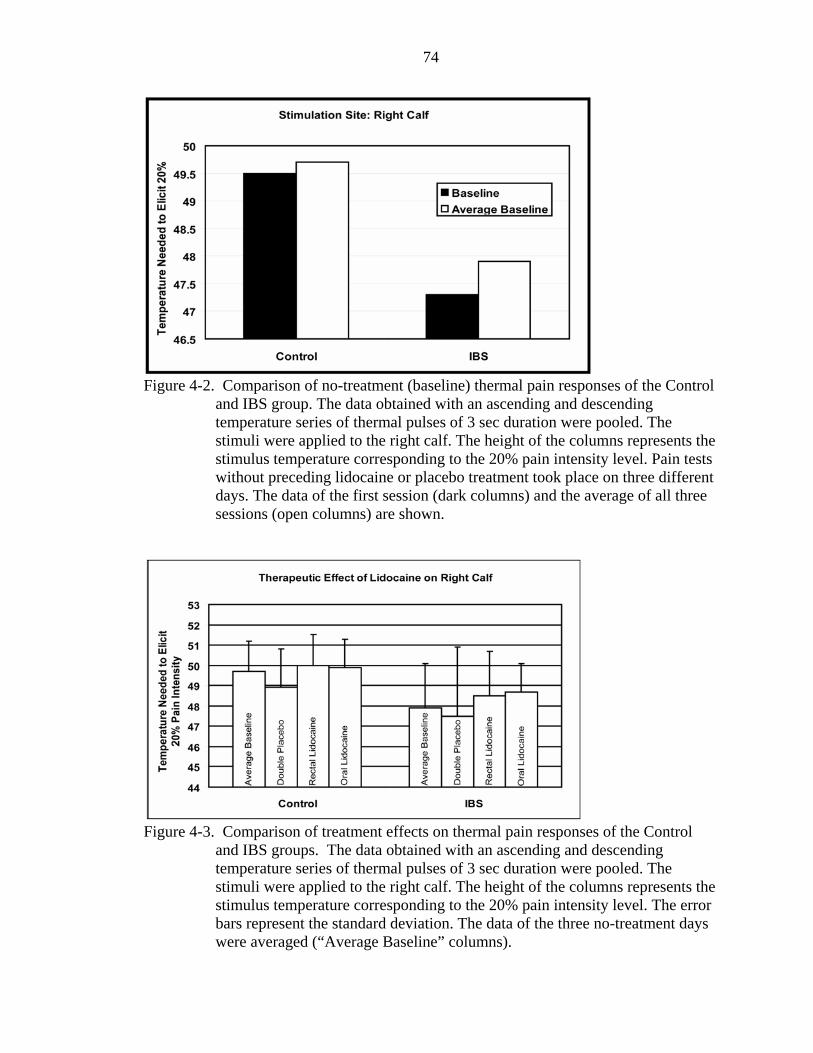
74
Figure 4-2. Comparison of no-treatment (baseline) thermal pain responses of the Control
and IBS group. The data obtained with an ascending and descending temperature series of thermal pulses of 3 sec duration were pooled. The stimuli were applied to the right calf. The height of the columns represents the stimulus temperature corresponding to the 20% pain intensity level. Pain tests without preceding lidocaine or placebo treatment took place on three different days. The data of the first session (dark columns) and the average of all three sessions (open columns) are shown.
Figure 4-3. Comparison of treatment effects on thermal pain responses of the Control
and IBS groups. The data obtained with an ascending and descending temperature series of thermal pulses of 3 sec duration were pooled. The stimuli were applied to the right calf. The height of the columns represents the stimulus temperature corresponding to the 20% pain intensity level. The error bars represent the standard deviation. The data of the three no-treatment days were averaged (“Average Baseline” columns).
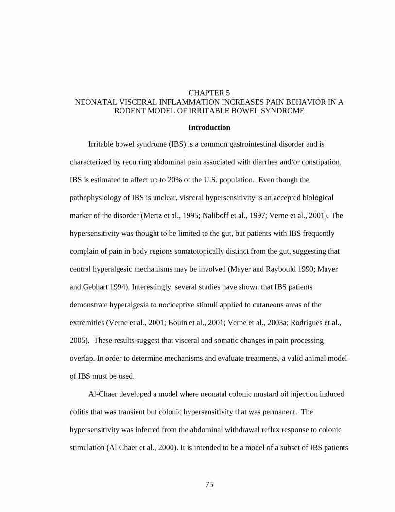
75
CHAPTER 5 NEONATAL VISCERAL INFLAMMATION INCREASES PAIN BEHAVIOR IN A
RODENT MODEL OF IRRITABLE BOWEL SYNDROME
Introduction
Irritable bowel syndrome (IBS) is a common gastrointestinal disorder and is
characterized by recurring abdominal pain associated with diarrhea and/or constipation.
IBS is estimated to affect up to 20% of the U.S. population. Even though the
pathophysiology of IBS is unclear, visceral hypersensitivity is an accepted biological
marker of the disorder (Mertz et al., 1995; Naliboff et al., 1997; Verne et al., 2001). The
hypersensitivity was thought to be limited to the gut, but patients with IBS frequently
complain of pain in body regions somatotopically distinct from the gut, suggesting that
central hyperalgesic mechanisms may be involved (Mayer and Raybould 1990; Mayer
and Gebhart 1994). Interestingly, several studies have shown that IBS patients
demonstrate hyperalgesia to nociceptive stimuli applied to cutaneous areas of the
extremities (Verne et al., 2001; Bouin et al., 2001; Verne et al., 2003a; Rodrigues et al.,
2005). These results suggest that visceral and somatic changes in pain processing
overlap. In order to determine mechanisms and evaluate treatments, a valid animal model
of IBS must be used.
Al-Chaer developed a model where neonatal colonic mustard oil injection induced
colitis that was transient but colonic hypersensitivity that was permanent. The
hypersensitivity was inferred from the abdominal withdrawal reflex response to colonic
stimulation (Al Chaer et al., 2000). It is intended to be a model of a subset of IBS patients
76
who develop their symptoms after experiencing an infectious colitis (McKendrick and
Read 1994). In addition to revealing significant increases in the rodent abdominal
withdrawal reflex, the model also demonstrated increases in firing of viscerosomatic
convergent neurons in spinal segments L6-S1 in response to rectal distension and somatic
nociceptive pinch (Al Chaer et al., 2000). Together these results suggest an increase in
both visceral and somatic sensitivity, although one could argue that reflex- and
electrophysiological measures are not necessarily a valid reflection of (cortically
mediated) pain perception. An assay that measures a cortically mediated response may be
a better predictor of cerebral manifestations of pain than reflexes or neuronal firing
patterns in the spinal dorsal horn. Therefore, the present study used learned operant
escape behavior from a thermal stimulus as a measure of somatic pain sensitivity. The
overall purpose of our current study was to see if rats that were neonatally treated with
mustard oil developed a lifelong increase in thermal sensitivity to nociceptive stimulation
of the paws. A demonstration of increased sensitivity to nociceptive stimulation of the
paws would parallel the cutaneous thermal hypesensitivity seen in human IBS patients
and thus contribute to the validation of the model.
Methods
Induction of Colitis:
A total of 22 neonatal pathogen-free female Long Evans rats were treated for 14
days, between the ages of 8 and 21 days, with either intracolonic injections of saline
(n=11) or mustard oil (0.2 mL, 5%) (n=11). Mustard oil produces a transmural colitis that
resolves within four weeks (Al Chaer et al., 2000). The animals were provided free
access to food and water throughout treatment and testing. All procedures were approved
by the University of Florida’s Institutional Animal Care and Use Committee and the
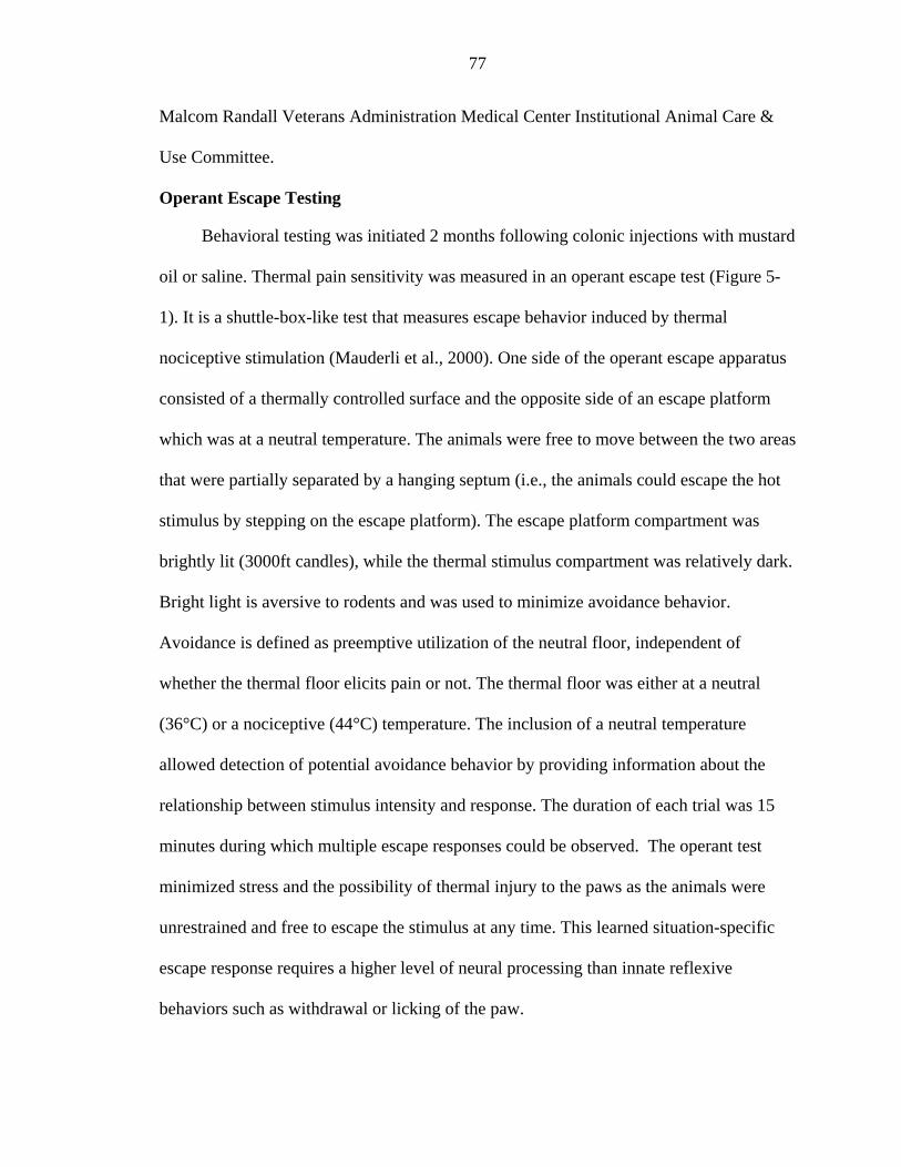
77
Malcom Randall Veterans Administration Medical Center Institutional Animal Care &
Use Committee.
Operant Escape Testing
Behavioral testing was initiated 2 months following colonic injections with mustard
oil or saline. Thermal pain sensitivity was measured in an operant escape test (Figure 5-
1). It is a shuttle-box-like test that measures escape behavior induced by thermal
nociceptive stimulation (Mauderli et al., 2000). One side of the operant escape apparatus
consisted of a thermally controlled surface and the opposite side of an escape platform
which was at a neutral temperature. The animals were free to move between the two areas
that were partially separated by a hanging septum (i.e., the animals could escape the hot
stimulus by stepping on the escape platform). The escape platform compartment was
brightly lit (3000ft candles), while the thermal stimulus compartment was relatively dark.
Bright light is aversive to rodents and was used to minimize avoidance behavior.
Avoidance is defined as preemptive utilization of the neutral floor, independent of
whether the thermal floor elicits pain or not. The thermal floor was either at a neutral
(36°C) or a nociceptive (44°C) temperature. The inclusion of a neutral temperature
allowed detection of potential avoidance behavior by providing information about the
relationship between stimulus intensity and response. The duration of each trial was 15
minutes during which multiple escape responses could be observed. The operant test
minimized stress and the possibility of thermal injury to the paws as the animals were
unrestrained and free to escape the stimulus at any time. This learned situation-specific
escape response requires a higher level of neural processing than innate reflexive
behaviors such as withdrawal or licking of the paw.
78
Data collection was preceded by a training period of approximately 2-3 weeks.
Training began at low temperatures with the light off, to facilitate acclimation to the test
environment. The temperature was increased to 36, 42, 44, and 47°C over a week to
allow the animal to learn to escape from the thermal plate to the thermally neutral
platform. Once the rat demonstrated stable escape behavior, training continued with the
light on. Once test-retest comparisons suggested that the learning curve had reached a
plateau, data collection commenced. Tests were conducted approximately 3 times a week
for 2 months. Each test session consisted of two phases of 15 minutes duration each.
During the first phase of 15 minutes the thermal plate temperature was either 36°C (skin
temperature) or 44°C. The 36oC conditioning phase allowed the paws of the animals to
equilibrate to a standard temperature, and exploratory behavior could subside prior to the
second phase. A 44oC conditioning phase allowed slowly-responding pain modulation
systems to be activated prior to the second phase of the test. During the second phase
escape behavior from either a 36°C or 44°C floor stimulus was measured. The response
variables were: (1) frequency of movement between the plate and escape platform, (2)
latency of each escape event, and (3) duration of each escape event. The animals shuttled
back and forth between the thermal plate and the escape platform. An escape latency
decrease (spent on thermal plate prior to escape) was interpreted as an increase in pain
sensitivity. An increase in escape duration (time spent on the escape platform) was
considered to be an indicator of the intensity of stimulus-related after-sensations. The
behavior could gradually change over the course of the 15 min test, e.g., due to pain
adaptation. Therefore, certain analyses focused on only the first four escape events.
79
Place Preference Testing
The place preference test consisted of two compartments with different floor
temperatures: compartment 1 had a heated floor (45oC), compartment 2 had a
nociceptively cold surface (10oC). The number of crossings and duration of time spent on
each side were recorded for each 15 minute trial. The rats quickly learned to apportion
their time according to the relative aversiveness of the two plates. Unlike in the escape
test, bright light was not used as a behavioral driving force in the place preference assay.
Paw Temperature Measurement
At the conclusion of 8 weeks of operant testing, each rat underwent a test to
measure paw temperature regulation in response to thermal heating. After induction of
general anesthesia (Nembutal), the animals’ core temperature was stabilized at 37.5 oC
with a heating blanket and monitored with a rectal probe. Temperature sensors were
placed on both forepaws, one hind paw and the tail to record thermoregulatory responses
in areas that were not directly stimulated. In order to improve accuracy, thermally
conductive paste was applied on the paw-side of the thermistors while the other side was
covered with insulation material. This assembly was secured to the paws with tape. The
remaining hind paw was exposed to a 10 minute 44oC contact heat stimulus. Throughout
the stimulus and the subsequent 10 minute recovery period, the core, tail, and paw
temperatures were recorded in 15 second intervals. After a 15 minute break, the
procedure was repeated in mirror-image fashion, i.e., with the other hind paw serving as
stimulation site.
Protocol
Thermal operant testing began two months following colonic injections. Five
different test protocols, each repeated 8 times over the course of two months, were used.
80
The number of days between sessions varied, but the order of escape and place
preference tests was kept constant (Table 5-1).
Statistical Analysis
Escape numbers, latencies, durations and temperature changes were analyzed with
a repeated measures ANOVA model. The first four escapes for the operant escape assay
and place preference test were used for the within and between group comparisons.
Results
Temperature Effect on Operant Escape
In order to assure that the measured responses represented escape rather than
avoidance, tests were conducted above and below pain threshold (i.e., 36oC and 44oC)
(Table 5-1). Avoidance behavior can be recognized when the stimulus-response function
is flat, i.e., when the responses to 36 and 44oC are similar. The temperature of 44oC is
known to excite nociceptors (Tillman et al., 1995a; Tillman et al., 1995b), while 36oC
neither activates nociceptors nor elicits pain. For both test conditions, the animals cycled
back and forth between the plate and platform, responding to the conflicting motivations
of escaping thermal pain and bright light. The total number of platform occupancies was
slightly increased for the suprathreshold floor temperature of 44oC compared to the
neutral 36oC floor temp. Within-group analysis showed that both groups spent a
significantly larger amount of time on the escape platform when it was at 44oC as
opposed to 36oC (p<0.005). Analysis of consecutive escape events revealed that the
temperature effect on escape duration is most visible during the second, third, and fourth
escapes and less so during the first escape of the trial when the drive to explore is likely
to still be a significant force. Baseline escape latencies and escape platform dwell times
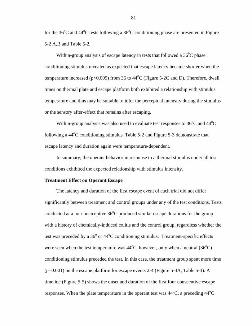
81
for the 36oC and 44oC tests following a 36oC conditioning phase are presented in Figure
5-2 A,B and Table 5-2.
Within-group analysis of escape latency in tests that followed a 360C phase 1
conditioning stimulus revealed as expected that escape latency became shorter when the
temperature increased (p<0.009) from 36 to 440C (Figure 5-2C and D). Therefore, dwell
times on thermal plate and escape platform both exhibited a relationship with stimulus
temperature and thus may be suitable to infer the perceptual intensity during the stimulus
or the sensory after-effect that remains after escaping.
Within-group analysis was also used to evaluate test responses to 36oC and 44oC
following a 44oC conditioning stimulus. Table 5-2 and Figure 5-3 demonstrate that
escape latency and duration again were temperature-dependent.
In summary, the operant behavior in response to a thermal stimulus under all test
conditions exhibited the expected relationship with stimulus intensity.
Treatment Effect on Operant Escape
The latency and duration of the first escape event of each trial did not differ
significantly between treatment and control groups under any of the test conditions. Tests
conducted at a non-nociceptive 36oC produced similar escape durations for the group
with a history of chemically-induced colitis and the control group, regardless whether the
test was preceded by a 36o or 44oC conditioning stimulus. Treatment-specific effects
were seen when the test temperature was 44oC, however, only when a neutral (36oC)
conditioning stimulus preceded the test. In this case, the treatment group spent more time
(p=0.001) on the escape platform for escape events 2-4 (Figure 5-4A, Table 5-3). A
timeline (Figure 5-5) shows the onset and duration of the first four consecutive escape
responses. When the plate temperature in the operant test was 44oC, a preceding 44oC
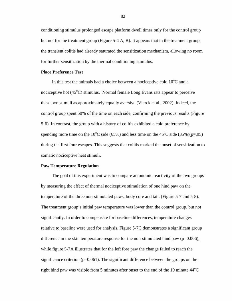
82
conditioning stimulus prolonged escape platform dwell times only for the control group
but not for the treatment group (Figure 5-4 A, B). It appears that in the treatment group
the transient colitis had already saturated the sensitization mechanism, allowing no room
for further sensitization by the thermal conditioning stimulus.
Place Preference Test
In this test the animals had a choice between a nociceptive cold 10oC and a
nociceptive hot (45oC) stimulus. Normal female Long Evans rats appear to perceive
these two stimuli as approximately equally aversive (Vierck et al., 2002). Indeed, the
control group spent 50% of the time on each side, confirming the previous results (Figure
5-6). In contrast, the group with a history of colitis exhibited a cold preference by
spending more time on the 10oC side (65%) and less time on the 45oC side (35%)(p=.05)
during the first four escapes. This suggests that colitis marked the onset of sensitization to
somatic nociceptive heat stimuli.
Paw Temperature Regulation
The goal of this experiment was to compare autonomic reactivity of the two groups
by measuring the effect of thermal nociceptive stimulation of one hind paw on the
temperature of the three non-stimulated paws, body core and tail. (Figure 5-7 and 5-8).
The treatment group’s initial paw temperature was lower than the control group, but not
significantly. In order to compensate for baseline differences, temperature changes
relative to baseline were used for analysis. Figure 5-7C demonstrates a significant group
difference in the skin temperature response for the non-stimulated hind paw (p=0.006),
while figure 5-7A illustrates that for the left fore paw the change failed to reach the
significance criterion (p=0.061). The significant difference between the groups on the
right hind paw was visible from 5 minutes after onset to the end of the 10 minute 44oC
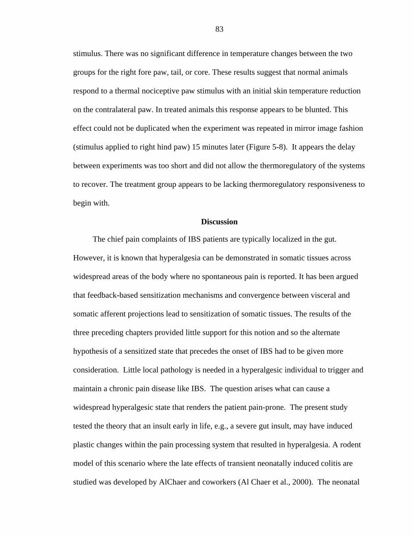
83
stimulus. There was no significant difference in temperature changes between the two
groups for the right fore paw, tail, or core. These results suggest that normal animals
respond to a thermal nociceptive paw stimulus with an initial skin temperature reduction
on the contralateral paw. In treated animals this response appears to be blunted. This
effect could not be duplicated when the experiment was repeated in mirror image fashion
(stimulus applied to right hind paw) 15 minutes later (Figure 5-8). It appears the delay
between experiments was too short and did not allow the thermoregulatory of the systems
to recover. The treatment group appears to be lacking thermoregulatory responsiveness to
begin with.
Discussion
The chief pain complaints of IBS patients are typically localized in the gut.
However, it is known that hyperalgesia can be demonstrated in somatic tissues across
widespread areas of the body where no spontaneous pain is reported. It has been argued
that feedback-based sensitization mechanisms and convergence between visceral and
somatic afferent projections lead to sensitization of somatic tissues. The results of the
three preceding chapters provided little support for this notion and so the alternate
hypothesis of a sensitized state that precedes the onset of IBS had to be given more
consideration. Little local pathology is needed in a hyperalgesic individual to trigger and
maintain a chronic pain disease like IBS. The question arises what can cause a
widespread hyperalgesic state that renders the patient pain-prone. The present study
tested the theory that an insult early in life, e.g., a severe gut insult, may have induced
plastic changes within the pain processing system that resulted in hyperalgesia. A rodent
model of this scenario where the late effects of transient neonatally induced colitis are
studied was developed by AlChaer and coworkers (Al Chaer et al., 2000). The neonatal
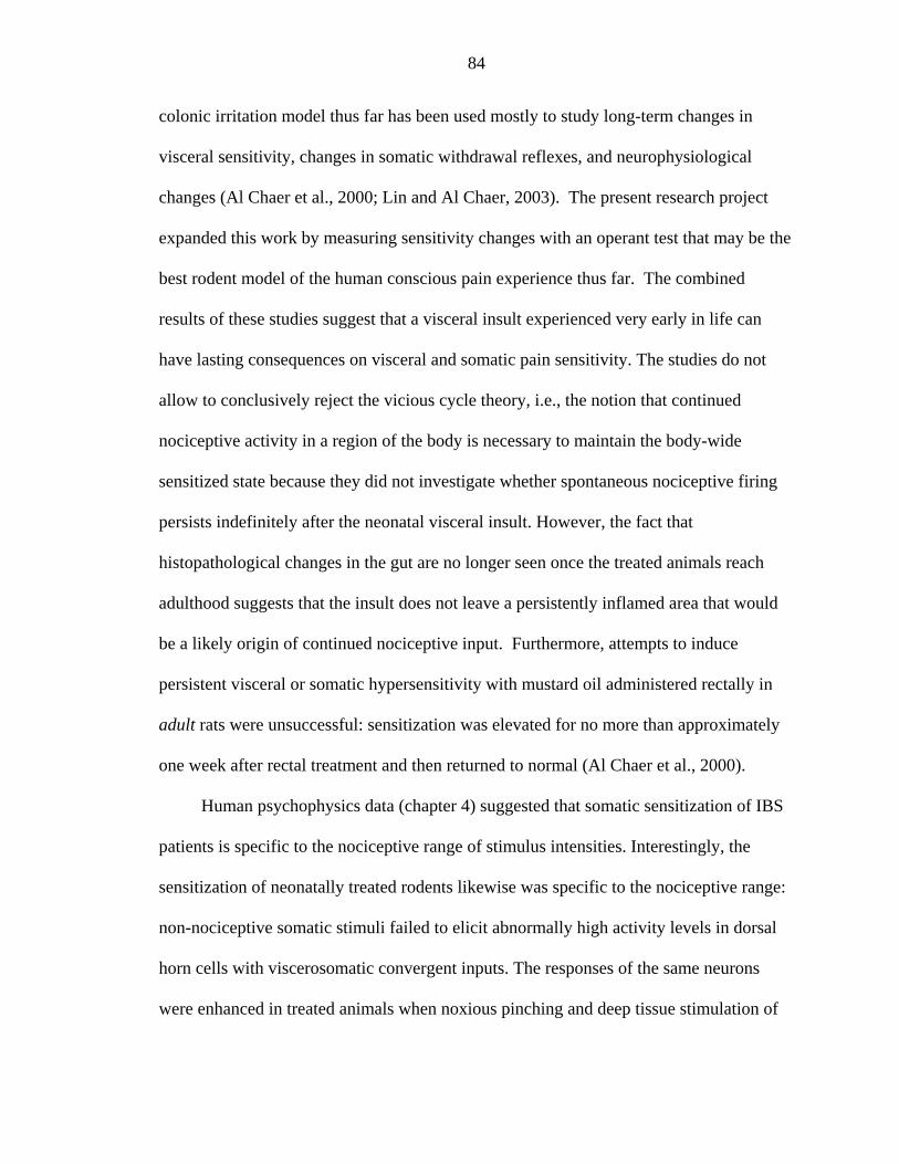
84
colonic irritation model thus far has been used mostly to study long-term changes in
visceral sensitivity, changes in somatic withdrawal reflexes, and neurophysiological
changes (Al Chaer et al., 2000; Lin and Al Chaer, 2003). The present research project
expanded this work by measuring sensitivity changes with an operant test that may be the
best rodent model of the human conscious pain experience thus far. The combined
results of these studies suggest that a visceral insult experienced very early in life can
have lasting consequences on visceral and somatic pain sensitivity. The studies do not
allow to conclusively reject the vicious cycle theory, i.e., the notion that continued
nociceptive activity in a region of the body is necessary to maintain the body-wide
sensitized state because they did not investigate whether spontaneous nociceptive firing
persists indefinitely after the neonatal visceral insult. However, the fact that
histopathological changes in the gut are no longer seen once the treated animals reach
adulthood suggests that the insult does not leave a persistently inflamed area that would
be a likely origin of continued nociceptive input. Furthermore, attempts to induce
persistent visceral or somatic hypersensitivity with mustard oil administered rectally in
adult rats were unsuccessful: sensitization was elevated for no more than approximately
one week after rectal treatment and then returned to normal (Al Chaer et al., 2000).
Human psychophysics data (chapter 4) suggested that somatic sensitization of IBS
patients is specific to the nociceptive range of stimulus intensities. Interestingly, the
sensitization of neonatally treated rodents likewise was specific to the nociceptive range:
non-nociceptive somatic stimuli failed to elicit abnormally high activity levels in dorsal
horn cells with viscerosomatic convergent inputs. The responses of the same neurons
were enhanced in treated animals when noxious pinching and deep tissue stimulation of
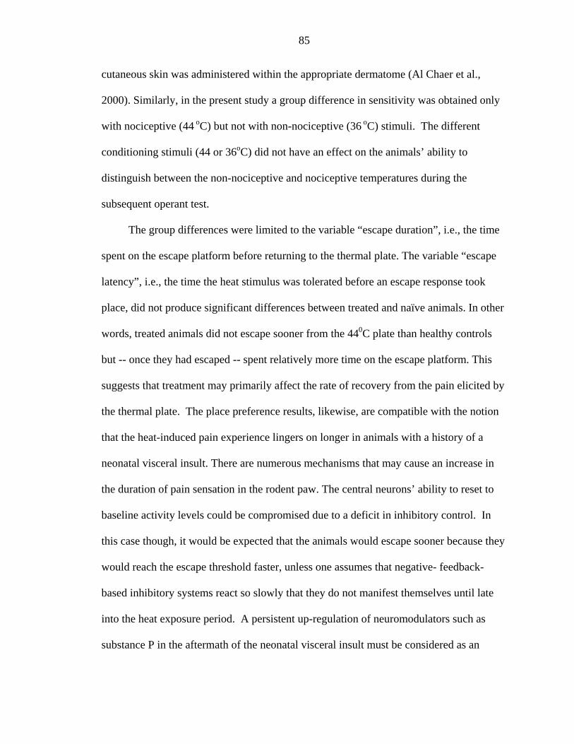
85
cutaneous skin was administered within the appropriate dermatome (Al Chaer et al.,
2000). Similarly, in the present study a group difference in sensitivity was obtained only
with nociceptive (44 oC) but not with non-nociceptive (36 oC) stimuli. The different
conditioning stimuli (44 or 36oC) did not have an effect on the animals’ ability to
distinguish between the non-nociceptive and nociceptive temperatures during the
subsequent operant test.
The group differences were limited to the variable “escape duration”, i.e., the time
spent on the escape platform before returning to the thermal plate. The variable “escape
latency”, i.e., the time the heat stimulus was tolerated before an escape response took
place, did not produce significant differences between treated and naïve animals. In other
words, treated animals did not escape sooner from the 440C plate than healthy controls
but -- once they had escaped -- spent relatively more time on the escape platform. This
suggests that treatment may primarily affect the rate of recovery from the pain elicited by
the thermal plate. The place preference results, likewise, are compatible with the notion
that the heat-induced pain experience lingers on longer in animals with a history of a
neonatal visceral insult. There are numerous mechanisms that may cause an increase in
the duration of pain sensation in the rodent paw. The central neurons’ ability to reset to
baseline activity levels could be compromised due to a deficit in inhibitory control. In
this case though, it would be expected that the animals would escape sooner because they
would reach the escape threshold faster, unless one assumes that negative- feedback-
based inhibitory systems react so slowly that they do not manifest themselves until late
into the heat exposure period. A persistent up-regulation of neuromodulators such as
substance P in the aftermath of the neonatal visceral insult must be considered as an
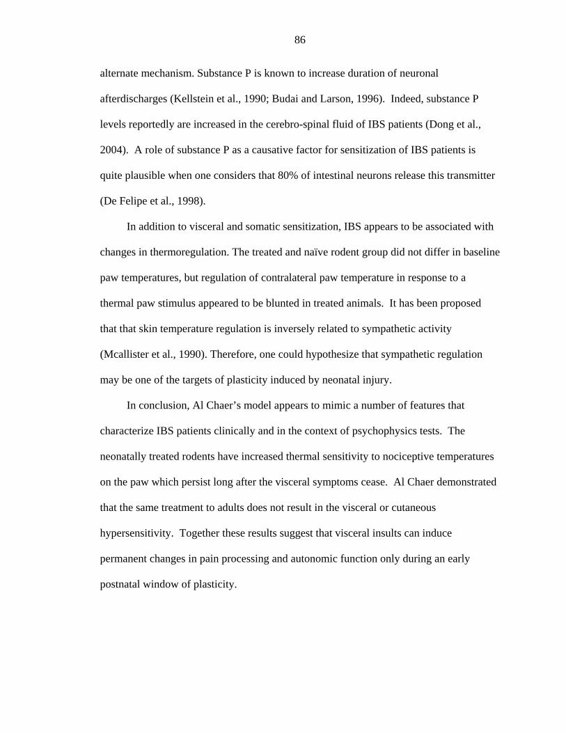
86
alternate mechanism. Substance P is known to increase duration of neuronal
afterdischarges (Kellstein et al., 1990; Budai and Larson, 1996). Indeed, substance P
levels reportedly are increased in the cerebro-spinal fluid of IBS patients (Dong et al.,
2004). A role of substance P as a causative factor for sensitization of IBS patients is
quite plausible when one considers that 80% of intestinal neurons release this transmitter
(De Felipe et al., 1998).
In addition to visceral and somatic sensitization, IBS appears to be associated with
changes in thermoregulation. The treated and naïve rodent group did not differ in baseline
paw temperatures, but regulation of contralateral paw temperature in response to a
thermal paw stimulus appeared to be blunted in treated animals. It has been proposed
that that skin temperature regulation is inversely related to sympathetic activity
(Mcallister et al., 1990). Therefore, one could hypothesize that sympathetic regulation
may be one of the targets of plasticity induced by neonatal injury.
In conclusion, Al Chaer’s model appears to mimic a number of features that
characterize IBS patients clinically and in the context of psychophysics tests. The
neonatally treated rodents have increased thermal sensitivity to nociceptive temperatures
on the paw which persist long after the visceral symptoms cease. Al Chaer demonstrated
that the same treatment to adults does not result in the visceral or cutaneous
hypersensitivity. Together these results suggest that visceral insults can induce
permanent changes in pain processing and autonomic function only during an early
postnatal window of plasticity.
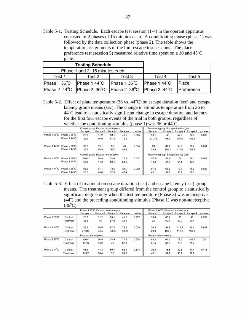
87
Table 5-1. Testing Schedule. Each escape test session (1-4) in the operant apparatus consisted of 2 phases of 15 minutes each. A conditioning phase (phase 1) was followed by the data collection phase (phase 2). The table shows the temperature assignments of the four escape test sessions. The place preference test (session 5) measured relative time spent on a 10 and 45oC plate.
Table 5-2. Effect of plate temperature (36 vs. 44oC) on escape duration (sec) and escape
latency group means (sec). The change in stimulus temperature from 36 to 44oC lead to a statistically significant change in escape duration and latency for the first four escape events of the trial in both groups, regardless of whether the conditioning stimulus (phase 1) was 36 or 44oC.
Table 5-3. Effect of treatment on escape duration (sec) and escape latency (sec) group
means. The treatment group differed from the control group to a statistically significant degree only when the test temperature (Phase 2) was nociceptive (44o) and the preceding conditioning stimulus (Phase 1) was non-nociceptive (36oC)
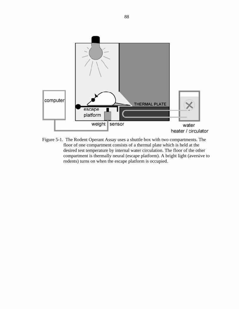
88
Figure 5-1. The Rodent Operant Assay uses a shuttle box with two compartments. The
floor of one compartment consists of a thermal plate which is held at the desired test temperature by internal water circulation. The floor of the other compartment is thermally neural (escape platform). A bright light (aversive to rodents) turns on when the escape platform is occupied.
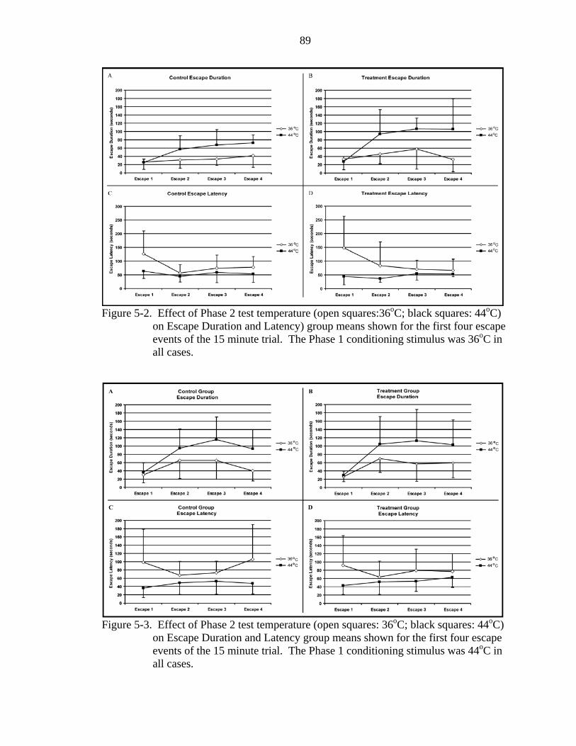
89
Figure 5-2. Effect of Phase 2 test temperature (open squares:36oC; black squares: 44oC)
on Escape Duration and Latency) group means shown for the first four escape events of the 15 minute trial. The Phase 1 conditioning stimulus was 36oC in all cases.
Figure 5-3. Effect of Phase 2 test temperature (open squares: 36oC; black squares: 44oC)
on Escape Duration and Latency group means shown for the first four escape events of the 15 minute trial. The Phase 1 conditioning stimulus was 44oC in all cases.
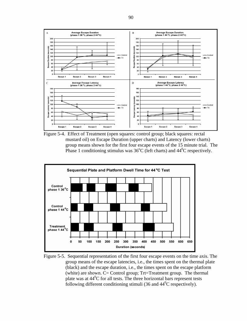
90
Figure 5-4. Effect of Treatment (open squares: control group; black squares: rectal
mustard oil) on Escape Duration (upper charts) and Latency (lower charts) group means shown for the first four escape events of the 15 minute trial. The Phase 1 conditioning stimulus was 36oC (left charts) and 440C respectively.
Figure 5-5. Sequential representation of the first four escape events on the time axis. The
group means of the escape latencies, i.e., the times spent on the thermal plate (black) and the escape duration, i.e., the times spent on the escape platform (white) are shown. C= Control group; Trt=Treatment group. The thermal plate was at 440C for all tests. The three horizontal bars represent tests following different conditioning stimuli (36 and 440C respectively).

91
Figure 5-6. Place Preference Test. Group means (pooled first four crossing events) for
time spent on the 10oC and 45oC sides. Error bars represent the standard deviation.
Figure 5-7. Skin temperature regulation. Group means of temperature recorded at four
sites during a 10 min thermal stimulus (440C) to the left hindpaw and for 10 minutes after the end of the stimulus. Recording sites: (A) left forepaw; (B) right forepaw; (C) right hindpaw; (D) rectum.
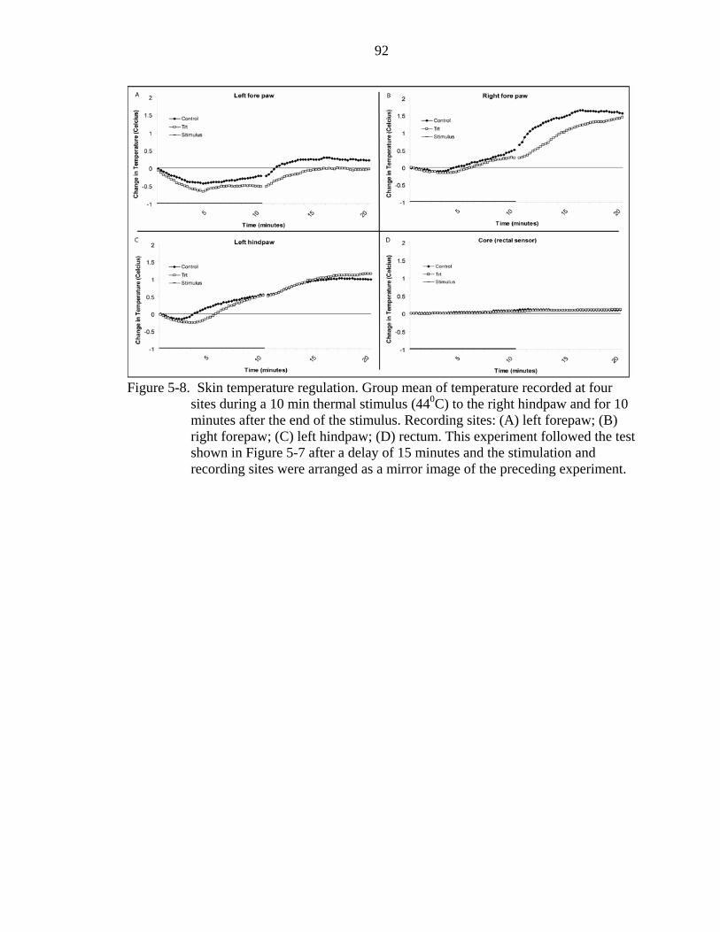
92
Figure 5-8. Skin temperature regulation. Group mean of temperature recorded at four
sites during a 10 min thermal stimulus (440C) to the right hindpaw and for 10 minutes after the end of the stimulus. Recording sites: (A) left forepaw; (B) right forepaw; (C) left hindpaw; (D) rectum. This experiment followed the test shown in Figure 5-7 after a delay of 15 minutes and the stimulation and recording sites were arranged as a mirror image of the preceding experiment.
93
CHAPTER 6 FINAL DISCUSSION
Localized or widespread pain complaints in somatic and visceral tissues are typical
for a number of chronic pain conditions. For instance, IBS, which is considered a
visceral disease and diagnostically defined by visceral symptoms, is often associated with
pain far removed from the gut, e.g., the incidence of headaches, back pain, etc, is higher
than average. Similar findings occur in patients with masticatory myofascial pain
syndrome (MPS) as they present with episodes of pain not only in masticatory muscles
but also in non-facial areas. Even when the clinical complaints are confined to a
localized area (e.g., the gut in IBS patients or the face in masticatory MPS), widespread
pain sensitization has been experimentally demonstrated in many of these patients
(Maixner et al., 1998; Verne et al., 2001; Bouin et al., 2001; present work). A sensitized
state may offer an explanation why these patients are generally more pain-prone
anywhere in the body considering that in the presence of hyperalgesia it takes a smaller
tissue insult to reach pain threshold. It is well known that prolonged exposure to a
nociceptive stimulus leads to an increase in pain sensitivity at least at the location of the
stimulus and –over time— even beyond. In fact, short lasting sensitization (wind-up) has
been induced in an experimental setting with brief repetitive thermal and mechanical
stimuli (Vierck et al., 1997; Maixner et al., 1998). It cannot be ruled out that a disease-
related nociceptive focus can do the same, and due to its persistent nature, over time lead
to states of sensitization that are longer lasting than wind-up and extend to more distant
areas of the body. The question addressed by this body of work is whether the
94
widespread sensitization or pain-proneness of IBS patients originates in the symptomatic
areas that diagnostically define the disease and gradually spreads from there (vicious pain
cycle). The data presented in chapters 2-4 suggest that a key role for the vicious pain
cycle as a basis for remote sensitization in IBS patients must be questioned. Other
possible mechanisms must be given serious consideration.
The first finding that put the vicious pain cycle hypothesis into question was that
remote pain sensitization did not occur in a gradient fashion (IBS patients were expected
to be most sensitive on the foot and less sensitive on the cheek compared to control
subjects), but instead the sensitization was equal along the entire segmental axis of the
body. Interestingly, the IBS patient’s thermal hypersensitivity was nociceptive-specific
but independent of the pain intensity level at which the test was conducted and similar
regardless whether brief or prolonged test stimuli were used.
The vicious pain cycle hypothesis was weakened further when it was found that at
least in the samples of this project, the IBS group’s thermal pain sensitivity was
independent of the magnitude of disease-related spontaneous pain. Since the
hypersensitivity induced by the disease are very stable: i.e., episodes without constant
clinical pain are not associated with appreciably lower sensitivity levels, it appears that
disease-related sensitization of somatic tissues of IBS patients is maintained by an
extremely long term mechanism. Granted, the lack of a relationship between disease-
related pain and experimentally measured sensitization does not necessarily eliminate the
vicious cycle theory as a viable hypothesis because it is known that low level activity of
nociceptors may not always induce consciously perceived pain. It cannot be ruled out
that nociceptors are firing in the viscera of IBS patients that do not currently have pain.
95
This made it necessary to include an experiment where nociceptor activity (whether
consciously perceived as pain or not) was silenced with topical local anesthetics. It
turned out that rectally administered topical anesthetic affected sensitization only
minimally and similarly in IBS and control subjects. According to the vicious pain cycle
hypothesis, IBS patients only but not healthy controls (who do not have a nociceptive
focus) should have been affected by the rectal lidocaine treatment. Again, this finding by
itself does not allow discounting completely the feedback-based sensitization theory
because one could argue that in the present sample of subjects the presumed nociceptive
focus was located proximal to the rectal area that is digitally accessible for topical drug
administration.
However, the fact that none of the results of the present series of studies provide
direct or indirect support for the nociceptive focus-based sensitization theory must at least
convince us to consider alternate hypotheses. The new theory should be able to
accommodate the findings that-- at least in a subpopulation-- sensitization is widespread
across the body, largely independent of intensity and unpleasantness of clinical pain and
not responsive to topical lidocaine intervention when control experiments for systemic
and placebo effects are included in the protocol. A possible alternate hypothesis could
postulate that widespread sensitization is not the result of IBS-related regional
nociceptive activity but that the hyperalgesia predates onset of IBS and facilitates the
development of the chronic pain disease. In other words, according to this theory,
widespread sensitization would be a primary factor that makes the individual pain-prone
across the body and thus susceptible to developing a chronic pain condition, such as IBS
or MPS, upon a minor regional insult. In this context it is interesting to note that some
96
IBS patients had normal sensitivity while a small number of healthy control subjects
exhibited hypersensitivity when probed with experimental stimuli. The heterogeneity
within the IBS group found in most psychophysics studies should not come as a surprise
when one considers that the diagnostic criteria for the disease are symptom-based only
and pain sensitivity of somatic tissues is not one of the criteria. The observation that
“healthy” control subjects can be hyperalgesic prompts the questions (1) whether the
hyperalgesic IBS subgroup is more likely to develop a generalized pain disorder such a
fibromyalgia than the subgroup without cutaneous sensitization; (2) whether the
hyperalgesic subgroup and the subgroup with normal sensitivity to thermal nociceptive
stimuli are characterized by different disease mechanisms and thus require different
treatments? If the latter turns out to be the case, psychophysics tests similar to those used
in this project may ultimately be of diagnostic value. Lastly, the question arises whether
control subjects with widespread cutaneous hyperalgesia are prime candidates for
developing a chronic pain disease later in life? Longitudinal studies to conclusively
answer this question are lacking. However, animal experiments, such as the one
described in chapter 5, suggest that visceral insults early in life can result in lifelong
visceral (Al Chaer et al., 2000) and somatic (chapter 5) sensitization. This, in
combination with the lack of convincing support for the vicious cycle hypothesis
(chapters 2, 3, and 4) suggests that our alternate hypothesis deserves serious
consideration in the design of future studies: the widespread pain sensitization seen in a
large number of chronic pain patients (including many IBS patients) is not induced by
nociceptive drive from a localized symptomatic region but may have preexisted before
the onset of the chronic pain syndrome. As possible causes for the generalized sensitized

97
state one must consider a plasticity-inducing injury or infection earlier in life (consistent
with the findings of the rodent study described in chapter 5), genetic factors, or a
combination of the two. The plastic changes responsible for sensitization could be
widespread and affect large parts of the somatosensory central nervous system.
Alternatively, they could affect a central component of the CNS where nociceptive
signals from a large part of the body converge. Lastly, it is possible that the plasticity
leads to diffuse widespread sensitization because it targets the neuroendocrine or
cytokine system. Longitudinal long-term studies in large populations of initially healthy
subjects and experiments in animal models where the late consequences of transient
neonatal and postnatal insults can be studied under controlled conditions will provide the
answers.
98
LIST OF REFERENCES
Accarino, A.M., Azpiroz, F., and Malagelada, J.R., Selective dysfunction of mechanosensitive intestinal afferents in irritable-bowel-syndrome. Gastroenterology, 108 (1995) 636-643.
Al Chaer, E.D., Kawasaki, M., and Pasricha, P.J., A new model of chronic visceral hypersensitivity in adult rats induced by colon irritation during postnatal development, Gastroenterology, 119 (2000) 1276-1285.
Bach, F.W., Jensen, T.S., Kastrup, J., Stigsby, B., and Dejgard, A., The effect of intravenous lidocaine on nociceptive processing in diabetic neuropathy, Pain, 40 (1990) 29-34.
Bouin, M., Meunier, P., Riberdy-Poitras, M., and Poitras, P., Pain hypersensitivity in patients with functional gastrointestinal disorders - a gastrointestinal-specific defect or a general systemic condition?, Digestive Diseases and Sciences, 46 (2001) 2542-2548.
Brose,W.G. and Cousins, M.J., Subcutaneous lidocaine for treatment of neuropathic cancer pain, Pain, 45 (1991) 145-148.
Budai, D. and Larson, A.A., Role of substance P in the modulation of C-fiber-evoked responses of spinal dorsal horn neurons, Brain Res., 710 (1996) 197-203.
Chang, L., Mayer, E.A., Johnson, T., FitzGerald, L.Z., and Naliboff, B., Differences in somatic perception in female patients with irritable bowel syndrome with and without fibromyalgia, Pain, 84 (2000) 297-307.
Cook, I.J., Vaneeden, A., and Collins, S.M., Patients with irritable-bowel-syndrome have greater pain tolerance than normal subjects, Gastroenterology, 93 (1987) 727-733.
De Felipe, C., Herrero, J.F., O'Brien, J.A., Palmer, J.A., Doyle, C.A., Smith, A.J., Laird, J.M., Belmonte, C., Cervero, F., and Hunt, S.P., Altered nociception, analgesia and aggression in mice lacking the receptor for substance P, Nature, 392 (1998) 394-397.
Deboer, A.G., Breimer, D.D., Mattie, H., Pronk, J., and Gubbensstibbe, J.M., Rectal bioavailability of lidocaine in man - partial avoidance of 1st-pass metabolism, Clinical Pharmacology & Therapeutics, 26 (1979) 701-709.
99
Dirks, J., Fabricius, P., Petersen, K.L., Rowbotham, M.C., and Dahl, J.B., The effect of systemic lidocaine on pain and secondary hyperalgesia associated with the heat/capsaicin sensitization model in healthy volunteers, Anesth.Analg., 91 (2000) 967-972.
Dong, W.Z., Zou, D.W., Li, Z.S., Zou, X.P., Zhu, A.Y., Xu, G.M., Yin, N., Gong, Y.F., Sun, Z.X., and Man, X.H., Study of visceral hypersensitivity in irritable bowel syndrome, Chin J Dig.Dis., 5 (2004) 103-109.
Fillingim, R.B., Fillingim, L.A., Hollins, M., Sigurdsson, A., and Maixner, W., Generalized vibrotactile allodynia in a patient with temporomandibular disorder, Pain, 78 (1998) 75-78.
Gallez, A., Rainville, P., Duncan, G.H., Habituation to heat pain: evidence of somatotopic specificity. Neuroscience . 2003.
Garvey, T.A., Marks, M.R., and Wiesel, S.W., A prospective, randomized, double-blind evaluation of trigger-point injection therapy for low-back pain, Spine, 14 (1989) 962-964.
Gebhart, G.F., Pathobiology of visceral pain: Molecular mechanisms and therapeutic implications - IV. Visceral afferent contributions to the pathobiology of visceral pain, American Journal of Physiology-Gastrointestinal and Liver Physiology, 278 (2000) G834-G838.
Gracely, R.H., Lota, L., Walter, D.J., and Dubner, R., A multiple random staircase method of psychophysical pain assessment, Pain, 32 (1988) 55-63.
Gracely, R.H., Lynch, S.A., and Bennett, G.J., Painful neuropathy - altered central processing maintained dynamically by peripheral input, Pain, 51 (1992) 175-194.
Grill, J.D. and Coghill, R.C., Transient analgesia evoked by noxious stimulus offset, Journal of Neurophysiology, 87 (2002) 2205-2208.
Groudine, S.B., Fisher, H.A., Kaufman, R.P., Jr., Patel, M.K., Wilkins, L.J., Mehta, S.A., and Lumb, P.D., Intravenous lidocaine speeds the return of bowel function, decreases postoperative pain, and shortens hospital stay in patients undergoing radical retropubic prostatectomy, Anesth.Analg., 86 (1998) 235-239.
Gwee, K.A., Graham, J.C., McKendrick, M.W., Collins, S.M., Marshall, J.S., Walters, S.J., and Read, N.W., Psychometric scores and persistence of irritable bowel after infectious diarrhoea, Lancet, 347 (1996) 150-153.
Isaacson, R.J., Ethics and economics, Angle Orthod., 72 (2002) iv.
Joranson, D.E., Are health-care reimbursement policies a barrier to acute and cancer pain management?, J Pain Symptom Manage., 9 (1994) 244-253.
100
Kellstein, D.E., Price, D.D., Hayes, R.L., and Mayer, D.J., Evidence that substance P selectively modulates C-fiber-evoked discharges of dorsal horn nociceptive neurons, Brain Res., 526 (1990) 291-298.
Koppert, W., Weigand, M., Neumann, F., Sittl, R., Schuettler, J., Schmelz, M., and Hering, W., Perioperative intravenous lidocaine has preventive effects on postoperative pain and morphine consumption after major abdominal surgery, Anesth.Analg., 98 (2004) 1050-5, table.
Koppert, W., Zeck, S., Sittl, R., Likar, R., Knoll, R., and Schmelz, M., Low-dose lidocaine suppresses experimentally induced hyperalgesia in humans, Anesthesiology, 89 (1998) 1345-1353.
Lamotte, R.H. and Campbell, J.N., Comparison of responses of warm and nociceptive C-fiber afferents in monkey with human judgments of thermal pain, Journal of Neurophysiology, 41 (1978) 509-528.
Lamotte, R.H., Thalhammer, J.G., and Robinson, C.J., Peripheral neural correlates of magnitude of cutaneous pain and hyperalgesia: a comparison of neural events in monkey with sensory judgments in human, J Neurophysiol., 50 (1983) 1-26.
Lembo, T., Munakata, J., Mertz, H., Niazi, N., Kodner, A., Nikas, V., and Mayer, E.A., Evidence for the hypersensitivity of lumbar splanchnic afferents in irritable bowel syndrome, Gastroenterology, 107 (1994) 1686-1696.
Lembo, T., Naliboff, B.D., Matin, K., Munakata, J., Parker, R.A., Gracely, R.H., and Mayer, E.A., Irritable bowel syndrome patients show altered sensitivity to exogenous opioids, Pain, 87 (2000) 137-147.
Lin, C. and Al Chaer, E.D., Long-term sensitization of primary afferents in adult rats exposed to neonatal colon pain, Brain Res., 971 (2003) 73-82.
Locke, G.R., III, Weaver, A.L., Melton, L.J., III, and Talley, N.J., Psychosocial factors are linked to functional gastrointestinal disorders: a population based nested case-control study, Am.J.Gastroenterol., 99 (2004) 350-357.
Maixner, W., Fillingim, R., Sigurdsson, A., Kincaid, S., and Silva, S., Sensitivity of patients with painful temporomandibular disorders to experimentally evoked pain: evidence for altered temporal summation of pain, Pain, 76 (1998) 71-81.
Mallon, W.J. and Thomas, C.W., Patient-controlled lidocaine analgesia for acromioplasty surgery, J Shoulder.Elbow.Surg., 9 (2000) 85-88.
Manning, A.P., Thompson, W.G., Heaton, K.W., and Morris, A.F., Towards positive diagnosis of the irritable bowel, Br.Med.J., 2 (1978) 653-654.
101
Mao, J., Price, D.D., Hayes, R.L., Lu, J., and Mayer, D.J., Intrathecal GM1 ganglioside and local nerve anesthesia reduce nociceptive behaviors in rats with experimental peripheral mononeuropathy, Brain Res., 584 (1992) 28-53.
Mauderli, A.P., Acosta-Rua, A., and Vierck, C.J., An operant assay of thermal pain in conscious, unrestrained rats, Journal of Neuroscience Methods, 97 (2000) 19-29.
Mayer, E.A. and Gebhart, G.F., Basic and clinical aspects of visceral hyperalgesia, Gastroenterology, 107 (1994) 271-293.
Mayer, E.A. and Raybould, H.E., Role of visceral afferent mechanisms in functional bowel disorders, Gastroenterology, 99 (1990) 1688-1704.
Mcallister, C., Mcgrath, F., and Fielding, J.F., Altered skin temperature and electromyographic activity in the irritable-bowel-syndrome, Biomedicine & Pharmacotherapy, 44 (1990) 399-401.
McClintock, M.K., Menstrual synchorony and suppression, Nature, 229 (1971) 244-245.
McKendrick, M.W. and Read, N.W., Irritable-bowel-syndrome - post salmonella infection, Journal of Infection, 29 (1994) 1-3.
Mertz, H., Naliboff, B., Munakata, J., Niazi, N., and Mayer, E.A., Altered rectal perception is a biological marker of patients with irritable-bowel-syndrome, Gastroenterology, 109 (1995) 40-52.
Munakata, J., Naliboff, B., Harraf, F., Kodner, A., Lembo, T., Chang, L., Silverman, D.H.S., and Mayer, E.A., Repetitive sigmoid stimulation induces rectal hyperalgesia in patients with irritable bowel syndrome, Gastroenterology, 112 (1997) 55-63.
Naliboff, B.D., Munakata, J., Fullerton, S., Gracely, R.H., Kodner, A., Harraf, F., and Mayer, E.A., Evidence for two distinct perceptual alterations in irritable bowel syndrome, Gut, 41 (1997) 505-512.
Price, D.D. and Harkins, S.W., The combined use of visual analogue scales and experimental pain in providing standardized measurement of clinical pain, Clinical Journal of Pain, 3 (1987) 1-8.
Price, D.D., Hu, J.W., Dubner, R., and Gracely, R.H., Peripheral suppression of 1st pain and central summation of 2nd pain evoked by noxious heat pulses, Pain, 3 (1977) 57-68.
Rodrigues, A.C., Nicholas, V.G., Schmidt, S., and Mauderli, A.P., Hypersensitivity to cutaneous thermal nociceptive stimuli in irritable bowel syndrome, Pain, 115 (2005) 5-11.

102
Sabate, J.M., Coffin, B., Jian, R., Le Bars, D., and Bouhassira, D., Rectal sensitivity assessed by a reflexologic technique: further evidence for two types of mechanoreceptors, Am.J.Physiol Gastrointest.Liver Physiol, 279 (2000) G692-G699.
Sandler, R.S., Epidemiology of irritable bowel syndrome in the United-States, Gastroenterology, 99 (1990) 409-415.
Staud, R., Vierck, C.J., Cannon, R.L., Mauderli, A.P., and Price, D.D., Abnormal sensitization and temporal summation of second pain (wind-up) in patients with fibromyalgia syndrome, Pain, 91 (2001) 165-175.
Thompson, W.G., Longstreth, G.F., Drossman, D.A., Heaton, K.W., Irvine, E.J., and Muller-Lissner, S.A., Functional bowel disorders and functional abdominal pain, Gut, 45 (1999) 43-47.
Tillman, D.B., Treede, R.D., Meyer, R.A., and Campbell, J.N., Response of C-fiber nociceptors in the anesthetized monkey to heat stimuli - correlation with pain threshold in humans, Journal of Physiology-London, 485 (1995a) 767-774.
Tillman, D.B., Treede, R.D., Meyer, R.A., and Campbell, J.N., Response of C-fiber nociceptors in the anesthetized monkey to heat stimuli - estimates of receptor depth and threshold, Journal of Physiology-London, 485 (1995b) 753-765.
Turk, D.C., Clinical effectiveness and cost-effectiveness of treatments for patients with chronic pain, Clinical Journal of Pain, 18 (2002) 355-365.
Veale, D., Kavanagh, G., Fielding, J.F., and Fitzgerald, O., Primary fibromyalgia and the irritable-bowel-syndrome - different expressions of a common pathogenetic process, British Journal of Rheumatology, 30 (1991) 220-222.
Verne, G.N. and Cerda, J.J., Streamlining the diagnosis of the irritable colon syndrome. Postgraduate Medicine, 102 (1997) 197-208.
Verne, G.N., Himes, N.C., Robinson, M.E., Gopinath, K.S., Briggs, R.W., Crosson, B., and Price, D.D., Central representation of visceral and cutaneous hypersensitivity in the irritable bowel syndrome, Pain, 103 (2003a) 99-110.
Verne, G.N., Robinson, M.E., and Price, D.D., Hypersensitivity to visceral and cutaneous pain in the irritable bowel syndrome, Pain, 93 (2001) 7-14.
Verne, G.N., Robinson, M.E., Vase, L., and Price, D.D., Reversal of visceral and cutaneous hyperalgesia by local rectal anesthesia in irritable bowel syndrome (IBS) patients, Pain, 105 (2003b) 223-230.
Vierck, C.J., Acosta-Rua, A., Nelligan, R., Tester, N., and Mauderli, A., Low dose systemic morphine attenuates operant escape but facilitates innate reflex responses to thermal stimulation, Journal of Pain, 3 (2002) 309-319.

103
Vierck, C.J., Cannon, R.L., Fry, G., Maixner, W., and Whitsel, B.L., Characteristics of temporal summation of second pain sensations elicited by brief contact of glabrous skin by a preheated thermode, Journal of Neurophysiology, 78 (1997) 992-1002.
Whitehead, W.E., Holtkotter, B., Enck, P., Hoelzl, R., Holmes, K.D., Anthony, J., Shabsin, H.S., and Schuster, M.M., Tolerance for rectosigmoid distension in irritable bowel syndrome, Gastroenterology, 98 (1990) 1187-1192.
Whitehead, W.E., Palsson, O., and Jones, K.R., Systematic review of the comorbidity of irritable bowel syndrome with other disorders: what are the causes and implications?, Gastroenterology, 122 (2002) 1140-1156.
Whorwell, P.J., Mccallum, M., Creed, F.H., and Roberts, C.T., Noncolonic features of irritable-bowel-syndrome, Gut, 27 (1986) 37-40.
Wolfe, F., Smythe, H.A., Yunus, M.B., Bennett, R.M., Bombardier, C., Goldenberg, D.L., Tugwell, P., Campbell, S.M., Abeles, M., Clark, P., and ., The American College of Rheumatology, 1990 Criteria for the classification of fibromyalgia. Report of the multicenter criteria committee, Arthritis Rheum., 33 (1990) 160-172.
Woolf, C.J., Windup and central sensitization are not equivalent, Pain, 66 (1996) 105-108.
Zighelboim, J., Talley, N.J., Phillips, S.F., Harmsen, W.S., and Zinsmeister, A.R., Visceral perception in irritable-bowel-syndrome - rectal and gastric responses to distension and serotonin type-3 antagonism, Digestive Diseases and Sciences, 40 (1995) 819-827.
104
BIOGRAPHICAL SKETCH
I was born in Providence, RI on March 7, 1978. By the age of one, I moved to
Smithfield, RI. I attended Smithfield High School from 1992 -1996. In August 1996, I
began my collegiate work at Assumption College in Worcester, MA. I graduated in May
2000 and received a Bachelor of Arts. The following August I started the
Interdisciplinary Program in Biomedical Sciences at UF, to work on my Ph.D. in
neuroscience.