Embed Size (px)
Citation preview
J Neurosurg Volume 122 • June 2015
cliNical articleJ Neurosurg 122:1380–1389, 2015
Rathke’s cleft cysts (RCCs) are nonneoplastic epi-thelial lesions of the sellar and suprasellar region. They are typically asymptomatic with a slow growth
rate. An asymptomatic RCC is most often diagnosed at autopsy with a reported incidence of 5%–33%.5,47–49 Quite often some RCCs grow over time and can become suffi-
ciently large to cause a compressive effect on surrounding structures, resulting in neurological and endocrine abnor-malities.7,28,37,42 A symptomatic RCC was first described by Goldzieher17 in 1913, and this type of cyst follows a variable natural history. Some cysts undergo spontaneous resolution,2 and others have reductions in cyst size and
abbreviatioNs DI = diabetes insipidus; RCC = Rathke’s cleft cyst; RFS = recurrence-free survival.submitted March 15, 2014. accepted December 18, 2014.iNclude wheN citiNg Published online February 13, 2015; DOI: 10.3171/2014.12.JNS14596.disclosure The authors report no conflict of interest concerning the materials or methods used in this study or the findings specified in this paper. This study was sup-ported by grants from the Natural Science Foundation of Guang Dong province (Grant No. 2011010003063 and Grant No. 2011020005179) and from the National Natural Science Foundation of China (Grant No. 81072067).* Drs. Chotai and Liu contributed equally to this work.
Characteristics of Rathke’s cleft cyst based on cyst location with a primary focus on recurrence after resection*silky chotai, md, Yi liu, md, phd, Jun pan, md, phd, and songtao Qi, md, phdDepartment of Neurosurgery, Nanfang Hospital, Southern Medical University, Guangzhou, Guangdong, People’s Republic of China
obJect Rathke’s cleft cysts (RCCs) are benign lesions with a location that is entirely intrasellar, intrasellar with supra-sellar extension (intrasuprasellar), or purely suprasellar. The recurrence of RCC is relatively uncommon. The present study was conducted to report clinical characteristics, histological features, and outcomes based on location of the cyst with a primary focus on analyzing the predictors of squamous metaplasia and recurrence in these 3 types of RCCs.methods A retrospective review of the medical records of patients with symptomatic RCCs who had undergone resection at the authors’ institution was conducted. Data points, including clinical presentation, preoperative endocrine status, operative details, imaging findings, pathology, and clinical outcomes, were reviewed. A multivariable regression model was used to identify predictors of recurrence.results The mean age of the 87 eligible patients, 64 females and 23 males, was 41 ± 14 years (range 10–73 years). Sixteen patients (18%) had an entirely intrasellar RCC, 21 (24%) had a purely suprasellar cyst, and 50 (58%) had an intrasuprasellar RCC. The mean cyst volume was 2.4 ± 0.9 cm3 (range 0.36–4.9 cm3). Headache was the most frequent initial symptom (76%) followed by visual disturbance (45%). The transsphenoidal approach was performed for all intra-sellar RCCs (16 cysts) and 33 of 50 intrasuprasellar RCCs. The transcranial route was used for all suprasellar cysts (21 cysts) and 17 of 50 intrasuprasellar RCCs. Squamous metaplasia was present in 27 (31%) of 87 RCCs. The occurrence of squamous metaplasia was associated with cyst location (p = 0.027), T1 signal intensity (p = 0.004) and ring enhance-ment on Gd-enhanced MRI (p = 0.017), and cyst volume (p = 0.045). A suprasellar location (p = 0.048, OR 3.89, 95% CI 1.010–15.020), ring enhancement on Gd-enhanced MRI (p = 0.028, OR 3.922, 95% CI 1.158–13.288), hypointensity on T1-weighted MRI (p = 0.002, OR 6.86, 95% CI 1.972–23.909), and cyst volume (p = 0.01, OR 0.367, 95% CI 0.170–0.789) were independent predictors of squamous metaplasia. The mean time to reaccumulation (11 [12.6%] of 87 cases) and recurrence (7 [8%] of 87 cases) was 14 ± 6 months. Recurrence-free survival was 84.5% at a mean of 98.2 ± 4.6 months after treatment. A suprasellar cyst location (p = 0.007, OR 7.7, 95% CI 1.75–34.54), the occurrence of squamous metaplasia (p = 0.007, OR 19.3, 95% CI 2.25–165.18), and isointensity on T2-weighted MRI (p = 0.041, OR 10.29, 95% CI 1.094–96.872) were the independent predictors of RCC recurrence.coNclusioNs A suprasellar cyst location, the occurrence of squamous metaplasia, and isointensity on T2-weighted MRI were independent predictors of RCC recurrence. The extent of resection and type of surgical approach used were not associated with recurrence. A tailored extent of resection based on cyst location and predictive factors is recommended.http://thejns.org/doi/abs/10.3171/2014.12.JNS14596KeY words Rathke’s cleft cyst; RCC; intrasellar; suprasellar; recurrence; squamous metaplasia; resection; oncology
1380 ©AANS, 2015
Unauthenticated | Downloaded 03/17/22 02:54 PM UTC
rathke’s cleft cyst
symptoms after treatment with glucocorticoids.32 Most of-ten the surgically treated RCC remains stable during the long-term follow-up; however, a good number of RCCs are prone to recurrence. Numerous authors have reported their experience in the management of RCCs and have speculated about an association between recurrence and various clinical or histopathological features.6,18,30,33,34,39 Nonetheless, the factors that predict recurrence after sur-gical treatment are still debatable.38,54
Rathke’s cleft cysts are believed to be derived from true remnants of the embryological Rathke’s pouch.3,11,16,23 As regards the development of RCC, theoretically, during the 3rd or 4th week of gestation, a rostral outpouching of the ectodermal primitive oral cavity meets a downward projec-tion from the neuroectodermal diencephalon. These struc-tures then give rise to the anterior lobe, pars tuberalis, and pars intermedia of the pituitary gland.3,19,44 The residual lu-men between the anterior and intermediate lobe constitutes Rathke’s cleft. The remnant of Rathke’s cleft between the anterior lobe and the pars intermedia can result in an ac-cumulation of fluid and cystic dilation leading to the purely intrasellar RCC or an intrasellar RCC with suprasellar (in-trasuprasellar) extension.44,51 The pars tuberalis lies above the diaphragm, and the Rathke’s pouch remnants in this location can give rise to an entirely suprasellar RCC.4 En-tirely suprasellar RCCs with a normal sella turcica are relatively rare (incidence 0%–20%).8,12,13,15,20,24,25,31,36,39 Su-prasellar, intrasuprasellar, and intrasellar RCCs are distinct entities with respect to their clinical presentation, resect-ability, postoperative recurrence, and outcomes. Few au-thors have classified RCCs based on cyst location and have compared the presentations, different surgical approaches, pathological types, and surgical outcomes.39 The present study was conducted to report clinical characteristics, histological features, and outcomes of surgically treated symptomatic RCCs. Our primary focus was to analyze the predictors of squamous metaplasia and recurrence in the 3 types of RCCs classified based on cyst location.
methodsA retrospective review of the medical records for all
patients with symptomatic RCCs who had undergone re-section at our institution between January 2002 and Janu-ary 2012 was conducted after obtaining approval from our local ethics committee. Data points, including clinical presentation, preoperative endocrine status, operative de-tails, imaging findings, pathology reports, and clinical out-comes, were reviewed. Preoperative pituitary function was evaluated through laboratory assessment of adrenocorti-cotropic hormone, growth hormone, insulin-like growth factor, prolactin levels, thyroid-stimulating hormone, thy-roxin, follicle-stimulating hormone, luteinizing hormone, testosterone, and morning cortisol levels. The precise cyst location, size (calculated using the Coniglobus formula), and cyst characteristics were evaluated on Gd-enhanced T1-weighted MRI. The variation in signal intensity of the cyst on T1- and T2-weighted MRI was noted.
surgical removalResection is performed in all symptomatic patients
with laboratory evidence of hypopituitarism, documented growth of the lesion on serial imaging, and an uncertain or alternative preoperative diagnosis. Selection of the surgical approach is based on the location of the cyst on preopera-tive MRI. Transsphenoidal or subfrontal, frontotemporal, and supraorbital approaches with endoscopic assistance or pure endoscopic technique were used in this study. The details of these approaches have been described in our previous publication.13 The essential principle during re-section of an RCC is to avoid manipulation of the normal pituitary gland to minimize the risk of gland injury. Com-plete excision of the cyst wall is attempted whenever pos-sible. Radical resection of the cyst wall with the intention of reducing the recurrence rate was performed in 15 cases treated early in the series. For later cases in the series, the cyst contents were totally removed, and the cyst wall was partially resected to avoid transgression of surrounding structures. For cases in which no defect in the suprasel-lar arachnoid is identified, hydrogen peroxide is placed in the cyst cavity for 2 minutes after complete removal of the cyst contents with the intention of decimating the cyst wall lining and theoretically decreasing the proliferative ability of any residual cyst wall. The extent of resection of the cyst wall was identified based on video recordings of the surgical procedure and postoperative imaging. Speci-mens obtained at surgery are fixed in 10% formalin and cut into sections, and the tissues are stained with H & E. The histopathological slides are re-reviewed by a patholo-gist, and the cysts are classified as having or not having squamous metaplasia. An immediate postoperative MRI study is obtained within 48 hours of surgery to evaluate the extent of removal.
Follow-upFollow-up is scheduled at 3 and 9 months after surgery
and annually thereafter. Magnetic resonance imaging is performed at each follow-up visit. Pituitary function is monitored by hormone level assessment at 1, 3, 6, and 12 months after surgery. Recurrence was defined as the reac-cumulation of cyst contents on follow-up imaging with or without symptomatic recurrence.
statistical analysisThe mean ± standard deviation, median, and range for
continuous variables and the frequency for discrete data were calculated for patient demographics. Univariate anal-ysis using the chi-square test for categorical variables and the Mann-Whitney U-test or Kruskal-Wallis test for con-tinuous variables was performed to evaluate the associa-tion of clinical parameters with the location of the RCC, the occurrence of squamous metaplasia, and recurrence. Multivariable binary logistic regression analysis was con-ducted to evaluate the predictors of squamous metaplasia. The Hosmer-Lemeshow statistic was used to assess the fit of the models. Kaplan-Meier analyses with the log-rank model were used to evaluate recurrence-free survival (RFS). Multivariable stepwise Cox proportional-hazards regression analysis was used to identify independent pre-dictors of recurrence. All variables with a p value ≤ 0.05 in the univariate analysis, in addition to age at the time of
J Neurosurg Volume 122 • June 2015 1381
Unauthenticated | Downloaded 03/17/22 02:54 PM UTC
s. chotai et al.
surgery and histology, were included as independent vari-ables. All the analyses were tested at the 0.05 level of sig-nificance, and the analysis was performed using the SPSS version 20 (IBM Inc.).
resultsThe mean age of the 87 patients eligible for this study
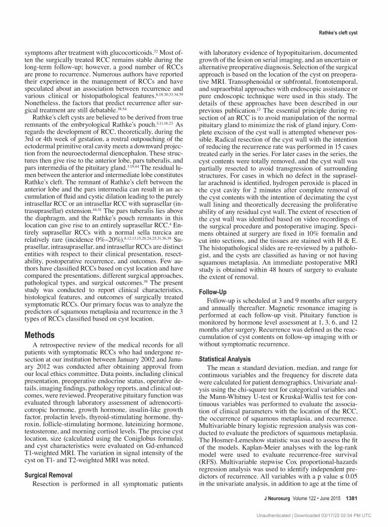
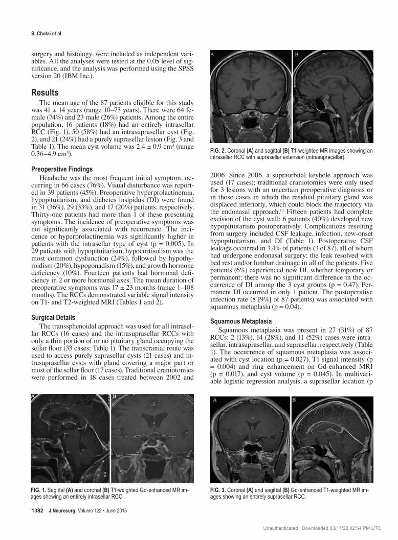
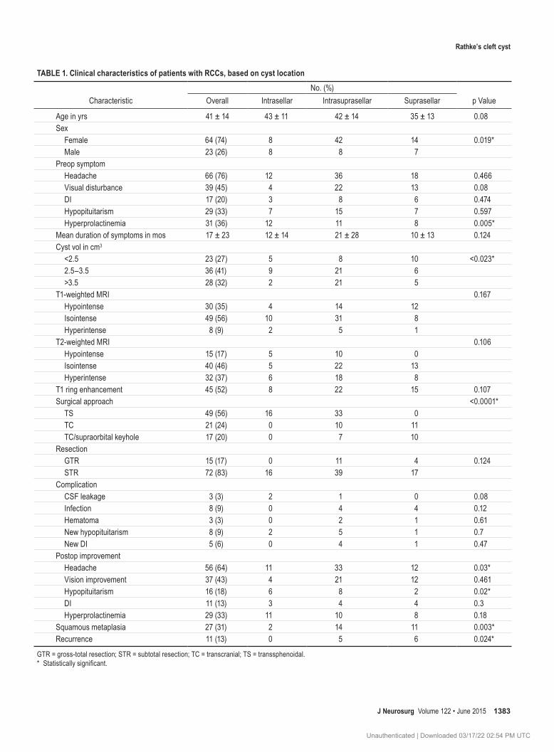
was 41 ± 14 years (range 10–73 years). There were 64 fe-male (74%) and 23 male (26%) patients. Among the entire population, 16 patients (18%) had an entirely intrasellar RCC (Fig. 1), 50 (58%) had an intrasuprasellar cyst (Fig. 2), and 21 (24%) had a purely suprasellar lesion (Fig. 3 and Table 1). The mean cyst volume was 2.4 ± 0.9 cm3 (range 0.36–4.9 cm3).
preoperative FindingsHeadache was the most frequent initial symptom, oc-
curring in 66 cases (76%). Visual disturbance was report-ed in 39 patients (45%). Preoperative hyperprolactinemia, hypopituitarism, and diabetes insipidus (DI) were found in 31 (36%), 29 (33%), and 17 (20%) patients, respectively. Thirty-one patients had more than 1 of these presenting symptoms. The incidence of preoperative symptoms was not significantly associated with recurrence. The inci-dence of hyperprolactinemia was significantly higher in patients with the intrasellar type of cyst (p = 0.005). In 29 patients with hypopituitarism, hypocortisolism was the most common dysfunction (24%), followed by hypothy-roidism (20%), hypogonadism (15%), and growth hormone deficiency (10%). Fourteen patients had hormonal defi-ciency in 2 or more hormonal axes. The mean duration of preoperative symptoms was 17 ± 23 months (range 1–108 months). The RCCs demonstrated variable signal intensity on T1- and T2-weighted MRI (Tables 1 and 2).
surgical detailsThe transsphenoidal approach was used for all intrasel-
lar RCCs (16 cases) and the intrasuprasellar RCCs with only a thin portion of or no pituitary gland occupying the sellar floor (33 cases; Table 1). The transcranial route was used to access purely suprasellar cysts (21 cases) and in-trasuprasellar cysts with gland covering a major part or most of the sellar floor (17 cases). Traditional craniotomies were performed in 18 cases treated between 2002 and
2006. Since 2006, a supraorbital keyhole approach was used (17 cases); traditional craniotomies were only used for 3 lesions with an uncertain preoperative diagnosis or in those cases in which the residual pituitary gland was displaced inferiorly, which could block the trajectory via the endonasal approach.13 Fifteen patients had complete excision of the cyst wall; 6 patients (40%) developed new hypopituitarism postoperatively. Complications resulting from surgery included CSF leakage, infection, new-onset hypopituitarism, and DI (Table 1). Postoperative CSF leakage occurred in 3.4% of patients (3 of 87), all of whom had undergone endonasal surgery; the leak resolved with bed rest and/or lumbar drainage in all of the patients. Five patients (6%) experienced new DI, whether temporary or permanent; there was no significant difference in the oc-currence of DI among the 3 cyst groups (p = 0.47). Per-manent DI occurred in only 1 patient. The postoperative infection rate (8 [9%] of 87 patients) was associated with squamous metaplasia (p = 0.04).
squamous metaplasiaSquamous metaplasia was present in 27 (31%) of 87
RCCs: 2 (13%), 14 (28%), and 11 (52%) cases were intra-sellar, intrasuprasellar, and suprasellar, respectively (Table 1). The occurrence of squamous metaplasia was associ-ated with cyst location (p = 0.027), T1 signal intensity (p = 0.004) and ring enhancement on Gd-enhanced MRI (p = 0.017), and cyst volume (p = 0.045). In multivari-able logistic regression analysis, a suprasellar location (p
Fig. 1. Sagittal (a) and coronal (b) T1-weighted Gd-enhanced MR im-ages showing an entirely intrasellar RCC.
Fig. 3. Coronal (a) and sagittal (b) Gd-enhanced T1-weighted MR im-ages showing an entirely suprasellar RCC.
Fig. 2. Coronal (a) and sagittal (b) T1-weighted MR images showing an intrasellar RCC with suprasellar extension (intrasupracellar).
J Neurosurg Volume 122 • June 20151382
Unauthenticated | Downloaded 03/17/22 02:54 PM UTC
rathke’s cleft cyst
table 1. clinical characteristics of patients with rccs, based on cyst location
CharacteristicNo. (%)
p ValueOverall Intrasellar Intrasuprasellar Suprasellar
Age in yrs 41 ± 14 43 ± 11 42 ± 14 35 ± 13 0.08Sex Female 64 (74) 8 42 14 0.019* Male 23 (26) 8 8 7Preop symptom Headache 66 (76) 12 36 18 0.466 Visual disturbance 39 (45) 4 22 13 0.08 DI 17 (20) 3 8 6 0.474 Hypopituitarism 29 (33) 7 15 7 0.597 Hyperprolactinemia 31 (36) 12 11 8 0.005*Mean duration of symptoms in mos 17 ± 23 12 ± 14 21 ± 28 10 ± 13 0.124Cyst vol in cm3
<2.5 23 (27) 5 8 10 <0.023* 2.5–3.5 36 (41) 9 21 6 >3.5 28 (32) 2 21 5T1-weighted MRI 0.167 Hypointense 30 (35) 4 14 12 Isointense 49 (56) 10 31 8 Hyperintense 8 (9) 2 5 1T2-weighted MRI 0.106 Hypointense 15 (17) 5 10 0 Isointense 40 (46) 5 22 13 Hyperintense 32 (37) 6 18 8T1 ring enhancement 45 (52) 8 22 15 0.107Surgical approach <0.0001* TS 49 (56) 16 33 0 TC 21 (24) 0 10 11 TC/supraorbital keyhole 17 (20) 0 7 10Resection GTR 15 (17) 0 11 4 0.124 STR 72 (83) 16 39 17Complication CSF leakage 3 (3) 2 1 0 0.08 Infection 8 (9) 0 4 4 0.12 Hematoma 3 (3) 0 2 1 0.61 New hypopituitarism 8 (9) 2 5 1 0.7 New DI 5 (6) 0 4 1 0.47Postop improvement Headache 56 (64) 11 33 12 0.03* Vision improvement 37 (43) 4 21 12 0.461 Hypopituitarism 16 (18) 6 8 2 0.02* DI 11 (13) 3 4 4 0.3 Hyperprolactinemia 29 (33) 11 10 8 0.18Squamous metaplasia 27 (31) 2 14 11 0.003*Recurrence 11 (13) 0 5 6 0.024*
GTR = gross-total resection; STR = subtotal resection; TC = transcranial; TS = transsphenoidal.* Statistically significant.
J Neurosurg Volume 122 • June 2015 1383
Unauthenticated | Downloaded 03/17/22 02:54 PM UTC
s. chotai et al.
= 0.048, OR 3.89, 95% CI 1.010–15.020), ring enhance-ment on Gd-enhanced MRI (p = 0.028, OR 3.922, 95% CI 1.158–13.288), hypointensity on T1-weighted MRI (p = 0.002, OR 6.86, 95% CI 1.972–23.909), and cyst volume (p = 0.01, OR 0.367, 95% CI 0.170–0.789) were the indepen-dent predictors of squamous metaplasia.
Follow-up and outcomesThe mean duration of follow-up was 54 ± 29 months
(median 60 months, range 9–131 months). Preoperative headaches improved in 56 (85%) of 66 patients. There was a significant difference in the rate of postoperative headache improvement among the RCC types; the rate of improvement was highest in the intrasellar type (92%) and lowest in the suprasellar (66%; p = 0.03). Of the 39 pa-tients presenting with visual dysfunction, 37 (94%) expe-rienced an improvement following surgery. Hyperprolac-tinemia improved remarkably in 29 (94%) of 31 patients. In contrast, hypopituitarism resolved in only 16 (55%) of 29 patients, and DI in 11 (65%) of 17 patients. There was a significant difference in the rate of postoperative improve-ment in hypopituitarism among the RCC types; all but 1 patient in the intrasellar group improved (86%), whereas only 29% of patients in the suprasellar group improved (p = 0.02). Hypocortisolism and hypogonadism resolved in 62% and 54% of patients, respectively. Growth hormone deficiency and hypothyroidism resolved in only 33% and 35% of patients, respectively. Eight patients (9.2%) had new hypopituitarism, and 5 patients (5.7%) had new DI after cyst evacuation/resection. New postoperative hypo-pituitarism was encountered in 1 patient with a suprasellar RCC, 2 with intrasellar RCC, and 5 with intrasuprasellar RCC. There was no significant difference in the occur-rence of new postoperative hypopituitarism among the 3 types of RCC.
recurrenceThe mean time to recurrence was 14 ± 6 months.
Eleven patients (12.6%) had MRI evidence of the reaccu-mulation of cyst fluid; 7 (63.6%) of these 11 patients had symptomatic recurrence. All but 1 recurrent case demon-strated homogeneous isointensity on T2-weighted MRI. Two patients underwent a traditional craniotomy for cyst recurrence, and the other 5 patients underwent repeat en-donasal surgery. The recurrent RCCs were treated based on the same principles as the primary cases. Aggressive complete resection of the cyst wall was attempted in all recurrent cases. No new deficits were noted in any pa-
table 2. univariate analysis of factors associated with rcc recurrence
Characteristic
No. (%)
p ValueOverallRecurrenceYes No
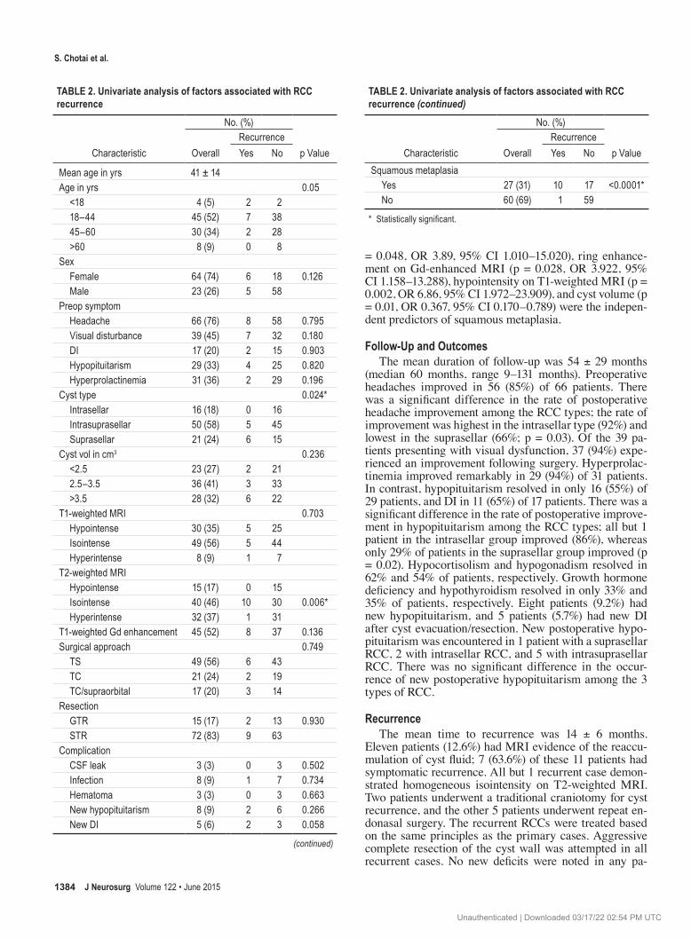
Mean age in yrs 41 ± 14Age in yrs 0.05 <18 4 (5) 2 2 18–44 45 (52) 7 38 45–60 30 (34) 2 28 >60 8 (9) 0 8 Sex Female 64 (74) 6 18 0.126 Male 23 (26) 5 58Preop symptom Headache 66 (76) 8 58 0.795 Visual disturbance 39 (45) 7 32 0.180 DI 17 (20) 2 15 0.903 Hypopituitarism 29 (33) 4 25 0.820 Hyperprolactinemia 31 (36) 2 29 0.196Cyst type 0.024* Intrasellar 16 (18) 0 16 Intrasuprasellar 50 (58) 5 45 Suprasellar 21 (24) 6 15Cyst vol in cm3 0.236 <2.5 23 (27) 2 21 2.5–3.5 36 (41) 3 33 >3.5 28 (32) 6 22 T1-weighted MRI 0.703 Hypointense 30 (35) 5 25 Isointense 49 (56) 5 44 Hyperintense 8 (9) 1 7T2-weighted MRI Hypointense 15 (17) 0 15 Isointense 40 (46) 10 30 0.006* Hyperintense 32 (37) 1 31T1-weighted Gd enhancement 45 (52) 8 37 0.136Surgical approach 0.749 TS 49 (56) 6 43 TC 21 (24) 2 19 TC/supraorbital 17 (20) 3 14 Resection GTR 15 (17) 2 13 0.930 STR 72 (83) 9 63Complication CSF leak 3 (3) 0 3 0.502 Infection 8 (9) 1 7 0.734 Hematoma 3 (3) 0 3 0.663 New hypopituitarism 8 (9) 2 6 0.266 New DI 5 (6) 2 3 0.058
(continued)
table 2. univariate analysis of factors associated with rcc recurrence (continued)
Characteristic
No. (%)
p ValueOverallRecurrenceYes No
Squamous metaplasia Yes 27 (31) 10 17 <0.0001* No 60 (69) 1 59
* Statistically significant.
J Neurosurg Volume 122 • June 20151384
Unauthenticated | Downloaded 03/17/22 02:54 PM UTC
rathke’s cleft cyst
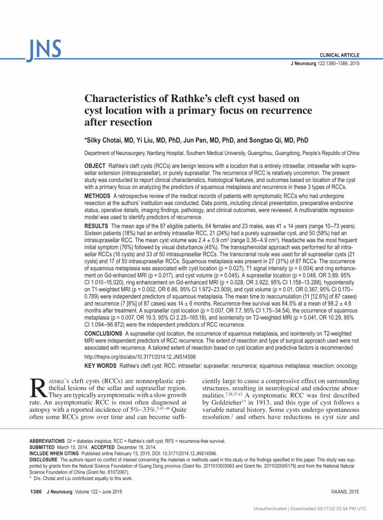
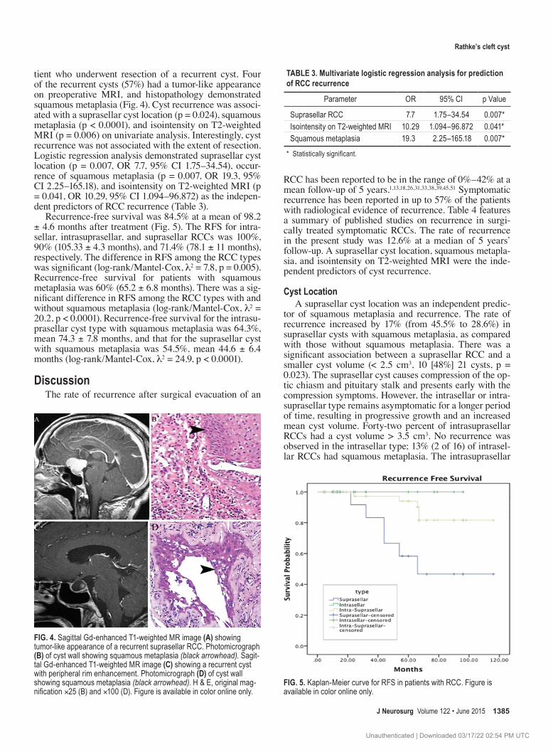
tient who underwent resection of a recurrent cyst. Four of the recurrent cysts (57%) had a tumor-like appearance on preoperative MRI, and histopathology demonstrated squamous metaplasia (Fig. 4). Cyst recurrence was associ-ated with a suprasellar cyst location (p = 0.024), squamous metaplasia (p < 0.0001), and isointensity on T2-weighted MRI (p = 0.006) on univariate analysis. Interestingly, cyst recurrence was not associated with the extent of resection. Logistic regression analysis demonstrated suprasellar cyst location (p = 0.007, OR 7.7, 95% CI 1.75–34.54), occur-rence of squamous metaplasia (p = 0.007, OR 19.3, 95% CI 2.25–165.18), and isointensity on T2-weighted MRI (p = 0.041, OR 10.29, 95% CI 1.094–96.872) as the indepen-dent predictors of RCC recurrence (Table 3).
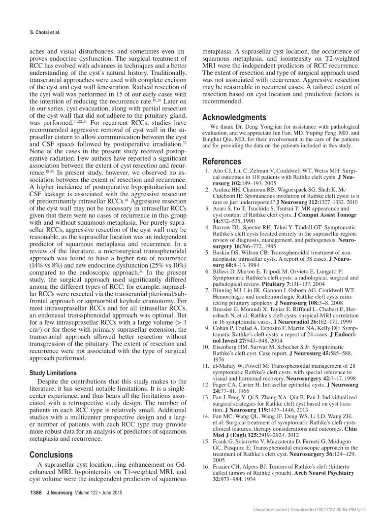
Recurrence-free survival was 84.5% at a mean of 98.2 ± 4.6 months after treatment (Fig. 5). The RFS for intra-sellar, intrasuprasellar, and suprasellar RCCs was 100%, 90% (105.33 ± 4.3 months), and 71.4% (78.1 ± 11 months), respectively. The difference in RFS among the RCC types was significant (log-rank/Mantel-Cox, l2 = 7.8, p = 0.005). Recurrence-free survival for patients with squamous metaplasia was 60% (65.2 ± 6.8 months). There was a sig-nificant difference in RFS among the RCC types with and without squamous metaplasia (log-rank/Mantel-Cox, l2 = 20.2, p < 0.0001). Recurrence-free survival for the intrasu-prasellar cyst type with squamous metaplasia was 64.3%, mean 74.3 ± 7.8 months, and that for the suprasellar cyst with squamous metaplasia was 54.5%, mean 44.6 ± 6.4 months (log-rank/Mantel-Cox, l2 = 24.9, p < 0.0001).
discussionThe rate of recurrence after surgical evacuation of an
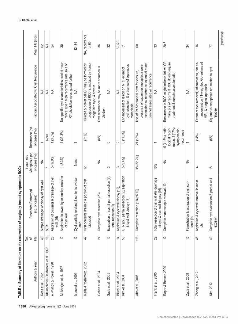
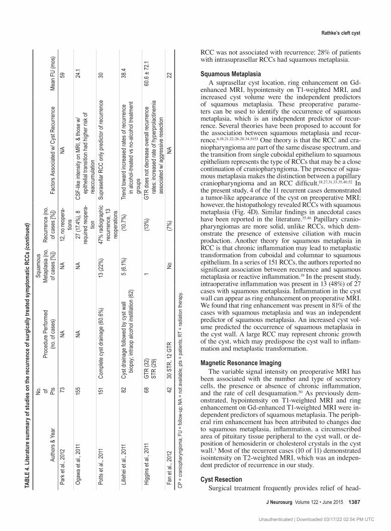
RCC has been reported to be in the range of 0%–42% at a mean follow-up of 5 years.1,13,18,26,31,33,38,39,45,51 Symptomatic recurrence has been reported in up to 57% of the patients with radiological evidence of recurrence. Table 4 features a summary of published studies on recurrence in surgi-cally treated symptomatic RCCs. The rate of recurrence in the present study was 12.6% at a median of 5 years’ follow-up. A suprasellar cyst location, squamous metapla-sia, and isointensity on T2-weighted MRI were the inde-pendent predictors of cyst recurrence.
cyst locationA suprasellar cyst location was an independent predic-
tor of squamous metaplasia and recurrence. The rate of recurrence increased by 17% (from 45.5% to 28.6%) in suprasellar cysts with squamous metaplasia, as compared with those without squamous metaplasia. There was a significant association between a suprasellar RCC and a smaller cyst volume (< 2.5 cm3, 10 [48%] 21 cysts, p = 0.023). The suprasellar cyst causes compression of the op-tic chiasm and pituitary stalk and presents early with the compression symptoms. However, the intrasellar or intra-suprasellar type remains asymptomatic for a longer period of time, resulting in progressive growth and an increased mean cyst volume. Forty-two percent of intrasuprasellar RCCs had a cyst volume > 3.5 cm3. No recurrence was observed in the intrasellar type; 13% (2 of 16) of intrasel-lar RCCs had squamous metaplasia. The intrasuprasellar
Fig. 4. Sagittal Gd-enhanced T1-weighted MR image (a) showing tumor-like appearance of a recurrent suprasellar RCC. Photomicrograph (b) of cyst wall showing squamous metaplasia (black arrowhead). Sagit-tal Gd-enhanced T1-weighted MR image (c) showing a recurrent cyst with peripheral rim enhancement. Photomicrograph (d) of cyst wall showing squamous metaplasia (black arrowhead). H & E, original mag-nification ×25 (B) and ×100 (D). Figure is available in color online only.
Fig. 5. Kaplan-Meier curve for RFS in patients with RCC. Figure is available in color online only.
table 3. multivariate logistic regression analysis for prediction of rcc recurrence
Parameter OR 95% CI p Value
Suprasellar RCC 7.7 1.75–34.54 0.007*Isointensity on T2-weighted MRI 10.29 1.094–96.872 0.041*Squamous metaplasia 19.3 2.25–165.18 0.007*
* Statistically significant.
J Neurosurg Volume 122 • June 2015 1385
Unauthenticated | Downloaded 03/17/22 02:54 PM UTC
s. chotai et al.ta
ble
4. su
mm
ary o
f lite
ratu
re o
n th
e rec
urre
nce o
f sur
gica
lly tr
eate
d sy
mpt
omat
ic rc
cs
Authors &
Year
No.
of Pts
Proc
edur
e Per
form
ed
(no. of cases)
Squamo
us
Metaplas
ia (no.
of cases [%])
Recurre
nce (no.
of cases [%])
Facto
rs Associated w/ C
yst R
ecurrence
Mean F
U (mos)
Ross et al., 1
992
43Simp
le drain
age w
/ biop
sy of cy
st wa
ll NA
1NA
62
Klein
schm
idt-DeM
aster
s et al., 1995
16ST
R1
None
NANA
el-Mahdy & Pow
ell, 1998
28As
piration of co
ntents
& dr
ainage o
f cyst
wall (26)
5 (17.9%)
1 (3.6%
)NA
24
Mukherjee e
t al., 1997
12As
piration followe
d by e
xtensive
excis
ion
of cyst wa
ll1 (8.3%
)4 (33.3%
)No
specific
cyst character
istics
predict re
cur-
rence; giv
en high re
curre
nce r
ate, role
of
RT sh
ould be investigated
furth
er
30
Isono et al., 2
001
12Cy
st partially e
xcise
d & co
ntents
evacu-
ated
None
1NA
12–8
4
Ikeda & Yoshim
oto, 2002
42Cy
st conte
nts dr
ained & po
rtion of cy
st bio
psied
12 (7.1%
) Cilia
ted & go
blet cell CP ma
y be forme
d by
progression
of RCC
, stim
ulated
by he
mor-
rhage into
cyst, & se
vere
NA, recurrence
at 60
Cohan e
t al., 2004
24Co
mplete c
yst resection (23)
NA (8%)
Cyst recurre
nce m
ay be
more c
ommo
n in
children
36
Sade et al., 2
005
10Evacuation o
f cyst &
partial resection (9),
total resection (1)
30
NA32
Billeci et al., 200
414
Aspiration & cy
st wa
ll biop
sy (12),
32
NA6–120
Kim et al., 200
453
GTR (27), partial re
section (9), aspiration
& cyst wa
ll biop
sy (17)
5 (9.4
%)6 (11.3%
)En
hancem
ent of le
sion o
n MRI, exte
nt of
cyst resection, &
presence of sq
uamo
us
metapla
sia
31
Aho e
t al., 2005
118
Comp
lete r
esection (114 [97%])
38 (32.2%
)21 (18%
) Us
e of fa
t &/or fascial gr
aft fo
r clos
ure,
presence of sq
uamo
us metaplas
ia we
re
associa
ted w/ recurrence; exten
t of resec-
tion n
ot associa
ted w/ recurrence
60
Frank e
t al., 2005
22Total re
section of cy
st wa
ll (6), draina
ge
of cyst & cyst wa
ll biop
sy (16)
18%
1 NA
33
Raper &
Besser, 2
009
12Co
mplete m
acroscopic remo
val (1
0)NA
5 (41.6%
) radio-
logica
l recur-
rence, 2 (17%)
symp
tomatic
recurre
nce
Recurre
nce in R
CC migh
t indic
ate lin
k w/ C
P;
many pts w
/ recurrent RC
C do no
t require
treatm
ent &
rema
in asym
ptoma
tic
20.5
Zada et al., 2
009
10Fenestration & as
piration of cy
st con-
tents (9)
NA1
NA34
Zhong e
t al., 2012
45As
piration & cy
st wa
ll rem
oval in mo
st pts
4
(14%
)Ex
tent of cyst rem
oval, inflamm
ation, rim en
-hancem
ent on T
1-we
ighted
Gd-enhanced
MRI, &
surgica
l approach
16
Kim, 20
1240
Comp
lete c
yst evacuation & pa
rtial wa
ll excis
ion18
(5%)
Squamo
us metaplas
ia not rela
ted to cy
st relap
se
77 (con
tinue
d)
J Neurosurg Volume 122 • June 20151386
Unauthenticated | Downloaded 03/17/22 02:54 PM UTC
rathke’s cleft cyst
RCC was not associated with recurrence; 28% of patients with intrasuprasellar RCCs had squamous metaplasia.
squamous metaplasiaA suprasellar cyst location, ring enhancement on Gd-
enhanced MRI, hypointensity on T1-weighted MRI, and increased cyst volume were the independent predictors of squamous metaplasia. These preoperative parame-ters can be used to identify the occurrence of squamous metaplasia, which is an independent predictor of recur-rence. Several theories have been proposed to account for the association between squamous metaplasia and recur-rence.6,18,21,22,26,28,34,5153 One theory is that the RCC and cra-niopharyngioma are part of the same disease spectrum, and the transition from single cuboidal epithelium to squamous epithelium represents the type of RCCs that may be a close continuation of craniopharyngioma. The presence of squa-mous metaplasia makes the distinction between a papillary craniopharyngioma and an RCC difficult.18,27,31,33,35,40,52 In the present study, 4 of the 11 recurrent cases demonstrated a tumor-like appearance of the cyst on preoperative MRI; however, the histopathology revealed RCCs with squamous metaplasia (Fig. 4D). Similar findings in anecdotal cases have been reported in the literature.35,46 Papillary cranio-pharyngiomas are more solid, unlike RCCs, which dem-onstrate the presence of extensive ciliation with mucin production. Another theory for squamous metaplasia in RCC is that chronic inflammation may lead to metaplastic transformation from cuboidal and columnar to squamous epithelium. In a series of 151 RCCs, the authors reported no significant association between recurrence and squamous metaplasia or reactive inflammation.39 In the present study, intraoperative inflammation was present in 13 (48%) of 27 cases with squamous metaplasia. Inflammation in the cyst wall can appear as ring enhancement on preoperative MRI. We found that ring enhancement was present in 81% of the cases with squamous metaplasia and was an independent predictor of squamous metaplasia. An increased cyst vol-ume predicted the occurrence of squamous metaplasia in the cyst wall. A large RCC may represent chronic growth of the cyst, which may predispose the cyst wall to inflam-mation and metaplastic transformation.
magnetic resonance imagingThe variable signal intensity on preoperative MRI has
been associated with the number and type of secretory cells, the presence or absence of chronic inflammation, and the rate of cell desquamation.50 As previously dem-onstrated, hypointensity on T1-weighted MRI and ring enhancement on Gd-enhanced T1-weighted MRI were in-dependent predictors of squamous metaplasia. The periph-eral rim enhancement has been attributed to changes due to squamous metaplasia, inflammation, a circumscribed area of pituitary tissue peripheral to the cyst wall, or de-position of hemosiderin or cholesterol crystals in the cyst wall.3 Most of the recurrent cases (10 of 11) demonstrated isointensity on T2-weighted MRI, which was an indepen-dent predictor of recurrence in our study.
cyst resectionSurgical treatment frequently provides relief of head-ta
ble
4. li
tera
ture
sum
mar
y of s
tudi
es o
n th
e rec
urre
nce o
f sur
gica
lly tr
eate
d sy
mpt
omat
ic rc
cs (c
ontin
ued)
Authors &
Year
No.
of Pts
Proc
edur
e Per
form
ed
(no. of cases)
Squamo
us
Metaplas
ia (no.
of cases [%])
Recurre
nce (no.
of cases [%])
Facto
rs Associated w/ C
yst R
ecurrence
Mean F
U (mos)
Park et al., 2
012
73NA
NA12, no r
eopera-
tions
NA59
Ogaw
a et al., 2011
155
NANA
27 (17.4
%), 8
required r
eopera-
tion
CSF-like inte
nsity on
MRI, &
those w
/ epithelial tra
nsitio
n had higher risk of
reaccumu
lation
24.1
Potts et al., 2
011
151
Comp
lete c
yst draina
ge (6
0.6%)
13 (2
2%)
47% ra
diographic
recurre
nce, 13
reoperations
Suprasellar RCC
only predictor of re
curre
nce
30
Lilleh
ei et al., 2011
82Cy
st drain
age follow
ed by
cyst wa
ll bio
psy; intraop alcohol in
stillation (62)
5 (6.1
%) (10.7
%)
Trend tow
ard increased ra
tes of re
curre
nce
in alc
ohol-tre
ated v
s no-alc
ohol tre
atment
groups
38.4
Higgins
et al., 2
011
68GT
R (32)
STR (29)
1 (13%
)GT
R does no
t decrease o
verall recurrence
rates
; incre
ased rate of hyperprolac
tinem
ia associa
ted w/ aggressive
resection
60.6 ± 72.1
Fan e
t al., 2012
4230 STR
, 12 G
TR
No
(7%)
NA22
CP = craniop
haryngiom
a; FU
= follow-up; N
A = not available
; pts = patients; RT
= ra
diation therapy.
J Neurosurg Volume 122 • June 2015 1387
Unauthenticated | Downloaded 03/17/22 02:54 PM UTC
s. chotai et al.
aches and visual disturbances, and sometimes even im-proves endocrine dysfunction. The surgical treatment of RCC has evolved with advances in techniques and a better understanding of the cyst’s natural history. Traditionally, transcranial approaches were used with complete excision of the cyst and cyst wall fenestration. Radical resection of the cyst wall was performed in 15 of our early cases with the intention of reducing the recurrence rate.10,26 Later on in our series, cyst evacuation, along with partial resection of the cyst wall that did not adhere to the pituitary gland, was performed.11,22,51 For recurrent RCCs, studies have recommended aggressive removal of cyst wall in the su-prasellar cistern to allow communication between the cyst and CSF spaces followed by postoperative irradiation.33 None of the cases in the present study received postop-erative radiation. Few authors have reported a significant association between the extent of cyst resection and recur-rence.18,26 In present study, however, we observed no as-sociation between the extent of resection and recurrence. A higher incidence of postoperative hypopituitarism and CSF leakage is associated with the aggressive resection of predominantly intrasellar RCCs.41 Aggressive resection of the cyst wall may not be necessary in intrasellar RCCs given that there were no cases of recurrence in this group with and without squamous metaplasia. For purely supra-sellar RCCs, aggressive resection of the cyst wall may be reasonable, as the suprasellar location was an independent predictor of squamous metaplasia and recurrence. In a review of the literature, a microsurgical transsphenoidal approach was found to have a higher rate of recurrence (14% vs 8%) and new endocrine dysfunction (25% vs 10%) compared to the endoscopic approach.30 In the present study, the surgical approach used significantly differed among the different types of RCC; for example, suprasel-lar RCCs were resected via the transcranial pterional/sub-frontal approach or supraorbital keyhole craniotomy. For most intrasuprasellar RCCs and for all intrasellar RCCs, an endonasal transsphenoidal approach was optimal. But for a few intrasuprasellar RCCs with a large volume (> 3 cm3) or for those with primary suprasellar extension, the transcranial approach allowed better resection without transgression of the pituitary. The extent of resection and recurrence were not associated with the type of surgical approach performed.
study limitationsDespite the contributions that this study makes to the
literature, it has several notable limitations. It is a single-center experience, and thus bears all the limitations asso-ciated with a retrospective study design. The number of patients in each RCC type is relatively small. Additional studies with a multicenter prospective design and a larg-er number of patients with each RCC type may provide more robust data for an analysis of predictors of squamous metaplasia and recurrence.
conclusionsA suprasellar cyst location, ring enhancement on Gd-
enhanced MRI, hypointensity on T1-weighted MRI, and cyst volume were the independent predictors of squamous
metaplasia. A suprasellar cyst location, the occurrence of squamous metaplasia, and isointensity on T2-weighted MRI were the independent predictors of RCC recurrence. The extent of resection and type of surgical approach used was not associated with recurrence. Aggressive resection may be reasonable in recurrent cases. A tailored extent of resection based on cyst location and predictive factors is recommended.
acknowledgments We thank Dr. Deng Yongjian for assistance with pathological
evaluation, and we appreciate Jun Fan, MD, Yuping Peng, MD, and Binghui Qiu, MD, for their involvement in the care of the patients and for providing the data on the patients included in this study.
references 1. Aho CJ, Liu C, Zelman V, Couldwell WT, Weiss MH: Surgi-
cal outcomes in 118 patients with Rathke cleft cysts. J Neu-rosurg 102:189–193, 2005
2. Amhaz HH, Chamoun RB, Waguespack SG, Shah K, Mc-Cutcheon IE: Spontaneous involution of Rathke cleft cysts: is it rare or just underreported? J Neurosurg 112:1327–1332, 2010
3. Asari S, Ito T, Tsuchida S, Tsutsui T: MR appearance and cyst content of Rathke cleft cysts. J Comput Assist Tomogr 14:532–535, 1990
4. Barrow DL, Spector RH, Takei Y, Tindall GT: Symptomatic Rathke’s cleft cysts located entirely in the suprasellar region: review of diagnosis, management, and pathogenesis. Neuro-surgery 16:766–772, 1985
5. Baskin DS, Wilson CB: Transsphenoidal treatment of non-neoplastic intrasellar cysts. A report of 38 cases. J Neuro-surg 60:8–13, 1984
6. Billeci D, Marton E, Tripodi M, Orvieto E, Longatti P: Symptomatic Rathke’s cleft cysts: a radiological, surgical and pathological review. Pituitary 7:131–137, 2004
7. Binning MJ, Liu JK, Gannon J, Osborn AG, Couldwell WT: Hemorrhagic and nonhemorrhagic Rathke cleft cysts mim-icking pituitary apoplexy. J Neurosurg 108:3–8, 2008
8. Brassier G, Morandi X, Tayiar E, Riffaud L, Chabert E, Her-esbach N, et al: Rathke’s cleft cysts: surgical-MRI correlation in 16 symptomatic cases. J Neuroradiol 26:162–171, 1999
9. Cohan P, Foulad A, Esposito F, Martin NA, Kelly DF: Symp-tomatic Rathke’s cleft cysts: a report of 24 cases. J Endocri-nol Invest 27:943–948, 2004
10. Eisenberg HM, Sarwar M, Schochet S Jr: Symptomatic Rathke’s cleft cyst. Case report. J Neurosurg 45:585–588, 1976
11. el-Mahdy W, Powell M: Transsphenoidal management of 28 symptomatic Rathke’s cleft cysts, with special reference to visual and hormonal recovery. Neurosurgery 42:7–17, 1998
12. Fager CA, Carter H: Intrasellar epithelial cysts. J Neurosurg 24:77–81, 1966
13. Fan J, Peng Y, Qi S, Zhang XA, Qiu B, Pan J: Individualized surgical strategies for Rathke cleft cyst based on cyst loca-tion. J Neurosurg 119:1437–1446, 2013
14. Fan MC, Wang QL, Wang JF, Deng WS, Li LD, Wang ZH, et al: Surgical treatment of symptomatic Rathke’s cleft cysts: clinical features, therapy considerations and outcomes. Chin Med J (Engl) 125:2919–2924, 2012
15. Frank G, Sciarretta V, Mazzatenta D, Farneti G, Modugno GC, Pasquini E: Transsphenoidal endoscopic approach in the treatment of Rathke’s cleft cyst. Neurosurgery 56:124–129, 2005
16. Frazier CH, Alpers BJ: Tumors of Rathke’s cleft (hitherto called tumors of Rathke’s pouch). Arch Neurol Psychiatry 32:973–984, 1934
J Neurosurg Volume 122 • June 20151388
Unauthenticated | Downloaded 03/17/22 02:54 PM UTC
rathke’s cleft cyst
17. Goldzieher M: Über Sektionsbefunde Bei Diabetes Insipidus. Verh Dtsch Ges Pathol 16:281–287, 1913
18. Han SJ, Rolston JD, Jahangiri A, Aghi MK: Rathke’s cleft cysts: review of natural history and surgical outcomes. J Neurooncol 117:197–203, 2014
19. Harrison MJ, Morgello S, Post KD: Epithelial cystic lesions of the sellar and parasellar region: a continuum of ectoder-mal derivatives? J Neurosurg 80:1018–1025, 1994
20. Higgins DM, Van Gompel JJ, Nippoldt TB, Meyer FB: Symp-tomatic Rathke cleft cysts: extent of resection and surgical complications. Neurosurg Focus 31(1):E2, 2011
21. Hofmann BM, Kreutzer J, Saeger W, Buchfelder M, Blümcke I, Fahlbusch R, et al: Nuclear beta-catenin accumulation as reliable marker for the differentiation between cystic cranio-pharyngiomas and rathke cleft cysts: a clinico-pathologic approach. Am J Surg Pathol 30:1595–1603, 2006
22. Ikeda H, Yoshimoto T: Clinicopathological study of Rathke’s cleft cysts. Clin Neuropathol 21:82–91, 2002
23. Ikeda H, Yoshimoto T, Suzuki J: Immunohistochemical study of Rathke’s cleft cyst. Acta Neuropathol 77:33–38, 1988
24. Isono M, Kamida T, Kobayashi H, Shimomura T, Matsuyama J: Clinical features of symptomatic Rathke’s cleft cyst. Clin Neurol Neurosurg 103:96–100, 2001
25. Kim E: Symptomatic Rathke cleft cyst: clinical features and surgical outcomes. World Neurosurg 78:527–534, 2012
26. Kim JE, Kim JH, Kim OL, Paek SH, Kim DG, Chi JG, et al: Surgical treatment of symptomatic Rathke cleft cysts: clini-cal features and results with special attention to recurrence. J Neurosurg 100:33–40, 2004
27. Kleinschmidt-DeMasters BK, Lillehei KO, Stears JC: The pathologic, surgical, and MR spectrum of Rathke cleft cysts. Surg Neurol 44:19–27, 1995
28. Komatsu F, Tsugu H, Komatsu M, Sakamoto S, Oshiro S, Fu-kushima T, et al: Clinicopathological characteristics in patients presenting with acute onset of symptoms caused by Rathke’s cleft cysts. Acta Neurochir (Wien) 152:1673–1678, 2010
29. Lillehei KO, Widdel L, Astete CA, Wierman ME, Klein-schmidt-DeMasters BK, Kerr JM: Transsphenoidal resection of 82 Rathke cleft cysts: limited value of alcohol cauterization in reducing recurrence rates. J Neurosurg 114:310–317, 2011
30. Mendelson ZS, Husain Q, Elmoursi S, Svider PF, Eloy JA, Liu JK: Rathke’s cleft cyst recurrence after transsphenoi-dal surgery: a meta-analysis of 1151 cases. J Clin Neurosci 21:378–385, 2014
31. Mukherjee JJ, Islam N, Kaltsas G, Lowe DG, Charlesworth M, Afshar F, et al: Clinical, radiological and pathological features of patients with Rathke’s cleft cysts: tumors that may recur. J Clin Endocrinol Metab 82:2357–2362, 1997
32. Munich SA, Leonardo J: Spontaneous involution of a Rathke’s cleft cyst in a patient with normal cortisol secretion. Surg Neurol Int 3:42, 2012
33. Ogawa Y, Watanabe M, Tominaga T: Prognostic factors of operated Rathke’s cleft cysts with special reference to re-accumulation and recommended surgical strategy. Acta Neu-rochir (Wien) 153:2427–2433, 2011
34. Ogawa Y, Watanabe M, Tominaga T: Rathke’s cleft cysts with significant squamous metaplasia—high risk of postoperative deterioration and close origins to craniopharyngioma. Acta Neurochir (Wien) 155:1069–1075, 2013
35. Oka H, Kawano N, Yagishita S, Suwa T, Yoshida T, Maezawa H, et al: Origin of ciliated craniopharyngioma: pathological relationship between Rathke cleft cyst and ciliated cranio-pharyngioma. Noshuyo Byori 12:97–103, 1995
36. Park JK, Lee EJ, Kim SH: Optimal surgical approaches for Rathke cleft cyst with consideration of endocrine function. Neurosurgery 70 (2 Suppl Operative):250–257, 2012
37. Pawar SJ, Sharma RR, Lad SD, Dev E, Devadas RV: Rathke’s cleft cyst presenting as pituitary apoplexy. J Clin Neurosci 9:76–79, 2002
38. Post KD: Rathke’s cleft cysts: unanswered questions. World Neurosurg 78:428–429, 2012
39. Potts MB, Jahangiri A, Lamborn KR, Blevins LS, Kunwar S, Aghi MK: Suprasellar Rathke cleft cysts: clinical presentation and treatment outcomes. Neurosurgery 69:1058–1068, 2011
40. Qi ST, Zhou J, Pan J, Zhang C, Silky C, Yan XR: Epithelial-mesenchymal transition and clinicopathological correlation in craniopharyngioma. Histopathology 61:711–725, 2012
41. Raper DM, Besser M: Clinical features, management and re-currence of symptomatic Rathke’s cleft cyst. J Clin Neurosci 16:385–389, 2009
42. Rosales MY, Smith TW, Safran M: Hemorrhagic Rathke’s cleft cyst presenting as diplopia. Endocr Pract 10:129–134, 2004
43. Ross DA, Norman D, Wilson CB: Radiologic characteristics and results of surgical management of Rathke’s cysts in 43 patients. Neurosurgery 30:173–179, 1992
44. Rottenberg GT, Chong WK, Powell M, Kendall BE: Cyst formation of the craniopharyngeal duct. Clin Radiol 49:126–129, 1994
45. Sade B, Albrecht S, Assimakopoulos P, Vézina JL, Mohr G: Management of Rathke’s cleft cysts. Surg Neurol 63:459–466, 2005
46. Sato K, Oka H, Utsuki S, Kondo K, Kurata A, Fujii K: Cili-ated craniopharyngioma may arise from Rathke cleft cyst. Clin Neuropathol 25:25–28, 2006
47. Shanklin WM: On the presence of cysts in the human pitu-itary. Anat Rec 104:379–407, 1949
48. Shin JL, Asa SL, Woodhouse LJ, Smyth HS, Ezzat S: Cystic lesions of the pituitary: clinicopathological features distin-guishing craniopharyngioma, Rathke’s cleft cyst, and arach-noid cyst. J Clin Endocrinol Metab 84:3972–3982, 1999
49. Steinberg GK, Koenig GH, Golden JB: Symptomatic Rathke’s cleft cysts. Report of two cases. J Neurosurg 56:290–295, 1982
50. Trifanescu R, Ansorge O, Wass JA, Grossman AB, Karavi-taki N: Rathke’s cleft cysts. Clin Endocrinol (Oxf) 76:151–160, 2012
51. Voelker JL, Campbell RL, Muller J: Clinical, radiographic, and pathological features of symptomatic Rathke’s cleft cysts. J Neurosurg 74:535–544, 1991
52. Wolfe SQ, Heros RC: Editorial. A Rathke cleft cyst to cranio-pharyngioma: is there a spectrum? J Neurosurg 112:1322–1323, 2010
53. Xin W, Rubin MA, McKeever PE: Differential expression of cytokeratins 8 and 20 distinguishes craniopharyngioma from Rathke cleft cyst. Arch Pathol Lab Med 126:1174–1178, 2002
54. Zada G: Rathke cleft cysts: a review of clinical and surgical management. Neurosurg Focus 31(1):E1, 2011
55. Zada G, Ditty B, McNatt SA, McComb JG, Krieger MD: Surgical treatment of rathke cleft cysts in children. Neuro-surgery 64:1132–1038, 2009
56. Zhong W, You C, Jiang S, Huang S, Chen H, Liu J, et al: Symp-tomatic Rathke cleft cyst. J Clin Neurosci 19:501–508, 2012
author contributionsConception and design: Chotai, Liu, Pan. Acquisition of data: Liu. Analysis and interpretation of data: Chotai, Liu. Drafting the article: Chotai. Critically revising the article: Chotai, Liu, Qi. Reviewed submitted version of manuscript: Chotai, Qi, Liu, Pan. Approved the final version of the manuscript on behalf of all authors: Chotai. Statistical analysis: Chotai, Liu. Administrative/technical/material support: Pan. Study supervision: Pan, Qi.
correspondenceSongtao Qi, Department of Neurosurgery, Nanfang Hospital, Southern Medical University, Guangzhou 510515, China. email: [email protected].
J Neurosurg Volume 122 • June 2015 1389
Unauthenticated | Downloaded 03/17/22 02:54 PM UTC