Embed Size (px)
Citation preview

Portland State UniversityPDXScholar
Dissertations and Theses Dissertations and Theses
1973
Characteristics of Arginase from the Terebellid Polychaete PistaPacifica BerkeleyKaren Laurel O'MalleyPortland State University
Let us know how access to this document benefits you.Follow this and additional works at: http://pdxscholar.library.pdx.edu/open_access_etds
Part of the Biology Commons
This Thesis is brought to you for free and open access. It has been accepted for inclusion in Dissertations and Theses by an authorized administrator ofPDXScholar. For more information, please contact [email protected].
Recommended CitationO'Malley, Karen Laurel, "Characteristics of Arginase from the Terebellid Polychaete Pista Pacifica Berkeley" (1973). Dissertations andTheses. Paper 1660.
10.15760/etd.1659

AN ABSTRACT OF THE THESIS OF Karen Laurel 0' ?1alley for the Mas ter of
Science in Biology presented September 27, 1973.
Title: Characteristics of Arginase from the Terebellid Polychaete
Piata pacifica Berkeley.
APPROVED BY M&~BERS OF THE THESIS COMMITTEE:
Leonard Simpson, Chairman
t'ln Loehr
Arginase has been found to occur in the tentacles, gut, and body
wall of Pista pacifica Berkeley_ Partially purified arginase from the
intestine has a molecular weight of 200,000, a Km of about 155 ~~, an
arginase/canavanase ratio of 22, a pH optimum of 10.5, and a temperature
optimum of 60°C. In addition, !. pacifica arginase is competitively
inhibited by ornithine but is not inhibited by high arginine concentra
tions, nor by sulfhydryl reagents. The enzyme is not stimulated by ex
ogenous manganese and breaks down into an active subunit under harsh
treatment. The subunit has a KID of about 118 m~1 and is also unaffected
by exogenous. manganese.

Polychaete arginase shows most of the properties characteristic
of arginases from other animal and plant species. However, none of the
characteristics observed to date can be correlated with a particular
mode of nitrotelism.

CHARACTERISTICS OF ARGINASE FROH THE TEREBELLID
POLYCHAETE ~ista pacifica Berkeley
by
KAREN LAUREL O'MALLEY
A thesis submitted in partial fulfillment of the requirements for the degree of
MASTER or SCIENCE in
BIOLOGY
Portland State University 1973

TO THE OFFICE OF GRADUATE STUDIES AND RESEARCH:
The members of the Committee approve the thesis of
Karen Laurel O'Malley presented September 27, 1973.
Leonard Simpson
J~mn Loehr
Earl Fisher, Jre, Head, Department of Biology

TABLE OF CONTENTS
PAGE
APPROVAL PAGE. ii
LIST OF TABLES • v
LIST OF FIGURES •• vi
PRIMARY DIVISION
I INTRODUCTION..... 1
II METHODS AND MATERIALS 3
Enzyme Source and Assay • 3
Molecular Weight ••••• 4
Polyacrylamide Gel Electrophoresis. 4
Sterile Sea Water 4
Reagents. 5
III RESULTS.... 6
Purification of Pista pacifica Arginase 6
Distribution of Arginase. • 10
pH Optimum. • • • • • 11
Effect of Temperature • • ••• •• 11
Substrate Kinetics. • • • • • 14
Effect of Mn2+ Concentration. 14
. Specificity • 14

iv
PRIMARY DIVISION PAGE
Effect of Sulfhydryl Compounds • • • • 17
Polyacrylamide Gel Electrophoresis
Effect of Mn2+ Concentration on
Molecular Weight • • 18
Appearance of an Active Subunit. • 18
of Subunit •••••• 22
Michaelis Constant of Subunit. 22
the Subunit. • • • • • • 22
IV DISCUSSION 26
REFERENCES 35

LIST OF TABLES
TABLE PAGE
I Purification of Pista pacifica Arginase 7
II Distribution of Arginase in the Tissues of Pista
pacifica. 10
III Distribution of Arginase in the Tissues of Pista
pacifica Maintained in Sterile Sea Water for
72 Hours. • • • • • • • • • • • • • • • • • • • 11
IV The Effect of Mn2+ Concentration on Pista pacifica
Arginase Activity • • • • • • • • • • • • 17
VI The Effect of Sulfhydryl Compounds on Pista
VII Effect of Mn2+ Concentration on Pista pacifica
VIII Characteristics of Arginase in Relation to
V Substrate Specificity of Pista pacifica Arginase. • 18
pacifica Arginase • • • • • • • • 19
Subunit Activity•••• 25
Nitrotelism • • • • • • • • • • • • 28

LIST OF FIGURES
FIGURE
1
2
3
4
5
6
7
8
9
10
11
PAGE
DEAE-Ce11u10se Chromatographic Fractionation
of !. pacifica Arginase After Ethanol
Fractionation • • • • • 8
Sephadex G-200 Gel Filtration of Polychaete Argin
ase After DEAE-Ce11u10se Chromatography • • 9
pH Optimum of Pista pacifica Arginase • • • • • 12
The Effect of Temperature on P. pacifica Arginase • • 13
Plot of log V versus lIT. • • • • • • • • • • • • • • 13
Kinetic Behavior of !. pacifica Arginase. • • • 15
Hill Plot of Piata pacifica Arginase Kinetics 16
Estimate of Molecular Weight of Pista pacifica
20Arginase. • • • • • • • • • • • • • •
Breakdown of P. pacifica Arginase After Acetone
21Fractionation • •
Appearance of Subunit on Sephadex G-200 • 23
Lineweaver-Burke Plot of Affinity of Subunit
for Substrate • • 24

INTRODUCTION
The terebellid polychaete Pista pacifica is commonly found in
sandy mud flats of relatively protected areas such as bays or estuaries.
These terbellid worms are easily located by the top of their tube, an
elaborately shaped hood which lies on the surface of the mud, presumably
protecting the animal from predation or clogging of the tube by debris.
The tubes, composed of mucous, sand and mud, extend vertically 70 - 80
cm into the substrate and are closed at the end by a small plug of sand
with holes resembling a button. The worm comes to the top of the tube
to feed by extending its long white tentacles out over the surface mud.
Nutrients are sent back to the mouth by means of a ciliated groove along
each tentacle. Defecation is accomplished by reversing positions in the
tube so that the anus is projected outside•. Other inhabitants of the
tube include the scale worm Halosydna brevisetosa and large colonies of
the bryozoan Tritecella elongata which live on the inner tube surfaces
and on the body wall of Pista itself.
Evidence is presented in this paper for the presence of an argin
ase in the tentacles, gut tissues, and body wall of Pista pacifica. L
arginase (L-arginine ureohydrolase. E.C. 3.5.3.1.) catalyzes the hydrol
ysis of L-arginine into L-ornithine and urea. This enzyme once thought
to be present only in ureotelic species (Clementi, 1937) now appears to
be widespread throughout the animal kingdom. Characterization data have
been reported for an oligochaete annelid (Bishop & Campbell, 1965: Red
dy & Campbell, 1968), a crayfish (Hartenstein, 1971), a mollusc (Camp

2
bell, 1966), insects (Reddy & Campbell, 1969a), amphibians (Soberbn ~
al., 1967; Carlisky & Sadnik, 1972), reptiles and birds (Mora et al.,
1965; Brown, 1966), and manunals (Greenberg, 1960; lIirsch-Kolb ~ al. ,
1970) •
Several research groups have reported significant differences in
arginases from ureotelic, uricotelic, and ammonotelic organisms with re
spect to antigenic properties, molecular weights, Michaelis constants,
substrate inhibition, and stability during dialysis (Mora et al., 1965,
1966; Soru, 1965; Brown, 1966; Soberon, et al., 1967; Reddy & Campbell,
1968, 1969a; Rossi &Grazi, 1969; Middlehoven, 1969; Hirsch-Kolb ~t al.,
1970). However, more recent studies (Reddy & Campbell, 1970;
Hartenstein, 1971) contradict the hypothesis of a distinction between a
species' mode of nitrogen excretion and its arginase characteristics.
In view of this apparent dichotomy the present study was undertaken to
characterize the arginase of a probable ammonotele, Pista pacifica.
Characterization data reported for this worm include molecular weight,
Michaelis constant, specificity, activation by sulfhydryl compounds and
the presence of an active subunit.

METHODS ~~D MATERIALS
Enzyme Source and Assay
Piata pacifica Berkeley was collected from the mudflats near
Charleston, Oregon. Only freshly collected specimens were used in
tissue preparations.
Preliminary tissue homogenates were prepared in either cetyltri
methylammoniumbromide for the localization of activity or in 50 mM Tris
chloride buffer, pH 7.5, containing 100 mM KCl and 50 ~1 }mC12 for the
enzyme purification.
Unless otherwise stated the principle assay medium used at 25°C
contained 0.85 M arginine, pH 9.5; 5 roM MnC12 ; 0.5 M sodium glycinate
buffer, pH 9.5; and a sample of the enzyme in a final volume of 1 mI.
The assay was stopped after ten minutes with 5 ml of 0.5 M HCl04 , and
urea determined according to Archibald (1945). The colorimetric deter
mination of urea is based on the red color formed when urea is heated in
an acid solution with isonitrosopropiophenone. One enzyme unit was de
fined as the amount of enzyme that produces one umole of urea per minute
at 25°C. Specific activity. was expressed in enzyme units per mg protein.
Protein was determined by the method of Lowry ~ al., (1951) as modified
by Linn & Lehmann (1965). Bovine serum albumin was used as a standard.
The possibility that the worm was producing urea was tested by
using Nessler's Reagent as described by Hult (1969), by the method of
Ternberg & Hershey (1964), and by the method of Fawcett & Scott (1960).
The tests for possible urea production included samples of fluid from

4
the tube itself and from animals placed in a small amount of sterile sea
water for several days. In all cases urea was not detected. From these
preliminary tests it appears that Pista pacifica is primarily an ammono
tele.
Molecular Weight
An estimate of the molecular weight of Pista pacifica arginase was
made on Sephadex G-200. The eluting solution was 100 mM KCl - SO mM
Tris-HCl - SO mM }mC12 , pH 7.5. With a 20 cm pressure head, the column
(2x150 cm) had a flow rate of 15 ml/hr at 10oc. Blue dextran (2,000,000
molecular weight), bovine catalase (232,000 molecular weight), bovine
serum albumin (68,000 molecular weight), ovalbumin (43,000 molecular
weight), e>l chymotrypsinogen A (25,700 molecular wei.ght) and sperm whale
metmyoglobin (17,200 molecular weight) were used as standards. Fractions
were assayed for arginase activity_
Polyacrylamide Gel Electrophoresis
Running and stacking gels were prepared as in Davis (1964) with the
exception that both Tris buffer solutions were altered to pH 9.5 and pH
7.5 respectively. Samples were dissolved in large pore solution and
photochemically polymerized on top of the stacking gel. Electrophoresis
was performed in Tris-glycine buffer at pH 9.2. Gels were stained with
Amido Schwartz, and destained with 7% acetic acid according to Davis
(1964).
Sterile Sea Water
To eliminate the possibility of bacterial contamination, fresh

5
worms were placed in filtered, autoclaved sea water containing strepto
mycin sulfate (1 mg/ml) and penicillin (0.3 mg/ml). Each worm was main
tained in a liter of water which was replaced twice a day with fresh,
sterile sea water.
Reagents
L-arginine (free base), L-ornithine, streptomycin sulfate, peni
eillin-G, bovine serum albumin, ~chymotrypsinogen A, bovine catalase,
sperm whale metmyoglobin, dithiothreitol, y-guanidinobutyric acid, p
hydroxymercuribenzoate, L-canavanine, D-arginine, agmatine sulfate, ~
guanidinopropionic acid, 2-mercap toethano I , argininic acid, L-cysteine,
Sephadex G-200-120, and DEAE-cellulose were purchased from Sigma Chern.
Co.; phenol reagent from Fisher; and blue dextran from Pharmacia.

RESULTS
Purification of Pista pacifica Arginase
The scheme outlined below represents steps described in the purifi
cation of rat liver arginase (Schimke, 1962) and rabbit liver arginase
(Vielle-Breitburd & Orth, 1972) adapted for the purification of Pista
pacifica arginase. The purification procedure is summarized in Table I
and illustrated in Figures 1 and 2. Unless otherwise stated all proced
ures were carried out in the cold.
Extraction. Several worms were cut open in cold sea water and the
intestine quickly removed, weighed and homogenized in a ground glass ho
mogenizer wi th 3 volumes of 50 mH Tris-HCl buffer containing 100 roti KCl
and 50 m.'1 HnCl2 , pH 7.5. The homogenate was centrifuged for 15 min at
13,000 x g in a Sorvall RC2-B refrigerated centrifuge and the pellet was
discarded.
Heat Treatment. The supernatant fluid containing the enzyme activ
ity was incubated at 60°C for 20 min, cooled and centrifuged for 10 min
at 13,000 x g. The pellet was discarded.
Ethanol Fractionation. Three volumes of absolute ethanol contain
ing 50 mH MnCl2 at -10°C were added to the supernatant. The reSUlting
precipitate was removed by centrifugation for 10 min at 15,000 x g at
-IOce. The pellet was, then resuspended in a small amount of 0.01 M Tris
He! buffer, pH 9.0 and dialyzed over night against the same buffer.
DUE-cellulose Chromatography. The dialyzed solution was placed
on a DEAE-cellulose column (2x10 cm) equilibrated lY'ith 0.01 M Tris-HCl,

7
pH 9.0. The column was then washed with two void volumes of the equili
bration buffer. Enzyme elution was performed with a linear gradient of
NaCl from 0 to 0.4 M in the same buffer. The activity was eluted at a
narrow range of molarity (approximately O.2M) in a single broad peak.
No additional arginase activity was eluted by washing the column with the
same buffer containing 1 M NaCI. The active fractions were pooled and
lyophilized (Figure 1).
Chromatography on Sephadex G-200. Lyophilized enzyme was redis
solved in 2 ml of 0.01 M Tris-Hel, pH 9.0, and chromatographed on Sepha
dex G-200. The column (2x130 cm) was previously equilibrated with the
same buffer. The enzyme eluted as a single symmetrical peak. The active
fractions were pooled and represented the primary enzyme source (Figure
2). In the following text it will be referred to as G-200 enzyme.
TABLE I
PURIFICATION OF Pista pacifica ARGINASE
. Procedure Vol Total Total Yield Specific ml activity protein % activity
units mg units/mg
Extract 10 25 310.2 100 0.082
Extract supernatant 9 23.6 21.87 94 1.07
Heat 8.5 17.34 7.73 69 2.24
ETOH 2 16.5 1.56 66 10.57
DEAE* 5 16 0.47 64 34.64
G-200 20 15.9 0.168 63 94.8
* After lyophilization and redissolution.

8
, 1
1·0 <!J. u f6 .0 t... o CJ)
..0 «
05
140
Figure 1. DEAE-cellulose chromatographic fractionation of !. pacifica arginase after ethanol fractionation. The active fractions were pooled as indicated by the bar and lyophilized. The arrow indicates the start of the salt gradient•• ·s represent protein concentration measured at 280 nm; ~'s represent arginase activity determined colorimetrically by absorbance at 540 nm.
Ve (mf)

9·
03
OJ U C2{ 0·2
b (/)
.n <{
0·1
.Ve (mJ)
Figure 2. Sephadex G-200 gel filtration of polychaete arginase after DEAE-cellulose chromatography. The active fractions were pooled as indicated by the bar. .' s represent protein concentration measured at 280 nm. ~'s represent arginase activity determined colorimetrically by absorbance at 540 nm.

10
The specific activity of the enzyme after chromatography on Sepha
dex G-200 was about 1000-fold greater than the specific activity of the
crude extract. If the G-200 enzyme is lyophilized and electrophoresed on
polyacrylamide gels at pH 9.2, 8 bands of varying intensities will appear
in the gels. Whether these bands represent impurities or breakdown pro
ducts of the enzyme itself has not yet been determined.
Distribution of Arginase
Arginase was present in the tentacles, body wall and gut tissues of
f. pacifica (Table II). Whether the activity in the tentacles and the
intestine could be attributed to bacterial origen cannot be said though
worms maintained in ·sterile sea water for 72 hours still exhibited argin
ase activity (Table III). The intestine was consistently the richest
source of arginase, particularly in terms of total units.
TABLE II
DISTRIBUTION OF ARGINASE IN THE TISSUES OF Pista pacifica
worm tentacles esophagus anterior posterior intestine body wall stomach stomach
1 216* 79.6 0.22 0.25 67.9 0.91
2 22.7 0.5 0.16 0.24 17.7 0.57
3 120.0 5.5 0.90 0.40 12.6 0.68
4 1.1 11.4 0.36 0.10 18.6 0.58
5 1.5 1.7 0.70 0.24 95.8 0.90
6 66.0 1.8 0.50 0~28 82.0 0.90
* umoles urea/min/mg protein x 1000 Worms were dissected and the tissues removed, weighed and homogen
ized in 9 vol of CTAB. 0.2 mls homogenate were added to the assay medium.

11
TABLE III
DISTRIBtrrION OF ARGINASE IN THE TISSUES OF IIPista pacifica HAINTAINED IN STERILE 'I
SEA WATER FOR 72 HOURS II I
worm tentacles esophagus anterior posterior intestine body wall stomach stomach
1* 83.0** 0 0.27 3.58 38.6 1.47
2 59 0.43 0.10 1.32 13.5 0.97
3 6.3 0 0.20 1.82 34.2 0.97
4 12.6 0.33 0.27 1.84 20.2 1.16 , I5 1.7 0 0.2 0.69 22.8 0.97 ~
* control ~
** umoles urea/min/mg protein x 1000 t Worms were dissected and the tissues removed, weighed and homogen
ized in 9 vol of CTAB. 0.2 ml of homogenate were added to the principle J assay medium.
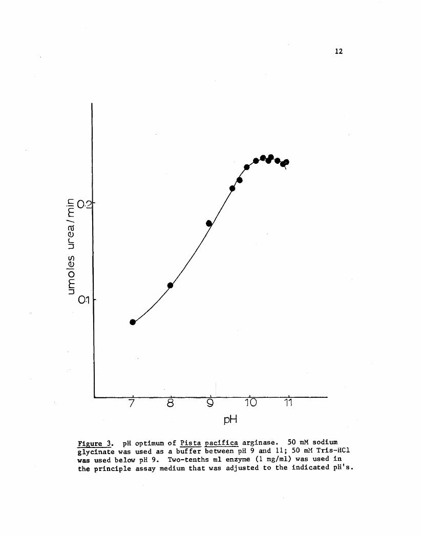
i~pH Optimum , I ~A pH optimum of 10.5 was observed in four different experiments. N
The results of one experiment are shown in Figure 3. Although the opti ~
~ ~ 1mal pH was 10.5 assays were conducted at pH 9.5 in this study so that II
-., ~ I'results could be compared with reported data (Hirsch-Kolb et al., 1970). ~ ~
Effect of Temperature
I I
The effect of temperature on!. pacifica arginase is shown in Fig
ure 4a Figure 5 shows a plot of log V versus liT for temperatures be-
I ~
tween 273 - 333°K. From this graph the energy of activation (E) was cal
culated according to: slope = -E/4.576. Energy of activation was found I to be 8.09 kcal/mole.
I I' 1
12
.c: 0.21 E --. n1 OJ L ::J
(/) <lJ o E ::J
01
'~
7 8 Q 10 11
pH
Figure 3. pH optimum of Pista pacifica arginase. 50 roM sodium glycinate was used as a buffer between pH 9 and 11; 50 Dll.'1 Tris-Hel was used below pH 9. Two-tenths ml enzyme (1 mg/ml) was used in the principle assay medium that was adjusted to the indicated pH's.

x
13
b ~
>
- 1
> Ol o
10
Temperature °C
Figure 4. The effect of temperature on P. pacifica arginase. A plot of temperature versus initial velocity. Two-tenths ml enzyme (1 mg/ml) was used in the assay medium equilibrated to the appropriate temperatures.
(1/Absolute Temperature) 10-3
Figure 5. Plot of log V versus lIT from which the energy of activation was obtained. Two-tenths ml G-200 enzyme (1 mg/ml) was used in the assay medium equilibrated to the appropriate temperatures.

14
Substrate Kinetics
The apparent Michaelis-Henten constant for L-arginine was 155
160 mM in several experiments. As shown in Figure 6 there was no indi
cation of inhibition by high substrate concentrations. The substrate
concentration used for conducting assays on ~. pacifica arginase was
0.85 M. At this concentration 95 - 100% of the calculated maximum velo
city-(Vm) was obtained.
On the same grapb L-ornithine (100 ~~) appears to be a competitive
inhibitor. Hill plots' for both enzyme and enzyme + inhibitor at the
concentrations shown yield n values of 1.034 and 1.040 respectively,
where n equals the slope of 10g(Vm - V~/Vo versus log S (Figure 7).
Effect of Mn2+ Concentration
Mn2+ has been shown to be a metallic cofactor for a number of ar
ginases although variable-effects have been reported. Enzyme incubated
- for 30 min in the principle assay system seemed unaffected by various
Mn2+ concentrations at 25°C and pH 9.5 (Table IV).
Specificity
Agmatine sulfate, D-arginine, L-arginine, argininic acid, L-canava
nine, y-guan1d1nobutyric acid, and ~-guan1dinoprop1onic acid were tested
as potential substrates. Only two were hydrolyzed to urea at an appre
ciable rate, L-arginine and L-canavanine (Table V). The ratio of ar
ginase to canavanase was about 22 at pH 9.5.

x
o o o ~
> ........ r
4 1 11 (Arginine)
o
Figure 6. Kinetic behavior of ~. pacifica arginase. A double reciprocal plot of 11v versus lIS for both enzyme and enzyme + inhibitor. The reaction mixture contained the concentration of arginine indicated + 0.1 ml of G-200 enzyme. e's represent the enzyme kinetics without ornithine; ~s represent enzyme activity in the presence of 0.1 ml 100 w~ ornithine.

16
2·5
o > ~20>
I
E .G Ol o
1·
-1 -2 log (Argi nine)
Figure 7. Hill plot of Pista pacifica arginase kinetics. .'s represent the reaction mixture + enzyme; ~'s represent the reaction mixture + enzyme + inhibitor.

17
TABLE IV
THE EFFECT OF Mn2+ CONCENTRATION ON Pist'a pacifica ARGINASE ACTIVITY
Mn2+ toM umole urea formed/30 min
0 0.312
0.025 0.315
0.05 0.310
0.10 0.315
0.50 0.309
1.0 0.310
2.5 0.312
5.0 0.310
One-tenth ml of enzyme (1 mg/ml) from the final purification step (G-200) was incubated for 30 min in the principle assay system except
2that the Mn + concentration was varied as indicated.
Effect of Sulfhydryl Compounds
Cysteine, dithiothreitol, reduced glutathione, 2-mercaptoethanol
were added to the assay medium, and their effect noted in Table VI. All
compounds and reagents tested had a stimulatory effect on polychaete ar
ginase.
The addition of one umole of p-hydroxymercuribenzoate (p~m), a
sulfhydryl inhibitor, had no effect when the enzyme and PMB were incu
bated for 60 min in the principle assay medium. When p~m was increased
to 5 toM a slight stimulatory effect was noticed.

18
TABLE V
SUBSTRATE SPECIFICITY OF Pista pacifica ARGINASE
Compound umoles urea released/hr pH 7.5 pH 9.5
Agamatine sulfate 0 0
D-arginine 0 0
L-arginine 0.072 0.620
Argininic acid 0 0
L-canavanine 0.014 0.028
~-guanidinobutyric acid 0 0
~-guanidinopropionic acid 0 0
One-tenth ml of G-200 enzyme (1 mg/ml) was incubated for 60 min with each of the listed substrates in the principle assay system. Each of the substrates were adjusted to the indicated pH. Sodium glycinate was used as a buffer at pH 9.5 and 50 roM Tri~-HCI was used at pH 7.5.
Molecular Weight
The molecular weight of Pista pacifica arginase, determined from
the chromatography of the enzyme through Sephadex G-200, was estimated as
200,000 (Figure 8).
Appearance of an Active Subunit
Following heat treatment, acetone fractionation was tried as a pos
sible purification method. One and a half sample volumes of acetone at
-IQOC were slowly added to the heat supernatant. The mixture was centri
fuged at 13,000 x g for 5 minutes at -10°C, and the pellet resuspended in
one volume 50 ~I Tris-HCI containing 100 mM KCI and 50 mM MnCI2 , pH 7.5.

19
TABLE VI
THE EFFECT OF SULFHYDRYL COMPOUNDS ON Pista pacifica ARGINASE
Compound umoles urea produced/hr
None 0.685
Cysteine 1.180
Dithiothreitol 1.365
Reduced glutathione 1.190
2-mercaptoethanol 2.500
Five umoles of each sulfhydryl compound were added to the reaction medium. The reaction was started with 0.1 ml G-200 enzyme (1 mg/ml) and incubated for 60 min. Standards were prepared in the presence of the same amount of sulfhydryl compound used for colorimetric correction.
The solution was then dialyzed for 2 hours against the same buffer. The
sample was then chromatographed on a Sephadex G-200 column (2x150 em)
equilibrated in the same buffer. The enzyme eluted in two peaks, one of
. approximately 200,000 molecular weight and an apparent dissociation pro
duct with a molecular weight about 42,000 (Figure 9).
An active subunit could also be produced when the active fractions
of the G-200 enzyme in the purification scheme of polychaete arginase
were pooled and lyophilized. The lyophilized material was redissolved in
Tris-HCl buffer, pH 7.5, placed on a G-200 column and eluted as before.
Fractions included two activity peaks, one corresponding to the large
molecular weight unit and the other to 34,000. The peak fraction of the
lower molecular weight was rechromatographed on the column and eluted at
15 ml/hr. There was no evidence of aggregation. Arginase activity ap

18
16
".,........
5120 Q)
>
100
""'.........
.... ......80 ......
....... --. Molecular Weight x 104
Figure 8. Estimate of molecular weight of Pista pacifica arginase. .'s from left to right: sperm whale metmyoglobin, chymotrypsinogen A, bovine serum albumin, lactate dehydrogenase, catalase, and blue dextran. ~: Pista pacifica arginase. N
o

21 6«
I ...!.cc T.;:9(!) « ro·- ~ (!)~ IJ)::1+-) roll) 0, >E ..co
uCU ---1 CD 0::1 U c i IJ)
..0 'til 0
j AT~
L +-I
Q) U C C'U ..c
'b 13 «
60 100 140 Vol ume el uted
Figure 9. Breakdown of !. pacifica arginase after acetone fractionation. .'s represent protein concentration measured at 280 nm. ~'s represent arginase activity determined colorimetrically by absorbance at 540 nm. Calibrants were: blue dextran, bovine catalase, bovine serum albumin, lactate dehydrogenase, ovalbumin, ~Chymotrypsinogen A, and sperm whale metmyoglobin.

22
peared in the same fractions as before: molecular weight of 34,000 (Fig
ure 10). Active fractions were pooled and lyophilized.
Polyacrylamide Gel Electrophoresis of Subunit
Lyophilized subunit (2 mg/ml) from the dissociation of the G-200
enzyme, migrated as a single dark band during polyacrylamide gel elec
trophoresis at pH 9.2. Another, lighter band was observed on several
occasions. Whether this band represents an impurity, closely allied
subunit structure, or an artifact of electrophoresis has not yet been
determined.
Michaelis Constant of Subunit
Values of 114 - 118 ~l were obtained for the Km of the subunit ar
ginase on a double reciprocal plot of 1/Vo versus 1/substrate concentra
tions. A Hill plot indicated an n value of 1.07 where n equals the
slope of 10g(Vm - Vd/Vo versus log S (Figure" 11) •
. Effect of Mn2+ Concentrations on the Subunit
The subunit, incubated for 30 min in the principle assay system,
appeared to be unaffected by Mn2+ concentrations (Table VII).

23 I
~<!J 0 ~V) c
(/)061- CU 0..
J ~ +>c omEO) >. ..c u
04· I I
w u z « en 0::
Ao (j) 100en 4::
01
B 50
Va..UME ELUTED
Figure 10. Appearance of subunit on Sephadex G-200. A, breakdown of enzyme after redissolution in buffer following lyophilization. Bt rechromatography of subunit from A showing the absence of aggregation. .'s represent protein concentration measured at 280 nm. ~'s represent arginase activity measured as urea production determined colorimetrically at 540 nm.

(Y)
o ~
x
>-r'"
'6
4
2
o 15 >-.. ~
I
24
-1 -2 log (Arginine)
40 12 1JArginine
Figure 11. Lineweaver-Burke plot of affinity of subunit for substrate. Inset: Hill plot of indicated data. Enzyme source was isolated subunit (1 mg/ml) added to the assay medium.

25
TABLE VII
EFFECT OF Mn2+ CONCENTRATION ON Pista pacifica SUBUNIT ACTIVITY
Mn2+ mM umole urea formed/30 min
0 0.368
0.025 0.368
0.050 0.396
0.100 0.370
0.500 0.365
1.000 0.387
2.500 0.376
5.000 0.370
One-tenth ml of subunit from dissociated G-200 enzyme was incubated in the principle assay medium with the above concentrations of manganous ion.

DISCUSSION
Despite the well established role of arginase in certain oligo
chaetes (Needham, 1957, 1960, 1962; Bishop & Campbell, 1965; Reddy &
Campbell, 1968), few marine annelids have been investigated with regard
to the presence of this enzyme, none with regard to its characteristics
(Hult, 1969). Studies of arginase in oligochaetes have primarily fo
cused on its presence, location, and role in connection with the other
enzymes of the Krebs-Henseleit cycle. Needham (1960) reported that ar
ginase activity in the gut of Lumbricus terrestris was maximal in the
anterior part of the intestine declining to a minimum in the crop-giz
zard region. In addition, he noted an inverse relationship between
activity and the amount of chloragogen tissue. This tissue, possibly
analogous to the vertebrate liver (Laverack, 1963), is characterized by
a bright yellow color. In P. pacifica the occurrence of the tissue con
taining the yellow pigment (chloragogen tissue) is restricted to the
anterior stomach and to the intestine. The anterior stomach exhibits
minimal arginase activity while the intestine has relatively high activ
ity (Table II). A cursory histological examination indicated equal
amounts of chloragogen tissue in both of these anatomical regions so
that in Pista there does not seem to be a correlation, either positively
or inversely, between the amount of chloragogen tissue and arginase
activity.
The possibility that the arginase detected in Pista pacifica tis
sues is of bacterial origin cannot be ruled out. Arginase has been re

27
ported in Escherichia coli, Proteus vulgaris, Staph. aureus, and Bacil
lis stabilis (Khramov &Galaev, 1971). Unfortunately, no molecu~ar
weights or Michaelis constants are available for comparison. Fluctua
tions in bacterial arginase could explain the large variations observed
in tissue activity (Tables II & III) though such variations could also
be due to the worm's arginase responding to feeding activities, metabol
ic functions, or tidal patterns. Worms could only be maintained three
days in sterile sea water without their tubes which may have been insuf
ficient to notice a significant difference in activity from fresh worms.
The characteristics of arginase have undergone extensive evalua
tion in the past few years, in particular, their relationship to a spe
cies' mode of nitrogen excretion. A comparison of !. pacifica arginase
to the enzymes of ureoteles and uricoteles summarized in Table VIII
shows some similarities and some differences.
The molecular weights of arginase from uricotelic species have
been found to be around 225,000 - 280,000 (Mora ~ al., 1965; Campbell,
1966; Reddy & Campbell, 1969a). Based on the probable subunit structure
of 30,000, molecular weights in this range have been referred to as oct
amers (Reddy & Campbell, 1970). The value of 200,000 reported here for
Pista pacifica arginase although it falls outside of this range probably
represents an octamer. No heptameric arginases have been reported nor
does it seem likely that they will be found as proteins containing an
odd number of subunits are rare (Klotz, 1967). liowever, the estimation
on gel filtration should be regarded as tentative until supplementary
data such as ultracentrifugal studies on completely purified arginase
are done. The difference in molecular weights of native Pista pacific~

TABLE VIII
CHARACTERISTICS OF ARGINASE IN RELATION TO NITROTELISM*
Nitrotelism Km Molecular Stability Arginase/ Inhibition and mM weight during canavanase ------....-------- Reference
source x 1000 dialysis PCMB Arginine
Ammonoteles crayfish body wall 2 225 + 3 + + Hartenstein, 1972 Pista pacifica 155 200 + 22 Present study axolotl liver 16 + Mora et al., 1965 earthworm 27 Reddy & Campbell,
1968 Ureoteles rat liver 10-40 118 + 1.5 ± Hora et al., 1966,
1967; Hirsch-Kolb & Greenberg,- 1968
beef liver 2.7 + 15 + Brown, 1966
Uricoteles chicken liver 100 276 7 + Mora et al., 1966 gull liver 2 134 + + Brown, 1966; Reddy
& Campbell, 1970 lizard liver 100-200 276 Mora et al., 1965 Helix aspersa 3.9 244 10 Campbell, 1966;
Reddy & Campbell, 1970; Baret et al., 1972
silkmoth 2.5 228 24 + Reddy & Campbell, 1969a
* table adapted from Hartenstein (1972rN 00

29
arginase (200,000) and that of Lumbricus terrestris (30,000) is notable
though unexplained at this time.
Molecular weights of 120,000 (tetramers) have been reported for
ureoteles (Greenberg ~ al., 1956; ~fora et al., 1966; Hirsch-Kolb et al.,
1970). Hirsch-Kolb & Greenberg (1968) have found the molecular weight
of rat liver arginase to be 118,000. The enzyme is composed of four
subunits each of 30,800 molecular weight. When isolated the molecular
weight of !. pacifica subunit on gel filtration studies is approximately
34,000, which is in close agreement with the rat liver subunit and the
monomeric arginase of the earthworm (Reddy & Campbell, 1968).
The molecular.weight of the subunit following acetone treatment
was approximately 42,000 (Figure 9) although the two activity peaks were
not completely separated as they were with the lyophilized subunit.
This could represent an association-dissociation equilibrium generated
by the harsh treatment. Harsh treatment may also account for the active
monomer of rabbit liver arginase. When purified at low pH's the native
arginase of 118,000 molecular weight breaks down to a subunit of 36,000
(Rogers &Moore, 1963; Vielle-Breitburd & Orth, 1972) •.
The high Km value obtained for polychaete arginase is in the range
of the values (> 100 ~~) found for chicken liver and lizard liver, both
uricoteles (Mora ~ al., 1965, 1966). As with chiCken liver, high con
centrations of arginine did not inhibit the enzyme. Competitive inhibi
tion by L-ornithine is characteristic of chicken liver, Neurospora
crassa (Mora et al., 1966), silkmoth arginases (Reddy & Campbell,
1969a), and Pista pacifica arginase.
Sulfhydryl compounds have had a variable effect on reported argin

30
ases. Glutathione has had a stimulatory effect on arginase on several
occasions (Campbell, 1966). Glutathione, dithiothreitol, 2-mercaptoeth
anol, and cysteine have a stimulatory effect on both bullfrog arginase
and bovine arginase (Carlisky & Sadnik, 1972). The same effect was ob
served with Pista. On the other hand these compounds were slightly in
hibitory in the case of insects (Reddy & Campbell, 1969a). The activity
toward L-arginine is inhibited by p-chloromercuribenzoate in the cray
fish (Hartenstein, 1971), chicken liver (Hora.!!. al., 1966), cockroach
fat body (Reddy & Campbell, 1969a), and bullfrog kidney and bovine liver
(Carlisky & Sadnik, 1972). Rat liver O'Iora et a!., 1966) and silkmoth
are unaffected (Reddy & Campbell, 1969a). Pista pacifica arginase is
unaffected by p-hydroxymercuribenzoate, the sodium salt of the free acid
form. (The "chloro" converts to "hydroxy" when the sodium salt is pre
pared). Effects of sulfhydryl reagents on arginase are quite variable.
This may indicate differences in enzyme structure or it may reflect ex
perimental conditions.
The arginases present in the livers of uricotelic and ureotelic
animals have a similar substrate specificity. All hydrolyze L-arginine
and to some extent L-canavanine (Mora.!!. al., 1965). Helix pomatia and
Helix aspersa also hydrolyze homoarginine (Baret et al., 1972). Argin
ase/canavanase ratios at pH 9 - 10 for reported species have no apparent
pattern. High ratios (> 10) as that obtained for Pista arginase have
been reported for rat liver (Mora et al., 1966), beef liver (Brown,
1966), and silkmoth (Reddy & Campbell, 1969a).
Few temperature optima have been reported. The arginase of the
sockeye salmon has a temperature optimum of 45°C (Cvancara, 1971),

31
Helix pomatia and Helix aspersa between 60 - 65°C (Baret!:! a1., 1972),
and horse liver arginase has a temperature optimum of 45 - 50°C (Green
berg &Mohamed, 1945). The temperature optimum of Pista pacifica is
approximately 60°C.
The majority of arginases appear to be activated by Mn2+. In these
studies P. pacifica does not exhibit this characteristic. This does not
prove that Mn2+ is not required by the enzyme. The divalent cation may
be tightly bound to the enzyme and thus be present in adequate amounts
even in the absence of added }m2+. The lack of activation of the native
enzyme is also observed with the subunit. Hirsch-Ko1b et a1., (1970)
reported that four molecules of manganese are bound to one molecule of
fully activated rat arginase with different binding affinities. ' Two are
loosely bound and two molecules of manganese are tightly bound. The re
moval of the two loosely bound ions results' in a 50% loss of activity
which can be reversed by an incubation at 55°C with t1nC1 • When the two2
tightly bound ions are removed irreversible inactivation occurs. It may
be that manganous ions are tightly bound to the arginase of P. pacifica.
This may explain the lack of activation. On the other hand, it may be
that Mn2+ is not required for enzyme activity or perhaps another diva
lent metal ion would be more effective. Experiments should be carried
2+out in EDTA (ethy1enediaminetetraacetic acid) to determine whether Mn
was required or not.
In the search to categorize arginases with respect to modes of ni
trogen excretion, reports on arginases of ureo-, ammono-, and uricote1es
served more to point out the disparities rather than the similarities
among arginase properties. Terms such as "ammonoureote1es" and "urico

32
teles with ureotelic ancestors" have been used to obscure differences
and to maintain the theory of a distinction between arginases from
ureotelic and uricotelic organisms. The information presented in this
paper on an arginase from a terebellid worm, together with other recent
data (Reddy & Campbell, 1970; Hartenstein, 1971) contradicts this theory
and points out that none of the characteristics of arginase observed to
date can be correlated with a particular mode of nitrotelism.
Perhaps a better way of distinguishing between arginases would be
in terms of their function. A physiological function of the liver ar
ginase is to form urea, a role which is clear from its participation in
the mUlti-enzyme ornithine-urea cycle. Other roles for the enzyme, es
pecially in nonhepatic tissues and in tissues where the other enzymes of
the urea cycle have not been demonstrated, would include catalysis of
the first step in the conversion of arginine to proline, a conversion
known to occur in the udder of goats (~tepham &Linzell, 1966), the mam
mary gland of rats (Yip &Knox, 1972), and the fat body of insects
(Reddy & Campbell, 1969b). Another functional arginase may serve to
catalyze the breakdown of ingested arginine or arginine obtained from
protein hydrolysis (Canedo et al., 1967; Soberon et al., 1967), as for
example, via lysozymes (Tabor &TabQr, 1969). Whether!. pacifica ar
ginase functions as an excretion arginase, a catabolic arginase, as a
precursor arginase, or in a manner as yet unkown has not been empirically
determined.
Although all of the ornithine cycle enzymes have not been assayed
for in this animal, ornithine transcarbamylase has been detected in the
intestine according to the method of Brown & Cohen (1959). Such activi

33
ty is suggestive of a functional ornithine-urea cycle. However, several
attempts to detect urea have been unsuccessful. A search for urease was
also unsuccessful. If the arginase is not functioning to produce urea,
perhaps it acts as a dietary arginase, or for the production of orni
thine as a precursor of proline, or in conjunction with arginine phospha
gens.
The appearance of an enzymatically active subunit is significant
in comparison with the earthworm arginase and in possible terms of regu
lation. As previously stated, Lumbricus terrestris has a monomeric ar
ginase of 27,000 molecular weight which is active in a functional urea
cycle (Bishop & Campbell, 1965; Reddy & Campbell, 1968). Apparently a
monomeric unit is sufficient for this catalysis but it raises the ques
tion of why the marine annelid developed an oligomeric arginase while
retaining an active subunit, a subunit which is capable of catalyzing
the reaction with a greater affinity for substrate than the intact pro~
tein. Unless, an arrangement of subunits is capable of regulating the
appearance of product. Such regulation usually appears as an atypical
plot (sigmoid curve) of substrate versus velocity and a Hill plot n
value greater than 1. The n value of 1.034 for f. pacifica arginase
would not indicate such regulation under the stated assay conditions.
Possibly the enzyme has been "desensitized" (Monod et al., 1963) during
purification procedures. Several research teams (Hochachka &Mostafa,
1973; Gerhart & Pardee, 1962) have noticed that the kinetics of the
reaction catalyzed by a desensitized enzyme obey the Michaelis-Henten
relation (hyperbolic curve) while in the presence of native enzyme the
rate-concentration curve is sigmoid. Another possibility is that the

34
regulation requires certain small molecules for activation. Cabello
(1967) has indicated that arginase modified by sodium dodecyl sulfate
exhibits a sigmoidal curve as a function of arginine concentration. He
also suggested that natural analogues of dodecyl sulfate, such as salts
of fatty acids, may modify the properties of arginase in vivo.
However, the existence of subunits is not a necessary indication
of regulation. Subunits provide stability, the conservation of DNA, and
lessen the chance of a lethal mutation; many nonregulatory enzymes are
composed of several subunits (Monod, 1969).

REFERENCES
Archibald, R.M. (1945) Colorimetric determination of urea. J. BioI. Chem. 157, 507-518.
Baret, R., Girard, C. and Riou, J. (1972) Sur certaines proprietes des arginases du tissu hepatopancreatique d'Helix pomatia Lin. et d'Helix aspersa Mull. Biochimie 54, 421-430.
Bishop, S.R. and Campbell, J.W. (1965) Arginine and urea biosynthesis in the earthworm Lumbricus terrestris. Comp. Biochem. Physiol. 15, 51-71.
Brown, G.W., Jr. and Cohen, P.R. (1959) Comparative biochemistry of urea synthesis I. Methods for the quantitative assay of urea cycle enzymes in liver. r. BioI. Chern. 234, 1769-1774.
Brown, G.W., Jr. (1966) Studies in comparative biochemistry and evolution. Archs. Biochem. Biophys. 114, 184-194.
Cabello, J. (1967) Discussion: enzymatic aspects of metabolic regulation. u.!. Nat. Cancer Inst. Honograph 27, 297-298.
Campbell, J.W. (1966) A comparative study of molluscan and mammalian arginases. Comp. Biochem. Physiol. 18, 179-199.
Canedo, L., Martuscelli, J. and Mora, J. (1967) Catabolism of L-arginine in Neurospora crassa. Q.~. Nat. Cancer Inst. Monograph 27, 273-283.
Carlisky, N.J. and Sadnik, I.L. (1972) Properties of amphibian renal arginase I. The effect of dialysis and sulfhydryl compounds. Comp. Biochem. Physiol. 41B, 785-792.
Clementi, A. (1937) L'arginase epatica I. Suoi rapporti con a genesi dell'urea durante l'autolisi del fegato nella serie de! vertebrati. Enzymologia 4, 205-216.
Cvancara, V.A. (1971) Liver arginase activity in the sockeye salmon, Oncorhynchus nerka. Comp. Biochem. Physiol. 40B, 819-822.
Davis, B.J. (1964) Disc electrophoresis, methods and applications to human serum proteins. Ann. li.!. Acad. Sci. 121, 404-427.
Fawcett, J.K. and Scott, J.E. (1960) A rapid and precise method for the determination of urea. J. Clin. Path. 13, 156-159.
Gerhart, J.C. and Pardee, G.B. (1962) The enzymology of control by feedback inhibition. ~. BioI. Chern. 237, 891-896.

36
Greenberg, D.M. and Mohamed M.S. (1945) Liver arginase II. Kinetic properties. Archs. Biochem. 8, 365-375.
Greenberg, D.M., Bagot, A.E. and Roholt, O.A., Jr. (1956) Liver arginase III. Properties of highly purified arginase. Archs. Biochem. Biophys. 62, 446-453.
Greenberg, D.M. (1960) Arginase. (in) The Enzymes (edited by Boyer, P.:, Lardy, H. and Myrback, K.) 2nd edn, vol. 4, 257-267. Academic Press, New York.
Hartenstein, R. (1971) Characteristics of arginase from the freshwater crayfish Cambarus bartoni. Comp. Biochem. Physiol. 40B, 781-795.
Hirsch-Kolb, H. and Greenberg, D.M. (1968) Molecular characteristics of rat liver arginase. l. BioI. Chem. 243, 6123-6129.
Hirsch-Kolb, H., Heine, J.P., Kolb, H.J. and Greenberg, D.M. (1970) Comparative physical-chemical studies of mammalian arginases. Comp. Biochem. Physiol. 37, 345-359.
Hirsch-Kolb, H., Kolb, H.J. and Greenberg, D.M. (1971) Nuclear magnetic resonance studies of manganese binding of rat liver arhinase. J. BioI. Chem. 246, 395-401.
Hochachka, P.W. and Mustafa, T. (1973) Enzymes in facultative anaerobis of molluscs I. Malic enzyme of oyster adductor muscle. Comp. Biochem. Physiol. 45B, 625,637.
Hult, J.E. (1969) Nitrogenous waste products and excretory enzymes in the marine polychaete Cirriformia spirabranchia (Moore). Comp. Biochem. Physiol. 31, 15-24.
Khramov, V.A. and Galaev, Y.V. (1971) Rasprostranenie arginazy u mikrobov. Lab. Delo. 1, 50-53.
Klotz, I.M. (1967) Protein subunits: a table. Science 155, 697-698.
Laverack, M.S. (1963) The Physiology of Earthworms. Macmillan, New York.
Linn, S. and Lehmann, I.R. (1965) An endonuclease from Neurospora crassa specific for polynucleotides lacking an ordered structure. ~. BioI. Chem. 240, 1287-1293.
Lowry, O.H., Rosebrough, N.J., Fair, A.L. and Randall, R.J. (1951) Protein measurement with the Folin phenol reagent. J. BioI. Chem. 193, 265-275. - -- --
Mepham, T.B. and Linzell, J.L. (1966) A quantitative assessment of the contribution of individual plasma amino acids to the synthesis of milk proteins by the goat mamary gland. Biochem. l. 101, 76-83.

37
Mlddlehoven, W.J. (1969) The ferrous ion as the cofactor of arginase in vivo I. Properties of yeast arginase metallocomplexes of known co~ position and of native arginase. BioChem. Biophys. Acta 118, 206209. .
Monod, J., Changeux, J.P. and Jacob, F. (1963) Allosteric proteins and cellular control systems. ~. Mol. Biol. 6, 306-329.
l'lonod, J. (1969) On symmetry and function in biological systems. (in) 11th Nobel Symposium (edited by Engstrom, A. and Stranberg, B.) 1527. Wiley Interscience Press, New York.
Mora, J., Tarrab, R., Martuscelli, J. and Sober6n, G. (1965) Characteristics of arginases from ureotelic and non-ureotelic animals. Biochem. ~. 96, 588-594.
Mora, J., Tarrab, R. and Bojalil, L.F. (1966) On the structure and function of different arginases. Biochem. Biophys. Acta 118, 206209.
Needham, A.E. (1957) Components of nitrogenous excreta in the earthworms Lumbricus terrestris L. and Eisenia foetida (Savigny). J. Exp. BioI. 34, 425-446. -
Needham, A.E. (1960) The arginase activity of the tissues of the earth, worms Lumbricus terrestris L. and Eisenia foetida (Savigny). J.
Exp. BioI. 37, 775-782. -
Needham, A.E. (1962) Distribution of arginase activity along the body of earthworms. Comp. Biochem. Physiol. 5, 69-82.
Reddy, S.R.R. and Campbell, J.W. (1968) A low molecular weight arginase in the earthworm. Biochem. Biophys. Acta 159, 557-560.
Reddy, S.R.R. and Campbell, J.W. (1969a) Arginine metabolism in insects: properties of insect fat body arginase. Comp. Biochem. Physiol. 28, 515-534.
Reddy, S.R.R. and Campbell, J.W. (1969b) Arginine metabolism in insects: role of arginase in proline formation during silkmoth development. Biochem. l. 115, 495-503.
Reddy, S.R.R. and Campbell, J.W. (1970) Molecular weights of arginase from different species. Comp. Biochem. Physiol. 32, 499-509.
Rogers, S. and Moore, M. (1963) Studies of the mechanism of action of the Shope rabbit papilloma virus I. Concerning the nature of the induction of arginase in the infected cells. ~. ~. Med. 117, 521545.

38
Rossi, N. and Grazi, G. (1969) Characterization of a new type of arginase from chicken liver. Eur. ~. Biochem. 7, 348-352.
Schimke, R.T. (1962) Adaptive characteristics of urea cycle enzymes in the rat. I. ~. Chern. 237, 459-468.
SoberOn, G., Ortiz-Pineda, J. and Tarrab, R. (1967) Characteristics of the ureotelic arginase and its role in the advent of ureotelism during metamorphosis of the Mexican axolotl. u.!. Nat. Cancer lnst. Monograph 27, 283-295.
Soru, E. (1965) Purification of bacterial arginase. J. Chromatography 20, 325-333.
Tabor, H. and Tabor, C.W. (1969) Partial separation of two pools of arginine in E. coli; preferential use of exogenous rather than endogenous arginine for the biosynthesis of 1,4 diamino butane. l. BioI. Chem. 244, 6383-6387.
Ternberg, J.L. and Hershey, F.B. (1964) Colorimetric determination of blood ammonia. ~. Lab. Clin. Med. 56, 766-776.
Vielle-Breitburd, F. and Orth, G. (1972) Rabbit liver L-arginase: purification, properties, and subunit structure. J. BioI. Chern. 247, 1227-1235. - -- -
Yip, M.C.M. and Knox, W.E. (1972) Function of arginase in lactating mammary gland. Biochem. l. 127, 893-899.