Embed Size (px)
Citation preview
Gut, 1990, 31, 294-299
Characterisation of the effector cells responsible forthe in vitro cytotoxicity of blood leucocytes fromaphthous ulcer patients for oral epithelial cells
D W Thomas, J Bagg, D M Walker
AbstractThis study was designed to identify the cellsresponsible for the spontaneous cell mediatedcytotoxic effect (SCMC) exerted by peripheralblood leucocytes from patients with recurrentaphthous ulceration, towards cultured oralepithelial cells. Peripheral blood leucocytesfrom recurrent aphthous ulceration patientsexerted a significantly greater (p<001) degreeof cytotoxicity towards the oral epithelialtarget cells than did peripheral blood leuco-cytes from healthy control subjects, orfrom patients with non-specific ulceration.Depletion of CD-5 positive cells (T-lympho-cytes) resulted in a significant decrease in theSCMC in aphthous patients. Depletion ofCD-16 positive cells (NK-cells) produced nosignificant change in cytotoxicity. T-lympho-cytes, therefore, appear to be intimatelyinvolved in the in vitro SCMC effect in recur-rent aphthous ulceration.
Department of OralSurgery, Medicine andPathology, DentalSchool, University ofWales College ofMedicine, Cardiff, SouthWalesD W ThomasJ BaggD M WalkerCorrespondence to:Dr J Bagg, Dept of OralSurgery, Medicine andPathology, Dental School,University of Wales College ofMedicine, Cardiff, S WalesAccepted for publication23 May 1989
Recurrent aphthous ulceration' is a form of focaloral ulceration, which affects up to 20% ofthe population. Although recurrent aphthousulceration may rarely be a marker of underlyingsystemic illness such as coeliac disease, or presentas a feature of Behcet's syndrome, in most cases
no additional body systems are affected, andpatients remain otherwise fit and well.An immunopathogenesis for recurrent aph-
thous ulceration was first suggested by Lehner,'who likened the histological appearance of aph-thous ulceration, with its early mononuclear cellinfiltrate, to that of a type IV hypersensitivityreaction. The finding of significant lympho-cytotoxicity in other immunologically mediateddiseases3 prompted Dolby4 to show that peri-pheral blood leucocytes from aphthous patientsexerted a significantly enhanced cytotoxicityin vitro, for allogeneic gingival epithelial cells.This finding has been substantiated by otherworkers.56 Greenspan et al7 and Burnett andWray,8 however, found that the increased cyto-toxicity was antibody dependent, presumablymediated by K orNK cells.
Suppressor/cytotoxic T cells, T helper cellsand NK cells have been identified in the infiltratein aphthae, in different proportions varying withthe duration of the lesion.9 Despite much specu-lation, however, the effector cells mediatingthe tissue damage seen in recurrent aphthousulceration remain unidentified.The aims of the present study were three-fold.
Firstly, the cytotoxic effect of peripheral bloodleucocytes from recurrent aphthous ulcerationsufferers on epithelial target cells derived from a
novel source, namely, actively growing longtermcultures of oral epithelium, was investigated.Secondly, the specificity of the raised SCMC inrecurrent aphthous ulceration was examined bydetermining the cytotoxic effect of peripheralblood leucocytes from patients with other typesof mouth ulcers. This part of the study wasdesigned to show whether or not the previouslydescribed cytotoxic process in recurrent aph-thous ulceration was merely an epiphenomenonrelated to oral epithelial cell breakdown. Finally,tentative identification of the cytotoxic effectorcell in our in vitro system was attempted bystudying the effect on cytotoxicity of leucocytesubset depletion.
Methods
PATIENTS WITH RECURRENT APHTHOUSULCERATIONThirty two patients were studied, each sufferingfrom minor aphthous ulceration, diagnosedaccording to the criteria of Lehner,'° of recurrentcrops of one to five small painful ulcers affectingthe non-keratinised oral mucosa and healingwithin four to 14 days, without scarring or asystemic mucocutaneous disorder. Seventeen ofthe patients were men and 15 were women, agedfrom 16 to 56 years (mean 30 7). Seventeen ofthepatients had ulcers at the time of study whilst theremainder were ulcer free (remission), thoughall patients were experiencing regular bouts ofulceration. All of the patients were haemato-logically normal and only two were tobaccosmokers.
DISEASED CONTROLSWe also investigated as diseased controls sixpatients with other types of active mouth ulcera-tion, due to trauma (four patients), erythemamultiforme (one) and erosive lichen planus (one)with ages ranging from 27 to 63 years (mean age44-6 years).
HEALTHY CONTROL SUBJECTSFor each aphthous or diseased control patient, anage and sex matched healthy volunteer wasstudied in the same experiment.
COLLECTION OF BLOOD AND SEPARATIONOF PERIPHERAL BLOOD MONONUCLEARCELLSTwenty millilitres ofvenous blood were collectedfrom the antecubital fossa of aphthous and
294
on 26 May 2018 by guest. P
rotected by copyright.http://gut.bm
j.com/
Gut: first published as 10.1136/gut.31.3.294 on 1 M
arch 1990. Dow
nloaded from
Characterisation ofthe effector cells responsiblefor the in vitro cytotoxicity ofblood leucocytesfrom aphthous ulcerpatientsfor oral epithelial cells
control patients into heparinised tubes (Vacu-tainer Systems UK, Becton Dickinson). Theperipheral blood mononuclear cells were separa-ted by density gradient centrifugation over FicollHypaque." The leucocytes were washed twicein phosphate buffered saline (PBS) and theirconcentration adjusted to 3 x 061/ml in growthmedium, before use in the cytotoxicity assay.
CULTURE OF ORAL EPITHELIAL CELLSOral mucosa was harvested from patients under-going routine minor oral surgical procedures.Within one to four hours after collection, thetissue was transported to the laboratory inDulbecco's modified Eagle's medium (lOx) con-taining fetal calf serum (10%), penicillin/strepto-mycin mixture, 1000 IU/ml and amphotericin,5000 IU/ml.Most of the underlying connective tissue was
removed and the epithelium and remainingconnective tissue were minced finely underaseptic conditions using Iris scissors in a laminarair flow cabinet. These diced explants wereresuspended in a growth medium identical withtransport medium, but with added hydrocorti-sone (1 mg/ml) and cholera toxin (0-002 mg/ml).The explants were seeded out in tissue cultureflasks containing a monolayer of 3T3 fibroblasts,which acted as a feeder layer. 12 Division ofthe 3T3 cells was halted by treatment withmitomycin C 12-24 hours before culturing theepithelial explants. The cultures were main-tained at 37°C in a 5% CO2 atmosphere, remov-ing any non-adherent explants and changing themedium after 72 hours and thereafter twiceweekly.
CHARACTERISATION OF TARGET CELLSThe cultured cells were characterised as epithelialcells by three independent methods:
Scanning electron microscopyCultured cells were fixed in 3% glutaraldehyde at4°C in Sorenson's buffer, dehydrated throughgraded alcohols, and critical point dried fromArklone (ICI plc, Runcorn, Cheshire, UK).They were mounted on stubs and sputter-coatedwith gold (thickness 48 nm), before examinationin an ISI Super III scanning electron microscope.
Indirect immunofluorescenceTwenty microlitres of pemphigus serum diluted1: 50 in PBS were added to cultured oral epithelialcells suspended in a minimal volume of PBS.After 45-60 minutes incubation at ambient tem-perature, the cells were washed by centrifugingat 3000 rpm in PBS and reincubated for a further45 minutes with FITC-labelled antihumanimmunoglobulin (Dakopatts Limited, HighWycombe, Buckinghamshire, UK).
Immunoperoxidase stainingSmear preparations of the epithelial cultures,with fibroblasts as negative controls, were fixedin acetone for five minutes at 4°C. All subsequent
incubations were carried out at ambient tem-perature and separated by three washes in PBS.The preparations were treated with 3% hydrogenperoxide in methanol for five minutes to blockendogenous peroxidase, followed by incubationwith normal swine serum for 20 minutes. Theslides were then treated sequentially with theprimary rabbit antihuman keratin antibody (20min), sheep antirabbit immunoglobulin (20 min)and peroxidase-rabbit antiperoxidase (20 min)(all from Orthodiagnostic Systems Inc, Raritan,New Jersey). Finally, the slides were incubatedfor 40 minutes with diaminobenzidine'3 tovisualise any labelled antigens.
CYTOTOXICITY ASSAYThe primary cultures of oral epithelial cells wereharvested by dispersion with a mixture of trypsin(0 05%) and EDTA (0 02%) for four to fiveminutes, or until the cells lost their adherence tothe flask. Trypsinisation was halted by theaddition of Dulbecco's modified Eagle's mediumcontaining 20% fetal calf serum. The cells werethen washed twice in medium and resuspendedin growth medium at a concentration of 7 5 x 10⁴cells/ml. Only preparations in which at least 80%of the epithelial cells were viable were used, andthe same preparation of target cells was used forboth control and experimental groups in any onerun. One hundred microlitres of the leucocytesuspension were added to 20 p.1 of the epithelialcells, thus providing a leucocyte: epithelial cellratio of 200:1. At this ratio, the largest differencein cytotoxicity between aphthous and controlleucocytes could be demonstrated (unpublisheddata). As a control, 100 p.1 of growth mediumalone was added to 20 ,u1 of the epithelial cellsuspension. Triplicate cultures were incubatedat 37°C in a humidified incubator with 5% CO2 inair for 17 hours. The cells were sedimented bycentrifugation, 80 R1 of the supernatant re-moved, and 40 pt1 of ethidium bromide andacridine orange added to the remaining 40 pt1 ofcell suspension. The viability of 200 of theepithelial cells per culture was assessed blind bya single examiner in coded cultures by directultraviolet microscopy.
LEUCOCYTE SUBSET DEPLETIONLeucocyte subset depletion was effected bycomplement dependent lysis of the peripheralblood leucocyte preparations with the mono-clonal antibodies anti Leu-l and anti Leu-1 1(Becton Dickinson ImmunocytochemistrySystems, Mountain View, California, 94039).Anti Leu-l is specific for peripheral blood leuco-cytes expressing the CD 5 antigen (T-lympho-cytes) while anti Leu- 1 recognises the CD16antigen (natural killer (NK) cells, and killer (K)cells).
Fifty microlitre aliquots of the leucocyte sus-pension were incubated with 500 p.1 of a 1:100dilution of the appropriate monoclonal antibodyfor 50 minutes on ice, and recovered by centri-fugation. Preliminary experiments showed thatcomplement at a dilution of 1:10 gave optimalcell lysis. The leucocytes were then incubated,therefore, with 100 p.1 of a 1:10 dilution of baby
295
on 26 May 2018 by guest. P
rotected by copyright.http://gut.bm
j.com/
Gut: first published as 10.1136/gut.31.3.294 on 1 M
arch 1990. Dow
nloaded from
Thomas, Bagg, Walker
rabbit complement at 37°C for 50 min, pelletedby centrifugation and washed twice in phosphatebuffered saline. The treated leucocytes werespun through 10% fetal calf serum, counted andthe concentration of viable cells adjusted ingrowth medium to the original concentration of3x 106 cells/ml. The viability of all leucocytepreparations after depletion was greater than80%. The peripheral blood leucocytes were thenincubated with the oral epithelial target cells inthe above cytotoxicity assay.
CYTOTOXICITY INDICES AND STATISTICALANALYSISThe data were presented as the mean numberof surviving cultured oral epithelial cells. Inaddition, a modified version of the 'difference incytotoxicity index' (/\ C T)5 was determined:
Number of epithelial Number of epithelialcells surviving in _ cells surviving inpresence of control presence ofaphthousleucocytes leucocytesNumber of epithelial cells surviving in presence
ofmedium alone
Thus, positive values for /\ CT indicate thataphthous leucocytes are more cytotoxic than arethe control leucocytes. A paired analysis ofvariance, the F test, was used to analyse the dataobtained. This allowed an analysis of intergroupand intertest variation.
Results
CHARACTERISATION OF TARGET CELLSScanning electron microscopy confirmed thatthe cultured oral mucosal explants had theultrastructural features of epithelial cells. Thecells formed a well defined monolayer withprominent nuclei. The cells also reacted posi-tively with the rabbit antihuman keratin usingthe immunoperoxidase method and demon-trated well defined positive surface immuno-fluorescence after i'ncubation with the humanpemphigus serum. The control fibroblastsreacted negatively in both these tests.
CYTOXICITY OF BLOOD LEUCOCYTES FROMAPHTHOUS PATIENTS AND CONTROLSUBJECTSThe leucocytes from 32 patients with aphthousulceration were significantly more cytotoxic fororal epithelial cells when compared with leuco-cytes from healthy control individuals (p<0l001)(Fig 1). By paired analysis of variance, thedifference in cytotoxicity between five aphthouspatients and their matched controls was found tobe significantly greater than that within eachtriplicate (p<005).
LEUCOCYTE CYTOTOXICITY AND ACTIVITYOF APHTHOUS ULCERATIONThe leucocyte cytotoxicity (/\ CT) values werepositive in 31/32 patients and were significantlyhigher (p<001) for aphthous patients with
200-
a)
4-
SC.)
a)
a,
40
o
=
'a-a
0.Ca,
C._
a0
C.)
.0
4-
D
zCa,a,
160-
120-
80-
40-
* III:
. . I*I
II
Il1l
S...
* a
p <0-001
A CFigure 1: Mean number ofcultured viable oral epithelial cellsafter incubation with leucocytesfrom aphthous patients (A)and control subjects (C) at an effector:target cell ratio of200:1. The horizontal bars represent the overall mean valuesand corresponding standard deviations.
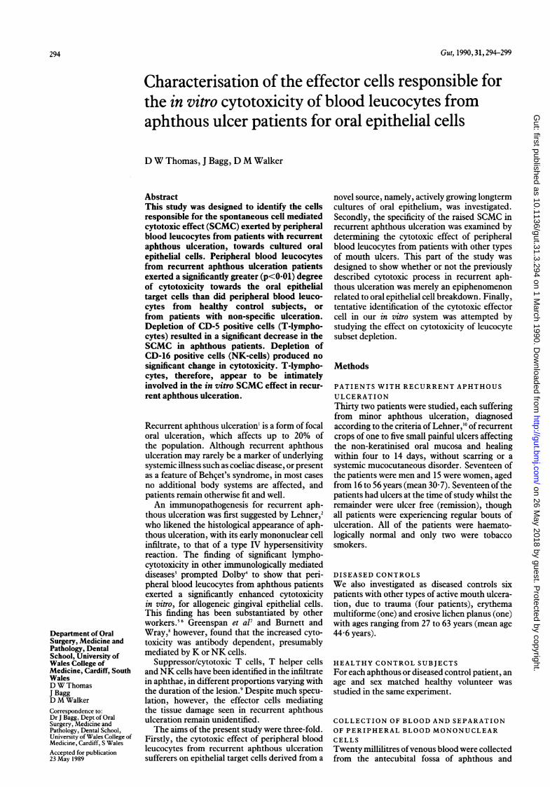
mouth ulcers at the time of sampling comparedwith those of patients in remission (Fig 2).
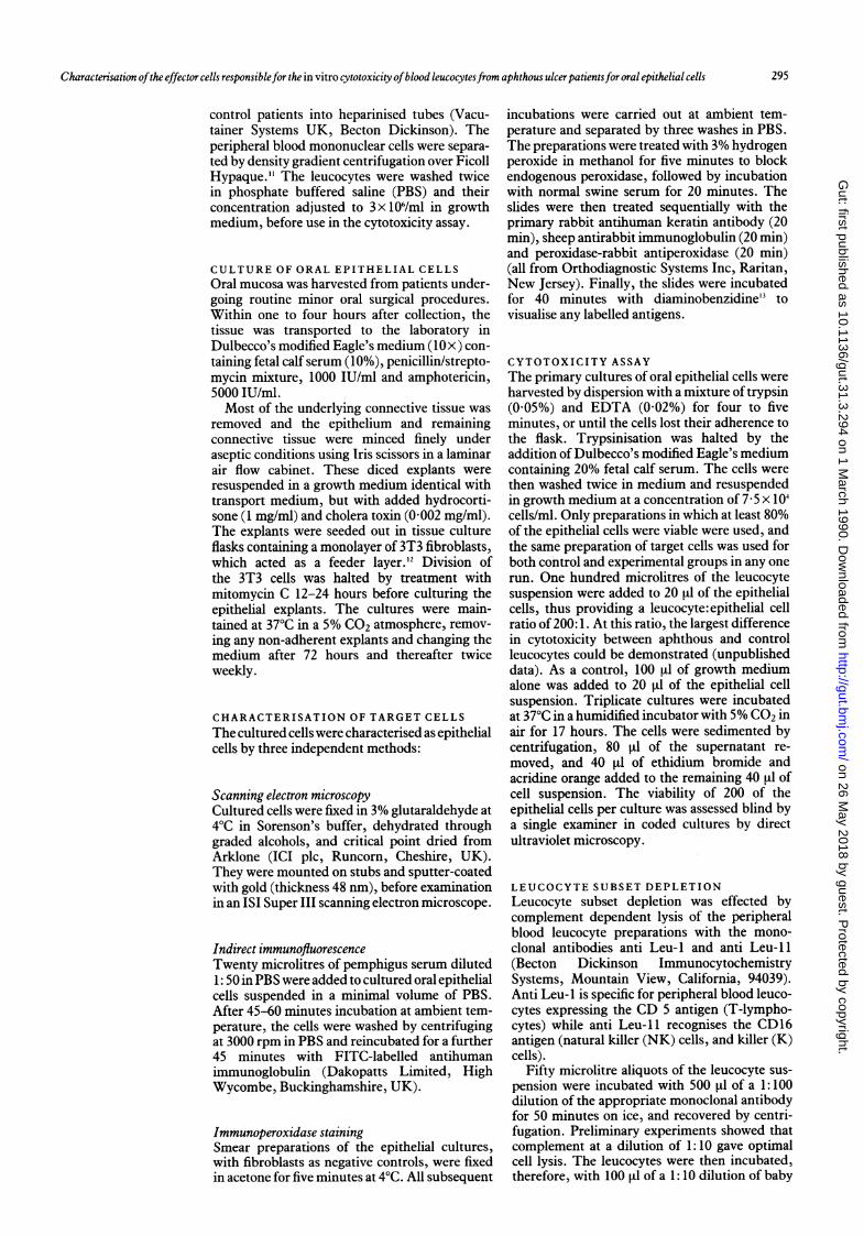
LEUCOCYTE CYTOTOXICITY IN PATIENTSWITH NON-APHTHOUS MOUTH ULCERSIn the six patients with other types of oralulceration there were no statistically significant
0-8-
*
'1x00
UCa,
a,
a,._
0-4-
0-2
* I* I
I: I
II
* IS. .I
*0 I
II
* I* I
*SI
I
.. 1ll
p 0-01
*o
2
A RFigure 2: Mean difference in cytotoxicity values (/\ CT)forleucocytesfrom aphthous patients with active ulceration (A),and aphthous patients in remission (R) towards cultured oralepithelial cells.
m
296
on 26 May 2018 by guest. P
rotected by copyright.http://gut.bm
j.com/
Gut: first published as 10.1136/gut.31.3.294 on 1 M
arch 1990. Dow
nloaded from
297Characterisation ofthe effector cells responsiblefor the in vitro cytotoxicity ofblood leucocytesfrom aphthous ulcerpatientsfor oral epithelial cells
0
*ii
a
-a._;
0s
.X!
0
.-
0z
0
co0cO
200 Non-specific ulceration subjects1 Control subjects
Experiment No
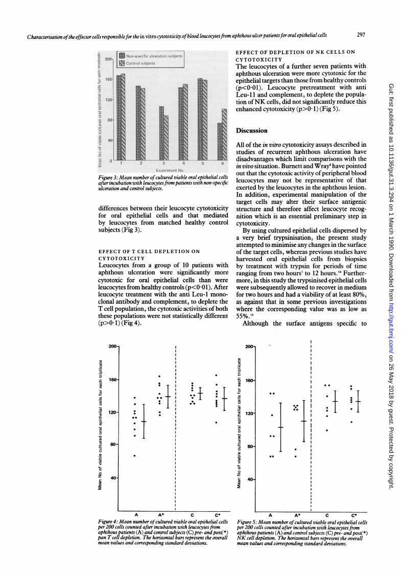
Figure 3: Mean number ofcultured viable oral epithelial cellsafter incubation with leucocytesfrom patients with non-specificulceration and control subjects.
differences between their leucocyte cytotoxicityfor oral epithelial cells and that mediatedby leucocytes from matched healthy controlsubjects (Fig 3).
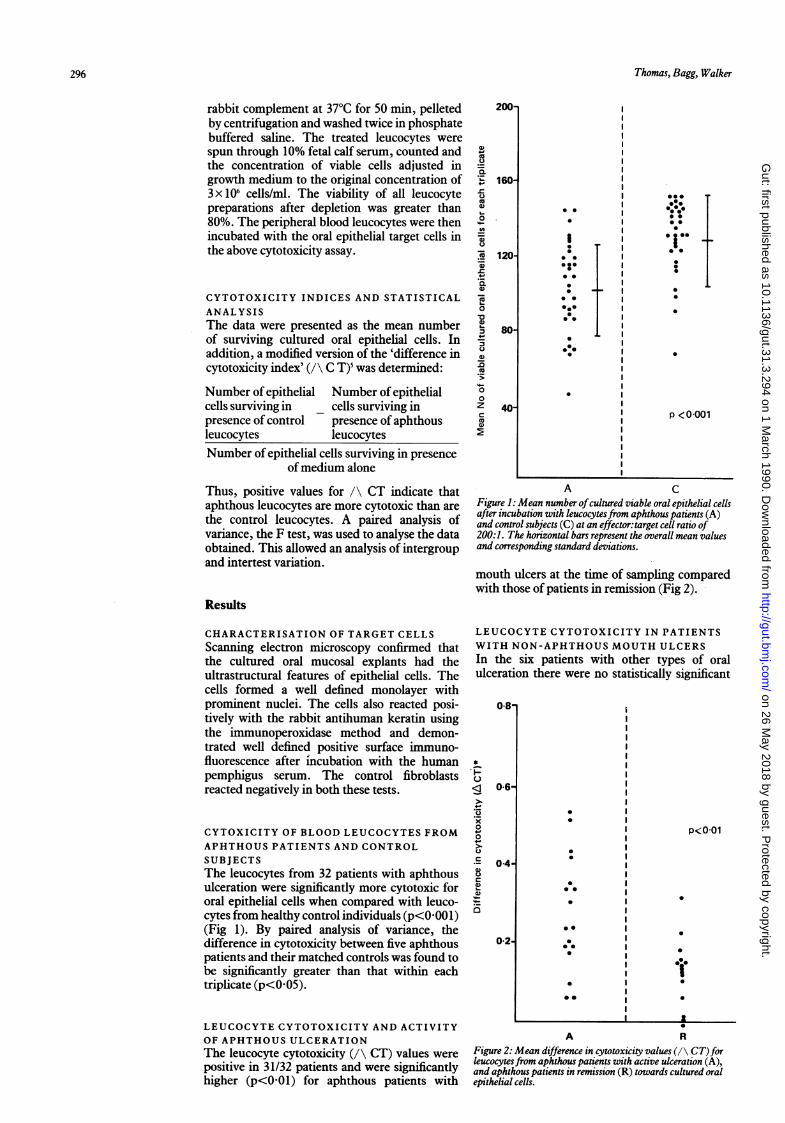
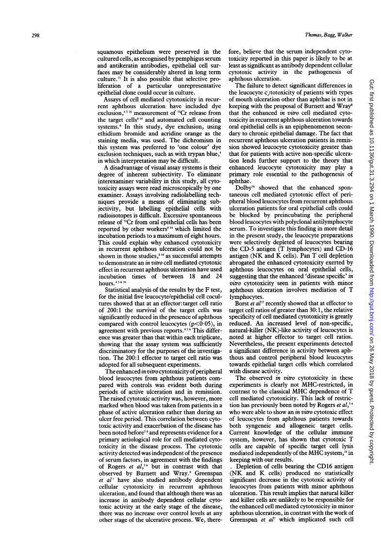
EFFECT OF T CELL DEPLETION ONCYTOTOXICITYLeucocytes from a group of 10 patients withaphthous ulceration were significantly morecytotoxic for oral epithelial cells than wereleucocytes from healthy controls (p<0 01). Afterleucocyte treatment with the anti Leu- 1 mono-clonal antibody and complement, to deplete theT cell population, the cytotoxic activities of boththese populations were not statistically different(p>0. 1) (Fig 4).
200-
41)
co-c
0
az
0
+0
0co
0
.C
160
120-
80-
40.
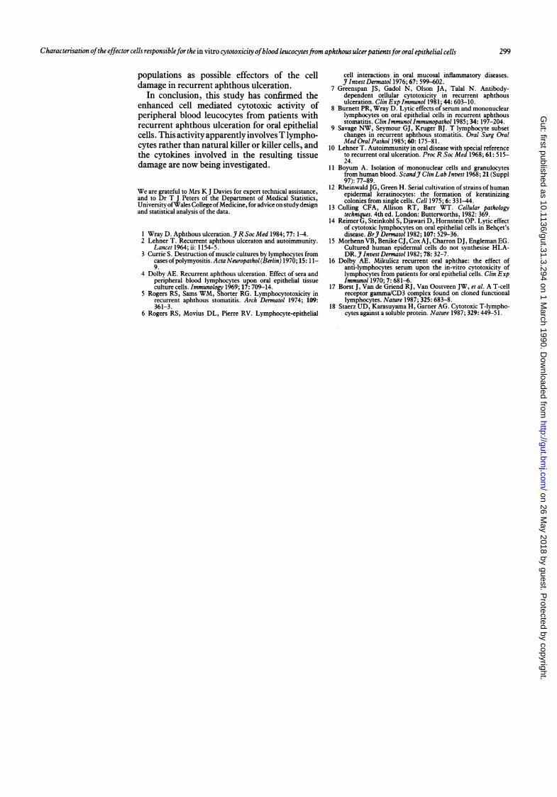
EFFECT OF DEPLETION OF NK CELLS ONCYTOTOXICITYThe leucocytes of a further seven patients withaphthous ulceration were more cytotoxic for theepithelial targets than those from healthy controls(p<001). Leucocyte pretreatment with antiLeu-l1 and complement, to deplete the popula-tion ofNK cells, did not significantly reduce thisenhanced cytotoxicity (p>0. 1) (Fig 5).
Discussion
All of the in vitro cytotoxicity assays described instudies of recurrent aphthous ulceration havedisadvantages which limit comparisons with thein vivo situation. Burnett and Wray8 have pointedout that the cytotoxic activity of peripheral bloodleucocytes may not be representative of thatexerted by the leucocytes in the aphthous lesion.In addition, experimental manipulation of thetarget cells may alter their surface antigenicstructure and therefore affect leucocyte recog-nition which is an essential preliminary step incytotoxicity.By using cultured epithelial cells dispersed by
a very brief trypsinisation, the present studyattempted to minimise any changes in the surfaceof the target cells, whereas previous studies haveharvested oral epithelial cells from biopsiesby treatment with trypsin for periods of timeranging from two hours' to 12 hours.'4 Further-more, in this study the trypsinised epithelial cellswere subsequently allowed to recover in mediumfor two hours and had a viability of at least 80%,as against that in some previous investigationswhere the corresponding value was as low as55%. 14
Although the surface antigens specific to
200-
++ *
0.0
I t
A A* C C*
Figure 4: Mean number ofcultured viable oral epithelial cellsper 200 cells counted after incubation with leucocytes fromaphthous patients (A) and control subjects (C) pre- and post(*)pan T cell depletion. The horizontal bars represent the overallmean values and corresponding standard deviations.
a)
-a0
C0
= 160-
oL-
o
1 1 20-4_a0
zC 40-D
C 0a,
*@ ~~~~~~~~~~~~~~~~I
_I
..I.
* ** ~~~~~~~~~~~~~~~~~~I
i
le
*
. 1 *1
A A* C C*
Figure 5: Mean number ofcultured viable oral epithelial cellsper 200 cells counted after incubation with leucocytes fromaphthous patients (A) and control subjects (C) pre- and post(*)NK cell depletion. The horizontal bars represent the overallmean values and corresponding standard deviations.
0
11
*-0
.
on 26 May 2018 by guest. P
rotected by copyright.http://gut.bm
j.com/
Gut: first published as 10.1136/gut.31.3.294 on 1 M
arch 1990. Dow
nloaded from
Thomas, Bagg, Walker
squamous epithelium were preserved in thecultured cells, as recognised by pemphigus serumand antikeratin antibodies, epithelial cell sur-faces may be considerably altered in long termculture.'5 It is also possible that selective pro-liferation of a particular unrepresentativeepithelial clone could occur in culture.
Assays of cell mediated cytotoxicity in recur-rent aphthous ulceration have included dyeexclusion,' I 16 measurement of 5Cr release fromthe target cells8'4 and automated cell countingsystems.6 In this study, dye exclusion, usingethidium bromide and acridine orange as thestaining media, was used. The dichromism inthis system was preferred to 'one colour' dyeexclusion techniques, such as with trypan blue,4in which interpretation may be difficult.A disadvantage of visual assay systems is their
degree of inherent subjectivity. To eliminateinterexaminer variability in this study, all cyto-toxicity assays were read microscopically by oneexaminer. Assays involving radiolabelling tech-niques provide a means of eliminating sub-jectivity, but labelling epithelial cells withradioisotopes is difficult. Excessive spontaneousrelease of 5Cr from oral epithelial cells has beenreported by other workers8 14 which limited theincubation periods to a maximum of eight hours.This could explain why enhanced cytotoxicityin recurrent aphthous ulceration could not beshown in those studies,8 '4 as successful attemptsto demonstrate an in vitro cell mediated cytotoxiceffect in recurrent aphthous ulceration have usedincubation times of between 18 and 24hours.4566
Statistical analysis of the results by the F test,for the initial five leucocyte/epithelial cell cocul-tures showed that at an effector: target cell ratioof 200:1 the survival of the target cells wassignificantly reduced in the presence ofaphthouscompared with control leucocytes (p<005), inagreement with previous reports.456 This differ-ence was greater than that within each triplicate,showing that the assay system was sufficientlydiscriminatory for the purposes of the investiga-tion. The 200:1 effector to target cell ratio wasadopted for all subsequent experiments.The enhanced in vitro cytotoxicity ofperipheral
blood leucocytes from aphthous patients com-pared with controls was evident both duringperiods of active ulceration and in remission.The raised cytotoxic activity was, however, moremarked when blood was taken from patients in aphase of active ulceration rather than during anulcer free period. This correlation between cyto-toxic activity and exacerbation of the disease hasbeen noted before5 6 and represents evidence for aprimary aetiological role for cell mediated cyto-toxicity in the disease process. The cytotoxicactivity detected was independent ofthe presenceof serum factors, in agreement with the findingsof Rogers et al,56 but in contrast with thatobserved by Burnett and Wray.8 Greenspanet al7 have also studied antibody dependentcellular cytotoxicity in recurrent aphthousulceration, and found that although there was anincrease in antibody dependent cellular cyto-toxic activity at the early stage of the disease,there was no increase over control levels at anyother stage of the ulcerative process. We, there-
fore, believe that the serum independent cyto-toxicity reported in this paper is likely to be atleast as significant as antibody dependent cellularcytotoxic activity in the pathogenesis ofaphthous ulceration.The failure to detect significant differences in
the leucocyte cytotoxicity of patients with typesof mouth ulceration other than aphthae is not inkeeping with the proposal of Burnett and Wray8that the enhanced in vitro cell mediated cyto-toxicity in recurrent aphthous ulceration towardsoral epithelial cells is an epiphenomenon secon-dary to chronic epithelial damage. The fact thatrecurrent aphthous ulceration patients in remis-sion showed leucocyte cytotoxicity greater thanthat for patients with active non-specific ulcera-tion lends further support to the theory thatenhanced leucocyte cytotoxicity may play aprimary role essential to the pathogenesis ofaphthae.
Dolby'6 showed that the enhanced spon-taneous cell mediated cytotoxic effect of peri-pheral blood leucocytes from recurrent aphthousulceration patients for oral epithelial cells couldbe blocked by preincubating the peripheralblood leucocytes with polyclonal antilymphocyteserum. To investigate this finding in more detailin the present study, the leucocyte preparationswere selectively depleted of leucocytes bearingthe CD-5 antigen (T lymphocytes) and CD-16antigen (NK and K cells). Pan T cell depletionabrogated the enhanced cytotoxicity exerted byaphthous leucocytes on oral epithelial cells,suggesting that the enhanced 'disease specific' invitro cytotoxicity seen in patients with minoraphthous ulceration involves mediation of Tlymphocytes.Borst et all7 recently showed that at effector to
target cell ratios of greater than 30:1, the relativespecificity of cell mediated cytotoxicity is greatlyreduced. An increased level of non-specific,natural-killer (NK)-like activity of leucocytes isnoted at higher effector to target cell ratios.Nevertheless, the present experiments detecteda significant difference in activity between aph-thous and control peripheral blood leucocytestowards epithelial target cells which correlatedwith disease activity.The observed in vitro cytotoxicity in these
experiments is clearly not MHC-restricted, incontrast to the classical MHC dependence of Tcell mediated cytotoxicity. This lack of restric-tion has previously been noted by Rogers et al, 6
who were able to show an in vitro cytotoxic effectof leucocytes from aphthous patients towardsboth syngeneic and allogeneic target cells.Current knowledge of the cellular immunesystem, however, has shown that cytotoxic Tcells are capable of specific target cell lysismediated independently of the MHC system,'8 inkeeping with our results.
Depletion of cells bearing the CD16 antigen(NK and K cells) produced no statisticallysignificant decrease in the cytotoxic activity ofleucocytes from patients with minor aphthousulceration. This result implies that natural killerand killer cells are unlikely to be responsible forthe enhanced cell mediated cytotoxicity in minoraphthous ulceration, in contrast with the work ofGreenspan et al7 which implicated such cell
298
on 26 May 2018 by guest. P
rotected by copyright.http://gut.bm
j.com/
Gut: first published as 10.1136/gut.31.3.294 on 1 M
arch 1990. Dow
nloaded from
Characterisation ofthe effector cells responsiblefor the in vitro cytotoxicity ofblood leucocytesfrom aphthous ulcerpatientsfor oral epithelial cells 299
populations as possible effectors of the celldamage in recurrent aphthous ulceration.
In conclusion, this study has confirmed theenhanced cell mediated cytotoxic activity ofperipheral blood leucocytes from patients withrecurrent aphthous ulceration for oral epithelialcells. This activity apparently involvesT lympho-cytes rather than natural killer or killer cells, andthe cytokines involved in the resulting tissuedamage are now being investigated.
We are grateful to Mrs K J Davies for expert technical assistance,and to Dr T J Peters of the Department of Medical Statistics,University ofWales College ofMedicine, for advice on study designand statistical analysis of the data.
1 Wray D. Aphthous ulceration. J R Soc Med 1984; 77: 1-4.2 Lehner T. Recurrent aphthous ulceraton and autoimmunity.
Lancet 1964; ii: 1154-5.3 Currie S. Destruction of muscle cultures by lymphocytes from
cases ofpolymyositis. Acta Neuropathol (Berlin) 1970; 15: 11-9.
4 Dolby AE. Recurrent aphthous ulceration. Effect of sera andperipheral blood lymphocytes upon oral epithelial tissueculture cells. Immunology 1969; 17: 709-14.
5 Rogers RS, Sams WM, Shorter RG. Lymphocytotoxicity inrecurrent aphthous stomatitis. Arch Dermatol 1974; 109:361-3.
6 Rogers RS, Movius DL, Pierre RV. Lymphocyte-epithelial
cell interactions in oral mucosal inflammatory diseases.j Invest Dermatol 1976; 67: 599-602.
7 Greenspan JS, Gadol N, Olson JA, Talal N. Antibody-dependent cellular cytotoxicity in recurrent aphthousulceration. Clin Exp Immunol 1981; 44: 603-10.
8 Burnett PR, Wray D. Lytic effects of serum and mononuclearlymphocytes on oral epithelial cells in recurrent aphthousstomatitis. Clin Immunol Immunopathol 1985; 34: 197-204.
9 Savage NW, Seymour GJ, Kruger BJ. T lymphocyte subsetchanges in recurrent aphthous stomatitis. Oral Surg OralMed Oral Pathol 1985; 60: 175-81.
10 Lehner T. Autoimmunity in oral disease with special referenceto recurrent oral ulceration. Proc R Soc Med 1968; 61: 515-24.
11 Boyum A. Isolation of mononuclear cells and granulocytesfrom human blood. ScandJ Clin Lab Invest 1968; 21 (Suppl97): 77-89.
12 Rheinwald JG, Green H. Serial cultivation of strains of humanepidermal keratinocytes: the formation of keratinizingcolonies from single cells. Cell 1975; 6: 331-44.
13 Culling CFA, Allison RT, Barr WT. Cellular pathologytechniques. 4th ed. London: Butterworths, 1982: 369.
14 Reimer G, Steinkohl S, Djawari D, Hornstein OP. Lytic effectof cytotoxic lymphocytes on oral epithelial cells in Behcet'sdisease. BrJ Dermatol 1982; 107: 529-36.
15 Morhenn VB, Benike CJ, Cox AJ, Charron DJ, Engleman EG.Cultured human epidermal cells do not synthesise HLA-DR. J Invest Dermatol 1982; 78: 32-7.
16 Dolby AE. Mikulicz recurrent oral aphthae: the effect ofanti-lymphocytes serum upon the in-vitro cytotoxicity oflymphocytes from patients for oral epithelial cells. Clin ExpImmunol 1970; 7: 681-6.
17 Borst J, Van de Griend RJ, Van Oostveen JW, et al. A T-cellreceptor gamma/CD3 complex found on cloned functionallymphocytes. Nature 1987; 325: 683-8.
18 Staerz UD, Karasuyama H, Garner AG. Cytotoxic T-lympho-cytes against a soluble protein. Nature 1987; 329: 449-51.
on 26 May 2018 by guest. P
rotected by copyright.http://gut.bm
j.com/
Gut: first published as 10.1136/gut.31.3.294 on 1 M
arch 1990. Dow
nloaded from