Embed Size (px)
Citation preview
HAL Id: hal-02368557https://hal.archives-ouvertes.fr/hal-02368557
Submitted on 18 Nov 2019
HAL is a multi-disciplinary open accessarchive for the deposit and dissemination of sci-entific research documents, whether they are pub-lished or not. The documents may come fromteaching and research institutions in France orabroad, or from public or private research centers.
L’archive ouverte pluridisciplinaire HAL, estdestinée au dépôt et à la diffusion de documentsscientifiques de niveau recherche, publiés ou non,émanant des établissements d’enseignement et derecherche français ou étrangers, des laboratoirespublics ou privés.
Characterisation of Peptide Microarrays for StudyingAntibody-Antigen Binding Using Surface Plasmon
Resonance ImageryClaude Nogues, Hervé Leh, Christopher Langendorf, Ruby Law, Ashley
Buckle, Malcolm Buckle
To cite this version:Claude Nogues, Hervé Leh, Christopher Langendorf, Ruby Law, Ashley Buckle, et al.. Characterisa-tion of Peptide Microarrays for Studying Antibody-Antigen Binding Using Surface Plasmon ResonanceImagery. PLoS ONE, Public Library of Science, 2010, �10.1371/journal.pone.0012152�. �hal-02368557�

Characterisation of Peptide Microarrays for StudyingAntibody-Antigen Binding Using Surface PlasmonResonance ImageryClaude Nogues1, Herve Leh1, Christopher G. Langendorf2, Ruby H. P. Law2, Ashley M. Buckle2*.,
Malcolm Buckle1*.
1 Dynamics of Macromolecular Complexes, Laboratoire de Biologie et Pharmacologie Appliquee, UMR 8113 du CNRS, Institut d’Alembert, Ecole Normale Superieure de
Cachan, Cachan, France, 2 Department of Biochemistry and Molecular Biology, Monash University, Clayton, Victoria, Australia
Abstract
Background: Non-specific binding to biosensor surfaces is a major obstacle to quantitative analysis of selective retention ofanalytes at immobilized target molecules. Although a range of chemical antifouling monolayers has been developed toaddress this problem, many macromolecular interactions still remain refractory to analysis due to the prevalent high degreeof non-specific binding. We describe how we use the dynamic process of the formation of self assembling monolayers andoptimise physical and chemical properties thus reducing considerably non-specific binding and allowing analysis of specificbinding of analytes to immobilized target molecules.
Methodology/Principal Findings: We illustrate this approach by the production of specific protein arrays for the analysis ofinteractions between the 65kDa isoform of human glutamate decarboxylase (GAD65) and a human monoclonal antibody.Our data illustrate that we have effectively eliminated non-specific interactions with the surface containing the immobilisedGAD65 molecules. The findings have several implications. First, this approach obviates the dubious process of backgroundsubtraction and gives access to more accurate kinetic and equilibrium values that are no longer contaminated bymultiphase non-specific binding. Second, an enhanced signal to noise ratio increases not only the sensitivity but alsoconfidence in the use of SPR to generate kinetic constants that may then be inserted into van’t Hoff type analyses toprovide comparative DG, DS and DH values, making this an efficient, rapid and competitive alternative to ITC measurementsused in drug and macromolecular-interaction mechanistic studies. Third, the accuracy of the measurements allows theapplication of more intricate interaction models than simple Langmuir monophasic binding.
Conclusions: The detection and measurement of antibody binding by the type 1 diabetes autoantigen GAD65 representsan example of an antibody-antigen interaction where good structural, mechanistic and immunological data are available.Using SPRi we were able to characterise the kinetics of the interaction in greater detail than ELISA/RIA methods.Furthermore, our data indicate that SPRi is well suited to a multiplexed immunoassay using GAD65 proteins, and may beapplicable to other biomarkers.
Citation: Nogues C, Leh H, Langendorf CG, Law RHP, Buckle AM, et al. (2010) Characterisation of Peptide Microarrays for Studying Antibody-Antigen BindingUsing Surface Plasmon Resonance Imagery. PLoS ONE 5(8): e12152. doi:10.1371/journal.pone.0012152
Editor: Peter Butko, University of Maryland-Baltimore, School of Pharmacy, United States of America
Received May 31, 2010; Accepted July 19, 2010; Published August 13, 2010
Copyright: ! 2010 Nogues et al. This is an open-access article distributed under the terms of the Creative Commons Attribution License, which permitsunrestricted use, distribution, and reproduction in any medium, provided the original author and source are credited.
Funding: The authors thank the National Health & Medical Research Council (NHMRC, Australia), and Australian Research Council for funding and support. AMB isan NHMRC Senior Research Fellow. CN and MB thank Eurotransbio (QUBIS) (Targeted functional genomics based on Mass Spectrometry (MS), Surface PlasmonResonance imaging (SPRi) and Stable Isotope(SI)-labelling: Application towards the understanding of the function of PDZ-domains as switch board for signaltransduction), ANVAR/OSEO (National Agency for the Valorisation of Research) (specific biochemical platform for customised biosensor surfaces) and ANR(National Research Agency) IMFOVIR (Structural and Functional Imagery of Retroviral Replication Complexes) for financial aid. The funders had no role in studydesign, data collection and analysis, decision to publish, or preparation of the manuscript.
Competing Interests: The authors have declared that no competing interests exist.
* E-mail: [email protected] (AMB); [email protected] (MB)
. These authors contributed equally to this work.
Introduction
Surface plasmon resonance imagery (SPRi) [1,2] is a label freetechnique that avoids the use of fluorescence or radioactivelabelling and offers a comparable dynamic range of detection, aswell as access to kinetic constants not obtained by end point assayssuch as Radioimmunoassay (RIA). Furthermore it is a trulymultiplexed assay, allowing the detection and measurement ofligand binding using a wide-range of immobilised target moleculessimultaneously and in real-time. Performing a large amount of
assays concurrently on one sensor surface offers a clear solution toproblems of variability [3]. Finally, the application of microfluidics opens up the exciting possibility of performing biosensor-based immunoassays using a range of antigens and tens-tohundreds of antibody/serum samples on one single chip.
A major stumbling block to the goal of achieving highthroughput, rapid, quantitative analysis of peptide microarraysusing SPRi is the difficulty of eliminating non-specific interactionswith the surface containing the microarray target molecules. Wehave devised a novel surface chemistry (Nogues et al., submitted) in
PLoS ONE | www.plosone.org 1 August 2010 | Volume 5 | Issue 8 | e12152

which target macromolecules are immobilised at the metallicinterface with the SPRi device in such a way that non-specificinteractions between solution bound macromolecules and thesurface are essentially non existent. This approach, optimised forprotein-nucleic acid interactions was thus applied here to study theinteraction between the 65kDa isoform of glutamic aciddecarboxylase (GAD65) and a monoclonal antibody.
GAD65, but not GAD67, is a major autoantigen andautoantibodies to GAD65 are detected at high frequency inpatients with newly diagnosed type 1 diabetes (T1D) (,80%) [4].Autoantibodies to GAD65 are considered an important diagnosticmarker and are highly effective for predicting the development ofT1D [5]. We reasoned that studying the interaction betweenGAD65 and monoclonal antibodies using SPRi might offer severalinsights. First, the recent crystal structure determination of bothGAD65 and GAD67 [6] allowed existing epitope mapping studies[7,8,9] to be placed in a molecular context [7,10,11]. Structuralknowledge of GAD65 also provides a higher level of control overits immobilisation on the biosensor surface. We are thus now in aposition to probe the precise location and composition of theseepitope sites. Second, establishing the binding kinetics of theGAD65-antibody interaction may pave the way for the develop-ment of a SPRi-based, high-throughput immunoassay for T1D.Measurement of autoantibodies against islet beta cell antigens isperformed in many laboratories throughout the world, andrepeated testing for combinations of b-cell autoantibodies is amajor component of studies examining the progression to diabetes.At present such immunoassays (typically RIA and ELISA) arecostly and labour intensive, as they are performed individually,antigen by antigen. However a single test for a diabetes-associatedautoantibody to a single autoantigen cannot form the basis of animportant clinical decision. Therefore screening for theseautoantibodies should be performed using a range of antigensduring different times in the disease process, and for diseaseprediction may have to be performed at distinct ages in order toachieve maximal predictive sensitivity and specificity. As suchthere is a pressing need for the development of novel validatedimmunoassays that are rapid, cheap, and high-throughput.
A multiplexed SPRi-based optical biosensor assay potentiallyoffers many advantages over RIA and ELISA and has thepotential to realize the high-throughput screening describedabove. We therefore decided to investigate the interaction betweenGAD65 and antibody using SPRi.
Methods
Preparation of protein chipsProtein chips were prepared on a glass prism (high refractive index
n 1.7) activated by Reactive Ion Etching (RIE) prior to the thermalevaporation of a 50 nm gold layer as described in Nogues et al.,(submitted). A mixed self assembled monolayer of two alkanethio-lates 2S(CH2)11(OCH2CH2)4OH, and 2S(CH2)11(OCH2CH2)6-COOH was adsorbed on the gold surface as described previously[12]. Prior to protein deposition the prisms were thoroughly rinsed inpure ethanol for 20 min. The thiol coupling surface was preparedusing a thiol coupling kit from Biacore following the standardBiacore protocol; the carboxylated surface was activated byincubation with a mixture of 1-ethyl-3-(3-dimethylaminopropyl)carbodiimide hydrochloride (EDC) (final concentration = 200 mM),N-hydroxysuccinimide (NHS) (final concentration = 50 nM) for5 min at room temperature. A second incubation with 2-(2-pyridinyldithio) ethaneamine hydrochloride (PDEA) (final concen-tration = 175 mM) for 5 min at room temperature produced a thiolactivated surface. Un-reacted activated carboxyl groups on the
surface were blocked by incubation with ethanolamine (1M atpH 8.5) for 10 minutes.
The GAD65 protein was dialysed against phosphate bufferedsaline (PBS) in order to remove mercaptoethanol in the storagebuffer and aliquots (100nL) at a final concentration of 0.5 mg/mlGAD65 were then deposited onto the fresh pre-treated prismsurface using the Hamilton starlet robot and a modified pin toolprotocol that minimised contact of the pin tool with the goldcoated surface. Following GAD65 immobilization on the pre-treated surface via the covalent coupling of the thiol groups of theprotein to the carboxylated-terminal of the surface, un-reactedgroups on the surface were blocked as described in [13]. Theprism was left for two minutes at room temperature in a sealedPetri dish at 100% relative humidity to prevent the spots fromdrying. Moreover, in order to reduce evaporation during theincubation, the protein solution contained 5% glycerol. The prismwas then directly inserted into the SPRi apparatus and PBS bufferwas immediately flowed across the surface at 25 ml/min.
Preparation of GAD65 and mutantsDetails of the expression and purification of recombinant
human GAD65 have been published previously [6]. We used anN-terminally truncated form of GAD65 that lacked the first 89residues; the N-terminal truncation facilitated purification becausethis region is hydrophobic and highly susceptible to proteolysis,and did not affect enzymatic properties [6] nor reactivity withspecific antisera [14,15,16]. The proteins were expressed inSaccharomyces cerevisiae as fusions to a C-terminal hexahistidinetag, and purified from the cell lysate by immobilized metal affinitychromatography and size exclusion chromatography in thepresence of glutamate and pyridoxal-59-phosphate (PLP).
Preparation of the monoclonal antibody GAD1The mouse mAb GAD1, prepared from a BALB/c mouse
immunized with partially purified chicken brain GAD [17] was agift from M. Rowley (Monash University).
SPR imaging setupThe SPRi machine was purchased from GenOptics. The
biological interface consists of a prism surface coated with a thinlayer (,50nm) of gold. An evanescent field referred to as aplasmon wave is created at the interface of this gold-coated surfaceand the dielectric from a light beam when the light beam arrives atthe interface at an angle of total internal reflection (TIR). At TIRthere is a resonance effect, leading to a decrease in reflectance at agiven angle. This is measured by imaging the entire reflected lightfrom a monochromatic polarized electroluminescent diode using acamera linked via a dedicated optical system. Thus the wholesurface of the imaged field containing many discrete spots withimmobilised ligands may be analysed simultaneously. A micro-cuvette system allows material to be flowed across the surface andthe SPR response at predetermined spots can be assessed inparallel by a time resolved CCD that captures changes inpercentage reflectivity at selected spots on the surface. Thesechanges, averaged across the surface of each spot as a function oftime, are related to changes in concentration of mass at each spot,and thus provide access to the kinetics of interactions at the surfaceat each immobilised ligand. A characteristic of this techniquecompared to other SPR based devices is that non-specificinteractions of the molecules directly with the surface aroundselected spots can be simultaneously quantified and compared withspecific interactions occurring with the target material in the spots,assuming that the amount of non specific interaction inside andoutside the spots are identical. A recent study has shown that
SPRi of GAD65-Antibody Binding
PLoS ONE | www.plosone.org 2 August 2010 | Volume 5 | Issue 8 | e12152
kinetic constants extracted from the kinetic curves collected with aBiacore SPR apparatus or from the SPRi apparatus arecomparable when using identical surfaces and conditions [2].
Interactions with anti-GAD antibodiesGAD1 antibody (200 ml at various concentrations) was flowed
across the immobilised GAD65 proteins in the SPRi apparatus at20ml/ml at room temperature in PBS buffer.
Data analysisPrism software was used to fit curves and obtain apparent rate
constants. Curves were fitted to a simple Langmuir binding modelto obtain kinetic constants. The apparent dissociation rateconstant koff may be obtained by fitting the dissociation phase toa simple exponential expression where the relative change inresonance response (R) as a function of time (t) with respect to Rat time t = 0, (R(0)), results from Rt~R0 exp {koff tð Þ and theapparent association rate constant kon is obtained fromRt~Rmax 1{exp{ kon C½ $zkoff )tð Þ
! "at a given protein concentra-
tion of C½ $, where Rmax is the response at steady state. The
calculated Kd is simply the ratio ofkoff
kon. Three spots were analysed
and the data represents the average of values obtained for thesethree spots.
Results and Discussion
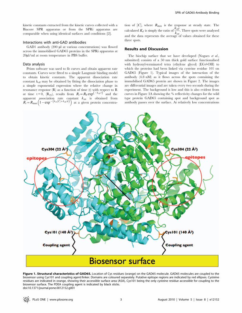
The biochip surface that we have developed (Nogues et al.,submitted) consists of a 50 nm thick gold surface functionalisedwith hydroxyl-terminated tetra (ethylene glycol) (EG4-OH) towhich the proteins had been linked via cysteine residue 101 onGAD65 (Figure 1). Typical images of the interaction of theantibody (4.8 nM) as it flows across the spots containing theimmobilised GAD65 protein are shown in Figure 2. The imagesare differential images and are taken every two seconds during theexperiment. The background is low and this is also evident fromcurves in Figure 3A showing the % reflectivity changes for the wildtype protein GAD65 containing spot and background spot asantibody passes over the surface. At relatively low concentrations
Figure 1. Structural characteristics of GAD65. Location of Cys residues (orange) on the GAD65 molecule. GAD65 molecules are coupled to thebiosensor using Cys101 and coupling agent/linker. Domains are coloured separately. Putative epitope regions are indicated by red ellipses. Cysteineresidues are indicated in orange, showing their accessible surface area (ASA), Cys101 being the only cysteine residue accessible for coupling to thebiosensor surface. The PDEA coupling agent is indicated by black sticks.doi:10.1371/journal.pone.0012152.g001
SPRi of GAD65-Antibody Binding
PLoS ONE | www.plosone.org 3 August 2010 | Volume 5 | Issue 8 | e12152
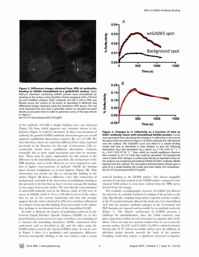
of the antibody (4.8 nM) a simple binding curve was observed(Figure 3A) from which apparent rate constants (shown in thelegend to Figure 3) could be calculated. At these concentrations ofantibody the protein GAD65-antibody interaction gave an overallapparent equilibrium dissociation constant (Kd ) of 1.37 nM. Wenote that these values are somewhat different from values reportedpreviously in the literature for this type of interaction [18]; weconsistently found lower equilibrium dissociation constantsessentially due to more rapid association rates that we measurehere. There may be many explanations for this related to thedifference in the immobilisation procedure, the architecture of theSPR machine, and so forth. However we were surprised to notethat at higher concentrations of antibody (48nM) the bindingphase became multiphasic or at least biphasic (Figure 3B). Thisobservation was clearly not due to non-specific binding to thesurface (Figure 3B shows a difference curve after subtraction ofbackground), and indeed the observation of multiphasic binding isdue precisely to the fact that we have very low non-specific bindingto non target areas on the surface. We note that the concentrationsof anti-GAD antibody used in the Biacore study of [18] were inexcess of 500nM, which is 10 to a 100 fold in excess of the valuesthat we use in the current manuscript and that this stronglysuggests that the values obtained in [18] were somehow influencedby a degree of non specific binding, if not necessarily to the surfacethen perhaps to an abnormal form of the immobilised GAD.
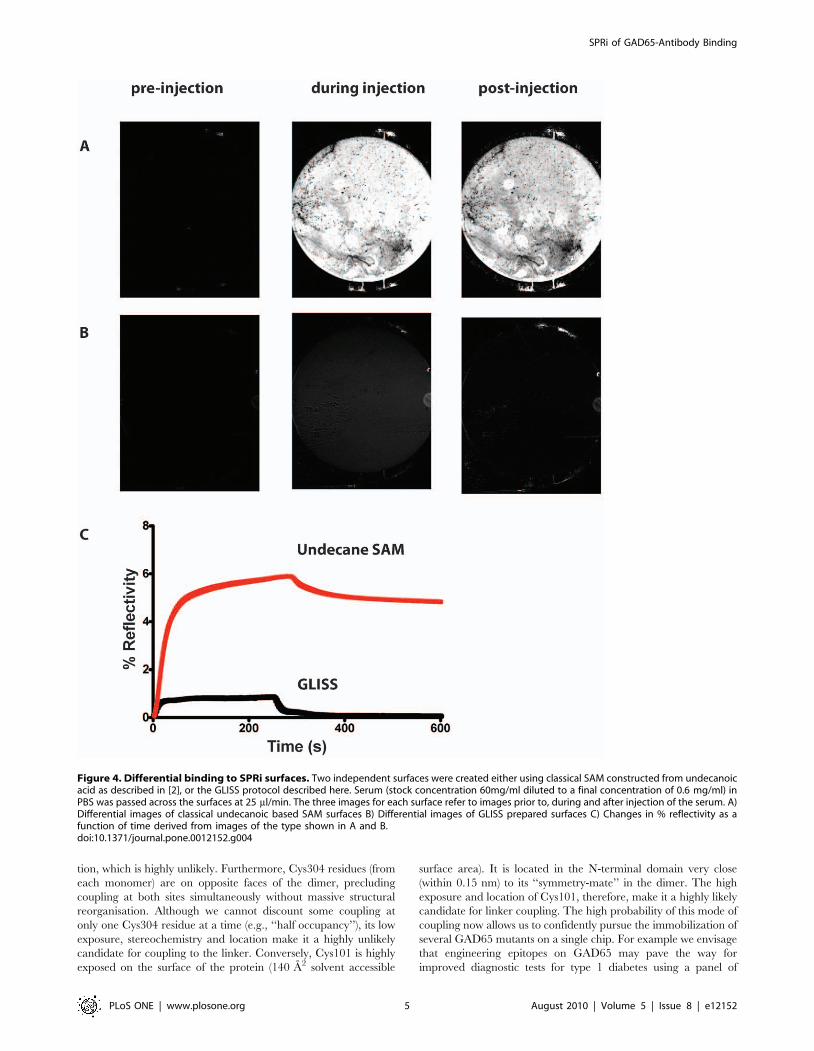
In order to illustrate the degree of antifouling conferred by theGeneral Liquid Interface Specific Surfaces (GLISS) we in factpassed human serum across two types of surface, one consisting ofa classical self assembling monolayer (SAM) constructed fromundecanoic acid as described in [2], and the other using theGLISS surfaces used in the current GAD65 study. As can be seenin Figure 4 there is a qualitative and quantitative differencebetween non-specific binding to the two surfaces with a much
reduced binding to the GLISS surface. The almost negligibleamount of material retained at the GLISS surface compared to theclassical SAM surface is even more evident from the SPR curvesderived from the images.
The available crystallographic structure of GAD65 [6] allowedthe selection of a suitable method of coupling of the protein to thechip. Specifically, coupling using surface-exposed cysteine residuesin the N-terminal domains allowed the molecule to be immobilisedsuch that the putative antibody epitopes in the C-terminal andPLP domains are exposed and accessible by an antibody molecule(Figure 1). The dimeric architecture of GAD65 presents achallenge for immobilization, since the 2-fold symmetry mayplace equivalent residues in each monomer on opposite sides of thedimer. There are only two cysteine residues that are on or near theprotein surface (Cys101 and Cys304). Cys304 is relatively buried,having only 22 A2 solvent accessible surface area. In addition, itssidechain points inwards towards the body of the protein.Coupling would thus require a significant structural reorganisa-
Figure 2. Difference images obtained from SPRi of antibodiesbinding to GAD65 immobilised at a gold/GLISS surface. Spots(400 mm diameter) containing GAD65 protein were immobilised byspotting at the surface using Hamilton Starlet equipped with a PinTooltip and modified software. GAD1 antibody (4.8 nM in 200 ml PBS) wasflowed across the surface at 20 ml/min as described in Methods anddifferential images obtained using the GenOptics SPRi device. The redcircle illustrates the area that is generally chosen to calculate the pixeldensity at any given time in order to generate curves of the type shownin Figure 3.doi:10.1371/journal.pone.0012152.g002
Figure 3. Changes in % reflectivity as a function of time asGAD1 antibody reacts with immobilised GAD65 protein. Curveswere generated from calculating the change in % reflectivity in time acrossthe spots of the sort shown in Figure 2. A) GAD1 antibody (4.8 nM) injectedover the surfaces. The wtGAD65 curve was fitted to a simple bindingmodel (red line) as described in Data Analysis to give the followingdissociation (koff) and association (kon) values: koff = 1.2960.02?1023 s21;kon = 9.4160.29?105 M21s21 from which an overall equilibrium dissocia-tion constant, Kd of 1.3760.06 nM, could be calculated. The backgroundcurve is taken from changes in surface pixel density at equivalent areas onthe surfaces not containing immobilised GAD65; B) GAD1 antibody (48nM)injected over the surfaces. The association and dissociation phases gave apoor fit to a single binding site model and were clearly not monophasic.doi:10.1371/journal.pone.0012152.g003
SPRi of GAD65-Antibody Binding
PLoS ONE | www.plosone.org 4 August 2010 | Volume 5 | Issue 8 | e12152
tion, which is highly unlikely. Furthermore, Cys304 residues (fromeach monomer) are on opposite faces of the dimer, precludingcoupling at both sites simultaneously without massive structuralreorganisation. Although we cannot discount some coupling atonly one Cys304 residue at a time (e.g., ‘‘half occupancy’’), its lowexposure, stereochemistry and location make it a highly unlikelycandidate for coupling to the linker. Conversely, Cys101 is highlyexposed on the surface of the protein (140 A2 solvent accessible
surface area). It is located in the N-terminal domain very close(within 0.15 nm) to its ‘‘symmetry-mate’’ in the dimer. The highexposure and location of Cys101, therefore, make it a highly likelycandidate for linker coupling. The high probability of this mode ofcoupling now allows us to confidently pursue the immobilization ofseveral GAD65 mutants on a single chip. For example we envisagethat engineering epitopes on GAD65 may pave the way forimproved diagnostic tests for type 1 diabetes using a panel of
Figure 4. Differential binding to SPRi surfaces. Two independent surfaces were created either using classical SAM constructed from undecanoicacid as described in [2], or the GLISS protocol described here. Serum (stock concentration 60mg/ml diluted to a final concentration of 0.6 mg/ml) inPBS was passed across the surfaces at 25 ml/min. The three images for each surface refer to images prior to, during and after injection of the serum. A)Differential images of classical undecanoic based SAM surfaces B) Differential images of GLISS prepared surfaces C) Changes in % reflectivity as afunction of time derived from images of the type shown in A and B.doi:10.1371/journal.pone.0012152.g004
SPRi of GAD65-Antibody Binding
PLoS ONE | www.plosone.org 5 August 2010 | Volume 5 | Issue 8 | e12152
antibodies as well as human sera. The ability to couple multipleGAD65 proteins on the same chip and measure antibody bindingin a reproducible and rapid fashion may ultimately lead to a novelSPRi-based diagnostic immunoassay for type 1 diabetes.
We clearly advocate the use of engineering surface exposedcysteines for immobilisation but recognize that this may not alwaysbe possible. A number of alternatives are available, and generallyin SPR, coupling using amines, for the most part throughaccessible lysines, is advocated. It must be noted however, that thisgenerally results in a reduced degree of activity of the immobilisedtarget that may render quantitative analysis difficult. The GLISSsurfaces used here may easily be functionalised with carboxyl, thiolor amine groups thus permitting a wide range of immobilisationtechniques. However we would like to stress that although theexpedient of engineering solvent accessible thiols is somewhatlimiting it does optimise accessibility and that whilst this mayrestrict general applicability we strongly suggest that immobilisa-tion strategies that aim at increasing accessibility and orientationbe elaborated rather than blind immobilisation through solventaccessible amines for example. Alternatively one can immobiliseon the GLISS surfaces, specific antibodies or haptens eitherthrough accessible cysteines or via other coupling techniques, thatthen allow mild capture of the target molecules.
Although the purpose of the present study was not to explorethe detection levels of the SPRi technique we could detect andquantify GAD65-anti-GAD65 interactions between approximately1010 molecules of target GAD65 on the surface and antibody at4nM concentration in solution. Our limit of detection as discernedfrom the signal to noise ratio suggests that we can detect a changeof approximately 0.01% reflectivity that corresponds to levels ofdetection of around 30 to 40 ng/ml of protein in solution.
We have characterised the binding of wild type GAD65 to theGAD1 monoclonal antibody using SPRi. Our data illustrate thatwe have effectively eliminated non-specific interactions with thesurface containing the immobilised GAD65 molecules. Theimplications of this are far reaching; in short not only does thisapproach obviate the dubious process of background subtractionbut gives access to more accurate kinetic and equilibrium values
that tend towards more affine measurements since any multiphasebehaviour can be separated from non-specific binding. On abroader level, an enhanced signal to noise ratio increases not onlythe sensitivity but also confidence in the use of SPR to generatekinetic constants that may then be inserted into van’t Hoff typeanalyses to provide comparative DG, DS and DH values, makingthis an efficient, rapid and competitive alternative to ITCmeasurements used in drug and macromolecular-interactionmechanistic studies. Finally, and this is particularly evident here,the accuracy of the measurements allows the application of moreintricate interaction models than simple Langmuir monophasicbinding. The observation that monoclonal antibodies can usemultiple binding modes is intriguing. We are currently applyingthe technology developed here to analyse monoclonal antibodybinding to microarrays containing selected mutants of GAD65(Buckle et al. in preparation). The implications are thatintramolecular rearrangements associated with antibody bindingmay be involved.
ConclusionsThe detection and measurement of antibody binding by the
type 1 diabetes autoantigen GAD65 represents an example of anantibody-antigen interaction where good structural, mechanisticand immunological data are available. Using SPRi we were able tocharacterise the kinetics of the interaction in greater detail thanELISA/RIA methods. Furthermore, our data indicate that SPRi iswell suited to a multiplexed immunoassay using GAD65 proteins,and may be applicable to other biomarkers.
Acknowledgments
We thank Merrill Rowley for the kind gift of GAD1 antibody.
Author Contributions
Conceived and designed the experiments: AMB MB. Performed theexperiments: CN HL MB. Analyzed the data: CN HL AMB MB.Contributed reagents/materials/analysis tools: CN HL CGL RHPL AMBMB. Wrote the paper: AMB MB.
References
1. Buckle M, Williams RM, Negroni M, Buc H (1996) Real time measurements ofelongation by a reverse transcriptase using surface plasmon resonance.Proceedings of the National Academy of Sciences of the United States ofAmerica 93: 889–894.
2. Bouffartigues E, Leh H, Anger-Leroy M, Rimsky S, Buckle M (2007) Rapidcoupling of Surface Plasmon Resonance (SPR and SPRi) and ProteinChip (TM)based mass spectrometry for the identification of proteins in nucleoproteininteractions. Nucleic Acids Research 35: e39.
3. Cherif B, Roget A, Villiers CL, Calemczuk R, Leroy V, et al. (2006) Clinicallyrelated protein-peptide interactions monitored in real time on novel peptidechips by surface plasmon resonance imaging. Clin Chem 52: 255–262.
4. Baekkeskov S, Aanstoot HJ, Christgau S, Reetz A, Solimena M, et al. (1990)Identification of the 64K autoantigen in insulin-dependent diabetes as theGABA-synthesizing enzyme glutamic acid decarboxylase. Nature 347: 151–156.
5. Tuomilehto J, Zimmet P, Mackay IR, Koskela P, Vidgren G, et al. (1994)Antibodies to glutamic acid decarboxylase as predictors of insulin-dependentdiabetes mellitus before clinical onset of disease. Lancet 343: 1383–1385.
6. Fenalti G, Law RH, Buckle AM, Langendorf C, Tuck K, et al. (2007) GABAproduction by glutamic acid decarboxylase is regulated by a dynamic catalyticloop. Nat Struct Mol Biol 14: 280–286.
7. Fenalti G, Hampe CS, Arafat Y, Law RH, Banga JP, et al. (2008) COOH-terminal clustering of autoantibody and T-cell determinants on the structure ofGAD65 provide insights into the molecular basis of autoreactivity. Diabetes 57:1293–1301.
8. Fenalti G, Hampe CS, O’Connor K, Banga JP, Mackay IR, et al. (2007)Molecular characterization of a disease associated conformational epitope onGAD65 recognised by a human monoclonal antibody b96.11. Mol Immunol 44:1178–1189.
9. Schwartz HL, Chandonia JM, Kash SF, Kanaani J, Tunnell E, et al. (1999)High-resolution autoreactive epitope mapping and structural modeling of the65 kDa form of human glutamic acid decarboxylase. J Mol Biol 287: 983–999.
10. Fenalti G, Buckle AM (2009) Structural biology of the GAD autoantigen.Autoimmun Rev 9: 148–152.
11. Arafat Y, Fenalti G, Whisstock JC, Mackay IR, Garcia de la Banda M, et al.(2009) Structural determinants of GAD antigenicity. Mol Immunol 47: 493–505.
12. Prime KL, Whitesides GM (1993) Adsorption of proteins onto surfacescontaining end-attached oligo(ethylene oxide): a model system using self-assembled monolayers. J Am Chem Soc 115: 10714–10721.
13. Frederix F, Bonroy K, Reekmans G, Laureyn W, Campitelli A, et al. (2004)Reduced nonspecific adsorption on covalently immobilized protein surfacesusing poly(ethylene oxide) containing blocking agents. J Biochem BiophysMethods 58: 67–74.
14. Teoh KL, Fida S, Rowley MJ, Mackay IR (1998) Autoantigenic reactivity ofdiabetes sera with a hybrid glutamic acid decarboxylase GAD67-65 moleculeGAD67(1-101)/GAD65(96-585). Autoimmunity 28: 259–266.
15. Powell M, Prentice L, Asawa T, Kato R, Sawicka J, et al. (1996) Glutamic aciddecarboxylase autoantibody assay using 125I-labelled recombinant GAD65produced in yeast. Clin Chim Acta 256: 175–188.
16. Primo ME, Anton EA, Villanueva AL, Poskus E, Ermacora MR (2003)Engineered variants of human glutamic acid decarboxylase (GAD) andautoantibody epitope recognition. Clin Immunol 108: 38–45.
17. Gottlieb DI, Chang YC, Schwob JE (1986) Monoclonal antibodies to glutamicacid decarboxylase. Proc Natl Acad Sci U S A 83: 8808–8812.
18. Lee JW, Sim SJ, Cho SM, Lee J (2005) Characterization of a self-assembledmonolayer of thiol on a gold surface and the fabrication of a biosensor chipbased on surface plasmon resonance for detecting anti-GAD antibody. BiosensBioelectron 20: 1422–1427.
SPRi of GAD65-Antibody Binding
PLoS ONE | www.plosone.org 6 August 2010 | Volume 5 | Issue 8 | e12152