Embed Size (px)
Citation preview
Original Article
Characterisation of novel strains of multiply antibiotic-resistant Salmonella recovered from poultry in Southern Senegal
Michel M. Dione1, Stanny Geerts2 and Martin Antonio1 1Vaccinology Theme, Medical Research Council, Unit, Banjul, The Gambia
2Institute of Tropical Medicine, Antwerp, Belgium
Abstract Introduction: Non-typhoidal Salmonella (NTS) contamination in poultry and poultry products is a major cause of food-borne disease in
humans. This study presents the molecular epidemiology of NTS isolated from poultry in Senegal.
Methodology: A total of 261 NTS recovered from broiler farms, chicken carcasses and street vendors were characterized using random
amplification of polymorphic DNA (RAPD) and multilocus sequence typing (MLST) techniques.
Results: We observed 20 distinct RAPD profiles corresponding to 18 different serotypes. Strains from each of these 20 groups were further
analysed using MLST. Consequently, 12 new MLST alleles and 17 new sequence types were discovered. Three sequence types (S. Kentucky
ST198, S. Agona ST13 and S. Istanbul ST33) have previously been described in Senegal and other countries, suggesting that these clones are
geographically widely distributed and are circulating in a wide range of hosts. Nine clones showed multi-resistance to the most commonly
used antibiotics in both humans and animals. However, a novel multi-resistant clone of S. Kentucky ST832 was found.
Conclusion: This study gives new insights into the genetic diversity of NTS in Senegal. Molecular tools remain essential to improve our
understanding of the epidemiology of NTS by tracking the sources of infection and/or contamination.
Key words: Salmonella; clones; antibiotic-resistant; Senegal
J Infect Dev Ctries 2012; 6(5):436-442.
(Received 31 August 2010 – Accepted 25 July 2011)
Copyright © 2012 Dione et al. This is an open-access article distributed under the Creative Commons Attribution License, which permits unrestricted use,
distribution, and reproduction in any medium, provided the original work is properly cited
Introduction Enteric diseases caused by Salmonella in
chickens are of great concern because they are an
important cause of mortality and morbidity [1].
Poultry and its products, in particular chicken meat
and eggs, are a major source of Salmonella infection
[2]. In Senegal, several studies have reported the
presence of Salmonella along the poultry production
supply chain and its public health impact [3-7],
withserotyping being used to characterise the
Salmonella [8]. Due to the ubiquitous nature of
Salmonella, a typing scheme capable of a more
detailed strain identification is essential for
epidemiological studies [9], because the ability to
distinguish isolates of Salmonella is very important to
trace the source of infection and outbreaks. Several
methods have been used for deciphering the
relatedness among isolates but some have low
discriminating power, whereas others demand a
considerable amount of expertise, time and
equipment [10]. Multilocus sequence typing (MLST)
was found to have a low discriminative power, but is
easier to interpret and to compare between
laboratories and provides the best phylogenetic-
relationship inferences [11]. Our study reports for the
first time in Senegal the characterization of the most
prevalent NTS strains isolated from poultry and
poultry products by using the random amplification
of polymorphic DNA (RAPD) and MLST techniques.
Methodology Bacterial isolates
From October 2007 to July 2008, a total of 261
strains of Salmonella were collected during a cross-
sectional study in 57 broiler farms, 285 chicken
carcasses from farms/sale points, and 42 dishes of
chicken meat from street restaurants in southern
Senegal [7]. Salmonella was cultured according to
the standard technique ISO 6579 (1998, International
Organization for Standardization, Geneva,
Switzerland). Serotyping was done by slide
agglutination using Salmonella polyvalent and
monovalent O and H antisera (Diagnostic Pasteur,
Paris, France) according to the Kauffmann-White
classification scheme [8]. The data of the
Dione et al. - Non-typhoidal Salmonella in West Africa J Infect Dev Ctries 2012; 6(5):436-442.
437
antimicrobial resistance tests were extracted from a
previous study [7].
RAPD-PCR reaction conditions
The RAPD reaction was performed on all 261
isolates using P1254 primer as previously described
[12]. The reaction was as follows: 3 µl of primer (20
mM) was added to 10 µl of DNA free water
(Invitrogen, Paisley, UK), 5 µl of 5x Buffer (Qiagen,
Crawley, UK), 2 µl of dNTPS (2 mM), and 3.5 µl of
MgCl2 (25 mM), 0.5 GoTaq polymerase (Promega,
Madisson, UK) and 1 µl of template (cells) to get the
final volume of 25 µl. Amplification was performed
in a PCR machine (Techne, Hatboro, USA) as
follows: one cycle at 94ºC (2 minutes); a series of 3
cycles at 94ºC (2 minutes), 35ºC (1 minute) and 72ºC
(2 minutes); a second series of 34 cycles at 94ºC (10
seconds), 40ºC (20 seconds) and 72ºC (2 minutes);
with final extension at 72ºC for 5 minutes. The
amplified product was stored at 4ºC until required.
The PCR product (10 µl each) was loaded on a 1.5%
agarose gel containing 0.5ul/ml ethidium bromide at
25 V overnight in 1X (SIGMA, Steinheim,
Germany). Two lanes of 50 bp and 100 bp of ladder
were included in each gel for reference. The RAPD
electrophoresis bands were photographed using UV
illumination with a gel documentation system (gel
Doc 2000, Bio-Rad, Hertfordshire, UK). Gel pictures
were analysed with Bionumerics software (version
4.0; Applied Maths, Saint-Martens-Latem, Belgium)
and finally similar profiles resolving into similar
clones were selected.
Multi locus sequence typing
MLST was performed on 20 randomly selected
Salmonella serotypes with unique RAPD profiles.
The protocol was followed as previously described
[13] . Briefly, the seven genes were targeted were
aroC, dnaN, hemD, hisD, purE, sucE and thrA.
Amplification of all genes was performed with a 25
µl reaction mixture of the following: 10x Buffer
(Applied Biosystems, Foster City, USA) with 1.5mM
MgCl2 (2.5 µl); 2 mM dNTP (0.5 µl); 12.5 mM
forward primer (1 µl); 12.5 mM reverse primer (1
µl); 5U/µl Qiagen Hotstart Taq Polymerase (Qiagen,
Crawley, UK) (0.25µl); template (cell lysate) (2 µl)
and 17.75 µl sterile DNA free water. PCR cycling
conditions were 10 minutes at 94°C, followed by 32
cycles of 94°C for 1 minute, 55°C for 1 minute and
72°C for 1 minute, with a final extension at 72°C for
5 minutes. From each PCR, 2 µl aliquots were
separated by 1% agarose gel electrophoresis and
visualized with ethidium bromide staining and UV
illumination, and using a gel documentation system
(gel Doc 2000, Bio-Rad, Hertfordshire, UK). PCR
products were purified using Qiagen kit (Qiagen,
Crawley, UK). Sequencing was done on both strands
with BigDye Terminator Cycle Sequencing kit
(Applied Biosystems, Warrington, UK). The labelled
fragments were separated by size using a 3130xl
Genetic Analyser (Applied Biosystems, UK).
Sequences were edited and complementary sense
antisense fragments were aligned using the Laser
Gene DNA star 7.1 software. Finally, the sequences
were submitted to the MLST database website [14]
and assigned to existing or novel allele type numbers
defined by the database. New sequence types were
arbitrarily assigned a number for the purpose of the
analysis.
Cluster analysis and mapping
To perform the cluster analysis of the serotypes,
MLST data were analysed with Bionumerics
software (version 6.5; Applied Maths, Sint-Martens-
Latem, Belgium). Analysis using a hierarchic
unweighted pair group method (UPGMA) with
averaging was used to generate a dendrogram
describing the relationship among Salmonella
serotypes. A minimum spanning tree was used to
compare Senegalese clones to Gambian and other
African clones. The mapping of the locations of the
cases was performed using Arc Gis 9.3 software.
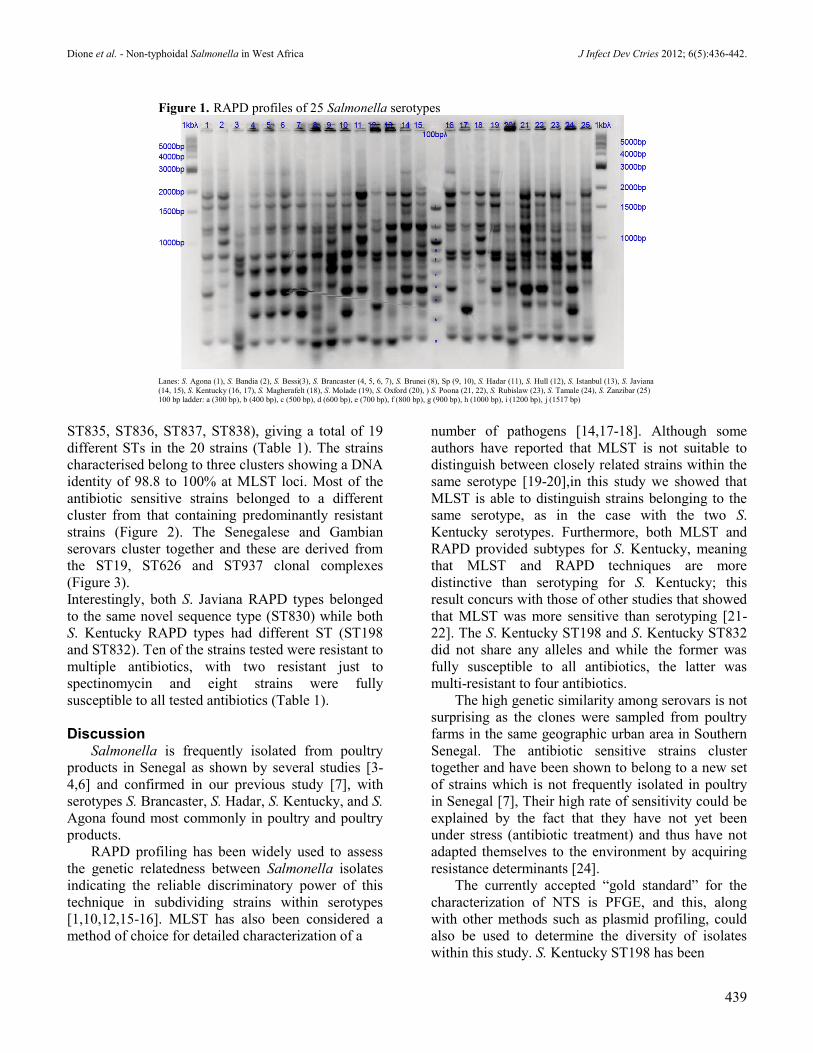
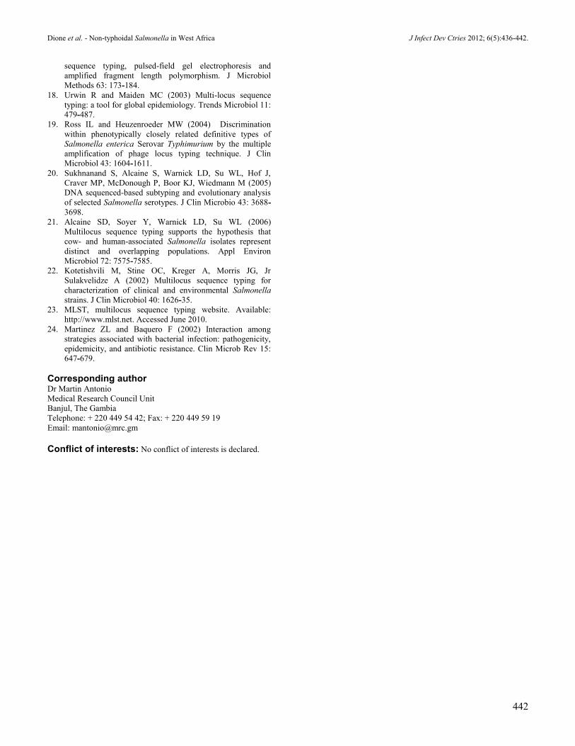
Results In this study, we analyzed the genomic
relatedness between 261 NTS belonging to 18
different serovars, initially using RAPD profiling. All
RAPD fragments amplified were between 200 bp and
3000 bp. Twenty different RAPD fingerprints were
observed among the 261 NTS (Figure 1). Each
serovar generated a unique RAPD fingerprints, with
the exception of two serovars, S. Javiana (Figure 1,
lanes 14 and line 15) and S. Kentucky (Figure 1,
lanes 16 and line 17) which each showed two
different fingerprints. To further characterise these
NTS, MLST was performed on one strain from each
unique RAPD type. Twelve new alleles were
discovered [aroC (203), aroC (204), aroC (205),
dnaN (195), dnaN (196), hisD (238), hisD (239),
hisD (241), purE (199), sucA (195), thrA (185), and
thrA (186)] as well as sixteen new sequence types
(ST923, ST824, ST825, ST826, ST827, ST828,
ST829, ST830, ST831, ST832, ST833, ST834,
Allele number
Strain ID Serotype aroC dnaN hemD hisD purE sucA thrA ST Source (chicken) Antibiotic resistance
A1001 Agona 3 3 7 4 3 3 7 13 skin SPT
A1002 Bandia 84 65 8 241 64 9 168 823 stool STX, TMP, CIP, SSS
A1003 Bessi 204 11 8 241 161 195 186 824 skin None
A1004 Brancaster 205 81 10 36 88 108 36 825 skin STX, TE, TMP, S, SSS
A1005 Brunei 19 81 8 20 5 9 185 826 stool AMX, TIC, STX, TE, TMP, SSS
A1006 Sp. 203 195 17 164 12 35 4 827 skin SPT
A1007 Hadar 17 5 78 7 5 7 12 828 muscle STX, TE, TMP, S, SSS
A1008 Hull 114 5 3 238 199 10 12 829 skin None
A1009 Istanbul 2 5 6 7 5 7 12 33 restaurant CF, STX, TE, GM, TMP, SPT, S, SSS
A1010 Javiana 11 196 78 74 40 13 4 830 skin None
A232 Javiana 11 196 78 74 40 13 4 830 muscle None
A1011 Kentucky 76 14 3 77 64 64 67 198
skin None
A112 Kentucky 115 65 8 115 2 116 110 832 stool STX, TMP, SPT, S, SSS
A1012 Magherafelt 84 65 3 241 64 9 110 833 skin None
A1013 Sp. 76 14 3 77 64 132 67 835 skin STX, TE, TMP, SPT, S, SSS
A1014 Sp. 203 14 17 164 12 35 4 836 skin None
A1015 Poona 13 127 92 157 40 35 137 838 skin AMX, TIC, STX, TE, TMP, SPT, S, SSS
A1016 Rubislaw 42 46 48 239 12 35 4 837 stool None
A1017 Tamale 205 46 8 42 88 108 36 834 stool STX, TE, TMP, SPT, S, SSS
A1018 Zanzibar 84 11 8 42 40 71 4 831 muscle SPT, S
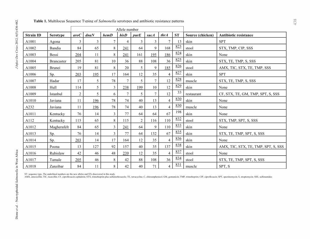
ST: sequence type, The underlined numbers are the new alleles and STs discovered in this study.
AMX, amoxicillin; TIC, ticarcillin; CF, ciprofloxacin cephalotin; STX, trimethoprim plus sulfamethoxazole; TE, tetracycline; C, chloramphenicol; GM, gentamicin; TMP, trimethoprim; CIP, ciprofloxacin; SPT, spectinomycin; S, streptomycin; SSS, sulfonamides
Dio
ne
et a
l. -
Non
-ty
pho
idal
Salm
on
ella
in
Wes
t A
fric
a
J
Infe
ct D
ev C
trie
s 20
12;
6(5
):4
36
-442
.
43
8
Table 1. Multilocus Sequence Typing of Salmonella serotypes and antibiotic resistance patterns
Strain
ID Serotype aroC dnaN hemD hisD purE sucA thrA ST
Source
(chicken) Antibiotic resistance
A1001 Agona 3 3 7 4 3 3 7 13 skin SPT
A1002 Bandia 84 65 8 241 64 9 168 823 stool STX, TMP, CIP, SSS
A1003 Bessi 204 11 8 241 161 195 186 824 skin None
A1004 Brancaster 205 81 10 36 88 108 36 825 skin STX, TE, TMP, S, SSS
A1005 Brunei 19 81 8 20 5 9 185 826 stool AMX, TIC, STX, TE, TMP, SSS
A1006 Sp. 203 195 17 164 12 35 4 827 skin SPT
A1007 Hadar 17 5 78 7 5 7 12 828 muscle STX, TE, TMP, S, SSS
A1008 Hull 114 5 3 238 199 10 12 829 skin None
A1009 Istanbul 2 5 6 7 5 7 12 33 restaurant CF, STX, TE, GM, TMP, SPT, S, SSS
A1010 Javiana 11 196 78 74 40 13 4 830 skin None
A232 Javiana 11 196 78 74 40 13 4 830 muscle None
A1011
Sp. Kentucky 76 14 3 77 64 64 67
198
skin None
A112 Kentucky 115 65 8 115 2 116 110 832 stool STX, TMP, SPT, S, SSS
A1012 Magherafelt 84 65 3 241 64 9 110 833 skin None
A1013 Sp. 76 14 3 77 64 132 67 835 skin STX, TE, TMP, SPT, S, SSS
A1014 Sp. 203 14 17 164 12 35 4 836 skin None
A1015 Poona 13 127 92 157 40 35 137
838
skin
AMX, TIC, STX, TE, TMP, SPT, S,
SSS
A1016 Rubislaw 42 46 48 239 12 35 4 837 stool None
A1017 Tamale 205 46 8 42 88 108 36 834 stool STX, TE, TMP, SPT, S, SSS
A1018 Zanzibar 84 11 8 42 40 71 4 831 muscle SPT, S
Dione et al. - Non-typhoidal Salmonella in West Africa J Infect Dev Ctries 2012; 6(5):436-442.
439
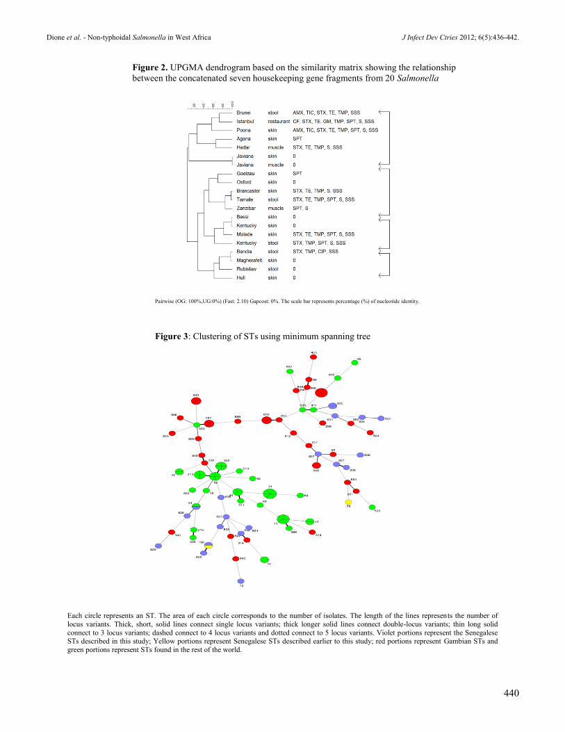
ST835, ST836, ST837, ST838), giving a total of 19
different STs in the 20 strains (Table 1). The strains
characterised belong to three clusters showing a DNA
identity of 98.8 to 100% at MLST loci. Most of the
antibiotic sensitive strains belonged to a different
cluster from that containing predominantly resistant
strains (Figure 2). The Senegalese and Gambian
serovars cluster together and these are derived from
the ST19, ST626 and ST937 clonal complexes
(Figure 3).
Interestingly, both S. Javiana RAPD types belonged
to the same novel sequence type (ST830) while both
S. Kentucky RAPD types had different ST (ST198
and ST832). Ten of the strains tested were resistant to
multiple antibiotics, with two resistant just to
spectinomycin and eight strains were fully
susceptible to all tested antibiotics (Table 1).
Discussion Salmonella is frequently isolated from poultry
products in Senegal as shown by several studies [3-
4,6] and confirmed in our previous study [7], with
serotypes S. Brancaster, S. Hadar, S. Kentucky, and S.
Agona found most commonly in poultry and poultry
products.
RAPD profiling has been widely used to assess
the genetic relatedness between Salmonella isolates
indicating the reliable discriminatory power of this
technique in subdividing strains within serotypes
[1,10,12,15-16]. MLST has also been considered a
method of choice for detailed characterization of a
number of pathogens [14,17-18]. Although some
authors have reported that MLST is not suitable to
distinguish between closely related strains within the
same serotype [19-20],in this study we showed that
MLST is able to distinguish strains belonging to the
same serotype, as in the case with the two S.
Kentucky serotypes. Furthermore, both MLST and
RAPD provided subtypes for S. Kentucky, meaning
that MLST and RAPD techniques are more
distinctive than serotyping for S. Kentucky; this
result concurs with those of other studies that showed
that MLST was more sensitive than serotyping [21-
22]. The S. Kentucky ST198 and S. Kentucky ST832
did not share any alleles and while the former was
fully susceptible to all antibiotics, the latter was
multi-resistant to four antibiotics.
The high genetic similarity among serovars is not
surprising as the clones were sampled from poultry
farms in the same geographic urban area in Southern
Senegal. The antibiotic sensitive strains cluster
together and have been shown to belong to a new set
of strains which is not frequently isolated in poultry
in Senegal [7], Their high rate of sensitivity could be
explained by the fact that they have not yet been
under stress (antibiotic treatment) and thus have not
adapted themselves to the environment by acquiring
resistance determinants [24].
The currently accepted “gold standard” for the
characterization of NTS is PFGE, and this, along
with other methods such as plasmid profiling, could
also be used to determine the diversity of isolates
within this study. S. Kentucky ST198 has been
Figure 1. RAPD profiles of 25 Salmonella serotypes
Lanes: S. Agona (1), S. Bandia (2), S. Bessi(3), S. Brancaster (4, 5, 6, 7), S. Brunei (8), Sp (9, 10), S. Hadar (11), S. Hull (12), S. Istanbul (13), S. Javiana
(14, 15), S. Kentucky (16, 17), S. Magherafelt (18), S. Molade (19), S. Oxford (20), ) S. Poona (21, 22), S. Rubislaw (23), S. Tamale (24), S. Zanzibar (25)
100 bp ladder: a (300 bp), b (400 bp), c (500 bp), d (600 bp), e (700 bp), f (800 bp), g (900 bp), h (1000 bp), i (1200 bp), j (1517 bp)
Dione et al. - Non-typhoidal Salmonella in West Africa J Infect Dev Ctries 2012; 6(5):436-442.
440
Figure 2. UPGMA dendrogram based on the similarity matrix showing the relationship
between the concatenated seven housekeeping gene fragments from 20 Salmonella
strains
Pairwise (OG: 100%,UG:0%) (Fast: 2.10) Gapcost: 0%. The scale bar represents percentage (%) of nucleotide identity.
Each circle represents an ST. The area of each circle corresponds to the number of isolates. The length of the lines represents the number of
locus variants. Thick, short, solid lines connect single locus variants; thick longer solid lines connect double-locus variants; thin long solid
connect to 3 locus variants; dashed connect to 4 locus variants and dotted connect to 5 locus variants. Violet portions represent the Senegalese STs described in this study; Yellow portions represent Senegalese STs described earlier to this study; red portions represent Gambian STs and
green portions represent STs found in the rest of the world.
Figure 3: Clustering of STs using minimum spanning tree
Dione et al. - Non-typhoidal Salmonella in West Africa J Infect Dev Ctries 2012; 6(5):436-442.
441
previously isolated from humans in Senegal and from
food and cattle in the United States (USA). Our study
revealed its presence in chicken skin in southern
Senegal. S. Agona ST13 found in chicken skin in our
study was also previously described in Denmark
from both human and veterinary sources, from milk
in Ireland, from humans in Scotland, and from
undetermined hosts in Australia, Germany, the USA,
Canada and Spain [23], while S. Hadar ST33 was
isolated from various sources in several European
and American countries and South Africa [23].
However, the ST33 found in southern Senegal in our
study belonged to serovar S. Istanbul. Collectively,
ST13, ST33 and ST198 clones are widely distributed
throughout the world and all three clones have been
simultaneously identified in both human and animal
sources, suggesting that the same clones are
circulating in both hosts. S. Istanbul ST33 was multi-
resistant to most commonly used antibiotics in
humans and animals in southern Senegal, contrary to
S. Kentucky ST198 and S. Agona ST13, which were
fully sensitive to all antibiotics tested. However, a
novel multi-resistant clone of S. Kentucky ST832
was found in poultry.
Conclusion Our study showed that a variety of serovars and
clones is circulating in southern Senegal;
furthermore, 17 out of the 20 clones characterised in
this study have never been reported elsewhere in the
world.
This is the first report describing the use of
MLST to characterise the most common NTS in
poultry and poultry products in Senegal. The
discovery of new alleles and sequence types is very
useful for a better understanding of the epidemiology
of NTS in Senegal.
Acknowledgments This work was supported by the Flemish Interuniversity
Council (VLIR-UOS, Brussels, Belgium) and the Medical
Research Council, United Kingdom. We acknowledge
Mark Achtman for the use of the Salmonella MLST
database (http://mlst.ucc.ie/mlst/dbs/Senterica), which is
hosted at the University College Cork and funded by the
Science Foundation of Ireland (05/FE1/B882).
References 1. Chansiripornchai N, Ramasoota P, Bangtrakulnonth A,
Sasipreeyajan J, Svenson SB(2000) Application of randomly
amplified polymorphic DNA (RAPD) analysis for typing
Avian Salmonella enterica subsp. enterica. FEMS Immunol
Med Microbiol 29: 221-225.
2. Rampling A (1993) Salmonella enteritidis five years on.
Lancet 342: 317-318.
3. Bada-Alambedji R, Fofana A, Seydi M, Akakpo AJ (2006)
Antimicrobial resistance of Salmonella isolated from poultry
carcasses in Dakar (Senegal). Braz J Microbiol 37: 510-515.
4. Cardinale E, Perrier Gros-Claude JD, Tall F, Cisse M,
Gueye EHF, Salvat G (2003) Prevalence of Salmonella and
Campylobacter in retail chicken carcasses in Senegal. Rev
Elev Vét Pays Trop 56: 13-16.
5. Cardinale E, Perrier Gros-Claude JD, Tall F, Gueye EF,
Salvat G (2005) Risk factors for contamination of ready-to-
eat street-vended poultry dishes in Dakar, Senegal. Int J
Food Microbiol 103: 157-65.
6. Cardinale E, Tall F, Gueye, EF, Cisse M, Salvat G (2004)
Risk factors for Salmonella enterica subsp. enterica
infection in Senegalese broiler-chicken flocks. Prev VetMed
63: 151-161.
7. Dione MM, Ieven M, Garin B, Marcotty T, Geerts S (2009)
Prevalence and antimicrobial resistance of Salmonella
isolated from broiler farms, chicken carcasses, and street-
vended restaurants in Casamance, Senegal. J Food Prot 72:
2423-2427.
8. Grimont PAD, Weill FX (2007) Antigenic formulae of the
Salmonella serovars. 9th Edition. WHO Collaboration
Centre for Reference and Research on Salmonella and
Institut Pasteur, 167p.
9. Betancor LF, Schelott A, Martinez M, Pereira G, Algorta
MA, Rodrigez F, Vignoli Chabalgoity JA (2004) Random
Amplified Polymorphic DNA and phenotyping analysis of
Salmonella enterica serovar enteritidis isolates collected
from humans and poultry in Uruguay from 1995-2002. J
Clin Microbiol 42: 1155-1162.
10. Maripandi A, Suresh SSR, Ponmurugan P,
Gurusubramanian G (2007) Random Amplification of
Polymorphic DNA (RAPD) of Salmonella enteritidis
Isolated from Chicken Samples. Biotechnology 6: 278-282.
11. Cooper JE, Feil EJ (2004) Multilocus sequence typing-what
is resolved? . Trends Microbiol 12: 373-377.
12. Lin AW, Usera MA, Barrett TJ, Goldsby RA (1996)
Application of random amplified DNA analysis to
differentiate strains of Salmonella enteritidis. J Clin
Microbiol34: 870-876.
13. Ikumapayi UN, Antonio M, Sonne-Hansen J, Biney E,
Enwere G, Okoko B, Oluwalana C, Vaughan A, Zaman SM,
Greenwood BM, Cutts FT, Adegbola RA (2007) Molecular
epidemiology of community-acquired invasive non-
typhoidal Salmonella among children aged 2-29 months in
rural Gambia and discovery of a new serovar, Salmonella
enterica Dingiri. J Med Microbiol 56: 1479-1484.
14. Aanensen DM and Spratt BG (2005) The multilocus
sequence typing network: mlst.net Nucleic Acids Res. 33:
728-733.
15. Tikoo A, Tripathi AK, Verma SC, Agrawal N, Nath G
(2001) Application of PCR finger printing techniques for
identification and discrimination of Salmonella isolates.
Curr Sci. 80: 1049-1052.
16. Yaqoob E, Hussain I, Rahman SU (2007) Molecular
characterization by using amplified polymorphic DNA
(RAPD) analysis of Salmonella enteritidis isolates recovered
from avian and human sources. Pakistan Vet J 27: 102-104.
17. Torpdahl M, Skov MN, Sandvang D, Baggesen DL (2005)
Genotypic characterization of Salmonella by multilocus
Dione et al. - Non-typhoidal Salmonella in West Africa J Infect Dev Ctries 2012; 6(5):436-442.
442
sequence typing, pulsed-field gel electrophoresis and
amplified fragment length polymorphism. J Microbiol
Methods 63: 173-184.
18. Urwin R and Maiden MC (2003) Multi-locus sequence
typing: a tool for global epidemiology. Trends Microbiol 11:
479-487.
19. Ross IL and Heuzenroeder MW (2004) Discrimination
within phenotypically closely related definitive types of
Salmonella enterica Serovar Typhimurium by the multiple
amplification of phage locus typing technique. J Clin
Microbiol 43: 1604-1611.
20. Sukhnanand S, Alcaine S, Warnick LD, Su WL, Hof J,
Craver MP, McDonough P, Boor KJ, Wiedmann M (2005)
DNA sequenced-based subtyping and evolutionary analysis
of selected Salmonella serotypes. J Clin Microbio 43: 3688-
3698.
21. Alcaine SD, Soyer Y, Warnick LD, Su WL (2006)
Multilocus sequence typing supports the hypothesis that
cow- and human-associated Salmonella isolates represent
distinct and overlapping populations. Appl Environ
Microbiol 72: 7575-7585.
22. Kotetishvili M, Stine OC, Kreger A, Morris JG, Jr
Sulakvelidze A (2002) Multilocus sequence typing for
characterization of clinical and environmental Salmonella
strains. J Clin Microbiol 40: 1626-35.
23. MLST, multilocus sequence typing website. Available:
http://www.mlst.net. Accessed June 2010.
24. Martinez ZL and Baquero F (2002) Interaction among
strategies associated with bacterial infection: pathogenicity,
epidemicity, and antibiotic resistance. Clin Microb Rev 15:
647-679.
Corresponding author
Dr Martin Antonio
Medical Research Council Unit
Banjul, The Gambia
Telephone: + 220 449 54 42; Fax: + 220 449 59 19
Email: [email protected]
Conflict of interests: No conflict of interests is declared.
![Molecular characterisation of equid alphaherpesvirus 1 strains … · 2018. 12. 3. · mares [4, 5]. Moreover, in recent years, an increase in the number of EHM cases has been observed](https://img.pdfslide.us/doc/110x75/60d998a192e6795ad427635f/molecular-characterisation-of-equid-alphaherpesvirus-1-strains-2018-12-3-mares.jpg)


![Molecular characterisation of rotavirus strains detected ... · Rotavirus is the most common cause of severe gastroenteritis in children under five years of age [1]. The currently](https://img.pdfslide.us/doc/110x75/5fff0da4b0f8833e00500254/molecular-characterisation-of-rotavirus-strains-detected-rotavirus-is-the-most.jpg)