Embed Size (px)
Citation preview
PDZ Binding peptides
Characterisation of NeuroprotectivePDZ binding peptides
by Jamie Al-NasirProject supervisor: Prof. Brian Austen
Presented as part of M.Pharm project at:Kingston university and St. Georges Hospital medical school, 24/01/2011
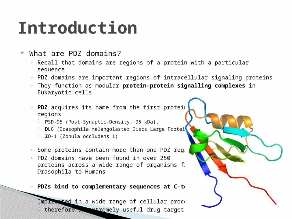
What are PDZ domains?◦ Recall that domains are regions of a protein with a particular sequence◦ PDZ domains are important regions of intracellular signaling proteins ◦ They function as modular protein-protein signalling complexes in Eukaryotic
cells
◦ PDZ acquires its name from the first proteins found to contain these regions PSD-95 (Post-Synaptic-Density, 95 kDa), DLG (Drasophila melangolaster Discs Large Protein) and ZO-1 (Zonula occludens 1)
◦ Some proteins contain more than one PDZ region◦ PDZ domains have been found in over 250
proteins across a wide range of organisms fromDrasophila to Humans
◦ PDZs bind to complementary sequences at C-termini
◦ Implicated in a wide range of cellular processes◦ – therefore an extremely useful drug target
Introduction
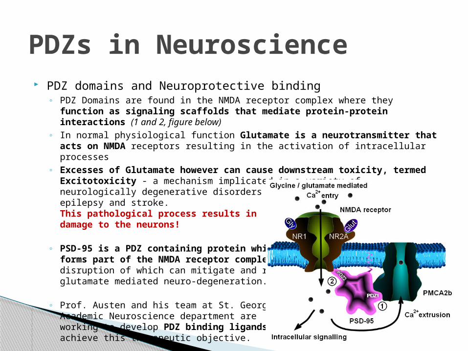
PDZ domains and Neuroprotective binding◦ PDZ Domains are found in the NMDA receptor complex where they function as
signaling scaffolds that mediate protein-protein interactions (1 and 2, figure below)
◦ In normal physiological function Glutamate is a neurotransmitter that acts on NMDA receptors resulting in the activation of intracellular processes
◦ Excesses of Glutamate however can cause downstream toxicity, termed Excitotoxicity - a mechanism implicated in a variety of neurologically degenerative disorders such as Alzheimers, Parkinsons, epilepsy and stroke.This pathological process results indamage to the neurons!
◦ PSD-95 is a PDZ containing protein whichforms part of the NMDA receptor complexdisruption of which can mitigate and reduceglutamate mediated neuro-degeneration.
◦ Prof. Austen and his team at St. GeorgesAcademic Neuroscience department areworking to develop PDZ binding ligands toachieve this therapeutic objective.
PDZs in Neuroscience
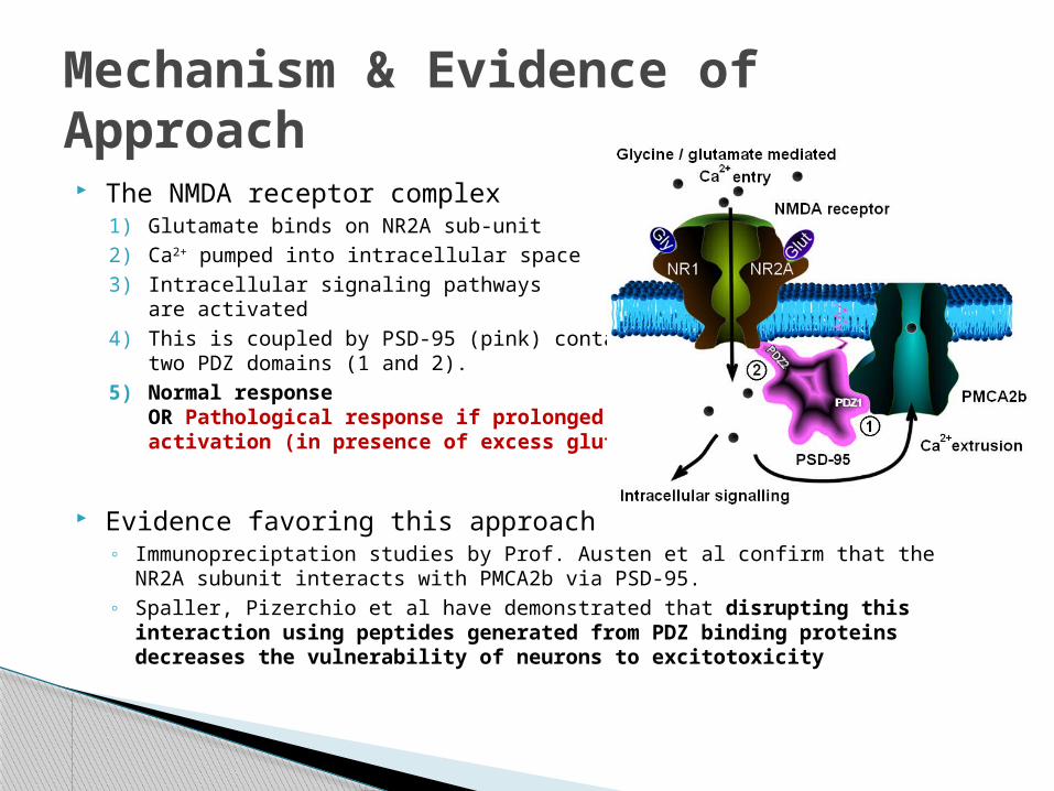
The NMDA receptor complex1) Glutamate binds on NR2A sub-unit2) Ca2+ pumped into intracellular space3) Intracellular signaling pathways
are activated4) This is coupled by PSD-95 (pink) containing
two PDZ domains (1 and 2).5) Normal response
OR Pathological response if prolongedactivation (in presence of excess glutamate)
Evidence favoring this approach◦ Immunopreciptation studies by Prof. Austen et al confirm that the NR2A subunit
interacts with PMCA2b via PSD-95.◦ Spaller, Pizerchio et al have demonstrated that disrupting this interaction
using peptides generated from PDZ binding proteins decreases the vulnerability of neurons to excitotoxicity
Mechanism & Evidence of Approach
Objectives◦ “To alter the function of the NMDA receptor complex by targeting PDZ1
domain of PSD-95 thereby disrupting it’s interaction with PMCA-2b◦ …to develop a PDZ binding ligand with improved binding characteristics
over an existing PDZ binding peptide ligand, R2…◦ …the putative ligand should therefore possess a smaller Kd dissociation
constant binding at nano-molar concentrations – R2 currently binds at micromolar concentrations.”
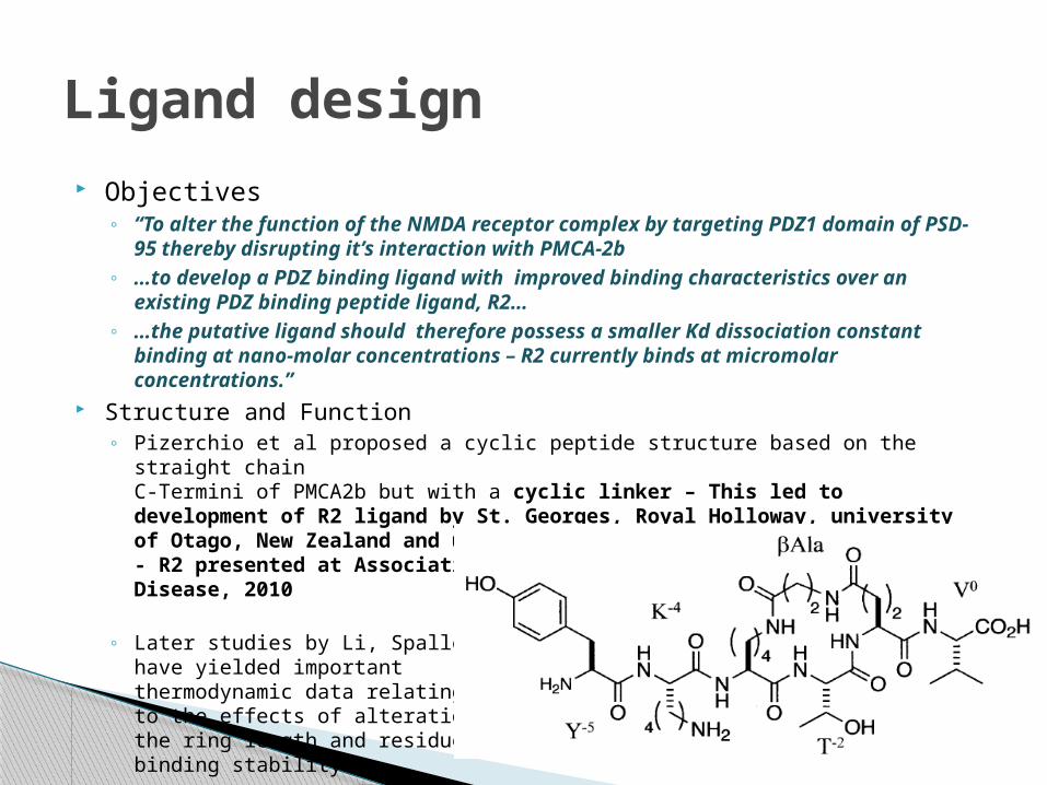
Structure and Function◦ Pizerchio et al proposed a cyclic peptide structure based on the straight chain
C-Termini of PMCA2b but with a cyclic linker – This led to development of R2 ligand by St. Georges, Royal Holloway, university of Otago, New Zealand and uni. of London- R2 presented at Association International Conference on Alzheimer's Disease, 2010
◦ Later studies by Li, Spaller et alhave yielded importantthermodynamic data relatingto the effects of alterations inthe ring length and residues onbinding stability
Ligand design
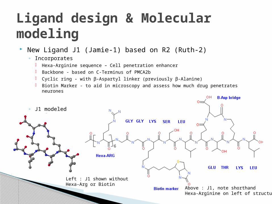
New Ligand J1 (Jamie-1) based on R2 (Ruth-2)◦ Incorporates
Hexa-Arginine sequence – Cell penetration enhancer Backbone - based on C-Terminus of PMCA2b Cyclic ring - with β-Aspartyl linker (previously β-Alanine) Biotin Marker - to aid in microscopy and assess how much drug penetrates neurones
◦ J1 modeledIn Silico
Ligand design & Molecular modeling
Left : J1 shown withoutHexa-Arg or Biotin
Above : J1, note shorthandHexa-Arginine on left of structure
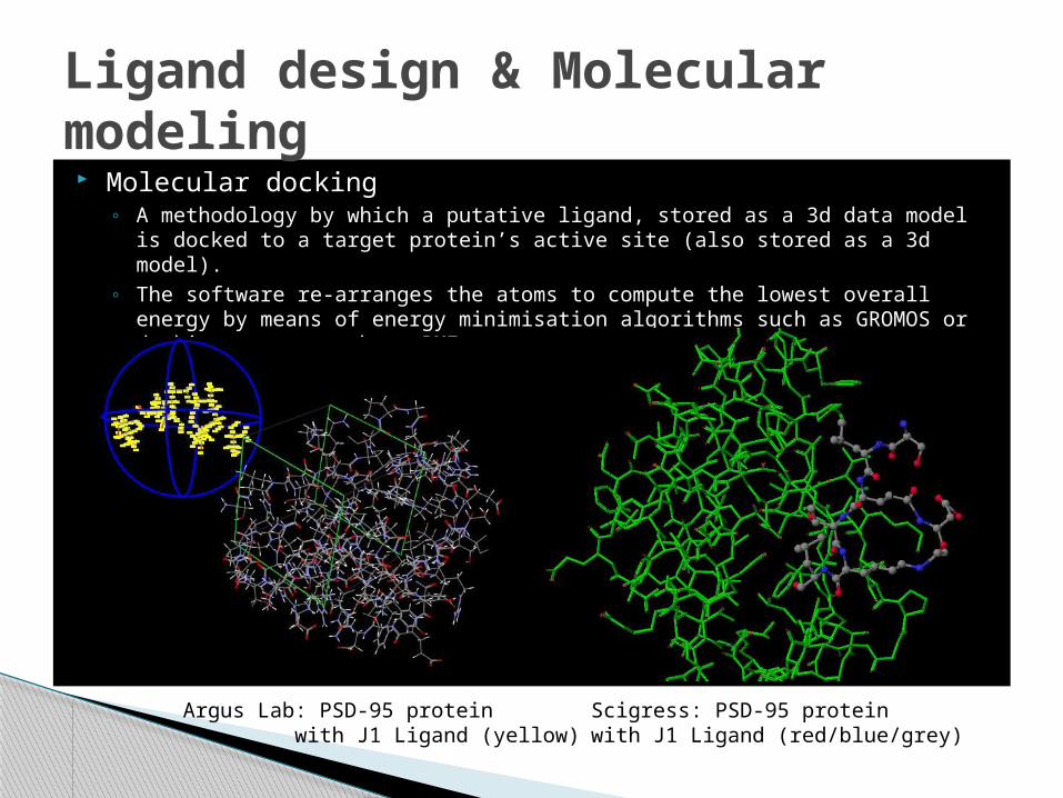
Molecular docking◦ A methodology by which a putative ligand, stored as a 3d data model is docked to
a target protein’s active site (also stored as a 3d model).◦ The software re-arranges the atoms to compute the lowest overall energy by
means of energy minimisation algorithms such as GROMOS or docking scores such as PMF
Ligand design & Molecular modeling
Argus Lab: PSD-95 protein with J1 Ligand (yellow)
Scigress: PSD-95 proteinwith J1 Ligand (red/blue/grey)
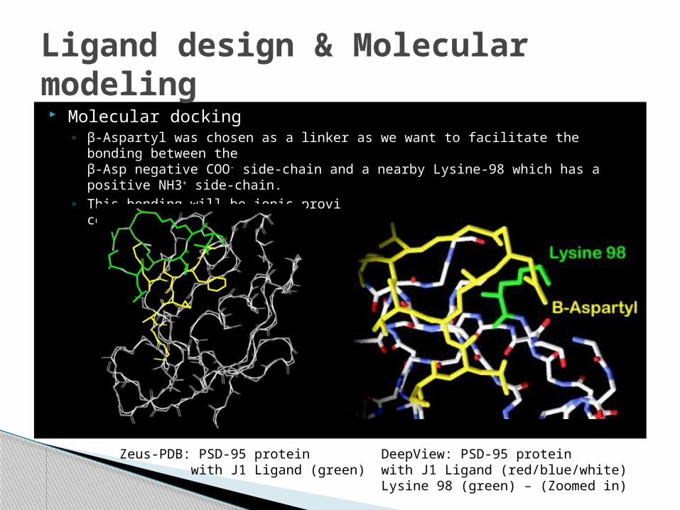
Molecular docking◦ β-Aspartyl was chosen as a linker as we want to facilitate the bonding between
theβ-Asp negative COO- side-chain and a nearby Lysine-98 which has a positive NH3+ side-chain.
◦ This bonding will be ionic providing extra stability to the bound complex
Ligand design & Molecular modeling
Zeus-PDB: PSD-95 protein with J1 Ligand (green)
DeepView: PSD-95 proteinwith J1 Ligand (red/blue/white)Lysine 98 (green) – (Zoomed in)
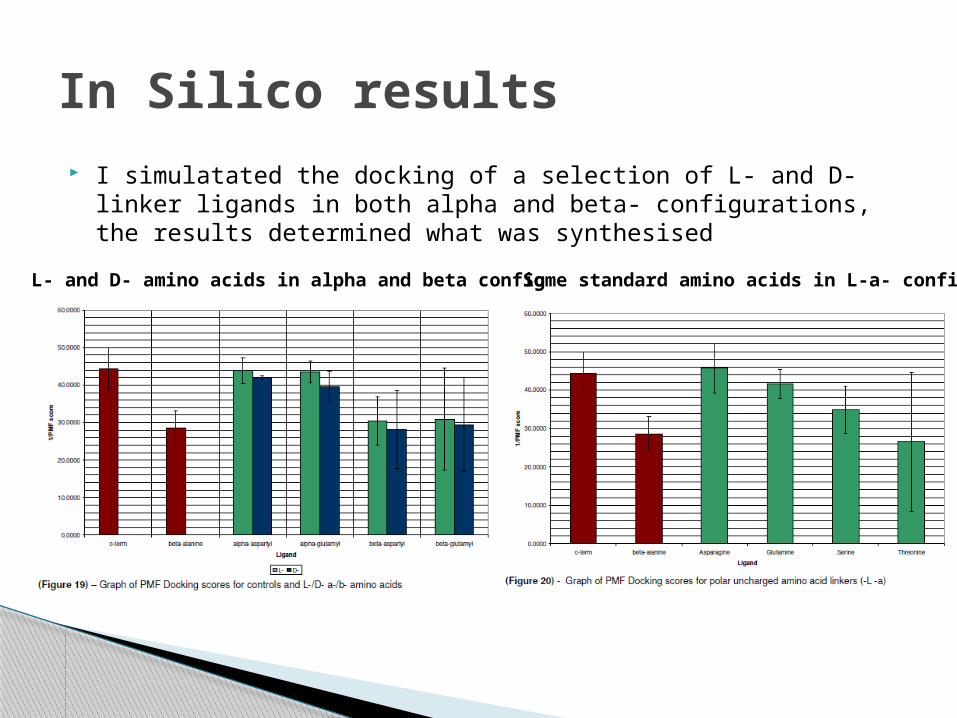
I simulatated the docking of a selection of L- and D- linker ligands in both alpha and beta- configurations, the results determined what was synthesised
In Silico results
L- and D- amino acids in alpha and beta config Some standard amino acids in L-a- config
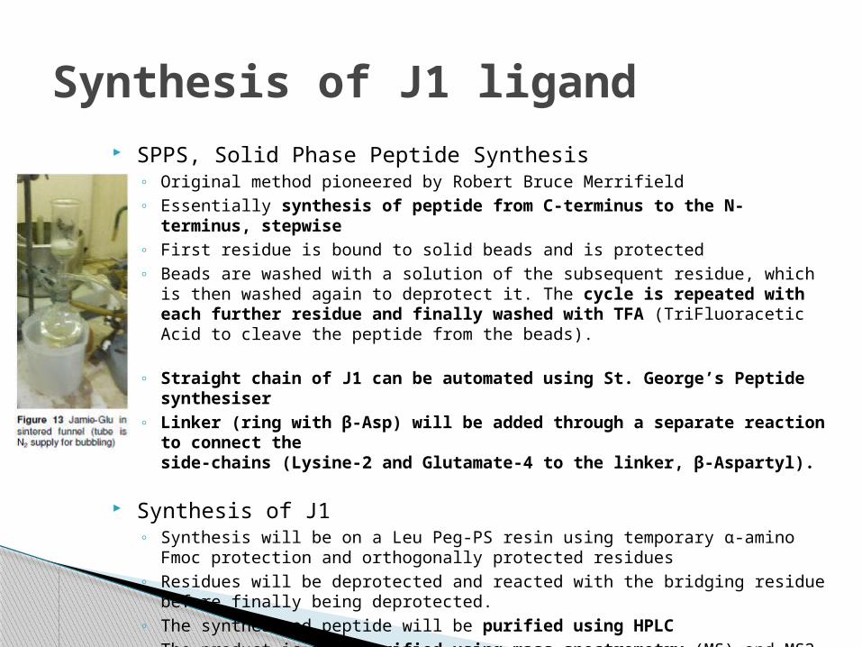
SPPS, Solid Phase Peptide Synthesis◦ Original method pioneered by Robert Bruce Merrifield◦ Essentially synthesis of peptide from C-terminus to the N-terminus,
stepwise◦ First residue is bound to solid beads and is protected◦ Beads are washed with a solution of the subsequent residue, which is then
washed again to deprotect it. The cycle is repeated with each further residue and finally washed with TFA (TriFluoracetic Acid to cleave the peptide from the beads).
◦ Straight chain of J1 can be automated using St. George’s Peptide synthesiser
◦ Linker (ring with β-Asp) will be added through a separate reaction to connect theside-chains (Lysine-2 and Glutamate-4 to the linker, β-Aspartyl).
Synthesis of J1◦ Synthesis will be on a Leu Peg-PS resin using temporary α-amino Fmoc
protection and orthogonally protected residues◦ Residues will be deprotected and reacted with the bridging residue before
finally being deprotected.◦ The synthesised peptide will be purified using HPLC◦ The product is then verified using mass spectrometry (MS) and MS2 by
Maldi
Synthesis of J1 ligand
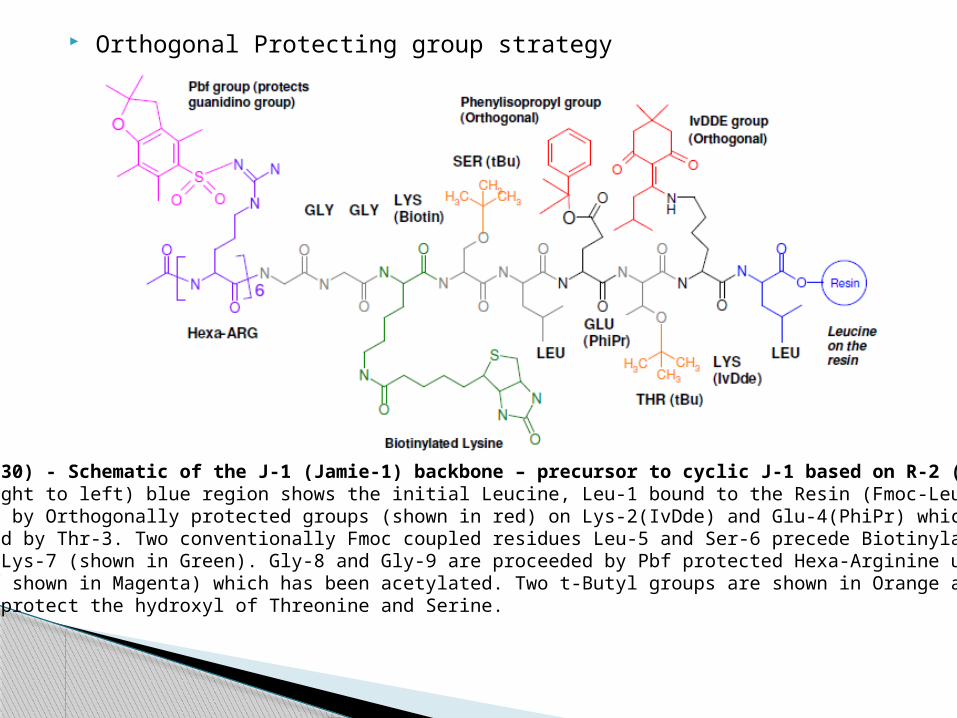
Orthogonal Protecting group strategy
(Figure 30) - Schematic of the J-1 (Jamie-1) backbone – precursor to cyclic J-1 based on R-2 (Ruth-2).(From right to left) blue region shows the initial Leucine, Leu-1 bound to the Resin (Fmoc-Leu-PEG-PS)followed by Orthogonally protected groups (shown in red) on Lys-2(IvDde) and Glu-4(PhiPr) which areseparated by Thr-3. Two conventionally Fmoc coupled residues Leu-5 and Ser-6 precede BiotinylatedLysine, Lys-7 (shown in Green). Gly-8 and Gly-9 are proceeded by Pbf protected Hexa-Arginine unit (Purplewith Pbf shown in Magenta) which has been acetylated. Two t-Butyl groups are shown in Orange and areused to protect the hydroxyl of Threonine and Serine.
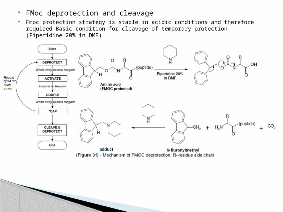
FMoc deprotection and cleavage Fmoc protection strategy is stable in acidic conditions and therefore required Basic
condition for cleavage of temporary protection (Piperidine 20% in DMF)
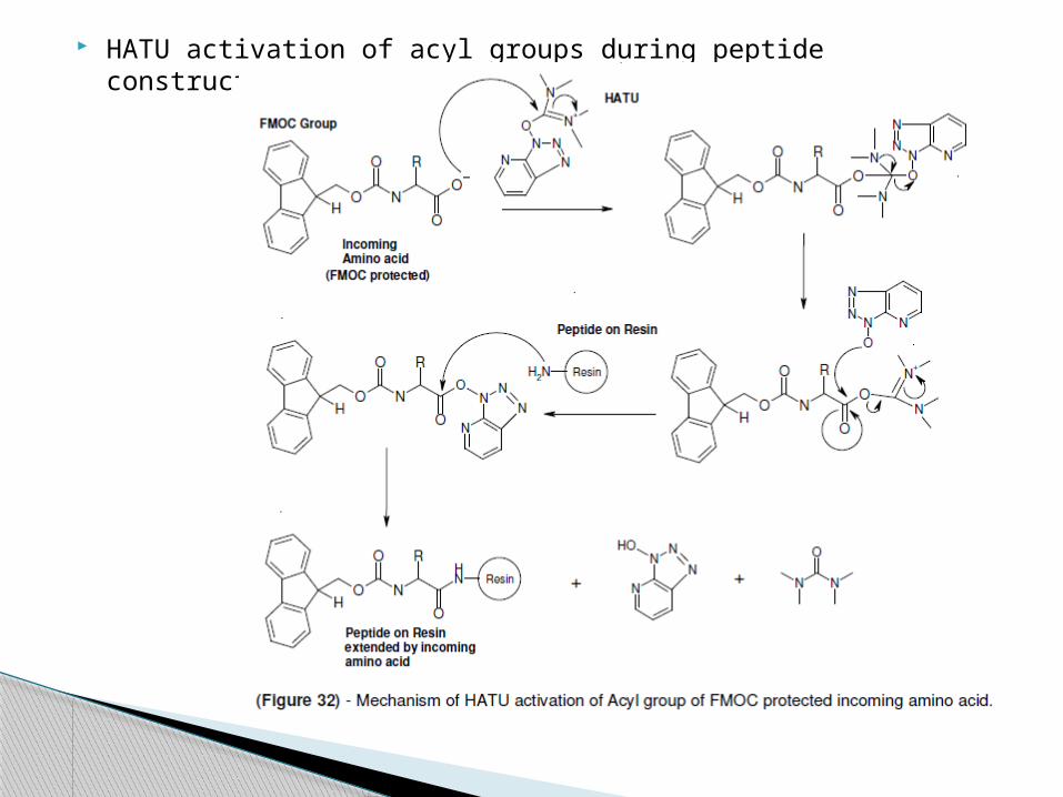
HATU activation of acyl groups during peptide construction
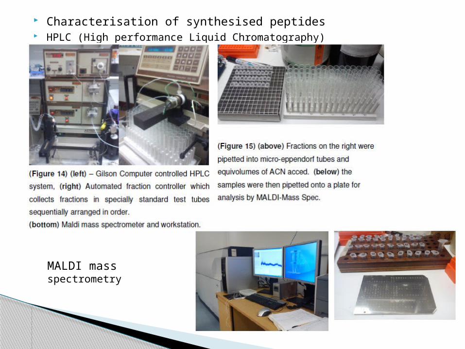
Characterisation of synthesised peptides HPLC (High performance Liquid Chromatography)
MALDI mass spectrometry
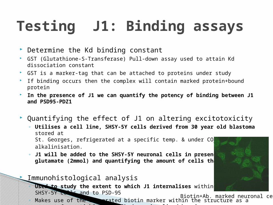
Determine the Kd binding constant GST (Glutathione-S-Transferase) Pull-down assay used to attain Kd dissociation
constant GST is a marker-tag that can be attached to proteins under study If binding occurs then the complex will contain marked protein+bound protein In the presence of J1 we can quantify the potency of binding between J1 and
PSD95-PDZ1
Quantifying the effect of J1 on altering excitotoxicity◦ Utilises a cell line, SHSY-5Y cells derived from 30 year old blastoma stored
atSt. Georges, refrigerated at a specific temp. & under CO2 to prevent alkalinisation.
◦ J1 will be added to the SHSY-5Y neuronal cells in presence of excess glutamate (2mmol) and quantifying the amount of cells that survive.
Immunohistological analysis◦ Used to study the extent to which J1 internalises within
SHSY-5Y cells and to PSD-95◦ Makes use of the integrated biotin marker within the structure as a
target for anti-biotin antibody - visualised by microscopy
Testing J1: Binding assays
Biotin+Ab. marked neuronal cells
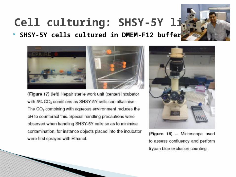
Cell culturing: SHSY-5Y line SHSY-5Y cells cultured in DMEM-F12 buffer
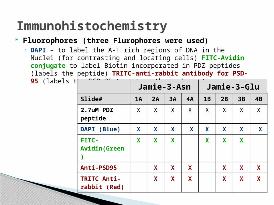
Immunohistochemistry Fluorophores (three Flurophores were used)
◦ DAPI – to label the A-T rich regions of DNA in the Nuclei (for contrasting and locating cells) FITC-Avidin conjugate to label Biotin incorporated in PDZ peptides (labels the peptide) TRITC-anti-rabbit antibody for PSD-95 (labels the PSD-95 target on the membrane)
Jamie-3-Asn Jamie-3-Glu
Slide# 1A 2A 3A 4A 1B 2B 3B 4B
2.7uM PDZ peptide
X X X X X X X X
DAPI (Blue) X X X X X X X X
FITC-Avidin(Green)
X X X X X X
Anti-PSD95 X X X X X X
TRITC Anti-rabbit (Red)
X X X X X X
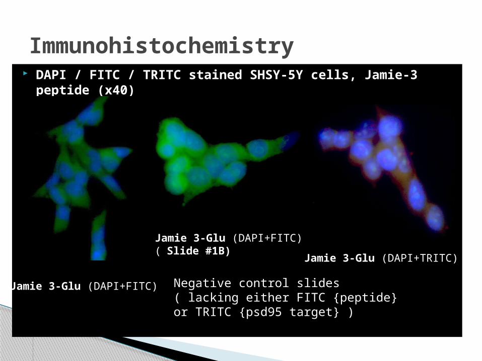
Immunohistochemistry DAPI / FITC / TRITC stained SHSY-5Y cells, Jamie-3 peptide
(x40)
Jamie 3-Glu (DAPI+FITC)
Jamie 3-Glu (DAPI+FITC)( Slide #1B)
Jamie 3-Glu (DAPI+TRITC)
Negative control slides ( lacking either FITC {peptide} or TRITC {psd95 target} )
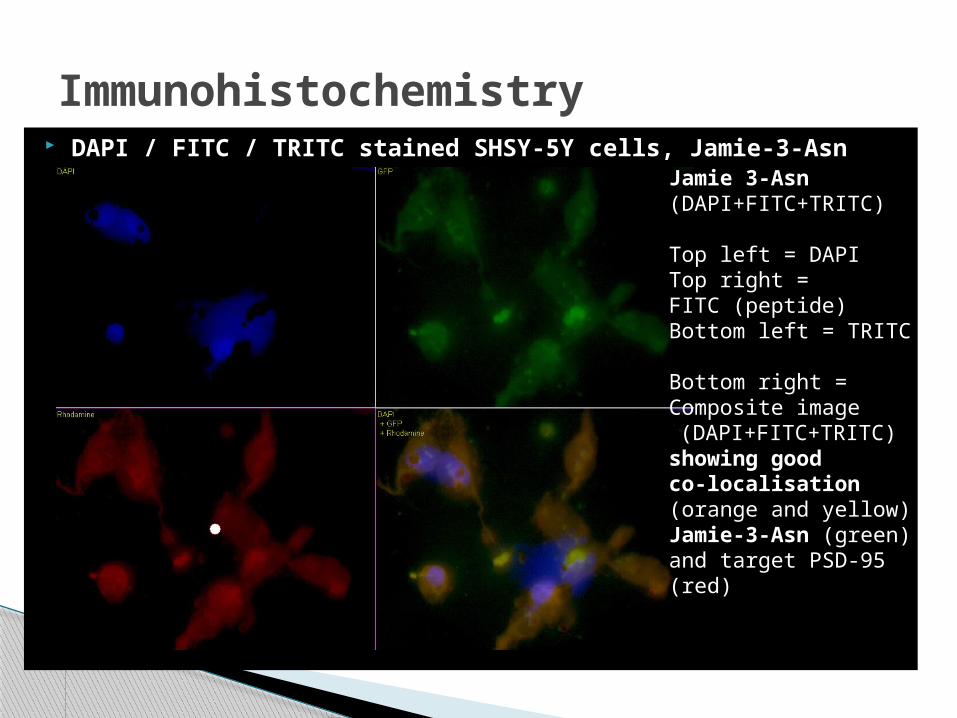
Immunohistochemistry DAPI / FITC / TRITC stained SHSY-5Y cells, Jamie-3-Asn (x63)
Jamie 3-Asn(DAPI+FITC+TRITC)
Top left = DAPITop right =FITC (peptide)Bottom left = TRITC
Bottom right =Composite image (DAPI+FITC+TRITC)showing goodco-localisation(orange and yellow) ofJamie-3-Asn (green)and target PSD-95(red)
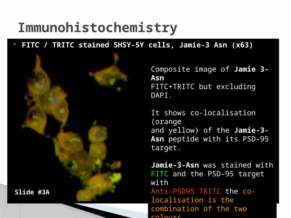
Immunohistochemistry FITC / TRITC stained SHSY-5Y cells, Jamie-3 Asn (x63)
Composite image of Jamie 3-AsnFITC+TRITC but excluding DAPI.
It shows co-localisation (orangeand yellow) of the Jamie-3-Asn peptide with its PSD-95 target.
Jamie-3-Asn was stained with FITC and the PSD-95 target withAnti-PSD95 TRITC the co-localisation is the combination of the two colours
Slide #3A
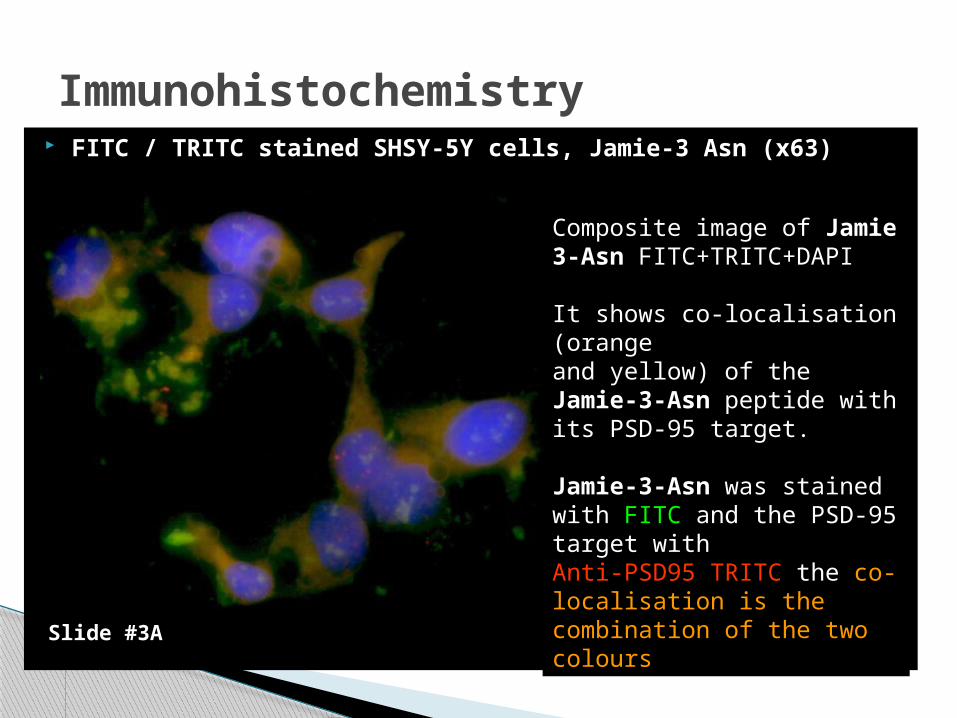
Immunohistochemistry FITC / TRITC stained SHSY-5Y cells, Jamie-3 Asn (x63)
Composite image of Jamie 3-Asn FITC+TRITC+DAPI
It shows co-localisation (orangeand yellow) of the Jamie-3-Asn peptide with its PSD-95 target.
Jamie-3-Asn was stained with FITC and the PSD-95 target withAnti-PSD95 TRITC the co-localisation is the combination of the two colours
Slide #3A
PDZ domains◦ PDZ domains are important protein-protein cell signaling mediators◦ Implicated in a variety of cellular processes not just neurological◦ Important drug target for novel drugs
Molecular modeling and In Silico methods◦ Molecular modeling and docking is extremely useful method of simulating
thermodynamic stability and binding between ligands and proteins◦ Data can be used to select a putative ligand prior synthesis vs the traditional
combinatorial approach
Immunohistochemical analysis◦ Jamie-3-Asn pdz binding peptide showed good co-localisation of peptide with PSD-95◦ Jamie-3-Glu showed some co-localisation but was less consistent across repeated imaging◦ Results consistent with MTT assay, in which Jamie-3-Asn pdz binding peptide performed
better at mitigating excitotoxic damage than Jamie-3-Glu
Implications◦ More favorable binding characteristics than R2 - further development may yield a
useful pharmacological agent that could be administered to slow down neurological degeneration in diseases such as Alzeimers, Parkinsons, epilepsy and stroke.
Conclusions
References used1. Harris BZ, Lim WA. Mechanism and role of PDZ domains in signaling complex
assembly. Journal of Cell Science, 2010; 114: 3219-32312. Cui H, Hayashi A, Sun HS, Belmares MP, Cobey C, Phan T et al. PDZ Protein
Interactions Underlying NMDA Receptor-Mediated Excitotoxicity and Neuroprotection by PSD-95 Inhibitors. The Journal of Neuroscience, 2007; 27(37): 9901-9915
3. Li T, Saro D, Spaller MR. Thermodynamic profiling of conformationally constrained cyclic ligands for the PDZ domain. Bioorganic & Medicinal Chemistry Letters, 2004; 14(6): 1385-1388
4. Austen BM, Duberley K, Turner P, Empson R. Cyclic hexa-arg PDZ-binding peptides that bind PSD95 inhibit glutamate-mediated toxicity. Alzheimer's Association International Conference on Alzheimer's Disease 2010, 6(4): Supp. 562
Software used (in order of images shown)
1. Argus Lab by Mark Thompson2. Scigress explorer by Fujitsu Siemens3. Zeus PDB viewer by Jamie al-nasir4. DeepView/Swiss-PDB viewer
References