Embed Size (px)
Citation preview
materials
Article
Polydopamine Linking Substrate for AMPs:Characterisation and Stability on Ti6Al4V
Zuzanna Trzcinska 1, Marc Bruggeman 1 , Hanieh Ijakipour 1, Nikolas J. Hodges 2 ,James Bowen 3 and Artemis Stamboulis 1,*
1 School of Metallurgy and Materials, University of Birmingham, Edgbaston, Birmingham B15 2TT, UK;[email protected] (Z.T.); [email protected] (M.B.); [email protected] (H.I.)
2 School of Biosciences, University of Birmingham, Edgbaston, Birmingham B15 2TT, UK;[email protected]
3 School of Chemical Engineering, University of Birmingham, Edgbaston, Birmingham B15 2TT, UK;[email protected]
* Correspondence: [email protected]
Received: 15 May 2020; Accepted: 19 August 2020; Published: 22 August 2020�����������������
Abstract: Infections are common complications in joint replacement surgeries. Eradicated infectionscan lead to implant failure. In this paper, analogues of the peptide KR-12 derived from the humancathelicidin LL-37 were designed, synthesised, and characterised. The designed antimicrobialpeptides (AMPs) were attached to the surface of a titanium alloy, Ti6Al4V, by conjugation to apolydopamine linking substrate. The topography of the polydopamine coating was evaluated byelectron microscopy and coating thickness measurements were performed with ellipsometry andAtomic Force Microscopy (AFM). The subsequently attached peptide stability was investigated withrelease profile studies in simulated body fluid, using both fluorescence imaging and High-PerformanceLiquid Chromatography (HPLC). Finally, the hydrophobicity of the coating was characterised bywater contact angle measurements. The designed AMPs were shown to provide long-term bondingto the polydopamine-coated Ti6Al4V surfaces.
Keywords: Ti6Al4V; polydopamine; antimicrobial peptides; cathelicidin; KR-12
1. Introduction
Infection are the most common complications of joint replacement surgery, with nosocomial orhospital-acquired infections ranking as the sixth leading cause of death, presenting a major healthcarechallenge [1]. Infection can lead to extended inflammation at the site of the surgery, thus causing therejection and failure of the implant [2]. Although the administration of antibiotics significantly reducesthe risk of postsurgical infections, bacterial biofilm production on the implant surface or untimelyadministration of antibiotics will reduce their effectiveness [3].
The use of titanium in dental and orthopaedic implants is well established due to titanium’sstrength, stiffness, and corrosion resistance. Titanium also shows seamless integration with thesurrounding tissues due to its excellent biocompatibility [4,5]. The drawback of using titaniumimplants is their susceptibility to bacterial colonisation on the surfaces of the implants [6]. To combatthe formation of biofilms on implant surfaces, the time-controlled release of various antibiotic coatingshas previously been investigated [7,8]. However, the release of the antibiotics below the level ofthe minimum inhibitory concentration (MIC) is known to produce antibiotic-resistant strains of bacteria.Higher levels of antibiotic release have been shown to be toxic to the surrounding tissues. The increase ofantibiotic-resistant bacteria has led to the search for an alternative method of antimicrobial protection [9].
Materials 2020, 13, 3714; doi:10.3390/ma13173714 www.mdpi.com/journal/materials
Materials 2020, 13, 3714 2 of 19
Antimicrobial peptides, which are a part of the innate immune system of all living organisms, havebroad-spectrum activity against many microorganisms, such as Gram-positive bacteria, Gram-negativebacteria, viruses, and fungi [10–13]. Moreover, they can inhibit biofilm formation and induceits dissolution, as well as attract phagocytes to further induce natural defence mechanisms [14].The mechanisms of action of antimicrobial peptides (AMPs) against bacteria are not fully understooddue to the high diversity of these peptides. Nevertheless, it is widely accepted that bacterial cell deathis due to the interaction of cationic AMPs with negatively charged phospholipids on the bacterialmembrane, which lead to the loss of membrane structural integrity, and eventually cell death [10–13].AMPs exhibit a strong preference for specific membrane compositions, allowing them to be selectivetowards bacterial cell membranes, but not mammalian or plant cells [14]. Currently, only a few AMPsare used clinically due to limiting factors, such as the high cost of peptide synthesis, their susceptibilityto proteolytic degradation, and their unknown long-term toxicology profiles [1,14,15]. Here, analoguesof the peptide KR-12 derived from the human cathelicidin LL-37 were designed due to its establishedantimicrobial activity and lack of mammalian cell toxicity, as originally found by Jacob et al. [16].
To introduce the antimicrobial peptides stably on a surface, different types of coatings can beemployed. A popular approach is the use of polydopamine (pDA), a strong adhesive mussel-inspiredpolymer, due to its low cost, simplicity of application, and improved biocompatibility [17–19].Similarly to mussel adhesive proteins, the adhesive properties of the pDA are owed to quinine andcatechol groups, which create chelating structures with metals. Additionally, after polymerisation,pDA can be further functionalised with amine-containing nucleophiles, such as proteins and peptides.This allows the application of a pDA coating to Ti6Al4V, where the in-house designed analogues of thepeptide KR-12 are subsequently covalently bonded to the pDA coating.
2. Materials and Methods
2.1. Peptide Design
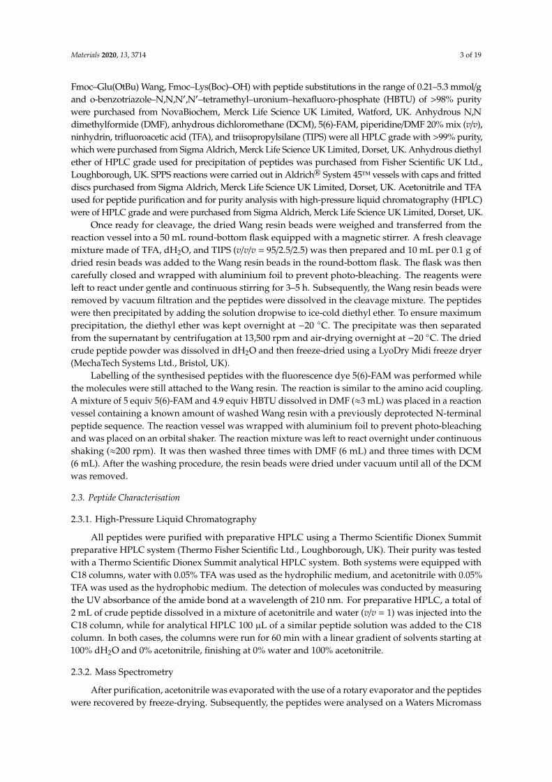
The shortest active peptide fragment derived from cathelicidin LL-37, KR12, was used as atemplate to design three peptides of varying antimicrobial activity. The peptide sequences and variousproperties are summarised in Table 1.
Table 1. Table showing the used peptides sequences, including their charge, hydrophobicity, andamphiphacity, determined by hydrophobic characteristics. Within the sequences, positively chargedresidues are marked in blue, the negatively charged in red, and highly hydrophobic sequences areunderlined.
Peptide Sequence Length ofSequence
NetCharge
ChargeDensity
MeanHydrophobicity
[H]
HelicalHydrophobic
KR12 KRIVQRIKDFLR 12 aa +4 0.33 0.193 0.782KR12/32 KIRVQRIKDFLR 12 aa +4 0.33 0.193 0.429
KR12-5911 KRIVRIKFR 9 aa +5 0.56 0.178 0.395KR12/32-5911 KIRVRIKFR 9 aa +5 0.56 0.178 0.092
2.2. Peptide Synthesis, Purification
Peptides dyed with carboxyfluorescein (5(6)-FAM) were synthesised in-house following conventionalsolid-phase peptide synthesis (SPPS). After synthesis, the peptides were purified to show a minimum purityof >95%. Peptides without the dye exhibited a purity of >98% and were purchased from ProteoGenix,Schiltigheim, France. All the amino acids used in SPPS (i.e., Fmoc–Ala–OH, Fmoc–Asp(OtBu)–OH,Fmoc–Glu(OtBu)–OH, Fmoc–Phe–OH, Fmoc–Gly–OH, Fmoc–Ile–OH, Fmoc–Lys(Boc)–OH, Fmoc–Leu–OH,Fmoc–Asn–OH, Fmoc–Gln–OH, Fmoc–Pro–OH, Fmoc–Arg(Pbf)–OH) were purchased from AGTCBioproducts Ltd., Itlings Lane Hessle, UK, with a purity of >98 % purity, which were side-chain protectedwhere appropriate. Preloaded Wang resin with Fmoc-protected amino acids (Fmoc–Arg(Pbf) Wang,
Materials 2020, 13, 3714 3 of 19
Fmoc–Glu(OtBu) Wang, Fmoc–Lys(Boc)–OH) with peptide substitutions in the range of 0.21–5.3 mmol/gand o-benzotriazole–N,N,N′,N′–tetramethyl–uronium–hexafluoro-phosphate (HBTU) of >98% puritywere purchased from NovaBiochem, Merck Life Science UK Limited, Watford, UK. Anhydrous N,Ndimethylformide (DMF), anhydrous dichloromethane (DCM), 5(6)-FAM, piperidine/DMF 20% mix (v/v),ninhydrin, trifluoroacetic acid (TFA), and triisopropylsilane (TIPS) were all HPLC grade with >99% purity,which were purchased from Sigma Aldrich, Merck Life Science UK Limited, Dorset, UK. Anhydrous diethylether of HPLC grade used for precipitation of peptides was purchased from Fisher Scientific UK Ltd.,Loughborough, UK. SPPS reactions were carried out in Aldrich® System 45™ vessels with caps and fritteddiscs purchased from Sigma Aldrich, Merck Life Science UK Limited, Dorset, UK. Acetonitrile and TFAused for peptide purification and for purity analysis with high-pressure liquid chromatography (HPLC)were of HPLC grade and were purchased from Sigma Aldrich, Merck Life Science UK Limited, Dorset, UK.
Once ready for cleavage, the dried Wang resin beads were weighed and transferred from thereaction vessel into a 50 mL round-bottom flask equipped with a magnetic stirrer. A fresh cleavagemixture made of TFA, dH2O, and TIPS (v/v/v = 95/2.5/2.5) was then prepared and 10 mL per 0.1 g ofdried resin beads was added to the Wang resin beads in the round-bottom flask. The flask was thencarefully closed and wrapped with aluminium foil to prevent photo-bleaching. The reagents wereleft to react under gentle and continuous stirring for 3–5 h. Subsequently, the Wang resin beads wereremoved by vacuum filtration and the peptides were dissolved in the cleavage mixture. The peptideswere then precipitated by adding the solution dropwise to ice-cold diethyl ether. To ensure maximumprecipitation, the diethyl ether was kept overnight at −20 ◦C. The precipitate was then separatedfrom the supernatant by centrifugation at 13,500 rpm and air-drying overnight at −20 ◦C. The driedcrude peptide powder was dissolved in dH2O and then freeze-dried using a LyoDry Midi freeze dryer(MechaTech Systems Ltd., Bristol, UK).
Labelling of the synthesised peptides with the fluorescence dye 5(6)-FAM was performed whilethe molecules were still attached to the Wang resin. The reaction is similar to the amino acid coupling.A mixture of 5 equiv 5(6)-FAM and 4.9 equiv HBTU dissolved in DMF (≈3 mL) was placed in a reactionvessel containing a known amount of washed Wang resin with a previously deprotected N-terminalpeptide sequence. The reaction vessel was wrapped with aluminium foil to prevent photo-bleachingand was placed on an orbital shaker. The reaction mixture was left to react overnight under continuousshaking (≈200 rpm). It was then washed three times with DMF (6 mL) and three times with DCM(6 mL). After the washing procedure, the resin beads were dried under vacuum until all of the DCMwas removed.
2.3. Peptide Characterisation
2.3.1. High-Pressure Liquid Chromatography
All peptides were purified with preparative HPLC using a Thermo Scientific Dionex Summitpreparative HPLC system (Thermo Fisher Scientific Ltd., Loughborough, UK). Their purity was testedwith a Thermo Scientific Dionex Summit analytical HPLC system. Both systems were equipped withC18 columns, water with 0.05% TFA was used as the hydrophilic medium, and acetonitrile with 0.05%TFA was used as the hydrophobic medium. The detection of molecules was conducted by measuringthe UV absorbance of the amide bond at a wavelength of 210 nm. For preparative HPLC, a total of2 mL of crude peptide dissolved in a mixture of acetonitrile and water (v/v = 1) was injected into theC18 column, while for analytical HPLC 100 µL of a similar peptide solution was added to the C18column. In both cases, the columns were run for 60 min with a linear gradient of solvents starting at100% dH2O and 0% acetonitrile, finishing at 0% water and 100% acetonitrile.
2.3.2. Mass Spectrometry
After purification, acetonitrile was evaporated with the use of a rotary evaporator and the peptideswere recovered by freeze-drying. Subsequently, the peptides were analysed on a Waters Micromass
Materials 2020, 13, 3714 4 of 19
LCT TOF spectrometer (Waters UK, Wilmslow, UK) using electrospray ionisation in the School ofChemistry Mass Spectrometry facility. Prior to mass analysis, the peptide samples were dissolved inwater. After the purification of the crude peptides, a purity of ≥95% was achieved and the theoreticalmass agreed with the calculated mass displayed in Table 2.
Table 2. Purity and mass analysis of peptides.
Peptide Theoretical MolecularWeight (g/mol)
Molecular Weight MMeasured by MS (g/mol)
Purity Calculatedby HPLC (%)
KR12 1572M = 1574
[M + 2H]2+ = 788[M + Na + H]2+ = 799
95
5(6)-FAM-labelled KR12 1904 M = 1903[M + 2H]2+ = 952 95
KR12/32 1572M = 1572
[M + 2H]2+ = 787[M + 3H]3+ = 525
99
5(6)-FAM-labelledKR12/32 1904
M = 1903[M + H]+ = 1904[M + 2H]2+ = 952
95
KR12-5911 1216 M = 1215[M + 2H]2+ = 609 97
5(6)-FAM-labelledKR12-5911 1548
M = 1547[M + H]+ = 1549[M + 2H]2+ = 775
95
KR12/32-5911 1216 M = 1215[M + 2H]2+ = 609 98
5(6)-FAM-labelledKR12/32-5911 1548 M = 1547
[M + 2H]2+ = 775 96
KR12 1572M = 1574
[M + 2H]2+ = 788[M + Na + H]2+ = 799
95
5(6)-FAM-labelled KR12 1904 M = 1903[M + 2H]2+ = 952 95
2.3.3. Minimum Inhibitory Concentration (MIC) Values of Peptides
The minimum inhibition concentration (MIC) is used to determine the lowest concentrationof antimicrobial agent needed to inhibit the visible growth of a bacteria strain after overnightincubation. All the bacteria strains were kindly provided by Dr Mark Webber of the Quadram InstituteBioscience (Norwich, UK; Escherichia coli (E. coli, I364), Pseudomonas aeruginosa (P. aeruginosa, PAO1) andStaphylococcus aureus (S. aureus, F77/NCTC8532)). Lysogen broth (LB broth) and agar were purchasedfrom Sigma-Aldrich, Merck Life Science UK Limited, Dorset, UK. Fresh LB agar culture plates wereprepared by pouring ≈10 mL of an autoclaved warm mixture of LB broth (2.5%) and bacteriologicalagar (1.5%) dissolved in dH2O. The agar plate was streaked and incubated overnight at 36 ◦C. A singlebacterial colony was chosen and grown in 5 mL broth overnight under agitation at 36 ◦C. Subsequently,50 µL of LB broth was added to wells 2–12 of a 96-well culture plate. AMPs were diluted to aconcentration of 256 µg/mL, added to well 1, and diluted two-fold down to column 11. Column 12 wasleft empty, with no AMPs added. Then, 50 µL of the diluted overnight bacteria culture was added tothe wells and incubated at 36 ◦C for 18 h to allow bacteria to grow. After incubation, the well plateswere examined for bacterial growth and the lowest concentration of AMPs where clear liquid wasobserved was assumed to be the minimum inhibitory concentration. Three measurements for eachpeptide and against each type of bacteria were performed and the average values were obtained.
Materials 2020, 13, 3714 5 of 19
2.4. Sample Preparation
Titanium alloy grade 5 (Ti6Al4V) plates with dimensions of 15 cm × 15 cm and a thickness of0.1 cm were purchased from William Gregor Ltd., London, UK, and cut into 1 cm × 1 cm plates. Then,the plates were mounted in conducting Bakelite and polished to mirror finish. Three steps were usedduring polishing of the plates. First, Bakelite-mounted Ti6Al4V plates were ground with MD-Piano of220 grit, with water used as a lubricant. Then, a DiaDuo-2 diamond of 9 µm grain size suspended inwater and an MD-Largo polishing plate were used. Finally, a colloidal suspension (OP-S) activatedwith ammonia solution was used and polished on MD-Chem polishing disc. All the polishing materialsand equipment were purchased from Struers Ltd., Rotherham, UK. After polishing the plates to amirror finish, the highly polished surfaces were secured with electrical tape to prevent the introductionof scratches and Bakelite was broken down to release the mounted Ti6Al4V plates. The tape was thenremoved from the plates and any impurities introduced on the Ti6Al4V surfaces during the previoussteps were removed by cleaning the plates in an ultrasonic bath with water (15 min) and acetone(15 min). The plates were dried overnight in a desiccator and were used within 48 h after cleaning.
Having been cleaned and polished to a mirror finish, Ti6Al4V plates were placed inside a 24-wellcell culture plate with the polished side facing upwards. Dopamine, purchased from Sigma-Aldrich,was dissolved to a final concentration of 5 mg/mL in 50 mM Tris buffer (Fisher Scientific UK Ltd.,Loughborough, UK) at pH = 8.5. Then, 1.5 mL of dopamine solution was transferred into the cellculture plates containing the Ti6Al4V plates. Subsequently, the prepared plates were placed in the darkfor 24 h without a cover to allow simultaneous dopamine polymerisation in air and metallic surfacecoating. Ti6Al4V plates coated with polydopamine (pDA) were washed to remove any loose pDAparticles and placed in a new set of 24-well cell culture plates. The AMP solution was then prepared bydissolving peptides in a concentration equal to the MIC value for each peptide in 50 mM Tris buffer atpH 7.4. Then, 1.5 mL of this solution was transferred into the cell culture plates with the pDA-coatedTi6Al4V plates. The cell culture plates were then kept in the dark without a cover for 24 h to allowconjugation of the peptides with the pDA. Finally, the plates were washed several times with dH2O toremove unconjugated peptides. The prepared plates were stored in the dark in a desiccator to dry andused within 7 days of preparation.
2.5. Topography of Coating
The topography analysis of the pDA- and AMP-conjugated pDA coatings was performedwith a Bruker Icon Atomic Force Microscope (Bruker UK Ltd., Coventry, UK). A silicone probewas passed over the surface of the coatings and its displacement was recorded. This generated athree-dimensional plot of the surface topography. In this research, images were recorded in staticmode over a 20 µm × 20 µm area. Ellipsometry was performed on a Jobin-Yvon UVISEL ellipsometer(HORIBA UK Ltd., Northampton, UK) with a xenon light source. First, the light reflection of theuncoated surface of the sample was measured to set the measurement baseline, then the height of thecoated surface was measured relative to the baseline.
2.6. Fluorescence Microscopy
Fluorescence microscopy was used to determine the conjugation of AMPs to the pDA with aLeica DM6000B widefield epifluorescence microscope (Leica Microsystems Ltd., Milton Keynes, UK),equipped with a 100 W short-arc epifluorescence mercury burner and a Leica DFL350 FX firewirecamera (Leica Microsystems Ltd., Milton Keynes, UK) located at the Institute of Biomedical Research(IBR) in the School of Medicine, University of Birmingham. Measurements were performed with anepifluorescence filter set at an excitation wavelength of 480 nm and a green emission wavelength of527 nm, corresponding to the green fluorescence associated with 5(6)-FAM. For each sample, a set offive random points were recorded for comparison and the brightness of the green light was analysedusing the ImageJ 1.46r analysis program.
Materials 2020, 13, 3714 6 of 19
2.7. Scanning Electron Microscopy
Imaging of the uncoated and pDA-coated surfaces was performed on a Zeiss Supra 55VP scanningelectron microscope (Carl Zeiss Ltd., Zeiss House, Cambridge, UK). All measurements were conductedon samples coated with ≈1 nm of the electrodeposited carbon film. A 10 kV electron beam and variousmagnifications were used.
2.8. Coating Stability
In order to prepare the simulated body fluid (SBF), sodium chloride (NaCl), sodium bicarbonate(NaHCO3), potassium chloride (KCl), potassium phosphate dibasic trihydrate (K2HPO4·3H2O),magnesium chloride hexahydrate (MgCl2·6H2O), hydrochloric acid (HCl) 32%, calcium chloride(CaCl2), sodium sulfate (Na2SO4), and tris(hydroxymethyl)aminomethane (Tris, (CH2OH)3CNH2)were purchased from Sigma Aldrich, Merck Life Science UK Limited, Dorset, UK. The SBF was preparedas described by Kokubo and Takadama [20]. The stability of the AMPs on the surface of Ti6Al4Vwas performed in SBF by submerging and keeping AMP-coated plates in SBF at 37 ◦C for a total of30 days. Analysis of the released AMPs to the solution was performed using two methodologies.Firstly, the change in green light intensity of the pDA Ti6Al4V surfaces coated with 5(6)-FAM-labelledpeptides was analysed after 1 and 6 h, then at 1, 3, 7, 14, and 30 days of submersion in SBF, imagedunder fluorescence microscopy. Secondly, plates with no labelled peptides were immersed in SBFsolution and analysed by HPLC at the same time intervals. However, in this method, 100 µL of the SBFsolution was withdrawn to determine the cumulative absorbance at the same retention time for everyindividual peptide.
2.9. Dynamic Contact Angle Analysis
Hydrophobicity was determined by a dynamic contact angle technique on a Dyne TechnologyThetaLite optical tensiometer (Dyne Testing Ltd., Lichfield, UK) located in the Science City Lab at theSchool of Chemical Engineering, University of Birmingham. Deionised water was used as a liquidmedium to perform the contact angle measurements. The measurements were performed by recordingthe contact and retraction angles of a 5 µL droplet released on and removed from the studied surface.The recording time of each incident was set to 20 s and the camera recorded 10 frames/s. The dropletswere released and then removed at a speed of 5 mL/min. All surfaces were kept in a desiccator prior tomeasurements and were each measured three times.
2.10. Cell Culture Studies
Human osteosarcoma cells (HOS) were purchased from the European Collection of AuthenticatedCell Cultures (catalogue number 87070202). Cultured cells were grown in RPMI medium supplementedwith 10% v/v fetal calf serum, 100 U/mL penicillin, 100 µg/mL streptomycin, and 2 mM glutamine as amonolayer in T75 cell culture flasks in a humidified atmosphere (5% CO2 incubator; 95% air) at 37 ◦C.Cells were sub-cultured at approximately 80% confluency twice-weekly using a standard trypsin-EDTAprotocol. All cell cultures were confirmed as being free from Mycoplasma sp. contamination using theEZ-PCR mycoplasma detection kit according to the manufacturer’s instructions (Biological IndustriesUSA, Cromwell, CT, USA). All cells were cultured up to passage 20 before being discarded. Beforecell culture, coated surfaces were placed flat with the coated surface facing upwards into a 6-well cellculture dish. Surfaces were sterilised with 2 mL of 70% v/v ethanol for 30 min. Subsequently, surfaceswere washed with sterile phosphate-buffered saline (PBS, 3 × 2 mL). HOS cells (100,000) were addedand left to attach for 4 h. The medium was changed and cells were incubated for 5 and 7 days beforebeing prepared for analysis by electron microscopy.
Materials 2020, 13, 3714 7 of 19
3. Results
3.1. Antimicrobial Characterisation
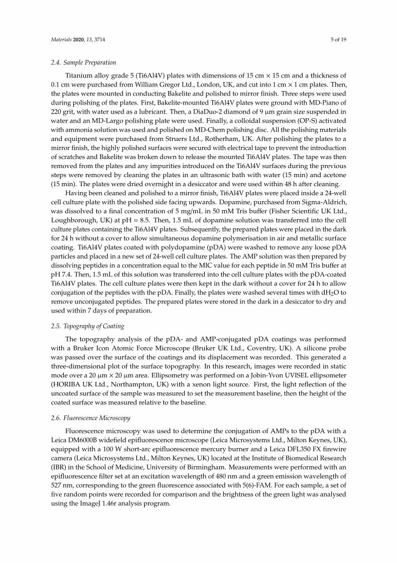
The MIC values of the peptides KR12, KR12/32, KR12-5911, and KR12/32-5911 against E. coli,P. aeruginosa, and S. aureus are shown in Figure 1 and compared against LL-37 in Table 3. The mosteffective peptide against E. coli was peptide KR12-5911 at 0.5 µM, followed by KR12 at 2 µM, andKR12/32 and KR12/32-5911 at 4 µM. The lowest MIC values against P. aeruginosa were observed forpeptides KR12 and KR12-5911 at 2 µM, whilst peptides KR12/32 and KR12/32-5911 showed MIC valuesof 4 µM. For S. aureus, the most effective was peptide KR12/32-5911 with an MIC of 2 µM, followed byKR12-5911 and KR12 at 8 µM and KR12/32 at 32 µM. All peptides show similar MIC values againstGram-negative bacteria. Additionally, the peptides were more effective against Gram-negative bacteriawhen compared to the Gram-positive bacteria, apart from KR12/32-5911.
Materials 2020, 13, x FOR PEER REVIEW 7 of 20
The MIC values of the peptides KR12, KR12/32, KR12-5911, and KR12/32-5911 against E. coli, P.
aeruginosa, and S. aureus are shown in Figure 1 and compared against LL-37 in Table 3. The most
effective peptide against E. coli was peptide KR12-5911 at 0.5 μM, followed by KR12 at 2 μM, and
KR12/32 and KR12/32-5911 at 4 μM. The lowest MIC values against P. aeruginosa were observed for
peptides KR12 and KR12-5911 at 2 μM, whilst peptides KR12/32 and KR12/32-5911 showed MIC
values of 4 μM. For S. aureus, the most effective was peptide KR12/32-5911 with an MIC of 2 μM,
followed by KR12-5911 and KR12 at 8 μM and KR12/32 at 32 μM. All peptides show similar MIC
values against Gram-negative bacteria. Additionally, the peptides were more effective against Gram-
negative bacteria when compared to the Gram-positive bacteria, apart from KR12/32-5911.
Figure 1. Minimum inhibitory concentration (MIC) values for the designed KR12 analogues.
Table 3. MIC values for the designed KR12 analogues compared to the LL-37 human cathelicidin.
Sequence MIC Values (μM)
E. coli (I364) P. aeruginosa (PAO1) S. aureus (F77) Geometric Mean
KR12 2 2 8 4.0
KR12/32 4 4 32 13.33
KR12-5911 0.5 2 8 3.5
KR12/32-5911 4 4 2 3.33
LL-37 [14] 8 8 4 6.67
3.2. Polymerisation of Dopamine
Dissolution of dopamine in the Tris buffer resulted in an immediate colour shift to light brown.
As the polymerisation proceeded, the colour became darker until the solution was black, as shown
in Figure 2. As can be observed, once the polymerisation took place for 96 h, a film formed between
the surface of the liquid and the air, resulting in a different reflection of light. Scanning electron
microscopy (SEM) micrographs of the pDA coatings are shown in Figure 3. Figure 3a shows a clear
boundary between the uncoated and uniformly coated Ti6Al4V surfaces, while Figure 3b shows the
bead-like structure of the polydopamine (pDA) coating. The beads display a circular shape with an
average diameter of 86 ± 20 nm. It can be observed that the beads are in fact constructed of clusters
of even smaller particles with an average diameter of 10 ± 1 nm. Even though the clusters are densely
packed, there are visible inequalities in the shapes of the grooves in between the clusters.
The thickness of the pDA coating was measured by both ellipsometry and AFM, the results of
which are shown in Table 4. The results for the thickness of the coating measured by both
0
5
10
15
20
25
30
35
40
KR12 KR12/32 KR12-5911 KR12/32-5911
MIC
val
ues
(μ
M)
E. coli P. aeruginosa S. aureus
Figure 1. Minimum inhibitory concentration (MIC) values for the designed KR12 analogues.
Table 3. MIC values for the designed KR12 analogues compared to the LL-37 human cathelicidin.
SequenceMIC Values (µM)
E. coli (I364) P. aeruginosa (PAO1) S. aureus (F77) Geometric Mean
KR12 2 2 8 4.0KR12/32 4 4 32 13.33
KR12-5911 0.5 2 8 3.5KR12/32-5911 4 4 2 3.33
LL-37 [14] 8 8 4 6.67
3.2. Polymerisation of Dopamine
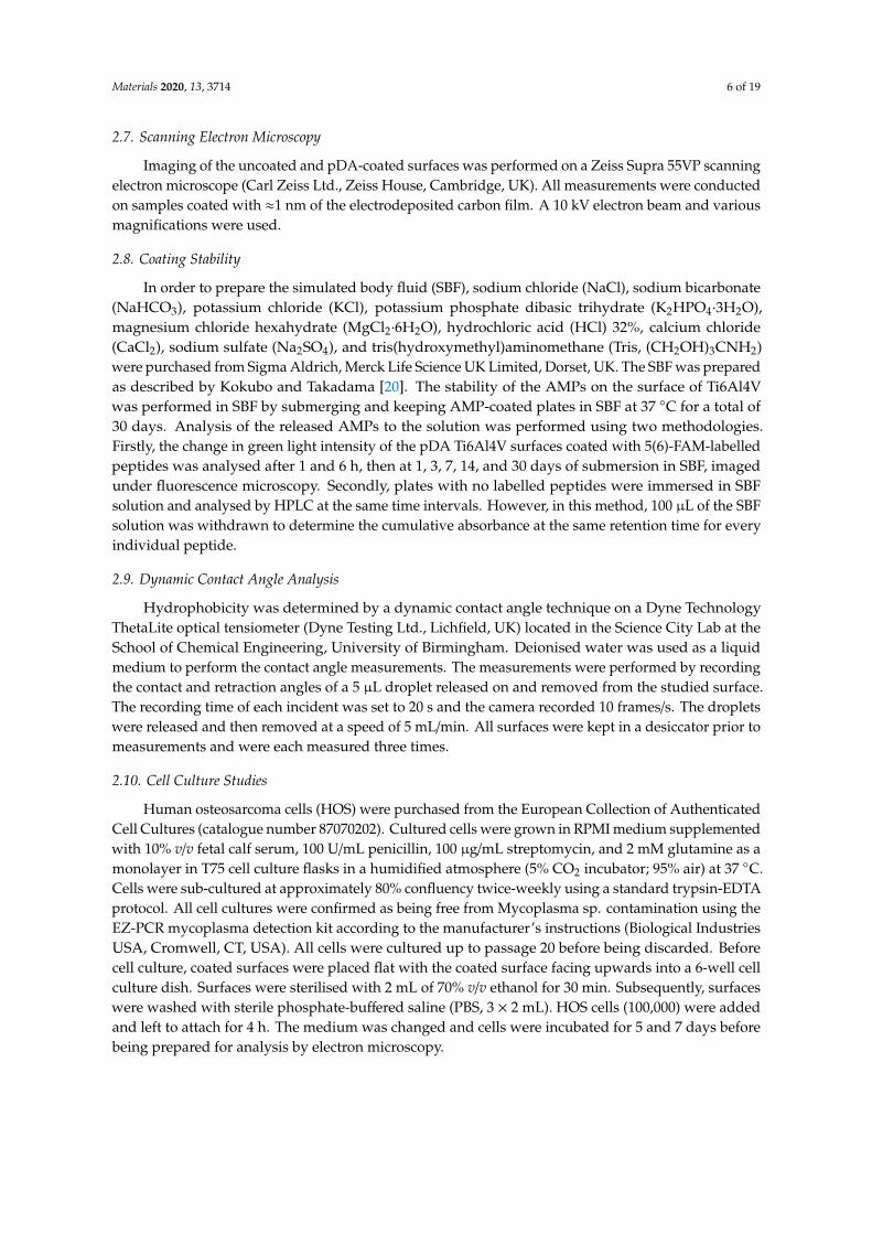
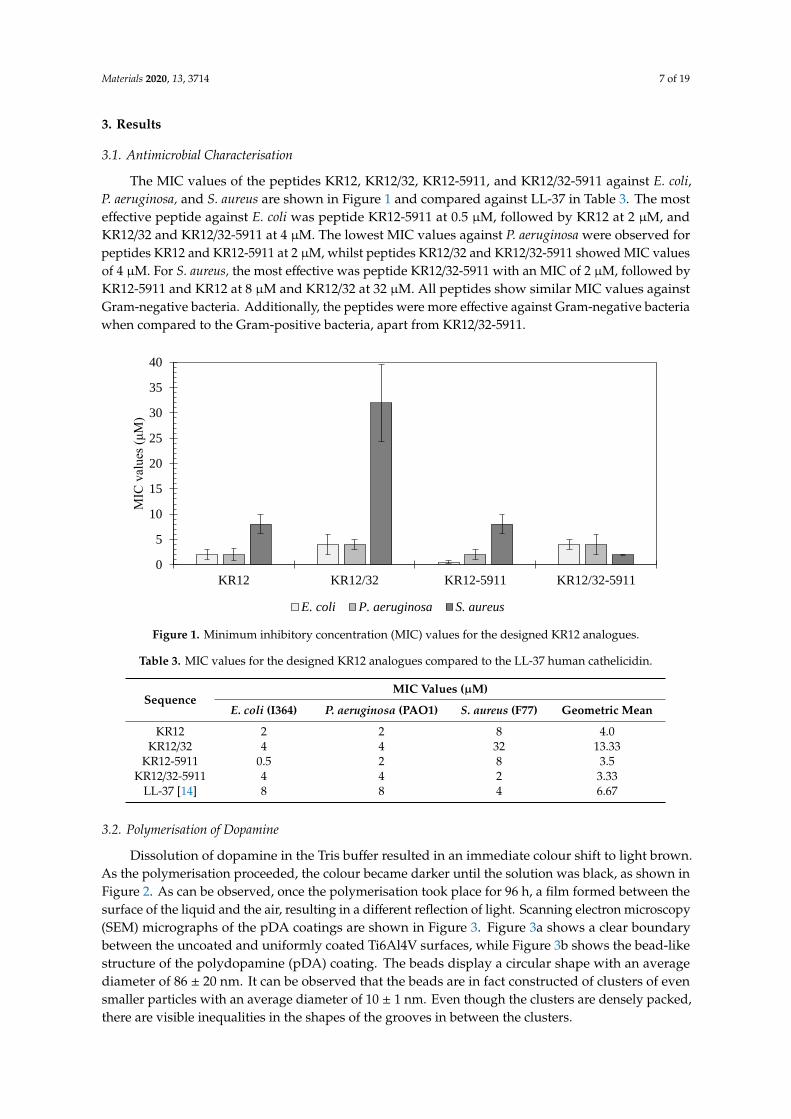
Dissolution of dopamine in the Tris buffer resulted in an immediate colour shift to light brown.As the polymerisation proceeded, the colour became darker until the solution was black, as shown inFigure 2. As can be observed, once the polymerisation took place for 96 h, a film formed between thesurface of the liquid and the air, resulting in a different reflection of light. Scanning electron microscopy(SEM) micrographs of the pDA coatings are shown in Figure 3. Figure 3a shows a clear boundarybetween the uncoated and uniformly coated Ti6Al4V surfaces, while Figure 3b shows the bead-likestructure of the polydopamine (pDA) coating. The beads display a circular shape with an averagediameter of 86 ± 20 nm. It can be observed that the beads are in fact constructed of clusters of evensmaller particles with an average diameter of 10 ± 1 nm. Even though the clusters are densely packed,there are visible inequalities in the shapes of the grooves in between the clusters.
Materials 2020, 13, 3714 8 of 19
Materials 2020, 13, x FOR PEER REVIEW 8 of 20
ellipsometry and AFM agreed with one another. A 24-h immersion of Ti6Al4V plates in the dopamine
solution resulted in the growth of an approximately 10-nm-thick layer. The thickness of the coating
steadily increased to reach around 55 nm after 72 h, after which the growth plateaued.
Figure 2. A photograph of s 24-well plate showing the change in colour of the alkaline dopamine
solutions over 6 h, 12 h, 24 h, 48 h, 72 h, and 96 h, shown from left to right, respectively.
Figure 2. A photograph of s 24-well plate showing the change in colour of the alkaline dopaminesolutions over 6 h, 12 h, 24 h, 48 h, 72 h, and 96 h, shown from left to right, respectively.Materials 2020, 13, x FOR PEER REVIEW 9 of 20
(a)
(b)
Figure 3. SEM micrographs showing (a) the Ti6Al4V surface with a polydopamine (pDA) coating
(left) and uncoated (right) at a magnification of 1000×, as well as (b) the pDA coating at 100,000×
magnification.
Table 4. Polydopamine coating thickness as measured by ellipsometry and Atomic Force M.icroscopy
(AFM).
Time of Immersion 24 h 48 h 72 h 96 h
Coating thickness measured by ellipsometry (nm) 10.2 ± 1.1 32.8 ± 1.1 52.4 ± 7.0 54.7 ± 6.4
Coating thickness measured by AFM (nm) 10.3 ± 0.5 34.1 ± 2.0 55.4 ± 9.0 57.1 ± 6.8
coated uncoated
Figure 3. SEM micrographs showing (a) the Ti6Al4V surface with a polydopamine (pDA) coating (left)and uncoated (right) at a magnification of 1000×, as well as (b) the pDA coating at 100,000×magnification.
Materials 2020, 13, 3714 9 of 19
The thickness of the pDA coating was measured by both ellipsometry and AFM, the results ofwhich are shown in Table 4. The results for the thickness of the coating measured by both ellipsometryand AFM agreed with one another. A 24-h immersion of Ti6Al4V plates in the dopamine solutionresulted in the growth of an approximately 10-nm-thick layer. The thickness of the coating steadilyincreased to reach around 55 nm after 72 h, after which the growth plateaued.
Table 4. Polydopamine coating thickness as measured by ellipsometry and Atomic Force M.icroscopy (AFM).
Time of Immersion 24 h 48 h 72 h 96 h
Coating thickness measured by ellipsometry (nm) 10.2 ± 1.1 32.8 ± 1.1 52.4 ± 7.0 54.7 ± 6.4Coating thickness measured by AFM (nm) 10.3 ± 0.5 34.1 ± 2.0 55.4 ± 9.0 57.1 ± 6.8
3.3. Fluorescence Microscopy
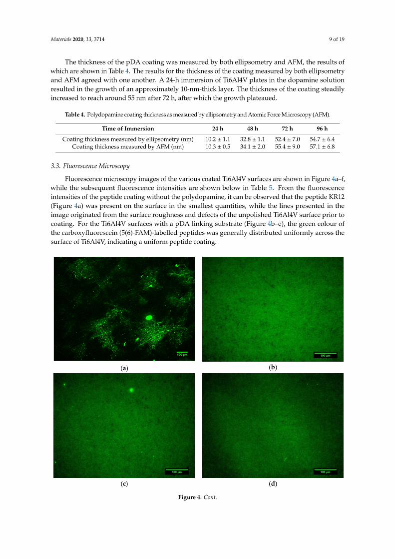
Fluorescence microscopy images of the various coated Ti6Al4V surfaces are shown in Figure 4a–f,while the subsequent fluorescence intensities are shown below in Table 5. From the fluorescenceintensities of the peptide coating without the polydopamine, it can be observed that the peptide KR12(Figure 4a) was present on the surface in the smallest quantities, while the lines presented in theimage originated from the surface roughness and defects of the unpolished Ti6Al4V surface prior tocoating. For the Ti6Al4V surfaces with a pDA linking substrate (Figure 4b–e), the green colour ofthe carboxyfluorescein (5(6)-FAM)-labelled peptides was generally distributed uniformly across thesurface of Ti6Al4V, indicating a uniform peptide coating.
Materials 2020, 13, x FOR PEER REVIEW 9 of 19
(b)
Figure 3. SEM micrographs showing (a) the Ti6Al4V surface with a polydopamine (pDA) coating (left) and uncoated (right) at a magnification of 1000×, as well as (b) the pDA coating at 100,000× magnification.
Table 4. Polydopamine coating thickness as measured by ellipsometry and Atomic Force M.icroscopy (AFM).
Time of Immersion 24 h 48 h 72 h 96 h Coating thickness measured by ellipsometry (nm) 10.2 ± 1.1 32.8 ± 1.1 52.4 ± 7.0 54.7 ± 6.4
Coating thickness measured by AFM (nm) 10.3 ± 0.5 34.1 ± 2.0 55.4 ± 9.0 57.1 ± 6.8
3.3. Fluorescence Microscopy
Fluorescence microscopy images of the various coated Ti6Al4V surfaces are shown in Figure 4a–f, while the subsequent fluorescence intensities are shown below in Table 5. From the fluorescence intensities of the peptide coating without the polydopamine, it can be observed that the peptide KR12 (Figure 4a) was present on the surface in the smallest quantities, while the lines presented in the image originated from the surface roughness and defects of the unpolished Ti6Al4V surface prior to coating. For the Ti6Al4V surfaces with a pDA linking substrate (Figure 4b–e), the green colour of the carboxyfluorescein (5(6)-FAM)-labelled peptides was generally distributed uniformly across the surface of Ti6Al4V, indicating a uniform peptide coating.
(a) (b)
(c) (d)
Figure 4. Cont.
Materials 2020, 13, 3714 10 of 19
Materials 2020, 13, x FOR PEER REVIEW 10 of 19
(e) (f)
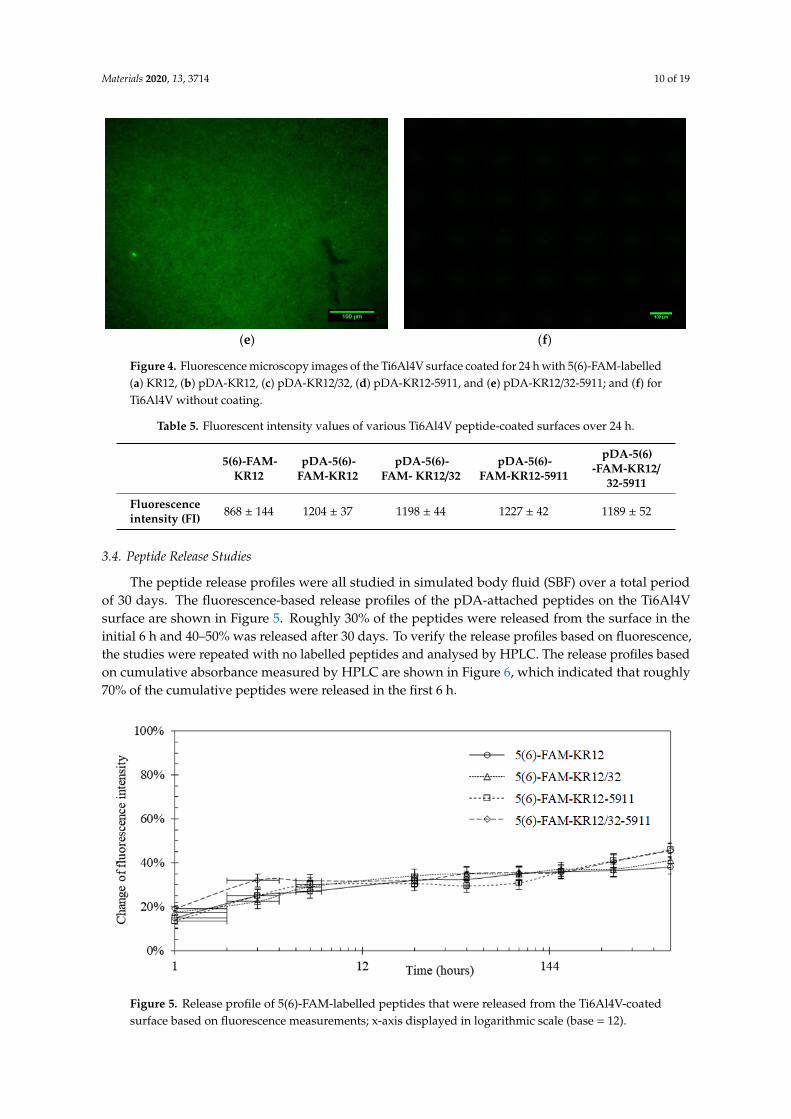
Figure 4. Fluorescence microscopy images of the Ti6Al4V surface coated for 24 hours with 5(6)-FAM-labelled (a) KR12, (b) pDA-KR12, (c) pDA-KR12/32, (d) pDA-KR12-5911, and (e) pDA-KR12/32-5911; and (f) for Ti6Al4V without coating.
Table 5. Fluorescent intensity values of various Ti6Al4V peptide-coated surfaces over 24 h.
5(6)-
FAM-KR12
pDA-5(6)-FAM-KR12
pDA-5(6)-FAM-KR12/32
pDA-5(6)-FAM-KR12
-5911
pDA-5(6)-FAM-KR12/32
-5911 Fluorescence intensity (FI)
868 ± 144 1204 ± 37 1198 ± 44 1227 ± 42 1189 ± 52
3.4. Peptide Release Studies
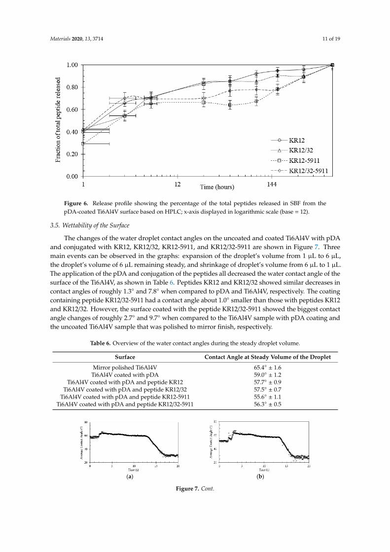
The peptide release profiles were all studied in simulated body fluid (SBF) over a total period of 30 days. The fluorescence-based release profiles of the pDA-attached peptides on the Ti6Al4V surface are shown in Figure 5. Roughly 30% of the peptides were released from the surface in the initial 6 hours and 40–50% was released after 30 days. To verify the release profiles based on fluorescence, the studies were repeated with no labelled peptides and analysed by HPLC. The release profiles based on cumulative absorbance measured by HPLC are shown in Figure 6, which indicated that roughly 70% of the cumulative peptides were released in the first 6 h.
Figure 5. Release profile of 5(6)-FAM-labelled peptides that were released from the Ti6Al4V-coated surface based on fluorescence measurements; x-axis displayed in logarithmic scale (base = 12).
Figure 4. Fluorescence microscopy images of the Ti6Al4V surface coated for 24 h with 5(6)-FAM-labelled(a) KR12, (b) pDA-KR12, (c) pDA-KR12/32, (d) pDA-KR12-5911, and (e) pDA-KR12/32-5911; and (f) forTi6Al4V without coating.
Table 5. Fluorescent intensity values of various Ti6Al4V peptide-coated surfaces over 24 h.
5(6)-FAM-KR12
pDA-5(6)-FAM-KR12
pDA-5(6)-FAM- KR12/32
pDA-5(6)-FAM-KR12-5911
pDA-5(6)-FAM-KR12/
32-5911
Fluorescenceintensity (FI) 868 ± 144 1204 ± 37 1198 ± 44 1227 ± 42 1189 ± 52
3.4. Peptide Release Studies
The peptide release profiles were all studied in simulated body fluid (SBF) over a total periodof 30 days. The fluorescence-based release profiles of the pDA-attached peptides on the Ti6Al4Vsurface are shown in Figure 5. Roughly 30% of the peptides were released from the surface in theinitial 6 h and 40–50% was released after 30 days. To verify the release profiles based on fluorescence,the studies were repeated with no labelled peptides and analysed by HPLC. The release profiles basedon cumulative absorbance measured by HPLC are shown in Figure 6, which indicated that roughly70% of the cumulative peptides were released in the first 6 h.
Materials 2020, 13, x FOR PEER REVIEW 11 of 20
Table 5. Fluorescent intensity values of various Ti6Al4V peptide-coated surfaces over 24 h.
5(6)-
FAM-
KR12
pDA-5(6)-
FAM-KR12
pDA-5(6)-
FAM-KR12/32
pDA-5(6)-
FAM-KR12-
5911
pDA-5(6)-
FAM-
KR12/32-5911
Fluorescence
intensity (FI) 868 ± 144 1204 ± 37 1198 ± 44 1227 ± 42 1189 ± 52
3.4. Peptide Release Studies
The peptide release profiles were all studied in simulated body fluid (SBF) over a total period of
30 days. The fluorescence-based release profiles of the pDA-attached peptides on the Ti6Al4V surface
are shown in Figure 5. Roughly 30% of the peptides were released from the surface in the initial 6 h
and 40–50% was released after 30 days. To verify the release profiles based on fluorescence, the
studies were repeated with no labelled peptides and analysed by HPLC. The release profiles based
on cumulative absorbance measured by HPLC are shown in Figure 6, which indicated that roughly
70% of the cumulative peptides were released in the first 6 h.
Figure 5. Release profile of 5(6)-FAM-labelled peptides that were released from the Ti6Al4V-coated
surface based on fluorescence measurements; x-axis displayed in logarithmic scale (base = 12).
Figure 5. Release profile of 5(6)-FAM-labelled peptides that were released from the Ti6Al4V-coatedsurface based on fluorescence measurements; x-axis displayed in logarithmic scale (base = 12).
Materials 2020, 13, 3714 11 of 19
Materials 2020, 13, x FOR PEER REVIEW 11 of 20
Table 5. Fluorescent intensity values of various Ti6Al4V peptide-coated surfaces over 24 h.
5(6)-
FAM-
KR12
pDA-5(6)-
FAM-KR12
pDA-5(6)-
FAM-KR12/32
pDA-5(6)-
FAM-KR12-
5911
pDA-5(6)-
FAM-
KR12/32-5911
Fluorescence
intensity (FI) 868 ± 144 1204 ± 37 1198 ± 44 1227 ± 42 1189 ± 52
3.4. Peptide Release Studies
The peptide release profiles were all studied in simulated body fluid (SBF) over a total period of
30 days. The fluorescence-based release profiles of the pDA-attached peptides on the Ti6Al4V surface
are shown in Figure 5. Roughly 30% of the peptides were released from the surface in the initial 6 h
and 40–50% was released after 30 days. To verify the release profiles based on fluorescence, the
studies were repeated with no labelled peptides and analysed by HPLC. The release profiles based
on cumulative absorbance measured by HPLC are shown in Figure 6, which indicated that roughly
70% of the cumulative peptides were released in the first 6 h.
Figure 5. Release profile of 5(6)-FAM-labelled peptides that were released from the Ti6Al4V-coated
surface based on fluorescence measurements; x-axis displayed in logarithmic scale (base = 12).
Figure 6. Release profile showing the percentage of the total peptides released in SBF from thepDA-coated Ti6Al4V surface based on HPLC; x-axis displayed in logarithmic scale (base = 12).
3.5. Wettability of the Surface
The changes of the water droplet contact angles on the uncoated and coated Ti6Al4V with pDAand conjugated with KR12, KR12/32, KR12-5911, and KR12/32-5911 are shown in Figure 7. Threemain events can be observed in the graphs: expansion of the droplet’s volume from 1 µL to 6 µL,the droplet’s volume of 6 µL remaining steady, and shrinkage of droplet’s volume from 6 µL to 1 µL.The application of the pDA and conjugation of the peptides all decreased the water contact angle of thesurface of the Ti6Al4V, as shown in Table 6. Peptides KR12 and KR12/32 showed similar decreases incontact angles of roughly 1.3◦ and 7.8◦ when compared to pDA and Ti6Al4V, respectively. The coatingcontaining peptide KR12/32-5911 had a contact angle about 1.0◦ smaller than those with peptides KR12and KR12/32. However, the surface coated with the peptide KR12/32-5911 showed the biggest contactangle changes of roughly 2.7◦ and 9.7◦ when compared to the Ti6Al4V sample with pDA coating andthe uncoated Ti6Al4V sample that was polished to mirror finish, respectively.
Table 6. Overview of the water contact angles during the steady droplet volume.
Surface Contact Angle at Steady Volume of the Droplet
Mirror polished Ti6Al4V 65.4◦ ± 1.6Ti6Al4V coated with pDA 59.0◦ ± 1.2
Ti6Al4V coated with pDA and peptide KR12 57.7◦ ± 0.9Ti6Al4V coated with pDA and peptide KR12/32 57.5◦ ± 0.7
Ti6Al4V coated with pDA and peptide KR12-5911 55.6◦ ± 1.1Ti6Al4V coated with pDA and peptide KR12/32-5911 56.3◦ ± 0.5
Materials 2020, 13, x FOR PEER REVIEW 11 of 19
Figure 6. Release profile showing the percentage of the total peptides released in SBF from the pDA-coated Ti6Al4V surface based on HPLC; x-axis displayed in logarithmic scale (base = 12).
3.5. Wettability of the Surface
The changes of the water droplet contact angles on the uncoated and coated Ti6Al4V with pDA and conjugated with KR12, KR12/32, KR12-5911, and KR12/32-5911 are shown in Figure 7. Three main events can be observed in the graphs: expansion of the droplet's volume from 1 µL to 6 µL, the droplet's volume of 6 µL remaining steady, and shrinkage of droplet's volume from 6 µL to 1 µL. The application of the pDA and conjugation of the peptides all decreased the water contact angle of the surface of the Ti6Al4V, as shown in Table 6. Peptides KR12 and KR12/32 showed similar decreases in contact angles of roughly 1.3° and 7.8° when compared to pDA and Ti6Al4V, respectively. The coating containing peptide KR12/32-5911 had a contact angle about 1.0° smaller than those with peptides KR12 and KR12/32. However, the surface coated with the peptide KR12/32-5911 showed the biggest contact angle changes of roughly 2.7° and 9.7° when compared to the Ti6Al4V sample with pDA coating and the uncoated Ti6Al4V sample that was polished to mirror finish, respectively.
Table 6. Overview of the water contact angles during the steady droplet volume.
Surface Contact Angle at Steady Volume of the Droplet Mirror polished Ti6Al4V 65.4° ± 1.6
Ti6Al4V coated with pDA 59.0° ± 1.2 Ti6Al4V coated with pDA and peptide KR12 57.7° ± 0.9
Ti6Al4V coated with pDA and peptide KR12/32 57.5° ± 0.7 Ti6Al4V coated with pDA and peptide KR12-5911 55.6° ± 1.1
Ti6Al4V coated with pDA and peptide KR12/32-5911 56.3° ± 0.5
(a) (b)
Figure 7. Cont.
Materials 2020, 13, 3714 12 of 19
Materials 2020, 13, x FOR PEER REVIEW 12 of 19
(c) (d)
(e) (f)
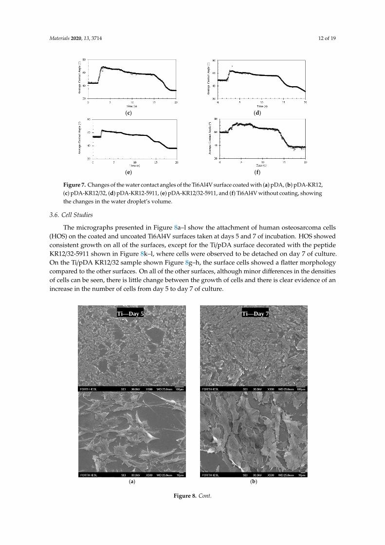
Figure 7. Changes of the water contact angles of the Ti6Al4V surface coated with (a) pDA, (b) pDA-KR12, (c) pDA-KR12/32, (d) pDA-KR12-5911, (e) pDA-KR12/32-5911, and (f) Ti6Al4V without coating, showing the changes in the water droplet's volume.
3.6. Cell Studies
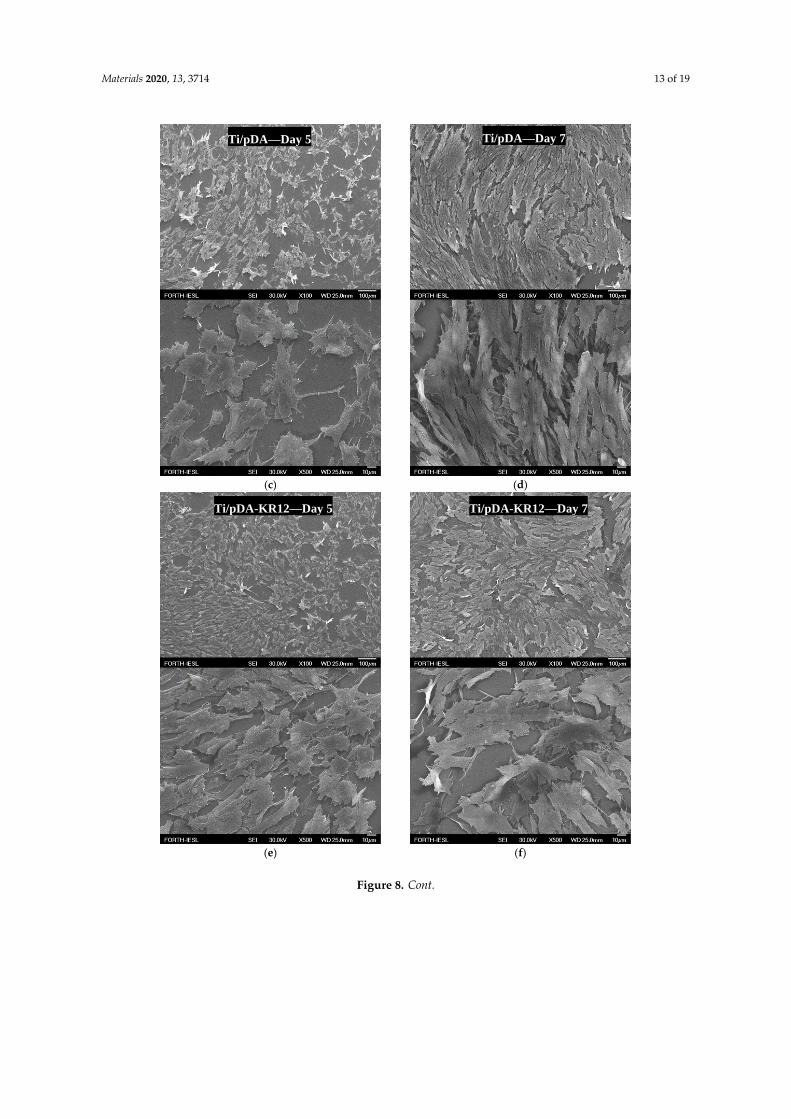
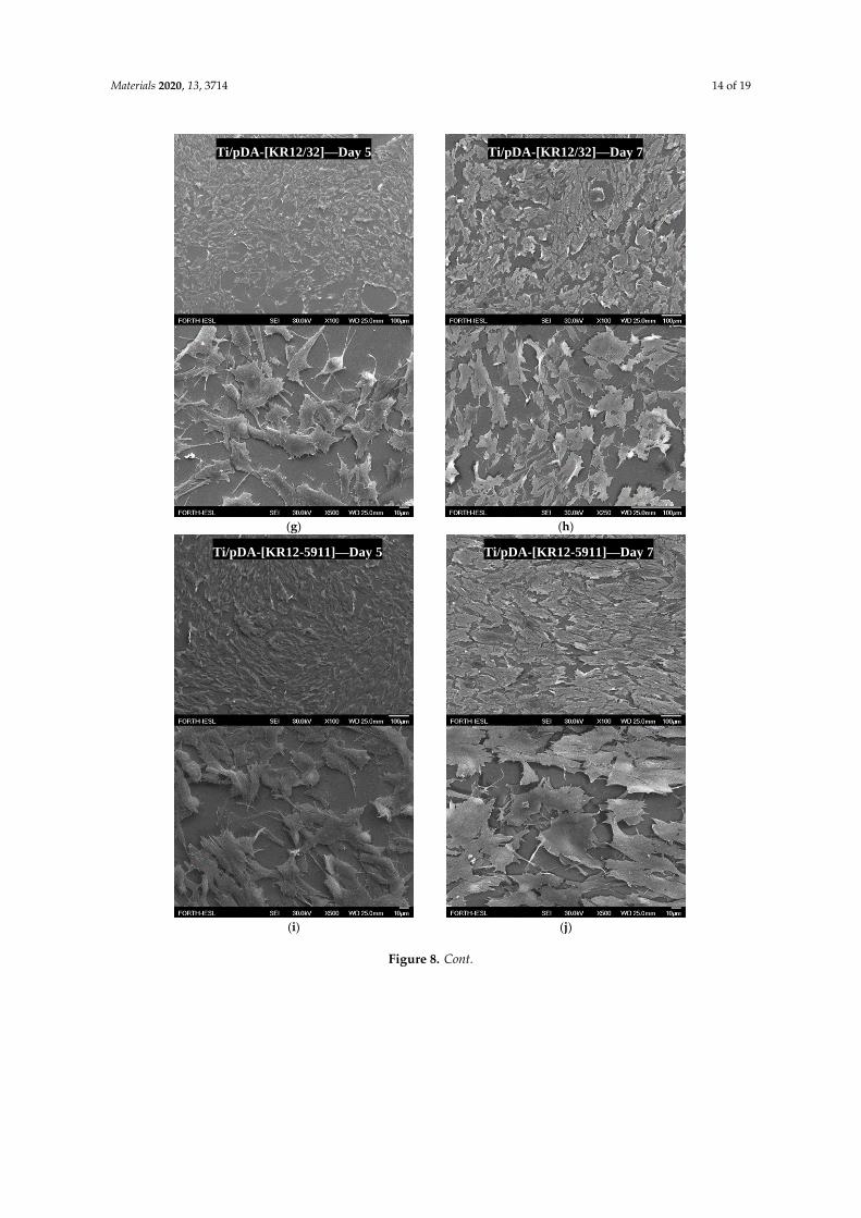
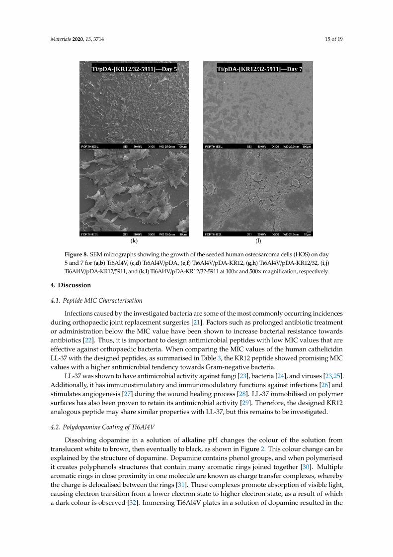
The micrographs presented in Figure 8a–l show the attachment of human osteosarcoma cells (HOS) on the coated and uncoated Ti6Al4V surfaces taken at days 5 and 7 of incubation. HOS showed consistent growth on all of the surfaces, except for the Ti/pDA surface decorated with the peptide KR12/32-5911 shown in Figure 8k–l, where cells were observed to be detached on day 7 of culture. On the Ti/pDA KR12/32 sample shown Figure 8g–h, the surface cells showed a flatter morphology compared to the other surfaces. On all of the other surfaces, although minor differences in the densities of cells can be seen, there is little change between the growth of cells and there is clear evidence of an increase in the number of cells from day 5 to day 7 of culture.
(a) (b)
Ti—Day 5 Ti—Day 7
Figure 7. Changes of the water contact angles of the Ti6Al4V surface coated with (a) pDA, (b) pDA-KR12,(c) pDA-KR12/32, (d) pDA-KR12-5911, (e) pDA-KR12/32-5911, and (f) Ti6Al4V without coating, showingthe changes in the water droplet’s volume.
3.6. Cell Studies
The micrographs presented in Figure 8a–l show the attachment of human osteosarcoma cells(HOS) on the coated and uncoated Ti6Al4V surfaces taken at days 5 and 7 of incubation. HOS showedconsistent growth on all of the surfaces, except for the Ti/pDA surface decorated with the peptideKR12/32-5911 shown in Figure 8k–l, where cells were observed to be detached on day 7 of culture.On the Ti/pDA KR12/32 sample shown Figure 8g–h, the surface cells showed a flatter morphologycompared to the other surfaces. On all of the other surfaces, although minor differences in the densitiesof cells can be seen, there is little change between the growth of cells and there is clear evidence of anincrease in the number of cells from day 5 to day 7 of culture.
Materials 2020, 13, x FOR PEER REVIEW 13 of 20
3.6. Cell Studies
The micrographs presented in Figure 8a–l show the attachment of human osteosarcoma cells
(HOS) on the coated and uncoated Ti6Al4V surfaces taken at days 5 and 7 of incubation. HOS showed
consistent growth on all of the surfaces, except for the Ti/pDA surface decorated with the peptide
KR12/32-5911 shown in Figure 8k–l, where cells were observed to be detached on day 7 of culture.
On the Ti/pDA KR12/32 sample shown Figure 8g–h, the surface cells showed a flatter morphology
compared to the other surfaces. On all of the other surfaces, although minor differences in the
densities of cells can be seen, there is little change between the growth of cells and there is clear
evidence of an increase in the number of cells from day 5 to day 7 of culture.
(a) (b)
Ti—Day 5 Ti—Day 7
Figure 8. Cont.
Materials 2020, 13, 3714 13 of 19
Materials 2020, 13, x FOR PEER REVIEW 14 of 20
(c) (d)
(e) (f)
Ti/pDA—Day 5 Ti/pDA—Day 7
Ti/pDA-KR12—Day 5 Ti/pDA-KR12—Day 7
Figure 8. Cont.
Materials 2020, 13, 3714 14 of 19
Materials 2020, 13, x FOR PEER REVIEW 15 of 20
(g) (h)
(i) (j)
Ti/pDA-[KR12-5911]—Day 5 Ti/pDA-[KR12-5911]—Day 7
Ti/pDA-[KR12/32]—Day 5 Ti/pDA-[KR12/32]—Day 7
Figure 8. Cont.
Materials 2020, 13, 3714 15 of 19
Materials 2020, 13, x FOR PEER REVIEW 16 of 20
(k) (l)
Figure 8. SEM micrographs showing the growth of the seeded human osteosarcoma cells (HOS) on
day 5 and 7 for (a,b) Ti6Al4V, (c,d) Ti6Al4V/pDA, (e,f) Ti6Al4V/pDA-KR12, (g,h) Ti6Al4V/pDA-
KR12/32, (i,j) Ti6Al4V/pDA-KR12/5911, and (k,l) Ti6Al4V/pDA-KR12/32-5911 at 100× and 500×
magnification, respectively.
4. Discussion
4.1. Peptide MIC Characterisation
Infections caused by the investigated bacteria are some of the most commonly occurring
incidences during orthopaedic joint replacement surgeries [21]. Factors such as prolonged antibiotic
treatment or administration below the MIC value have been shown to increase bacterial resistance
towards antibiotics [22]. Thus, it is important to design antimicrobial peptides with low MIC values
that are effective against orthopaedic bacteria. When comparing the MIC values of the human
cathelicidin LL-37 with the designed peptides, as summarised in Table 3, the KR12 peptide showed
promising MIC values with a higher antimicrobial tendency towards Gram-negative bacteria.
LL-37 was shown to have antimicrobial activity against fungi [23], bacteria [24], and viruses
[23,25]. Additionally, it has immunostimulatory and immunomodulatory functions against infections
[26] and stimulates angiogenesis [27] during the wound healing process [28]. LL-37 immobilised on
polymer surfaces has also been proven to retain its antimicrobial activity [29]. Therefore, the designed
KR12 analogous peptide may share similar properties with LL-37, but this remains to be investigated.
4.2. Polydopamine Coating of Ti6Al4V
Dissolving dopamine in a solution of alkaline pH changes the colour of the solution from
translucent white to brown, then eventually to black, as shown in Figure 2. This colour change can
be explained by the structure of dopamine. Dopamine contains phenol groups, and when
polymerised it creates polyphenols structures that contain many aromatic rings joined together [30].
Multiple aromatic rings in close proximity in one molecule are known as charge transfer complexes,
Ti/pDA-[KR12/32-5911]—Day 5 Ti/pDA-[KR12/32-5911]—Day 7
Figure 8. SEM micrographs showing the growth of the seeded human osteosarcoma cells (HOS) on day5 and 7 for (a,b) Ti6Al4V, (c,d) Ti6Al4V/pDA, (e,f) Ti6Al4V/pDA-KR12, (g,h) Ti6Al4V/pDA-KR12/32, (i,j)Ti6Al4V/pDA-KR12/5911, and (k,l) Ti6Al4V/pDA-KR12/32-5911 at 100× and 500×magnification, respectively.
4. Discussion
4.1. Peptide MIC Characterisation
Infections caused by the investigated bacteria are some of the most commonly occurring incidencesduring orthopaedic joint replacement surgeries [21]. Factors such as prolonged antibiotic treatmentor administration below the MIC value have been shown to increase bacterial resistance towardsantibiotics [22]. Thus, it is important to design antimicrobial peptides with low MIC values that areeffective against orthopaedic bacteria. When comparing the MIC values of the human cathelicidinLL-37 with the designed peptides, as summarised in Table 3, the KR12 peptide showed promising MICvalues with a higher antimicrobial tendency towards Gram-negative bacteria.
LL-37 was shown to have antimicrobial activity against fungi [23], bacteria [24], and viruses [23,25].Additionally, it has immunostimulatory and immunomodulatory functions against infections [26] andstimulates angiogenesis [27] during the wound healing process [28]. LL-37 immobilised on polymersurfaces has also been proven to retain its antimicrobial activity [29]. Therefore, the designed KR12analogous peptide may share similar properties with LL-37, but this remains to be investigated.
4.2. Polydopamine Coating of Ti6Al4V
Dissolving dopamine in a solution of alkaline pH changes the colour of the solution fromtranslucent white to brown, then eventually to black, as shown in Figure 2. This colour change can beexplained by the structure of dopamine. Dopamine contains phenol groups, and when polymerisedit creates polyphenols structures that contain many aromatic rings joined together [30]. Multiplearomatic rings in close proximity in one molecule are known as charge transfer complexes, wherebythe charge is delocalised between the rings [31]. These complexes promote absorption of visible light,causing electron transition from a lower electron state to higher electron state, as a result of whicha dark colour is observed [32]. Immersing Ti6Al4V plates in a solution of dopamine resulted in the
Materials 2020, 13, 3714 16 of 19
formation of a thin film on the surface of the metallic plate, which agrees with Messersmith et al. [33],who reported that pDA was able to adhere to virtually any surface and that its polymerisation inslightly basic solution resulted in a film formation on the surface. The SEM morphology and topologystudies showed that the surface of the pDA coating was built from small round particles of ≈100 nmdiameter. It was proposed by Jiang et al. that the pDA first polymerises in nanoaggregates, which overtime attach themselves on the surface, creating a uniform coating [34,35].
The attachment of the nanoaggregates to the surface could potentially have been driven by thesedimentation of the particles towards the bottom of the container where the substrate to be coatedwas located. The coating then accumulated with more beads and increased the thickness of the coating,which varied depending on the size of the beads accumulated. This could explain the ≈10 nm coatingthickness after the initial 24 h, and the increases of the thickness to 32 nm after 48 h and to 57 nmafter 52 h of pDA polymerisation. Similar film thickness growth was reported by Jiang et al. [33,35].In this study, a thickness plateau was reached after 72 h of polymerisation. This pattern of growth wasalso previously reported by Bensmann et al. [36], where the pDA film reached 62.8 nm in thicknessafter 72 h and did not increase in thickness afterwards. Formation of the plateau can be explained bythe lack of oxygen after the formation of the first pDA layer. Since oxidation is a common reactionof polyphenols [37], it can be assumed that the polymerisation required oxygen to continue and thepresence of the film limited the accessibility to oxygen. In addition, as oxidation on the surface willcontinue as oxygen is available on the surface, polymerisation will eventually stop when dopamineis consumed.
4.3. Peptide Attachment to Polydopamine
The KR12 analogous peptide labelled with 5(6)-FAM at the N-terminal was conjugated to analready pDA-coated Ti6Al4V surface. The presence of the peptides was confirmed by the emittedfluorescence, showing only small variations in the measured fluorescence intensities between thedifferent peptides. The quinone group of pDA is known to undergo a Schiff base reaction or Michaeladdition without the use of any other reagent or catalyst, allowing the conjugation of biomolecules topDA [38]. The catechol group of the pDA is known to oxidise to quinone at a pH above 7.5 in excessof oxygen, which is necessary for successful conjugation to nucleophiles [39]. Here, the designedpeptides contained two lysine residues that should be able to conjugate to pDA, but the first lysine waspresumed to be shielded by the bulky 5(6)-FAM label at the N-terminus. Therefore, it was expectedthat pDA was conjugated with the second lysine residue relative to the N-terminus. It was observedthat the initial burst of peptides released approximately 25–35% of the peptides from the surface after6 h, and approximately 40–50% was released after 30 days. The fluorescence release profile studies inSBF indicated that 50–60% of the peptides potentially remained conjugated to the pDA after 30 days.The initial burst of peptide release could be attributed to the release of unattached peptides to thepDA that were not successfully removed during the wash step. After the initial burst, the rate ofrelease of the peptides decreased and was only reduced further by roughly 5–10% after the final30 days. The release profile from the fluorescence was confirmed by HPLC using non-fluorescentpeptides. An initial ≈70% burst of cumulative peptide was released in the first 6 h, and a slowersubsequent release for the remaining period was observed. These release studies clearly showed thatthese peptides were conjugated to the pDA-coated Ti6Al4V surface in a very stable manner throughthe lysine residues.
4.4. Wettability of the Surface
New implants within the body come into immediate contact with extracellular fluid and moieties,such as proteins, which are some of the first molecules to interact with the surfaces of an implant [40,41].The optimal water contact angle of a biomaterial reported for bone-forming cells to attach to a surfacewas reported to be 55◦ [42]. Metallic surfaces are very hydrophobic, resulting in slow integrationrates of the implant with the body. A water contact angle study of pDA-coated titanium surfaces
Materials 2020, 13, 3714 17 of 19
by Nijhuis et al. reported that coated Ti6Al4V surfaces were much less hydrophobic (≈47◦) than theuncoated surfaces (≈75◦) [43], presumably resulting in an improvement of the implant integration [44].In this paper, we report that the introduction of pDA coating on the surface of Ti6Al4V resulted ina decrease in the water contact angle from 65.4◦ ± 1.6 to 59.0◦ ± 1.2. The experimental values are inagreement with prior research performed by Luo et al. [45], who reported on the water contact angle ofa pDA-coated Ti6Al4V surface. The water contact angle was further reduced to approximately 56.8◦
when the pDA coating was decorated with the peptides—the results varied depending on the usedpeptide, but they all showed a reduction relative to the pDA-coated Ti6Al4V surface.
4.5. Cell Culture Studies
All surfaces showed attachment of cells on day 5, with no evidence of toxicity (which is determinedby any change of cell morphology or density compared to the plain titanium surface), with one exceptionfor the KR12/32-5911 surface. The cell morphology was generally "flattened" and fibroblast-like, typicalof the described HOS morphology in the literature [46]. The density of attached cells increased overtime, confirming that cells were viable and able to proliferate. In contrast, there was evidence that thesurface of KR12/32-5911 was toxic to the cells, and at day 7 there were no longer any cells.
5. Conclusions
In the present research, KR12 and three new analogous peptides were successfully synthesised andshowed promising antimicrobial activity against E. coli, P. aeruginosa, and S. aureus, with MIC valuesgenerally being lower than that of their native peptide, the human cathelicidin LL-37. The titaniumsurfaces were coated with pDA and characterised. Subsequently, the pDA-coated surfaces weredecorated with KR12 and the other three designed analogous peptides. The pDA coating provided along-term linking substrate for the peptides, which was confirmed by fluorescence microscopy andHPLC. Cultured HOS cells showed good attachment and cell growth on the material surfaces, with novisible toxicity towards the cells, except for the KR12/32-5911 peptide. This surface treatment showspotential to provide long-term antimicrobial activity on many metallic and organic material surfacesand could be used in biomedical materials and implants.
Author Contributions: Conceptualization, A.S. and Z.T.; methodology, A.S., J.B., N.J.H.; software, M.B.; validation,A.S. and Z.T.; formal analysis Z.T. and A.S.; investigation Z.T.; resources, Z.T., M.B. and H.I.; data curation, Z.T.,M.B., H.I., J.B., N.J.H. and A.S.; writing—original draft preparation, Z.T., M.B. and H.I.; writing—review andediting, A.S., N.J.H.; visualization, Z.T., M.B., H.I.; supervision, A.S., J.B. and N.J.H.; project administration, A.S.;funding acquisition, A.S. All authors have read and agreed to the published version of the manuscript.
Funding: This project has been partially funded by the European Union’s Horizon 2020 research and innovationprogramme under the Marie Sklodowska-Curie grant agreement No. 645749. The project has been also financiallysupported by a studentship provided by the School of Metallurgy and Materials, University of Birmingham, UK.
Conflicts of Interest: The authors declare no conflict of interest.
References
1. Cloutier, M.; Mantovani, D.; Rosei, F. Antibacterial Coatings: Challenges, Perspectives, and Opportunities.Trends Biotechnol. 2015, 33, 637–652. [CrossRef]
2. Goodman, S.B.; Yao, Z.; Keeney, M.; Yang, F. The future of biologic coatings for orthopaedic implants.Biomaterials 2013, 34, 3174–3183. [CrossRef]
3. Romanò, C.L.; Scarponi, S.; Gallazzi, E.; Romanò, D.; Drago, L. Antibacterial coating of implants inorthopaedics and trauma: A classification proposal in an evolving panorama. J. Orthop. Surg. Res. 2015,10, 157. [CrossRef]
4. Geetha, M.; Singh, A.K.; Asokamani, R.; Gogia, K.A. Ti based biomaterials, the ultimate choice for orthopaedicimplants—A review. Prog. Mater. Sci. 2009, 54, 397–425. [CrossRef]
5. Bauer, J.; Siala, W.; Tulkens, P.M.; Van Bambeke, F. A combined pharmacodynamic quantitative and qualitativemodel reveals the potent activity of daptomycin and delafloxacin against Staphylococcus aureus biofilms.Antimicrob. Agents Chemother. 2013, 57, 2726–2737. [CrossRef]
Materials 2020, 13, 3714 18 of 19
6. Mishra, B.; Wang, G. Titanium surfaces immobilised with the major antimicrobial fragment FK-16 of humancathelicidin LL-37 are potent against multiple antibiotic-resistant bacteria. Biofouling 2017, 33, 544–555.[CrossRef] [PubMed]
7. Stigter, M.; Bezemer, J.; De Groot, K.; Layrolle, P. Incorporation of different antibiotics into carbonatedhydroxyapatite coatings on titanium implants, release and antibiotic efficacy. J. Control. Release 2004, 99,127–137. [CrossRef]
8. Rathbone, C.R.; Cross, J.D.; Brown, K.V.; Murray, C.; Wenke, J.C. Effect of various concentrations of antibioticson osteogenic cell viability and activity. J. Orthop. Res. 2011, 29, 1070–1074. [CrossRef]
9. Mor, A. Peptide-based antibiotics: A potential answer to raging antimicrobial resistance. Drug Dev. Res.2000, 50, 440–447. [CrossRef]
10. Alves, D.; Olívia Pereira, M. Mini-review: Antimicrobial peptides and enzymes as promising candidates tofunctionalise biomaterial surfaces. Biofouling 2014, 30, 483–499. [CrossRef]
11. Bazaka, K.; Jacob, M.V.; Chrzanowski, W.; Ostrikov, K. Anti-bacterial surfaces: Natural agents, mechanismsof action, and plasma surface modification. RSC Adv. 2015, 5, 48739–48759. [CrossRef]
12. Zasloff, M. Antimicrobial peptides of multicellular organisms. Nature 2002, 415, 389–395. [CrossRef][PubMed]
13. Guilhelmelli, F.; Vilela, N.; Albuquerque, P.; Derengowski, L.D.S.; Pereira, I.S.; Kyaw, C.M. Antibioticdevelopment challenges: The various mechanisms of action of antimicrobial peptides and of bacterialresistance. Front. Microbiol. 2013, 4, 353. [CrossRef]
14. Fjell, C.D.; Hiss, J.A.; Hancock, R.E.W.; Schneider, G. Designing antimicrobial peptides: Form followsfunction. Nat. Rev. Drug Discov. 2011, 11, 37–51. [CrossRef]
15. Lazzaro, B.P.; Zasloff, M.; Rolff, J. Antimicrobial peptides: Application informed by evolution. Science 2020,368, eaau5480. [CrossRef]
16. Jacob, B.; Park, I.-S.; Bang, J.-K.; Shin, S.Y. Short KR-12 analogs designed from human cathelicidin LL-37possessing both antimicrobial and antiendotoxic activities without mammalian cell toxicity. J. Pept. Sci. 2013,19, 700–707. [CrossRef]
17. Jia, L.; Han, F.; Wang, H.; Zhu, C.; Guo, Q.; Li, J.; Zhao, Z.; Zhang, Q.; Zhu, X.; Li, B. Polydopamine-assistedsurface modification for orthopaedic implants. J. Orthop. Transl. 2019, 17, 82–95. [CrossRef]
18. Zhu, Y.; Liu, D.; Wang, X.; He, Y.; Luan, W.; Qi, F.; Ding, J. Polydopamine-mediated covalent functionalisationof collagen on a titanium alloy to promote biocompatibility with soft tissues J. Mater. Chem. B 2019, 7,2019–2031. [CrossRef]
19. Wang, Y.; Qi, H.; Miron, R.J.; Zhang, Y. Modulating macrophage polarisation on titanium implant surface bypoly(dopamine)-assisted immobilisation of IL4. Clin. Implant Dent. Relat. Res. 2019, 21, 977–986. [CrossRef]
20. Kokubo, T.; Takadama, H. How useful is SBF in predicting in vivo bone bioactivity? Biomaterials 2006, 27,2907–2915. [CrossRef]
21. Ribeiro, M.; Monteiro, F.J.; Ferraz, M.P. Infection of orthopedic implants with emphasis on bacterial adhesionprocess and techniques used in studying bacterial-material interactions. Biomaterials 2013, 2, 176–194.[CrossRef] [PubMed]
22. Levison, M.E.; Levison, J.H. Pharmacokinetics and Pharmacodynamics of Antibacterial Agents. Infect. Dis.Clin. N. A. 2009, 23, 791–815. [CrossRef] [PubMed]
23. Wong, J.H.; Łegowska, A.; Rolka, K.; Ng, T.B.; Hui, M.; Cho, C.H.; Lam, W.W.L.; Au, S.W.-N.; Gu, O.W.;Wan, D.C.C. Effects of cathelicidin and its fragments on three key enzymes of HIV-1. Peptides 2011, 32,1117–1122. [CrossRef]
24. Overhage, J.; Campisano, A.; Bains, M.; Torfs, E.C.W.; Rehm, B.H.A.; Hancock, R.E.W. Human Host DefensePeptide LL-37 Prevents Bacterial Biofilm Formation. Infect. Immun. 2008, 76, 4176–4182. [CrossRef]
25. Barlow, P.G.; Svoboda, P.; Mackellar, A.; Nash, A.A.; York, I.A.; Pohl, J.; Davidson, D.J.; Donis, R.O. AntiviralActivity and Increased Host Defense against Influenza Infection Elicited by the Human Cathelicidin LL-37.PLoS ONE 2011, 6, e25333. [CrossRef]
26. Davidson, D.J.; Currie, A.J.; Reid, G.S.D.; Bowdish, D.M.E.; MacDonald, K.L.; Ma, R.C.; Hancock, R.E.W.;Speert, D.P. The Cationic Antimicrobial Peptide LL-37 Modulates Dendritic Cell Differentiation and DendriticCell-Induced T Cell Polarization. J. Immunol. 2004, 172, 1146–1156. [CrossRef]
Materials 2020, 13, 3714 19 of 19
27. Koczulla, R.; Von Degenfeld, G.; Kupatt, C.; Krötz, F.; Zahler, S.; Gloe, T.; Issbrücker, K.; Unterberger, P.;Zaiou, M.; Lebherz, C.; et al. An angiogenic role for the human peptide antibiotic LL-37/hCAP-18.J. Clin. Investig. 2003, 111, 1665–1672. [CrossRef]
28. Duplantier, A.J.; Van Hoek, M.L. The Human Cathelicidin Antimicrobial Peptide LL-37 as a PotentialTreatment for Polymicrobial Infected Wounds. Front. Immunol. 2013, 4, 1–14. [CrossRef]
29. Dutta, D.; Kumar, N.; Willcox, M.D.P. Antimicrobial activity of four cationic peptides immobilised topoly-hydroxyethylmethacrylate. Biofouling 2016, 32, 429–438. [CrossRef]
30. Alfieri, M.L.; Panzella, L.; Oscurato, S.L.; Salvatore, M.; Avolio, R.; Errico, M.E.; Maddalena, P.M.;Napolitano, A.; D’Ischia, M. The Chemistry of Polydopamine Film Formation: The Amine-QuinoneInterplay. Biomimetics 2018, 3, 26. [CrossRef]
31. Kim, J.H.; Lee, M.; Park, C.B. Polydopamine as a Biomimetic Electron Gate for Artificial Photosynthesis.Angew. Chem. Int. Ed. 2014, 53, 6364–6368. [CrossRef] [PubMed]
32. Dreyer, D.R.; Miller, D.J.; Freeman, B.D.; Paul, D.R.; Bielawski, C.W. Perspectives on poly(dopamine).Chem. Sci. 2013, 4, 3796. [CrossRef]
33. Lee, H.; Dellatore, S.M.; Miller, W.M.; Messersmith, P.B. Mussel-Inspired Surface Chemistry forMultifunctional Coatings. Science 2007, 318, 426–430. [CrossRef] [PubMed]
34. Jiang, J.; Zhu, L.; Zhu, L.; Zhu, B.; Xu, Y. Surface Characteristics of a Self-Polymerised Dopamine CoatingDeposited on Hydrophobic Polymer Films. Langmuir 2011, 27, 14180–14187. [CrossRef] [PubMed]
35. Zhang, W.; Yang, F.K.; Han, Y.; Gaikwad, R.; Leonenko, Z.; Zhao, B. Surface and Tribological Behaviors of theBioinspired Polydopamine Thin Films under Dry and Wet Conditions. Biomacromolecules 2013, 14, 394–405.[CrossRef] [PubMed]
36. Bernsmann, F.; Ponche, A.; Ringwald, C.; Hemmerle, J.; Raya, J.; Bechinger, B.; Voegel, J.C.; Schaaf, P.; Ball, V.Characterisation of Dopamine−Melanin Growth on Silicon Oxide. J. Phys. Chem. C 2009, 113, 8234–8242.[CrossRef]
37. Lynge, M.E.; Van Der Westen, R.; Postma, A.; Städler, B. Polydopamine-a nature-inspired polymer coatingfor biomedical science. Nanoscale 2011, 3, 4916. [CrossRef]
38. Shin, Y.M.; Jun, I.; Lee, J.Y.; Rhim, T.; Shin, H. Bio-inspired Immobilization of Cell-Adhesive Ligands onElectrospun Nanofibrous Patches for Cell Delivery. Macromol. Mater. Eng. 2012, 298, 555–564. [CrossRef]
39. Li, H.; Cui, D.; Cai, H.; Zhang, L.; Chen, X.; Sun, J.; Chao, Y. Use of surface plasmon resonance to investigatelateral wall deposition kinetics and properties of polydopamine films. Biosens. Bioelectron. 2013, 41, 809–814.[CrossRef]
40. Lee, H.J.; Koo, A.N.; Lee, S.W.; Lee, M.H.; Lee, S.C. Catechol-functionalised adhesive polymer nanoparticlesfor controlled local release of bone morphogenetic protein-2 from titanium surface. J. Control. Release 2013,170, 198–208. [CrossRef]
41. Ku, S.H.; Ryu, J.; Hong, S.K.; Lee, H.; Park, C.B. General functionalisation route for cell adhesion onnon-wetting surfaces. Biomaterials 2010, 31, 2535–2541. [CrossRef] [PubMed]
42. Ponsonnet, L.; Reybier, K.; Jaffrezic, N.; Comte, V.; Lagneau, C.; Lissac, M.; Martelet, C. Relationship betweensurface properties (roughness, wettability) of titanium and titanium alloys and cell behaviour. Mater. Sci.Eng. C 2003, 23, 551–560. [CrossRef]
43. Nijhuis, A.W.G.; Van den Beucken, J.J.J.P.; Boerman, O.C.; Jansen, J.A.; Leeuwenburgh, S.C.G. 1-Step Versus2-Step Immobilisation of Alkaline Phosphatase and Bone Morphogenetic Protein-2 onto Implant SurfacesUsing Polydopamine. Tissue Eng. Part C Methods 2013, 19, 610–619. [CrossRef] [PubMed]
44. Liu, X.; Chu, P.K.; Ding, C. Surface modification of titanium, titanium alloys, and related materials forbiomedical applications. Mater. Sci. Eng. R Rep. 2004, 47, 49–121. [CrossRef]
45. Luo, R.; Tang, L.; Zhong, S.; Yang, Z.; Wang, J.; Weng, Y.; Tu, Q.; Jiang, C.; Huang, N. In Vitro Investigationof Enhanced Hemocompatibility and Endothelial Cell Proliferation Associated with Quinone-RichPolydopamine Coating. ACS Appl. Mater. Interfaces 2013, 5, 1704–1714. [CrossRef]
46. Jun, I.-K.; Jang, J.-H.; Kim, H.-W.; Kim, H.-E. Recombinant osteopontin fragment coating on hydroxyapatitefor enhanced osteoblast-like cell responses. J. Mater. Sci. 2005, 40, 2891–2895. [CrossRef]
© 2020 by the authors. Licensee MDPI, Basel, Switzerland. This article is an open accessarticle distributed under the terms and conditions of the Creative Commons Attribution(CC BY) license (http://creativecommons.org/licenses/by/4.0/).


![Characterisation of Large Disturbance Rotor Angle and Voltage … · Stability studies presented in [11] argued that transient and voltage stability improved by high voltage network](https://img.pdfslide.us/doc/110x75/60c67244766c713781605937/characterisation-of-large-disturbance-rotor-angle-and-voltage-stability-studies.jpg)