Embed Size (px)
Citation preview

CHARACTERISATION AND PUBLIC HEALTH SIGNIFICANCE OF SELECTED
ENTEROPATHOGENIC BACTERIA ISOLATED FROM JAPANESE QUAILS
(Cortunix cortunix japonica) IN LUSAKA, ZAMBIA
By
MUKACHIKWIKWI HAMAKOKO
A dissertation submitted to the University of Zambia in fulfillment of the requirements
Of the Degree in Master of Science in Epidemiology
THE UNIVERSITY OF ZAMBIA
2017

DECLARATION
I, Mukachikwikwi Hamakoko, do hereby declare that the contents of this thesis being
submitted herein are my original work and they have not been previously submitted to any
University for the award of a degree or any other qualification.
Signature: ………………………………… Date:……….……………………….

COPYRIGHT
© 2017 By Mukachikwikwi Hamakoko. All rights reserved. No part of this dissertation may
be reproduced, stored in any retrieved system, or transmitted in any form or by any means-
electronic, mechanical, photocopying, recording or otherwise without prior with permission
of the author or the University of Zambia

CERTIFICATE OF APPROVAL
The Board of Examiners has approved the dissertation of MUKACHIKWIKWI
HAMAKOKO as partial fulfilment of the requirements for the award of the Degree of
Master of Science in Epidemiology of the University of Zambia.
Examiner 1: ……………………… Sign: …………………… Date: ……………………
Examiner 2: ……………………… Sign: …………………… Date: ……………………
Examiner 3: ……………………… Sign: …………………… Date: ……………………
Principal Supervisor: ……………………… Sign: ……………… Date: ………………..
Head of Department: ……………………… Sign: ……………… Date: ………………..

iv
ABSTRACT
Animals are known to harbour different pathogenic bacteria with potential for zoonosis
especially with food producing animals like poultry. Currently quail farming is rapidly
gaining momentum in Zambia, as a source of protein in the form of meat and eggs. The study
aimed at evaluating the prevalence of enteropathogenic bacteria (i.e. Proteus spp.,
Escherichia coli, Salmonella spp.). This work was a cross sectional study in Lusaka where
consented fifteen quail farmers were sampled. These fifteen farms were selected from within
Lusaka which had the quails going into market for sale. The aim of the study was to identify
Salmonella and other entero-pathogenic bacteria on their prevalence in quails. The specific
objectives were to characterise isolates and determine the public health significance for
Salmonella, E. coli and Proteus bacteria positive farms. The sampling method done was
probability proportional to size and systematic sampling at individual farms at set intervals.
The study findings indicate that Salmonella was absent from the faecal samples collected
however other bacteria of public health significance such as Proteus and E. coli were
isolated. From the E. coli species isolated, six isolates were identified and shown to have
resistant genes CTX-M. Extended Spectrum Beta-Lactamase gram negative organisms are
associated with antimicrobial resistance, which are part of an emerging problem worldwide.
E.coli isolates were resistant to cephalosporins 100% Cefotaxime and 86% Cefoxitin. The
findings also suggested that quail farmers that take up veterinary services experience very
low isolates of bacterial contamination among their birds.. The study showed no Salmonella
recovered from the faecal samples collected however other bacteria of public health
significance such as ESBL E.coli with resistant genes are present that could become an
important threat to food safety.

v
DEDICATION
This work is dedicated to my loving and caring husband Chapita Mbuzi, my children
Nkosazana and Mazuba for their patience, encouragement and all the support they rendered
to me during my studies.
I would also like to thank my beautiful parents for their care and encouragement through the
whole process of my thesis.
Above all, I say thank you to God Jehovah for helping me through my studies. All the glory
belongs to God Almighty.

vi
ACKNOWLEGEMENTS
I would like to sincerely thank my supervisors Prof. Charles Michelo and Ms Rosaria Dambe
for their guidance and support throughout the research work.
Special thanks to my Co-supervisor Prof. Benard Mudenda Hang’ombe for the moral support
and encouragement in the execution of the study.
My heartfelt thanks also go out to my parents Mr Jethro Hamakoko and Mrs. Keziah
Constance Muchangani Hamakoko for their love and support through the research.
My gratitude goes out to the Schools of Public Health and Veterinary Medicine in particular
the Department of Paraclinical Studies, Microbiology unit for their technical assistance.
Lastly I would like to thank all my colleagues at School of Public Health, who helped me
through until the completion of the study.

vii
TABLE OF CONTENTS
DECLARATION COPYRIGHT CERTIFICATE OF APPROVAL ABSTRACT............................................................................................................................. iv DEDICATION ......................................................................................................................... v ACKNOWLEGEMENTS ...................................................................................................... vi
TABLE OF CONTENTS ...................................................................................................... vii LIST OF FIGURES ................................................................................................................ ix LIST OF TABLES ................................................................................................................... x LIST OF APPEDICES ........................................................................................................... xi
ABBREVIATIONS ................................................................................................................ xii
CHAPTER ONE : INTRODUCTION ................................................................................... 1 1.1 Background Information .................................................................................................. 1
1.2 Salmonella enterica serotype enteritidis as a Zoonotic pathogen.................................... 2 1.3 Other Enteropathogenic Organisms in Poultry ................................................................ 3 1.4 Statement of the Problem ................................................................................................. 4 1.5 Justification ...................................................................................................................... 5
1.6. Main Objective ................................................................................................................ 6 1.7 Specific Objectives .......................................................................................................... 6
1.8 Conceptual Framework .................................................................................................... 6
CHAPTER TWO : LITERATURE REVIEW...................................................................... 8 2.1 Overview .......................................................................................................................... 8
2.2 Non-Typhoid Salmonellosis as an Emerging Problem .................................................. 10 2.3 Quails ............................................................................................................................. 11 2.4 Bacteria and Antimicrobial Resistance in quails ........................................................... 13
2.5 Virulence and Rapid Detection of Salmonella ............................................................... 13
CHAPTER THREE : RESEARCH METHODOLOGY ................................................... 15
3.1 Study Design .................................................................................................................. 15 3.2 Study Setting .................................................................................................................. 15
3.3 Sample size determination (Selection of participants) ................................................... 16 3.4 Data Collection .............................................................................................................. 16
3.4.1 Data analysis ............................................................................................................ 17
3.4.2 Sample Collection ................................................................................................... 17
CHAPTER FOUR : RESULTS ............................................................................................ 20 4.1 Clinical Bacterial Strains ............................................................................................... 20
4.2 Antimicrobial Susceptibility .......................................................................................... 20 4.3 PCR Findings ................................................................................................................. 23 4.4 Farm Level Characteristics Influencing Bacterial Contamination ................................. 24
4.4.1 Descriptive Statistics of Variables .......................................................................... 25

viii
CHAPTER FIVE : DISCUSSION ....................................................................................... 28
5.1 Determination of Salmonella ......................................................................................... 28 5.2 E. coli and Proteus Isolates ............................................................................................ 29 5.3 Antibiotic Resistance of E.coli and Proteus .................................................................. 29 5.4 Possible attributes and factors on the Public health significance of Identified bacteria 31
CHAPTER SIX : CONCLUSION ........................................................................................ 33
CHAPTER SEVEN : RECOMMENDATIONS ................................................................. 34
REFERENCES ...................................................................................................................... 35
APPENDICES ........................................................................................................................ 39

ix
LIST OF FIGURES
Figure 1.1: Conceptual Framework ...................................................................................... 7
Figure 2.2: Quail Species ................................................................................................... 12
Figure 4.3: Electrophoresis pattern of isolates after PCR using CTX-M genes ................ 23
Figure 4.4: Electrophoresis pattern of isolates after PCR using SHV genes ..................... 23

x
LIST OF TABLES
Table 4.1: Number of Bacterial Isolates and Percentages ................................................ 20
Table 4.2: E. coli isolates producing ESBL ...................................................................... 20
Table 4.3: Bacterial Sensitivities ...................................................................................... 22
Table 4.4: Study farms and Sample sizes ......................................................................... 22
Table 4.5: Results Bi-variate and Multivariate Regression analysis investigating
Determinants of infection with Proteus spp. Bacteria in farmed quail in
Lusaka, Zambia ................................................................................................ 26
Table 4.6: Results of the Bi-variate and Multivariate Regression analysis investigating
Determinants of infection with E.coli bacteria in farmed quail in Lusaka,
Zambia ............................................................................................................. 27

xi
LIST OF APPEDICES
Appendix 1: Participant Information sheet & Consent Forms ............................................. 39
Appendix 2: Data Collection tools ....................................................................................... 47

xii
ABBREVIATIONS
AIDS Acquired immunodeficiency Syndrome
ELISA Enzyme-linked immunosorbent assay
HIV Human Immunodeficiency Virus
LPS Lipopolysaccharides
NTS Non- typhoid Salmonellosis
P.C.R Polymerase Chain Reaction
TT Tetrathionate
XLD Xylose Lysine Deoxycholate agar
W.H.O World Health Organisation

1
CHAPTER ONE
INTRODUCTION
1.1 Background Information
Enterobacteria are a public health concern of great importance around the world affecting
both humans and animals with considerable economic impact. Several organisms considered
in this group of gram negative bacteria include Salmonella, E. coli, Proteus, klebsiella and
Campylobacter. Other organisms include gram positive such as Staphylococcus and
Streptococcus species. It is widely reported that these organisms Salmonella and E. coli are
widely responsible for enteric infections known in man after consumption of poultry meat
products. Foodborne pathogens including bacteria with zoonotic potential are in focus
worldwide because of immense health loss and costs that arise from foodborne infection
associated with bacteria, such as Salmonella, E. coli and Campylobacter species (Reich,
2013; Chen et al., 2010)
Salmonellosis is a commonly and widely distributed food borne illness according to Botti et
al., (2013). It is a condition that refers to infections that are caused by Salmonella species
according to Kauffmann-White scheme (Hendriksen et al., 2011). The strains of Salmonella
are classified into serovars on the basis of lipopolysaccharide (LPS) antigens (O) and
flagellar protein (H) and currently over 2600 serovars are recognized (Hendriksen et al.,
2011; Suez et al., 2013). Bacteria of the genus Salmonella are gram negative, facultative
anaerobic, non-spore forming, usually motile rods (peritrichous flagella) belonging to the
Enterobacteriaceae family, which are associated with alimentary tract of animals. Salmonella
can also be considered a common commensal of the gut micro flora of animals including
mammals, birds, reptiles, amphibians, fish and shell fish. Meat animals can be infected and
act as reservoirs of Salmonella.
It is also known now that enterobacteria are a major cause of foodborne diarrhoeal illness in
humans and are the most common bacteria that cause gastroenteritis worldwide (WHO 2013;
Crump and Heyderman, 2014; Rothick et al ., 2015). Many infections are due to ingestion of
food contaminated with Salmonella species. A variety of foods have been implicated as
vehicles transmitting salmonellosis to humans including poultry, beef, pork, eggs, cheese,
resh vegetables and sea food that affect public health and food safety (Espi et al., 2005;
Crump and Heyderman , 2005 McEntire et al., 2014). Salmonella can still be divided into

2
two groups typhoidal and non typhoidal Salmonella serovars. Typhoidal serovars include
Salmonella typhi and Salmonella paratyphi A, which are adapted to humans and do not occur
in animals (Suez et al., 2013). However there are two main types of systemic avian
salmonellosis: the chicken adapted Salmonella- adapted Salmonella serovar gallinarum
biovars pullorum and gallinarum which are responsible for pullorum disease and fowl
typhoid, respectively all over the world (Barrow and Neto, 2011). In addition to these two
clinically systemic salmonella, birds will be infected by other Salmonella serovars like
Salmonella enterica serotype enteriditis, in this case, these birds may become asymptomatic
carriers and potential sources of human salmonellosis
1.2 Salmonella enterica serotype enteritidis as a Zoonotic pathogen
Salmonella enterica is a zoonotic pathogen which can readily pass from animal to humans
through the consumption of contaminated meat, animal products or other food products after
contamination with animal faecal material. Infections caused by Salmonella enterica serovar
enteriditis have increased worldwide beginning as early the 1970s and subsequently by the
1990s it was reported by Baumer et al (2000) as the primary cause of salmonellosis in the
world. The ability of salmonella species to cause human infection involves attachment and
colonization of intestinal columnar epithelial cells and specialized microfold cells overlying
Peyers patches. Symptoms of salmonellosis include vomiting, diarrhoea, abdominal pain and
nausea lasting 1 to 7 days and the condition is self- limiting in healthy adults with a mortality
of < 1% (Berkely et al., 2005). In severe cases, infection may progress to septicaemia and
death unless the person is promptly treated with appropriate antimicrobials presently of
fluoroquinolones, macrolides and third generation cephalosporins.
Antibiotics which are very essential drugs for human and animal health are often used by
veterinary surgeons and farmers on pets and farm animals for therapeutic and prophylactic
treatment and also to promote growth (Graham et al., 2002). These routine practices are
important factors in the emergence of antibiotic- resistant bacteria that subsequently can be
transmitted from animals to humans through the food chain. Most antimicrobial- resistant
Salmonella infections are acquired from eating contaminated foods of animal origin.
Infections with antimicrobial- resistant strains may comprise treatment outcomes thus
resulting in increased morbidity and mortality.
In Africa , multi drug resistant Non- typhoidal Salmonellosis are one of the leading causes of
morbidity and mortality in children under 5 years of age (Kariuki et al., 2006). Individuals
3
infected with NTS experience mild forms of diarrhoea, abdominal cramps, fever and
vomiting. Infections are acquired as food poisoning and are self- limiting but can lead to
severe illness in immune-compromised individuals.
Poor hygiene and sanitation as well as close proximity to animals in developing countries all
contribute to easy acquisition of the enteric pathogen. Reports of salmonellosis in developing
countries (Padungton et al., 2003; Uaboi-Egbenni et al., 2013), point to an urgent need to
explore prevalence rates and antibiograms activities in animals because of the zoonotic nature
of infections and for proper planning of effective prevention and control measures (Oporto et
al., 2009; Botti et al., 2013).
1.3 Other Enteropathogenic Organisms in Poultry
Food products of animal origin are thought to be the main source of zoonoses. Salmonella
and Campylobacter species are the main pathogens contributing to zoonosis regarding
poultry and responsible to gastroenteritis in human populations (Mead et al., 1999). In other
studies it was shown that Salmonella and E. coli were isolated from raw poultry meat (Doyle
and Schoeni, 1987; Chen et al., 2010). Salmonellosis appears to be most prevalent in
intensive animal husbandry such as in pigs and calves as well as poultry reared in
confinement according to O.I.E 2010. Enteropathogenic infections of food animals play an
important role in both public health and food safety. In Netherlands, it was found the most
resistant faecal E. coli of food animals is related to antibiotics given on veterinary
prescription (Bogaard et al., 2001). Its common practice to use antimicrobials in feed
supplementation to decrease disease incidents and enhance growth performance in poultry.
These antimicrobials tend to favour the growth of antimicrobial resistant bacteria in animals.
A source of concern is carcass contamination from these pathogens that may occur hence
infect man directly or indirectly. These resistant organisms may then colonize the intestinal
tract and contribute resistant genes to the endogenous flora in man, posing a challenge to
antimicrobial therapeutic treatments in man. Contamination of carcasses can occur at
different stages through the food chain at production, processing, retail sales and handling
itself. Campylobacter, Salmonella, and pathogenic E. coli all colonize the gastrointestinal
tracts of a wide range of wild and domestic animals, especially animals raised for human
consumption. When these organisms contaminate raw or undercooked poultry and red meats
then become particularly important in transmitting these food-borne pathogens (Zhao et
al.2003)

4
In an increasing concern on the emergence of multi-resistant food borne pathogens from food
sources including poultry, the study aimed to evaluate enterobacteria specifically in quails
where there is limited information on the new poultry meat in Zambia.
1.4 Statement of the Problem
In Zambia, the prevalence rates of Salmonella and other entero-pathogenic organisms are not
well known in quail species and how wide the distribution. Little is known about the role the
quails play in the spread of zoonotic pathogens. This is despite reports that most foodborne
infections acquired by man is through poultry products such as meat and eggs. Studies have
shown that quails have played a role in the circulation of some zoonotic pathogens
threatening human health and domestic animals (Abulreeshet et al.,2007; Benskin et al.,
2009). Zoonotic pathogens such as Staphylococcus spp and Proteus spp have been isolated
from migratory quails (Mohamed et al., 2001; Effat and Morsi 2005). Other bacterial
organisms such as Escherichia coli and Salmonella are considered to cause severe losses
through mortality in the poultry industry and potentially could be found also in the other
species of poultry such as quails.
Currently, quail farming is rapidly gaining momentum in Zambia (Kanyinji and Chionile
2014) as this source of protein requires little feed in production. The increase in production
comes with its own pressure hence the possible indiscriminate use of antimicrobials as
growth promoters and hence reduction of disease incidence in the flocks with better market
weights of birds. On the other side, development of antimicrobial resistance of zoonotic
bacteria suggests a far greater public health risk giving rise to treatment failures in human
populations.
Studies done in Zambia show prevalence rates of Salmonella in chickens and no information
is given in other species such as quails. However, anecdoctal evidence suggests that this
could also be a problem of Salmonella and other entero-pathogenic organisms in these quail
poultry species. The economic and health effects of the condition are considerable. WHO
(2013) Annual reports in Zambia, show that diarrhoea is one of the leading causes of death in
children under 5 years of age. However, it is made difficult by the lack of human
salmonellosis information in the population. Most diarrheal conditions are not investigated to
determine the causative agents whether biological or otherwise, responsible for the diarrhoea.
The regional epidemiological picture within Africa also tells very little about the status quo of
salmonellosis and the extent of the problem. The cost of medical expenses and time lost by

5
adults to look after the sick children is considerable. Adults that are sick may be affected by
their ability to work and in some cases even result in the loss of life. In a study done by
Mshana et al., (2013), the study showed that there is an urgent need for sustainable
surveillance on antimicrobial resistance in human and animal pathogens. The study was done
in Zambia, DRC, Tanzania and Mozambique which showed an increasing trend in the
incidence of antimicrobial resistance for organisms such as Escherichia coli, Klebsiella
pneumonia, Staphylococcus aureus and Vibrio cholera as well as non-typhoid Salmonella.
The bacteria Salmonella continues to lead as a cause of non-typhoidal Salmonellosis in the
developing countries. It was seen as a cause of bacteraemia and septicaemia in children in
tropical Africa particularly in a study done in Rwanda which showed up to 47% of isolates
from S. enterica enteriditis and S. enterica tyhimurium (Graham et al., 2002, Maclannen et
al., 2008). The condition was a source of concern in children less than 5 years of age and
indeed immuno-comprised individuals such as those suffering from HIV-AIDS (Magwedere
et al., 2015).
1.5 Justification
Zoonotic pathogens such as Staphylococcus spp and Proteus spp have been isolated from
migratory quails (Mohamed et al., 2001; Effat and Morsi 2005). In other studies, it was
shown that Salmonella and E. coli were isolated from raw poultry meat (Hang’ombe et al.,
1999; Kilonzo-Nthenge et al., 2008). While these organisms colonize the gut of these
animals, they could potentially contaminate raw or undercooked poultry meats and become
particularly important in the transmission of food borne pathogens (Reich and Klein, 2013).
Furthermore, an emerging problem now is the antimicrobial resistant organisms such as E.
coli found in quails (Roy et al., 2006; Paulsen et al., 2012). Therefore there is need to
understand that to effectively control salmonellosis or other emergent infections,
epidemiological surveys largely need to be employed to understand the infection status in
flocks of quails with reference to entero bacteria. The status of entero pathogens in quail
farms and possible sources at small scale production needs to be assessed and documented for
better control strategies. In other studies it has been reported the existence of quails especially
those that are now avian species for commercial purposes may have potential pathogens of
zoonotic nature that present as a serious public health risk (Youssef and Mansour, 2014).
While this is documented, currently in Zambia, information on entero bacteria in quail
species is limited.

6
Most surveys that have been conducted in Zambia have generally looked at prevalence rates
of Salmonella in chicken broiler poultry units as well as layer breeding units otherwise
information on entero bacteria in other poultry species is unknown. The purpose of this study
is in identifying Salmonella and other selected enteropathogens strains present in quails from
selected farms in Lusaka. This information would then help identify for possible zoonotic
strains of the pathogens with their molecular characterisation in quails and the risk factors
associated with entero pathogens infected quails entering the market for sale.
The study will generate the knowledge on selected enteropathogens, in quails and if there is
any antimicrobial resistance of the organism. The information will show an important update
on the status of entero-pathogens in quails and if it presents as an emerging problem in these
avian species and bring into play the deliberate actions to control antimicrobial resistance in
animal populations that may spill over into the human population. It will be important to
identify resistant strains of Salmonella, E. coli and Proteus organisms that can spread from
this birds to humans and if this can complicate the treatment of the disease in humans.
Consequently NTS is a target of integrated surveillance system of foodborne pathogens and
taking a One Health Approach and implemented along the farm-to-folk continuum
(Magwedere, 2015).
1.6. Main Objective
1. To characterize and determine the public health significance of selected entero
pathogenic bacteria isolated from Quails in Lusaka, Zambia.
1.7 Specific Objectives
1. To isolate and identify Salmonella, E. coli and Proteus from quails in Lusaka.
2. To identify molecular characteristics of Salmonella, E. coli and Proteus isolates
from quails in Lusaka.
3. To assess for antimicrobial susceptibility of Salmonella, E. coli and Proteus
isolates from quails in Lusaka.
4. To establish the public health significance of the selected enterobacteria identified
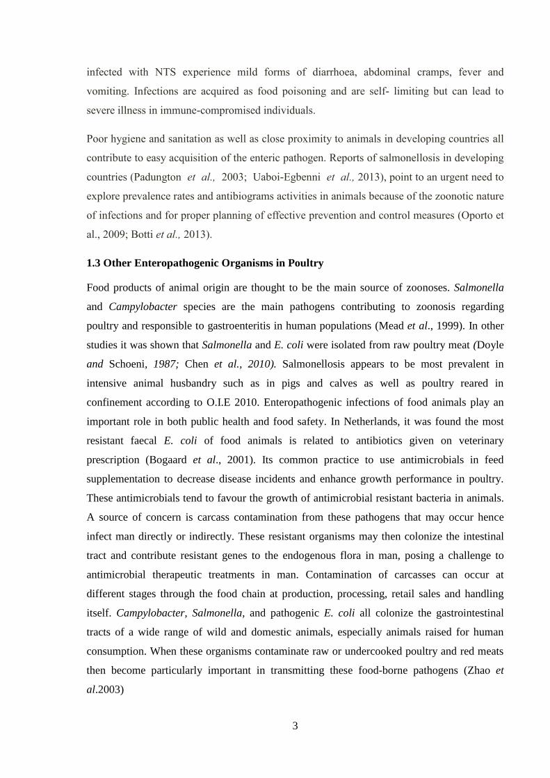
1.8 Conceptual Framework
To answer the objectives of the study, it is also important to consider the risks factors
associated with Salmonella and other enteropathogenic isolates infected quails at farm level.
It is known in developed countries, increase in demand of meat products has led countries to
7
produce animal or their products with higher efficiency and in turn, massive production has
caused the increase in food borne pathogens (Koluman 2012). Risk factors have been
documented in chicken poultry species as suggested by Mollenhorst et al., (2005) that risks
for Salmonella in laying hens include housing, flock size and different ages of birds.
Managerial skills, general hygiene and environmental status were assessed in a study
conducted in French commercial broiler flocks that showed an association of risk of
Salmonella in birds (Rose et al., 1999). By using the mentioned set of factors, a proposed
frame work was generated as shown in Appendix II.
Figure1. Conceptual framework

8
CHAPTER TWO
LITERATURE REVIEW
2.1 Overview
Enterobacteria are pathogens that will inhabit both humans and animals which are limited to
their digestive tracts. These animals include livestock, poultry, birds, reptiles, rodents and
pets. These species of pathogens can cause a number of foodborne and waterborne diseases.
These include food poisoning (gastroenteritis), typhoid (enteric fever) as well as bacteraemia
and septicaemia (Bell and Kyriakides, 2002)
Campylobacters are relatively ‘new’ zoonotic pathogens and the two species which are most
important in food-borne infections of humans with Campylobacter are C. jejuni and C. coli.
The pathogens are ubiquitous in nature and in domestic animals and, as a consequence, are
found frequently in the environment and on many raw foods, of both plant and animal origin
and bacterial numbers can be very high on certain key foods like raw poultry meat. Although
all commercial poultry species can carry campylobacters, the risk is greater from chicken
because of the high levels of consumption (Humphrey et al., 2007).
Escherichia coli is a bacterial commensal of the intestinal microflora of a variety of animals,
including humans. However, not all E. coli strains are harmless, as some are able to cause
diseases in humans as well as in mammals and birds (Dho-Moulin & Fairbrother, 1999;
Kaper et al., 2004). Pathogenic E. coli strains fall into two categories: those that cause
intestinal pathologies and those that cause extraintestinal pathologies. Intestinal pathologies
mostly consist of more or less severe diarrhoea or enteritis caused by different E. coli
pathotypes such as enterotoxinogenic, enteropathogenic or enterohaemorragic E. coli (ETEC,
EPEC and EHEC, respectively), potentially evolving into a haemolytic uremic syndrome
(HUS) in the case of EHEC infections in the case of EHEC infections. Comparison of E. coli
isolates can be a considerable challenge because of the wide genetic diversity resulting from
genome remodelling and horizontal acquisition from other pathogenic bacteria according to
Ron, 2010.
The enteropathogenic serotypes of E. coli (018, 044, 055, 086, 0111, 0114, 0119, 0126, 0127,
0128ab, 0142, 0158) produce toxins adhere to intestinal mucosa, disturbing the function of
microvilli, and cause diarrhea. The entero invasive serotypes (028ac, 029, 0124, 0136, 0143,
0144, 0152, 0164, 0167) invade and proliferate within epithelial cells, eventually causing cell

9
death. The enteroxigenic serotypes (06, 08, 020, 025, 027, 063, 078, 080, 085, 0115, 0128ac,
0139, 0148, 0153, 0159, 0167) and the entero haemorrhagic serotypes (01, 026, 091, 0111,
0113, 0121, 0128, 0145, 0157) of E. coli are associated with diarrheal illness.
Escherichia coli has implicated as an etiological agent of food poisoning involving different
foods such as raw milk, vegetables, cheese, potatoes, uncooked or poorly cooked meats and
poultry. Several strains of E. coli have emerged has potent food pathogens. One particular
strain such as (0157:H7) has been identified as one of the most dangerous in humans causing
a bloody diarrhea and even responsible for kidney failure in children (Ron, 2010)
Salmonella is rod shaped gram negative facultative anaerobic bacteria belonging to the family
Enterobacteriaceae that causes Salmonellosis. It’s an intracellular bacterium that is primarily
intestinal in nature in both animals and man. The bacterium is characterized by the O, H and
Vi antigens. The taxonomic classification of Salmonella is based on two species which
Salmonella enterica and Salmonella bongori. Salmonella enterica is further divided into six
subspecies which are I, II, IIa, IIIb, IV and IV (Noyal et al., 2009). Most of the Salmonellosis
in warm blooded animals in most cases is associated with serovars of Salmonella enterica
belonging to sub type I including the typhoid and paratyphoid bacilli. The most common type
of infection is the carrier state, in which healthy animals carry the pathogen without showing
any clinical signs. Clinical signs may take two forms systemic septicaemia and enteritis.
Numerous serovars of non-typhoid Salmonella (NTS) cause a self-limiting gastroenteritis in
healthy humans but may cause serious illness in immune-compromised persons. The most
common serovars isolated from humans in the developed world are serovars typhimurium,
enteritidis, virchow and hadar. They are all considered to be of zoonotic origin and all
exhibit resistance to commonly used antibiotics (Phillips et al., 2004). In the developing
world this is a great challenge, as little is known about bacterial populations in animal hosts.
The basic nature of these pathogens is to invade the intestinal mucosa and associated
lymphoid tissue. From the infected intestinal tissues the pathogens are drained to the
surrounding lymph nodes where macrophages from the lymphoid tissues form the first line of
barrier or defence against systemic infection. If the host is able to limit the infection to the gut
then it remains localized. In humans, non- typhoidal Salmonella serovars typically cause a
localized infection which manifests itself as acute gastroenteritis. On the other hand if the
macrophages located in the lymph nodes are unable to localize the infection, Salmonella can
cause a systemic infection. The systemic disease caused by human adapted serovar

10
Typhimurium causes typhoid fever. Some Salmonella serovars that cause typhoid fever like
disease in animals include S. gallinarum in poultry (fowl typhoid), S. choleraesuis in pigs
(porcine paratyphoid), S. tyhphimurium and S. enteriditis in mice (Wray and Wray 2002).
Salmonella serovars causing localized or systemic illness is complicated by the fact the
disease outcome is dependent on the immune status of the host. Most Salmonella serovars are
able to cause systemic disease in immune-compromised individuals that those at extreme
ages, the young and the old as well as those with underlying conditions such as HIV infection
(Magwedere et al.,2015)
2.2 Non-Typhoid Salmonellosis as an Emerging Problem
The poultry industry is a fast growing sector for both small and large scale commercial
farmers over the recent years in Zambia. The industry provides a cheap source of protein in
the form of meat as well as eggs. The industry in Zambia largely comprises the chickens
readily available on the market. However, due to the demand for other poultry species such as
quails, these species of poultry are now sold in supermarkets, local trading markets and farms
at point of sale for consumption and as source of revenue for poultry keepers.
In a study conducted by Hangombe et al., (1999), the prevalence rate of Salmonella in
poultry was estimated at 23%. The study also went to show that the presence of Salmonella
enteritidis was much higher for Zambia as compared to other parts of the world for chicken
poultry products at 4.7%. However, a recent study done by Ulaya (2013) found prevalence
rates of Salmonella in poultry species in particular chickens at 16.9%
An emerging problem now is that of antimicrobial resistance globally and in particular Africa
(Thakar et al., 2005, Kariuki et al., 2006). Routine practices of giving antimicrobial agents to
domestic livestock as a means of preventing and treating diseases, as well as promoting
growth, is an important factor in the emergence of antibiotic-resistant bacteria that are
subsequently transferred to humans through the food chain. Most infections with
antimicrobial-resistant Salmonella are acquired by eating contaminated foods of animal
origin (White et al., 2001).
The burden of invasive Salmonella disease in Africa stands at 227 per 100,000 in population
according to Gibani 2015. Invasive NTS disease was estimated to cause 3.4 million illnesses
and approximately 690 000 deaths in 2010 alone according to Crump 2015. NTS stands as
one of the leading enteric pathogens causing bacteraemia in young children in many parts of

11
the world including Africa (Berkley et al., 2006; Ikumapayi et al., 2007,). In a meta-analysis
of studies done in Africa, Salmonella serovars accounted for at least 33.1% of all invasive
NTS infections (Keddy et al., 2015). In a study done in Congo, they had shown Salmonella
enteritidis isolates of up to 79.7% showing multidrug resistance (Kalonji et al., 2015). Whilst
in South Africa, NTS is on the increase as an emerging pathogen associated with meningitis
among HIV infected persons according to Keddy et al., 2015. Likewise a study done in
Zambia by Mwansa et al., 2002, it was found that of 124 adults presented with persistent
diarrhea caused by non typhoidal Salmonella was accounted for at 5% of the individuals
whilst in the same study HIV related persistent diarrhoea in relation to NTS were at 11% and
16% in Rwanda and Kenya respectively. This is in agreement with W.H.O. 2013 reports
suggesting that infectious diarrhoea is a frequently occurring worldwide disease and
incidence of the condition is high in developing countries. From the findings mentioned
above we can see how NTS is a problem in foodborne illness and the challenges it presents. It
is for this reason that this study will look at the Salmonella strains and other bacterial strains
their molecular characteristics present in quail species and how sensitive they are to
antibiotics including their risk factors in quail species.
2.3 Quails
Quails have been farmed for a long time in many parts of the world and over the past few
decades commercial quail farming has grown in many parts if the world including Africa .
This has been seen in response to a fast growing market for the meat and the eggs they
produce. It has also been documented that quails are now been used as experimental animals
in biological research and in particular vaccine production for some diseases which they are
seen to be resistant to like Newcastle disease (Alderton, 1992; Shanaway, 1994). The
common quail is about 17.5 cm in length and can weigh about 70 to 155 grams in weight
(Figure 2).

12
Figure 2. Quail species
Common quail are terrestrial, temperate and tropical birds. Grasslands are the general habitat
of common quail. Dense, tall vegetation is preferred, while forest edges and hedgerows are
avoided. Cultivated fields of winter wheat, clover, and small grain crops are also used as
nesting cover (Johnsgard, 1988). The diet of quails consists of grains, seeds, nuts and insects
These birds have been considered as game animals for a long time until more recently
commercialized for their meat and eggs for consumption. The common quail is closely
related to the Japanese quail from the east which has well documented in other studies.
According to Chege (2014) protein deficiency remains a major challenge all over the world
and quail farming has been shown to be cheap and this has been used to fill up the nutrition
gap.
These birds are known to produce rapidly and are easy to keep in confinement although they
have not been commercially grown on a large scale as compared to broiler or layer chickens
here in Zambia.

13
Commercialized quails such as Japanese quail (Cortunix japonica) are raised for meat and
eggs. They reach market weight at 5- 6 weeks of age and they begin to lay their eggs at 6-7
weeks of age much earlier than broiler chickens at 22-24 weeks (Farooq et al., 2014) .
In developing countries, quail farming offers a viable and sustainable way of addressing the
problem of animal protein shortage hence offering an alternative source of protein other than
chicken production (Chege, 2014).
2.4 Bacteria and Antimicrobial Resistance in quails
In a study done by Roy et al., (2006), bacteria E. coli was isolated from diseased quails and
the bacteria was shown to be resistant to antimicrobials such as tetracyclines,
ampicillin/cloxacillin, cotrimoxazole, chloramphenicol and nitrofuratoin. In other studies
done in Iran isolates of Campylobacter were seen in quails at 43% of raw carcasses examined
with antimicrobial resistance to tetracyclines and nalidixic acid. These studies then show that
even as quails have been commercialized they are susceptible to bacterial infections and the
organisms been shown to be resistant to antimicrobials, presenting as a serious concern.
The global food-products trade is expected to increase in the future. Thus, attempts to
improve food safety must emphasize detection of antimicrobial drug–resistant bacteria
Salmonella species resistant to multiple antimicrobials agents have emerged worldwide
according to Jiang et al., 2005. This was shown with 3 Salmonella isolates that were isolated
from quails as recovered under the Danish Institute for Veterinary Research in October, 2003.
The serotypes isolated were of Virchow showing antimicrobial resistance to ampicillin,
ceftiofur, nalidixic acid, and tetracycline and with reduced susceptibility to ciprofloxacin
(MICs >0.125 µg/mL). Other studies have been done in quails located in the wild in Europe
as opposed to quails commercialized for sale of their meat and their eggs. In a study done by
Paulsen et al., 2012, a number of Salmonella enterica serovar isolates were recovered with
highest prevalence rates been S. typhimurium and S. enteriditis. The study also showed that
breeding game animals under intensive farming like the quails can create new
epidemiological situations of Salmonella in the birds and transmission of these pathogens into
other farm animal species.
2.5 Virulence and Rapid Detection of Salmonella
Characterisation of mechanisms underlying invasive manifestations by NTS is essential to
understanding the real depth in the biology and pathogenicity of Salmonella (Suez et al.,

14
2013). Laboratory techniques such as PCR assays have now been recognised as means of
detecting Salmonella and other pathogens. The molecular methods such as PCR were
standardised as a tool for detection of food borne pathogens including Salmonella species as
reported in study done by Malorny et al., 2002. The PCR can also be used to amplify
segments of particular genes such as invA that is responsible for the invasive characteristic of
Salmonella to cause disease (Mercanoglu et al., 2005). Other methods of identifying
Salmonella include dot blot hybridization and Enzyme Linked Immuno-sorbent Assay
(ELISA).
There is an epidemiologically important connection between poultry products and human
infections because many of the serotypes that are most prevalent in humans (such as
Salmonella typhimurium and Salmonella enteritidis) are similarly common in poultry Richard
(2007). Serotyping is important in determining what strains of the organism are present and
put in place appropriate interventions to control the disease in poultry species. A changing
epidemiological pattern and their dynamics can also be addressed by knowing the specific
strains present in specific poultry species. These bacteria can contaminate animal carcasses at
slaughter leading to potential human illness through raw consumption of eggs or undercooked
meat.

15
CHAPTER THREE
RESEARCH METHODOLOGY
3.1 Study Design
The study used a quantitative, analytical cross sectional study design in order to determine
the prevalence and risk factors associated with Salmonella and selected enterobacteria in
quails in Lusaka.
3.2 Study Setting
The study was conducted in Lusaka, the capital city of Zambia. Lusaka city lies on a plateau
1280 metres above sea level covering an estimated area of 360km2 and is located at 15º30'
latitude south and 28º17' longitude east. It was conducted among small holder poultry farms
that keep quails in production for consumption and sale. A total of 15 farms from across the
city were sampled. They had the required age for birds going into the market and also were
farmers that consented to answer the questionnaires.
Selection of participants (quail farmers)
Fifteen quail farms in Lusaka were identified and the consented farmers given questionnaires
to answer.
Quails.
Inclusion criteria- farms with quails that enter the market for sale at 6 to 8 weeks and
farmers that had consented.
Exclusion criteria-all poultry species except for quails, birds on recent antimicrobial therapy
in last 2-3 weeks and farmers that have not consented.
Study Variables
Dependent Variable- Salmonella, E. coli and Proteus positive on culture
Independent Variable- Age of birds, veterinary services, other poultry species, recent use of
antimicrobials and educational attainment of farmer, .

16
3.3 Sample size determination (Selection of participants)
The prevalence of Salmonella and other enteropathogenic isolates in quails in Zambia is
unknown. However this study took the prevalence rate of Salmonella in chickens as an
estimate at 16.9% (Ulaya et al., 2015). This was considered this way because most poultry
farmers may still keep chicken species on the same farm premises as quails. Then prevalence
was set at 16.9% to yield the maximum value of sample size (n). Assuming that we would
require the estimate to be within 5% of the true value in either direction at 95% CI, using a
sample size formula by Kish Leslie for cross sectional studies, sample size was given by
𝑠𝑠 =𝑍2 ∗ (𝑝) ∗ (1 − 𝑝)
𝑑2
This was done at confidence Level of 0.95 and desired precision of 0.05
Where:
ss= Sample size
Z = Z value for 95% confidence level (1.96)
p = 0.17
d = confidence interval (0.05)
Sample size will be given at 217 quails.
Probability Proportional to size sampling was then used for number of samples taken at each
farm. The probability proportional to size sampling frame was used to arrive at the total
number of 217 samples collected. This Sampling method was used to take into account the
varying sample sizes in account. The total number of quails was recorded at each individual
farm and the total sum arrived at for all fifteen farms. Thereafter, the number of quails
sampled was derived from dividing the individual farm number of quails by the total number
of fifteen farms multiplied by sample required. This study took the prevalence rate of
Salmonella in chickens as an estimate at 16.9% cite to yield a maximum sample size of 217
The birds from individual farms were then selected through systematic sampling method at
set intervals of ten.
3.4 Data Collection
Faecal samples were collected from anal openings of the birds at the farms. The samples were
the subjected to standard laboratory diagnostic culture and biochemical tests. This was
supplemented with questionnaires administered to the quail farmers. The questionnaire

17
included questions about management, use of antimicrobials, flock size and from which
breeding farms they acquire quails. To reduce bias and enhance performance of the
questionnaires, questions on Salmonella and other entero-pathogenic isolates were disguised
among other health related conditions.
3.4.1 Data analysis
These were interview guided questionnaires. The factors included the socio-economic status
and environmental conditions around the birds and the slaughtering process of birds going
into the markets. The questionnaire had epidemiological questions which included the age of
birds at point of sale, any history of sick birds. Others were husbandry practices such as
vaccination, any medication used in birds, veterinary services sought and any diseases present
or previously seen in birds. The information was used to generate knowledge, altitudes and
practices of quail farming. Information from questionnaire was cleaned, checked for
accuracy, consistency and completeness and then put on data base created in excel which was
exported into STATA Version 12 for analysis. Summary statistics were then calculated.
Association between possible factors such as socio-economic and environmental
characteristics with the positive outcome of isolates in quails from the quail farms were
analysed. All variables were included in the initial multiple logistic regression model and
using the backward elimination method, variables which showed independent association at a
significant level of p-value <0.05 were retained in the model.
3.4.2 Sample Collection
Faecal samples were collected aseptically from the anal regions of the birds using Amies
sterile swabs. The faecal samples were kept cool at 4˚C in cooler boxes with ice. Double
packaging of faecal swabs in sealable containers and later within sealable plastics was done
to prevent cross-contamination of samples and associated packaging materials (OIE
Terrestrial Manual, 2013). Salmonella may be isolated using various techniques that may
include pre enrichment to resuscitate sub-lethally damaged Salmonella, enrichment media
that may contain inhibitory substances to suppress competing organisms, and selective
plating agars to differentiate Salmonella from other enter bacteria (OIE Terrestrial Manual,
2013) Then various biochemical and molecular tests were applied to the pure culture to
provide a definitive confirmation of the isolated strain. Genetic technique such as PCR
analysis was used to identify specific resistant genes as well as providing additional

18
information on the virulence of the isolates (Herrera-Leon et al., 2004; Porwollik et al., 2004;
Batchelor et al., 2008; Wattiau et al., 2008).
3.4.2.1 Sample Preparation and Salmonella Isolation
The collected swabs were placed in Buffered Peptone Water as pre-enrichment medium. The
samples were then incubated at 37°C for 24hrs. One ml of each pre-enriched culture were
transferred into Tetrathionate (TT) Enrichment Broth (Oxoid Basingstoke, UK) and incubated
aerobically at 42˚C for 48 hrs. Then two to three loopfuls of each enriched broth culture were
streaked onto the surface of a selective medium Xylose Lysine Deoxycholate Agar (XLD)
(Merck 2005), and then all plates were incubated at 37˚C for 24 to 48 hrs.
3.4.2.2 E.coli and Proteus Isolation and PCR
The swabs were labelled appropriately and collection of the swab samples was aseptically
done. This involved the use of sterile swab sticks (Oxoid, Basingstoke, UK) which were
placed in tubes containing a Carry-Blair transport medium (Oxoid, Basingstoke, UK). The
poultry samples were inoculated on MacConkey agar (Oxoid, Basingstoke, UK) containing
2mg/L of cefotaxime (Sigma-Aldrich, Munich, Germany) for preliminary screening of ESBL
producing bacteria (Rayamajhi et al.,2008). The plates were later incubated at 37°C for 24
hours.
Colonies that grew on MacConkey agar were identified as lactose fermenters or non-lactose
fermenters. Identification of E. coli lactose-fermenting positive colonies was done using
phenotypic characteristics and confirmed by the Triple Sugar Iron (TSI) and IMViC tests as
described by Rayamajhi et al.,2008 and Batchelor et al.,2005. For genetic detection, E. coli
isolates were cultured on brain-heart-infusion broth (Nissui, Tokyo, Japan) at 37°C for 24
hours. After incubation, DNA was extracted by boiling methods (Reich and Klein 2013). The
E. coli isolates were subjected to PCR for confirmation of resistance genes TEM
(Temoniera), SHV (Sulphydryl Variable) and CTX-M (Cefotaxime –Munich) using primers
previously used by other workers (Batchelor et al.,2005 and Ranjbar et al.,2008). The PCR
(Finnzymes Oy, Finland) was performed in a total reaction volume of 10µl consisting of 5µl
Phusion master mix, 2µl sterile distilled water, 2µl primers (forward and reverse) and 1µl
bacterial DNA template. The PCR was performed using the rapid cycle DNA amplification
method comprising of an initial denaturation step at 98ºC for 30 seconds, followed by 35
cycles of template denaturation at 98ºC for 1 second, primer annealing at 60ºC for 5 seconds
and 72ºC for 1 second with final extension at 72ºC for 10 seconds. The PCR products were

19
later viewed with ethidium bromide after electrophoresis through 1.5% agarose gel (Clinical
and Laboratory Standards Institute, 2009).
3.4.2.3 Antimicrobial Sensitivity Identification
The antimicrobial susceptibility testing was done using the Kirby-Bauer disc diffusion
method on Mueller Hinton Agar (Becton, Dickinson and Company, MD, USA) based on the
Clinical Laboratory Standard Institute (CLSI) guidelines, 2009. The antibiotic discs (Becton,
Dickinson and Company, MD, USA) used included sulfamethoxazole/trimethoprim
(1.25/23.75μg), ciprofloxacin (5μg), tetracycline (30μg), gentamicin (10μg), chloramphenicol
(30μg), ceftazidime (30μg), norfloxacin (10μg) and cefotaxime (30μg). The phenotypic
confirmation of ESBL isolates was done by the combination of disc approximation method
using either ceftazidime (30μg) or cefotaxime (30μg) alone followed by over- night
incubation at 37°C for 18 – 24 hrs. Interpretation of susceptibility patterns on other anti-
microbial discs was done using guidelines laid down in the CLSI, which provides break
points corresponding to zone of inhibition diameter. An increase in antibiotic zone diameter
(5 – 12 mm) for either ceftazidime or cefotaxime indicated ESBL production (Clinical and
Laboratory Standards Institute, 2009). Quality control standard laboratory procedures were
strictly adhered to avoid contamination. Escherichia coli ATCC 25922 were used as a quality
control organism
20
CHAPTER FOUR
RESULTS
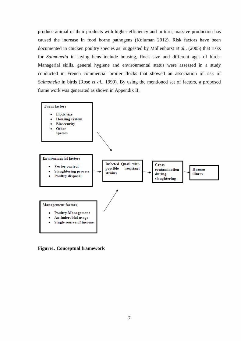
4.1 Clinical Bacterial Strains
During the study period, no species of Salmonella isolates were recovered from the 217 birds
sampled. However, 23 E. coli species and 30 Proteus species were isolated from the birds
(Table 1). The overall proportions of isolates observed in the study were 10.6% E. coli and
Proteus 13.8% as shown below
TABLE 1: Number of Bacterial Isolates and percentages
Bacterial isolates n(217) Number of isolates Percentage % 95% CI
Salmonella 0 0
E.coli 23 10.6 7-15
Proteus 30 13.8 9-19
Total 53 24.4
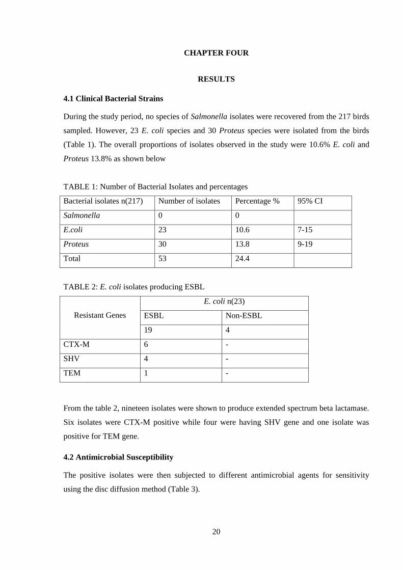
TABLE 2: E. coli isolates producing ESBL
Resistant Genes
E. coli n(23)
ESBL Non-ESBL
19 4
CTX-M 6 -
SHV 4 -
TEM 1 -
From the table 2, nineteen isolates were shown to produce extended spectrum beta lactamase.
Six isolates were CTX-M positive while four were having SHV gene and one isolate was
positive for TEM gene.
4.2 Antimicrobial Susceptibility
The positive isolates were then subjected to different antimicrobial agents for sensitivity
using the disc diffusion method (Table 3).
21
E. coli species showed 86.9% and 100% sensitivity to Norfloxacin and Ciprofloxacin
respectively while showing resistance to 91.3%, 100% and 86.9% to Co-trimoxazole,
Cefotaxime and Ceftazidime respectively. There was 78% resistance to Tetracycline. All E.
coli were sensitive to Gentamicin and 82% sensitive to Chloramphenicol.
Proteus species were 100% sensitive to Norfloxacin, Ciprofloxacin and Cefotaxime.
However, sensitivities to Co-trimoxazole and Ceftazidime were 70% and 93% respectively.
Proteus species showed more sensitivity to the antibiotics although 30% of the isolates
showed resistance to Co-trimoxazole and 6 % resistant to Ceftazidime. All Proteus species
were resistant to Tetracyline. Proteus were 90% sensitive to both Gentamicin and
Chloramphenicol.

22
TABLE 3: Bacterial Sensitivities
Bacterial
Isolates
Norfloxacin
10 μg
Sulfamethoxazole-
trimethroprim
1.25/23.75 μg
Ciprofloxacin
5 μg
Tetracyline
30 μg
Cefotaxime
30 μg
Ceftazidime
30 μg
Gentamicin
10 μg
Chloramphenicol
30 μg
E coli (n=23)
sensitive 20 2 23 5 - 3 - 19
resistant 3 21 18 23 20 23 4
Proteus (n=30)
sensitive 30 21 30 - 30 28 27 28
resistant - 9 - 30 - 2 3 2

23
4.3 PCR Findings
PCR amplification of target resistance genes was also investigated. Six isolates were found to
have the epidemiologic plasmid encoding gene for the CTX-M of extended beta lactamases.
While four were found to have SHV and one isolate for the TEM plasmids that encode for
antibiotic resistance.
Figure 3: Electrophoresis pattern of isolates after PCR using CTX genes. Isolate No 9 and 10
were positive isolates for the CTX gene, while No 17 is a positive control. Lane 16 is a
negative control and Lane 1 is a molecular weight marker. The positive amplicon is indicated
by the arrow.

24
Figure 4: Electrophoresis pattern of isolates after PCR using SHV genes. Isolate No 12 and
13 were positive isolates for the SHV gene, while number 4 is a positive control. Lane 5 is a
negative control and lane 1 is a molecular weight marker. The positive isolates are indicated
by the arrow.
4.4 Farm Level Characteristics Influencing Bacterial Contamination
To identify the farm-level characteristics that were influencing the bacterial
contamination/infection of quail meat in quail farms around Lusaka the study investigated the
association between having a contamination/infection with a particular species of bacteria of
public health significance and five variables for bacterial contamination/infection risk factors
were assessed. These were age of the birds, use of veterinary services, other poultry species,
employment status and education attainment.

25
4.4.1 Descriptive Statistics of Variables
TABLE 4: Study farms and sample sizes
Farm Id N % of Sample
1 18 8.3
2 11 5.1
3 15 6.9
4 21 9.7
5 13 6
6 8 3.7
7 25 11.5
8 12 5.5
9 10 4.6
10 13 6
11 10 4.6
12 8 3.7
13 10 4.6
14 13 6
15 30 13.8
1. Measuring the Effects of the use of Veterinary Services on likelihood of Proteus
After adjusting for the confounding effects of all the hypothesised risk factors, we found that
compared to quails on farms which did not use a veterinarians service, quail on farms which
used a veterinarian’s services were 85% less likely to be infected with Proteus spp. of
bacteria [Adjusted Odds Ratio: 0.15 (95% C l, 0.04 – 0.56, p<0.004].
In contrast, quail on farms that employed the use of a veterinarian’s services where 5 times
more likely to be infected with E. coli bacteria compared to those on farms which didn’t use
the service [Adjusted Odds Ratio: 4.85 (95% C l, 1.25 – 18.83, p<0.022].

26
Table 5: Results of the bi-variate and multivariate regression analysis investigating
determinants of infection with Proteus spp. bacteria in farmed quail in Lusaka, Zambia.
*Predictors of Proteus spp. infection with statistically significant effects (p<0.05)
2. Measuring the Effects of E. coli on likelihood of Proteus
After adjusting for the confounding effects of all the hypothesised risk factors, we found that
compared to quails which were not infected with E. coli, quail which were infected with E.
coli were 4 time more likely to be infected with Proteus spp. of bacteria [Adjusted Odds
Ratio: 4.15 (95% Cl, 2.0 – 18.13, p<0.002]. Similarly, After adjusting for the confounding
effects of all the hypothesised risk factors, we found that compared to quails which were not
infected with Proteus species,, quail which were infected with Proteus spp were 4 time more
likely to be infected with E. coli bacteria. [Adjusted Odds Ratio: 4.15 (95% Cl, 2.0 – 18.13,
p<0.002]
3. Measuring the Effects of farmers Education level on likelihood of E . coli
After adjusting for the confounding effects of all the hypothesised risk factors, we found that
compared to quails on farms whose owners only had primary school-level formal education,
quails on farms whose owners had secondary school-level education were 84% less likely to
Independent Predictors of Infection
Bivariate Analysis Multivariate Analysis
Crude
OR p value 95% CI
Adjusted
OR p value 95% CI
1. Flock Age At Testing (Ref: 6 Weeks)
8 Weeks Old 1.47 0.330 0.68 3.20 1.14 0.895 0.16 7.98
2. Used Veterinary Services (Ref: Don’t Use)
Uses Veterinary Services 0.21 0.005* 0.07 0.61 0.15 0.004* 0.04 0.56
3. Rearing of Other Poultry (Ref: Only Quail)
Rears Other Poultry 0.47 0.059 0.22 1.03 0.80 0.812 0.13 5.07
4. Employment of Owner (Ref: No other Employment)
Has formal Employment 0.45 0.057 0.20 1.02 0.66 0.485 0.20 2.15
5. Owner’s Formal Education (Ref: Primary)
Secondary School Education 1.12 0.812 0.43 2.93 2.00 0.314 0.51 7.86
6. E. coli Infection Status (Ref: Not Infected)
E. coli infected 4.15 0.004* 1.58
10.89 5.99 0.002* 2.00 18.13

27
be infected with E. coli bacteria [Adjusted Odds Ratio: 0.16 (p<0.001, 95% Cl 0.03 – 0.49,].
In contrast, education was not a factor for Proteus infection
Table 6: Results of the bi-variate and multivariate regression analysis investigating
determinants of infection with E. coli bacteria in farmed quail in Lusaka, Zambia.
*Predictors of E. coli infection with statistically significant effects (p<0.05)
Independent Predictors of Infection
Bivariate Analysis Multivariate Analysis
Crude
OR p value 95% CI
Adjusted
OR p value 95% CI
1. Flock Age At Testing (Ref: 6 Weeks)
8 Weeks Old 0.79 0.603 0.33 1.90 0.83 0.847 2.20 21.23
2. Used Veterinary Services (Ref: Don’t Use)
Received Training 1.26 0.599 0.53 3.03 4.85 0.022* 1.25 18.83
3. Rearing of Other Poultry (Ref: Only Quail)
Rears Other Poultry 1.00 0.995 0.40 2.47 0.85 0.874 0.12 6.29
4. Employment of Owner (Ref: No other Employment)
Has formal Employment 0.75 0.509 0.31 1.78 0.43 0.181 0.13 1.47
5. Owner’s Formal Education (Ref: Primary)
Secondary School Education 0.59 0.290 0.23 1.55 0.16 0.024* 0.03 0.49
6. E. coli Infection Status (Ref: Not Infected)
Proteus infected 4.15 0.004* 1.58 10.89 6.82 0.001* 2.20 21.23

28
CHAPTER FIVE
DISCUSSION
As an emerging problem worldwide, gram negative bacterial organisms are increasingly
becoming resistant to antimicrobials, in particular the extended-spectrum beta-lactamase
(ESBL)-producing enterobacteriaceae. Several studies have documented organisms isolated
from poultry in chickens, pathogens that have been associated with foodborne illness and that
the occurrence of antimicrobial resistant strains of zoonotic bacteria constitutes a public
health risk, increasing the risk of treatment failures as reported by Middleton and Ambrose,
2005. Quails previously considered as wild birds and a known delicacy in middle-eastern
parts of the world but now we see this source of protein farmed for commercial production
increasingly on menus in food outlets and indeed hotels sold out to the public in Zambia. The
findings of this study reveal the occurrence of Proteus and E. coli species. Out of the 217
samples examined, the prevalence rate of Proteus organisms was found to be slightly higher
at 13.8% than E. coli spp. at 10% from the domestic quails examined.
5.1 Determination of Salmonella
No Salmonella spp. were recovered from the 217 birds sampled as test results were
consistently negative. In this cross sectional study, results revealed that despite none of tested
quails showed characteristic clinical symptoms or pathological lesions, there were of some
zoonotic bacterial species that were detected among them indicating asymptomatic or carrier
infections. Bacterial strains of E. coli and Proteus species were isolated from the faecal swabs
taken from quails however no Salmonella strains were recovered from the birds. This agrees
with Teixeira et al., 2013 whose studies had shown also that no Salmonella isolates were seen
but recovered other enterobacteria such as E. coli and Proteus among others. Other studies
such Matankari (2014) have shown bacterial isolates of Escherichia coli, Salmonella species
and Pasteurella species were highly prevalent in the investigated quail egg shells. For the
Salmonella organisms alone, this is a remarkable result in the sense that it shows that it may
not be a worrying danger for these food pathogens as a source of enteric infections associated
consumption of protein contaminated at slaughter. Salmonellosis is an important disease
worldwide and it remains important that surveillance of poultry species at farms. Monitoring
of these zoonotic organisms, may not necessarily cause any health problems in animals but
pose as a health challenge in humans through enteric infections.
29
5.2 E. coli and Proteus Isolates
Avian pathogenic E. coli strains that are responsible for Colibacillosis and is one of the most
important economic loss through carcass rejection at slaughter facilities (FAO, 2002).
However, commensal organisms such as faecal E. coli constitute a reservoir of resistance
genes for (potentially) pathogenic bacteria. Resistant commensal bacteria of food animals
might contaminate, like zoonotic bacteria, meat (products) and so reach the intestinal tract of
humans (Bogaard and Stobberingh, 2000). From this study, 10.6% of E. coli isolates were
accounted for. Antimicrobial susceptibility and PCR tests confirmed the isolates for having
the antimicrobial resistance and the genes CTX-M (Cefoxatime-M). E. coli isolates were also
subjected to PCR for confirmation of resistance genes TEM (Temoniera), SHV (Sulphydryl
Variable). Six isolates were positive for CTX-M (Cefoxatime-M) genes, 4 isolates were
confirmed having SHV (Sulphydryl Variable) genes and one isolate positive for TEM
(Temoniera) gene. This TEM and SHV plasmids encode the genes responsible for
antimicrobial resistance. This study did not establish how this six isolates had the CTX-M
gene while other 13 did not have. This can be argued to imply a possible horizontal
transmission of these resistant genes among the commensal bacteria in the gut (Ron, 2010).
5.3 Antibiotic Resistance of E.coli and Proteus
In avian species, antimicrobial agents are often continuously provided as antimicrobial
growth promoters and this has resulted in increased antibiotic selection pressure for resistant
bacteria, resulting in their faecal flora containing a relatively high proportion of resistant
bacteria. The use of antibiotics has become the most important factor promoting the
emergence, selection and dissemination of antibiotic-resistant microorganisms in both
veterinary and human medicine (Bogaard et al., 2001)
E. coli species were resistant to this group of cephalosporins antibiotics and in particular
cefoxatime at 100% and ceftazidime 86%. Proteus species isolates did not show ESBL
properties but on antimicrobial tests there was 30% resistance to co-trimoxazole and 6%
resistance to ceftazidime. All Proteus species were resistant to tetracylines while 78% of E.
coli were resistant as were seen also in a study done by Roy et al., (2006). Proteus species or
bacilli as there are well known now, are classified as opportunistic pathogens that cause
illness in man. The results regarding the antimicrobial resistance pattern of E. coli isolates
were in agreement with Roy et al., (2006) who found that the antimicrobial resistance pattern
was 50% or more of that isolates were multi-drug resistant against oxtetracycline and

30
gentamicin. Sensitivity of bacterial strains to antimicrobials in the case of E. coli was higher
for the third generation cephalosporins antibiotics cefotaxime and ceftazidime compared to
Proteus species for as high as 100%. Use of cephalosporins in food producing animals could
be a selective factor for the appearance of extended spectrum beta lactamases producing
bacteria (Omoshaba et al., 2017). In addition, the bacterial organisms were highly resistant to
broad spectrum antibiotic such as tetracycline favoured for its use in animal feeds to enhance
growth and reduce disease incidence. It was further observed that E. coli was highly resistant
to Sulfamethoxazole trimethoprim and Gentamicin as opposed to Proteus spp. These two
antibiotics are fairly used more often for human treatments for urinary tract infections
(Rodríguez-Ban˜o et al., 2004). The organisms were sensitive to other antibiotics such as
chloramphenicol, norfloxacin and ciprofloxacin suggestive that they can actually be used in
treatments to control for these bacterial infections. Prevalence rates of these organisms in this
study are low compared to other studies; the findings of this research suggest that quails
could emerge as new population reservoirs for antibiotic resistant pathogens in the case of E.
coli.
There are three kinds of these opportunistic species P. vulgaris, P. penneri and P. mirabilis.
It is now said that Proteus mirabilis has been implicated in urinary tract infections, wounds
and infections of the gastrointestinal form arising from the consumption of meat products
(Rozalski et al., 1997). Although the study did not identify which Proteus sub species they
were and because of the potential virulence factors of Proteus bacilli, it still suggests that
screening of these organisms and their potential for ESBL production should be routinely
considered in quail species.
The significance of these findings is that, these organisms carry these enzymes to help in not
breaking them down by these antimicrobials. These organisms become resistant to
antimicrobial therapy hence the public health concern of resistant bacterial organisms that
may find their way onto poultry carcasses at slaughter hence into the food chain thereby
posing a health risk to mankind. Use of antimicrobial therapy in poultry as antimicrobial
growth promoters in feed to enhance productivity and reduce infections may favour
introduction of new resistant pathogens and if they find their way on meat products may
present as a challenge for human treatments when consumed raw or undercooked.

31
5.4 Possible attributes and factors on the Public health significance of Identified
bacteria
Several factors were hypothesized to assess whether certain attributes such veterinary service,
age of birds, employment, education and the presence of other poultry species have on the
risk of bacterial organisms isolated from the quails. Quails on farms which used a
veterinarian’s services were 85% less likely to be infected with Proteus spp. of bacteria
adjusted for age of the birds, other poultry flock, employment and education status [Adjusted
Odds Ratio: 0.15 (p<0.004 95% Cl, 0.04 – 0.56]. This shows a significant correlation
between use of veterinary services and Proteus status in the birds. A farmer who sought
veterinary assistance was more likely to use therapeutic methods to reduce enteric organisms
in the birds and reduce bacterial loads as seen in the isolates. This may also be supported by
the correlation seen where education at secondary level reduced the likelihood of bacterial
infection in the birds for E. coli but was not significant for Proteus. Several enteric organisms
can be isolated from birds at different times and the interaction of these organisms may differ
in proportions and the kind of sensitivity against antimicrobials. Proteus species was more
sensitive to the antimicrobials as opposed to E. coli species.
In contrast, quail on farms that employed the use of a veterinarian’s services where five times
more likely to be infected with E. coli bacteria compared to those on farms which didn’t use
the service adjusted for other risk factors [Adjusted Odds Ratio: 4.85 (p<0.022 95% C l, 1.25
– 18.83]. This does not tell as much about where the organisms are coming from except to
show frequent use of veterinary services increased the E. coli count on the farms from the
isolates recovered from the birds. It may also be that E. coli species increased in these birds
because they were no longer sensitive to antimicrobials given through commercial feeds to
enhance growth and hence flourish in the guts of these birds.
Veterinary services were then an important factor for less likelihood of Proteus infection
which implies that farms that engaged veterinary input were less likely to suffer Proteus
infection in birds. Therefore, this information emphasizes use of veterinary services help
inform farmers on curbing infection through biosecurity measures but may be supported in
that the species of Proteus isolated from these birds were sensitive to antimicrobials.
The results from this study may suggest interventions directed at the routine surveillance of
these enteric organisms that may become opportunistic pathogens if meat products are
contaminated. Secondly more studies that look at how quail keepers can use veterinary

32
services to help enhance production with the appropriate use of antimicrobials. The spread of
zoonotic bacteria resistant to antibiotics is an important concern for the treatment of human
infections, because it can compromise the effectiveness of the therapy (Kilonzo-Nthenge et
al., 2008). The study has shown that there are opportunities that exist for new health
problems in quail species commercialized for meat or indeed their eggs emerging as new
reservoirs of zoonotic entero bacteria.
Strengths and Limitations of the Study
The study only looked at quail farmers based in Lusaka and therefore generalizations of the
study were limited to Lusaka town. However this the first time we have had a base line study
on selected enteric microbes isolated from quail species and it would be important to screen
this species, especially that they are now reared for commercial purposes, regularly to
identify bacterial organisms that may carry resistant genes or even considered opportunistic
pathogens. The results from this study provide useful information for surveillance purposes.
Methodological challenges
A lot of these quail farmers are small holder poultry keepers. This presents a challenge with
keeping track of where the birds are sold to for consumption and also to the markets they go
to such as supermarkets, restaurants, open markets and hotels as owners may not have written
records. Written records for the purposes of tracing from which farms the birds are coming
from is important in surveillance of zoonotic pathogens.

33
CHAPTER SIX
CONCLUSION
The aim of the study was to characterise and determine the public health significance of
selected entero-pathogens in quails. The study findings revealed no Salmonella isolates. E.
coli and Proteus species were isolated. The ESBL producing E. coli isolates were of
significance in the role they play in resistance in poultry that may find their way into meat
products on the market. This study has shown that multidrug resistance of E. coli species in
poultry in particular quails may largely contribute to the wider and broad challenge of
antimicrobial resistance and at the same time provide useful information for surveillance
purposes. The use of antimicrobials does not necessarily lead to the selection of virulent
strains but rather to the spread of resistance genes among commensal strains. It is also
important to continue monitoring pathogens especially those of zoonotic nature such as
Salmonella and maintain the status quo through effective surveillance.

34
CHAPTER SEVEN
RECOMMENDATIONS
There are research and policy implications for the study. This study has highlighted on the
importance of conducting surveillance on enteric pathogens seen in other poultry species such
as quails as opposed to chickens alone.
1. It is also important to investigate antimicrobial resistance seen in these poultry species and
its impact on human health treatments.
2. There is need to look at prospective cohort studies and include all other quail farms in the
Zambia.
3. The Ministry of Livestock and Fisheries at policy level, should control antimicrobial
growth promoters in feed that are used to enhance productivity and their use monitored
closely to discourage resistance of organisms that may find their way along the food chain.it
is important to bring in veterinary services very close to the farmers that may help educate
them on relevant use of antimicrobials.

35
REFERENCES
Alderton, D. 1992. atlas of quails, TFH Publications.
Ao, T. T., Feasey, N. A., Gordon, M. A., Keddy, K. H., Angulo, F. J. & Crump, J. A. 2015.
Global burden of invasive nontyphoidal Salmonella disease, 2010. Emerging
infectious diseases, 21, 941.
Baird-Parker, A. 1990. Foodborne salmonellosis. The Lancet, 336, 1231-1235.
Barrow, P. & Neto, O. F. 2011. Pullorum disease and fowl typhoid—new thoughts on old
diseases: a review. Avian pathology, 40, 1-13.
Batchelor, M., Hopkins, K. L., Liebana, E., Slickers, P., Ehricht, R., Mafura, M., Aarestrup,
F., Mevius, D., Clifton-Hadley, F. A. & Woodward, M. J. 2008. Development of a
miniaturised microarray-based assay for the rapid identification of antimicrobial
resistance genes in Gram-negative bacteria. International journal of antimicrobial
agents, 31, 440-451.
Bell, C., Kyriakides, A., Salmonella, C. B. & Mcclure, P. 2002. Foodborne pathogens:
Hazards, risk analysis and control. Ch.
Berkley, J. A., Lowe, B. S., Mwangi, I., Williams, T., Bauni, E., Mwarumba, S., Ngetsa, C.,
Slack, M. P., Njenga, S. & Hart, C. A. 2005. Bacteremia among children admitted to a
rural hospital in Kenya. New England Journal of Medicine, 352, 39-47.
Botti, V., Navillod, F. V., Domenis, L., Orusa, R., Pepe, E., Robetto, S. & Guidetti, C. 2013.
Salmonella spp. and antibiotic-resistant strains in wild mammals and birds in north-
western Italy from 2002 to 2010. Vet Ital, 49, 195-202.
Chege, L. M. 2014. Factors Influencing Quail Farming In Nyeri Central Constituency, Nyeri
County, Kenya. University of Nairobi.
Crump, J. A. & Heyderman, R. S. 2015. A Perspective on Invasive Salmonella Disease in
Africa. Clinical Infectious Diseases, 61, S235-S240.
Doyle, M. P. & Schoeni, J. L. 1987. Isolation of Escherichia coli O157: H7 from retail fresh
meats and poultry. Applied and Environmental Microbiology, 53, 2394-2396.
Edward, B. 2005. Black’s Veterinary Dictionary. A & C Black Publishers, London.
Espié, E., De Valk, H., Vaillant, V., Quelquejeu, N., Le Querrec, F. & Weill, F. 2005. An
outbreak of multidrug-resistant Salmonella enterica serotype Newport infections
linked to the consumption of imported horse meat in France. Epidemiology and
infection, 133, 373-376.
Farooq, U. 2014. Investigation of factors controlling fertility in Japanese quail (Coturnix
Japonica).
Feasey, N. A., Cain, A. K., Msefula, C. L., Pickard, D., Alaerts, M., Aslett, M., Everett, D.
B., Allain, T. J., Dougan, G. & Gordon, M. A. 2014. Drug Resistance in Salmonella
enterica ser. Typhimurium Bloodstream Infection, Malawi. Emerging infectious
diseases, 20, 1957.
Gast, R. K. 2007. Serotype-specific and serotype-independent strategies for preharvest
control of food-borne Salmonella in poultry. Avian diseases, 51, 817-828.
Gibani, M. M., Jin, C., Darton, T. C. & Pollard, A. J. 2015. Control of Invasive Salmonella
Disease in Africa: Is There a Role for Human Challenge Models? Clinical Infectious
Diseases, 61, S266-S271.
Gordon, M. A. 2011. Invasive Non-typhoidal Salmonella Disease–epidemiology,
pathogenesis and diagnosis. Current opinion in infectious diseases, 24, 484.
Graham, S. M. 2002. Salmonellosis in children in developing and developed countries and
populations. Current opinion in infectious diseases, 15, 507-512.

36
Hang'ombe, B. M., Sharma, N. R., Skjerve, E. & Tuchili, L. M. 1999. Isolation of bacteria
during processing of chicken carcasses for the market in Lusaka, Zambia.
Veterinarski arhiv, 69, 191-197.
Hendriksen, R. S., Joensen, K. G., Lukwesa-Musyani, C., Kalondaa, A., Leekitcharoenphon,
P., Nakazwe, R., Aarestrup, F. M., Hasman, H. & Mwansa, J. C. 2013. Extremely
drug-resistant Salmonella enterica serovar Senftenberg infections in patients in
Zambia. Journal of clinical microbiology, 51, 284-286.
Hendriksen, R. S., Vieira, A. R., Karlsmose, S., Lo Fo Wong, D. M., Jensen, A. B., Wegener,
H. C. & Aarestrup, F. M. 2011. Global monitoring of Salmonella serovar distribution
from the World Health Organization Global Foodborne Infections Network Country
Data Bank: results of quality assured laboratories from 2001 to 2007. Foodborne
Pathogens and Disease, 8, 887-900.
Herrera-León, S., Mcquiston, J. R., Usera, M. A., Fields, P. I., Garaizar, J. & Echeita, M. A.
2004. Multiplex PCR for distinguishing the most common phase-1 flagellar antigens
of Salmonella spp. Journal of clinical microbiology, 42, 2581-2586.
Humphrey, T., O'brien, S. & Madsen, M. 2007. Campylobacters as zoonotic pathogens: a
food production perspective. International journal of food microbiology, 117, 237-
257.
Ikumapayi, U. N., Antonio, M., Sonne-Hansen, J., Biney, E., Enwere, G., Okoko, B.,
Oluwalana, C., Vaughan, A., Zaman, S. M. & Greenwood, B. M. 2007. Molecular
epidemiology of community-acquired invasive non-typhoidal Salmonella among
children aged 2–29 months in rural Gambia and discovery of a new serovar,
Salmonella enterica Dingiri. Journal of medical microbiology, 56, 1479-1484.
Johnsgard, P. A. & Jones, H. 1988. Quails, Partridges, and Francolins of the World, Oxford
University Press.
Kalonji, L. M., Post, A., Phoba, M.-F., Falay, D., Ngbonda, D., Muyembe, J.-J., Bertrand, S.,
Ceyssens, P.-J., Mattheus, W. & Verhaegen, J. 2015. Invasive Salmonella Infections
at Multiple Surveillance Sites in the Democratic Republic of the Congo, 2011–2014.
Clinical Infectious Diseases, 61, S346-S353.
Kariuki, S., Revathi, G., Kariuki, N., Kiiru, J., Mwituria, J., Muyodi, J., Githinji, J. W.,
Kagendo, D., Munyalo, A. & Hart, C. A. 2006. Invasive multidrug-resistant non-
typhoidal Salmonella infections in Africa: zoonotic or anthroponotic transmission?
Journal of medical microbiology, 55, 585-591.
Khan, C. 2005. The Merck Veterinary Manual 9th ed. Merck and C. Inc. NJ. USA, 2125-
2136.
Kilonzo-Nthenge, A., Nahashon, S., Chen, F. & Adefope, N. 2008. Prevalence and
antimicrobial resistance of pathogenic bacteria in chicken and guinea fowl. Poultry
science, 87, 1841-1848.
Kingsley, R. A., Msefula, C. L., Thomson, N. R., Kariuki, S., Holt, K. E., Gordon, M. A.,
Harris, D., Clarke, L., Whitehead, S. & Sangal, V. 2009. Epidemic multiple drug
resistant Salmonella Typhimurium causing invasive disease in sub-Saharan Africa
have a distinct genotype. Genome research, 19, 2279-2287.
Kish, L. 2004. Statistical design for research, John Wiley & Sons.
Koluman, A. & Dikici, A. 2013. Antimicrobial resistance of emerging foodborne pathogens:
status quo and global trends. Critical reviews in microbiology, 39, 57-69.
Maclennan, C. A., Gondwe, E. N., Msefula, C. L., Kingsley, R. A., Thomson, N. R., White,
S. A., Goodall, M., Pickard, D. J., Graham, S. M. & Dougan, G. 2008. The neglected
role of antibody in protection against bacteremia caused by nontyphoidal strains of
Salmonella in African children. The Journal of clinical investigation, 118, 1553.

37
Magwedere, K., Rauff, D., De Klerk, G., Keddy, K. H. & Dziva, F. 2015. Incidence of
Nontyphoidal Salmonella in Food-Producing Animals, Animal Feed, and the
Associated Environment in South Africa, 2012–2014. Clinical Infectious Diseases,
61, S283-S289.
Malorny, B., Hoorfar, J., Bunge, C. & Helmuth, R. 2003. Multicenter validation of the
analytical accuracy of Salmonella PCR: towards an international standard. Applied
and environmental microbiology, 69, 290-296.
Matankari, R. M. 2014. Antimicrobial Susceptibility Studies Of Escherichia Coli, Salmonella
Spp, Pasturella Spp Isolated From Quail Egg Shells In Some Farms In Kaduna State,
Nigeria.
McEntire, J., Acheson, D., Siemens, A., Eilert, S. & Robach, M. 2014. The public health
value of reducing salmonella levels in raw meat and poultry. Food Protection Trends,
34, 386-392.
Mercanoğlu, B. & Griffiths, M. W. 2005. Combination of immunomagnetic separation with
real-time PCR for rapid detection of Salmonella in milk, ground beef, and alfalfa
sprouts. Journal of Food Protection®, 68, 557-561.
Mollenhorst, H., Van Woudenbergh, C., Bokkers, E. & De Boer, I. 2005. Risk factors for
Salmonella enteritidis infections in laying hens. Poultry Science, 84, 1308-1313.
Mshana, S. E., Matee, M. & Rweyemamu, M. 2013. Antimicrobial resistance in human and
animal pathogens in Zambia, Democratic Republic of Congo, Mozambique and
Tanzania: an urgent need of a sustainable surveillance system. Annals of clinical
microbiology and antimicrobials, 12, 1.
Muthumbi, E., Morpeth, S. C., Ooko, M., Mwanzu, A., Mwarumba, S., Mturi, N., Etyang, A.
O., Berkley, J. A., Williams, T. N. & Kariuki, S. 2015. Invasive Salmonellosis in
Kilifi, Kenya. Clinical Infectious Diseases, 61, S290-S301.
Mwansa, J., Mutela, K., Zulu, I., Amadi, B. & Kelly, P. 2002. Antimicrobial sensitivity in
enterobacteria from AIDS patients, Zambia.(Dispatches). Emerging infectious
diseases, 8, 92-94.
Noyal, M., Menezes, G., Harish, B., Sujatha, S. & Parija, S. 2009. Simple screening tests for
detection of carbapenemases in clinical isolates of nonfermentative Gram-negative
bacteria. Indian Journal of Medical Research, 129, 707.
Organization, W. H. 2013. Sustaining the drive to overcome the global impact of neglected
tropical diseases: second WHO report on neglected tropical diseases: summary.
Padungton, P. & Kaneene, J. B. 2003. Campylobacter spp. in Human, Chickens, Pigs and
Their Antimicrobial Resistance. Journal of veterinary medical science, 65, 161-170.
Paulsen, P., Smulders, F. & Hilbert, F. 2012. Salmonella in meat from hunted game: A
Central European perspective. Food Research International, 45, 609-616.
Phillips, I., Casewell, M., Cox, T., De Groot, B., Friis, C., Jones, R., Nightingale, C., Preston,
R. & Waddell, J. 2004. Does the use of antibiotics in food animals pose a risk to
human health? A critical review of published data. Journal of Antimicrobial
Chemotherapy, 53, 28-52.
Porwollik, S., Boyd, E., Choy, C., Cheng, P., Florea, L., Proctor, E. & Mcclelland, M. 2004.
Characterization of Salmonella enterica subspecies I genovars by use of microarrays.
Journal of bacteriology, 186, 5883-5898.
Rose, N., Beaudeau, F., Drouin, P., Toux, J., Rose, V. & Colin, P. 1999. Risk factors for
Salmonella enterica subsp. enterica contamination in French broiler-chicken flocks at
the end of the rearing period. Preventive veterinary medicine, 39, 265-277.
Rotimi, V. O., Jamal, W., Pal, T., Sovenned, A. & Albert, M. J. 2008. Emergence of CTX-M-
15 type extended-spectrum β-lactamase-producing Salmonella spp. in Kuwait and the
United Arab Emirates. Journal of medical microbiology, 57, 881-886.

38
Roy, P., Purushothaman, V., Koteeswaran, A. & Dhillon, A. 2006. Isolation, characterization,
and antimicrobial drug resistance pattern of Escherichia coli isolated from Japanese
quail and their environment. The Journal of Applied Poultry Research, 15, 442-446.
Rózalski, A., Sidorczyk, Z. & Kotełko, K. 1997. Potential virulence factors of Proteus bacilli.
Microbiology and Molecular Biology Reviews, 61, 65-89.
Schroeder, C. M., White, D. G., Ge, B., Zhang, Y., Mcdermott, P. F., Ayers, S., Zhao, S. &
Meng, J. 2003. Isolation of antimicrobial-resistant Escherichia coli from retail meats
purchased in Greater Washington, DC, USA. International journal of food
microbiology, 85, 197-202.
Shanaway, M. 1994. Quail production systems: a review, Food & Agriculture Org.
Suez, J., Porwollik, S., Dagan, A., Marzel, A., Schorr, Y. I., Desai, P. T., Agmon, V.,
Mcclelland, M., Rahav, G. & Gal-Mor, O. 2013. Virulence gene profiling and
pathogenicity characterization of non-typhoidal Salmonella accounted for invasive
disease in humans. PLoS One, 8, e58449.
Susan, E. A. & Aiello, B. 1998. The Merck veterinary manual. Merck and Company Inc.,
USA., ISBN-13, 1271377300, 165-173.
Teixeira, R., Cardoso, W., Lopes, E., Rocha-E-Silva, R., Albuquerque, A., Horn, R. & Salles,
R. 2013. Bacteriological investigation of microorganisms (Salmonella sp. and other
Enterobacteriaceae) in common quails (Coturnix coturnix) submitted to different
forced-molting procedures. Revista Brasileira de Ciência Avícola, 15, 47-52.
Uaboi-Egbenni, P., Bessong, P., Samie, S. & Obi, C. 2013. Prevalence and antimicrobial
susceptibility profiles of Campylobacter jejuni and coli isolated from diarrheic and
non-diarrheic goat faeces in Venda region, South Africa. African Journal of
Biotechnology, 10, 14116-14124.
Ulaya, W. D. 2013. Determination of Virulence factors in Salmonella isolates of Human,
Poultry and Dog origin in Lusaka District, Zambia. UNIVERSITY OF ZAMBIA.
Van Den Bogaard, A. E. & Stobberingh, E. E. 2000. Epidemiology of resistance to
antibiotics: links between animals and humans. International journal of antimicrobial
agents, 14, 327-335.
Wain, J., Keddy, K. H., Hendriksen, R. S. & Rubino, S. 2013. Using next generation
sequencing to tackle non-typhoidal Salmonella infections. The Journal of Infection in
Developing Countries, 7, 001-005.
Wattiau, P., Weijers, T., Andreoli, P., Schliker, C., Vander Veken, H., Maas, H. M.,
Verbruggen, A. J., Heck, M. E., Wannet, W. J. & Imberechts, H. 2008. Evaluation of
the Premi® Test Salmonella, a commercial low-density DNA microarray system
intended for routine identification and typing of Salmonella enterica. International
journal of food microbiology, 123, 293-298.
White, D. G., Zhao, S., Sudler, R., Ayers, S., Friedman, S., Chen, S., Mcdermott, P. F.,
Mcdermott, S., Wagner, D. D. & Meng, J. 2001. The isolation of antibiotic-resistant
Salmonella from retail ground meats. New England Journal of Medicine, 345, 1147-
1154.
Wray, C. & Wray, A. 2000. Salmonella in domestic animals, Cabi

39
APPENDICES
Appendix 1: Participant Information sheet & Consent Forms
INFORMATION SHEET DOCUMENT FOR PARTICIPANTS
Study Title: isolation, Antimicrobial Susceptibility and Associated Risk Factors of
Salmonella in Quails
Principal Investigator : Dr. Mukachikwikwi Hamakoko, 121 C Close Avondale,
Lusaka. +260 977454945
Purpose of research project
This study is part of my research thesis in Masters in Public Health. The purpose of the
research is to check the presence of bacteria Salmonella in faeces. The study aims at
collecting fecal samples. After collection of samples, it is intended for the samples to undergo
laboratory tests at the University Of Zambia. This is to confirm if the bacteria Salmonella are
present or not. I also want to learn from the quail keepers if there is any more information
that can help find out why the birds may have or may not have the bacterium by asking a set
of questions.
Why you are being asked to participate?
Participants are farmers who are keeping quails and selling these quails on the market. You
have been asked to participate because you fit these descriptions.
Procedures
If you agree to participate in the research:
I will ask you to fill in a questionnaire. I will ask questions on what you know on
health related topics on quails. Your name or your personal details will not be
included in the typed documents. The questionnaire may take about 30 minutes to fill
in.
Risks/discomforts There are no physical risks to participating in the research. However, I recognize some
information you may tell me or enter in the questionnaires may be personal. Some
discomforts may be seen in the birds when taking fecal samples.
Benefits
There are no direct benefits to you.
Payment
There is no payment for participating in the research. However, I will inform you on the
results.

40
Protecting data confidentiality
I have put up steps to protect the information I will get from you. First, I will make sure that
your name is not included with any information. Only my research assistants and my
supervisors at University of Zambia will have knowledge to the study information. The
collected information will be locked in a secure place. I will keep copies of typed information
on a flash disc in case i have a problem with the computer.
What happens if you do not want to participate?
You are free to decide whether you want to participate or not. Participants are free to
withdraw from the study at any time and you may skip questions that you may not wish to
answer.

41
Certificate of Consent
STUDY TITLE: Isolation, Antimicrobial Susceptibility and Associated Risk Factors of
Salmonella in Quails
I have been asked to give consent to take part in the research study entitled “Prevalence,
Antimicrobial Susceptibility, Molecular Characterization and Associated Risk Factors for
Salmonella in Quails”. This will involve taking part in answering questions from a
questionnaire about knowledge, attitude and practices of quail keeping. I will also provide
information on quail keeping. I have understood, read or it has been read to me and I fully
understand all the information. I had the opportunity to ask questions and I was given
satisfactory answers to my questions.
If the respondent is illiterate, a literate witness must sign and participants who are illiterate
should include their thumb print as well. I have witnessed the accurate reading of the consent
from potential participant and the individual has had the opportunity to ask questions. I
confirm that the individual has given consent freely.
You are free to decide whether you want to participate or not. Participants are free to
withdraw from the study at any time and you may skip questions that you may not wish
to answer.
Who do I call if I have questions or problems?
Call me, <<Dr. Mukachikwikwi Hamakoko, 121 C Close Avondale, Lusaka>>, at
<<+260-977-454945>> if you have questions and complaints about the research.
Call or contact ERES office for any ethical queries. The Ethics Committee contact
information is:
Address: 33 Joseph Mwilwa Road
Rhodes Park, Lusaka
Telephone : +260 955 155 633
: + 260 955 155 634
E-mail: [email protected]
What does your signature (or thumbprint/mark) on this consent form mean?
Your signature (or thumbprint/mark) on this form means:

42
You have been informed about the research’s purpose, procedures, possible benefits and
risks.
You have been given the chance to ask questions before you sign.
You have voluntarily agreed to be in this research program
________________________ _____________________________ __________
Print name of Adult Participant Signature of Adult Participant Date
________________________ _____________________________ __________
Print name of Person/Witness Obtaining Signature of Person/Witness Obtaining
Consent Date
Left Thumb Print below, if cannot put your signature

43
Translation to Chinyanja Kadunswa ka 3: Pepala ilangiza otenga mbali
nakubvomekedza.
NKHANI YOLEMBEDWA YA OTENGA MBALI.
Mutu Wachobelenga: Kusankula,kugonja ku tudoyo todwalitsa matenda ndi chiopsezo cha
Salmonella mu mbalani za nkwali.
Ofufudza: Dr. Mukachikwikwi Hamakoko, 121 Close, Avondale, Lusaka. +260 0977454945.
Cholinga chanchito yakufufudza.
Uku kubelenga nikwina kwambali yina yanga ya pepala ya masitala imene ninayanganapo
zaulele za anthu. Cholinga chakubelenga kumene uku niku funa kuona ngati khungapedzeke
kadoyo ka Samonella mamatubvi ya nkwali. Uku kufufudza kusayetsa-yetsa kusakila
nakutenga matubvi ya nyoni zosiyana-siyana. Pambuyo potenga matubvi amenewa,
yadzankhala yalikuikidwa mukupimiwa mumakina kusukulu lali khulu ya University of
Zambia. Uku nikusimikidza kuona kuti ngati kadoyo ka Samonella kapedzekamo olo
sikapedzekamo. Nidza funatso kufutsa kuli amene asunga nkwali ngati kuli nkhani yina inga
tandidzile mukufufudza kwachifukwa nyoni zina zipedzeka nakadoyo aka kupyolela
mukufutsa mafutso.
Chifukwa ninji mukupedwa kutenga mbali?
Otenga mbali ni alimi osunga nkwali ndipo ogulitsa kumutsika. Mwapedwa kutengako mbali
chifukwa mwakwanilitsa zimene zilikulankhulindwa umu.
Mochitila.
Ngati mwabvomera kutengako mbali mukufufudza uku:
• Nidzakufutsani pali zamene mudziba pazaumoyo za nkwali. Khoma, dzindikilani kuti
dzina lanu silidza lembedwapo pachipelala. Kuyankha mafudza amenewa kukatenga chabe
nthawi itali monga pindi zili makhumi atathu (30 minutes).
Ziopezo/ Kusanzibvera bwino
44
Dzibani kuti kulibe ziopezo zilizonse mukuyankha mafutso. Khoma nidzindikila kuti nkhani
yamene mudzalembamo kapena kuyika umu muchipepala chamafutso niya paumoyo wanu
noka. Dzina zache zoipitsa dzidzaokela mutunyoni pa nthawi yotenga matubvi yachitsadzo.
Phindu.
Munchito imeneyi, kulibe phindu yomwe mudzapedzamo.
Malipilo.
Kulibe malipilo mukutenga mbali muku fufudza uku. Ngankhale telo, nidza ku udzani
zimene zidzapedzeka pambuyo kwakusila kwakufufudza kumene uku.
Kuchingilidzidwa kwa nkhani yachitsitsi.
Nayika ndondomeko yo chingilidza nkhani yamene nidzatenga kwa inu. Choyamba nichakuti
sinidza ika dzina lanu papepala na nkhani ili yonse. Oka adzadziba nkhani iyi ni aja
otandidzila mukufufudza kuyikilapo na aja akulu-akulu akumupando oyatiya ganila
mukubelenga uku ku Universitt of Zambia. Nkhani iza tengedwa idza nkhala yokomewa
nakusungiwa apo pamene munthu wina sanga iwone. Nidzasungatso zina zipepala dza nkhani
yamene iyi kuchitila kuti mwina mwache napedzeka nabvuto na computer, sininga sobe
pogwila.
Nichani chingachitike ngati simufuna kutenga mbali?
Muli na ufulu obvomela kutengako mbali olo kusatenga. Otenga mbali ali na ufulu
kuchokamo mukubelenga uku pa nthawi ili yonse. Mafutso amene simukwanisa kuyanka
munga yasiye yosayankhiwa.

45
CHIGAWO CHACHIWILI
Pepala limene lilangidza kubvomekedza.
Mutu Wachobelenga: Kusankula,kugonja ku tudoyo todwalitsa matenda ndi chiopsezo
cha Salmonella mu mbalani za nkwali.
Napempedwa kuti nisonyedze kubvomekedza kwanga kwa kutenga mbali mukubelenga uku
ko fufudza na mutu uyu: “kuchuluka kwakadoyo, kaonekedwe kakadoyo nadzina zoipitsa za
kadoyo ka Samonella mu nkwali.” Ichi chidzachitika kupyolela mukuyankha mafutso
kuchokela muchipepala cholembewamo mafutso pali kudziba, maganidzo namachitidwe ya
osunga nkwali. Nidzakudzani futi nkhani yo sunga nkwali. Nabvetsa-tsa, nabelenga kapena
kuti anibelengela nkhani iyi ndipo nabvela mokwana zonse izi zili umu. Ninali nayo danga
lofutsa mafutso ndipo ananiyankha mafutso yonse mokwana.
Ngati boyankha sa ziba kubelenga, opedzekako odziba kubelenga ayenela kudinda chidindo
ndipo amene zadziba kubelenga ayenela kudinda chidindo nachiikumo chawo. Naonetsa-tsa
kubelenga kwabwino kwakubvomekedza kuchoka kuli otenga mbali ndipo munthu uyu anali
na danga lo futsa mafutso. Nasimikidza kuti munthu uyu abvomela kutenga mbali. Muli na
ufulu otengako mbali olo kusatenga mbali. Otenga mbali ali na ufulu kuchoka mukubelenga
uku pa nthawi ili yonse ndipo mafutso amene si akwanilitsa kuyankha anga asiye kulibe
kuyankha.
Nindani ningatumile lamya ngati napedzeka mumabvuto?
• Tumani: Dr. Mukachikwikwi Hamakoko, 121, C Close, Avondale, Lusaka @ 260
0977454945. Munga futse ngati mwapedzeka namafutso.
• Tumani olo bwelani ku ERES Office ngati mwapedzeka mamafutso monga pali
zamalamulo. Aka kabungwe ka malamulo kapedzeka ku: 33 Joseph Mwilwa Road, Rhodes
Park, Lusaka. Telephone: 260 955155 633. 260 955 155634

46
Email: [email protected]
Kodi chidindo chachikumo chanu nakudinda kwanu pachipepala chobvomekedza kutandiza
nji?
Ichi chidindo chako pachipepala chilangidza kuti:
• Waudzidwa cholinga chakufudza uku, mochitila, phindu nazoipitsa.
• Wapadzidwa nthawi yo futsa mafutso musane yike chidindo
• Mwabvomela nakudzipeleka mwa ufulu kutenga mbali mundondomeko yaku fufudza
uku.
Lemba dzina/ opedzekako:
Chidindo cha otenga mbali mukulu:
Siku:
Lemba dzina/opedzekako obvomekedza:
Chidindo/opedzekako:

47
Appendix 2: Data Collection tools
Questionnaire
Dear respondent,
My name is Mukachikwikwi Hamakoko; I am a Masters student at the University of Zambia
studying Masters in Public Health. I am carrying out a research on the Prevalence,
Antimicrobial Susceptibility, Molecular Characterisation and Associated Risk Factors for
Salmonella in Quail species in Lusaka. You have been randomly selected to participate in this
research. The findings of this research are purely for academic purposes. Be assured that your
responses will be treated with utmost confidentiality. Nothing that can reveal your identity
will be published. Respond as sincere as possible. Please write, circle or tick appropriate
responses in the spaces provided. For any concerns on this questionnaire, please contact
myself on this number: 0977454945.
Farm Id number:
Location:
Questionnaire #
Demographic Information
Sex female or Male
Age
Level of education: a. primary b. secondary c. tertiary
Employment status: Employed or Self employed

48
General information
1. How many quail birds do you have on your premises?
2. Do you have any other poultry species on the farm?
3. List the poultry species…………………………………………………………………
4. Where do you get the quails from?
5. What age are the quails taken for sale?
6. Do you slaughter the quails yourself? Yes or No
7. If not who does the slaughter of the quails?
Farm management
1. Do you have assistants who help you with the quails? Yes or No
2. Do you buy feed commercially or make your own?
3. How often do you disinfect the housing units?
4. What disinfectants do you use?
5. Do you have challenges with rodents? Yes or No
6. How do you control them?
7. Have you had any specific training with poultry management? Yes or No
Vaccinations
1. Do you vaccinate your birds? Yes or No
2. How often do you vaccinate your birds?
3. Who does the vaccinations?
4. What vaccines have you used?

49
Drugs
1. Do you have any disease challenges?
2. What disease has affected your quails?
..............................................................................................................................................
3. Do you have access to veterinary services?....................................
4. What drugs do you
use?................................................................................................................
5. Are you guided by the vets or other specialists on how to use the drugs? Yes or No
6. Do you have the syringes and needles needed for use? Yes or

![Knowing your Public(s)—The Significance of Audiences in Public History [Annual Meeting Program]](https://img.pdfslide.us/doc/110x75/557211c1497959fc0b8f7337/knowing-your-publicsthe-significance-of-audiences-in-public-history-annual-meeting-program.jpg)