Embed Size (px)
Citation preview
CHAPTER 11.2
Management of osmotic stress by Bacillus subtilis:genetics and physiology
Tamara Hoffmann1 and Erhard Bremer1,2
1Laboratory for Microbiology, Department of Biology, Philipps-University Marburg, Marburg, Germany2LOEWE-Center for Synthetic Microbiology, Philipps-University Marburg, Marburg, Germany
11.2.1 Counteracting environmentalosmotic changes is key for cell integrity andgrowth
Water is the foundation of life (Stevenson et al., 2015). Thedevelopment of a semipermeable membrane through whichwater can pass freely, but ions, nutrients, and metabolites can-not, was a key event in the evolution of microbial proto-cells(Booth et al., 2015). Due to the considerable osmotic potential ofthe cytoplasm caused by nucleic acids, proteins, and metabolites,water enters the cell and creates an outward-directed hydrostaticpressure, the turgor (Bremer and Kramer, 2000; Wood, 2011).Turgor is rather difficult to measure experimentally, and valuesbetween 3 and 5 atm have been reported for Gram-negative bac-teria such as Escherichia coli and between 20 and 30 atm forGram-positive bacteria such as Bacillus subtilis and Staphylo-coccus aureus. However, recently published data suggest that themagnitude of turgor, at least for E. coli, might have been substan-tially overestimated (Deng et al., 2011).
Essentially, all free-living bacteria have to cope in theirnatural habitats with fluctuations in the osmolarity of theirsurroundings. Caused by the biophysical properties of the cyto-plasmic membrane, fluctuations in the environmental osmolar-ity will inevitably trigger water fluxes along the osmotic gradi-ent into or out of the cell; hence, the magnitude of turgor willbe affected (Booth, 2014; Booth et al., 2015). Turgor is consid-ered to be critical for the expansion of microbial cells duringgrowth and for their viability. Consequently, cellular adjustmentprocesses that aim to maintain turgor and the hydration of thecytoplasm within physiological acceptable boundaries are cor-nerstones of the stress response to osmotic changes (Bremer andKramer, 2000; Wood, 2011). This is true for members of the Bac-teria and Archaea alike (Csonka, 1989; Kempf and Bremer, 1998;Roesser and Muller, 2001; Wood et al., 2001).
Water influx at low external osmolarity can potentially driveup turgor in milliseconds to such an extent that the stability ofthe stress-bearing peptidoglycan sacculus is no longer sufficientto resist the internal hydrostatic pressure; hence, the integrity ofthe cell is threatened (Booth, 2014). Conversely, water efflux inhigh-osmolarity habitats causes dehydration of the cytoplasm,and an ensuing drop in turgor, and the cell will experience plas-molysis; hence, growth is restricted or even prevented (Bre-mer and Kramer, 2000; Wood, 2011). Although a considerablenumber of microorganisms possess AqpZ-type aquaporins thatmediate accelerated water fluxes across the cytoplasmic mem-brane (Calamita, 2000; Delamarche et al., 1999), it is impor-tant to recall that no bacterium can actively and vectoriallytransport water. Accordingly, microorganisms can only achievecontrol of water fluxes across their cytoplasmic membrane indi-rectly. They do so by actively modulating the intracellular con-centration of osmotically active solutes (Bremer and Kramer,2000; Csonka, 1989; Wood, 2011), and corresponding osmoti-cally driven water fluxes will then ensue (Booth, 2014). Underhypotonic conditions, the cell rapidly jettisons ions and organiccompounds through the transient opening of mechanosensi-tive channels (Naismith and Booth, 2012) and thereby reducesthe osmotic potential of the cytoplasm; as a consequence, waterinflux and the concomitant raise in turgor is curbed. Conversely,it actively accumulates ions or organic solutes to increase theosmotic potential of the cytoplasm and thereby creates a drivingforce for water influx to stabilize turgor (Csonka, 1989; Galinskiand Truper, 1994; Kempf and Bremer, 1998).
Here, we present an overview on the genetic and cellular adap-tation mechanisms of B. subtilis, the model organism for Gram-positive bacteria, to fluctuating osmolarities (Figure 11.2.1). B.subtilis can be found widely in nature and in many differenthabitats (Logan and De Vos, 2009), but the upper layers ofthe soil comprise one of the prime ecological niches in which
Stress and Environmental Regulation of Gene Expression and Adaptation in Bacteria, First Edition. Edited by Frans J. de Bruijn.© 2016 John Wiley & Sons, Inc. Published 2016 by John Wiley & Sons, Inc.
657
658 Section 11: Adaptation to osmotic stress
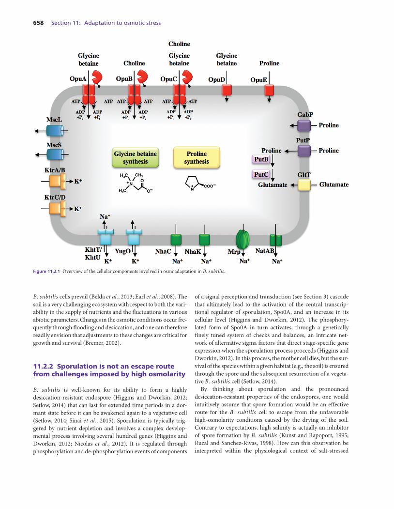
Figure 11.2.1 Overview of the cellular components involved in osmoadaptation in B. subtilis.
B. subtilis cells prevail (Belda et al., 2013; Earl et al., 2008). Thesoil is a very challenging ecosystem with respect to both the vari-ability in the supply of nutrients and the fluctuations in variousabiotic parameters. Changes in the osmotic conditions occur fre-quently through flooding and desiccation, and one can thereforereadily envision that adjustments to these changes are critical forgrowth and survival (Bremer, 2002).
11.2.2 Sporulation is not an escape routefrom challenges imposed by high osmolarity
B. subtilis is well-known for its ability to form a highlydesiccation-resistant endospore (Higgins and Dworkin, 2012;Setlow, 2014) that can last for extended time periods in a dor-mant state before it can be awakened again to a vegetative cell(Setlow, 2014; Sinai et al., 2015). Sporulation is typically trig-gered by nutrient depletion and involves a complex develop-mental process involving several hundred genes (Higgins andDworkin, 2012; Nicolas et al., 2012). It is regulated throughphosphorylation and de-phosphorylation events of components
of a signal perception and transduction (see Section 3) cascadethat ultimately lead to the activation of the central transcrip-tional regulator of sporulation, Spo0A, and an increase in itscellular level (Higgins and Dworkin, 2012). The phosphory-lated form of Spo0A in turn activates, through a geneticallyfinely tuned system of checks and balances, an intricate net-work of alternative sigma factors that direct stage-specific geneexpression when the sporulation process proceeds (Higgins andDworkin, 2012). In this process, the mother cell dies, but the sur-vival of the species within a given habitat (e.g., the soil) is ensuredthrough the spore and the subsequent resurrection of a vegeta-tive B. subtilis cell (Setlow, 2014).
By thinking about sporulation and the pronounceddesiccation-resistant properties of the endospores, one wouldintuitively assume that spore formation would be an effectiveroute for the B. subtilis cell to escape from the unfavorablehigh-osmolarity conditions caused by the drying of the soil.Contrary to expectations, high salinity is actually an inhibitorof spore formation by B. subtilis (Kunst and Rapoport, 1995;Ruzal and Sanchez-Rivas, 1998). How can this observation beinterpreted within the physiological context of salt-stressed
Chapter 11.2: Stress responses of B. subtilis to osmotic challenges 659
cells? The commitment of the cell to sporulate is a dicey deci-sion since sporulation is an energy-demanding and complexdevelopmental process that requires, even under favorablelaboratory conditions, several hours for its completion (Higginsand Dworkin, 2012). Increases in the external salinity negativelyimpinge on many cellular processes and slow the growth of B.subtilis considerably (Boch et al., 1994). One is thus temptedto speculate that the stressed B. subtilis cell might not possessthe energetic and biosynthetic resources required to completespore formation under high-osmolarity conditions. Since pro-gression of the sporulation process is irreversible after a certaintime point (Higgins and Dworkin, 2012), committing the B.subtilis cell to this developmental pathway under high-salinityconditions bears the risk of the death of the mother cell withouthaving produced a fully stress-resistant spore. It would thereforemake sense physiologically to block the sporulation processat an early stage, and this is actually what has been observedunder high-salinity conditions (Ruzal and Sanchez-Rivas, 1998;Widderich et al., 2016).
The activity of the master regulator of sporulation in B.subtilis, the phosphorylated form of Spo0A, is regulated by asophisticated phosphorelay integrating multiple positively andnegatively acting signals through the activities of kinases andphosphatases. One of these phosphatases (Spo0E) specificallytargets Spo0A–phosphate and thereby abrogates the sporula-tion process (Higgins and Dworkin, 2012). Recently, an inter-esting connection between the genetics of the sporulation net-work and the operation of the SigB-controlled general stressresponse system of B. subtilis (Hecker et al., 2007; Price, 2011)has been uncovered by the finding that a SigB-dependent pro-moter contributes to spo0E expression (Reder et al., 2012). Sud-denly imposed severe salt stress is a strong inducer of the SigBregulon (Nannapaneni et al., 2012; Young et al., 2013), and SigBactivity is therefore integrated via Spo0E into the finely tuneddecision-making process during the onset and progression ofsporulation (Reder et al., 2012). Since SigB activity increasesonly transiently after a severe salt shock (Young et al., 2013)and is not triggered by sustained high salinity (Spiegelhalter andBremer, 1998), the observations made by Reder et al. (2012)address a phenomenon and regulatory circuit that must be dif-ferent from the strong decrease in sporulation frequency thathas been observed in B. subtilis cells exposed to sustained highsalinity (Kunst and Rapoport, 1995; Lopez et al., 1998).
In addition to the repressing influence on sporulation, vari-ous effects of high salinity on the germination of spores havealso been reported (Nagler et al., 2014). Increases in salinitycause progressive, albeit reversible, inhibition of germinationefficiency and heterogeneity of germination initiation in a givenspore population, and it slows germination kinetics of individ-ual spores. Interestingly, part of the spore population can be trig-gered to initiate spore germination even in solutions containingnear-saturated NaCl concentrations (about 5.4 M). This obser-vation by Nagler et al. suggests that spores lack a sensory systemthat would prevent them from germinating under conditions
where the awakened vegetative cells cannot grow at all (Bochet al., 1994; Nagler et al., 2014).
11.2.3 Contributions of the SigB-controlledgeneral stress regulon to cellularadjustment to acute osmotic stress
High-salinity shocks are one of the most effective triggers toonset the general stress response of B. subtilis (Nannapaneniet al., 2012; Young et al., 2013). This emergency response sys-tem comprises several hundred genes (Nannapaneni et al., 2012;Nicolas et al., 2012) and provides the cell with a preemp-tive resistance against a multitude of environmental insults andagainst energy stress. It is controlled through the biochemicalactivation of the alternative transcription factor SigB (Heckeret al., 2007; Price, 2011). Disruption of sigB causes sensitivityagainst severe salt shocks. Indeed, a systematic inactivation ofa large subset of SigB-controlled genes of B. subtilis revealeda salt-sensitive phenotype for many of them under conditionswhere the imposed osmotic stress (with 1.5 M NaCl) was severeand acute (Hoper et al., 2005, 2006). Unfortunately, thus far abioinformatics analysis of the predicted functions of the geneswith a salt-sensitive phenotype has not provided truly informa-tive clues about how the encoded proteins might participate inthe development of salt-shock resistance. While SigB is criti-cal for the ability of the B. subtilis cell to withstand growth-restricting salt shocks (Hoper et al., 2005), it is dispensable forcells that are growing under sustained high-salinity conditions(e.g., in a chemically defined medium with 1.2 M NaCl).
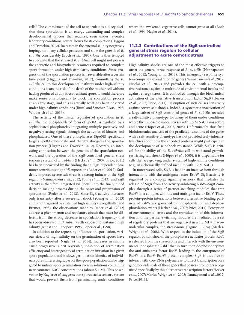
In nonstressed cells, SigB is held in an inactive form throughinteractions with the antisigma factor RsbW. SigB activity isregulated by a complex signaling network that mediates therelease of SigB from the activity-inhibiting RsbW–SigB com-plex through a series of partner-switching modules that trapRsbW in a complex with the anti-antisigma factor RsbV. Theseprotein–protein interactions between alternative binding part-ners of RsbW are governed by phosphorylation and dephos-phorylation events (Hecker et al., 2007; Price, 2011). Perceptionof environmental stress and the transduction of this informa-tion into the partner-switching modules are mediated by a setof regulatory proteins that are organized in a 1.8 MDa macro-molecular complex, the stressosome (Figure 11.2.2a) (Marles-Wright et al., 2008). With respect to the induction of the SigBregulon by salt shocks, the phosphatase activator protein RbsTis released from the stressosome and interacts with the environ-mental phosphatase RsbU that in turn then de-phosphorylatesthe anti-antisigma factor RsbV, leading to the entrapment ofRsbW in a RsbV–RsbW protein complex. SigB is thus free tointeract with core RNA polymerase to direct transcription on agenome-wide scale of those genes that possess promoters recog-nized specifically by this alternative transcription factor (Heckeret al., 2007; Marles-Wright et al., 2008; Nannapaneni et al., 2012;Price, 2011).
660 Section 11: Adaptation to osmotic stress
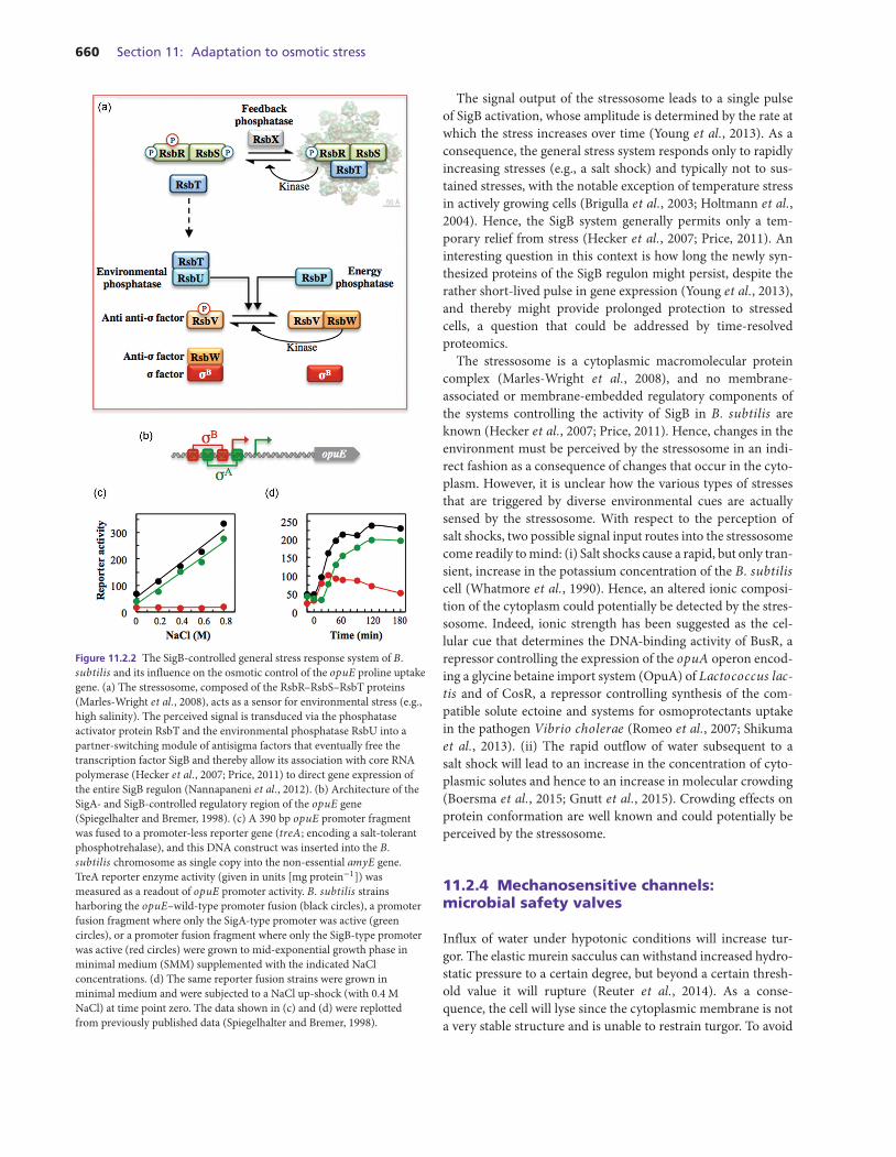
Figure 11.2.2 The SigB-controlled general stress response system of B.subtilis and its influence on the osmotic control of the opuE proline uptakegene. (a) The stressosome, composed of the RsbR–RsbS–RsbT proteins(Marles-Wright et al., 2008), acts as a sensor for environmental stress (e.g.,high salinity). The perceived signal is transduced via the phosphataseactivator protein RsbT and the environmental phosphatase RsbU into apartner-switching module of antisigma factors that eventually free thetranscription factor SigB and thereby allow its association with core RNApolymerase (Hecker et al., 2007; Price, 2011) to direct gene expression ofthe entire SigB regulon (Nannapaneni et al., 2012). (b) Architecture of theSigA- and SigB-controlled regulatory region of the opuE gene(Spiegelhalter and Bremer, 1998). (c) A 390 bp opuE promoter fragmentwas fused to a promoter-less reporter gene (treA; encoding a salt-tolerantphosphotrehalase), and this DNA construct was inserted into the B.subtilis chromosome as single copy into the non-essential amyE gene.TreA reporter enzyme activity (given in units [mg protein−1]) wasmeasured as a readout of opuE promoter activity. B. subtilis strainsharboring the opuE–wild-type promoter fusion (black circles), a promoterfusion fragment where only the SigA-type promoter was active (greencircles), or a promoter fusion fragment where only the SigB-type promoterwas active (red circles) were grown to mid-exponential growth phase inminimal medium (SMM) supplemented with the indicated NaClconcentrations. (d) The same reporter fusion strains were grown inminimal medium and were subjected to a NaCl up-shock (with 0.4 MNaCl) at time point zero. The data shown in (c) and (d) were replottedfrom previously published data (Spiegelhalter and Bremer, 1998).
The signal output of the stressosome leads to a single pulseof SigB activation, whose amplitude is determined by the rate atwhich the stress increases over time (Young et al., 2013). As aconsequence, the general stress system responds only to rapidlyincreasing stresses (e.g., a salt shock) and typically not to sus-tained stresses, with the notable exception of temperature stressin actively growing cells (Brigulla et al., 2003; Holtmann et al.,2004). Hence, the SigB system generally permits only a tem-porary relief from stress (Hecker et al., 2007; Price, 2011). Aninteresting question in this context is how long the newly syn-thesized proteins of the SigB regulon might persist, despite therather short-lived pulse in gene expression (Young et al., 2013),and thereby might provide prolonged protection to stressedcells, a question that could be addressed by time-resolvedproteomics.
The stressosome is a cytoplasmic macromolecular proteincomplex (Marles-Wright et al., 2008), and no membrane-associated or membrane-embedded regulatory components ofthe systems controlling the activity of SigB in B. subtilis areknown (Hecker et al., 2007; Price, 2011). Hence, changes in theenvironment must be perceived by the stressosome in an indi-rect fashion as a consequence of changes that occur in the cyto-plasm. However, it is unclear how the various types of stressesthat are triggered by diverse environmental cues are actuallysensed by the stressosome. With respect to the perception ofsalt shocks, two possible signal input routes into the stressosomecome readily to mind: (i) Salt shocks cause a rapid, but only tran-sient, increase in the potassium concentration of the B. subtiliscell (Whatmore et al., 1990). Hence, an altered ionic composi-tion of the cytoplasm could potentially be detected by the stres-sosome. Indeed, ionic strength has been suggested as the cel-lular cue that determines the DNA-binding activity of BusR, arepressor controlling the expression of the opuA operon encod-ing a glycine betaine import system (OpuA) of Lactococcus lac-tis and of CosR, a repressor controlling synthesis of the com-patible solute ectoine and systems for osmoprotectants uptakein the pathogen Vibrio cholerae (Romeo et al., 2007; Shikumaet al., 2013). (ii) The rapid outflow of water subsequent to asalt shock will lead to an increase in the concentration of cyto-plasmic solutes and hence to an increase in molecular crowding(Boersma et al., 2015; Gnutt et al., 2015). Crowding effects onprotein conformation are well known and could potentially beperceived by the stressosome.
11.2.4 Mechanosensitive channels:microbial safety valves
Influx of water under hypotonic conditions will increase tur-gor. The elastic murein sacculus can withstand increased hydro-static pressure to a certain degree, but beyond a certain thresh-old value it will rupture (Reuter et al., 2014). As a conse-quence, the cell will lyse since the cytoplasmic membrane is nota very stable structure and is unable to restrain turgor. To avoid
Chapter 11.2: Stress responses of B. subtilis to osmotic challenges 661
such a catastrophic event, most microorganisms have devel-oped safety valves whose gating is triggered by an upshot inturgor. Mechanosensitive channels embedded in the cytoplas-mic membrane are the molecular basis of these safety valves,and their transient opening allows the cell to rapidly jettisonwater-attracting ions and organic compounds to reduce the driv-ing force for the osmotically instigated rapid water influx thatfollows an osmotic downshift. The ability of different types ofthese channels to gate at different pressure thresholds allows agraded response of the cell coupled to the severity of the osmoticdownshift (Booth, 2014; Naismith and Booth, 2012). Electro-physiological studies with giant spheroplasts derived from bac-terial cells and reconstituted in vitro systems set up for patch-clamp analysis allowed the classification of mechanosensitivechannels into three classes: MscM (mini), MscS (small), andMscL (large). The MscS and MscL channels are best understoodwith respect to their gating behavior, biochemistry, and crystalstructure (Booth, 2014; Naismith and Booth, 2012). MscL is themeasure of last resort; it gates just shortly before the osmoticallydownshifted cell would rupture and thereby transiently opens achannel in the cytoplasmic membrane with a diameter of about30 A (Booth, 2014; Naismith and Booth, 2012), substantiallylarger than the permanently open pores (porins) in the outermembrane of Gram-negative bacteria. Fully opened MscS-typechannels possess a diameter of about 6 A (Booth, 2014; Naismithand Booth, 2012). Mechanosensitive channels typically do notpossess substrate specificity, and hence both ions and organiccompounds can pass freely. It is obvious that the cell must verycarefully control their numbers and gating behavior.
For a soil bacterium such as B. subtilis, a rapid osmotic down-shift can simply be caused through the wetting of the dried-out upper layers of soil by rainfall. Electrophysiological stud-ies revealed that different types of mechanosensitive channelsmust operate in B. subtilis, and the inspection of its genomesequence suggested the presence of one MscL- and three MscS-type channels (Figure 11.2.1) (Hoffmann et al., 2008; Wahomeet al., 2009). Nothing is known about the potential operation ofMscM-type channels in B. subtilis; the molecular basis of thesechannels has only recently been discovered in E. coli (Booth,2014; Booth et al., 2015). The systematic inactivation of themscL and the three mscS channel genes (ykuT, yfkC, and yhdY)reveals that neither the single mutants nor the quadruple mutantstrain (mscL, ykuT, yfkC, and yhdY) have a growth disad-vantage in either low-osmolarity or high-osmolarity media incomparison to the wild-type strain. However, osmotic down-shock experiments demonstrated that the MscL channel is theprinciple solute release system of B. subtilis. The activity ofthe YkuT MscS-type channel aids MscL in protecting the cellsfrom the detrimental effects of a severe osmotic downshift.Indeed, cells of an mscL ykuT double mutant are almost allkilled when cells grown at high osmolarity are rapidly shifted tolow-osmolarity conditions (Hoffmann et al., 2008). No evidencefor mechanosensitive channel activity has been found with thisassay for the MscS-type proteins YfkC and YhdY.
Interestingly, in a genome-wide transcriptional profilingstudy of B. subtilis, cells subjected to a severe osmotic upshiftexpress the mscL and ykuT genes at a significantly higher levelthan in their non-osmotically stressed counterparts; in con-trast, expression of the mscS-type genes yfkC and yhdY is notaffected (Hahne et al., 2010). The observation of the transcrip-tional induction of the mscL and ykuT genes by high salinitycould potentially be rationalized by invoking the idea that cellssubjected to high-osmolarity stress already prepare themselvesfor an osmotic downshock that inevitably will follow at onepoint in time. One can thus summarize that tension-activatedmechanosensitive channels of the MscS and MscL types (Booth,2014; Naismith and Booth, 2012) are key for managing the tran-sition of B. subtilis cells from high- to low-osmolarity environ-ments (Hoffmann et al., 2008; Wahome et al., 2009).
11.2.5 Salt-in and salt out: strategies tocope with high osmolarity
Selected groups of Archaea (representatives of Halobacteria,halophilic methanogenic Archaea, and Nanohaloarchaea) and afew Bacteria (representatives of Bacterioidetes and Haloanaero-biales) that live permanently in high-salinity environments pref-erentially accumulate molar concentrations of potassium andchloride, to balance the osmotic gradient between the cell’s inte-rior and that of the surroundings (Oren, 2011). The lasting accu-mulation of ions is an energetically favorable cellular adjustmentto cope with the challenges of high osmolarity (Oren, 2011).However, it comes at an evolutionary price since the high ionicstrength of the cytoplasm requires the adaptation of macro-molecular structures and biochemical processes on a genome-wide scale to maintain the solubility and structural integrity ofboth cell surface exposed and cytoplasmic proteins. The solute-exposed surfaces of proteins become more acidic, and at thesame time their hydrophobicity is reduced (Coquelle et al., 2010;Talon et al., 2014).
Microorganisms that use this salt-in strategy (Galinski andTruper, 1994; Oren, 2011) typically can only tolerate moder-ate fluctuations in the osmolarity of their environment. Thus,a more flexible osmostress response is needed for bacteria thatlive in environments exhibiting either more frequent or moreextreme changes in the external osmolarity. These microorgan-isms pursue an osmostress adaptation strategy that aims at keep-ing the permanent intracellular ion concentrations low (salt-out) (Galinski and Truper, 1994). To accomplish this, a selectedclass of organic osmolytes, the compatible solutes, are amassed,and their intracellular concentration is set by the osmolarityprevalent at a given time in the environment of the micro-bial cells (Csonka, 1989; Kempf and Bremer, 1998; Roesser andMuller, 2001; Wood, 2011; Wood et al., 2001). Since compati-ble solutes are highly compliant with cellular functions (Ignatovaand Gierasch, 2006; Street et al., 2006), this obviated, on an evo-lutionary timescale, the need to adapt cellular components to a
662 Section 11: Adaptation to osmotic stress
high-ionic-strength cytoplasm in the way that was dictated bythe salt-in strategy (Oren, 2013; Talon et al., 2014). It suffices tostate here that B. subtilis uses the salt-out strategy to physiolog-ically cope with prolonged high-osmolarity conditions.
11.2.6 Uptake of potassium and extrusionof sodium: the first line of defense
As in many other bacteria (Csonka, 1989; Kempf and Bremer,1998; Wood et al., 2001), in B. subtilis, osmotic upshifts triggera rapid import of potassium ions (Whatmore et al., 1990). Thebuildup of this elevated potassium pool serves as the first line ofcellular defense against loss of water and a reduction in turgor(Holtmann et al., 2003). Studies conducted by Whatmore et al.(1990) have revealed a potassium pool of about 350 mM in cellsgrown in a minimal medium, which increases about twofold (upto about 720 mM) over 3 h subsequent to a moderate salt shockwith 0.4 M NaCl. The slowness of this increase is probably dueto the use of cells for these measurements that had been grownat a suboptimal temperature (25 ◦C) for B. subtilis (Whatmoreet al., 1990) and the fact that the expression of the genes encod-ing its major potassium uptake system (KtrAB) (Holtmann et al.,2003) (discussed further in this chapter) is downregulated atlow-growth temperatures (Nicolas et al., 2012).
B. subtilis lacks a true high-affinity potassium transport sys-tem and instead possesses two representatives of the Ktr potas-sium uptake family (Figure 11.2.1) (Hanelt et al., 2011). TheKtrAB and KtrCD systems of B. subtilis possess only moder-ate affinities for the potassium ion and exhibit Km values ofapproximately 1 mM and 10 mM, respectively (Holtmann et al.,2003). Loss of the KtrAB system causes a severe salt sensitiv-ity in cells continuously challenged by high osmolarity, a phe-notype that is augmented by the simultaneous inactivation ofthe KtrCD transporter. In a salt shock experiment, cells lackingKtrAB cannot recover from an increase in salinity (with 0.6 MNaCl) in a growth medium containing 2 mM potassium. Higherconcentrations of potassium (50 mM), however, allow recoveryfrom the suddenly imposed salt stress, as the KtrCD transporterwill permit potassium uptake under these conditions (Holtmannet al., 2003). Collectively, these experiments illustrate how criti-cal potassium uptake is for cells that are either suddenly exposedto high salinity or continuously challenged by it.
Ktr-type potassium transporters consist of a membrane-embedded protein (e.g., KtrB and KtrD) and a regulatory com-ponent (e.g., KtrA and KtrC) (Figure 11.2.1) that is periph-erally associated with the membrane and interacts with themembrane-embedded potassium translocation subunit (Haneltet al., 2011). The KtrA and KtrC proteins serve to control theactivity of the potassium translocating subunit and possess eachtwo RCK domains (regulator of conductance of K+), mod-ules that bind nucleotides and thereby affect potassium import(Corrigan et al., 2013). The crystal structure of the B. subtilisKtrAB system has recently been solved and reveals a striking
arrangement of a dimer of the membrane-embedded KtrB sub-unit that is decorated on the cytoplasmic side by an octamericring formed by KtrA (Vieira-Pires et al., 2013).
In contrast to the structural genes for the high-affinity potas-sium uptake Kdp system of E. coli (Laermann et al., 2013),transcription of the ktrAB, ktrC, and ktrD genes from B. sub-tilis is not induced by potassium limitation or high osmolarity(Holtmann et al., 2003). However, the ktrAB operon is regu-lated in a highly interesting fashion since its transcription is con-trolled by an ydaO-type riboswitch that recognizes as its effec-tor molecule the recently discovered second messenger, cyclicdiadenylate monophosphate (c-di-AMP) (Nelson et al., 2013).Strikingly, c-di-AMP is also an effector for the RCK-containingKtrA subunit of Staphylococcus aureus, and its binding servesto downregulate KtrAB potassium transporter activity (Corri-gan et al., 2013). It will therefore be highly interesting in futurestudies to probe how the control of potassium homeostasis inB. subtilis is affected by the genetic and biochemically controlmechanisms setting the c-di-AMP pool (Mehne et al., 2013) andhow this might affect the physiology of the overall osmostressadaptation process of the cell.
A mutant strain lacking both the KtrAB and KtrCD systemshas a residual potassium transport activity that exhibits a Kmvalue of around 110 mM, suggesting the operation of otherpotassium uptake systems in B. subtilis. Indeed, the inspec-tion of the B. subtilis genome sequence revealed the presenceof another transporter, YugO (Figure 11.2.1), which is related tothe MthK channel from Methanobacterium thermoautotroph-icum (Ye et al., 2010). The arrangements of the membrane-spanning segments in YugO are different from the potassiumtranslocating subunit (KtrB) of the Ktr system, and it carries atits C-terminus an RCK domain. Interestingly, disruption of theYugO channel abolishes biofilm formation of B. subtilis in a reg-ulatory loop that involves KinC, a histidine kinase that seems tobe activated by potassium leakage (Lopez et al., 2009a). Consis-tent with the role of YugO in biofilm formation (Lundberg et al.,2013), expression of the mstX–yugO operon is negatively con-trolled by SinR, the central regulator of matrix and expolysac-charide synthesis during biofilm formation by B. subtilis (Lopezet al., 2009b; Vlamakis et al., 2013).
For microorganisms that use the salt-out strategy, prolongedhigh levels of potassium ions are detrimental to cellular physi-ology. Hence, the osmotically stressed cells replace part of theinitially accumulated potassium with organic osmolytes (e.g.,proline and glycine betaine) that are highly compliant with itsbiochemistry and physiology (Ignatova and Gierasch, 2006;Street et al., 2006; Whatmore et al., 1990). In this way, the ionicstrength of the cytoplasm is reduced without compromisingturgor. Potassium extrusion in B. subtilis is only incompletelyunderstood, but one of the systems that mediate potassiumexport has been identified. It is the cation–proton antiporterKhtTU (also known as YhaTU) (Figure 11.2.1) (Fujisawa et al.,2007). The involvement of this potassium extrusion system inthe cell’s overall osmotic adjustment process is evident from the
Chapter 11.2: Stress responses of B. subtilis to osmotic challenges 663
upregulation of the khtSTU (yhaSTU) gene cluster in responseto a salt shock (Fujisawa et al., 2004; Hahne et al., 2010). Itis highly likely that other potassium exporters exist in B. sub-tilis since a mutant carrying a deletion of the khtSTU (yhaSTU)operon is viable (our unpublished data).
Sodium ions are highly cytotoxic, and B. subtilis keeps itscytoplasmic concentration very low (Gorecki et al., 2014). Dur-ing salt challenges, Na+ might enter the cell in various ways,one of which occurs during the import of osmostress protec-tants such as glycine betaine and proline via the OpuD and OpuEtransporters, respectively. The proline transporter OpuE (vonBlohn et al., 1997) and the glycine betaine transporter OpuD(Figure 11.2.1) (Kappes et al., 1996) belong to different types oftransporter superfamilies, but both of them import their sub-strates in concert with Na+ ions. The imported sodium mustbe speedily exported, and B. subtilis possesses four Na+ extru-sion systems that serve to keep intracellular Na+ levels low: themulticomponent Mrp transporter, the single-component NhaCand NhaK systems, and the adenosine triphosphate (ATP)-dependent NatAB system (Figure 11.2.1). A severe salt shocktriggers the expression of the mrp operon, of nhaK, and, to alesser extent, of nhaC (Hahne et al., 2010). The ATP-bindingcassette (ABC)-type transporter NatAB is primarily involved inNa+ extrusion at alkaline pH, and the expression of its structuralgenes is controlled by the two-component NatK–NatR regula-tory system (Ogura et al., 2007). The transcriptional inductionof the Mrp, NhaK, and NhaC systems subsequent to a salt shockin cells grown at neutral pH highlights the importance of sodiumhomeostasis in salt-stressed cells and buttresses the physiologi-cal role of Na+ extrusion in the cellular response of B. subtilisto osmotic stress. Indeed, the genetic disruption of the Mrp sys-tem causes a strong salt-sensitive growth phenotype and drivesup the Na+ content of the cells from a practically nonmeasurablelevel to 11 mM (Gorecki et al., 2014).
11.2.7 Uptake of compatible solutes
As outlined in this chapter, the accumulation of compatiblesolutes is a key event in the well-orchestrated cellular responsesof many Bacteria and Archaea to osmotic stress (Csonka, 1989;Kempf and Bremer, 1998; Roesser and Muller, 2001; Wood et al.,2001). This is true for B. subtilis as well (Bremer, 2002; Bre-mer and Kramer, 2000). The amassing of compatible solutes byosmotically stressed cells can occur either via synthesis or byuptake (Kempf and Bremer, 1998), and both processes are usedby B. subtilis to achieve osmostress resistance (Bremer, 2002;Bremer and Kramer, 2000). Extensive physiological studies haverevealed that B. subtilis uses primarily compatible solutes thatare chemically related to either proline or glycine betaine. Sofar, 14 naturally occurring compatible solutes have been iden-tified to serve as osmostress protectants for B. subtilis, all ofwhich can be found in habitats populated by this bacterium (e.g.,the soil or marine sediments) (Bashir et al., 2014a; Broy et al.,
2015; Hoffmann and Bremer, 2011; Nau-Wagner et al., 1999).With the notable exception of proline (Moses et al., 2012), noneof these compounds can be metabolized by B. subtilis and arethus amassed exclusively as stress protectants, to fend off eitherthe detrimental effects of high osmolarity on cellular physiol-ogy (Bremer, 2002), or extremes in high- and low-growth tem-peratures (Hoffmann and Bremer, 2011; Holtmann and Bremer,2004).
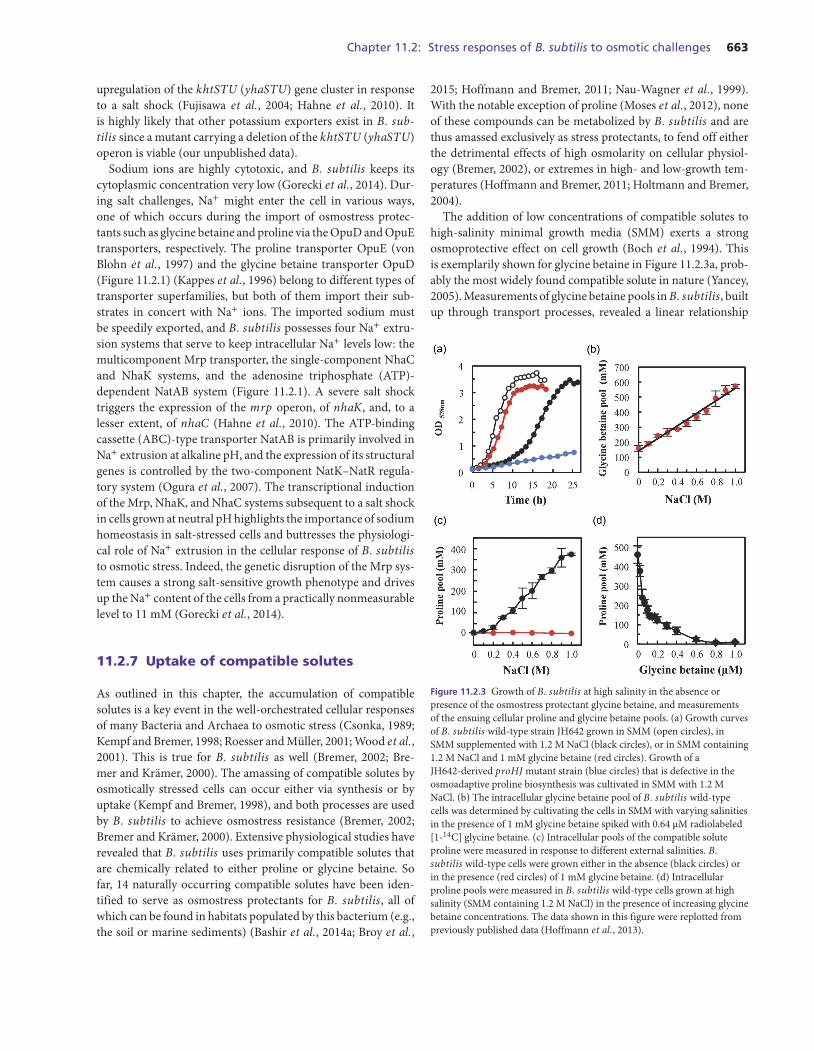
The addition of low concentrations of compatible solutes tohigh-salinity minimal growth media (SMM) exerts a strongosmoprotective effect on cell growth (Boch et al., 1994). Thisis exemplarily shown for glycine betaine in Figure 11.2.3a, prob-ably the most widely found compatible solute in nature (Yancey,2005). Measurements of glycine betaine pools in B. subtilis, builtup through transport processes, revealed a linear relationship
Figure 11.2.3 Growth of B. subtilis at high salinity in the absence orpresence of the osmostress protectant glycine betaine, and measurementsof the ensuing cellular proline and glycine betaine pools. (a) Growth curvesof B. subtilis wild-type strain JH642 grown in SMM (open circles), inSMM supplemented with 1.2 M NaCl (black circles), or in SMM containing1.2 M NaCl and 1 mM glycine betaine (red circles). Growth of aJH642-derived proHJ mutant strain (blue circles) that is defective in theosmoadaptive proline biosynthesis was cultivated in SMM with 1.2 MNaCl. (b) The intracellular glycine betaine pool of B. subtilis wild-typecells was determined by cultivating the cells in SMM with varying salinitiesin the presence of 1 mM glycine betaine spiked with 0.64 μM radiolabeled[1-14C] glycine betaine. (c) Intracellular pools of the compatible soluteproline were measured in response to different external salinities. B.subtilis wild-type cells were grown either in the absence (black circles) orin the presence (red circles) of 1 mM glycine betaine. (d) Intracellularproline pools were measured in B. subtilis wild-type cells grown at highsalinity (SMM containing 1.2 M NaCl) in the presence of increasing glycinebetaine concentrations. The data shown in this figure were replotted frompreviously published data (Hoffmann et al., 2013).
664 Section 11: Adaptation to osmotic stress
between the degree of the imposed osmotic stress and the cellu-lar levels of this solute (Figure 11.2.3b). This is quite an amaz-ing relationship because it shows that the B. subtilis cell candetect rather small increases in the external salinity and thenset its osmostress-protective glycine betaine pool very preciselyto maintain a physiologically perfect balance between the intra-cellular and environmental osmolarity. In severely osmoticallystressed cells (e.g., with 1 M NaCl) (2200 mOsmol kg−1), theglycine betaine pool increases from a basal level of 150 mM incells cultivated in SMM (350 mOsmol kg−1) about 3.8-fold to acellular content of about 570 mM (Figure 11.2.3b) (Hoffmannet al., 2013).
The glycine betaine content of the cells is already remarkablyhigh (about 150 mM) in cells grown in SMM, a minimal mediumthat is widely used in physiological studies with B. subtilis; itsosmolarity (350 mOsmol kg−1) is considered not to confer anyosmotic stress onto the cells.
The uptake of glycine betaine by B. subtilis is mediatedby three distinct transport systems that belong to the Opu(osmoprotectants uptake) family of transporters (Bremer, 2002)and that comprise the ABC-type uptake systems OpuA andOpuC and the single-component BCCT-type transporter OpuD(Figure 11.2.1) (Kappes et al., 1996, 1999; Kempf and Bre-mer, 1995). Each of these transporters imports glycine betainewith high affinity (Km values in the low μM range), but theirtransport capacity (Vmax) varies. OpuA is the dominant glycinebetaine transporter operating in B. subtilis due to its substan-tial uptake velocity (Kappes et al., 1996; Kempf and Bremer,1995), but its substrate spectrum is rather restricted (Bashiret al., 2014a,b; Broy et al., 2015; Hoffmann and Bremer, 2011).In contrast, the OpuC system has a very broad substrate profileand, with the exception of proline, can mediate the import of allosmostress protectants known so far for B. subtilis. Remarkably,it can also catalyze the import of several synthetic compatiblesolutes derived from the ecologically abundant marine osmolytedimethlylsulfoniopropionate (DMSP) (Broy et al., 2015) and atoxic derivative of glycine betaine (Cosquer et al., 2004).
An ABC transporter (OpuB) very closely related to the OpuCsystem exists in B. subtilis (Figure 11.2.1); the genes encodingthese two transporters have in all likelihood arisen through agene duplication event (Kappes et al., 1999). However, the sub-strate specificity of OpuB and OpuC is strikingly different. OpuBmediates only the import of choline and glycine betaine alde-hyde, the precursor and intermediate, respectively, for the syn-thesis of glycine betaine (Boch et al., 1996; Kappes et al., 1999),whereas OpuC mediates the import of essentially all osmopro-tectants (with the exception of proline) known to date for B. sub-tilis (Bashir et al., 2014a; Broy et al., 2015; Hoffmann and Bre-mer, 2011).
The OpuA, OpuB, and OpuC systems are all members ofthe ABC superfamily and possess extracellular substrate bindingproteins (OpuAC, OpuBC, and OpuCC) that are tethered witha lipid anchor to the outer surface of the cytoplasmic membrane(Figure 11.2.1) (Kappes et al., 1999; Kempf and Bremer, 1995).
These lipoproteins serve as the primary ligand recognition com-ponent of the OpuA, OpuB, and OpuC transporters and deter-mine their overall affinity and substrate specificity (Berntssonet al., 2010). Hence, the question arose how the OpuAC, OpuBC,and OpuCC solute receptor proteins can recognize ligands withhigh affinity (Horn et al., 2006; Pittelkow et al., 2011) thatare otherwise effectively excluded from protein surfaces (Streetet al., 2006). Crystallographic analysis has provided the answerand showed that each of them possessed a ligand-binding sitethat accommodates the positively charged trimethlyammoniumhead group of the various ligands (e.g., glycine betaine, choline,and carnitine) in a cage formed by aromatic side chains and sta-bilizes them via cation–π interactions. The “tails” of the vari-ous ligands protrude out of these aromatic cages and are furtherstabilized within the binding site via contacts with specific sidechains, with the protein backbone, or through intricate waternetworks (Du et al., 2011; Horn et al., 2006; Pittelkow et al.,2011). The ligand-binding site present in the OpuCC ligand-binding protein has proven to be particularly structurally flex-ible (Du et al., 2011) and thereby provides the molecular under-pinnings for the remarkably broad substrate specificity of theOpuC transport system (Bashir et al., 2014a,b; Broy et al., 2015;Hoffmann and Bremer, 2011).
Intensive studies with the OpuA system from Lactococcuslactis have shown that this glycine betaine import system (alsosometimes referred to as BusA) is osmotically regulated notonly at the transcriptional level through the ionic strength–dependent BusR repressor protein (Romeo et al., 2007) butalso at the level of its transport activity (Poolman et al., 2004).Detailed studies by Poolman and coworkers have revealed thatthe osmotic activity control of the L. lactis OpuA transporteris dependent on two cystathionine β-synthase (CBS) domainspresent within the extended C-terminal domain of the OpuAAATPase. These serve as sensors of the ionic strength of the cyto-plasm and thereby couple information on the properties of thecytoplasm (changes in ionic strength and molecular crowdingsubsequent to an osmotic upshift) to conformational changesin the nucleotide-binding domain (OpuAA) of the ABC trans-porter, and thereby affect the overall transport activity of theOpuA system (Karasawa et al., 2011). Data derived from in sil-ico assessments indicate that such CBS domains are also presentin the ATPases (OpuAA, OpuBA, and OpuCA) of the OpuA,OpuB, and OpuC systems of B. subtilis (Chen and Beattie,2007). This finding suggests that these transporters are also sub-jected to activity control of their transport capacity in responseto increase in the external osmolarity. Such an enhancement oftransport activity would allow the cell to react instantaneouslywith increased uptake of compatible solutes to relieve osmoticstress (Poolman et al., 2004). However, a possible activity con-trol of the B. subtilis OpuA, OpuB, and OpuC systems has notyet been studied experimentally.
The third glycine betaine transporter operating in B. sub-tilis is OpuD (Figure 11.2.1), a single-component system (Fig-ure 11.2.1) and a member of the BCCT family of permeases
Chapter 11.2: Stress responses of B. subtilis to osmotic challenges 665
(Ziegler et al., 2010). These widely found types of transportersare involved in the uptake of various kinds of compatible solutesin many microbial species. OpuD is closely related to the glycinebetaine transporter BetP from Corynebacterium glutamicum,the biochemically, mechanistically, and structurally best stud-ied member of the BCCT family (Maximov et al., 2014; Perezet al., 2012; Ziegler et al., 2010). BetP imports glycine betainein symport with two Na+ ions (Perez et al., 2014) and has beencrystallized in the presence and absence of its ligand. Not onlyis the expression of the betP gene upregulated in response tohigh osmolarity, a process that involves the osmostress-sensingtwo-component MtrAB regulatory system (Moker et al., 2004,2007), but also the activity of the BetP transporter itself is mod-ulated both by a trans-membrane osmotic gradient and by thecytoplasmic potassium concentration (Maximov et al., 2014).Given the close amino acid sequence identity of BetP and OpuDand the conservation of ligand- and sodium-contacting aminoacid residues, it is highly likely that the two proteins functionmechanistically in the same fashion with respect to the over-all transport process. Strikingly, the architecture of the glycinebetaine–binding site present in the membrane-embedded BetPprotein (Perez et al., 2012, 2014), and hence in all likelihood alsoin OpuD, resembles that of the soluble glycine betaine–bindingprotein OpuAC (Horn et al., 2006). Hence, nature has founda common solution for providing a high-affinity binding sitein transport proteins to a solute that is otherwise preferentiallyexcluded from protein surfaces (Street et al., 2006).
The fifth osmostress protectant uptake system found in B.subtilis is the proline transporter OpuE (Figure 11.2.1) (vonBlohn et al., 1997). It is a member of the solute sodium sym-porter (SSS) family, and is sequence related to the PutP trans-porter that is used by E. coli and B. subtilis for the acquisi-tion of proline as a nutrient (Moses et al., 2012; Olkhova et al.,2011). In contrast to the osmotic transcriptional control of opuE(Spiegelhalter and Bremer, 1998), putP expression in B. sub-tilis is upregulated in response to the availability of proline inthe growth medium, a genetic control mechanism that is depen-dent on the proline-responsive activator protein PutR (Belit-sky, 2011; Moses et al., 2012). Proline is the only compatiblesolute used by B. subtilis for osmostress protective purposes thatcan also be exploited as a nutrient through its PutBC-mediatedcatabolism to glutamate (Figure 11.2.1). This limits the effective-ness of exogenously provided proline as an osmostress protec-tant (Zaprasis et al., 2013a) and requires genetic precautions toprevent the degradation of newly synthesized proline by high-osmolarity-challenged cells (Moses et al., 2012).
Osmostress protective levels of proline can also be achievedby B. subtilis through the import of proline-containing di-and oligo-peptides via the Opp, Dpp, App, and DtpT trans-porters and their subsequent hydrolysis to release the compatiblesolute proline (Zaprasis et al., 2013a). Furthermore, B. subtiliscan replenish its osmostress-protective proline pool to deriveosmostress protection by importing proteogenic (Glu, Gln, Asp,Asn, and Arg) and non-proteogenic (Orn, Cit) amino acids that
can be metabolically converted into proline. One example isthe import of glutamate (Glu) via the GltT transporter (Fig-ure 11.2.1) (Zaprasis et al., 2015); Glu serves as the direct precur-sor for proline biosynthesis in B. subtilis (Brill et al., 2011a,b).
If one views the considerable number of osmostress adaptivetransporters that are present in B. subtilis, the breadth of theirsubstrate profile for compatible solutes, and the varied ways inwhich this bacterium can acquire osmoprotective levels of pro-line (Figure 11.2.1), one can readily see that B. subtilis is wellprepared to exploit many osmoprotectants present in its variedhabitats. Furthermore, the use of importers that either are ener-getically coupled to ATP hydrolysis (OpuA, OpuB, and OpuC)or are dependent for their functioning on ion gradient (OpuDand OpuE) provides additional flexibility to osmotically stressedB. subtilis cells.
With respect to the osmotically regulated OpuE proline trans-porter (von Blohn et al., 1997), an interesting additional physio-logical function was discovered when its structural gene (opuE)was disrupted. In such a mutant strain, part of the newly syn-thesized proline produced as an osmostress protectant (dis-cussed further in this chapter) is found in the supernatant of thecells grown in high-salinity medium (Hoffmann et al., 2012).Because the osmostress adaptive proline pool is reduced in anopuE mutant, such a strain is at a significant growth disadvan-tage. Hence, under high-osmolarity growth condition, B. sub-tilis engages in a cycle of synthesis–release–recapture of pro-line. Although this cycle appears energetically wasteful at firstsight, it might actually provide a useful physiological functionsince it could potentially aid the osmotically stressed cell to fine-tune turgor when it elongates and eventually divides (Hoffmannet al., 2012). It is unclear how proline is released from the B. sub-tilis cells under steady-state high-salinity growth conditions. InC. glutamicum, a mechanosensitive channel of the MscS typehas been implicated in the release of glycine betaine that hadbeen imported from the environment (Borngen et al., 2010).In contrast, in B. subtilis the involvement of MscL- and MscS-type channels (Figure 11.2.1) in the release of proline was firmlyruled out (Hoffmann et al., 2012). Therefore, the question ariseswhether dedicated export systems for compatible solutes exist inmicroorganisms, similar to those mediating the efflux of aminoacids (Eggeling and Sahm, 2003).
11.2.8 Synthesis of compatible solutes
Natural-abundance 13C-NMR (carbon-13 nuclear magnetic res-onance) spectroscopy has been used to assess the types of com-patible solutes synthesized de novo by members of the Bacilli inresponse to high salinity. Three large groups were detected: (i)those that synthesized only glutamate, (ii) those that synthesizedproline, and (iii) those that synthesized ectoine. In this lattergroup, ectoine production could be combined with the synthesisof the ectoine derivative 5-hydroxyectoine, or with the synthe-sis of proline (Bursy et al., 2007; Kuhlmann and Bremer, 2002).
666 Section 11: Adaptation to osmotic stress
Although not studied in detail, these three groups differed intheir ability to withstand salt stress: The glutamate producerswere the most salt-sensitive Bacillus species, proline produc-tion conferred an intermediate degree of salt stress resistance,whereas those Bacilli that synthesized ectoines could withstandthe highest levels of salt stress. None of the studied Bacilli syn-thesized glycine betaine de novo, and none produced the com-patible solute trehalose (Bursy et al., 2007), an osmostress pro-tectant that is otherwise synthesized by many bacterial speciesin response to high salinity (Bremer and Kramer, 2000; Csonka,1989; Kempf and Bremer, 1998; Wood et al., 2001). We will notcover here the osmotically controlled synthesis of ectoine and5-hydroxyectoine since B. subtilis does not produce these com-patible solutes (Bursy et al., 2007; Kuhlmann and Bremer, 2002),but we refer the reader to recent overviews that trace the synthe-sis of ectoines on genome-wide scales in members of the Bacte-ria and a few selected Archaea and that summarize the genetics,biochemistry, and biotechnological applications of these versa-tile stress protectants (Hoppner et al., 2014; Widderich et al.,2014). Here, we will focus on the only compatible solute that B.subtilis can produce de novo, the amino acid proline, and thesynthesis of glycine betaine from the precursor choline. All othercompatible solutes that B. subtilis can acquire via transport can-not be synthesized by this bacterium (Bashir et al., 2014a; Broyet al., 2015; Hoffmann and Bremer, 2011).
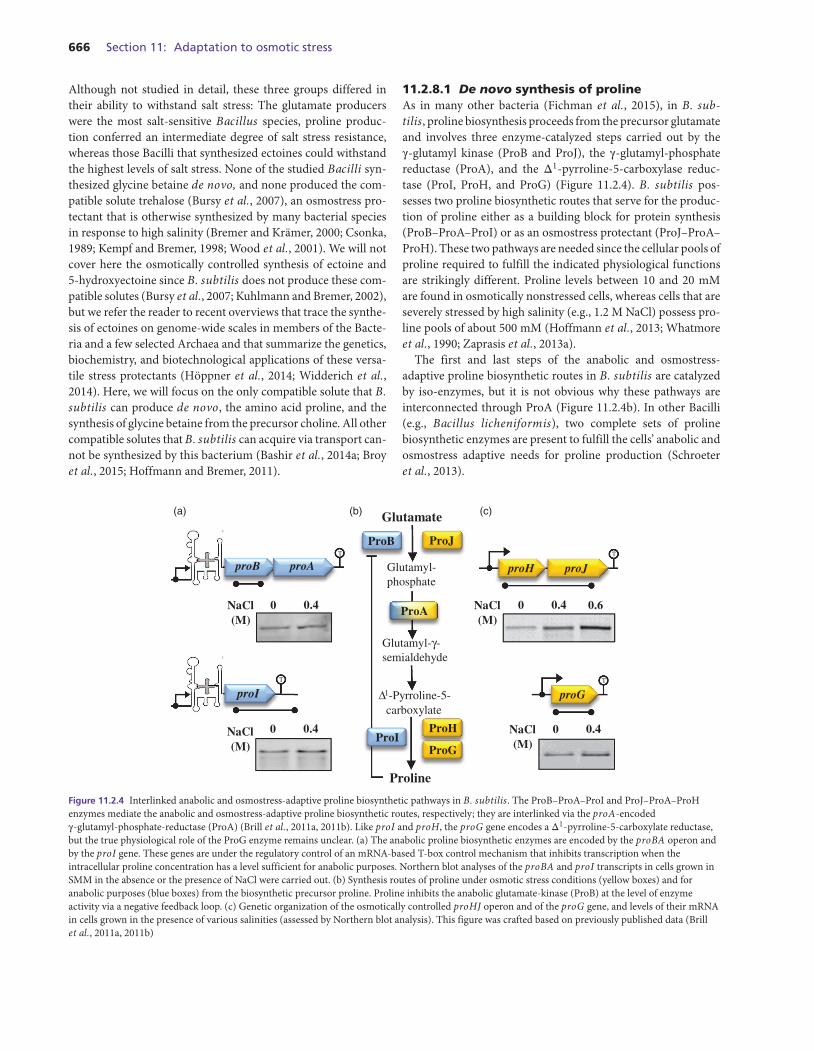
11.2.8.1 De novo synthesis of prolineAs in many other bacteria (Fichman et al., 2015), in B. sub-tilis, proline biosynthesis proceeds from the precursor glutamateand involves three enzyme-catalyzed steps carried out by theγ-glutamyl kinase (ProB and ProJ), the γ-glutamyl-phosphatereductase (ProA), and the Δ1-pyrroline-5-carboxylase reduc-tase (ProI, ProH, and ProG) (Figure 11.2.4). B. subtilis pos-sesses two proline biosynthetic routes that serve for the produc-tion of proline either as a building block for protein synthesis(ProB–ProA–ProI) or as an osmostress protectant (ProJ–ProA–ProH). These two pathways are needed since the cellular pools ofproline required to fulfill the indicated physiological functionsare strikingly different. Proline levels between 10 and 20 mMare found in osmotically nonstressed cells, whereas cells that areseverely stressed by high salinity (e.g., 1.2 M NaCl) possess pro-line pools of about 500 mM (Hoffmann et al., 2013; Whatmoreet al., 1990; Zaprasis et al., 2013a).
The first and last steps of the anabolic and osmostress-adaptive proline biosynthetic routes in B. subtilis are catalyzedby iso-enzymes, but it is not obvious why these pathways areinterconnected through ProA (Figure 11.2.4b). In other Bacilli(e.g., Bacillus licheniformis), two complete sets of prolinebiosynthetic enzymes are present to fulfill the cells’ anabolic andosmostress adaptive needs for proline production (Schroeteret al., 2013).
ProJ
Glutamyl-γ-semialdehyde
Δ1-Pyrroline-5-carboxylate
Glutamyl-phosphate
Glutamate
ProH
ProB
ProA
Proline
NaCl(M)
proH proJ
0 0.4 0.6
proB proA
NaCl(M)
0 0.4
proI
NaCl(M)
0 0.4
ProG
proG
NaCl(M)
0 0.4ProI
(a) (b) (c)
T T
TT
Figure 11.2.4 Interlinked anabolic and osmostress-adaptive proline biosynthetic pathways in B. subtilis. The ProB–ProA–ProI and ProJ–ProA–ProHenzymes mediate the anabolic and osmostress-adaptive proline biosynthetic routes, respectively; they are interlinked via the proA-encodedγ-glutamyl-phosphate-reductase (ProA) (Brill et al., 2011a, 2011b). Like proI and proH, the proG gene encodes a Δ1-pyrroline-5-carboxylate reductase,but the true physiological role of the ProG enzyme remains unclear. (a) The anabolic proline biosynthetic enzymes are encoded by the proBA operon andby the proI gene. These genes are under the regulatory control of an mRNA-based T-box control mechanism that inhibits transcription when theintracellular proline concentration has a level sufficient for anabolic purposes. Northern blot analyses of the proBA and proI transcripts in cells grown inSMM in the absence or the presence of NaCl were carried out. (b) Synthesis routes of proline under osmotic stress conditions (yellow boxes) and foranabolic purposes (blue boxes) from the biosynthetic precursor proline. Proline inhibits the anabolic glutamate-kinase (ProB) at the level of enzymeactivity via a negative feedback loop. (c) Genetic organization of the osmotically controlled proHJ operon and of the proG gene, and levels of their mRNAin cells grown in the presence of various salinities (assessed by Northern blot analysis). This figure was crafted based on previously published data (Brillet al., 2011a, 2011b)
Chapter 11.2: Stress responses of B. subtilis to osmotic challenges 667
The biochemistry and genetics of the anabolic ProB–ProA–ProI route in B. subtilis are geared to prevent a wasteful overpro-duction of proline and to adjust the proline pool with the ongo-ing protein biosynthetic activities of the cell. This is achieved(i) through a sensitive feedback regulation of the enzyme activ-ity of the γ-glutamyl kinase (ProB) by proline (Figure 11.2.4b)(Chen et al., 2007), and (ii) through transcriptional control ofthe proBA and proI genes through a T-box regulatory mech-anism (Figure 11.2.4a) (Brill et al., 2011b). T-box-controlledgenes possess long nontranslated 5′ messenger RNA (mRNA)sequences that can fold in two mutually exclusive secondarystructures and thereby affect the transcription of the full-lengthcoding sequence (Gutierrez-Preciado et al., 2009). When theproline pool is sufficient to fuel protein biosynthesis, proBA andproI expression is limited through the folding of the 5′UTR-mRNA region into a terminator structure that thereby pre-vents the transcription of the coding regions. When the cellsstarve for proline, the 5′UTR-mRNA region assumes an anti-terminator structure that allows the transcription of the full-length proBA and proI genes. This antiterminator structure isstabilized by the specific binding of the uncharged prolyl-tRNAvia a proline-specific specifier codon; hence, the loading status ofa tRNAPro is used by the cell as readout of an insufficient prolinepool (Gutierrez-Preciado et al., 2009). In contrast, the prolyl-tRNA charged with proline cannot make such interactionswith the T-box device and thus promote premature transcrip-tion termination of the proBA and proI mRNAs (Brill et al.,2011b).
It is obvious that the biochemistry and genetics of the ProB–ProA–ProI route are unable to provide the very large poolsof proline (up to 0.5 M) needed for osmostress protection(Brill et al., 2011a; Whatmore et al., 1990; Zaprasis et al.,2013b). Hence, B. subtilis has developed a second route (ProJ–ProA–ProH) (Figure 11.2.4b and 11.2.4c) that produces prolineunder osmotic stress conditions (Brill et al., 2011a). Althoughnot proven experimentally, the enzyme activity of the ProJ γ-glutamyl kinase, unlike that of ProB, cannot be strongly influ-enced by proline-mediated feedback control since this type ofposttranslational control sets in already at μM concentrationsof the effector molecule (Chen et al., 2007). The major osmoticcontrol of the ProJ–ProA–ProH biosynthetic route occurs at thelevel of transcription of the proHJ operon (Figure 11.2.4c) (Brillet al., 2011a). It is expressed from a SigA-responsive and osmot-ically controlled promoter. Detailed reporter gene fusion anal-ysis of proH–treA constructs has shown that proHJ expres-sion is strongly increased both subsequent to sudden osmoticupshifts and during sustained increases in the external osmolar-ity. Once the external osmolarity has exceeded a certain thresh-old value, proHJ expression increases linearly in response tostepwise increases in the osmolarity (Brill et al., 2011a) andthereby satisfies the need of the B. subtilis cell for an increasedproline biosynthetic capacity as a defense against the detrimen-tal effects of high osmolarity on cellular hydration and turgor(Figure 11.2.3c).
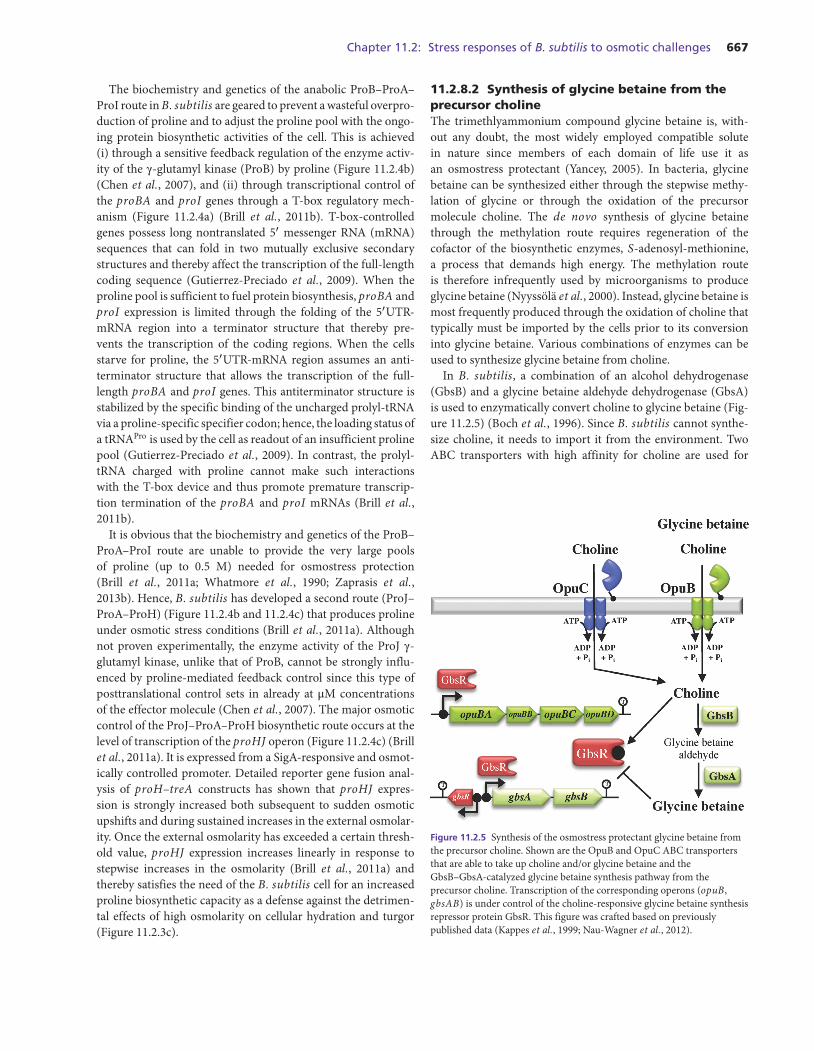
11.2.8.2 Synthesis of glycine betaine from theprecursor cholineThe trimethlyammonium compound glycine betaine is, with-out any doubt, the most widely employed compatible solutein nature since members of each domain of life use it asan osmostress protectant (Yancey, 2005). In bacteria, glycinebetaine can be synthesized either through the stepwise methy-lation of glycine or through the oxidation of the precursormolecule choline. The de novo synthesis of glycine betainethrough the methylation route requires regeneration of thecofactor of the biosynthetic enzymes, S-adenosyl-methionine,a process that demands high energy. The methylation routeis therefore infrequently used by microorganisms to produceglycine betaine (Nyyssola et al., 2000). Instead, glycine betaine ismost frequently produced through the oxidation of choline thattypically must be imported by the cells prior to its conversioninto glycine betaine. Various combinations of enzymes can beused to synthesize glycine betaine from choline.
In B. subtilis, a combination of an alcohol dehydrogenase(GbsB) and a glycine betaine aldehyde dehydrogenase (GbsA)is used to enzymatically convert choline to glycine betaine (Fig-ure 11.2.5) (Boch et al., 1996). Since B. subtilis cannot synthe-size choline, it needs to import it from the environment. TwoABC transporters with high affinity for choline are used for
Figure 11.2.5 Synthesis of the osmostress protectant glycine betaine fromthe precursor choline. Shown are the OpuB and OpuC ABC transportersthat are able to take up choline and/or glycine betaine and theGbsB–GbsA-catalyzed glycine betaine synthesis pathway from theprecursor choline. Transcription of the corresponding operons (opuB,gbsAB) is under control of the choline-responsive glycine betaine synthesisrepressor protein GbsR. This figure was crafted based on previouslypublished data (Kappes et al., 1999; Nau-Wagner et al., 2012).
668 Section 11: Adaptation to osmotic stress
this purpose, OpuB and OpuC. The OpuB system only importscholine and the intermediate in glycine betaine synthesis, glycinebetaine aldehyde, whereas OpuC cannot only import these twocompounds but exhibits a very broad substrate specificity (for anoverview of the types of compatible solutes imported via OpuC,see Hoffmann and Bremer (2011) and Broy et al. (2015)). Thesynthesis of glycine betaine from choline confers a considerabledegree of osmotic stress resistance; it should be noted in this con-text that choline is no compatible solute per se, and its importtherefore does not confer osmostress protection unless it is con-verted into glycine betaine (Boch et al., 1994, 1996).
B. subtilis is a bacterium that frequently lives in associationwith plants or plant detritus (Belda et al., 2013) and thereforecan acquire choline from degraded phospholipids of eukary-otic cell membranes. Since neither choline nor glycine betainecan be used as nutrients by B. subtilis (Boch et al., 1994),enhanced import of the glycine betaine precursor makes physi-ological sense only under conditions when the cells need to syn-thesize glycine betaine for osmoprotective purposes and whencholine is present in the environment. The regulatory circuitthat governs glycine betaine synthesis from choline reflects thisfact. The GbsR repressor, a member of the MarR-type familyof transcriptional regulators (Figure 11.2.5), controls both thegbsAB glycine betaine synthesis genes and the operon encod-ing the choline-specific OpuB transporter. Choline serves asthe effector molecule for the GbsR repressor protein, and itsbinding prevents DNA binding by GbsR to its cognate oper-ator sequences (Nau-Wagner et al., 2012). A model has beenproposed that envisions a structural rearrangement of the N-terminal DNA-reading head relative to the dimerization domainof GbsR once the effector molecule choline has been bound(Nau-Wagner et al., 2012). Interestingly, glycine betaine alde-hyde (Figure 11.2.5) also serves as an effector molecule forGbsR and thereby ensures that this toxic intermediate in glycinebetaine synthesis (Boch et al., 1996; Kappes et al., 1999) neveraccumulates to significant intracellular levels.
B. subtilis sets its intracellular glycine betaine pool very pre-cisely and in tune with the degree of the osmotic stress thatis experienced by the cell (Hoffmann et al., 2013). The GbsR-mediated regulatory circuit described in this chapter will leadto continuous glycine betaine synthesis as long as the inducercholine is present in the environment, regardless of the osmoticneeds of the cell. This would be very wasteful, as the cell would beeventually forced to expel the newly synthesized glycine betaineto avoid physiologically inadequate values of turgor. Therefore,in the GbsR-mediated regulatory system, a negative feedbackloop has been built in that re-establishes GbsR-mediated repres-sion of the gbsAB and opuB operons once the cell has producedenough glycine betaine to balance the osmotic gradient acrossthe cytoplasmic membrane. Not surprisingly, glycine betaineserved as the cellular cue to accomplish this, and GbsR is the tar-get of this genetic negative feedback loop. The GbsR regulatoryprotein thus records and integrates cellular and environmentalsignals for both the onset and the repression of the synthesis of
the osmoprotectant glycine betaine in B. subtilis (Nau-Wagneret al., 2012).
11.2.9 Osmotic control of gene expression
Due to the transient nature of the pulse in SigB-controlledgene expression (Young et al., 2013), implementation of specificstress-adaptive pathways, such as the synthesis and uptake ofosmoprotectants, is required for cellular adaptation to sustainedenvironmental challenges (Bremer, 2002; Bremer and Kramer,2000). However, there is an interesting interlink between thegeneral stress response system and specific adaptation reactionsof the B. subtilis cell exposed to sustained high-osmolarity envi-ronments. This interlink is manifested in the genetic control ofthe opuE and opuD genes.
OpuE mediates the import of proline as an osmostress protec-tant (von Blohn et al., 1997). Genetic control of the opuE geneintegrates general and specific stress responses via two inter-twined and osmotically controlled promoters: One is controlledby the housekeeping sigma factor of B. subtilis, SigA, and theother is responsive to SigB (Figure 11.2.2b) (Spiegelhalter andBremer, 1998). In the wild-type opuE gene, the level of tran-scription increases linearly in response to incremental, but last-ing, increases in the external salinity (Figure 11.2.2c). The inac-tivation of either the SigA- or the SigB-type promoters revealedtheir individual contributions to the osmotic control of opuEexpression in either salt-shocked or salt-adapted cells. The SigA-type promoter mediated a dose-dependent response in cells sub-jected to sustained salt stress, whereas the SigB-type promoteris completely inactive under these conditions (Figure 11.2.2c);however, it permitted a rapid but transient induction of opuEtranscription subsequent to a salt shock. The SigA-type pro-moter was also responsive to a salt shock, but the kinetics of itsactivation was somewhat delayed in comparison with the SigB-type promoter (Figure 11.2.2d). Hence, the combined activitiesof these two promoters allow both a rapid and well-graded phys-iological response to salt challenges through the OpuE-mediatedimport of the osmoprotectant proline.
Interestingly, the opuD glycine betaine transporter gene(Kappes et al., 1996) is also part of the SigB-controlled gen-eral stress response system (Nicolas et al., 2012). As detailed foropuE, it is expressed from closely spaced SigA- and SigB-typepromoters, whose activity is enhanced in response to osmoticstress (our unpublished data). As a consequence, the doubleosmotic control of opuD expression provides enhanced glycinebetaine transport capacity both of osmotically upshocked cellsand to B. subtilis cells continuously challenged by high osmo-larity (Kappes et al., 1996).
Genome-wide transcriptional analysis has been performedwith both salt-shocked and continuously osmotically stressedB. subtilis cells (Hahne et al., 2010; Kohlstedt et al., 2014;Nicolas et al., 2012; Steil et al., 2003), and thereby provides acomprehensive overview of the transcriptional landscape of
Chapter 11.2: Stress responses of B. subtilis to osmotic challenges 669
osmotically stressed B. subtilis cells. It is beyond the scope ofthis overview to discuss in detail each of these changes in geneexpression; instead, we focus on a few selected examples of geneswhere more detailed information is already available from tar-geted genetic and physiological studies.
In the transcriptome analysis reported by Steil et al. (2003),approximately 5% of the then annotated 4107 protein-codinggenes differ significantly (at least threefold) in their expressionlevels between high- and low-salinity-grown cells. Transcrip-tion of 101 genes is downregulated, and most of these genesare functionally associated with either the synthesis of the flag-ellum, chemotaxis, or (unknown at that time) the productionof an extracellular polysaccharide (EPS) matrix. Indeed, high-salinity-challenged B. subtilis cells can no longer swim (Steilet al., 2003); the synthesis of the Hag protein, the major com-ponent of the flagellum, is drastically reduced (Hoffmann et al.,2002); and the production of the sugar matrix involved in build-ing the B. subtilis biofilm is downregulated (Rubinstein et al.,2012). Expression of 123 genes is upregulated in the study con-ducted by Steil et al. (2003), and of these 21 are only indirectlyaffected by high salinity since such growth conditions triggeredin the B. subtilis strain used a severe iron limitation (Hoffmannet al., 2002). The group of 21 salt-inducible genes is function-ally annotated either in connection with iron acquisition or withthe synthesis of the iron chelator bacillibactin; a bioinformaticsanalysis revealed that they are all members of the Fur regulon(Hoffmann et al., 2002; Steil et al., 2003). Hence, the expres-sion of 102 genes has been found to be truly induced by highsalinity, and genes encoding either uptake or synthesis systemsfor compatible solutes are prominently represented in this group.A substantial number of genes involved in cell wall synthesis ormodification are also represented among the salt-induced genes,indicating that B. subtilis modifies its cell wall when it is contin-uously exposed to high salinity (Steil et al., 2003). Genes fallinginto this functional group have also been identified as osmot-ically controlled in other studies (Fischer and Bremer, 2012;Lopez et al., 1998, 2000; Palomino et al., 2009).
A substantial overlap (20 out of 102) of osmotically inducedgenes (Steil et al., 2003) with members of the DegSU regu-lon (Mader et al., 2002) has been observed. This includes (i)the structural genes for the DegS sensor kinase and its cog-nate response regulator DegU, and (ii) an operon (rapG–phrG)that encodes an exported regulatory pro-peptide (PhrG) anda PhrG-responsive regulator (RapG) that controls the DNA-binding activity of DegU-P via protein–protein interactions(Ogura et al., 2003). This implies that the level of DegSU isupregulated in high-salinity-grown cells and that a fine-tuningregulatory circuit exists that operates via the RapG–PhrG sig-naling system and sets the level and activity of DegU–DegU-Pat high salinity. These findings are of special interest for two rea-sons: (i) The DegSU two-component regulatory system has beenpreviously implicated in the salt stress response of B. subtilisthrough functional studies (Kunst and Rapoport, 1995; Ruzaland Sanchez-Rivas, 1998); and (ii) the DegS sensor kinase is
one of only two sensor kinases out of 36 sensor kinases iden-tified in B. subtilis that are localized in the cytoplasm (Fab-ret et al., 1999). As we have discussed here in the context ofthe stressosome controlling the expression of the SigB generalstress response system (Marles-Wright et al., 2008), the cyto-plasmic localization of DegS requires that the sensing of saltstress imposed by the environment has to rely on a derived cyto-plasmic signal that then can be perceived by DegS. The natureof this signal and the way it might be recognized by the DegSsensor kinase are unknown.
The DegSU-regulated genes found by Steil et al. (2003) com-prise only about one-fifth of the B. subtilis genes whose tran-scription is upregulated in response to sustained high osmo-larity. Hence, the DegSU two-component system is definitelynot the overarching osmotic stress-sensing and response sys-tem of B. subtilis, as has been suggested previously (Ruzaland Sanchez-Rivas, 1998). This conclusion is substantiated bydetailed reporter studies and degSU mutant analysis in theframework of studies addressing the properties of the SigA-dependent opuA promoter, a promoter that is strongly inducibleby high osmolarity but is not influenced by the DegSU two-component system (Hoffmann et al., 2013). It also should benoted in this context that the DegSU system is involved in theregulation of a diverse set of cellular processes that probablygenerate different types of environmental and cellular signals towhich the DegS sensor kinase will then respond (Cairns et al.,2013). In no case have the precise type(s) of signal(s) and themolecular mechanism(s) of their perception by DegS been elu-cidated.
Here, we describe the osmotic control of the opuA, proHJ,and yqiHIK operons from B. subtilis since detailed transcrip-tional and mutational studies have been reported for their pro-moters. The opuA operon (opuAA–opuAB–opuAC) encodesthe components of the OpuA ABC transport system for glycinebetaine (Figure 11.2.1) (Hoffmann et al., 2013; Kempf and Bre-mer, 1995), proHJ encodes the key enzymes for the osmostressadaptive proline biosynthetic route (Figure 11.2.4b,c) (Brillet al., 2011a), and the yqiHIK operon encodes a lipopro-tein exposed at the cell surface (YqiH), an extracellular N-acetyl-muramyl-L-alanine amidase (YqiI), and a cytoplasmicglycerophosphodiester phospodiesterase (YqiK) (Fischer andBremer, 2012). In each of these three operons, the osmoticallycontrolled promoter has been mapped by primer extension anal-ysis (Figure 11.2.6a), and its activity has been studied geneticallyvia reporter gene fusion analysis and site-directed mutagenesisstudies. The sequences of the −10 and −35 regions of all threepromoters resemble those of SigA-type housekeeping promotersfrom B. subtilis but deviate in key positions from the consen-sus sequence (Figure 11.2.6a). Mutations that improve the matchof the opuA, proHJ, and yqiHIK promoters to the consensussequence lead to enhanced gene expression both at low and athigh salinity, indicating that the deviations from the SigA-typeconsensus sequence serve to keep the activity of these osmot-ically controlled promoters low in cells grown in low-salinity
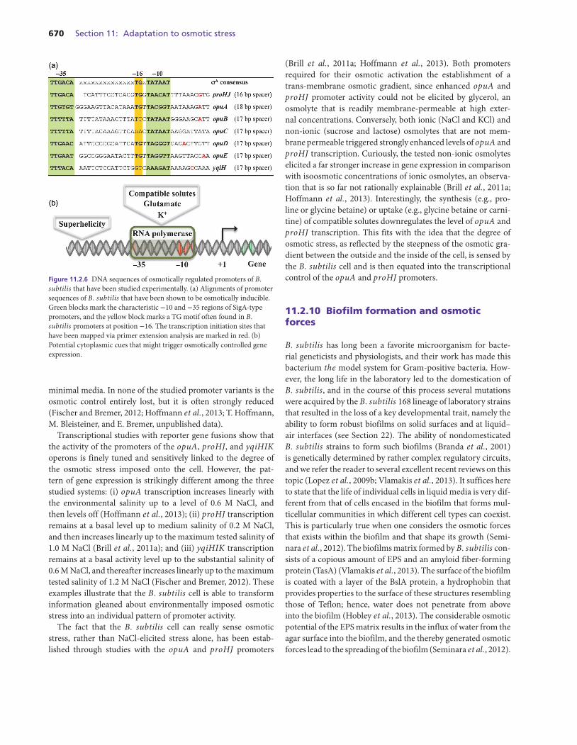
670 Section 11: Adaptation to osmotic stress
Figure 11.2.6 DNA sequences of osmotically regulated promoters of B.subtilis that have been studied experimentally. (a) Alignments of promotersequences of B. subtilis that have been shown to be osmotically inducible.Green blocks mark the characteristic −10 and −35 regions of SigA-typepromoters, and the yellow block marks a TG motif often found in B.subtilis promoters at position −16. The transcription initiation sites thathave been mapped via primer extension analysis are marked in red. (b)Potential cytoplasmic cues that might trigger osmotically controlled geneexpression.
minimal media. In none of the studied promoter variants is theosmotic control entirely lost, but it is often strongly reduced(Fischer and Bremer, 2012; Hoffmann et al., 2013; T. Hoffmann,M. Bleisteiner, and E. Bremer, unpublished data).
Transcriptional studies with reporter gene fusions show thatthe activity of the promoters of the opuA, proHJ, and yqiHIKoperons is finely tuned and sensitively linked to the degree ofthe osmotic stress imposed onto the cell. However, the pat-tern of gene expression is strikingly different among the threestudied systems: (i) opuA transcription increases linearly withthe environmental salinity up to a level of 0.6 M NaCl, andthen levels off (Hoffmann et al., 2013); (ii) proHJ transcriptionremains at a basal level up to medium salinity of 0.2 M NaCl,and then increases linearly up to the maximum tested salinity of1.0 M NaCl (Brill et al., 2011a); and (iii) yqiHIK transcriptionremains at a basal activity level up to the substantial salinity of0.6 M NaCl, and thereafter increases linearly up to the maximumtested salinity of 1.2 M NaCl (Fischer and Bremer, 2012). Theseexamples illustrate that the B. subtilis cell is able to transforminformation gleaned about environmentally imposed osmoticstress into an individual pattern of promoter activity.
The fact that the B. subtilis cell can really sense osmoticstress, rather than NaCl-elicited stress alone, has been estab-lished through studies with the opuA and proHJ promoters
(Brill et al., 2011a; Hoffmann et al., 2013). Both promotersrequired for their osmotic activation the establishment of atrans-membrane osmotic gradient, since enhanced opuA andproHJ promoter activity could not be elicited by glycerol, anosmolyte that is readily membrane-permeable at high exter-nal concentrations. Conversely, both ionic (NaCl and KCl) andnon-ionic (sucrose and lactose) osmolytes that are not mem-brane permeable triggered strongly enhanced levels of opuA andproHJ transcription. Curiously, the tested non-ionic osmolyteselicited a far stronger increase in gene expression in comparisonwith isoosmotic concentrations of ionic osmolytes, an observa-tion that is so far not rationally explainable (Brill et al., 2011a;Hoffmann et al., 2013). Interestingly, the synthesis (e.g., pro-line or glycine betaine) or uptake (e.g., glycine betaine or carni-tine) of compatible solutes downregulates the level of opuA andproHJ transcription. This fits with the idea that the degree ofosmotic stress, as reflected by the steepness of the osmotic gra-dient between the outside and the inside of the cell, is sensed bythe B. subtilis cell and is then equated into the transcriptionalcontrol of the opuA and proHJ promoters.
11.2.10 Biofilm formation and osmoticforces
B. subtilis has long been a favorite microorganism for bacte-rial geneticists and physiologists, and their work has made thisbacterium the model system for Gram-positive bacteria. How-ever, the long life in the laboratory led to the domestication ofB. subtilis, and in the course of this process several mutationswere acquired by the B. subtilis 168 lineage of laboratory strainsthat resulted in the loss of a key developmental trait, namely theability to form robust biofilms on solid surfaces and at liquid–air interfaces (see Section 22). The ability of nondomesticatedB. subtilis strains to form such biofilms (Branda et al., 2001)is genetically determined by rather complex regulatory circuits,and we refer the reader to several excellent recent reviews on thistopic (Lopez et al., 2009b; Vlamakis et al., 2013). It suffices hereto state that the life of individual cells in liquid media is very dif-ferent from that of cells encased in the biofilm that forms mul-ticellular communities in which different cell types can coexist.This is particularly true when one considers the osmotic forcesthat exists within the biofilm and that shape its growth (Semi-nara et al., 2012). The biofilms matrix formed by B. subtilis con-sists of a copious amount of EPS and an amyloid fiber-formingprotein (TasA) (Vlamakis et al., 2013). The surface of the biofilmis coated with a layer of the BslA protein, a hydrophobin thatprovides properties to the surface of these structures resemblingthose of Teflon; hence, water does not penetrate from aboveinto the biofilm (Hobley et al., 2013). The considerable osmoticpotential of the EPS matrix results in the influx of water from theagar surface into the biofilm, and the thereby generated osmoticforces lead to the spreading of the biofilm (Seminara et al., 2012).
Chapter 11.2: Stress responses of B. subtilis to osmotic challenges 671
Within the biofilm, microscopic water channels exist that trans-port nutrients and waste products.
The increase in osmotic pressure caused by the formationof the EPS is a cue that downregulates the gene cluster (epsA-O) required for the synthesis of the sugar component of theextracellular matrix and the operon (tapA–sipW–tasA) thatdirects the formation of the amyloid-like fibers (Nicolas et al.,2012; Rubinstein et al., 2012). These EPS-elicited nonspecificosmotic pressure effects activate the histidine kinase KinD,which in turn directs the phosphorylation of the master reg-ulatory protein of sporulation and biofilm formation, Spo0A,which at high levels represses matrix gene expression (Rubin-stein et al., 2012). The physiological and genetic consequencesof high osmotic pressure within the biofilm are not yet fully com-prehended, but Rubinstein et al. (2012) suggested that the sens-ing of this physical cue might be a strategy to coordinate multi-cellular behavior in the confines of the biofilm (Rubinstein et al.,2012).
An interesting connection between the genetic system thatdirects biofilm formation in B. subtilis and osmotically con-trolled expression of genes encoding uptake systems for com-patible solutes has recently been uncovered in a study conductedby Kearns and coworkers (Winkelman et al., 2013). A key regu-lator of biofilm formation, SinR, negatively controls expressionof the eps and tapA–sipW–tasA operons. The RemA activatorprotein antagonizes the repressing effects of SinR on eps andtapA–sipW–tasA expression and thereby stimulates biofilm for-mation. In connection with a DNA-profiling study of a remAmutant strain, the expression of the opuA, opuB, and opuCoperons in osmotically nonstressed cells was found to be affected(Winkelman et al., 2013). DNA-footprinting studies with thepurified RemA protein at the opuA promoter region revealedan extended RemA-binding region upstream of the opuA −35sequence; it is composed of five distinct subsites (Winkelmanet al., 2013). The opuA regulatory region is thus a direct targetof a transcription control protein that is essential for biofilm for-mation by B. subtilis (Winkelman et al., 2009). Reporter genestudies showed that RemA, like its actions at the eps and tapA–sipW–tasA regulatory regions (Winkelman et al., 2009, 2013),serves as an activator protein and is crucial for opuA expressionunder both low- and high-salinity growth conditions (T. Hoff-mann and E. Bremer, unpublished data). However, it is not yetclear whether RemA transduces the osmotic signal to the opuApromoter, or whether it just serves a role of a general activator ofopuA transcription, a function that would be independent fromthe signal transduction pathway that confers osmotic control ofpromoter activity.
We find the observation quite remarkable that the structuralgenes for three osmostress protectant uptake systems (OpuA,OpuB, and OpuC) are members of the RemA regulon (Winkel-man et al., 2013). It bears the question of how B. subtilis cellsembedded in a biofilm might actually profit for their physiologyand well-being from the import of compatible solutes.
11.2.11 The big challenge: sensing osmoticchanges
Since the 1990s, the basic cellular adjustment strategies of B.subtilis to fluctuations in the external salinity have been workedout in quite some detail. However, many questions remain withrespect to the precise physiological functions of those geneswhose transcription is upregulated either under acute osmoticupshifts or under sustained salt stress (Hahne et al., 2010; Hoperet al., 2005., 2006; Kohlstedt et al., 2014; Nannapaneni et al.,2012; Nicolas et al., 2012; Steil et al., 2003). The most press-ing question that needs to be answered is how increases inthe external osmotic pressure are perceived, genetically pro-cessed, and then transduced to the transcription apparatus sothat altered gene expression will ensue. Players in such a processhave been implicated in the case of the SigB-controlled stressresponse (via the stressosome), biofilm formation (via KinD),and the DegSU two-component regulatory system. KinD is amembrane-embedded sensor kinase, whereas the stressosomeand the DegS sensor kinase are located in the cytoplasm. Asdiscussed in this chapter in the context of these latter two sys-tems, both the stressosome and DegS must perceive a cytoplas-mic signal that is indirectly derived from changes in the externalosmolarity. Although KinD, the stressosome, and DegS have allbeen implicated in the cellular osmostress response, it is unclearhow the B. subtilis cell actually uses these devices to measure –either directly or indirectly – a change in an external physicalparameter, environmental osmolarity. It should be noted herethat the systems directly involved in ameliorating the dire conse-quences of high osmolarity on cellular physiology (e.g., systemsfor uptake and synthesis of compatible solutes) (Figure 11.2.1)are either fully independent of (e.g., opuA and proHJ), or areonly partially dependent on (e.g., opuE and opuD), these rathergeneral regulatory systems.
An overarching osmosensory and regulatory system thatmight operate in B. subtilis has not been detected. This mightbe due to the fact that such a system might be essential, thatthe screening procedures used to search for it might be unsuit-able, or that it simply does not exist. If one just considers the lastscenario to be correct, how might osmotic control of the SigA-type promoters of the opuA, opuB, opuC, opuD, opuE, proHJ,and yqiHIK genes (Figure 11.2.6a) be achieved in the absenceof an osmostress-specific regulatory protein? High osmolarityalters the ionic composition and biophysical properties of thecytoplasm (Cayley and Record, 2003; Whatmore et al., 1990;Wood, 2011). Such changes might be sensed directly by the RNApolymerase, and in combination with altered DNA-superhelicity(Higgins et al., 1988; Krispin and Allmansberger, 1995), specificpromoter sequences and their flanking regions might change thetranscriptional activity of a given osmoregulated promoter (Fig-ure 11.2.6b). It will be a challenge to experimentally verify orrefute such a model. The proHJ promoter with its essentially lin-ear dependence for its activity on the external osmolarity (Brill
672 Section 11: Adaptation to osmotic stress
et al., 2011a) might provide a suitable test system for the outlinedhypothesis.
Acknowledgements
We greatly appreciate the kind help of Vickie Koogle in the lan-guage editing of our manuscript. Funding for the work in ourlaboratory on osmotic stress responses, synthesis, uptake, andcatabolism of compatible solutes is generously supported by theDeutsche Forschungsgemeinschaft (DFG) through the Collab-orative Research Centre SFB-987, the LOEWE program of theState of Hessen (via the Centre for Synthetic Microbiology; Syn-micro, Marburg), and the Fonds der Chemischen Industrie.
References
Bashir A, Hoffmann T, Kempf B, Xie X, Smits SH, et al. 2014a. Theplant-derived compatible solutes proline betaine and betonicine con-fer enhanced osmotic and temperature stress tolerance to Bacillussubtilis. Microbiology 160(10): 2283–2294.
Bashir A, Hoffmann T, Smits SH, Bremer E. 2014b. Dimethlyglycineprovides salt and temperature stress protection to Bacillus subtilis.Applied Environmental Microbiology 80(9): 2773–2785.
Belda E, Sekowska A, Le Fevre F, Morgat A, Mornico D, et al. 2013. Anupdated metabolic view of the Bacillus subtilis 168 genome. Micro-biology 159(4): 757–770.
Belitsky BR. 2011. Indirect repression by Bacillus subtilis CodY via dis-placement of the activator of the proline utilization operon. Journalof Molecular Biology 413(2): 321–336.
Berntsson RP, Smits SH, Schmitt L, Slotboom DJ, Poolman B. 2010. Astructural classification of substrate-binding proteins. FEBS Letters584(12): 2606–2617.
Boch J, Kempf B, Bremer E. 1994. Osmoregulation in Bacillus subtilis:synthesis of the osmoprotectant glycine betaine from exogenouslyprovided choline. Journal of Bacteriology 176(17): 5364–5371.
Boch J, Kempf B, Schmid R, Bremer E. 1996. Synthesis of the osmo-protectant glycine betaine in Bacillus subtilis: characterization of thegbsAB genes. Journal of Bacteriology 178(17): 5121–5129.
Boersma AJ, Zuhorn IS, Poolman B. 2015. A sensor for quantificationof macromolecular crowding in living cells. Nature Methods 12(3):227–229.
Booth IR. 2014. Bacterial mechanosensitive channels: progress towardsan understanding of their roles in cell physiology. Current Opinionin Microbiology 18: 16–22.
Booth IR, Miller S, Muller A, Lehtovirta-Morley L. 2015. The evolutionof bacterial mechanosensitive channels. Cell Calcium 57(3): 140–150.
Borngen K, Battle AR, Moker N, Morbach S, Marin K, et al. 2010. Theproperties and contribution of the Corynebacterium glutamicumMscS variant to fine-tuning of osmotic adaptation. Biochimica etBiophysica Acta 1798(11): 2141–2149.
Branda SS, Gonzalez-Pastor JE, Ben-Yehuda S, Losick R, Kolter R. 2001.Fruiting body formation by Bacillus subtilis. Proceedings of theNational Academy of Sciences of the USA 98(20): 11621–11626.
Bremer E. 2002. Adaptation to changing osmolarity. In Bacillus sub-tilis and its closest relatives: from genes to cells (ed. SonensheinAL, Hoch JA, Losick R). Washington, DC: ASM Press, pp. 385–391.
Bremer E, Kramer R. 2000. Coping with osmotic challenges: osmoreg-ulation through accumulation and release of compatible solutes. InBacterial stress responses (ed. Storz G, Hengge-Aronis R). Washing-ton, DC: ASM Press, pp. 79–97.
Brigulla M, Hoffmann T, Krisp A, Volker A, Bremer E, et al. 2003. Chillinduction of the SigB-dependent general stress response in Bacillussubtilis and its contribution to low-temperature adaptation. Journalof Bacteriology 185(15): 4305–4514.
Brill J, Hoffmann T, Bleisteiner M, Bremer E. 2011a. Osmotically con-trolled synthesis of the compatible solute proline is critical for cellulardefense of Bacillus subtilis against high osmolarity. Journal of Bac-teriology 193(19): 5335–5346.
Brill J, Hoffmann T, Putzer H, Bremer E. 2011b. T-box-mediated con-trol of the anabolic proline biosynthetic genes of Bacillus subtilis.Microbiology 157(4): 977–987.
Broy S, Chen C, Hoffmann T, Brock NL, Nau-Wagner G, et al. 2015.Abiotic stress protection by ecologically abundant dimethylsulfonio-propionate and its natural and synthetic derivatives: insights fromBacillus subtilis. Environmental Microbiology 17(7): 2362–2378.
Bursy J, Pierik AJ, Pica N, Bremer E. 2007. Osmotically induced synthe-sis of the compatible solute hydroxyectoine is mediated by an evolu-tionarily conserved ectoine hydroxylase. The Journal of BiologicalChemistry 282(43): 31147–31155.
Cairns LS, Marlow VL, Bissett E, Ostrowski A, Stanley-Wall NR. 2013.A mechanical signal transmitted by the flagellum controls signallingin Bacillus subtilis. Molecular Microbiology 90(1): 6–21.
Calamita G. 2000. The Escherichia coli aquaporin-Z water channel.Molecular Microbiology 37(2): 254–262.
Cayley S, Record MT Jr. 2003. Roles of cytoplasmic osmolytes, water,and crowding in the response of Escherichia coli to osmotic stress:biophysical basis of osmoprotection by glycine betaine. Biochemistry42(43): 12596–12609.
Chen C, Beattie GA. 2007. Characterization of the osmoprotectanttransporter OpuC from Pseudomonas syringae and demonstra-tion that cystathionine-beta-synthase domains are required for itsosmoregulatory function. Journal of Bacteriology 189(19): 6901–6912.
Chen M, Wei H, Cao J, Liu R, Wang Y, et al. 2007. Expression of Bacillussubtilis proBA genes and reduction of feedback inhibition of prolinesynthesis increases proline production and confers osmotolerance intransgenic Arabidopsis. Journal of Biochemistry Molecular Biology40(3): 396–403.
Coquelle N, Talon R, Juers DH, Girard E, Kahn R, et al. 2010. Gradualadaptive changes of a protein facing high salt concentrations. Journalof Molecular Biology 404(3): 493–505.
Corrigan RM, Campeotto I, Jeganathan T, Roelofs KG, Lee VT, et al.2013. Systematic identification of conserved bacterial c-di-AMPreceptor proteins. Proceedings of the National Academy of Sciencesof the USA 110(22): 9084–9089.
Cosquer A, Ficamos M, Jebbar M, Corbel JC, Choquet G, et al. 2004.Antibacterial activity of glycine betaine analogues: involvement ofosmoporters. Bioorganic & Medicinal Chemistry Letters 14(9):2061–2065.
Csonka LN. 1989. Physiological and genetic responses of bacteria toosmotic stress. Microbiological Reviews 53(1): 121–147.
Chapter 11.2: Stress responses of B. subtilis to osmotic challenges 673
Delamarche C, Thomas D, Rolland JP, Froger A, Gouranton J,et al. 1999. Visualization of AqpZ-mediated water permeability inEscherichia coli by cryoelectron microscopy. Journal of Bacteriol-ogy 181(14): 4193–4197.
Deng Y, Sun M, Shaevitz JW. 2011. Direct measurement of cell wallstress stiffening and turgor pressure in live bacterial cells. PhysicalReview Letters 107(15): 158101.
Du Y, Shi WW, He YX, Yang YH, Zhou CZ, et al. 2011. Structuresof the substrate-binding protein provide insights into the multi-ple compatible solute binding specificities of the Bacillus subtilisABC transporter OpuC. The Biochemical Journal 436(2): 283–289.
Earl AM, Losick R, Kolter R. 2008. Ecology and genomics of Bacillussubtilis. Trends in Microbiology 16(6): 269–275.
Eggeling L, Sahm H. 2003. New ubiquitous translocators: amino acidexport by Corynebacterium glutamicum and Escherichia coli.Archives of Microbiology 180(3): 155–160.
Fabret C, Feher VA, Hoch JA. 1999. Two-component signal transduc-tion in Bacillus subtilis: how one organism sees its world. Journal ofBacteriology 181(7): 1975–1983.
Fichman Y, Gerdes SY, Kovacs H, Szabados L, Zilberstein A, et al. 2015.Evolution of proline biosynthesis: enzymology, bioinformatics, genet-ics, and transcriptional regulation. Biological Reviews 90(4): 1065–1099.
Fischer KE, Bremer E. 2012. Activity of the osmotically regulatedyqiHIK promoter from Bacillus subtilis is controlled at a distance.Journal of Bacteriology 194(19): 5197–5208.
Fujisawa M, Ito M, Krulwich TA. 2007. Three two-component trans-porters with channel-like properties have monovalent cation/protonantiport activity. Proceedings of the National Academy of Sciencesof the USA 104(33): 13289–13294.
Fujisawa M, Wada Y, Ito M. 2004. Modulation of the K+ efflux activity ofBacillus subtilis YhaU by YhaT and the C-terminal region of YhaS.FEMS Microbiology Letters 231(2): 211–217.
Galinski EA, Truper HG. 1994. Microbial behaviour in salt-stressedecosystems. FEMS Microbiological Reviews 15(2–3): 95–108.
Gnutt D, Gao M, Brylski O, Heyden M, Ebbinghaus S. 2015. Excluded-volume effects in living cells. Angewandte Chemie InternationalEdition English 54(8): 2548–2551.
Gorecki K, Hagerhall C, Drakenberg T. 2014. The Na+ transport ingram-positive bacteria defect in the Mrp antiporter complex mea-sured with 23Na nuclear magnetic resonance. Analytical Biochem-istry 445: 80–86.
Gutierrez-Preciado A, Henkin TM, Grundy FJ, Yanofsky C, Merino E.2009. Biochemical features and functional implications of the RNA-based T-box regulatory mechanism. Microbiology Molecular Biol-ogy Reviews 73(1): 36–61.
Hahne H, Mader U, Otto A, Bonn F, Steil L, et al. 2010. A comprehen-sive proteomics and transcriptomics analysis of Bacillus subtilis saltstress adaptation. Journal of Bacteriology 192(3): 870–882.
Hanelt I, Tholema N, Kroning N, Vor der Bruggen M, Wunnicke D,et al. 2011. KtrB, a member of the superfamily of K+ transporters.European Journal of Cell Biology 90(9): 696–704.
Hecker M, Pane-Farre J, Volker U. 2007. SigB-dependent general stressresponse in Bacillus subtilis and related gram-positive bacteria.Annual Review of Microbiology 61: 215–236.
Higgins CF, Dorman CJ, Stirling DA, Waddell L, Booth IR, et al. 1988. Aphysiological role for DNA supercoiling in the osmotic regulation ofgene expression in S. typhimurium and E. coli. Cell 52(4): 569–584.
Higgins D, Dworkin J. 2012. Recent progress in Bacillus subtilis sporu-lation. FEMS Microbiology Reviews 36(1): 131–148.
Hobley L, Ostrowski A, Rao FV, Bromley KM, Porter M, et al. 2013.BslA is a self-assembling bacterial hydrophobin that coats the Bacil-lus subtilis biofilm. Proceedings of the National Academy of Sci-ences of the USA 110(33): 13600–13605.
Hoffmann T, Schutz A, Brosius M, Volker A, Volker U, et al. 2002.High-salinity-induced iron limitation in Bacillus subtilis. Journal ofBacteriology 184(3): 718–727.
Hoffmann T, Boiangiu C, Moses S, Bremer E. 2008. Responses of Bacil-lus subtilis to hypotonic challenges: physiological contributions ofmechanosensitive channels to cellular survival. Applied and Envi-ronmental Microbiology 74(8): 2454–2460.
Hoffmann T, Bremer E. 2011. Protection of Bacillus subtilis againstcold stress via compatible-solute acquisition. Journal of Bacteriol-ogy 193(7): 1552–1562.
Hoffmann T, von Blohn C, Stanek A, Moses S, Barzantny S, et al. 2012.Synthesis, release, and recapture of the compatible solute proline byosmotically stressed Bacillus subtilis cells. Applied and Environ-mental Microbiology 78(16): 5753–5762.
Hoffmann T, Wensing A, Brosius M, Steil L, Volker U, et al. 2013.Osmotic control of opuA expression in Bacillus subtilis and its mod-ulation in response to intracellular glycine betaine and proline pools.Journal of Bacteriology 195(3): 510–522.
Holtmann G, Bakker EP, Uozumi N, Bremer E. 2003. KtrAB and KtrCD:two K+ uptake systems in Bacillus subtilis and their role in adapta-tion to hypertonicity. J Bacteriol 185(4): 1289–1298.
Holtmann G, Bremer E. 2004. Thermoprotection of Bacillus subtilis byexogenously provided glycine betaine and structurally related com-patible solutes: involvement of Opu transporters. Journal of Bacteri-ology 186(6): 1683–1693.
Holtmann G, Brigulla M, Steil L, Schutz A, Barnekow K, et al. 2004.RsbV-independent induction of the SigB-dependent general stressregulon of Bacillus subtilis during growth at high temperature. Jour-nal of Bacteriology 186(18): 6150–6158.
Hoper D, Volker U, Hecker M. 2005. Comprehensive characterizationof the contribution of individual SigB-dependent general stress genesto stress resistance of Bacillus subtilis. J Bacteriol 187(8): 2810–2826.
Hoper D, Bernhardt J, Hecker M. 2006. Salt stress adaptation of Bacil-lus subtilis: a physiological proteomics approach. Proteomics 6(5):1550–1562.
Hoppner A, Widderich N, Lenders M, Bremer E, Smits SHJ. 2014. Crys-tal structure of the ectoine hydroxylase, a snapshot of the active site.Journal of Biological Chemistry 289(43): 29570–29583.
Horn C, Sohn-Bosser L, Breed J, Welte W, Schmitt L, et al. 2006. Molec-ular determinants for substrate specificity of the ligand-binding pro-tein OpuAC from Bacillus subtilis for the compatible solutes glycinebetaine and proline betaine. Journal of Molecular Biology 357(2):592–606.
Ignatova Z, Gierasch LM. 2006. Inhibition of protein aggregation invitro and in vivo by a natural osmoprotectant. Proceedings ofthe National Academy of Sciences of the USA 103(36): 13357–13361.
Kappes RM, Kempf B, Bremer E. 1996. Three transport systems for theosmoprotectant glycine betaine operate in Bacillus subtilis: charac-terization of OpuD. Journal of Bacteriology 178(17): 5071–5079.
Kappes RM, Kempf B, Kneip S, Boch J, Gade J, et al. 1999. Two evo-lutionarily closely related ABC transporters mediate the uptake of
674 Section 11: Adaptation to osmotic stress
choline for synthesis of the osmoprotectant glycine betaine in Bacil-lus subtilis. Molecular Microbiology 32(1): 203–216.
Karasawa A, Erkens GB, Berntsson RP, Otten R, Schuurman-WoltersGK, et al. 2011. Cystathionine beta-synthase (CBS) domains 1 and 2fulfill different roles in ionic strength sensing of the ATP-binding cas-sette (ABC) transporter OpuA. The Journal of Biological Chemistry286(43): 37280–37291.
Kempf B, Bremer E. 1995. OpuA, an osmotically regulated bindingprotein-dependent transport system for the osmoprotectant glycinebetaine in Bacillus subtilis. The Journal of Biological Chemistry270(28): 16701–16713.
Kempf B, Bremer E. 1998. Uptake and synthesis of compatible solutes asmicrobial stress responses to high osmolality environments. Archivesof Microbiology 170 319–330.
Kohlstedt M, Sappa PK, Meyer H, Maass S, Zaprasis A, et al. 2014. Adap-tation of Bacillus subtilis carbon core metabolism to simultaneousnutrient limitation and osmotic challenge: a multi-omics perspective.Environmental Microbiology 16(6): 1898–1917.
Krispin O, Allmansberger R. 1995. Changes in DNA supertwist as aresponse of Bacillus subtilis towards different kinds of stress. FEMSMicrobiology Letters 134(2–3): 129–135.
Kuhlmann AU, Bremer E. 2002. Osmotically regulated synthesis of thecompatible solute ectoine in Bacillus pasteurii and related Bacil-lus spp. Applied and Environmental Microbiology 68(2): 772–783.
Kunst F, Rapoport G. 1995. Salt stress is an environmental signal affect-ing degradative enzyme synthesis in Bacillus subtilis. Journal of Bac-teriology 177(9): 2403–2407.
Laermann V, Cudic E, Kipschull K, Zimmann P, Altendorf K. 2013. Thesensor kinase KdpD of Escherichia coli senses external K+. Molecu-lar Microbiology 88(6): 1194–1204.
Logan N, De Vos P. 2009. Bacillus. In Bergey’s manual of systematicbacteriology (ed. De Vos P, Garrity GM, Jones D, Krieg NR, LudwigW, et al. Heidelberg: Springer, pp. 21–128.
Lopez CS, Heras H, Ruzal SM, Sanchez-Rivas C, Rivas EA. 1998. Varia-tions of the envelope composition of Bacillus subtilis during growthin hyperosmotic medium. Current Microbiology 36(1): 55–61.
Lopez CS, Heras H, Garda H, Ruzal S, Sanchez-Rivas C, et al. 2000. Bio-chemical and biophysical studies of Bacillus subtilis envelopes underhyperosmotic stress. International Journal of Food Microbiology55(1–3): 137–142.
Lopez D, Fischbach MA, Chu F, Losick R, Kolter R. 2009a. Struc-turally diverse natural products that cause potassium leakage trig-ger multicellularity in Bacillus subtilis. Proceedings of the NationalAcademy of Sciences of the USA 106(1): 280–285.
Lopez D, Vlamakis H, Kolter R. 2009b. Generation of multiple celltypes in Bacillus subtilis. FEMS Microbiology Reviews 33(1): 152–163.
Lundberg ME, Becker EC, Choe S. 2013. MstX and a putative potassiumchannel facilitate biofilm formation in Bacillus subtilis. PLoS One8(5): e60993.
Mader U, Antelmann H, Buder T, Dahl MK, Hecker M, et al. 2002.Bacillus subtilis functional genomics: genome-wide analysis of theDegS-DegU regulon by transcriptomics and proteomics. MolecularGenetics Genomics 268(4): 455–467.
Marles-Wright J, Grant T, Delumeau O, van Duinen G, Firbank SJ, et al.2008. Molecular architecture of the “stressosome,” a signal integrationand transduction hub. Science 322(5898): 92–96.
Maximov S, Ott V, Belkoura L, Kramer R. 2014. Stimulus analysis ofBetP activation under in vivo conditions. Biochimica et bBiophysicaActa 1838(5): 1288–1295.
Mehne FMP, Gunka K, Eilers H, Herzberg C, Kaever V, et al. 2013.Cyclic di-AMP homeostasis in Bacillus subtilis: Both lack and highlevel accumulation of the nucleotide are detrimental for cell growth.Journal of Biological Chemistry 288(3): 2004–2017.
Moker N, Brocker M, Schaffer S, Kramer R, Morbach S, et al. 2004.Deletion of the genes encoding the MtrA-MtrB two-component sys-tem of Corynebacterium glutamicum has a strong influence oncell morphology, antibiotics susceptibility and expression of genesinvolved in osmoprotection. Molecular Microbiology 54(2): 420–438.
Moker N, Reihlen P, Kramer R, Morbach S. 2007. Osmosensing proper-ties of the histidine protein kinase MtrB from Corynebacterium glu-tamicum. Journal of Biological Chemistry 282(38): 27666–27677.
Moses S, Sinner T, Zaprasis A, Stoveken N, Hoffmann T, et al. 2012.Proline utilization by Bacillus subtilis: uptake and catabolism. Jour-nal of Bacteriology 194(4): 745–758.
Nagler K, Setlow P, Li YQ, Moeller R. 2014. High salinity alters thegermination behavior of Bacillus subtilis spores with nutrient andnonnutrient germinants. Applied and Environmental Microbiology80(4): 1314–1321.
Naismith JH, Booth IR. 2012. Bacterial mechanosensitive channels –MscS: evolution’s solution to creating sensitivity in function. AnnualReviews Biophysics 41: 157–177.
Nannapaneni P, Hertwig F, Depke M, Hecker M, Mader U, et al. 2012.Defining the structure of the general stress regulon of Bacillus sub-tilis using targeted microarray analysis and random forest classifica-tion. Microbiology 158(3): 696–707.
Nau-Wagner G, Boch J, Le Good JA, Bremer E. 1999. High-affinitytransport of choline-O-sulfate and its use as a compatible solute inBacillus subtilis. Applied and Environmental Microbiology 65(2):560–568.
Nau-Wagner G, Opper D, Rolbetzki A, Boch J, Kempf B, et al. 2012.Genetic control of osmoadaptive glycine betaine synthesis in Bacillussubtilis through the choline-sensing and glycine betaine-responsiveGbsR repressor. Journal of Bacteriology 194(10): 2703–2714.
Nelson JW, Sudarsan N, Furukawa K, Weinberg Z, Wang JX, et al. 2013.Riboswitches in eubacteria sense the second messenger c-di-AMP.Nature Chemical Biology 9(12): 834–839.
Nicolas P, Mader U, Dervyn E, Rochat T, Leduc A, et al. 2012.Condition-dependent transcriptome reveals high-level regulatoryarchitecture in Bacillus subtilis. Science 335(6072): 1103–1106.
Nyyssola A, Kerovuo J, Kaukinen P, von Weymarn N, ReinikainenT. 2000. Extreme halophiles synthesize betaine from glycine bymethylation. The Journal of Biological Chemistry 275(29): 22196–22201.
Ogura M, Shimane K, Asai K, Ogasawara N, Tanaka T. 2003. Bind-ing of response regulator DegU to the aprE promoter is inhibited byRapG, which is counteracted by extracellular PhrG in Bacillus sub-tilis. Molecular Microbiology 49(6): 1685–1697.
Ogura M, Tsukahara K, Hayashi K, Tanaka T. 2007. The Bacillus sub-tilis NatK-NatR two-component system regulates expression of thenatAB operon encoding an ABC transporter for sodium ion extru-sion. Microbiology 153(3): 667–675.
Olkhova E, Raba M, Bracher S, Hilger D, Jung H. 2011. Homologymodel of the Na+/proline transporter PutP of Escherichia coli and
Chapter 11.2: Stress responses of B. subtilis to osmotic challenges 675
its functional implications. Journal of Molecular Biology 406(1): 59–74.
Oren A. 2011. Thermodynamic limits to microbial life at high salt con-centrations. Environmental Microbiology 13(8): 1908–1923.
Oren A. 2013. Life at high salt concentrations, intracellular KCl concen-trations, and acidic proteomes. Frontiers Microbiology 4: 315.
Palomino MM, Sanchez-Rivas C, Ruzal SM. 2009. High salt stress inBacillus subtilis: involvement of PBP4∗ as a peptidoglycan hydrolase.Research in Microbiology 160(2): 117–124.
Perez C, Faust B, Mehdipour AR, Francesconi KA, Forrest LR, et al.2014. Substrate-bound outward-open state of the betaine transporterBetP provides insights into Na+ coupling. Nature Communications5: 4231.
Perez C, Koshy C, Yildiz O, Ziegler C. 2012. Alternating-access mech-anism in conformationally asymmetric trimers of the betaine trans-porter BetP. Nature 490(7418): 126–130.
Pittelkow M, Tschapek B, Smits SH, Schmitt L, Bremer E. 2011. Thecrystal structure of the substrate-binding protein OpuBC from Bacil-lus subtilis in complex with choline. Journal of Molecular Biology411(1): 53–67.
Poolman B, Spitzer JJ, Wood JM. 2004. Bacterial osmosensing: roles ofmembrane structure and electrostatics in lipid-protein and protein-protein interactions. Biochimica et Biophysica Acta 1666(1–2): 88–104.
Price CW. 2011. General stress responses in Bacillus subtilis and relatedGram-positive bacteria. In Bacterial stress responses (ed. Storz G,Hengge R). Washington, DC: ASM Press, pp. 301–318.
Reder A, Gerth U, Hecker M. 2012. Integration of sigma B activity intothe decision-making process of sporulation initiation in Bacillus sub-tilis. Journal of Bacteriology 194(5): 1065–1074.
Reuter M, Hayward NJ, Black SS, Miller S, Dryden DT, et al. 2014.Mechanosensitive channels and bacterial cell wall integrity: does lifeend with a bang or a whimper? Journal Royal Society Interface11(91): 20130850.
Roesser M, Muller V. 2001. Osmoadaptation in bacteria and archaea:common principles and differences. Environmental Microbiology3(12): 743–754.
Romeo Y, Bouvier J, Gutierrez C. 2007. Osmotic regulation of transcrip-tion in Lactococcus lactis: ionic strength-dependent binding of theBusR repressor to the busA promoter. FEBS Letters 581(18): 3387–3390.
Rubinstein SM, Kolodkin-Gal I, McLoon A, Chai L, Kolter R, et al. 2012.Osmotic pressure can regulate matrix gene expression in Bacillussubtilis. Molecular Microbiology 86(2): 426–436.
Ruzal SM, Sanchez-Rivas C. 1998. In Bacillus subtilis DegU-P is a pos-itive regulator of the osmotic response. Current Microbiology 37(6):368–372.
Schroeter R, Hoffmann T, Voigt B, Meyer H, Bleisteiner M, et al. 2013.Stress responses of the industrial workhorse Bacillus licheniformisto osmotic challenges. PLoS One 8(11): e80956.
Seminara A, Angelini TE, Wilking JN, Vlamakis H, Ebrahim S, et al.2012. Osmotic spreading of Bacillus subtilis biofilms driven by anextracellular matrix. Proceedings of the National Academy of Sci-ences of the USA 109(4): 1116–1121.
Setlow P. 2014. Germination of spores of Bacillus species: what we knowand do not know. Journal of Bacteriology 196(7): 1297–1305.
Shikuma NJ, Davis KR, Fong JNC, Yildiz FH. 2013. The transcrip-tional regulator, CosR, controls compatible solute biosynthesis and
transport, motility and biofilm formation in Vibrio cholerae. Envi-ronmental Microbiology 15(5): 1387–1399.
Sinai L, Rosenberg A, Smith Y, Segev E, Ben-Yehuda S. 2015. The molec-ular timeline of a reviving bacterial spore. Molecular Cell 57(4): 695–707.
Spiegelhalter F, Bremer E. 1998. Osmoregulation of the opuE prolinetransport gene from Bacillus subtilis: contributions of the sigmaA- and sigma B-dependent stress-responsive promoters. MolecularMicrobiology 29(1): 285–296.
Steil L, Hoffmann T, Budde I, Volker U, Bremer E. 2003. Genome-widetranscriptional profiling analysis of adaptation of Bacillus subtilis tohigh salinity. Journal of Bacteriology 185(21): 6358–6370.
Stevenson A, Cray JA, Williams JP, Santos R, Sahay R, et al. 2015. Is therea common water-activity limit for the three domains of life? ISMEJournal 9(6): 1333–1351.
Street TO, Bolen DW, Rose GD. 2006. A molecular mechanism forosmolyte-induced protein stability. Proceedings of the NationalAcademy of Sciences of the USA 103(38): 13997–14002.
Talon R, Coquelle N, Madern D, Girard E. 2014. An experimentalpoint of view on hydration/solvation in halophilic proteins. FrontiersMicrobiology 5: 66.
Vieira-Pires RS, Szollosi A, Morais-Cabral JH. 2013. The structure of theKtrAB potassium transporter. Nature 496(7445): 323–328.
Vlamakis H, Chai Y, Beauregard P, Losick R, Kolter R. 2013. Stick-ing together: building a biofilm the Bacillus subtilis way. NatureReviews Microbiology 11(3): 157–168.
von Blohn C, Kempf B, Kappes RM, Bremer E. 1997. Osmostressresponse in Bacillus subtilis: characterization of a proline uptake sys-tem (OpuE) regulated by high osmolarity and the alternative tran-scription factor sigma B. Molecular Microbiology 25(1): 175–187.
Wahome PG, Cowan AE, Setlow B, Setlow P. 2009. Levels and local-ization of mechanosensitive channel proteins in Bacillus subtilis.Archives of Microbiology 191(5): 403–414.
Whatmore AM, Chudek JA, Reed RH. 1990. The effects of osmoticupshock on the intracellular solute pools of Bacillus subtilis. Jour-nal of General Microbiology 136(12): 2527–2535.
Widderich N, Hoppner A, Pittelkow M, Heider J, Smits SHJ, et al. 2014.Biochemical properties of ectoine hydroxylases from extremophilesand their wider taxonomic distribution among microorganisms.PLoS One 9(4):e93809.
Widderich N, Rodrigues ChDA, Commichau FM, Fischer KE, Ramirez-Guadiana FH, et al. 2016. Salt sensitivity of SigH and Spo0A preventssporulation of Bacillus subtilis at high osmolarity avoiding death dur-ing cellular differentiation. Molecular Microbiology 100(1): 108–124.
Winkelman JT, Blair KM, Kearns DB. 2009. RemA (YlzA) and RemB(YaaB) regulate extracellular matrix operon expression and biofilmformation in Bacillus subtilis. Journal of Bacteriology 191(12):3981–3991.
Winkelman JT, Bree AC, Bate AR, Eichenberger P, Gourse RL, et al.2013. RemA is a DNA-binding protein that activates biofilm matrixgene expression in Bacillus subtilis. Molecular Microbiology 88(5):984–997.
Wood JM. 2011. Bacterial osmoregulation: a paradigm for the studyof cellular homeostasis. Annual Review of Microbiology 65: 215–238.
Wood JM, Bremer E, Csonka LN, Kramer R, Poolman B, et al. 2001.Osmosensing and osmoregulatory compatible solute accumulation
676 Section 11: Adaptation to osmotic stress
by bacteria. Comparative Biochemistry and Physiology Part A,Molecular & integrative Physiology 130(3): 437–460.
Yancey PH. 2005. Organic osmolytes as compatible, metabolic andcounteracting cytoprotectants in high osmolarity and other stresses.The Journal of Experimental Biology 208(15): 2819–2830.
Ye S, Li Y, Jiang Y. 2010. Novel insights into K+ selectivity from high-resolution structures of an open K+ channel pore. Nature StructuralMolecular Biology 17(8): 1019–1023.
Young JW, Locke JC, Elowitz MB. 2013. Rate of environmental changedetermines stress response specificity. Proceedings of the NationalAcademy of Sciences of the USA 110(10): 4140–4145.
Zaprasis A, Bleisteiner M, Kerres A, Hoffmann T, Bremer E. 2015.Uptake of amino acids and their metabolic conversion into the com-
patible solute proline confers osmoprotection to Bacillus subtilis.Applied and Environmental Microbiology 81(1): 250–259.
Zaprasis A, Brill J, Thuring M, Wunsche G, Heun M, et al. 2013a.Osmoprotection of Bacillus subtilis through import and proteolysisof proline-containing peptides. Applied and Environmental Micro-biology 79(2): 567–587.
Zaprasis A, Hoffmann T, Wunsche G, Florez LA, Stulke J, et al. 2013b.Mutational activation of the RocR activator and of a cryptic rocDEFpromoter bypass loss of the initial steps of proline biosynthesis inBacillus subtilis. Environmental Microbiology 16(3): 701–717.
Ziegler C, Bremer E, Kramer R. 2010. The BCCT family of carriers: fromphysiology to crystal structure. Molecular Microbiology 78(1): 13–34.