Embed Size (px)
Citation preview
CHAPTER VII
THE SYSTEMATIC ANATOMYOF INVERTEBRATE EYES
From the morphological point of view we have seen that the
visual organs of Invertebrates show an astonishing range in structure,
varying in complexity from the simple eye-spot or the single visual cell
to the elaborate organs characteristic of Cephalopods or Insects ; from
the functional point of view the variation is equally great, evolving
from a primitive and j^erhaps undifferentiated sentiency which mayinfluence metabolic and motorial reactions, to the capacity to form
elaborate images whereby intensity, hue, form and spatial relationships
can be differentiated with sufficient exactitude and appreciation to
determine behaviour. The curious thing, however, is that in their
distribution the eyes of Invertebrates form no series of contiguity andsuccession. Without obvious phylogenetic sequence, their occurrence
seems haphazard ; analogous photoreceptors appear in unrelated
species, an elaborate organ in a primitive species ^ or an elementary
structure high in the evolutionary scale, ^ and the same animal may be
provided with two different mechanisms with different spectral
sensitivities subserving different types of behaviour.
A striking example of this is seen in the flat-worm, Planaria lugubris, whichhas both positive and negative photo -reactions (Viaud and Medioni, 1949) ; if
this aniinal is bisected the photo -positive reactions appear in the posterior
segment before the nerves regenerate suggesting that these responses are due to
dermal sensitivity, while it has been shown that the photo-negative reactions
are due to the eyes ; photokinesis is dependent on the skin, positional orientation
to light on the eyes. In the earthworm, Lurnbricus terrestris, on the other hand,
the photo-negative reactions in bright light are controlled by the head -ganglion,while the photo-jDOsitive reactions in dim light are nnediated by ; the ventral
cord ; the two activities are mutually antagonistic but normally the cephalic
mechanism is dominant (Prosser, 1934). Again, the possession of both ocelli
and compound eyes by many insects, the first sometimes reacting to polarized
light and orientative in function, and the second to ordinary light as well andalso subserving form vision, is an example of two mechanisms which are
supplementary in function and not antagonistic (Wellington, 1953).
We shall now discuss the occurrence of these organs in the inverte-
brate phyla, referring back to the previous chapter for a description of
their ii: nute structure.
^ Such as the complex eye of the jelly-fish, Charybdea (p. 183).
Such as the simple eyes of Insects (p. 224).
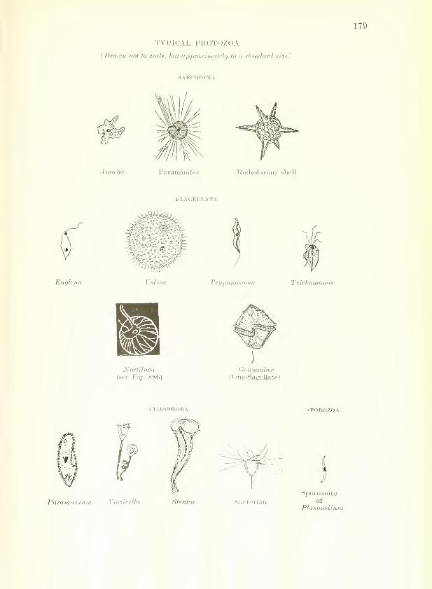
TYPICAL PROTOZOA
[Drawn not to scale, but approximately to a starulard size.']
SARCODINA
179
A mceba Foraminifei Radiolariaii shell
FLAGELT.ATA
Euglena Trypanosoma Trichomonas
Xoctiluca(see Fig. 886)
Gonyaulax(Dinoflagellate)
Paramcpcium Vorlicella
SPOROZOA
Suctorian
Sporozoiteof
Plasmodium

180 THE EYE IN EVOLUTION
Protozoa
PROTOZOA are the most primitive and simplest of animals, some of
which might with equal justification be considered as plants ; they are
essentially single-celled but sometimes form loose colonies by budding
or by cell-division, showing some degree of co-ordination but never
forming differentiated tissues. Of all animal types they are the most
numerous, being found in every continent, on land, in fresh water, in
the seas and impartially distributed as parasites within all animals
(including some of their own kind), among which not the least fre-
quented is Man ; their skeletons contribute largely to the oozes of the
seas and to the composition of the rocks of which the land-masses are
made.
Within the phylum four methods of activity are evident
—
amoeboid movement, flagellate and ciliary progression, and encystment
with spore-formation, characteristics under which the upwards of
15,000 species may be conveniently grouped into 4 classes (see p. 179).
SABCODiNA (or rhizopoda), Organisms which progress by sending out
finger-Hke pseudopodia into which the protoplasm of the cell pours itself. This
class comprises such types as the fresh-water Amoeba, the parasitic Entamceha
or the marine Foraminifera with chalky shells and Radiolaria with siliceous
shells which after death enter largely into the formation of the oozes of the bed
of the ocean.
FLAGELLATA (or mastigophora), Organisms which swim by the lashing
movements of one or a few whip-like flagella. The class comprises such types
as the common Euglena and colonial forms such as Volvox almost universal in
fresh-water ponds, the parasitic, disease-producing Trypanosomes and Tricho-
monads, Dinoflagellates including Noctiluca which gives luminescence to the seas,^
and Cystoflagellates, important constituents of the plankton of lakes and the
oceans.
ciliophora, organisms which progress by the coordinated movements of
many hair-like cilia. The class comprises the Ciliates (such common types as
the slipper-shaped Paramoscium, the bell-shaped Vorticella or the trumpet-
shaped Stentor) and the Suctorians which lose their cilia in adult life and in their
place develop tentacles used as suckers by which they capture and suck out the
bodies of their protozoan prey.
sporozoa, encysted organisms without a locomotive mechanism ; they are
parasitic on almost every species of animal and are spore-forming in habit
(Coccidia, Hsemosporidia, Plasmodium, etc.).
In view of the fact that the response to light in these primitive
forms is motorial, it is not surprising that receptors are not found in the
passive parasitic Sporozoa ; in the first three classes responses to light
are found among the freely-swimming active types, but as would be
expected in imicellular organisms, the receptor mechanisms are of the
most pr i itive nature. In the Sarcodina (Amceba) and some Ciliates
1 p. 738.

PARAZOA 181
{Paramoecmm) sensitivity to light is diffuse ; in other Cihates {Stentor)
it is localized to a part of the organism but without apparent specific
mechanism ; but even at this primitive unicellular stage an obvious
localization of function may be attained by the development of an
EYE-SPOT and the efficiency of the organelle increased, particularly in
the acquirement of a crude directional appreciation, by the provision
of pigment (as in Euglena) ^ or even of a primitive refractile mechanism
(as in some Dinoflagellates).^
Parazoa
The SPONGES (porifera), sessile marine animals which form living
thickets in the sea, represent a cul-de-sac in evolution between Protozoa
and Metazoa dating back almost to the beginning of geological records.
They are the simplest multicellular animals and show the beginnings
of the development of a "" body " composed of tissues ; but although
there is cellular differentiation there is little cellular co-ordination.
Being vegetative and sedentary in habit they have no need of sense-
organs as they lie moored to rocks or sea-weed. They possess no nerve
cells but the body cells retain properties of an irritability of a low
level ; and in the active larval forms of certain types (the simple
sponge, Leucosolenia) apolar light-sensitive cells of the most elementary
type have been described (Minchin, 1896).
Invertebrate Metazoa
In Metazoa—which includes all animal species apart from the
Protozoa and Parazoa—-the development of specialized cells and their
eventual co-ordination into distinct organs allow the evolution of
specific sensory activities as the term is generally understood. These
we shall now study, but it must be remembered that the Invertebrates
(or Non-chordates) do not form a homogeneous sub-kingdom but rather
represent an assemblage of unrelated groups of animals which have
little in common except the negative attribute of not being provided
with a dorsal nerve-cord with its supporting axis or with gill-slits.
From our restricted point of view there is the dramatic diff"erence that
(with few exceptions) the eye when present is developed from the skin,
while in Vertebrates it originates as an outgrowth of the brain.
cgelexterata
CCELENTERATES are simply formed animals with a body-cavity
(coelom) and digestive cavity(enteron) combined so that the body is formed
as a sac with an opening at one end only. They show the beginnings of
separate organs witli a consequent division of labour, and among them
Leucosolenia
Sycon
126. - p. 126.

182 THE EYE IN EVOLUTION
Hydra
Obelia medusoid
Obelia polyp
Sea-anemone
Comb jelly
(see Figs. 887-8)
visual structures of some complexity first make their appearance. Thephylum may be divided into two sub-phyla—the cnidaria, provided
with numerous stinging cells (kvlSy), a nettle), and acnidaria, wherein
these are replaced by adhesive cells. The first-sub-phylum is divided
into 3 classes :
HYDROZOA, comprising solitary polyps such as tlie fresh-water Hydra, the
marine Hydroids, branching colonial polyps of vegetative appearance liberating
freely-swimming Hydromedusie {Obelia, Sarsia, etc.) and some j^elagic colonial
forms.
SCYPHOZOA (" cup animals "), marine jellyfish, free-swimming medusae,
typically umbrella-shaped with the important organs situated on the margin or
under-surface.
ANTHOZOA (" flower animals "), sessile marine polyjas with no medusa-forms, such as sea-anemones, sea-fans, sea-pens and corals.
ACNIDARIA, comprising the Ctenophnra (comb-jellies or sea-gooseberries),
delicate freely-swimming globular organisms, pelagic in habit, gelatinous andtransparent, beautifully iridescent in the sunlight and often luminescent in the
dark,^ provided with comb-like rows of cilia.
The degree of elaboration of the visual receptors varies with the
motility of the organism, and many Coelenterates are sessile, plant-like
zoophytes ; eyes are therefore confined to the mobile medusae and these
are of a very primitive nature,^ while the sessile polyps of this phylum(hydroid forms and all Anthozoa) have no sense organs or, at most,
contact photoreceptors of the most elementary type.^
The Ctenophora are provided with a sense organ at the upper pole of the
organism consisting of a mass of limestone particles sup-
ported on cilia associated with sensory cells communicatingby nerve fibrils with the swimming-combs; this is considered
to act as a statocyst or balancing device and visual organs
are absent.
Among the Hydrozoa, some fresh-water forms
are sensitive to light but possess no detectable visual
organs ; a hydra, for example, will migrate towards
the lighted side of its container where, incidentally,
there are usually more food-organisms. In somefreely-swimming Hydromedusae, however, externally
visible light-sensitive organs provided with sensory
cells and pigment and sometimes a refringent appara-
tus may be found in the tentacular bulbs at the bases
of the tentacles (Fig. 162) ; these take the form of
' p. 739.
^'ir detailed information, see O. and R. Hertwig (1877)Ber^ S98), Linko (1900), v. Uexkull (1909), Lehmann (1923).
il6.
Fig. 162.— TheMedusoid FormOF BoviiAi.sriL-
LEA (Margel/.s).M, manubrium;
R, radial canal;
S, sense organ(after Allman).
Schewiakoff (1889),

CCELENTERATA 183
a primitive fiat eye, as in Turris or Lizzia (Fig. 96), or are invaginated
as an elementary cupulate eye, as in Sarsia (O. and R. Hertwig, 1878;
Jourdan, 1889). These organisms are light-sensitive and extirpation of
the tentacular bulbs with the ocelli completely abolishes the response
to light.
Among the jellyfish (Scyphozoa) more elaborate organs are seen.
In the common jellyfish. Aurelia aurita, which is found in great shoals
around the British coast, eight sense-organs (tentaculocysts) arise
as modifications of tentacles ; each, lying in the protection of a marginal
Figs. 163 and 1(54.
—
The Common Jellyj^sh, Acrelia aurita.
Fig. 163.—Side view of the jellyfish, showing thenumerous marginal tentacles hanging from theborder of the convex umbrella, and the dependentoral arms. The margin of the umbrella is brokenby 8 notches, the marginal lappets (L).
Fig. 164.—A marginalnotch, showing a ten-
taculocyst comprised oftwo olfactory pits, OP,a calcareous concre-tion, C, and an ocellus,
OC (modified fromLankester).
niche, has three types of sensory cells—red or black pigmented cells
responding to light, " olfactory " cells with a chemical appreciation,
and club -like cells containing calcareous concretions with a balancing
function (Figs. 163-4).
Exceptionally, as in the Cubomedusan, Charybdea, a large ocellus has been
reported with a cellular lens, a vitreous structure and a complex retina—anorgan structurally capable of some degree of visual imagery (Fig. 102) (Schewia-
koff, 1889 ; Berger, 1898). The biological value of this elaboration in a brainless
organism is somewhat speculative.
ECHINODERMATA
Among ECHiNODERMS (" spiny skinned "), a phylum characterized
by its radial symmetry, visual organs are rudimentary. This would

184 THE EYE IN EVOLUTION
Starfish
Brittle-star
be anticipated from the absence of centralization in the nervous
system, associated presumably with the absence of a head region,
and from the characteristically sluggish and sedentary habits of its
members. The phylum is divided into 5 extant classes :
ASTEROiDEA, or starfishes, motile but sluggish organisms.
OPHiUKOiDEA or brittle-stars, resembling starfishes but with the armssharply marked off from the central disc.
ECHINOIDEA or sea-urchins, living off rocky coasts, with a round pin-
cushion-like body covered with plates and provided with long sharp spines.
HOLOTHUEOiDEA Or sea-cucumbers, worm-like creatures with calcareous
plates, occurring in most seas.
CRiNOiDEA, sea-lilies or feather-stars, stalked forms anchored on rocks or
Free-swimmingfeather-star
Fig. 165.
—
The Iridophokes ix the .Sea-urchin, Djadema aatjllarum.
Section through a cluster of iridophores, I ; E, epidermal layer ; M,melanophores, underneath which lies the superficial nerve layer (fixed Bouin ;
stained Masson's argentaffine reaction ; counter-stained Mallory's triple
stain. (Approx. X 500) (N. Millott).
in mud usually at great depths, with appendages (cirri) and branching armsgrowing from a central cujd ; feather-stars become free-swimming in adult life.
In most Echinoderms the skin is diffusely sensitive to light,
particularly in sea-cucumbers (Crozier, 1914-15) ; in brittle-stars andfeather-stars there are no special sense organs ; in sea-cucumbers
sense organs are represented by statocysts sometimes present at the
bases of the tentacles, and tactile processes sometimes present on the
dorsal surface of some of the creeping forms ;
" eyes " are present only
in starfishes.
The diffuse dermatoptic sense shows interesting variations. Thus in somestarfishes the body-surface is said to be sensitive to changes in intensity, thepodia and skin gills to steady light ; in some sea-cucumbers {Synapta) the wholesn; • is sensitive to both, while in others {Holothuria surinamensis) the rim ofthe •'' ica is particularly sensitive, the posterior end and tentacles less so andthe
) ia least. In the sea-urchin, Paracentrotus lividus, the apical poles are

ECHINODERMATA 185
Fig. 166.
—
Diagram of a Very Young Asteroid
At the base of the 5 terminal tentacles is an optic cushion with a bright redocellus, Oc, connected by an epidermal radial nerve which runs to the centralnerve pentagon surrounding the mouth (after Lang). Compare Plate I.
said to show the most rapid reactions (Scheer, 1956). In the Echinoid, Diadema,the distribution of sensitivity corresponds to the distribution of the nerveelements and it may be that these are directly stimulated bj- light as we haveseen to occur in the apolar light-sensitive cells of w-orms (Millott, 1954). On theother hand, photosensitive pigments may be present in minute quantities, butthere is yet no evidence as to their nature.
Many sea-urchins have the same primitive sensitivity associated particu-larly with their pigmented spicules which move on the stimulus of light (v.
Uexkiill, 1900), and in some types characteristic iridescent bodies associated withmelanin pigment lie near the spines {Diadema antillarutti) (P. and F. Sarasin,1887 ; Dahlgren, 1916; Millott, 1950-54). These represent clusters of regularlyarranged plates resembling iridoi^hores ^ in their arrangement, which presumablyact by reflecting the light onto the sensitive
spines (Millott, 1953) (Fig. 165). It is of historical
interest that the Sarasins (1887), in a muchquoted paper, described similar structures in
Diadema setosum, an allied species inhabiting
the Indian Ocean, as being "eyes" composedof several hundred polygonal corneal facets, a
vitreous-like jelly and a '' retina", but without
nerve fibres.
Cuf
In STARFISHES (Asteroids such as the
common five-rayed Asterias), although the
skin is often diffusely light-sensitive, on
the tip of each of the five arms a visual
1 Compare iridocytes, p. 89.
Fig. 167.—The optic cushion ofthe Asteroid
Cut, cuticle ; CT, connectivetissue ; Ep, epithelium ; NN,nerve-net ; P, pigment cells.
Sea-cucumber,Holothuria
The sea-urchin,
Diadema

186 THE EYE IN EVOLUTION
organ is formed as a modified tube-foot lying on a slight elevation (the
" optic cushion ") on the dorsal surface of the terminal ossicle (Fig. 166).
The organ is bright red due to the presence of ^-carotene and esterified
astaxanthin and consists of an aggregation of several cupulate ocelli of
the simplest type covered by cuticle and lined by sensory and pig-
mented cells (Plate I ; Fig. 167) (PfefiFer, 1901) ; a central lenticular
body may serve to concentrate light upon the receptive elements (van
Weel, 1935 ; Smith, 1937). The optical function of this organ in
Asterias has been convincingly demonstrated by Hartline and his co-
workers (1952) who recorded the electric impulses following stimulation
by light. The terminal tube-foot appears to be olfactory in function.
Berger. J. comp. Neurol. Psychol., 8, 223 Philos. Trans. B, 238, 187 (1954).
(1898). Minchin. Proc. roy. Soc. B, 60, 42 (1896).
Crozier. ^mer. J. P/i^sioZ., 36, 8 (1914). Pfeffer. Zool. Jb., Abt. Anat., 14, 523Zool. Jb., Abt. Zool. Physiol., 35, 233 (1901).
(1915). Prosser. J. cell. comp. Physiol., 4, 363Dahlgren. J. Franklin Inst., 181, 377 (1934).
(1916). J. comp. iVewroZ., 59, 61 (1934).
Hartline, Wagner and MacNichol. Cold Sarasin, P. and F. Ergebn. naturwiss.
Spr. Harb. Symp. Quant. Biol., 17, 125 Forsch.Ceylon,V^\esh&den, 1, 1 (1887).
(1952). Scheer. Naturwissenschaften, 43, 501Hertwig, O. and R. Jena. Z. Naturwiss., (1956).
11, 355 (1877). Schewiakoff. Morphol. Jb., 15, 21 (1889).
Das Nervensysteyn u. die Sinnesorgane Smith. Philos. Trans. B., 227, HI (1937).
d. Medusen, Leipzig (1878). von Uexkiill. Z. Biol., 40, 447 (1900).
Jourdan. Les sens chez les animaux Umwelt u. Innenwelt d. Tiere, Berlin
inferieurs, Paris (1889). (1909).
Lehmann. Zool. Jb., Abt. Zool. Physiol., Viaud and Medioni. C. R. Soc. Biol.
39, 321 (1923). (Paris), 143, 1221 (1949).
Linko. Acad. Imp. Sci. St. Petersburg, van Weel. Arch, neerl. Zool., 1, 347Mem. Ser. 8, 10 (1900). (1935).
Millott. Biol. Bull., 99, 'S29 (1950). Wellington. Nature (Lond.), 172, 1177Nature (Lond.), 170, 325 (1952) ; 171, (1953).
973 (1953).
WORMS
The large group of " worms " shows a variety of visual organs as
pleomorphic as the multitude of forms which constitute this loose
grouping of animals, showing every variation from a unicellular eye to
a relatively complex organ. In some cases the surface of the whole bodyseems to be sensitive to light and it has not been possible to identify
specific sensory cells ; in most cases, however, specialized sensory
structures occur, for the elucidation of which we are largely indebted
to the classical work of Richard Hesse (1899-1908). Their presence,
their number, and the degree of their differentiation vary with the
animal's mode of life. This is the lowest group in the animal kingdomto show l>ilateral symmetry and the sense organs share in this general
scheme distribution ; moreover, these organs are usually concen-
trated 1 rds the head-end of the animal where they are of greatest
biologic; lue.

PLATE I
The Light-sensitive Apparatus of the Starfish
^^imf>^
Fitt. 1.—Maiih<istcrliis (/Idruilis, showing th(> ]>nsiti()ii of the eye-spot, e.s.
one of which is jjresent at the tip of each of the five arms.
Fig. 2.—The excised eye-spot (optic cushion) showing the
o])tic cups, o.c. They have a striking red colom- due to
L'-carotene and esterified astaxanthin; it is to be noted
that some of the colour of the body-wall, which is also
light-sensitive, is due to the same ])igments (X. ]\Iillott,
Endeavour, 1957).
S.O.—VOL.1 [To face 2>- 186.


WORMS 187
These photoreceptors are of the most varied types and many species
possess eyes of more than one variety. The neuro-sensory cells may be
either apolar in type provided with an internal optic organelle, or
bipolar, provided with a ciliated or striated border ^: they may occur
as single cells or in groups forming an eye of either the subepithelial or
epithelial variety, in which case it may show a flat, cupulate or vesicular
arrangement. Pigment is a constant association, situated within the
sensory cells or in special supporting cells. If a refractive medium is
present it may be formed either from the retinal or the epidermal cells,
while light-refracting structures are usually cuticular in origin. As a
general rule their function can only be the primitive ability to detect
light, but the visual organs of some types, such as some polychaete
worms, are structurally capable of some degree of localization andresolution (a directional eye) and perhaps even of visual imagery.
UNSEGMEXTED WORMS
The unsegmented worms may be divided into three phyla—flat-
worms, ribbon-worms, and thread-worms.
1. PLATYHELMINTHES or FLAT-WORMS Constitute a gi(jup of very simplyorganized creatures the members of which show the progressive degeneration
associated with parasitism. It is divided into 3 main classes :
(a) TURBELLARiANS, freely-Swimming leaf-shaped aquatic creatures of
carniv^orous habit, frequenting brackish or salt water or moist places on land;
the name is derived from the turbulence caused in the water by the beating of
their cilia when they swim. They are classified accoi'ding to the arrangementof the gut—the minute marine Actf-la (without intestine), the small salt andfresh-water Rhabdocojla (rod-shaped intestine), the (mainly) marine Alloeocoela
(irregular intestine), the small, flat, elongated Tricladida (3-branched intestine)
found in fresh or salt water or on land (including the Planaria), and the large,
leaf-like, marine Polycladida (many-branched intestine).
(6) TREMATODES or FLUKES, leaf-like parasites, external or internal, foundon or in all types of \'ertebrates, clinging to their hosts with suckers. Examplesare the liver-fluke, Fasciola hepatica, which infests the livers of sheep andcattle, or the Schistosoma Juematobia which causes bilharziasis.
(c) CESTODES or TAPE-WORMS, endoparasites, frequenting the alimentary
canal of Vertebrates, including domestic animals and man, such as Taenia eckino-
coccus, or T. solium.
2. NEMERTiNES or RIBBON-WORMS, ribbon- or thread-like in shape, often
vividly multi-coloured, varying in size from under an inch to enormous lengths
(25 metres in Linens) and provided with cilia and a remarkable retractile pro-
boscis forming a tactile organ used to capture prey. Most are marine in habitat,
creeping in the mud and under stones ; a few are found in fresh-water (Prostoma);
some are terrestrial (Geonemertes) ; and a few live commensally with bivalves
or ascidians.
3. NEMATODES, ROUND- Or THREAD-WORMS, Cylindrical in shape and often
minute, which teem in the soil or in water and are often endojjarasitic in plants
and animals (Ascaris, Trichinella, Ankylostoma, Filaria, etc.) ; but free-living
forms occur at any rate in part of the life-cycle.
1 p. 127.
Polyclad,Leptoplana
Schistosoma
Teen in
echinococcus

THE EYE IN EVOLUTION
The PLATYHELMiNTHES have sense organs only of the most
rudimentary type—if any. The freely-hving turbellarians (Plana-
rians, etc.) are the most adequately equipped with eyes (Figs. 168 to
170). These may be merely two or four in number, in which case they
lie on the dorsal aspect of the head-end associated with the tentacles
near the cerebral ganglion, as in the fresh-water Rhabdocoela ; but
others such as the marine Polycladida may possess several hundred.
A common arrangement, well seen in the Tricladida, is that these
multiple ocelli are distributed around the circumference of the bodyconcentrated particularly at the anterior margin (Figs. 168 and 170)
(Busch. 1851 ; Hyman, 1938-51). The eyes are always very elemen-
FiGS. 168 TO 170.
—
The Eyes of Turbellarian Worms.
\k s- „M
Fig. 168.—A land pla-
narian, Geoplana mexi-cana.
There is a row of eyesalong the entire marginof the animal (after
Hyman).
Fig. 169.—The eyes ofthe pelagic Rhabdo-coele, Alaurina proli-
fera.
S, papillated snout;
M, mouth ; E, paired eye(after Busch).
Fig. 170.—The eyes ofthe fresh-water pla-
narian, Polycelis coro-
nata. They are concen-trated at the head-end(after Hyman).
Dendrocoelum
tary, and lacking a dioptric apparatus are capable only of light
perception although a directional appreciation may be evident
(Taliaferro, 1920). The number of visual cells is said to vary between
1 and 200 (Hesse, 1896 ; Schmidt, 1902). Occasionally, as in
Dendroccehim, they are of the flat epithelial type (Fig. 95). Usually
they are of the subepithelial type, appearing as minute pigmented
spots about 0"1 mm. in diameter and consisting of a pigmented goblet
enclosing the sensory cells (Figs. 91 and 92). In these the sensory
ce-lr- .T,re of the bipolar type with a striated margin facing away from
tlK direction of light to form an inverted retina. When the eyes are
nea ''hi^ cerebral ganglion the sensory fibres enter the latter directly;
othc -e they enter the peripheral nerve-net.

WORMS 189
In some Rhabdocoela {Stenostonuni) curious hemispherical bodies consisting
of refringent granules lying underneath a bowl-shaped mass have been credited
with a photosensitive function ; there is no good evidence, however, for this
assumption.
Eyes are lacking in the cave-dwelling planarians (Kenkiidae) and in endo-
parasitic Rhabdocoela.^
TREMATODES may possess simple ocelli in the larval stage (as in
the liver-fluke. Fasciola hepatica), but the adults, leading an essentially
parasitic existence, rarely possess sense organs. If they are present
they are of the simjDlest type, usually consisting of a single cell with a
striated border invested by a cup of pigment (Hesse, 1897 ; Andre,
1910 ; Faust, 1918) ; a typical example is seen in the luiicellular eye
Fasciola hepatica
Figs. 171 and 172.
—
The Eyes of Xemertine Worms.
E
Fig. 171.
—
Lineus ruber.
E. eyes (after Hyman)Fig. 172.—The head of Ampkiporus
angulatus. E, eyes (after Hyman).
of Tristomum impiUosum. a marine Trematode jiarasitic on fishes
(Fig. 87).
CESTODES, in keeping with their endoparasitic life, are without sense
organs.
-
Among the nemertines, most of which are freely-living and marine
in habitat, rudimentary eyes of the same subepithelial type as occur in
flat-worms are general and occasionally are very numerous (Figs. 17 1-72).
They are always limited to the anterior end of the animal. Somespecies possess two eyes, others four or six on the prostomium ; others
up to 250 eyes [Amphiporus) arranged in clusters or rows, while the
number may vary in different individuals of the same species. The
eyes are nearly always subepithelial in type consisting of bipolar cells
terminating in a brush border enclosed within a pigment cup of
epithehum (Hilton, 1921) (Fig. 93). The eyes of the terrestrial genus,
1 pp. 724, 733. "^ p. 734.
Amphiporus

190 THE EYE IN EVOLUTION
Nematode,Ascaris
Arenirola
Hcemadipsa
Luinbricus
Geonemertes, differ from the usual type. In these the pigmented
epithehum forms a complete circle within which is a mass of refractile
material ; the nuclei of the sensory cells are arranged outside the circle
of pigment and their distal terminations pass through it into the
central refractile mass (Schroder, 1918).
In the NEMATODES, the majority of which are endoparasitic, sense organs
are Hmited to papillae on the lips ; in the free-living sexual state, however,
rudimentary eyes may exist, consisting of a lens-like cuticular body resting on a
cup of pigmented cells (Steiner, 1916 ; Hilton, 1921 ; Schulz, 1931).
SEGMENTED WOBMS (ANNELIDS)
The segmented worms exhibit much diversity in habit and
structure but their essential characteristics are segmentation of the
body with paired appendages on each segment and a closed vascular
system. Annelids are found both in marine and fresh water and on
land, and in the entire phylum more than 6,500 species are known.
These are divided into 4 classes, the first two of which are provided with
chitinous bristles or setae for locomotion.
1. OLiGOCH^TES (with few setne), hermaphroditic creatures, essentially
terrestrial in habit, typified in the common earthworm, Lumhricus terrestris, or
the tiny aquatic mud-worms living in brooks or between tide-marks.
2. polycHjEtes (with many setae), essentially marine in habit ; in them
the sexes are separate. Two types exist, distinguished by their habits. The
more active forms (errantia) are typified in the common lob-worm, Arenicola
marina, found burrowing in sandy beaches, or the freely-swimming types, such
as the rag-worm. Nereis. The sedentary forms (sedentaria) are tubicolous in
habit leading a sluggish life within tubes, limy, sandy or gelatinous ; as an
adaptive characteristic the tentacles, gills and sensory organs are aggregated in
the anterior part of the woi-m which protrudes from the tube.
3. ARCHiANNELiDS Comprise a small and anomalous class of simple marine
worms with juvenile characteristics and without seta?, freely swimming or
burrowing in sand and gravel.
4. HiRUDiNES or LEECHES form a highly specialized and much modified
class, most of which live in fresh water in ponds or sluggish streams although
a few are marine and others (the wiry land-leeches of the Far Eastern jungles,
Hcemadi])sa) are terrestrial, living in inoist places. In habit they are greedily
suctorial, sucking the blood of fishes, amphibians or other victims.
Eyes are usually lacking in the oligoch^tes ; of those possessing
visual organs, the most typical example is the earthworm, Lumhricus
terrestris. Its unicellular light-sensitive organs distributed in the
epithelium and aggregated around subepithelial nerves have already
])een fully described ^ (Figs. 86, 88). These visual elements are situated
\N iiere they are of the greatest biological value, being concentrated at
til; vo extremities, particularly the anterior.
131.

WORMS 191
Thvis W. N. Hess (1925) found that in the prostomium there were some440 light-sensitive cells in the epidermis and 700 sitviated in nearby nerve
enlargements, while in subsequent segments they were much fewer. Their rela-
tive numbers in corresponding sinall areas (200 x SOOti.) on the dorsal surface of
the animal are as follows—in the prostomium, 18 ; 1st seginent, 10 ; 2nd segment,
5 ; 3rd segment, 3 ; 40th segment, ; antepenultimate segment, 1;penultimate
segment, 1 ; last segment, 4. The segiuental photic sensitivity varies directly
with the number of receptors, and the distribution of light-sensitive elements
conforms with the habits of the earthworm.
^
Among POLYCH.ETE woEMS. the burrowing lob-worm, Are^iicola
marina, is not provided with visual organs although the prostomial
Fig. 173.
—
The Head of Nereis, Showing the Four Eyes.
e, eyes; j, jaw ; p, palp ; pe, peristomium (first two segments fused)
;
ph, pharynx; pp, first ordinaiy paraijodium ;\ pr, prostomium; t, accessory
teeth ; tc, tentacular cirri ; te, tentacle. (From Borradaile's Manual of
Elementary Zoology ; Oxford University Press.)
lobes are diffusely sensory. In contrast with the burrowing type,
however, the freely-swimming marine polychaetes show a much richer
development (Fig. 173). Of these, Nereis is a typical example. This
worm has four prominent eyes situated on the prostomium, each of
the cupulate type with a cuticle externally and a retina internally
formed of well-developed sensory cells with rod-like receptor endings
(Fig. 101). Other forms, such as Polyoplithalmus, have in addition to
the prostomial eyes similar pairs of subepithelial organs in manysegments of the body ; such eyes ^ are formed sometimes on each
segment {Myxicola (esfhetica ; Eunice), and occasionally on the anal
segment {Fabricia).
A much more complex type of e}e of the vesicular type is found
1 p. 572.^ These organs, usually considered to be " eyes " are said by some to be liglit-
producing (p. 736) (Benham, 1896).
Nereis

192 THE EYE IN EVOLUTION
Branchiomma
in certain pelagic polychsetes such as Alciopa and Eupolyodonfes, the
intimate structure of which has already been described. ^ These worms
have two eyes, sometimes facing forwards {Eupolyodontes), sometimes
diverging widely (Alciopa) (Fig. 174). Each organ is provided with
an elaborate retina, a lens, an accom-
modative mechanism and extra-ocular
muscles suggesting the potentiality for
binocular vision, an equipment which
seems capable of considerable visual
powers approximating those of the
Cephalopods.2 Little, however, is knownof the habits of these worms.
In the sedentary tubicolous poly-
chsetes (Potamilla, Branchiomma, Dasy-
chone, etc.) the ocelli are frequently
grouped in masses on the branchial fila-
ments to form a composite simple eye
of great complexity (Brunotte, 1888;
Andrews, 1891 ; Hesse, 1896) (Figs. 175 and 176) ; Vermilia infundi-
bulum has at least 220 ocelli on the external aspect of each branchium,
a total of some 11,000 eyes (Parker and Haswell, 1940). These
creatures live within their tubes from out of which extend the branchial
plumes bearing the filaments on each of which there is one or more
such eyes (Figs. 128, 129). The curious thing, however, is that in
Figs. 175 and 176.
—
The Complex Eyes of Tubicolous Polych^tes.
e
Fig. 174.—The Anterior End ofTHE Polych^te Wokm, Alciopa.
Showing the two large eyes (after
Greeff).
Fig. 175. Fig. 176.
The secondary filaments are seen issuing horizontally from the central
axis of the branchial filament. Fig. 175, Branchiomma, showing the single
complex eye, e, near the termination of the central axis. Fig. 176, Dasychone,
showing the row of complex eyes (2 of which are marked e) running up and';osvn the central axis (after Benham, Camh. Nat. Hist.).
143. Fig. 112.

WORMS 193
Branchiomma, at any rate, these structures do not seem to be essential
for the most characteristic responses of the worm to changes in tlie
intensity of hght (Millott, 1957) ; the position is therefore somewhatanomalous.
In the simple marine archiannelids, eyes of a similar
type are found. In Dinophilus, for example, a minute wormfound among alga^, two kidney-shaped pigmented eyes are
found on the prostomium (Hilton, 1924) (Fig. 177).
LEECHES (hirudinea) may be provided with
visual organs of a simple type varying in numberfrom 2 to 10 (Hesse, 1897 ; Herter, 1932) ; they are
incajDable of optical imagery although highly light-
sensitive, but in some species may be absent. Theyare found near the anterior extremity of the bodyand vary considerably in their morphology, but the
visual cells are always of the spherical apolar type
with a central optic organelle (Figs. 178-9).Fig. 177.—The
rchiannelid,DlXOPUlLUS.
Showing thepaired ocelli, Oc(after Sheldon-Harmer).
In Branchellion these organs are unicellular; in Piscicola
they consist of 12 cells arranged in a row surrounded bypigment. In Hcemopis both unicellular and multicellular
ocelli are found (Fig. 179). In the common medicinal leech,
Hirudo medictnalis, there are segmental papilhe with asensory function on the middle ring of each of the 26 segments. Although all
the sense organs are serially homologous the pairs on the dorsal surface of the
first five segments are purely visual, constituting ten " eyes " (Fig. 90), provided
with a rich nerve supply to the cerebral ganglia. At the other extremity the
Branchellion
Hirudo
Figs. 178 and 179.
—
The Eyes of Leeches.
Fig. 178.—The head end of the medicinal leech,
Hirudo niedicinalis.
The dorsal aspect. The body is divided into
segments, each of which contains 5 rings
(annulae). In the middle ring of each segmentthe segmental papillte have a sensory function.
The first 7 (and the last 3) segments have less
than the normal number of rings, and the first 5
show two paired eyes as larger black spots.
El to Eg, serially homologous with the sensory
papillae (see Figs. 89-90) (after Parker andHaswell).
S.O. VOL. I.

194 THE EYE IN EVOLUTION
organs are probably purely tactile, and between these two regions the sense organs
are compound since they contain both visual and tactile cells (Fig. 89).^
Chsetognath,Sagitta
Subsidiary Invertebrate Phyla
For convenience, four small and subsidiary phyla of the Inverte-
brates are most usefully considered here.
CH^TOGNATHA (" bristle-jawed ") or arrow-worms, delicate, translucent
torpedo-shaped creatures comprising some 30 species which swim in incredible
numbers in great shoals among the plankton of all seas, have well-developed
eyes. Spadella, for example, or Sagitta, has two composite simple eyes at the
anterior extremity of its body, formed by the union of 5 ocelli, the structure of
which has already been described (Fig. 132) ; although presumably tripartite,
the nerve fibre from each eye is gathered into a single optic nerve trunk.
Figs. 180 and 181.
—
The Eyes of Rotifera,
L
Fig. 180.—The cerebral eye.
Section through the cerebral gan-glion of Synchceta, showing two cere-
bral eyes, E (after Peters).
Fig. 181.—The frontal
eye.
The eye of Rhinoglenawith pigment spot, P, andrefractile lens, L (after
Stossberg).
Rotifer
Bryozoa
BOTiFERA (" wheel-bearers "), the beautiful minute wheel-animalcules,
sometimes of fantastic shape, which swim so abvindantly with the aid of a crown
of cilia like revolving wheels in fresh water, damp moss or the sea all the
world over, are usually highly light-sensitive. There is a generalized dermatoptic
sense which evokes a positive phototaxis, but exact orientation is determined
by the eyes and varies with their morphological development (Viaud, 1938-43).
Frequently there is a single or paired cerebral eye embedded in the dorsal
nerve ganglion {Synchceta) (Fig. 180). In other species, sometimes in addition
to the cerebral eyes, there is one or two frontal or lateral eyes (Fig. 181). Thecerebral eye consists of a single cell resembling a brain cell ; the lateral or
frontal eyes are epidermal cells inside which is a lens-like body associated with
a mass of red pigment (Peters, 1931 ; Stossberg, 1932). Branchionus, one of
the commonest members of this class which inhabits ponds and ditches in
abundance, has a simple unpaired eye surrounded by red pigment and associated
with tufts of sensory hairs, situated where the cerebral ganglion comes into
contact with the body-wall just behind the wheel of cilia at the anterior endof the animal.
POLYZOA (bryozoa), very ancient plant-like organisms which include fresh-
W:ter and marine forms (sea-mats, etc.) are sessile colonial corallines or " mossari Is " which grow in tufts on the shores or in pools all over the world encrust-
1 p. 133.
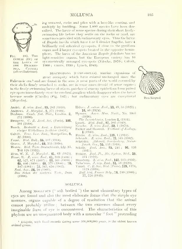
MOLLUSCA 195
Fig. 182.—TheOcelli (Oc) ofthe l.\rva ofTHE BrACHIO-POD, ClSTELLA(afterGladstone).
ing seaweed, rocks and piles with a lace-like coating, andmultiply by budding. Some 1,800 species have been des-
cribed. The larva? of some species during their short freely-
swimming life before they settle on the rocks or mud, are
sometimes provided with rudimentary eyes. Thus the larvae
of Bugula turrila which have 4 or 5 slender flagellae, have 4
brilliantly red spherical eye-spots, 2 close to the pyriform
organ and 2 larger eye-spots located in the opposite hemis-
phere. The larva of the American Bugula flahellata has no
light-sensitive organs, but the European variety has 10
symmetrically arranged eye-spots (Xit.sche, 1870 ; Calvet,
1900 ; Grave, 1930 ; Lynch, 1949).
BRACHIOPODA (lamp-shells), marine organisms of
great anticiuitj^ which have existed unchanged since the
Palaeozoic era ^ and are found in the seas in most parts of the world covered bytheir shells firmly attached to rocks, are in some cases devoid of sense organs ;
in the freely-swimming larvte of others, patches of sensory epithelium form paired
eye-spots immediately over the cerebral ganglion which disappear when the larvie
become sessile (Cistella) (Fig. 182) ; but rudimentary eyes are exceptional
{Megerlia).
Andre. Z. wiss. Zool., 95, 203 (1910).
Andrews. J. MorphoL, 5, 271 (1891).
Benham. Camb. Nat. Hist., London, 2,
272 (1896).
Brunette. C. R. Acad. Sci. (Paris), 106,301 (1888).
Busch. Beobacht i'l Anat. u Entwicklungeiniger Wirbellosen Seethiere (18.51).
Calvet. Trav. Inst. Zool., Montpellier, 8,
22 (1900).
Faust. Biol. Bull, 35, 117 (1918).
Grave. J. MorphoL, 49, 3.55 (1930).
Herter. Biol. Tiere Deutsclilands, Lfg. 35,
Teil 12b (1932).
Hess, W. N. J. Morplwl., 41, 63 (192.5).
Hesse, R. Z. wiss. Zool., 61, 393 (1896) ;
62, 527, 671 (1897) ; 63, 361 (1898) ;
65, 446 (1899) ; 68, 379 (1900) ; 70,
347 (1901) ; 72, 565. 656 (1902).
Zool. Anz., 24, 30 (1901).
Das Sehen der niederen Tiere, Jena(1908).
Hilton. J. entom. Zool., 13, 49, 55 (1921) ;
16, 89 (1924).
Hj'man. Ainer. Mus. Xovit., No. 1005
(1938).
The Invertebrates, London 2, (1951).
Lynch. Biol. Bull., 97, 302 (1949).
Nitsche. Z. wiss. Zool., 20, 1 (1870).
Parker and Haswell. Te.vtbook of Zoology,
1 (1940).
Peters. Z. wiss. Zool., 139, 1 (1931).
Schmidt. Z. wiss. Zool., 72, 545 (1902).
Schroder. Abliandl. Senckenberg. Xatur-forsch. Ges., 35, 153 (1918).
Schulz. Zool. Anz., 95, 241 ; 96, 159(1931).
Steiner. Zool. Jb., Abt. System. Biol., 39,511 (1916).
Stossberg. Z. wiss. Zool.. 142, 313 (1932).
Taliaferro. J. e.vp. Zool., 31, 59 (1920).
Viaud. C. R. Soc. Biol. (Paris), 129, 1174,
1178 (1938).
Bidl. biol. France Belg., 74, 249 (1940) ;
77, 224 (1943).
Brachiopod
MOLLUSCA
Among MOLLUSCS ('' soft bodied ") the most elementary types of
eyes are found and also the most elaborate forms that the simple eye
assimies, organs capable of a degree of resolution that the animal
cannot probably utilize ; between the two extremes almost every
imaginable form of eye is encountered. The characteristics of this
phylum are an unsegmented body with a muscular " foot " protruding
1 Lingula, with fossil records dating some 500,000,000 years, is the oldest knownanimal genus.

196 THE EYE IN EVOLUTION
Solenogastre
Nudibranch
Pulmonate,Limnoea
Nautilus
from the ventral surface serving for locomotion, a dorsal or lateral
fold of the body-wall to form a mantle or pallium within which lie the
gills, and frequently a shell. As a general rule, two cephalic eyes
subserve the visual function, but these may be replaced by more
rudimentary organs in the dorsal region or around the margin of the
mantle or at the end of the tentacles or the siphons. Occasionally
eyes are lacking, in which case the skin has usually some sensitivity
to light.
The large phylum of Molluscs is conveniently divided into six classes ;
three are relatively unimportant, sluggish in habit, and live in the mud or sand
of the sea-bottom—the shelled placophorans and scaphopods, and the worm-
like soLENOGASTRES. The remaining three classes contain an enormous number
of species of great variety—Gastropods, Lamellibranchs (Bivalves) and
Cephalopods.
The GASTROPODS (" belly-footed ") constitute a very varied group comprising
some 40,000 species and include three main classes :
(a) OPiSTHOBRANCHS : sea-hares, Pteropods (transparent marine plankton
forms), and the brilliantly coloured Nudibranchs or sea-slugs which have no
shell ;
(h) PROSOBRANCHS, an enormous and varied grouj^ including sea-snails,
whelks, limpets, Heteropods, etc. ;
(c) PULMONATES. The abundant and universally distributed fresh-water
and terrestrial snails and slugs.
The BIVALVES : shell -fish such as cockles, mvxssels, clams, scallops and
oysters which live within a rigid hinged shell often at the bottom of the sea.
They comprise some 11,000 species.
The remaining class, the cephalopods, are the
most interesting ; they are usually active, moving byjet propulsion with a jet of water expelled from the
siphon. Two orders are recognized : the Tetra-
branchiates, with two pairs of gills, represented by a
single living species, the Pearly Nautilus of the South
Pacific, and the Dibranchiates, with a single pair of
gills and remarkably well-developed eyes (cuttlefish,
sqviid, octojxis).
In the most primitive type of molluscs, the
PLACOPHORANS, cycs may be lacking althovigh some of
their sensory organs may be sensitive to light (Plate,
1899; Nowikoff, 1907). Some ofthem possess a multi-
tude of minute ocelli ; Corephiutn, for example, mayhave as many as 8,500. The most interesting in this
class are the Chitons (" coats -of-mail") ; these possess
cephalic eyes in the larval stage which, however, dis-
appear as the advilt becomes clothed by its eight-
plated dorsal shell, thus rendering them useless. In
the- place numerous innervated papillis appear con-
tai: i.ij sensory organs {aesthetes) which perforate the
shei. :^)earing in rows as minute black dots (" shell-
eyes loseley, 1884) (Fig. 183). The larger of these
Fig. 183.
—
The Mollusc,Chitox.
The sense organs, aes-
thetes, perforate the shell,
appearing as minute black
dots ; the larger of these
contain an ocellus (Thom-son's Zoology, JamesRitchie ; Oxford Univer-
sity Press).

MOLLUSCA 197
are light -sensitive, containing an ocellus composed ofa deep retinal cup surroundedby pigment lying beneath a lens, the whole organ being covered by a cornea. It
is to be remembered, however, that Crozier (1920) could find merely a generalphotosensitivity in Chiton, most pronounced where ocelli are lacking. AmongSOLENOGASTKES, these organs are replaced by simple epithelial papillte. In theSCAPHOPODA (" tusk-shells "), a small class of molluscs which burrow in the sand{Dentalium, elephant's-tooth shell, etc.) the sensory organs are represented onlyby statocysts.
Most members of the large class of gastropods, the eyes of whichwere studied at an early date by J. Miiller (1831), are provided withocelh of a relatively primitive kind often associated with the tentacles.
In the extremely passive limpet, Patella , the eyes at the base of thetentacles are very elementary, being merely lepresented by simple
Dentalium
Fig. 185.
—
The Common Whelk or Buckie,buccisum vsdatum.
Note the two simple eyes (e) at the base ofthe tentacles, s, respiratory siphon ; o, oper-
culum; /, foot (Thomson's Zoology, James
Ritchie ; Oxford University Press).
Fig. 184.
—
The Limpet,Patella vulgata
Ventral surface. Note thesimple eyes (appearing asblack dots) at the base of tlie
2 tentacles. The star-shapedmedian structure is themouth (Thomson's Zoology,
James Ritchie ; Oxford Uni-versity Press).
cupiilate depressions of sensory and pigmented cells (Figs. 97 and 184).
More usually, however, the eyes are vesicular in type. These are typified
in the two simple vesicular eyes of the grey slug, Limax, or the snail,
Helix (Fig. 110), perched on the tips of the two longer (and jjosterior)
tentacles (" horns '") and innervated from the cerebral ganglion
(Galati-Mosella, 1915) ; on exjDosure to light the tentacle is capable of
retraction like the finger of a glove so that the eye can be drawn within
it (Figs. 186 to 188). The common whelk, Bticcinum, has eyes of a
somewhat similar vesicular type situated near the base of the tentacles
(Fig. 185), as also has Murex.
The most elaborate eye of this type, however, is seen in the spider-shell,
Pterocera lambis, a gastropod found in quantity on tropical reefs. According to
Shell of Murex

198 THE EYE IN EVOLUTION
Figs. 186 to 188.
—
The Common Garden Snail, Helix aspebsa.
Fig. 186.—The two eyes arc situated on the tijD of each of the long posteriorhorns.
Fig. 187—The eye at the tip of the ex-panded horn.
Shell of Pterocera
Fig. 188.—The eye (E) retracted intothe horn. The horn invaginates like
the inturned finger of a glove ; theobliquity of this section gives theappearance of a double cavity(Norman Ashton).
Prince (1955), the two vesicular eyes, which have an elaborate neural structure,
^
are mounted on the tip of stalks (ommatophores) which also carry an olfactory
tentacle and a sensory node (Fig. 189). These, supplied with muscles arrangedround a central sinus, are retractile partly by muscular activity and partly byfluid engorgement by heemolymph. Retraction can be slow and voluntary or
r;i!)id and reflex in response to stimuli such as touch, odour or the cutting off ofI':--"' i : the reaction is thus the opposite of that seen in the snail. It appearsals -hat a certain amount of convergence upon an object is possible.
1 p. 142.
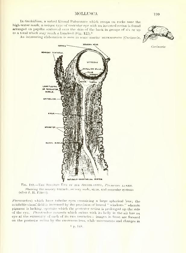
MOLLUSCA 199
In Onchidium, a naked littoral Pulmonate which creeps on rocks near thehigh-water mark, a unique type of vesicular eye with an inverted retina is foundarranged on papillae scattered over the skin of the back in groups of six or upto a total which inay reach a hundred (Fig. 122).^
An interesting elaboration is seen in some marine heteropods {Carinaria,
tCMBORY NOOC
NAOIAL MU«CL£
Carinaria
RITIOULO-CHOOTHELIAL SYCTIM.
Fig. 189.
—
The Stalked Eye of the Spider-shell, Pterocera lambis.
Showing the sensory tentacle, sensory node, sinus, and muscular systems(after J. H. Prince).
Pterotrachea) which ha\e tubular eyes containing a large spherical lens; theavailable visual field is increased by the provision of lateral " windows " whereinpigment is lacking, opposite which the posterior retina is prolonged up the sideof the eye. Pterotracliea coronata which swims with its belly in the air has aneye at the extremity of each of its two tentacles ; images in front are focusedon the posterior retina by the enormovis lens, while movements and changes in
1 p. 148.

200 THE EYE IN EVOLUTION
Avicula
Mi/tilus
Pholas
Cardium
illumination above and below are probably appreciated through the dorsal andventral " windows " (Hesse, 1908 ; v. Hess and Gerwerzhagen, 1914). Suchfenestrated eyes are also seen in abyssal fishes.^
LAMELLiBRANCHS or BIVALVES have ail Undeveloped head-region,
and the two lobes of the mantle which secrete the two valves of the
shell are frequently united posteriorly to form exhalant and inhalant
siphons. Anterior eyes are therefore rare. Such cephalic eyes are
sometimes seen in larval forms but in the adult they tend to becomevestigial remnants, a cupulate depression of bipolar sensory and
>..7.J-.i_
X--^'y^^^
Fig. 190.
—
The Common Scallop, Pectes.
The pallial ocelli, Oc, are seen in a single row i-ound the margin of themantle. For section of the eye, see Fig. 123 (after Pelseneer).
pigmented cells as occurs in the j^earl-oyster, Avicula, or the edible
mussel, Mytilus. More usually they are replaced by ocelli located in
situations where they are of greater biological value such as the
siphons, the tentacles or the mantle (Fig. 190).
Thus the ocelli are found on the inner surface of the siphons in clams which
habitually lie buried in the sand or mud (Mya) or bore into soft rocks (Pholas)
(Light, 1930) ; as they lie buried these molluscs extend the siphon to the surface
to feed and at daybreak or whenever the illumination increases the siphon is
withdrawn (Wenrich, 1916 ; Hecht, 1919-20 ; Pieron, 1925 ; Folger, 1927;
and others). It will be remembered that these visual organs are of the most
simple type resembling those of the earthworm, being merely single cells of the
apolar type with a refractive organelle in the cell-body richly supplied with
nerves.- In the cockle, Cardium,, small ocelli are situated at the tips of the
tentacles, about 100 in number, which are arranged around the siphonal
apertvires ; the eye is of a simple cupulate form, the cuj^-shaped retinal cells
resting on a layer of double pigmented cells underneath a large ectodermal
cell^iJar lens and cornea (Kishinouye, 1894). As in the pallial eyes of Pecten,
the !ct ina is inverted.
Pecten 1 p. 323. p. 131.

MOLLUSCA 201
Most bivalves, however, have numerovis oceUi arranged Hke a coronet
around the margin of the mantle (pallial eyes) ; these may be numbered in
hundreds and are probably to be looked upon as modified tentacles. In some
foi-ms, such as Lima, they are very primitive. This bivalve is provided with
30 simple cup-shaped depressions, 0-3 mm. in diameter, lined with sensory and
pigmented cells forming primitive cu2:)ulate eyes ; in others such as the fresh-
water mussel, Anodonta, eyes are completely absent. Most of these types are
relatively shiggish and quiescent, but in actively swimming forms the eyes maybe more elaborate. This development is well exemplified in svich bivalves as
the comnion scallop, Pecten, and Spondylus, both of which possess eyes
unique among IMolluscs. The pallial eyes are arranged in a single row around the
edge of the mantle ; when they are exposed as the
shell gapes they shine as brilliant emerald green or
purple spots, 0-6 to 0-8 mm. in diameter ; 28 to 46
have been counted in the upper half of the mantle,
15 to 36 in the lower, and each is borne on a con-
tractile pedicle (Fig. 190). These are of remarkable
complexity with a well-formed inverted retina
which appears to be much more elaborate than the
visual demands of the shell-fish would seem to
warrant (Fig. 123). Each is comiected by means of
its optic nerve with a large circumpallial nerve andso with the branchial ganglion.^ An anomalous
occurrence in certain lamellibranch molluscs (the
Noah's-ark shell. Area ; Pectunculus), is that of
unicellular ocelli grouped together in a spherical
mass constituting an aggiegate eye which
bears a superficial resemblance to a compound eye -
(Carriere, 1885 ; Patten, 1886 : Hesse, 1900).
PearlyNautilus
Fig. 191. — TheNautilus,pompilius.
The animal is seen in
section. Above is the spiral
shell. E, the eye, whichopens to the exterior ; Si,
siphon ; T, tentacles (after
Owen).
The CEPHALOPODS (cuttlefish, etc.)
usually exhibit the most elaborate visual
organs found among Molluscs, a characteristic
understandable in view of their active be-
haviour and carnivorous habits ; only one species living at abyssal
ocean depths is knoAMi to lack eyes, Cirrofhauma murrayi? They are
the most specialized of the molluscs and i:)resent considerable diversities
of type, but most of them are freely SAvimming and they all have a
\vell-develoj)ed head furnished with numerous "arms" bearing tentacles
or suckers and provided with eyes and other sensory structures.
In the pearly nautilus of the seas of the Far East, the sole survivor of the
primitive and almost extinct tetrabranchiate Cephaloi^ods which were largely
Palaeozoic in distribution, the eye retains its ancestral simplicity and consists
merely of an epithelial depression with a tiny aperture 2 mm. in diameter
(Figs. 100 and 191) ; it is situated on a raised flat peduncle which is also provided
witli two " ocular tentacles "', probably olfactory in function.
In the more recent and voraciously carnivorous dibranchiate
Cephalopods, however, such as the common cuttlefish, Sepia, the
Ayiodonta
Spondylus
Sepia
p. 1.51. J23.

202 THE EYE IN EVOLUTION
squid, Loligo, and the octopus, the two eyes are large and prominent
(Figs. 192-3). They are situated conspicuously on either side of the
head behind the main body of tentacles, protected in part by the
cartilage surrounding the brain and in part by cartilages in their own
Fig. 192.
—
Octopus vulgaris (J. Z. Young).
walls, and provided with rudimentary lids and a set of 4 extra-ocular
muscles which confer a wide range of movement on the globe (Hesse,
1908 ; Tompsett, 1939) (Figs. 113 and 114). The complex structure of
these organs has already been described, ^ and although they rival the
eyes of Vertebrates in their morphology, they
are simple in type, derived from the epithelium.
The close resemblance of the eyes of these
molluscs to the cerebral " camera " eyes of
Vertebrates is a striking examjDle of convergent
evolution whereby Nature achieves comparable
results by travelling along entirely different
routes. The nervous connections are promi-
nent ; in the posterior wall of each eye is a
large optic ganglion from which the thick optic
lobes lead directly to the closely associated
cerebral ganglion ^ (Fig. 698). There is a well-
developed olfactory sac behind each eye as well
as two statocysts and organs of general sensa-
tion, but it would seem that vision plays a
dominant part in the behaviour of the animal.^
The CommonLoligo vul-
the two large
f^yes, one onthe head
in).
143.
575.
2 p. 5:

MOLLUSCA 203
Anomalous types of eyes are seen among Cephalopods found at great ocean
depths (Chun, 1903). Stalked eyes comparable to those found in some deep-sea
fishes, are exemplified in Bathothauma (Fig. 194) and Srindalops (Fig. 195) ; both
of these live at great depths in the South Atlantic and the eyes of the latter are
unic£ue in that they point obliquely downwards, a curious configvu'ation said to be
explained by the fact that the squid swims with its body slanting upwards.
Figs. 19-1 to 196.
—
The Eyes of Dkep-sea Cephalopods.
Fig. 19.5.
Fig. 194. Fig. 196.
Fig. 194.—The deep-sea squid, Bathothauma. There are luminous organsbeside the eyes which are perched on the end of stalks. Found at a depthof 3,000 m. (from the Valdtvia Reports).
Fig. 19.5.—The deep-sea squid, Sandalops melancholicus. The stalked eyes
are unique in that they point obliquely downwards, possibly because the
animal swims with its body slanting upward (from the Vahlivia^ Reports).
Fig. 196.—The pelagic octopus, Amphitretus. The tubular eyes point
upwards and the whole body, including the eyes, is covered with a delicate
gelatinous covering (from the Valdivia Reports).
Another curious arrangement is seen in Amphitretus (Fig. 196) found in the Indian
and Pacific oceans. The eyes of this octopod resemble the tubular organs of
some deep-sea fishes, i pointing directly upM^ards and enclosed, as is the entire
body of the animal, in a delicate and transparent gelatinous covering.
Boulet. C. B. Soc. Biol. (Paris), 148, I486
(1954).
Carriere. Die Sehorgane der Ticre, Miin-
chen(1885).Arch, niikr. Anat., 33, 378 (1889).
Chun. Verhdl. dtsrh. Zool. Oes., 13, 67
(1903).
Crozier. J. gen. Physiol.. 2, 627 (1920).
Folger. Anat. Rec, 34, 1 b5 (1927).
Galati-Mosella. Motiit. Zool. ital., 26, 75
(1915).Hecht. J. gen. Phi/xioL, 1, 545, 657
(1919) ; 2, 337 (1920).
v. Hess and Gerwerzliagen. ^4rc/(. vergl.
Ophthal.,^, 300 (1914).
He.sse, R. Z. ^ris.<.. Zool.. 68, 379 (1900) ;
70, 347 (1901) ; 72, 565, 656 (1902).
Das Sehen der niederen Tiere, Jena
(1908).
' p.
Kishinouye. J. Coll. Scl. Imp. Univ.Japan. 4, 55 (1891) ; 6, 279 (1894).
Light. J. Morphol. Phi/siol.. 49, 1 (1930).
Moseley. Ann. Mag. not. Hist., 14, 141
(1884).
Mtiller, J. Ann. Sci. nat., 22, 5 (1831).
Nowikoff. Z. wiss. Zool., 88, 153 (1907).
Patten. Mitt. zool. Stat. Neapel, 6, 546,
568, 605 (1886).
Pieron. C. R. Soc. Biol. (Paris), 93, 1235(1925).
Plate. Zool. Jb., Suppl. 4, 1 (1899).
Prince. Te.ras J. Biol. Med., 13, 323(1955).
Tompsett. Liverpool marine biol. Comm.Mem., 32, 1 (1939).
Wenrich. J. anim. Behav., 6, 297 (1916).
Willem. Arch. Biol.. Gand. 12, 57 (1892).
322.

204 THE EYE IN EVOLUTION
ARTHROPODA
ARTHROPODS embrace more than three-quarters of the knownspecies of animals, and in view of their number and variety and the
diversity of their habits, it is not surprising that an extraordinary
variation occurs in their visual organs, while the intense and purposive
activity of many of them accounts for the complexity and efficiency of
their eyes. Arthropods are characterized by their bilateral symmetry,
their cegmental structure with jointed appendages, their chitinous
cuticle, a distinct head where the sense organs are aggregated, and a
nervous system consisting of a dorsal brain-ganglion connected by a
ring round the gullet with a double chain of ventral ganglia. Fromthe ocular point of view, although simple eyes often of quite a rudi-
mentary type are frequent, and may indeed be the sole visual organs
(as in Arachnids), the characteristic feature of the phylum is the
presence of compound eyes of elaborate structure and frequently with
highly developed functional abilities.
The Arthropods may conveniently be divided into five sub-phyla :
(1) the primitive worm-like onychophora, unique in having a soft, velvety
skin, and provided with a separate head, one pair of antennae
and 20 legs all alike;
(2) the CRUSTACEANS, comprising some 25,000 species,
with the head fused with the thorax, 2 pairs of antennae andat least 5 dissimilar pairs of legs
;
(3) the MYRiAPODS (centipedes, millipedes, etc.), of some2,000 species, with a distinct head, one pair of antennae andnumerous legs all alike
;
(4) the ARACHNIDS, of some 36,000 species, having 2
body-segments with a fused cephalothorax, without antennae
or wings, and 4 pairs of legs;
(5) the INSECTS, of which more than 577,000 species
have now been scientifically described and probably several
times as many await investigation, with a body divided
sharply into 3 segments, head, thorax and abdomen, bear-
ing one pair of antennae, 3 pairs of legs and (usually) one or
two pairs of wings in the adult.
^
Fig. 197. — TheOnychophore,Peripatus.
Note the twosimple eyes on topof the head at thebase of the anten-nae (Thomson'sZoologij, JamesRitchie ; OxfordUniver.^ity Press).
ONYCHOPHORA
The most primitive class of Arthropods, the
ONYCHOPHORA {Peripatus and its allies), inhabiting
the forests of the Southern Hemisphere, represent an
archaic type, differing widely from other members of
the phylum. Seeking out damj) places under leaves
^ In oue acre of farm-land in England it has been estimated that there are from700,000,or^:; to 800,000,000 Insects and as many Arachnids. They would usurpMan's do. .11 it ion of the earth were their numbers not kept in check by voracious
predators ^ parasites of their own kind.

ARTHROPODA 205
and in rotting wood, they are shy and nocturnal in habit with a marked
dishke of hght. They are beautiful, velvety, caterpillar-like creatures
with paired eyes set like diamonds (0- 2 to 0- 3 mm. ) on the side of the head
behind the two sensitive antenna?, looking upwards and outwards, not
forwards (Fig. 197) ; the eyes, like those of marine Polychsetes, are of
Figs. 198 TO 200.
—
The Eyes of the Large Crustaceans (Decapods)(Specimens from Natural History Museum, London).
Fig. 199.
Fig. 198. Fig. 200.
Fig. 198.—The common shrimp, Crangon vulgaris. The short eye-stalks
bearing the compound eyes lie in sockets in the carapace.
Fig. 199.—The fiddler crab, Gelasimus arcuatus. There are two com-
pound eyes, C, each standing out prominently on a muscular eye-stalk andprotruding on either side of the median rostrum. The left claw is repi-esented
by a small stump ; the huge right claw gives the animal its name.
Fig. 200.—The racing crab, Ocyilpoda ippens. Two j^rominent elongated
compound ejes, C, are set on eye-stalks, in sockets on the carapace.

206 THE EYE IN EVOLUTION
the simple type, cupulate in form with a corneal lens formed by the
cuticle and hypodermal cells (Fig. 103). Eyes so simple as this serve
merely as a means of orientation away from light, and two cave-
dwelling species are blind ^ (Dakin, 1921).
CRUSTACEA
The CRUSTACEANS (lobsters, crabs, shrimps, water-fleas, barnacles,
etc.) with few exceptions (land-crabs, wood-lice, sand-hoppers) are
aquatic in habit and in most the eyes are prominent ; some pelagic
forms are transparent except for the eyes which are highly coloured or
phosphorescent. Compound eyes are usually present, occasionally
supplemented by eyes of the simple type, but in sessile or parasitic
forms the visual organs may be vestigial or lacking. Most forms
Q
Fig. 201.
—
The Woodlouse, Sph.sroma lanceolata.
The compound eyes, C, are sessile, lying on the extreme lateral aspects
of the head segment (specimen from Natural History Museum, London).
Homams
Phronima
commence life as a nauplius larva with an oval body, three pairs of
limbs and a single eye in the middle of the head.
Of the larger forms (the sub-class malacostraca) the Decapods
(lobsters, shrimps, prawns, crabs) have the most elaborate eyes ; of
these the common lobster, Homarus vulgaris, may be taken as repre-
sentative. It possesses two typical compound eyes, each with a multi-
tude of ommatidia, associated with the procephalic lobes of the cerebral
ganglion. They stand out prominently on muscular eye-stalks to
protrude on either side of the median rostrum and are capable of some
degree of movement (Fig. 198). In crabs a similar pair of compoundeyes with relatively few but large ommatidia are set on eye-stalks in
sockets in the carapace (Figs. 199-200). The fact that the eye-stalks
L. ^Hi in the crab and in the crayfish exhibit optomotor reactions as whentJir- animal turns or is confronted by a black and white striped rotating
dr^ ', indicates that their movements are optically determined
1 p. 724.

ARTHROPODA 207
(v. Buddenbrock ei ah. 1954 ; Dijkgraaf, 1956). One group, the
Eryonidea, confined to the deep seas, are blind, the eyes being reduced
to stalks only. In other species the eyes are sessile, both in terrestrial
Isopods (such as woodlice, Fig. 201) and in pelagic Amphipods :
among the latter in the smaller forms the eyes may be minute{CapreUa, Fig. 202), while in the larger forms they may assumeenormous dimensions (the " wondrous-eyed hopper," Thaumatojis
magna. Fig. 203). Sedentary types such as Asellus, an Isopod whichlives in holes, are completely blind.
Fig. 202.
—
The Amphipod, CaprellaLjyEJItl.s.
Two ocelli are seen on the dorsalsurface of the head.
Fig. 203.
—
The " Wondrous-eyedHopper," Thaumatops magxa.
The largest known hyperiid Crusta-cean, found at a depth of 2,500 in.,
with enormous compound eyes (to
the right) (f natural size) (afterBrehm).
EuphausiidCrustacean
Asellus
The smaller Crustaceans (branchiopods, cojDepods, ostracods,
cirripedes) include a vast number of types in which the active swimmingforms are provided with eyes, while in most sessile and j)arasitic formsthe organs become degenerate. They comprise four diverse and little
related orders :
(a) BKANCHiOPODS—protected by a shell and provided with 4 pairs of leaf-
like swimming feet. They comprise tw'O groups : (1) the phvllopoda such as
the brine-shrimp, Artemia, which can survive even in Salt Lake, and the large
fresh-water Apus, of world-wide distribution, and (2) the laterally compressedminute water-fleas (cladocera), Daphnia, Polyphemus and Leptodora, so abund-ant in fresh water.
(6) OSTRACODS—small laterally compressed creatures with a bivalve shell
and indistinct segmentation, breeding parthenogenetically. Typical examplesare the fresh-water Cypris and the salt-water Cypridina.
(c) COPEPODS—elongated segmented creatures without a protective shell.
Typical examples are the beautiful fresh-water Cyclops and the salt-water
Calanus. Copepods occur in vast numbers in the seas and constitute the most
Artemia
Leptodora
Calanus

208 THE EYE IN EVOLUTION
Nauplius larva
abundant animal constituent of the plankton. The group also contains some
parasites, as the common fish-louse, Caligus.
{d) ciKRiPEDE.i—with an indistinctly segmented body and usually provided
with a calcareous shell. They have a complex life-history. They are born as
actively swimming nauplius larvae, develop into a pupal cypris-like stage, again
swimming freely with appendages, but in the adult condition lead an entirely
sessile or parasitic life. Typical examples are the barnacle, Lepas, which attaches
itself to the bottoms of ships or floating logs, the acorn-shell, Balanus, which
Figs. 204 to 206.
—
The Eyes of Small Crustaceans(Specimens from Natural History Museum, London).
c X^^
i^
Fig. 204.
Fig. 204.—The dorsal surface of a Branchiopod, Triops [Apus) cancri-
formis. In the anterior region are two compound eyes, C, and behind them a
median eye of the composite simple type, S.
Fig. 205.—An Ostracod, Cypria ophthalmica. The single deeply jjigmented
eye, E, is seen shining through the semi-transparent shell.
Fig. 206.—The water-flea, Daphnia. Prominently in the head region
(at the junction of the arrows) is the compound eye, ajDijearing as a mass
of pigment with little facets romid it. Behind and underneath lies the minute
composite median ej^e (see also Fig. 145).j
encrusts the rocks between tidal marks in enormous numbers, and Sacculina,
.!ie of the most degenerate of parasites which becomes an endoparasite in the
i.-' •' anen of crabs.
of
rhe characteristic ociilar feature of the whole group is the presence
uedian unpaired eye ; it is sometimes unique, as in Cyclops^

ARTHROPODA 209
sometimes associated with a single compound eye, as in Daphnia,sometimes with paired lateral eyes which may be either simple, as in
PonteUopsis, or comp>ound in type, as in the Phyllopod, Apus (Fig. 204).
In Apus the median eye is really a paired organ but the two are so
closely situated that they appear on examination to be a single spot.
The median e^-e of these small Crustaceans is situated either dorsal
or ventral to the cervical ganglion and is of the composite simple
type ^; it is comjDosed of the fusion of a number of constituent
ocelli (usually 3). Such a median eye is present in most of the
Branchiopods and Ostracods, only occasionally degenerating whenthe compound eyes are particularly well developed {Polyphemus,
Leptodora).
The ocular arrangements in these actively swimming small Crustaceans is
therefore very varied. The eyes of the water-flea, Daphnia, may be taken as
representative of the Branchiopods and Ostracods. There is a single compoundeye in the mid-line composed of 22 relatively rudimentary ommatidia (Fig. 206).
Behind and below this, buried in the central nervous system, is the small
composite ocellus (Figs. 131 and 145). It is interesting that the compoundeye is actively motile, being kept in a state of continual vibration by 4 musclessomewhat resembling in their arrangement the rectus muscles of vertebrates
(Rabl, 1901 ; Hess, 1912). It would seem that the small composite ocellus is
of little functional value. The phototactic responses exhibited by the animaldepend entirely upon the more elaborate compound eye ; when this has beenremoved the phototactic responses fail although the more primitive generalized
sensitivity to light persists (Schulz, 1928 ; Harris and ^lason, 1956).
The eyes of some of the actively swimming Copepods take on another form.
In the female PonteUopsis regalis, there are two very small dorsal ocelli sym-metrically placed and a large unpaired median eye situated fronto -vent rally
underneath the rostrum ; it has a large cuticular lens and 6 retinal cells arrangedin an inverted position in two groups of 3, forming an intermediate step betweena simple eye and an ommatidiura (Vaissiere, 1954-55). The elongated, actively
motile eyes of Copilia are of the same general structure with a retinule of 3
sensory cells (Fig. 139) (Grenacher, 1880-95 ; Exner, 1891). This animal has twosuch eyes facing forwards and widely separated ; in Sapphirina they are
close together ; and in Corycceus so close that the lenses ai-e fused in the
mid-line.
Polyphemus
Copilia
Balanus
In sessile forms eyes are usually present in the actively swimmingnauplius stage ; thus in the acorn-shell, Balanus. there is initially a
median unpaired eye but after several moults in the pupal stage twolateral composite eyes are acquired. In adult life, however, these
become vestigial, as also does the unpaired eye of the ship-barnacle,
Lepas (Fales, 1928). In some parasitic forms such as the fish-louse,
Caligus, both median (sim^^le) and lateral (composite) eyes are also
present, but in degenerate types such as SaccuJina eyes and other
sense organs are lost.
1 p. 152.
Lepas
S.O. —VOL. I.
210 THE EYE IN EVOLUTION
MYRIAPODA
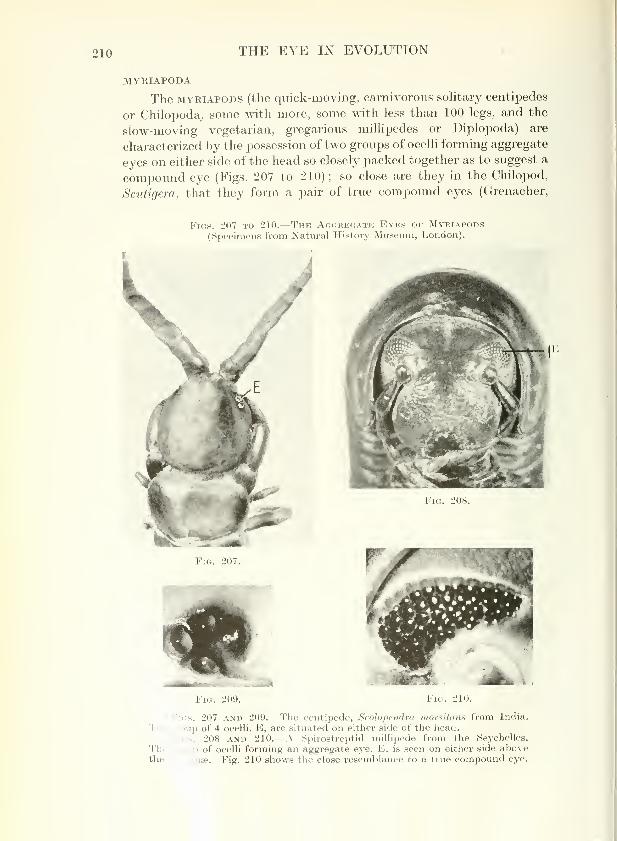
The MYEiAPODS (the quick-moving, carnivorous solitary centipedes
or Chilopoda^ some with more, some with less than 100 legs, and the
slow-moving vegetarian, gregarious millipedes or Diplopoda) are
characterized by the possession of two groups of ocelli forming aggregate
eyes on either side of the head so closely packed together as to suggest a
compound eye (Figs. 207 to 210); so close are they in the Chilopod,
Scutigera, that they form a pair of true compound eyes (Grenacher,
Figs. 207 to 210.
—
The Aggreg.a.te Eyes or Myriapods(Specimens from Natural History Museum, London).
Fig. 208.
Fig. 209. Fig. 210.
Figs. 207 and 209.—The centipede, ScoJopendra morsitans from India.
The jToup of 4 ocelli, E, are situated on either side of the head.
':s. 208 AND 210.—A iSpirostreptid millipede from the Seychelles.
Thi .iip of ocelli forming an aggregate eye, E, is seen on either side abovethe a , iinae. Fig. 210 shows the close resemblance to a true compound eye.

ARTHROPODA 211
1880; Graber, 1880 ; Caesar, 1913 ; Constantineanu, 1930). In sometypes, such as Pauropus, which live in moist debris in the woods andforests, eyes are lacking.
ARACHNIDA
The ARACHNIDS form a large and loosely associated group whichincludes scorpions, king-crabs, spiders, pseudo-scorpions, whip-tailed
scorpions, harvest-men, jerrymanders, mites and ticks. With the
single exception of the king-crab they do not possess conii^ound eyes
Fig. 211.
—
The Kikg-crab, Limvlus polypbeml's.
A .simple ej'e, S, is seen as a dark spot situated on either side of the medianspine. The two compound eyes, C, are situated on the external aspect ofeach of the first lateral spines (specimen from Natural Historj^ Museum,London).
but all are provided with ocelli sometimes of considerable size andcomplexity.
SCORPIONS (scorpionidea), venomous animals up to 8 in. in length
with a long stinging tail, are restricted to warm countries ; in habit
they are essentially solitary and nocturnal, being active during the
night and spending the day lurkmg under stones or in crevices. Theyare provided with a pair of large median eyes situated about the middleof the cephalothorax, and 2 to 6 pairs of lateral ocelli placed on its
antero-lateral margins, the more anterior being simpler in structure
than the posterior.^ The lateral eyes are simple ocelli in which the1 p. 141.
Scorpion

212 THE EYE IN EVOLUTION
borders of the visual cells unite with their neighbours to form rhabdomes
(Fig. 109) ; the median eyes are also of the simple type with the
sensory cells arranged in groups each centred on a rhabdome. These
cells, however, are peculiar in that they are doubled upon themselves
to form a semi-inverted retina ^ (compare Fig. 127).
^
The KING-CRABS (xiphosuea), a very ancient type dating to the
Silurian, w^hich live in shallow water on the sandy shores of North
America {Limulus) or Asia, have two large lateral compound eyes and
two median ocelli (Fig. 211). The compound eye is of a unique and
elementary type ^ (Fig. 143) ; it is not faceted but is covered by a
chitinous thickening of the cuticle which sends projections inwards as
Ki( 'I'll i: .1 I Mi'iN(. Si'i 1 'i:i!, /'/ / A // SIM'ATL'f!.
From the Dutch East Indies. The 2 large and 6 small simple eyes are
seen surrounding the anterior and lateral aspects of the carapace (specimen
from Natural History Museum, London).
conical papillse over each ommatidium to form a corneal lens. The
small median eyes are of the simple type wherein the sensory cells are
associated with rhabdomes (Fig. 142).
In addition, a third pair of ventral eyes is present in the larva on
either side of the frontal organ of the hypostoma. an olfactory organ;
in the adult these eyes become degenerate but it is possible that they
may participate in the olfactory function (Patten, 1893 ;Hanstrom,
1926).
2 F ' details, see J. Miiller (1826), Lankester and Bourne (1883), Parker (1887),
Petrun! -li (1907), Police (1908), Scheuring (1913-14). Biitschli (1921), Versluys
and Deu: 1923).
"p.

ARTHROPOD
A
213
SPIDERS (araneida) are of widespread distribution and, although
comprising some 14,000 species, are conveniently divided according to
their habits into two types, the relatively sedentary " web-spinners"
and the more active " wanderers " which hunt their prey ; all, how-ever, sjiin silk, either as a web, or for snaring or tying up their victims,
for protection of their cocoons or for making bridges for travelling. In
both types on the cephalothorax there are G or more usually 8 simple
eyes arranged in two or three rows (Fig. 212) ; these have received a
c«j:>'' ^'^rable amount of study. ^ The arrangement of these ocelli varies
remarkably (Figs. 213 to 216). Among the web-spinners the ocelli are
rudimentary and their effective range is short. The common house
Figs. 213 to 216.
—
The Arrangement of the Ocelli in DifferentSpecies of Spiders.
Fig. 213.—The ocelli of tlu* comtnonhouse spider, Te'jcu'irio doinestica.
Fic;. 214.—The ocelli of the common gardenspider, Araueus dldilcmatus.
Y\Q. 21.5.—The ocelli of the wolf FiG. 216.— The ocelli of the jumping spider,
spider, Li/ro.sd lujricoht. Salticus scenicu.s.
1 For details, see Hentschel (1899), Widmann (1908), Petrunkevitch (1911), Scheuring
(1914), Versluys and Demoll (1923), Savory (1928), Homanu (1928-53), Millot (1949).

214 THE EYE IN EVOLUTION
Tegenaria
Aratieus
Salticus
Whip-scorpiou
spider, Tegenaria domestica, has two rows of 4 ocelli, those of the
anterior row being slightly smaller than those of the posterior (Fig. 213);
the common garden spider, Araneus diadematus, has 4 median and 4
small lateral eyes (Fig. 214). The more active hunting species which
construct no web have larger eyes ; thus the wolf-spider, Lycosa,
has an anterior row of 4 small ocelli, two large posterior median
and two smaller posterior lateral ocelli (Fig. 215) ; while the jumping-
spider, Salticus, with a visual capacity more fully developed than
the wolf-spider, has an anterior row of two large and two smaller
ocelli on the front of its square-shaped cephalothorax, and two very
small posterior median and two posterior lateral ocelli on the top
(Fig. 216). With all its variations the general plan is thus consistent;
the anterior median eyes (the two central eyes in the front row) have a
verted retina, the remainder are inverted provided with a crystalline
tapetum ^ and since these latter glow in the dark the former are some-
times called " diurnal eyes." The nerve-fibres from the two anterior
median eyes travel—with a partial decussation at a chiasma—to the
ganglion of the first cephalic segment, from the remaining eyes to that
of the second (Figs. 107, 126).
It is interesting that the anterior median eyes of spiders are
equipped with muscles attached to their posterior aspect rendering them
motile so that they can increase their visual field ; thus web-spiders
have one muscle, Lycosids two, and Salticids six. These are absent in
the lateral and posterior median eyes.
Curious anomalies to this general arrangement exist, but they are rare;
thvis in the female of a spider found in France, Walckenaera acuminata, the eyes
are arranged on a dumpy tubercle on the cephalothorax, while in the inale they
are perched on a long stalk-like periscope, 4 on the tip and 4 half-way down(Millot, 1949). It is interesting that among spiders the lens, which is part of the
outside covering of the animal, is cast at the time of moulting and thtis it would
appear that the spider may be ren-dered temporarily blind.
PSEUDO-SCORPIONS (pseudoscorpionidea), minute animals resem-
bling miniature scorpions but without the long tail and sting, found
burrowing in books or under stones, the bark of trees and the wing-
covers of insects, are provided with two pairs of simple eyes (when
they exist) on either side of the cephalothorax ; the^e are typically
equipped with an inverted retina and a tapetum (Scheuring, 1913)
(Fig. 217).
w^HiP-TAiLED SCORPIONS (PEDiPALPi). The eycs of this order are
not well known (Scheuring, 1913 ; Versluys and Demoll, 1923 ; Millot,
H'49). They are entirely absent in some species ; in others there are
two median eyes only ; but the typical arrangement consists of two
median (principal) eyes and two groups of 3 lateral eyes.
' Except Salticus, the eyes of which lack a tapetum and are therefore " diurnal ".
See fr '- p. 1.50.

ARTHROPODA 215
The median eyes are of the cupulate type with a semi-inverted
retina the cells of which are doubled upon themselves ^ (Fig. 127). Thelateral eyes have an inverted retina with a tapetum (Fig. 124).
Fig. 217.
—
The Pseudoscorpion, Carffosius ischsocbules.
Showing two simple eyes, S, on either side (specimen from Natural HistoryIMuseum )
.
HARVESTMEN (PHALAXGIDA ; OPILIONES), minute spider-like
Arachnids with extremely long legs, which avoid the glare of daylight,
have two simple ocelli mounted one on either side of an oculiferous
tubercle (ocularium) (Fig. 218). It would seem that with its laterally
directed eyes the animal has no frontal vision. Each ocellus is a simple
Harvestman
As^. ***
Fig. 218.
—
The Eye of the Harvestman, Megabusus diadema.
The smiple eye is seen on a siDecial oculiferous tubercle (specimen fromNatural History Museum).
cupulate eye with a large cuticular lens and a simple row of visual cells
from which the fibres emerge in several branches to form the optic
nerve (Purcell, 1894).
^ p. 1.50.

216 THE EYE IN EVOLUTION
Jerrymander
JEBRYMANDERS (soLiFUG^)—active, pugiiacious, non-venomous,
nocturnal creatures found in warm countries—possess a pair of median
(principal) eyes situated on a small tubercle and one or two pairs of
lateral eyes usually rudimentary, difficult to see and probably function-
less. Both types are simple cupulate ocelli with direct (verted) retinse
(Scheuring, 1913; Demoll, 1917).
MITES and TICKS (acarina). mites are minute Arachnids of which
over 20,000 species are known, found almost universally in the earth or
in water, salt and fresh, often of parasitic habit on or within animals
(including man) and plants whether alive
or decaying after death : well-known
human j^arasites are SarcojJtes scabiei (the
itch-mite) causing scabies, and Dernodex
foUiculorum found in the hair follicles;
the harvest-mite (chigger) is a virulent
pest to both man and animals (particularly
rodents), while others infest insects (Isle
of Wight bee disease) and others plants
(gall mites, red spiders, etc.). Many, such
as SarcojJtes are without eyes (Fig. 219) ;
others, such as the Prostigmata and the
Hydracarina (fresh-water mites) are pro-
vided with 2, 4 or 6 ocelli on the front and
lateral aspects of the head depending on
the species, the individual organs being
sometimes fused (Fig. 220 and 221) (Lang,
1905). Each possesses a convex lens often
difficult to distinguish from the surround-
ing skin.
TICKS (ixoDiDEs) are larger than mites and are frequently of
biological importance as causing disease (tick-fevers) in man ^ and
animals. 2 Most types are without eyes, but such species may have
thin transparent areas on the dorsal surface which perhaps respond to
differences in the intensity of light. When visual organs are present
they are extremely rudimentary, being minute ocelli mounted curiously
on the animal's shoulder (Fig. 222).
Figs. 220 and 221.
—
The Eyes of Fresh-water Mites (Hydracarina).
Fig. 219. -The Mite, SarcoptesSCABIEI.
(Female) ( X 125) (Sutton andSutton, Hh. o/ Dis. of the Skin,
Mosby).
Fig. 220.—The 4 separate ocelli ofLimnesia.
Fig. 221.
—
Hijgrobates, showing fusion
of the anterior and posterior ocelli
(after P. Lang).
^ Texas fever. Rocky Mountain spotted fever, etc.
2 Red -water fever in cattle, heart -water in sheep, etc.

ARTHROPODA 217
SEA-SPIDERS (PYCNOGONiDA ; pantopoda). marine species related to the
Arachnids, inhabit the shores or the depths of the seas, Hving on seaweed,
hydroids and sponges. They are provided with 4 primitive oceUi perched in
two pairs on an ocuHferovis tubercle on the cephalothorax ; as we have already
noted, the retinse are of a jieculiar and characteristic inverted type ^ (Morgan,
1891 ; Korschelt and Heider, 1893 ; Sokolow. 1911 ; Schlottke. 1933) (Fig. 125).
Fig. 222.
—
The Tick, Amblfomma pompo.^vm.
The two simple eyes, S, lie well posteriorly on the shoulder of the animal(specimen from Xatural History Museum).
Pj'cnogonid
IXSECTA
INSECTS form the largest class of Arthropods and their multitude
of types is subdivided with reference to their possession of wings ; it is
interesting that the complexity of their eyes varies directly with this
characteristic, an association only natural in view of the demands madeupon vision by a high degree of mobility.
(1) Sub-class APTERYGOTA (d, privative ; -nrepv^ a wing), wingle.gs forms,
in which through a series of moults the adult differs little from the newly hatched
insect except in size. They are the most primitive of insects, some species being
marine, and when eyes are present they are simple in type.
THYSAXURA—bristle-tails, of wide distribution in damp soil, son"ie living
between tide-marks or under stones or bark ; others (silver-fish) in
bread-bins or books. Closely related are the eye-less diplura.
PROTURA—ininute creatures (2 mm.) living in moist soils vmder stones
and bark, without wings, antennae or eyes of anj^ kind.
COLLEMBOLA—.sjiringtails, living under stones and leaves ; one species
lives between tide-marks.
(2) Sub-class pterygota, provided with wings which, however, may be
secondarily lost through highly evolved specialization. The sub-class is divided
1 p. 150.

218 THE EYE IN EVOLUTION
TYPICAL INSECTS : I
(Draivn not to scale but approxitnately to a standard size.)
THYSANURA
APTERYGOTAPROTURA COLLEMBOLA
Silver-fish Acerentonion
EXOPTERYGOTAORTHOPTERA
Springtail
DERMAPTERA
Cockroach Grasshopper Stick-insect Earwig
PLECOPTERA ISOPTERA PSOCOPTERA ANOPLURA
Stone-fly Termite Book-louse Pediculus
EPHEMEROPTERA THYSANOPTERA HEMIPTERA
Thrip Bed-bug

ARTHROPODA 219
TYPICAL INSECTS : II
(Drcuun not to scale but approximately to a standard size.)
EXDOPTERYGOTA
NEUROPTEHA TRICHOPTERA LEPIDOPTERA
Lacewing Caddis-fly Butterfly Moth
COLEOPTERA
^kyA.
Colorado beetle,
LeptinotarsaBurying beetle,
NecrophorusRose-chafer,
Cetonia
Fire-fly,
Photinus
HYMENOPTERA
Bee, Bombus Wasp, Vesjm Ant
APHANIPTERA
Blue-bottle,CalUphora
Gad-fly,
TabanusBee-fly,
Bombiiliiis
Flea,
Pulex irritans

220 THE EYE IN EVOLUTION
into two, depending on whether their wings are developed externally (Exoptery-
gota) or internally (Endopterygota) ; in the latter the wings become evident
only in the adult (imago) stage.
(a) EXOPTERYGOTA, insects which undergo a series of moults marked by
the gradual development of wings. The more important orders are :—
•
ORTHOPTERA—cockroaches, locusts, grasshoppers, crickets, stick-
insects, praying mantis.
DERMAPTERA—earwigs.
PLECOPTERA—stone-flies, a small and little known order, the aquatic
larvte being found beneath the stones of mountain streams, and the
slow-flying adults having a very short life.
ISOPTERA—termites living under grovmd without eyes.
EMBIOPTERA—a few species of insignificant tropical insects.
ZORAPTERA—a few species of minute insects resembling termites.
psocoPTERA—small plump, book-lice (winged or wingless).
ANOPLURA—biting or sucking lice, wingless, parasitic on man and
animals and frequently disease-producing {Pediculus, Phthirus, etc.).
EPHEMEROPTERA—mayflies, the aquatic larva3 living up to 3 years, the
delicate adult a few hours.
ODONATA—brilliantly colovired dragonflies and demoiselle flies with
aquatic larvae, the former unusually active, swift-flying and
voracious, the latter more delicate.
THYSANOPTERA—the minute thrips, vegetarian in habit, living on
flowers, leaves and decayed vegetation.
HEMiPTERA—bugs with a specially developed proboscis (rostrum)
adapted for piercing and sucking, many of them beautiful and slender
despite their name : land bugs including the bed-bug, water bugs
varying from the giant flsh-killer or the water-scorpion to the water
boatman (Notonecta), the cicadas, the frog-hoppers, tree-hoppers,
leaf-hoppers, the aphids (or green-flies) and the scale-insects.
(b) ENDOPTERYGOTA, winged insects which have a complete metamorphosis
(egg, larva, pupa, adult) with a resting pupa (or chrysalis).
NEUROPTERA—lace-wings, alder-flies, scorpion-flies.
TRiCHOPTERA—caddis-flies, with aquatic larvae and moth-like adults
with hair-covered bodies and wings.
LEPIDOPTERA—butterflies and moths.
COLEOPTERA—beetles, including over 200,000 known species, both
terrestrial and water-beetles.
STREPSiPTERA
—
Stylops, miuvitc insects, parasitic on other insects,
particularly wasps and bees.
HYMENOPTERA—gall-flies, saw-flies, ichneumon-flies, bees, wasps, ants.
DIPTERA—two-winged flies, midges, gnats, mosquitoes and frviit-flies.
APHANiPTERA—the secondarily wingless fleas (jiggers, etc.), blood-
sucking in habit and parasitic on birds and mammals.
Ill the larval form all insects possess simple lateral eyes (stemmata;
ardixixy., a garland). The adult also frequently possesses simple eyes
(DOES.\f. ocelli), although they are absent or vestigial in many species,
as in rt.ost beetles and mosquitoes, some families of flies, and noctuid
moths nt in addition it is provided with multifaceted compound
EYES. his generalization there are some exceptions in degenerative
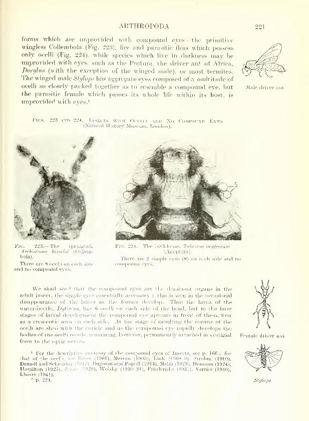
ARTHROPODA 221
forms which are unprovided with compound eyes—the primitivewingless Collembola (Fig. 223), hce and parasitic fleas which possessonly ocelli (Fig. 224), while species which live in darkness may beiniprovided with eyes, such as the Protura, the driver ant of Africa,
Dorylus (with the exception of the winged male), or most termites.
The winged male Stylops has aggregate eyes composed of a multitude of
ocelli so closely packed together as to resemble a compound eye, butthe parasitic female Mhich passes its whole life within its host, is
unprovided with eyes.^
jVIale driver ant
Figs. 22.3 and 224.
—
Insects with Ocelli and Xo Compound Eyes(Xatural History Museum, London).
Fig. 2l':;. Tlic springtail,
Arrhi.stoiuti brs-selsi (Collem-bola).
There are 8 ocelli on each side
and no compound eyes.
Fig. 224.—The bird-louse, Trinoton acidenlioii
(Anoplura).
There are 2 simple eyes (S) on each side and nocompoimd eyes.
We shall see ^ that the compound eyes are the dominant organs in the
adult insect, the simple eyes essentially accessory ; this is .seen in the occasional
disappearance of the latter as the former develop. Thus the larva of the
water-beetle, Dytiscus, has 6 ocelli on each side of the head, but in the later
stages of larval development the compound eye appears in front of them, first
as a crescentic area on each side. At the stage of moulting the cornea^ of the
ocelli are shed with the cuticle and as the compound eye rapidly develojDs the
bodies of the ocelli recede, remaining, however, permanently attached in vestigial
form to the optic nerves,
1 For the descriptive anatomy of the compound eyes of Insects, see p. 166 ; for
that of the ocelli, see Hesse (1901), Merton (1905), Link (1908-9), Strohm (1910),Demoll and Scheurins (1912). Bugnion and Poiwff ( 1914), Melin (1923). Homann (1924),Hamihon (1925), Zikan (1929), Wolsky (1930-31), Friederichs (1931). Verrier (1940),Lhoste (1941).
^ p. 224.
Female driver ant
Stylops

222 THE EYE IN EVOLUTION
Figs. 225 to 227.
—
The Compound Eyes of Insects.
Fig. 225.—The head of the dragon-fly, jEschna californica (Odonata) capped
by two enormovis crescent-like comiDound eyes (James Needham).
Fig. l&.- -The male gadfly, Ancnia fasciata
nilotica (Diptera).
The immense compound eyes occupy the
whole of the surface of the head (Natural HistoryMuseum, London).
Fig. 227.—The cave-bug, Leotichius
glaucopis (Hemiptera).
From Malaya. Dorsal surface. The2 prominent compound eyes (C) are
largely spread over the ventral surface.
There are 2 median ocelli (S) (Natural
History Museum, London).
THE STEMMATA (OR LATERAL OCELLi) OF LARVAL OR PUPAL FORMS
can in general be classified into two main types. The most elaborate
organs are seen in the larvae of Lepidoptera and Trichoptera;
these are arranged in a group of variable size ^ on either side of
the Jjcnd, each separate individual of which takes the form of the
single itnatidium of a compound eye with a cuticular corneal lens,
g., 6 on either side of the head in the caterpillars of butterflies.

ARTHROPODA 223
Figs. 228 and 229.
—
Stalked Compound Eyes.
Fig. 228.—A grouse locust, Ophiotettix limosina (Orthoptera).
' The compound eyes (C) are placed on either side at the end of the stalk-
like head. (One antenna is missing.) (Natural History Museum, London.)
Fig. 229.—The stalk-eyed fly, Achias rothschildi (Diptera).
The lai'ge compound eyes (C) are at the end of unusually long stalks(Natural History Museum, London).
a crystalline cone and a retinule of 7 sensory cells grouped around a
rhabdome (Fig. 138) (Dethier, 1942-43). A more simple variety is
seen in the larvae of Tenthredinidffi (saw-flies) and Coleoptera. These
usually have two lateral eyes of cupulate shape with a retma formed as
a palisade of sensory cells under a lens-like thickening of the cuticle.
The retinular cells are arranged in groups of two or three, each
group around an elementary rhabdome which is not constructed for
the reception of images. More rudimentary forms occur such as the
Sawfly

224 THE EYE IN EVOLUTION
simple pair of visual cells with two overlying pigment cells which form
the eye of the larva of the midge, Ceratojjogon, or the few light-sensitive
cells lying in a shaded pocket in the pharyngeal skeleton of the larva of
the house-fly, Musca (Fig. 99) (Welsh, 1937 ; Debaisieux, 1939).
THE DORSAL OCELLI OF ADULTS were described and figured as early
as 1678 by the French scientist, de la Hire (Figs. 149, 227, 230). Theyare usually three in number arranged in triangular form, one median
and anterior and two lateral and posterior on the dorsal aspect of the
Figs. 230 and 23 L—Unusual Compound Eyes in Insects (NaturalHistory Museum, London).
Fig. 230.—The aphid, Dnctynotusobscuras (Homoptera).
There are 2 compound eyes (C) oneon each side of the head, and, in
winged forms, 3 ocelh on tlie vertexof the head, the median one of whichis marked S. In the family Aphididaethere is in addition a prominence, thetriommatidion (T), of unknown func-tion, bearing 3 facets at the base ofeach compound eye. This organ is
always present, even in those forms in
which a comjjound eye is lacking.
Fig. 231.—The Whirligig beetle,
Dineutus grossus (Coleoptera).
There are 2 compound eyes on eachside of the head, one dorsal (D) for
aerial vision, and one ventral (V) for
vision under water.
head between the compound eyes ; but they are small and incon-
spicuous, being often hidden by scales as in moths or hairs as in bees.
In some species of ants belonging to the sub-family Myrmicinae, the
anterior ocelli are double or binary in type (Weber, 1947). In others,
such as Orthoptera, the ocelli are vestigial ; in general, their degree of
development shows some correlation with that of the wings (Kalmus,
1945). As a rule they resemble in structure the more simple type of
stemmata, being comjirised merely of a group of visual cells associated
with rb-'bdomes lying beneath a common cuticular lens (Fig. 108).
T OMPOUND EYES OF ADULTS are laterally situated on the head
and foi be essential visual organ (Fig. 149). They are large and

ARTHROPODA 225
prominent and vary in complexity from the small organ of the worker
of the ant, Solenopsis. which lives underground and is provided with
6 or 8 facets, to the elaborate organ of dragon-flies (Odonata) with upto 28,000 1 ommatidia (Imms, 1935) (Figs 225 to 227). Occasionally
the compound eyes are enormous, literally occupying the whole
surface of the head, as is seen in the gad-flies (Tabanida?) (Fig. 226) ;
usually they are situated on the surface of the head, sometimes they
stand out prominently as in the praying mantis (Fig. 734), but occasion-
ally they are perched on long stalks (Figs. 228 and 229). Exceptionally
two compound eyes are differentiated in function, such as in the
whirligig beetle, Dineutus, which has a dorsal compound eye for aerial
vision and a ventral for vision under water (Fig. 231).- A unique organ
is seen in the Aphid family (" green-fly ") in which an additional tri-
faceted organ, the triommatidion, is found at the base of each
compound eye (Fig. 230) ; the function of this organ is imknown but
it is present even in those forms of aphids which have no compoundeyes.
von Buddenbrock, Moller-Racke andSchaller. Experientia, 10, 333 (1954).
Biitschli. Vorlesungen ii. vergl. Anrit.,
Berlin, 872 (192'l).
Bugnion and Popoff. Arch. Anat. inter.,
16, 261 (1914).
Caesar. Zool. Jb., Abt. Anat., 35, 161
(1913).Constantineanu. Zool. Jb., Abt. Annt., 52,
253 (1930).
Dakin. Quart.J.»ilcr.Sci..6b, 163(1921).Debaisieux. Ann. Soc. Sci., Bruxelles, 59,
9 (1939).DenioU. Die Sinnesorgane der A rthropoden,
Braunschweig (1917).
Demoll and Scheuring. Zool. Jb., Abt.
Zool. Physiol., 31, 519 (1912).
Dethier. J. cell. camp. Pltysiol., 19, 301
(1942) ; 22, 115 (1943).'
Dijkgraaf. Z. vergl. Phi/siol., 38, 491
(1956).Exner. Die Physiologic d. Jacetlicrten
Augen von Krebsen u. Insekten,
Leipzig (1891).
Fales. Biol. Bull., 54, 534 (1928).
Friederichs. Z. Murphol. Oekol. Tiere, 21,
1 (1931).Graber. Arch. mikr. Anat., 17, 58 (1880).
Grenacher. .-^rch. mikr. Anat., 18, 415(1880).
Zool. An:Hamilton.
(1925).Hanstrom.
(1926).
Harris and Mason280 (1956).
, 18, 280 (1895).
Publ. U.S. nat. Mus., 65, 1
Lunds I Aarssr, 22
Proc. toy. Soc. B., 145,
1 p. 172.
Hentsfhel. Zool. Jb., Abt. Anat., 12, 509(1899).
von Hess. Vergl. Physiol, d. Gesichtssinnes,
Jena, 79 (1912).
Hesse, R. Zool. Anz., 24, 30 (1901).Das Sehen der niederen Tiere, Jena
(1908).
de la Hire. Mem. Acad. roy. Sci. Paris(1666-1699), 10, 609 (1730).
Homann. Z. vergl. Physiol., 1, 541 (1924) ;
7, 201 (1928) ; 14, 40 (1931).Zool. Jb.. Abt. Anat., 71, 56 (1950) ; 72,
345 (1952).Biol. Zbl., 72, 373 (1953).
Imms. Textbook of Entomology , London(1935).
Kalmus. Proc. roy. entom. Soc. Lond., A,20,84(1945).
Kor.schelt and Heider. Vergl. Entwicklung.d. Wirbellosen Tiere, Jena, 664 (1893).
Lang. Zool. Jb., Abt. Anat., 21, 453(1905).
Lankester and Bourne. Quart. J. micr.Sci., 23, 177 (1883).
Lhoste. Bull. Soc. zool. Fr., 66, 62 (1941).Link. Zool. Anz., 33, 445 (1908).
Zool. Jb., Abt. Anat. ,2,1, -m, 281 (1909).Melin. Zool. Bidrag Uppsala, 8, 1 (1923).Merton. Z. iviss. Zool., 79, 325 (1905).Millot. Grasse's Traite de Zool., Paris, 6,
295, 533, 589, 698 (1949).Morgan. Biol. Stud. Johns Hopk. Univ.,
5, 49 (1891).
Muller, J. Zur vergl. Pliysiol. des Gesichts-
sinnes des Mensclien it. d. Thiere,
Leipzig (1826).
Dragon-fly
Whirligig beetle
Aphid
- Compare the eye of Anableps, p. 324.
S.O.—VOL. I. 15

226 THE EYE IN EVOLUTION
Parker. Bull. Mus. Comp. Zool., 13, 173
(1887).
Patten. Quart. J. micr. Set., 35, 1 (1893).
Petrunkevitch. J. exp. Zool., 5, 275
(1907).
Zool. Jb.,Abt.Syst. Biol., Zl, 355 {Idll).
Police. Zool. Jb., Abt. Anat., 25, 1 (1908).
Purcell. Z. wiss. Zool., 58, 1 (1894).
Radl. Pflugers Arch. ges. Physiol., 87,
418 (1901).
Savory. The Biology of Spiders, London(1928).
Scheuring. Zool. Jb., Abt. Anat., 33, 553
(1913) ; 37, 369 (1914).
Schlottke. Z. mikr. Anat. Forsch., 32, 633
(1933).
Sehulz. Z. vergl. Physiol. ,1, 488 (1928).
Sokolow. Z. wiss. Zool., 98, 339 (1911).Strohrn. Zool. Anz., 36, 156 (1910).
Tompsett. Liverpool marine biol. Comm.Mem., 32, 1 (1939).
Vaissiere. C. R. Acad. Sci. (Paris), 238,942 (1954) ; 240, 345 (1955).
Verrier. Bull. Biol. France Belg., 74, 309(1940).
Versluys and Demoll. Ergebn. Fortsch.
Zool. Jena, 5, 67 (1923).Weber. Biol. Bull., 93, 112 (1947).
Welsh. .Sczence, 85, 430 (1937).
Widmann. Z. wiss. Zool., 90, 258 (1908).
Wolsky. Z. vergl. Physiol., 12, 783 (1930)
;
14, 385 (1931).
Biol. Bev., 8, 370 (1933).
Zikan. Zoo/, ylnz., 82, 269 (1929).