Embed Size (px)
Citation preview
CHAPTER TWO
PATHOLOGY OF THE REPIRATORY SYSTEM
THE UPPER RESPIRATORY TRACT
Allergic nasal polyp
Not true neoplasms. They are associated with inflammation and allergy. Generally, they
are multiple (bunch of grapes), and nearly always bilateral.
Microscopically:
composed of loose mucoid stroma and mucous glands covered by respiratory epithelium
which often shows foci of squamous metaplasia. They are infiltrated by lymphocytes,
plasma cells, mast cells, neureophils and eosinophils.
Benign tumors of the Nose and Nasal Cavity:
ANGIOFIBROMA
It occurs almost exclusively in males. It is androgen dependent.
It presents as polypoidal mass that bleeds severely on manipulation.
Microscopically: Composed of matrix of blood vessels and fibrous tissue stroma which may be loose and
edematous or dense acellular and highly collagenized.
The vessels range from capillary size which are particularly common at the growing edge
of the tumor, they have got plump endothelial cells.
Larger venous size vessels are located at the base of the lesion.
Malignant Tumors: NASOPHARYNGEAL CARCINOMA
This rare neoplasm has a strong epidemiologic links to EBV & a high frequency in China. These
facts raise the possibility of viral oncogenesis on a background of genetic susceptibility. The
cancers are either squamous cell carcinoma (keratinizing or nonkeratinizing) or undifferentiated
carcinoma. The latter is the most common and the one most closely linked with EBV. In
nasopharyngeal carcinomas a striking infiltration of mature lymphocytes can often be seen.
These neoplasms are therefore referred to as "lymphoepitheliomas," although the lymphocytes
are not part of the neoplastic process. Nasopharyngeal carcinomas invade locally, spread to
cervical lymph nodes, and then metastasize to distant sites. They are, however, radiosensitive,
and 5-year survival rates of 50% are reported for even advanced cancers.
LARYNGEAL TUMORS
Benign Lesions
Vocal cord nodules ("polyps") are smooth protrusions (usually less than 0.5 cm in diameter)
located, most often, on the true vocal cords. The nodules are composed of fibrous tissue and
covered by stratified squamous mucosa. These lesions occur chiefly in heavy smokers or singers
(singer's nodes), suggesting that they are the result of chronic irritation or voice abuse.
Laryngeal papilloma (squamous papilloma) of the larynx is a benign neoplasm, usually on the
true vocal cords, that forms a soft, raspberry-like excrescence rarely more than 1 cm in diameter.
Microscopically, it consists of multiple, slender, finger-like projections supported by central
fibrovascular cores and covered by benign, stratified squamous epithelium.
Papillomas are usually single in adults but are often multiple in children, in whom they are
referred to as recurrent respiratory papillomatosis (RRP), since they typically tend to recur after
excision. These lesions are caused by human papillomavirus (HPV) types 6 and 11, do not
become malignant, and often spontaneously regress at puberty.
Malignant Lesions
Carcinoma of the Larynx
Carcinoma of the larynx most commonly occurs after age 40 years and is more common in men
(7: 1) than in women. Nearly all cases occur in smokers, and alcohol and asbestos exposure may
also play roles. The vast majority (95%) are squamous cell carcinomas. The tumor develops
mostly on the vocal cords (glottic tumors), but it may arise above the cords (supraglottic) or
below the cords (subglottic). They begin as in situ lesions that later appear as pearly gray,
wrinkled plaques on the mucosal surface, ultimately ulcerating and fungating. As expected with
lesions arising from recurrent exposure to environmental carcinogens, adjacent mucosa may
demonstrate squamous cell hyperplasia with foci of dysplasia, or even carcinoma in situ.
Clinically there is persistent hoarseness. Supraglottic cancers have a worse prognosis than glottic
tumors because of their earlier metastasis to regional (cervical) lymph nodes. The subglottic
tumors, like the supraglottic ones, tend to remain clinically silent, & thus usually present as
advanced disease. With surgery, radiation, or combined therapeutic treatments,only one third die
of the disease. The usual cause of death is infection of the distal respiratory passages or
widespread metastases and cachexia.
THE LUNG ATELECTASIS (COLLAPSE) is loss of lung volume caused by inadequate expansion of
airspaces; this leads to shunting of inadequately oxygenated blood from pulmonary arteries into
veins, thus giving rise to hypoxia.
Pathogenetically atelectasis is classified into three forms (Fig. 2-1)
1. Resorption atelectasis complicating obstruction. The air already present distally gradually
becomes absorbed, and alveolar collapse follows. Depending on the level of airway obstruction,
an entire lung, a complete lobe, or a segment may be involved. A mucous or muco-purulent plug
is the most common cause of such obstruction for e.g. following surgical operations or bronchial
asthma, bronchiectasis, chronic bronchitis, or the aspiration of foreign bodies, particularly in
children.
2. Compression atelectasis is usually due to mechanical compression of the lung by pleural
distension as in pleural effusion (congestive heart failure) or pneumothorax. Basal atelectasis is
another example & is due to elevated diaphragm as that occurs in bedridden patients, in those
with ascites, and during and after surgery.
3. Contraction atelectasis occurs in the presence of focal or generalized pulmonary fibrosis or
pleural fibrosis; in these situations there is interference with expansion and an increase in elastic
recoil during expiration.
Atelectasis (except contraction type) is reversible and should be treated quickly to prevent
hypoxemia and infection of the collapsed lung.
ADULT RESPIRATORY DISTRESS SYNDROME (ARDS) (previously “Shock lung”) is
“progressive respiratory insufficiency caused by diffuse alveolar damage”
The clinical setting associated with ARDS include
A. Respiratory
1. Diffuse infections (viral, bacterial)
2. Aspiration
3. Inhalation (toxic gases, near drowning)
4. O2 therapy
B. Non-respiratory
1. Sepsis (septic shock) 2. Trauma (with hypotension) 3. Burns
4. Pancreatitis 5. Ingested toxins (e.g. paraquat)
There is an acute onset of dyspnea, hypoxemia (refractory to O2 therapy), and radiographic
bilateral pulmonary infiltrates (noncardiogenic pulmonary edema). The condition may progress
to multisystem organ failure.
Pathogenesis In ARDS there is damage to alveolar capillary membrane by endothelial &/or epithelial injury.
This leads to three consequences
1. Increased vascular permeability (endothelial damage)
2. Loss of diffusion capacity of the gases
3. Widespread surfactant deficiency (damage to type II pneumocytes).
Nuclear factor κB, is suspected of tilting the balance in favor of pro-inflammatory rather than
anti-inflammatory mediators, which causes the endothelial damage. This leads to fluid
accumulation. Following the insult, there is increased synthesis of a potent neutrophil
chemotactic and activating agent IL-8 & TNF by pulmonary macrophages. The recruited,
activated neutrophils release oxidants, proteases, etc. that cause damage to the alveolar
epithelium leading to loss of surfactant that interferes with alveolar expansion.
Gross features: in the acute phase the lungs are dark red, airless, and heavy.
Microscopic features: (Fig. 2-2)
The histologic reflection of ARDS in the lungs is known as diffuse alveolar damage.
Early stage is characterized by
Capillary congestion and stuffing by neutrophils
Necrosis of alveolar epithelial cells
Interstitial and intra-alveolar edema and sometimes hemorrhage
The presence of hyaline membranes is characteristic. They particularly line the distended
alveolar ducts & consist of fibrin admixed with necrotic epithelial cells.
Overall, the picture is very similar to that seen in respiratory distress syndrome in the
newborn.
Organizing stage is characterized by
Marked regenerative proliferation of type II pneumocytes
Organization of the fibrin exudates. This eventuates in intra-alveolar fibrosis.
Marked fibrotic thickening of the alveolar septa.
The prognosis of ARDS is gloomy and mortality rates are around 60% despite improvements in
supportive therapy. However, in most patients who survive the acute insult normal respiratory
function returns. Alternatively, diffuse interstitial fibrosis occurs with permanent impairment of
respiratory function.
OBSTRUCTIVE PULMONARY DISEASE
Under this heading come four entities
1. Emphysema
2. Chronic bronchitis
3. Asthma
4. Bronchiectasis
Although chronic bronchitis may exist without emphysema, and pure emphysema may occur
(with inherited α1-antitrypsin deficiency), the two diseases usually coexist. This is because long-
term cigarette smoking is a common underlying agent in both disorders. Emphysema and chronic
bronchitis are often clinically grouped together under the term chronic obstructive pulmonary
disease (COPD), which is one of the leading causes of death. The irreversibility of airflow
obstruction of COPD distinguishes it from asthma (reversible obstruction)
EMPHYSEMA is defined as "abnormal permanent enlargement of the airspaces distal to the
terminal bronchioles, accompanied by destruction of their walls without obvious fibrosis".
Classification of Emphysema is according to its anatomic distribution within the lobule; the
acinus is the structure distal to terminal bronchioles, and a cluster of 3 to 5 acini is called a
lobule. There are four major types of emphysema:
1. Centriacinar
2. Panacinar
3. Distal acinar
4. Irregular
Only the first two cause clinically significant airway obstruction, with centriacinar emphysema
being about 20 times more common than panacinar disease.
Centriacinar (Centrilobular) Emphysema: the central parts of the acini i.e. the respiratory
bronchioles are affected, while distal alveoli are spared (Fig. 2-3). The lesions are more common
and severe in the upper lobes. This type is most commonly associated with cigarette smoking.
Panacinar (Panlobular) Emphysema: the acini are uniformly enlarged from the level of the
respiratory bronchiole to the terminal blind alveoli (Fig. 2-4). It tends to occur more commonly
in the lower lobes and is the type that occurs in α1-antitrypsin deficiency.
Distal Acinar (Paraseptal) Emphysema: the distal part of the acinus is primarily involved
especially adjacent to the pleura and the lobular connective tissue septa. Characteristically, there
are multiple, adjacent, enlarged airspaces up to 2 cm or more in diameter, sometimes forming
cystic structures referred to as bullae. This type of emphysema probably underlies many of the
cases of spontaneous pneumothorax in young adults.
Irregular Emphysema: the acinus is irregularly involved; it is associated with scarring.
Pathogenesis of centriacinar & panacinar forms of emphysema
These are thought to arise as a result of imbalances of protease-antiprotease and oxidant-
antioxidant (Fig. 2-5). The protease-antiprotease imbalance hypothesis is supported by the
enhanced tendency of emphysema development in patients with genetic α1-antitrypsin
deficiency, which is aggrvated by smoking. α1-Antitrypsin, is a major inhibitor of proteases
(particularly elastase) secreted by neutrophils during inflammation. Emphysema seems to result
from the destructive effect of high protease activity in subjects with low antiprotease action.
Smoking seems to play a decisive role in the pathogenesis of emphysema
1. It causes accumulation of neutrophils & macrophages within the alveoli through its direct
chemoattractant effects and through the reactive oxygen species contained in it. These activate
the transcription factor NF-κB, which switches on genes that encode TNF and IL-8. These, in
turn, attract and activate more neutrophils. Accumulated active neutrophils release their granules,
which are rich in a variety of proteases (elastase, proteinase, etc.) that result in tissue damage.
2. Smoking also enhances elastase activity in macrophages; this elastase is not inhibited by α1-
antitrypsin; additionally, it can digest this antiprotease.
3. Tobacco smoke contains abundant reactive oxygen free radicals, which deplete the antioxidant
mechanisms, thereby inciting tissue damage. A secondary consequence of oxidative injury is
inactivation of native antiproteases, resulting in "functional" α1-antitrypsin deficiency even in
patients without enzyme deficiency.
Gross features
The diagnosis and classification of emphysema depend on the gross appearance of the lung.
Panacinar emphysema produces pale, voluminous lungs that obscure the heart at autopsy.
(Fig. 2-6)
In centriacinar emphysema the lungs are less voluminous and deeper pink. Generally, in this
type the upper two-thirds of the lungs are more severely affected.
Microscopic features
There is thinning and destruction of alveolar walls.
With advanced disease, adjacent alveoli coalesce, creating large airspaces (Fig. 2-7).
The capillaries within alveolar walls are reduced in number due to stretching.
Course & prognosis
With the loss of elastic tissue in the surrounding alveolar septa, there is reduced radial traction on
the small airways. As a result, they tend to collapse during expiration-an important cause of
chronic airflow obstruction in severe emphysema. The patient is barrel-chested and dyspneic,
with obviously prolonged expiration. Hyperventilation is prominent thus gas exchange is
adequate and blood gas values are relatively normal i.e. there is no cyanosis. Patients with
emphysema and chronic bronchitis usually have less prominent dyspnea and respiratory drive, so
they retain carbon dioxide, become hypoxic, and are often cyanotic. The eventual outcome of
emphysema is the gradual development of secondary pulmonary hypertension, arising from both
hypoxia-induced pulmonary vascular spasm and loss of pulmonary capillary surface area from
alveolar destruction and stretching. Death from emphysema is related to either pulmonary
failure, or right-sided heart failure (cor pulmonale).
Conditions Related to Emphysema
Several conditions resemble but are not really emphysema, these include
a. Compensatory emphysema refers to dilation of alveoli in response to loss of lung substance
elsewhere, as in residual lung tissue after surgical removal of a diseased lung or lobe.
b. Obstructive overinflation: the lung expands because air is trapped within it. A common cause
is subtotal obstruction by a tumor or foreign object, & mucous plugs in asthmatic patients (Fig.
2-10). It can be a life-threatening emergency if the affected portion extends sufficiently to
compress the remaining normal lung.
c. Bullous emphysema refers to any form of emphysema that produces bullae (spaces >1 cm in
diameter) (Fig. 2-8). They represent localized accentuations of one of the four forms of
emphysema, are most often subpleural that with rupture leads to pneumothorax.
d. Mediastinal (interstitial) emphysema signifies the entrance of air into the connective tissue of
the lung, mediastinum, and subcutaneous tissue. It may spontaneous as with a sudden increase in
intra-alveolar pressure (associated with vomiting or violent coughing) that causes a tear, with
dissection of air into the connective tissue. It is also likely to occur in patients on respirators who
have partial bronchiolar obstruction or in persons who suffer a perforating injury (e.g., a
fractured rib). When the interstitial air enters the subcutaneous tissue, the patient may blow up
like a balloon, with marked swelling of the head and neck.
CHRONIC BRONCHITIS is common among cigarette smokers and in smog-ridden cities. The
diagnosis of chronic bronchitis is clinical; it is defined as "a persistent productive cough for at
least 3 consecutive months in at least 2 consecutive years."
Pathogenesis
The distinctive feature of this disease is hypersecretion of mucus, beginning in the large airways.
Although the single most important cause is cigarette smoking, other air pollutants, such as
sulfur dioxide and nitrogen dioxide, may contribute. These environmental irritants induce
hypertrophy of mucous glands in the trachea and main bronchi and a marked increase in mucin-
secreting goblet cells in the surface epithelium of smaller bronchi and bronchioles. In addition,
these irritants cause inflammation with infiltration of CD8+ T cells, macrophages, and
neutrophils. Microbial infection is often present but has a secondary role, chiefly by maintaining
the inflammation.
Airflow obstruction in chronic bronchitis results from
1. Small airway disease, (chronic bronchiolitis) induced by goblet cell metaplasia with mucus
plugging of the bronchiolar lumen, inflammation, and bronchiolar wall fibrosis
2. Coexistent emphysema: while small airway disease is important in the early and mild airflow
obstruction, chronic bronchitis with significant airflow obstruction is almost always complicated
by emphysema.
Gross features
The mucosal lining of the larger airways is usually hyperemic and edematous. It is often
covered by a layer of mucus or mucopurulent secretions.
The smaller bronchi and bronchioles may also be filled with similar secretions.
Microscopic features
The diagnostic feature of chronic bronchitis in the trachea and larger bronchi is enlargement
of the mucus-secreting glands (Fig. 2-9).
A variable density of inflammatory cells, largely mononuclear but sometimes admixed with
neutrophils, is frequently present in the bronchial mucosa. Neutrophils increased markedly
during superimposed acute exacerbations.
Chronic bronchiolitis (small airway disease), characterized by goblet cell metaplasia, mucus
plugging, inflammation, and fibrosis. In severe cases, there may be complete obliteration of
the lumen due to fibrosis (bronchiolitis obliterans).
Patients with chronic bronchitis presents with a prominent productive cough that may persist
indefinitely without ventilatory dysfunction. However, some may develop significant COPD
with outflow obstruction. This is accompanied by hypercapnia, hypoxemia, and (in severe cases)
cyanosis. With progression, chronic bronchitis is complicated by pulmonary hypertension and
cardiac failure. Recurrent infections and respiratory failure are constant threats.
ASTHMA is "a chronic inflammatory disorder of the airways that causes recurrent attacks of
breathlessness". The inflammation seems to cause an increase in airway responsiveness
(bronchospasm) to a variety of stimuli, which would cause no such ill effects in nonasthmatic
individuals. In two-thirds of the cases, the disease is "extrinsic" (atopic) due to IgE and TH2-
mediated immune responses to environmental antigens. In the remaining one-third, asthma is
"intrinsic" (non-atopic) and is triggered by non-immune stimuli such as aspirin; pulmonary
infections, especially viral (common cold); psychological stress, etc.
Pathogenesis
The major etiologic factors of asthma are
1. Genetic predisposition to type I hypersensitivity ("atopy"),
2. Airway inflammation
3. Bronchial hyper-responsiveness to a variety of stimuli.
The role of TH2: the "atopic" form of asthma is associated with an excessive TH2 reaction
against environmental antigens. Three cytokines produced by TH2 cells are, in particular,
responsible for most of the features of asthma;
1. IL-4 stimulates IgE production
2. IL-5 activates eosinophils
3. IL-13 stimulates mucus production
In addition, epithelial cells are activated to produce chemokines that recruit more TH2 cells,
eosinophils, & other leukocytes, thus amplifying the inflammatory reaction.
The role of genetics: in asthma the bronchial smooth muscle hypertrophy and the deposition of
subepithelial collagen may be the result of a genetically inherited predisposition. ADAM33 is
one of the genes implicated.
The role of mast cells: these are part of the inflammatory infiltrate & contribute by secreting
growth factors that stimulate smooth muscle proliferation.
Atopic asthma, which usually begins in childhood, is triggered by environmental antigens (dusts,
pollen, animal dander, and foods). In the airways the inhaled antigens stimulates induction of
TH2-type cells and release of interleukins IL-4 and IL-5. This leads to synthesis of IgE that binds
to mucosal mast cells. Subsequent exposure of IgE-coated mast cells to the same antigen causes
the release of chemical mediators. In addition, direct stimulation of subepithelial vagal receptors
provokes reflex bronchocospasm. These occur within minutes after stimulation thus called acute,
or immediate, response. Mast cells release other cytokines that cause the influx of other
leukocytes, including eosinophils. These inflammatory cells set the stage for the late-phase
reaction, which starts 4 to 8 hours later.
The role of eosinophils: these cells are particularly important in the late phase. Their effects are
mediated by
1. Major basic protein and eosinophil cationic protein, which directly damage airway
epithelial cells.
2. Eosinophil peroxidase causes tissue damage through oxidative stress.
3. Leukotriene C4, which contribute to bronchospasm.
Viral infections of the respiratory tract and inhaled air pollutants such as sulfur dioxide increase
airway hyper-reactivity in both normal & non-atopic asthmatics. In the latter, however, the
bronchial spasm is much more severe and sustained. It is thought that virus-induced
inflammation of the respiratory mucosa renders the subepithelial vagal receptors more sensitive
to irritants.
The ultimate humoral and cellular mediators of airway obstruction are common to both atopic
and non-atopic variants of asthma, and hence they are treated in a similar way.
Drug-Induced Asthma: several drugs provoke asthma, aspirin being the most striking example.
Presumably, aspirin inhibits the cyclooxygenase pathway of arachidonic acid metabolism
without affecting the lipoxygenase route, thereby shifting the balance toward bronchoconstrictor
leukotrienes.
Occupational Asthma: this form is stimulated by fumes (epoxy resins, plastics), organic and
chemical dusts (wood, cotton, platinum), gases (toluene), and other chemicals. Asthma attacks
usually develop after repeated exposure to the inciting antigen(s).
Pathologic features (Fig. 2-10)
Gross features: in fatal cases, the lungs are overdistended because of overinflation.
Microscopic features:
There is occlusion of bronchi and bronchioles by thick, tenacious mucous plugs, which
contain numerous eosinophils.
Structural changes of airways include
- Thickening of the basement membrane of the bronchial epithelium
- Edema and an inflammatory infiltrate in the bronchial walls, with a prominence of
eosinophils and mast cells.
- An increase in the size of the submucosal glands.
- Hypertrophy of the bronchial muscle cells.
Course & prognosis
An attack of asthma is characterized by severe dyspnea with wheezing; the chief difficulty lies in
expiration. The victim struggles to get air into the lungs and then cannot get it out, so that there is
progressive hyperinflation of the lungs with air trapped distal to the bronchi, which are
constricted and filled with mucus and debris. In the usual case, attacks last from 1 to several
hours and subside either spontaneously or with therapy. Occasionally a severe paroxysm occurs
that does not respond to therapy and persists for days and even weeks (status asthmaticus). The
associated hypercapnia, acidosis, and severe hypoxia may be fatal.
BRONCHIECTASIS refers to "the permanent dilation of bronchi and bronchioles caused by
destruction of the musclulo- elastic supporting tissues, resulting from or associated with chronic
necrotizing infections." The disease is secondary to persisting infection or obstruction caused by
a variety of conditions. Once developed, it gives rise to symptoms dominated by cough and
expectoration of copious amounts of purulent, foul sputum. Diagnosis depends on an appropriate
history along with radiographic demonstration of bronchial dilation. The conditions that most
commonly predispose to bronchiectasis include the following:
1. Bronchial obstruction e.g. by tumors, foreign bodies. Under these conditions, the
bronchiectasis is localized to the obstructed segment. Bronchiectasis can also complicate atopic
asthma and chronic bronchitis through mucus impaction. In cystic fibrosis, widespread severe
bronchiectasis results from obstruction and infection caused by the secretion of abnormally
viscid mucus. Kartagener syndrome, an autosomal recessive disorder, is frequently associated
with bronchiectasis and sterility in males. Structural abnormalities of the cilia impair mucociliary
clearance in the airways, leading to persistent infections, and reduce the mobility of spermatozoa.
2. Necrotizing, or suppurative, pneumonia, particularly with virulent organisms such as
Staphylococcus aureus or Klebsiella spp., may predispose to bronchiectasis. In the past,
postinfective bronchiectasis was sometimes a sequel to the childhood pneumonias that
complicated measles, whooping cough, and influenza, but this has substantially decreased with
the advent of successful immunization. Post-tubercculosis bronchiectasis continues to be a
significant cause in endemic areas.
Pathogenesis
Two processes are crucial and tangled in the pathogenesis of bronchiectasis: obstruction and
chronic persistent infection. Either of these two processes may come first.
Normal clearance mechanisms are impaired by obstruction, so secondary infection soon
follows; conversely, chronic infection in time causes damage to bronchial walls, leading to
weakening and dilation. For example, obstruction caused by a bronchogenic carcinoma or a
foreign body impairs clearance of secretions, providing a fertile soil for superimposed
infection. The resultant inflammatory damage to the bronchial wall and the accumulating
exudate further distend the airways, leading to irreversible dilation.
A persistent necrotizing inflammation in the bronchi or bronchioles may cause obstructive
secretions, inflammation throughout the wall (with peribronchial fibrosis and traction on the
walls), and eventually dilation.
Gross features
Usually there is bilateral involvement of the lower lobes
When tumors or aspiration of foreign bodies lead to bronchiectasis, involvement may be
sharply localized to a single segment of the lungs.
The airways may be dilated up to 4 times their usual diameter and can be followed almost to
the pleural surfaces (Fig. 2-11)
Microscopic features (Fig. 2-12)
There is intense acute and chronic inflammatory exudate within the walls of the bronchi and
bronchioles.
The desquamation of lining epithelium causes extensive areas of ulceration. When healing
occurs, the lining epithelium may regenerate completely.
In chronic cases there is fibrosis of the bronchial and bronchiolar walls and peribronchiolar
areas.
In some instances, the necrotizing inflammation destroys the bronchial or bronchiolar walls
and forms a lung abscess.
In cases of severe, widespread bronchiectasis hypoxemia, hypercapnia, pulmonary hypertension,
and (rarely) cor pulmonale occur. Metastatic brain abscesses and reactive amyloidosis are other,
less frequent complications.
DIFFUSE INTERSTITIAL (RESTRICTIVE) LUNG DISEASES
These are a heterogeneous group of disorders characterized by diffuse and usually chronic
involvement of the pulmonary connective tissue, principally the delicate alveolar walls. The
hallmark of these disorders is reduced compliance (because of stiff lungs), which necessitates
increased effort of breathing. There are, in addition, abnormalities in the ventilation-perfusion
ratio, leading to hypoxia. Chest radiographs show diffuse infiltration by small nodules or
"ground-glass shadows." With progression respiratory failure may develop, often in association
with pulmonary hypertension and cor pulmonale. The end stage of most of these diseases,
irrespective of etiology, is diffuse interstitial pulmonary fibrosis with or without honeycombing.
Idiopathic Pulmonary Fibrosis (IPF) (cryptogenic fibrosing alveolitis) is characterized
histologically by diffuse interstitial fibrosis, which in advanced cases results in severe
hypoxemia and cyanosis. Males (usually over 60 years) are more often affected. Grossly, the
pleural surfaces of the lung have cobblestone appearance because of the retraction of scars along
the interlobular septa. The histologic hallmark is patchy interstitial fibrosis, which varies in
intensity (Fig. 2-13 A). The dense fibrosis causes collapse of alveolar walls and formation of
cystic spaces lined by hyperplastic type II pneumocytes (honeycomb fibrosis) (Fig 2-13 B). The
interstitial inflammation is usually patchy and consists of lymphocytes. Secondary pulmonary
hypertensive changes are often present.
Pulmonary Involvement in Collagen Vascular Diseases
Many collagen vascular diseases (e.g., SLE, rheumatoid arthritis, systemic sclerosis) are
associated with pulmonary manifestations. The histologic changes are in part similar to that of
IPF, vascular sclerosis, organizing pneumonia, and bronchiolitis. Pleural involvement may also
be present. Pulmonary involvement in these diseases is usually associated with a poor prognosis.
Pneumoconioses refer to the non-neoplastic lung reactions to inhalation of mineral dusts
(Organic and inorganic). The three most common of these result from exposure to coal dust,
silica, and asbestos; nearly always due to exposure in the workplace. However, the increased risk
of cancer as a result of asbestos exposure extends to family members of asbestos workers and to
other individuals exposed to asbestos outside the workplace.
Pathogenesis
The reaction of the lung to mineral dusts depends on the size, shape, solubility, and reactivity of
the particles; particles that are 1 to 5 μm are the most dangerous, because they lodge at the
bifurcation of the distal airways. Coal dust is relatively inert, and large amounts must be
deposited in the lungs before the disease is clinically apparent. Silica, asbestos, and beryllium are
more reactive than coal dust, resulting in fibrotic reactions at lower concentrations. The
pulmonary alveolar macrophage play central role in the initiation and progression of lung
injury and fibrosis. The more reactive particles stimulate the macrophages to release mediators
of inflammation and fibroblast proliferation with collagen deposition. Some of the inhaled
particles may reach the lymphatics. This leads to an amplification and extension of the local
reaction. Tobacco smoking worsens the effects of all inhaled mineral dusts, but particularly
asbestos particle.
1. Coal Workers' Pneumoconiosis
The spectrum of lung findings in coal workers includes
a. Asymptomatic anthracosis, in which pigment accumulates without cellular reaction. It is also
commonly seen in all urban dwellers and tobacco smokers. Inhaled carbon pigment is engulfed
by alveolar or interstitial macrophages, which then accumulate in the connective tissue along the
lymphatics, including the pleural lymphatics, or in lymph nodes.
b. Simple coal workers' pneumoconiosis is characterized by nodules that consist of dust-laden
macrophages with small delicate network of collagen fibers.
c. Progressive massive fibrosis develops in 10% of those with the above; it occurs through the
coalescence of the fibrotic nodules. The fibrosis is extensive and lung function is impaired with
subsequent pulmonary hypertension, and cor pulmonale. There is no increased frequency of
bronchogenic carcinoma (cf. silicosis & asbestosis).
2. Silicosis is the most common chronic occupational disease in the world. It is caused by
inhalation of silica crystals mostly quartz, usually in occupational settings. The condition is
characterized by the formation of silicotic nodules, at first tiny, discrete, pale or black (when
mixed with carbon) involving the upper zones of the lungs. Microscopically, the silicotic nodule
consists of concentrically arranged hyalinized collagen fibers surrounding an amorphous center
(Fig. 2-14). Polarized microscopy reveals weakly birefringent silica particles, primarily in the
center of the nodules. With progression, the individual nodules coalesce into large collagenous
scars, and eventually progressive massive fibrosis. Silicosis is associated with an increased
susceptibility to tuberculosis presumably due depression of cell-mediated immunity. Silica from
occupational sources is carcinogenic in humans. However, this subject continues to be
controversial.
3. Asbestosis and Asbestos-Related Diseases Asbestos is a family of silicate crystals with a fibrous spatial arrangement. Occupational
exposure to asbestos is associated with
1. Interstitial pulmonary fibrosis (asbestosis) 2. Localized fibrous pleural plaques
3. Pleural effusions 4. Bronchogenic carcinoma
5. Malignant mesotheliomas (pleural, peritoneal) 6. Laryngeal carcinoma
An increased incidence of asbestos-related cancers is noted in family members of asbestos
workers.
Asbestosis signifies diffuse pulmonary interstitial fibrosis & characteristically shows the
presence of asbestos bodies, which are seen as golden brown, beaded rods. They consist of
asbestos fibers coated with an iron-protein material (Fig. 2-15). In contrast to coal workers
pneumoconiosis and silicosis, asbestosis begins in the lower lobes and subpleural regions, but the
entire lungs become affected as fibrosis progresses. Simultaneously, the visceral pleura
undergoes fibrous thickening.
Pleural plaques are the most common manifestation of asbestos exposure and are well-
circumscribed patches of dense collagen that develop most frequently on the parietal pleura and
over the domes of the diaphragm.
The risk of bronchogenic carcinoma is increased about five times for asbestos workers. The risk
for mesotheliomas, normally a very rare tumor, is more than 1000 times greater. Both pleural and
peritoneal mesotheliomas have an association with asbestos exposure. Concomitant cigarette
smoking greatly increases the risk of bronchogenic carcinoma but not that of mesothelioma. The
carcinoma & mesothelioma associated with asbestos exposure have a particularly poor
prognosis.
Granulomatous Diseases
Sarcoidosis is a systemic granulomatous disease of unknown etiology characterized by
noncaseating granulomas in many tissues and organs. Other diseases, including mycobacterial
or fungal infections and berylliosis, sometimes also produce noncaseating granulomas; therefore,
the histologic diagnosis of sarcoidosis is one of exclusion. Bilateral hilar lymphadenopathy &/or
lung involvement is the major presenting manifestations in most cases. Eye and skin involvement
are also frequent and may occasionally be the presenting feature of the disease.
Sarcoidosis occurs throughout the world, affecting both sexes and all races and ages. There is a
predilection for adults younger than 40 years of age. Sarcoidosis is one of the few pulmonary
diseases with a higher prevalence among nonsmokers. Although the etiology of sarcoidosis
remains unknown, it is probably a disease of disordered immune regulation in genetically
predisposed individuals exposed to certain environmental agents.
Pathologic features
Noncaseating epithelioid granulomas are the histopathologic marker of sarcoidosis (Fig. 2-
16). A thin layer of fibroblasts is present peripheral to the granuloma; over time, these
proliferate and lay down collagen that replaces the entire granuloma with a hyalinized scar.
Two other microscopic features are sometimes seen in the granulomas: 1. Schaumann bodies,
laminated concretions composed of calcium and proteins; and 2. Asteroid bodies, stellate
inclusions enclosed within giant cells. They are neither specific nor required to make the
diagnosis. Caseation necrosis (typical of tuberculosis) is absent.
The lungs are involved in 90% of patients. The granulomas predominantly involve the
interstitium rather than airspaces. In up to 15% of patients, the granulomas are eventually
replaced by diffuse interstitial fibrosis resulting in a honeycomb lung. (Fig. 2-13 B)
Intrathoracic hilar and paratracheal lymph nodes are enlarged in the majority of patients.
Unlike in tuberculosis, lymph nodes in sarcoidosis are "nonmatted" (nonadherent) and do not
ulcerate.
Course & prognosis: in about two-thirds of symptomatic cases there is a gradual appearance of
dyspnea with or without fever, fatigue, weight loss, etc. Because of the variable and
nondiagnostic clinical features, frequently lung or lymph node biopsy is performed. The presence
of noncaseating granulomas is suggestive of sarcoidosis, but other identifiable causes of
granulomatous inflammation must be excluded. Sarcoidosis follows an unpredictable course.
Overall, 70% of affected individuals recover with minimal or no residual manifestations; 20%
develop permanent lung dysfunction or visual impairment. Of the remaining 10%, most die of
progressive pulmonary fibrosis and cor pulmonale.
Other granulomatous lung diseases including infectious e.g. TB, fungal, etc. will be covered
below.
PULMONARY INFECTIONS
Pneumonias are common cause of death. Defects in natural immunity and humoral
immunodeficiency lead to an increased incidence of infections with pyogenic bacteria whereas
cell-mediated immune defects lead to increased infections with intracellular microbes (as
mycobacteria and herpes viruses) as well as with microorganisms of very low virulence (as
Pneumocystis jiroveci). Cigarette smoke impairs mucociliary clearance and pulmonary
macrophage activity, while alcohol impairs cough and epiglottic reflexes, thereby increasing the
risk of aspiration, and also interferes with neutrophil mobilization and chemotaxis. Pneumonia
may be acute or as chronic disease. The histologic spectrum of pneumonia may vary from a
fibrinopurulent alveolar exudate seen in acute bacterial pneumonias, to mononuclear interstitial
infiltrates in viral and other atypical pneumonias, to granulomas and cavitation seen in many of
the chronic pneumonias (e.g. tuberculous).
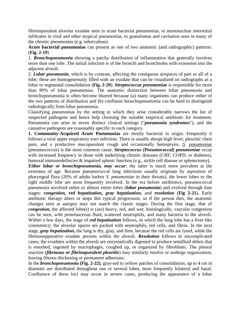
Acute bacterial pneumonias can present as one of two anatomic (and radiographic) patterns:
(Fig. 2-19)
1. Bronchopneumonia showing a patchy distribution of inflammation that generally involves
more than one lobe. The initial infection is of the bronchi and bronchioles with extension into the
adjacent alveoli.
2. Lobar pneumonia, which is by contrast, affecting the contiguous airspaces of part or all of a
lobe; these are homogeneously filled with an exudate that can be visualized on radiographs as a
lobar or segmental consolidation (Fig. 2-20). Streptococcus pneumoniae is responsible for more
than 90% of lobar pneumonias. The anatomic distinction between lobar pneumonia and
bronchopneumonia is often become blurred because (a) many organisms can produce either of
the two patterns of distribution and (b) confluent bronchopneumonia can be hard to distinguish
radiologically from lobar pneumonia.
Classifying pneumonias by the setting in which they arise considerably narrows the list of
suspected pathogens and hence help choosing the suitable empirical antibiotic for treatment.
Pneumonia can arise in seven distinct clinical settings ("pneumonia syndromes"), and the
causative pathogens are reasonably specific to each category.
1. Community-Acquired Acute Pneumonias are mostly bacterial in origin. Frequently it
follows a viral upper respiratory tract infection. There is usually abrupt high fever, pleuritic chest
pain, and a productive mucopurulent cough and occasionally hemoptysis. S. pneumoniae
(pneumococcus) is the most common cause. Streptococcus (Pneumococcal) pneumoniae occur
with increased frequency in those with underlying chronic diseases (CHF, COPD, or diabetes),
humoral immunodefincies & impaired splenic function (e.g., sickle cell disease or splenectomy).
Either lobar or bronchopneumonia, may occur; the latter is much more prevalent at the
extremes of age. Because pneumococcal lung infections usually originate by aspiration of
pharyngeal flora (20% of adults harbor S. pneumoniae in their throats), the lower lobes or the
right middle lobe are most frequently involved. In the era before antibiotics, pneumococcal
pneumonia involved entire or almost entire lobes (lobar pneumonia) and evolved through four
stages: congestion, red hepatization, gray hepatization, and resolution (Fig 2-21). Early
antibiotic therapy alters or stops this typical progression, so if the person dies, the anatomic
changes seen at autopsy may not match the classic stages. During the first stage, that of
congestion, the affected lobe(s) is (are) heavy, red, and wet; histologically, vascular congestion
can be seen, with proteinaceous fluid, scattered neutrophils, and many bacteria in the alveoli.
Within a few days, the stage of red hepatization follows, in which the lung lobe has a liver-like
consistency; the alveolar spaces are packed with neutrophils, red cells, and fibrin. In the next
stage, gray hepatization, the lung is dry, gray, and firm, because the red cells are lysed, while the
fibrinosuppurative exudate persists within the alveoli. Resolution follows in uncomplicated
cases; the exudates within the alveoli are enzymatically digested to produce semifluid debris that
is resorbed, ingested by macrophages, coughed up, or organized by fibroblasts. The pleural
reaction (fibrinous or fibrinopurulent pleuritis) may similarly resolve or undergo organization,
leaving fibrous thickening or permanent adhesions.
In the bronchopneumonia (Fig. 2-22), gray-red to yellow patches of consolidation, up to 4 cm in
diameter are distributed throughout one or several lobes, most frequently bilateral and basal.
Confluence of these foci may occur in severe cases, producing the appearance of a lobar
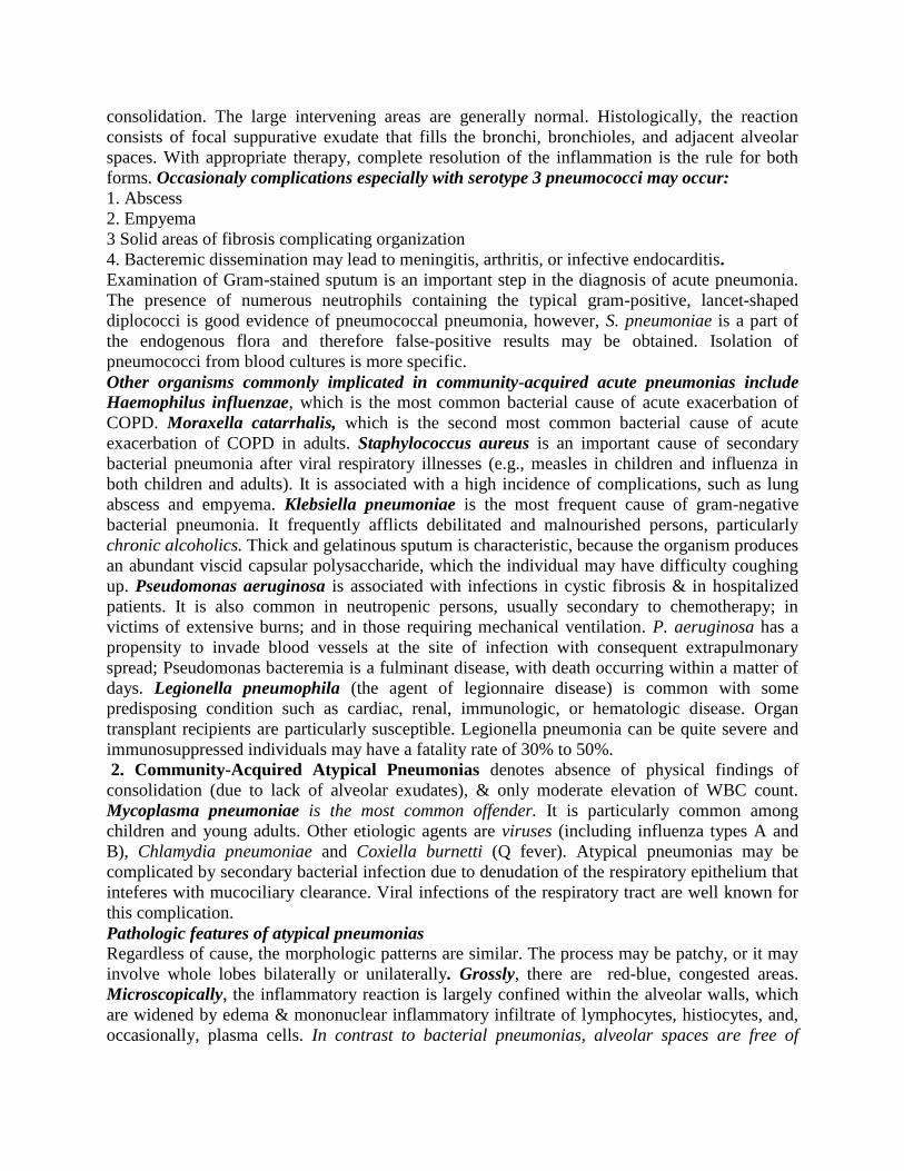
consolidation. The large intervening areas are generally normal. Histologically, the reaction
consists of focal suppurative exudate that fills the bronchi, bronchioles, and adjacent alveolar
spaces. With appropriate therapy, complete resolution of the inflammation is the rule for both
forms. Occasionaly complications especially with serotype 3 pneumococci may occur:
1. Abscess
2. Empyema
3 Solid areas of fibrosis complicating organization
4. Bacteremic dissemination may lead to meningitis, arthritis, or infective endocarditis.
Examination of Gram-stained sputum is an important step in the diagnosis of acute pneumonia.
The presence of numerous neutrophils containing the typical gram-positive, lancet-shaped
diplococci is good evidence of pneumococcal pneumonia, however, S. pneumoniae is a part of
the endogenous flora and therefore false-positive results may be obtained. Isolation of
pneumococci from blood cultures is more specific.
Other organisms commonly implicated in community-acquired acute pneumonias include
Haemophilus influenzae, which is the most common bacterial cause of acute exacerbation of
COPD. Moraxella catarrhalis, which is the second most common bacterial cause of acute
exacerbation of COPD in adults. Staphylococcus aureus is an important cause of secondary
bacterial pneumonia after viral respiratory illnesses (e.g., measles in children and influenza in
both children and adults). It is associated with a high incidence of complications, such as lung
abscess and empyema. Klebsiella pneumoniae is the most frequent cause of gram-negative
bacterial pneumonia. It frequently afflicts debilitated and malnourished persons, particularly
chronic alcoholics. Thick and gelatinous sputum is characteristic, because the organism produces
an abundant viscid capsular polysaccharide, which the individual may have difficulty coughing
up. Pseudomonas aeruginosa is associated with infections in cystic fibrosis & in hospitalized
patients. It is also common in neutropenic persons, usually secondary to chemotherapy; in
victims of extensive burns; and in those requiring mechanical ventilation. P. aeruginosa has a
propensity to invade blood vessels at the site of infection with consequent extrapulmonary
spread; Pseudomonas bacteremia is a fulminant disease, with death occurring within a matter of
days. Legionella pneumophila (the agent of legionnaire disease) is common with some
predisposing condition such as cardiac, renal, immunologic, or hematologic disease. Organ
transplant recipients are particularly susceptible. Legionella pneumonia can be quite severe and
immunosuppressed individuals may have a fatality rate of 30% to 50%.
2. Community-Acquired Atypical Pneumonias denotes absence of physical findings of
consolidation (due to lack of alveolar exudates), & only moderate elevation of WBC count.
Mycoplasma pneumoniae is the most common offender. It is particularly common among
children and young adults. Other etiologic agents are viruses (including influenza types A and
B), Chlamydia pneumoniae and Coxiella burnetti (Q fever). Atypical pneumonias may be
complicated by secondary bacterial infection due to denudation of the respiratory epithelium that
inteferes with mucociliary clearance. Viral infections of the respiratory tract are well known for
this complication.
Pathologic features of atypical pneumonias
Regardless of cause, the morphologic patterns are similar. The process may be patchy, or it may
involve whole lobes bilaterally or unilaterally. Grossly, there are red-blue, congested areas.
Microscopically, the inflammatory reaction is largely confined within the alveolar walls, which
are widened by edema & mononuclear inflammatory infiltrate of lymphocytes, histiocytes, and,
occasionally, plasma cells. In contrast to bacterial pneumonias, alveolar spaces are free of
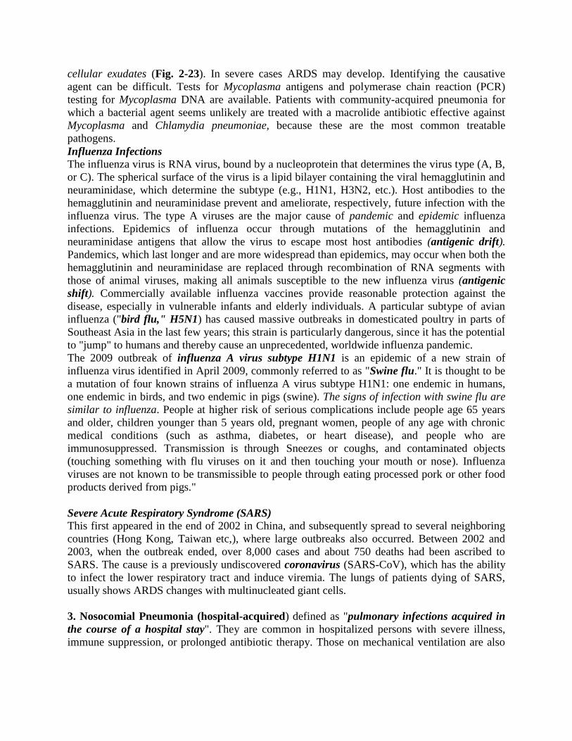
cellular exudates (Fig. 2-23). In severe cases ARDS may develop. Identifying the causative
agent can be difficult. Tests for Mycoplasma antigens and polymerase chain reaction (PCR)
testing for Mycoplasma DNA are available. Patients with community-acquired pneumonia for
which a bacterial agent seems unlikely are treated with a macrolide antibiotic effective against
Mycoplasma and Chlamydia pneumoniae, because these are the most common treatable
pathogens.
Influenza Infections
The influenza virus is RNA virus, bound by a nucleoprotein that determines the virus type (A, B,
or C). The spherical surface of the virus is a lipid bilayer containing the viral hemagglutinin and
neuraminidase, which determine the subtype (e.g., H1N1, H3N2, etc.). Host antibodies to the
hemagglutinin and neuraminidase prevent and ameliorate, respectively, future infection with the
influenza virus. The type A viruses are the major cause of pandemic and epidemic influenza
infections. Epidemics of influenza occur through mutations of the hemagglutinin and
neuraminidase antigens that allow the virus to escape most host antibodies (antigenic drift).
Pandemics, which last longer and are more widespread than epidemics, may occur when both the
hemagglutinin and neuraminidase are replaced through recombination of RNA segments with
those of animal viruses, making all animals susceptible to the new influenza virus (antigenic
shift). Commercially available influenza vaccines provide reasonable protection against the
disease, especially in vulnerable infants and elderly individuals. A particular subtype of avian
influenza ("bird flu," H5N1) has caused massive outbreaks in domesticated poultry in parts of
Southeast Asia in the last few years; this strain is particularly dangerous, since it has the potential
to "jump" to humans and thereby cause an unprecedented, worldwide influenza pandemic.
The 2009 outbreak of influenza A virus subtype H1N1 is an epidemic of a new strain of
influenza virus identified in April 2009, commonly referred to as "Swine flu." It is thought to be
a mutation of four known strains of influenza A virus subtype H1N1: one endemic in humans,
one endemic in birds, and two endemic in pigs (swine). The signs of infection with swine flu are
similar to influenza. People at higher risk of serious complications include people age 65 years
and older, children younger than 5 years old, pregnant women, people of any age with chronic
medical conditions (such as asthma, diabetes, or heart disease), and people who are
immunosuppressed. Transmission is through Sneezes or coughs, and contaminated objects
(touching something with flu viruses on it and then touching your mouth or nose). Influenza
viruses are not known to be transmissible to people through eating processed pork or other food
products derived from pigs."
Severe Acute Respiratory Syndrome (SARS)
This first appeared in the end of 2002 in China, and subsequently spread to several neighboring
countries (Hong Kong, Taiwan etc,), where large outbreaks also occurred. Between 2002 and
2003, when the outbreak ended, over 8,000 cases and about 750 deaths had been ascribed to
SARS. The cause is a previously undiscovered coronavirus (SARS-CoV), which has the ability
to infect the lower respiratory tract and induce viremia. The lungs of patients dying of SARS,
usually shows ARDS changes with multinucleated giant cells.
3. Nosocomial Pneumonia (hospital-acquired) defined as "pulmonary infections acquired in
the course of a hospital stay". They are common in hospitalized persons with severe illness,
immune suppression, or prolonged antibiotic therapy. Those on mechanical ventilation are also
susceptible; infections acquired in this setting are designated ventilator-associated pneumonia.
Gram-negative rods and S. aureus are the most common offenders.
4. Aspiration Pneumonia occurs in markedly debilitated patients or those who aspirate gastric
contents either while unconscious (e.g., after a stroke) or during repeated vomiting. The resultant
pneumonia is partly chemical, resulting from the extremely irritating effects of the gastric acid,
and partly bacterial. Recent studies implicate aerobes (S. pneumoniae, S. aureus, H. influenzae,
and Pseudomonas aeruginosa) more commonly than anaerobes (such as Bacteroides). This type
of pneumonia is often necrotizing with a fulminant clinical course. In those who survive, abscess
formation is a common complication.
5. Necrotizing pneumonia & Lung Abscess
Lung Abscess refers to "a localized area of suppurative necrosis within the pulmonary
parenchyma, resulting in the formation of one or more large cavities". Necrotizing pneumonia
often coexists or evolves into lung abscess, making the distinction between the two somewhat
subjective. The causative organism may be introduced into the lung by any of the following
mechanisms:
a. Aspiration of infective material from carious teeth or infected sinuses or tonsils, as during oral
surgery, anesthesia, coma, or alcoholic intoxication and in debilitated patients with depressed
cough reflexes.
b. Aspiration of gastric contents, usually accompanied by infectious organisms from the
oropharynx.
c. As a complication of necrotizing bacterial pneumonias, particularly those caused by S. aureus,
Streptococcus pyogenes, K. pneumoniae, Pseudomonas spp. etc.
d. Mycotic infections and bronchiectasis
e. Bronchial obstruction, particularly with bronchogenic carcinoma.
f. within a necrotic portion of a tumor
g. Septic embolism, from septic thrombophlebitis or from infective endocarditis of the right side
of the heart.
h. hematogenous spread of bacteria in disseminated pyogenic infection as with staphylococcal
bacteremia.
Anaerobic bacteria are present in almost all lung abscesses, sometimes in vast numbers, and
they are the exclusive isolates in one-third to two-thirds of cases.
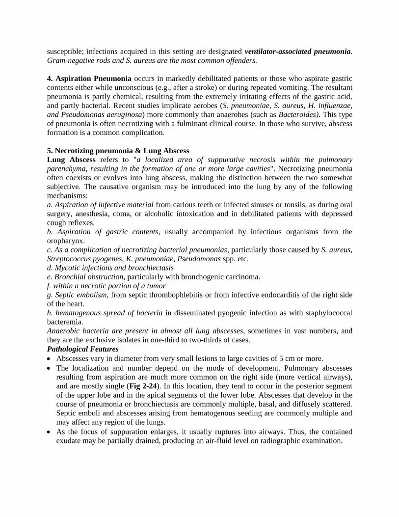
Pathological Features
Abscesses vary in diameter from very small lesions to large cavities of 5 cm or more.
The localization and number depend on the mode of development. Pulmonary abscesses
resulting from aspiration are much more common on the right side (more vertical airways),
and are mostly single (Fig 2-24). In this location, they tend to occur in the posterior segment
of the upper lobe and in the apical segments of the lower lobe. Abscesses that develop in the
course of pneumonia or bronchiectasis are commonly multiple, basal, and diffusely scattered.
Septic emboli and abscesses arising from hematogenous seeding are commonly multiple and
may affect any region of the lungs.
As the focus of suppuration enlarges, it usually ruptures into airways. Thus, the contained
exudate may be partially drained, producing an air-fluid level on radiographic examination.
Microscopic features
There is suppurative liquefactive necrosis (Fig. 2-24)
Depending on the chronicity, the above may be surrounded by variably thickened fibrous
tissue and mononuclear infiltration by variable amounts of (lymphocytes, plasma cells,
macrophages).
Complications
1. Rupture into the pleural cavity producing bronchopleural fistulas, the consequence of which is
pneumothorax or empyema.
2. Embolization of septic material to the brain, gives rise to meningitis or brain abscess.
3. Secondary amyloidosis may develop in chronic cases
Course & prognosis
The manifestations of a lung abscess are similar to those of bronchiectasis (productive cough of
copious, foul sputum). Abscesses occur in up to15% of persons with bronchogenic carcinoma;
thus, when a lung abscess is suspected in an older person, underlying carcinoma must be
considered. Overall, the mortality rate is in the range of 10%.
6. Chronic Pneumonia is mostly a localized lesion in an immunocompetent person, with or
without regional lymph node involvement. There is typically granulomatous inflammation,
which may be due to bacteria (e.g., M. tuberculosis) or fungi. In the immunocompromised, there
is usually systemic dissemination of the causative organism, accompanied by widespread
disease.
7. Pneumonia in the Immunocompromised Host
The appearance of a pulmonary infiltrate and signs of infection (e.g., fever) are some of the most
common and serious complications in immunocompromised persons whose immune and defense
systems are suppressed by disease, immunosuppression for organ transplants, malignancy, or
irradiation. A wide variety of opportunistic microorganisms, many of which rarely cause
infection in normal hosts, can cause these pneumonias.
Examples of pulmonary opportunistic pathogens include 1. Bacteria (P. aeruginosa, Mycobacterium spp., etc)
2. Viruses (cytomegalo and herpesvirus viruses)
3. Fungi (P. jiroveci, Candida spp., Aspergillus spp., and Cryptococcus neoformans).
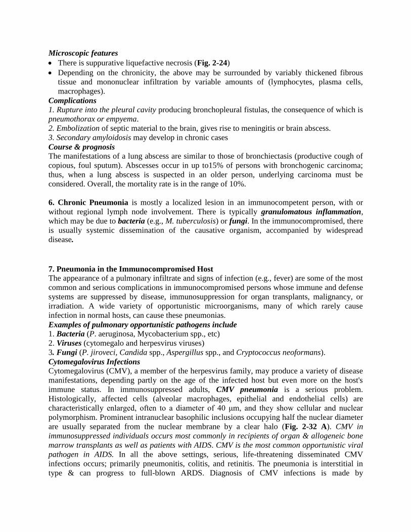
Cytomegalovirus Infections Cytomegalovirus (CMV), a member of the herpesvirus family, may produce a variety of disease
manifestations, depending partly on the age of the infected host but even more on the host's
immune status. In immunosuppressed adults, CMV pneumonia is a serious problem.
Histologically, affected cells (alveolar macrophages, epithelial and endothelial cells) are
characteristically enlarged, often to a diameter of 40 μm, and they show cellular and nuclear
polymorphism. Prominent intranuclear basophilic inclusions occupying half the nuclear diameter
are usually separated from the nuclear membrane by a clear halo (Fig. 2-32 A). CMV in
immunosuppressed individuals occurs most commonly in recipients of organ & allogeneic bone
marrow transplants as well as patients with AIDS. CMV is the most common opportunistic viral
pathogen in AIDS. In all the above settings, serious, life-threatening disseminated CMV
infections occurs; primarily pneumonitis, colitis, and retinitis. The pneumonia is interstitial in
type & can progress to full-blown ARDS. Diagnosis of CMV infections is made by
demonstration of characteristic morphologic alterations in tissue sections, viral culture, rising
antiviral antibody titer, and PCR-based detection of CMV DNA.
Pneumocystis Pneumonia
P. jiroveci (formerly P. carinii), an opportunistic infectious agent long considered to be a
protozoan, is now believed to be more closely related to fungi. Virtually all persons are exposed
to Pneumocystis during early childhood, but in most the infection remains latent. Reactivation
and clinical disease occurs almost exclusively in those who are immunocompromised (very
commonly in AIDS patients but also in the severely malnourished infants). Pneumocystis
produce an interstitial pneumonitis with a characteristic intra-alveolar foamy, pink-staining
exudate with H&E stains ("cotton candy" exudate). Silver stains of tissue sections reveal cup-
shaped cyst in the alveolar exudates (Fig. 2-32 B).The most sensitive and effective method of
diagnosis is to identify the organism in bronchoalveolar lavage fluids or in a transbronchial
biopsy specimen. Immunofluorescence antibody kits and PCR-based assays have also become
available for use on clinical specimens.
Opportunistic Fungal Infection
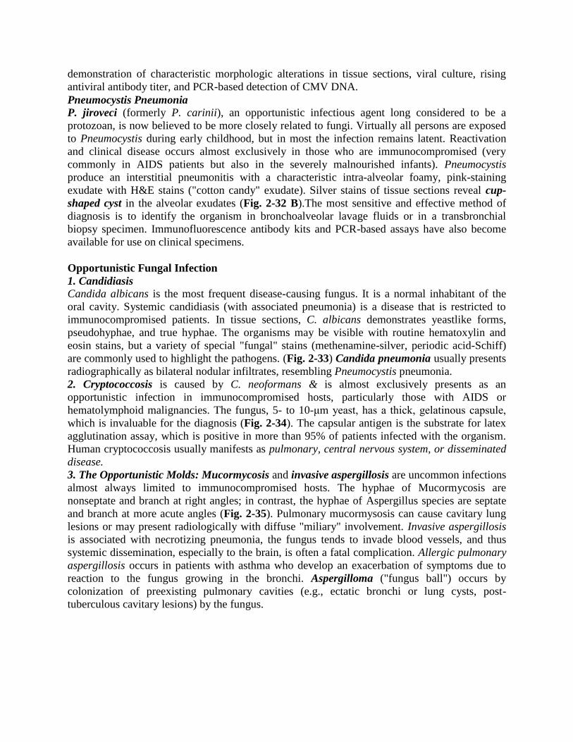
1. Candidiasis
Candida albicans is the most frequent disease-causing fungus. It is a normal inhabitant of the
oral cavity. Systemic candidiasis (with associated pneumonia) is a disease that is restricted to
immunocompromised patients. In tissue sections, C. albicans demonstrates yeastlike forms,
pseudohyphae, and true hyphae. The organisms may be visible with routine hematoxylin and
eosin stains, but a variety of special "fungal" stains (methenamine-silver, periodic acid-Schiff)
are commonly used to highlight the pathogens. (Fig. 2-33) Candida pneumonia usually presents
radiographically as bilateral nodular infiltrates, resembling Pneumocystis pneumonia.
2. Cryptococcosis is caused by C. neoformans & is almost exclusively presents as an
opportunistic infection in immunocompromised hosts, particularly those with AIDS or
hematolymphoid malignancies. The fungus, 5- to 10-μm yeast, has a thick, gelatinous capsule,
which is invaluable for the diagnosis (Fig. 2-34). The capsular antigen is the substrate for latex
agglutination assay, which is positive in more than 95% of patients infected with the organism.
Human cryptococcosis usually manifests as pulmonary, central nervous system, or disseminated
disease.
3. The Opportunistic Molds: Mucormycosis and invasive aspergillosis are uncommon infections
almost always limited to immunocompromised hosts. The hyphae of Mucormycosis are
nonseptate and branch at right angles; in contrast, the hyphae of Aspergillus species are septate
and branch at more acute angles (Fig. 2-35). Pulmonary mucormysosis can cause cavitary lung
lesions or may present radiologically with diffuse "miliary" involvement. Invasive aspergillosis
is associated with necrotizing pneumonia, the fungus tends to invade blood vessels, and thus
systemic dissemination, especially to the brain, is often a fatal complication. Allergic pulmonary
aspergillosis occurs in patients with asthma who develop an exacerbation of symptoms due to
reaction to the fungus growing in the bronchi. Aspergilloma ("fungus ball") occurs by
colonization of preexisting pulmonary cavities (e.g., ectatic bronchi or lung cysts, post-
tuberculous cavitary lesions) by the fungus.
Tuberculosis
Tuberculosis is by far the most important of the chronic penumonia; it causes 6% of all deaths
worldwide. Tuberculosis is "a communicable chronic granulomatous disease caused by
Mycobacterium tuberculosis". It usually involves the lungs but may affect any organ or tissue.
Tuberculosis thrives wherever there is poverty, crowding, and chronic debilitating illness;
elderly, with their weakened defenses, are also susceptible. Certain disease states also increase
the risk:
1. Diabetes mellitus
2. Hodgkin lymphoma
3. Chronic lung disease (particularly silicosis)
4. Chronic renal failure
5. Malnutrition & Alcoholism
6. Immunosuppression including HIV infection.
Most of these predisposing conditions are related to impairment of T cell-mediated immunity
against the Mycobacteria. The latter are slender rods that are acid fast, thus stained positively
with ZN stain. M. tuberculosis hominis is responsible for most cases of tuberculosis.
Oropharyngeal and intestinal tuberculosis contracted by drinking milk contaminated with
Mycobacterium bovis is now rare in developed nations. Other mycobacteria, particularly M.
avium-intracellulare, are much less virulent than M. tuberculosis and rarely cause disease.
However, it complicates up to 30% of patients with AIDS.
Pathogenesis
Primary TB
In the previously unexposed immunocompetent individual, the source of the organism is
exogenous; this leads to the development of cell mediated immunity; primarily mediated by
TH1 cells primarily mediated by TH1 cells, which stimulate macrophages to kill bacteria but
this is associated simultaneously with the development of destructive tissue hypersensitivity in
the form of caseation necrosis.
The virulent organisms once inside macrophages impair effective phagolysosomal digestion,
which in turn leads to unrestricted mycobacterial proliferation. Thus, the earliest phase of
primary tuberculosis is characterized by bacillary proliferation within alveolar macrophages,
with resulting bacteremia and seeding of multiple sites. Nevertheless, most persons at this
stage are asymptomatic; only about 5% of the infected develop significant disease.
The activated macrophages release a variety of mediators including secretion of TNF, which is
responsible for recruitment of monocytes, which in turn undergo activation and differentiation
into the "epithelioid histiocytes" that characterize the granulomatous response.
About 3 weeks are needed for the development of the hypersensitivity reaction.
Pathological features of primary TB
The inhaled bacilli are embedded in the distal airspaces of the lower part of the upper lobe or
the upper part of the lower lobe, usually close to the pleura.
As sensitization develops, a bout1 cm area of gray-white inflammatory consolidation develops
(the Ghon focus). The center of this focus undergoes caseous necrosis.
Tubercle bacilli, either free or within phagocytes, drain to the regional nodes, which also often
caseate. This combination of Ghon focus and nodal involvement is referred to as the Ghon
complex (Fig. 2-25).
During the first few weeks, there is also lymphatic and hematogenous dissemination to other
parts of the body.
In approximately 95% of cases, development of cell-mediated immunity controls the infection.
Hence, the Ghon complex undergoes progressive fibrosis, often followed by radiologically
detectable calcification, and, despite seeding of other organs, no lesions develop.
Microscopically, there is the characteristic granulomatous inflammatory reaction that forms
both caseating and noncaseating tubercles. Individual tubercles are microscopic; it is only
when multiple granulomas coalesce that they become macroscopically visible. The
granulomas are usually enclosed within a fibroblastic rim with lymphocytes. Multinucleate
giant cells are present in the granulomas. (Fig. 2-26)
The chief potential harmful outcomes of primary tuberculosis are
1. Induction of destructive tissue hypersensitivity, which is more damaging on subsequent
infection (secondary TB)
2. Healed foci of scarring may harbor viable bacilli for years, and thus be a potential nidus for
reactivation when host defenses are compromised
3. The disease progresses relentlessly into progressive primary tuberculosis (uncommon). This
occurs in immunocompromised individuals e.g. AIDS patients or in those with nonspecific
impairment of host defenses (malnourished children or elderly). Immunosuppression results in
the absence of a tissue hypersensitivity reaction and thus there are no granulomas but only sheets
of foamy histiocytes packed with the bacilli (nonreactive tuberculosis). Progressive primary
tuberculosis often resembles acute bacterial pneumonia, with lower and middle lobe
consolidation, hilar lymphadenopathy, and pleural effusion; cavitation is rare. Lympho-
hematogenous dissemination may result in the development of tuberculous meningitis and
miliary tuberculosis.
Secondary Tuberculosis (Postprimary) (Reactivation Tuberculosis)
Secondary tuberculosis is the pattern of disease that arises in a previously sensitized host.
Pathogenesis
Reactivation of the dormant primary infection (as in nonendemic, low-prevalence areas) or re-
exposure to the bacilli in a previously sensitized host (as in endemic areas) results in rapid
recruitment of defensive reactions but also tissue necrosis (caseation). This occurs when the
protection (immunity) offered by the primary infection is weakened.
Whatever the source, only less than 5% with primary disease subsequently develop secondary
tuberculosis.
Secondary pulmonary tuberculosis is classically localized to the apex of one or both upper
lobes. This may relate to high oxygen tension in the apices.
Because of the preexistence of hypersensitivity, the bacilli excite a marked tissue response
that tends to wall off the focus. As a result of this localization, the regional lymph nodes
involvement is less prominently than they are in primary tuberculosis.
Cavitation occurs readily in the secondary form, & is almost inevitable in neglected cases.
As a result erosion of airways occurs; this converts the patient into a source of infection to
others; he now raises sputum containing bacilli.
Gross features of secondary TB
The initial lesion is usually a small focus of consolidation, less than 2 cm in diameter, near the
apical pleura. Such foci are sharply circumscribed, firm, and gray-white to yellow areas that
have a variable amount of central caseation and peripheral fibrosis. This, if neglected
progresses to cavitations.
Microscopic features
The active lesions show characteristic coalescent tubercles with central caseation. TB bacilli
can be demonstrated by specific staining methods. (Fig. 2-27)
Progression of secondary TB
In favorable cases, the initial localized apical parenchymal damage undergoes progressive
healing by fibrosis & eventually represented by fibrocalcific scars. This happy outcome occurs
either spontaneously or after therapy. Alternatively, the disease may progress and extend along
several different pathways:
A. Progressive pulmonary tuberculosis: the apical lesion enlarges with expansion of the area of
caseation. Erosion into a bronchus evacuates the caseous center, creating a ragged, irregular
cavity lined by caseous material (Fig. 2-28); whereas erosion of blood vessels results in
hemoptysis. The pleural cavity is always involved and serous pleural effusions, tuberculous
empyema, or fibrous obliteration may develop.
B. Miliary pulmonary disease occurs when organisms drain through lymphatics into the
lymphatic ducts, which empty into the venous return to the right side of the heart and thence into
the pulmonary arteries. Individual lesions are either microscopic or small (2-mm) foci of yellow-
white; these scatter diffusely through the lungs (miliary is derived from the resemblance of these
foci to millet seeds). (Fig. 2-29) Miliary lesions may expand and coalesce to yield almost total
consolidation of large regions or even whole lobes of the lung.
C. Endobronchial, endotracheal, and laryngeal tuberculosis may develop when infective
material is spread either through lymphatic channels or from expectorated infectious material.
The mucosal lining may be studded with minute granulomatous lesions.
D. Systemic miliary tuberculosis occurs when infective foci in the lungs invade the pulmonary
venous return to the heart; the organisms subsequently disseminate through the systemic arterial
system. Almost every organ in the body may be seeded. The appearances are similar to miliary
pulmonary disease. Miliary tuberculosis is most prominent in the liver, bone marrow, spleen
(Fig. 2-30), adrenals, meninges, kidneys, fallopian tubes, and epididymis.
E. Isolated-organ tuberculosis may appear in any one of the organs or tissues seeded
hematogenously and may be the presenting manifestation of tuberculosis. Organs typically
involved include the meninges (tuberculous meningitis), kidneys (renal tuberculosis), adrenals
(formerly an important cause of Addison disease), bones (tuberculous osteomyelitis), and
fallopian tubes (tuberculous salpingitis). When the vertebrae are affected, the disease is referred
to as Pott disease. Paraspinal "cold" abscesses in persons with this disorder may track along the
tissue planes to present as an abdominal or pelvic mass.
F. Tuberculous Lymphadenitis is the most frequent form of extrapulmonary tuberculosis,
usually occurring in the cervical region ("scrofula"). It tends to be unifocal, and most individuals
do not have evidence of ongoing extranodal disease.
H. Intestinal tuberculosis was fairly common as a primary focus of tuberculosis contracted by
the drinking of contaminated milk. In developed countries today, intestinal tuberculosis is more
often a complication of protracted advanced secondary tuberculosis, secondary to the swallowing
of coughed-up infective material. Typically, the organisms are trapped in mucosal lymphoid
aggregates of the small and large bowel, which then undergo inflammatory enlargement with
ulceration of the overlying mucosa, particularly in the ileum.
The diagnosis of pulmonary disease is based in part on the history and on physical and
radiographic findings of consolidation or cavitation in the apices of the lungs. However, tubercle
bacilli must be identified to establish the diagnosis.
The most common method for diagnosis of tuberculosis remains demonstration of acid-fast
organisms in sputum by special stains e.g. acid-fast stain; most protocols require at least two
sputum examinations before labeling the case as sputum negative. Conventional cultures for
mycobacteria require up to 10 weeks. PCR amplification of M. tuberculosis DNA allows for
even greater rapidity of diagnosis and is currently approved for use. PCR assays can detect as
few as 10 organisms in clinical specimens, compared with greater than 10,000 organisms
required for smear positivity. However, culture remains the gold standard because it also
allows testing of drug susceptibility. Prognosis is generally favorable if infections are localized to the lungs, but it worsens
significantly when the disease occurs in the setting of aged, debilitated, or immunosuppressed
persons, who are at high risk for developing miliary tuberculosis, and in those with multi-drug
resistant-TB. Amyloidosis may appear in persistent cases.
Nontuberculous Mycobacterial Disease is mostly in the form of chronic but clinically localized
pulmonary disease in immunocompetent individuals. Strains implicated most frequently include
M. avium-intracellulare. Nontuberculous mycobacteriosis may present as upper lobe cavitary
disease, mimicking tuberculosis, especially in individuals with a long-standing history of
smoking or alcoholism. The presence of concomitant chronic pulmonary disease e.g. COPD, is
an important risk factor. In immunosuppressed individuals (primarily, HIV-positive patients), M.
avium-intracellulare presents as disseminated disease, associated with systemic symptoms.
LUNG TUMORS Bronchial carcinomas constitute 95% of primary lung tumors; the remaining 5% includes
bronchial carcinoids, sarcomas, lymphomas, and a few benign lesions.
Pulmonary Hamartoma is the most common benign lesion; it is rounded, small (3-4 cm),
discrete mass that often displayed as "coin" lesion on chest radiographs. They consist mainly of
mature cartilage with a scattered of bronchial glands that are often admixed with fat, fibrous
tissue, and blood vessels in varying proportions (Fig. 2-36)
Carcinomas
Carcinoma of the lung is the commonest cause of cancer-related deaths in industrialized
countries. The rate of increase among males is slowing down, but it continues to accelerate
among females; it has overrun breast cancer as a cause of death since 1987. This is undoubtedly
related to the strong relationship of cigarette smoking and lung cancer. Most patients are in the
age group of 50-60 years. The prognosis of lung cancer is very poor: the 5-year survival rate for
all stages combined is about 15%.
There are four major histologic types of lung carcinomas 1. Squamous cell carcinoma
2. Adenocarcinoma
3. Small-cell carcinoma
4. Large-cell carcinoma.
Adenocarcinomas are the most common primary tumors arising in women, in lifetime
nonsmokers, and in persons younger than 45 years. For therapeutic purposes, carcinomas of the
lung are divided into two groups: small-cell lung cancer (SCLC) and non-small-cell lung
cancer (NSCLC). The latter category includes squamous cell, adenocarcinomas, and large-cell
carcinomas. The reason for this division is that virtually all SCLCs have metastasized by the time
of diagnosis and hence are not curable by surgery. Therefore, they are best treated by
chemotherapy, with or without radiation. In contrast, NSCLCs usually respond poorly to
chemotherapy and are better treated by surgery. In addition, these two groups show genetic
differences. SCLCs are characterized by a high frequency of RB gene mutations, while the p16
gene is commonly inactivated in NSCLCs.
Etiology and Pathogenesis It seems that large areas of the respiratory mucosa have undergone mutation after exposure to
carcinogens ("field effect"). On this fertile soil, those cells that accumulate additional mutations
ultimately develop into cancer. The role of Cigarette smoking is the main agent responsible for the genetic changes that give
rise to lung cancers. About 90% of lung cancers occur in active smokers (or those who stopped
recently). The increased risk is 60 times greater among habitual heavy smokers (two packs a day
for 20 years) compared with nonsmokers. Women have a higher susceptibility to carcinogens in
tobacco than men. Although cessation of smoking decreases the risk of developing lung cancer
over time, genetic changes that predate lung cancer can persist for many years in the bronchial
epithelium of ex-smokers. Passive smoking (proximity to cigarette smokers) increases the risk to
twice that of nonsmokers. There is a linear correlation between the intensity of smoking and the
appearance of squamous metaplasia that progresses to squamous dysplasia and carcinoma in situ,
before culminating in invasive cancer. Squamous and small-cell carcinomas show the strongest
association with tobacco exposure. The role of occupation-related environmental agents; these may act alone or synergistically
with smoking to be pathogenitically related to some lung cancers, for e.g. radioactive ores; dusts
containing arsenic, chromium, uranium, nickel, vinyl chloride, and mustard gas. Exposure to
asbestos increases the risk of lung cancer 5 times in nonsmokers. However, heavy smoking with
asbestos exposure increases the risk to 50 times. The role of hereditary (genetic) factors: not all persons exposed to tobacco smoke develop
cancer. It seems that the effect of carcinogens is modulated by hereditary (genetic) factors. Many
procarcinogens require metabolic activation via the P-450 enzyme system for conversion into
carcinogens. Evidences support this scenario in that persons with specific genetic abnormalities
of P-450 genes have an increased capacity to metabolize procarcinogens derived from cigarette
smoke and thus sustain the greatest risk of developing lung cancer. The role of bronchioalveolar stem cells (BASCs) in the development of peripheral
adenocarcinoma: the "cell of origin" for peripheral adenocarcinomas is the BASCs. Following
peripheral lung injury, the multipotent BASCs undergo expansion to replenish the normal cell
types found in this location, thereby facilitating epithelial regeneration. BASCs are considered
the tumor initiating cells i.e. the first to acquire the somatic K-RAS mutation that enables their
daughter cells to escape normal "checkpoint" mechanisms and result in pulmonary
adenocarcinomas.
Gross Features
Carcinomas of the lung begin as small mucosal lesions that are usually firm and gray-white.
Further enlargement result in either intraluminal masses, or invasion of the bronchial wall to
form large bulky masses pushing into adjacent lung parenchyma. Obstruction of the bronchial
lumen often produces distal atelectasis and infection. (Fig. 2-37)
Some tumors tend to a rise centrally near the hilum i.e. in major brochi, these are exemplified
by squamous cell & small cell carcinomas. Adenocarcinomas may occur centrally but are
usually more peripherally located, many arising in relation to peripheral lung scars ("scar
carcinomas"). (Fig. 2-38)
Large tumors may undergo cavitation; this is caused by central necrosis (Fig. 2-39)
These tumors may extend to the pleura, invade the pleural cavity and chest wall, and spread to
adjacent intrathoracic structures. (Fig. 2-38) (Fig. 2-40)
Metastasis occurs first to peribronchial, hilar and mediastinal lymph nodes
More distant spread can occur via the lymphatics or the hematogenous route. Microscopic features
Squamous cell carcinomas are often preceded for years by squamous metaplasia or dysplasia in
the bronchial epithelium, which then transforms to carcinoma in situ, a phase that may last for
several years. These tumors range from well-differentiated showing keratin pearls and
intercellular bridges to poorly differentiated neoplasms having only minimal residual squamous
cell features. (Fig. 2-41)
Adenocarcinomas assume a variety of forms, including gland forming (acinar), papillary, and
solid types. The solid variant often requires demonstration of intracellular mucin production by
special stains to establish its adenocarcinomatous nature. (Fig. 2-42) Bronchioloalveolar carcinomas (BACs) are a subtype of adenocarcinomas. They involve
peripheral parts of the lung, either as a single nodule or, more often, as multiple diffuse nodules
that may coalesce to produce pneumonia-like consolidation. The key feature of BACs is their
growth along preexisting structures and preservation of alveolar architecture. (Fig. 2-43) Large-cell carcinomas are undifferentiated malignant epithelial tumors that lack the cytologic
features of glandular or squamous differentiation. The cells have large nuclei, prominent
nucleoli. (Fig. 2-44) Large-cell carcinomas probably represent undifferentiated examples of
squamous cell or adenocarcinomas at the light microscopic level. Ultrastructurally, however,
minimal glandular or squamous differentiation is common. Small-cell lung carcinomas are composed of small tumor cells with a round to fusiform nuclei
with finely granular chromatin, and scant cytoplasm. Mitotic figures are frequently seen (Fig. 2-
45). Despite the term of "small," the neoplastic cells are usually twice the size of resting
lymphocytes. Necrosis is invariably present and may be extensive. These tumors are derived
from neuroendocrine cells of the lung, and hence they express a variety of neuroendocrine
markers in addition to many polypeptide hormones that may result in paraneoplastic syndromes
(see below). Combined tumors: a minority of bronchogenic carcinomas reveal more than one line of
differentiation, sometimes several, suggesting that all are derived from a multipotential
progenitor cell.
All subtypes of lung cancer show involvement of successive chains of nodes about the carina, in
the mediastinum, and in the neck (scalene nodes) and clavicular regions and, sooner or later,
distant metastases. Involvement of the left supraclavicular node (Virchow node) is particularly
characteristic and sometimes calls attention to an occult primary tumor. These cancers, when advanced, often extend into the pericardial or pleural spaces, leading to
inflammation and effusions. They may compress or infiltrate the superior vena cava to cause
either venous congestion or the full-blown vena caval syndrome. Apical neoplasms may invade
the brachial or cervical sympathetic plexus to cause severe pain in the distribution of the ulnar
nerve or to produce Horner syndrome (ipsilateral enophthalmos, ptosis, miosis, and anhidrosis).
Such apical neoplasms are sometimes called Pancoast tumors, and the combination of clinical
findings is known as Pancoast syndrome. Pancoast tumor is often accompanied by destruction of
the first and second ribs and sometimes thoracic vertebrae.
As with other cancers, tumor-node-metastasis (TNM) categories have been established to
indicate the size and spread of the primary neoplasm.
Course & prognosis
Carcinomas of the lung are silent lesions that more often than not have spread beyond curable
resection at the time of diagnosis. Too often, the tumor presents with symptoms related to
metastatic spread to the brain (mental or neurologic changes), liver (hepatomegaly), or bones
(pain). Overall, NSCLCs have a better prognosis than SCLCs. When NSCLCs (squamous cell
carcinomas or adenocarcinomas) are detected before metastasis or local spread, cure is possible
by lobectomy or pneumonectomy. SCLCs, on the other hand, is almost always have spread by
the time of the diagnosis, even if the primary tumor appears small and localized. Thus, surgical
resection is not a practical treatment. They are very sensitive to chemotherapy but invariably
recur. Median survival even with treatment is 1 year.
Paraneoplastic syndromes
Up to10% of all patients with lung cancer develop clinically overt paraneoplastic syndromes.
These include
1. Hypercalcemia caused by secretion of a parathyroid hormone-related peptide.
2. Cushing syndrome (from increased production of ACTH);
3. Inappropriate secretion of ADH
4. Neuromuscular syndromes, including a myasthenic syndrome, peripheral neuropathy, and
polymyositis
5. Clubbing of the fingers and hypertrophic pulmonary osteoarthropathy
6. Hematologic manifestations, including migratory thrombophlebitis, nonbacterial endocarditis,
and disseminated intravascular coagulation.
Hypercalcemia is most often encountered with squamous cell carcinomas, the hematologic
syndromes with adenocarcinomas. The remaining syndromes are much more common with
small-cell neoplasms, but exceptions occur.
Bronchial Carcinoids are thought to arise from the Kulchitsky cells (neuroendocrine cells) of
the bronchial mucosa and represent about 5% of all pulmonary neoplasms. The mean age of
occurrence is 40 years. Ultrastructurally, the neoplastic cells contain cytoplasmic dense-core
neurosecretory granules and may secrete hormonally active polypeptides. In contrast to the more
ominous small-cell carcinomas, carcinoids are often resectable and curable.
Gross features
Most bronchial carcinoids originate in main bronchi and grow in one of two patterns
1. An Obstructing spherical, intraluminal mass (Fig. 2-46)
2. A mucosal plaque penetrating the bronchial wall to fan out in the peribronchial tissue.
Some tumors, (about 10%) metastasize to hilar nodes but distant metastasis is rare.
Microscopic features
They are composed of nests of uniform cells that have regular round nuclei with "salt-and
pepper" chromatin, absent or rare mitoses (Fig. 2-46).
A subset designated atypical carcinoid that differ from the typical by
1. Having higher mitotic rate, cytologic pleomorphism , and focal necrosis
2. Having a higher incidence of lymph node and distant metastasis.
Typical carcinoid, atypical carcinoid, and small-cell carcinoma can be considered to represent a
continuum of increasing histologic aggressiveness and malignant potential within the spectrum
of pulmonary neuroendocrine neoplasms.
Course & prognosis
Most clinical features are related to their intraluminal growth (cough, hemoptysis, and recurrent
bronchial and pulmonary infections). Some are asymptomatic and discovered by chance on
imaging studies. Only rarely do they induce the carcinoid syndrome, characterized by
intermittent attacks of diarrhea, flushing, and cyanosis. The reported 10-year survival rates for
typical carcinoids are 85%, but these drop to 35% for atypical carcinoids. Only 5% of patients
with SCLC are alive at 10 years.
THE PLEURA Pleural Effusion refers to the presence of fluid in the pleural space. The fluid can be either a
transudate or an exudate. A transudate is termed hydrothorax, which is mostly due to CHF. An
exudates is characterized by protein content of 3.0 gm/dl or more and, often, inflammatory cells.
The four principal causes of pleural exudate are
1. Microbial invasion; either direct extension of a pulmonary infection or blood-borne seeding
(suppurative exudates or empyema)
2. Cancer (lung carcinoma, metastatic cancers to the lung or pleural mesothelioma
3. Pulmonary infarction
4. Viral pleuritis
Less common causes include SLE, rheumatoid arthritis, uremia.
Cancer should be suspected as the underlying cause of an exudative effusion in any patient older
than the age of 40, particularly when there is no febrile illness, no pain, and a negative tuberculin
test result. These effusions characteristically are large and frequently bloody (hemorrhagic).
Cytologic examination may reveal malignant and inflammatory cells.
Pneumothorax refers to air in the pleural sac. It may occur in young, healthy adults, usually
men without any known pulmonary disease (spontaneous pneumothorax), or as a result of some
thoracic (e.g. a fractured rib) or lung disorder (secondary pneumothorax). Secondary
pneumothorax occurs with rupture of any pulmonary lesion situated close to the pleural surface
that allows inspired air to gain access to the pleural cavity. Such pulmonary lesions include
emphysema, lung abscess, tuberculosis, carcinoma, etc. Supportive mechanical ventilation with
high pressure may also trigger secondary pneumothorax.
A serious complication may occur through a ball-valve leak that creates a tension
pneumothorax. This shifts the mediastinum, which may be followed by impairment of the
pulmonary circulation that may be fatal.
With prolonged collapse, the lung becomes vulnerable to infection, as does the pleural cavity.
Empyema is thus an important complication of pneumothorax (pyopneumothorax).
Hemothorax is the collection of whole blood (in contrast to bloody effusion) in the pleural
cavity, is a complication of a ruptured intrathoracic aortic aneurysm that is almost always fatal.
Chylothorax is a pleural collection of a milky lymphatic fluid containing microglobules of lipid.
It implies obstruction of the major lymph ducts, usually by an intrathoracic cancer (e.g., a
primary or secondary mediastinal neoplasm, such as a lymphoma).
Malignant Mesothelioma is a rare cancer of mesothelial cells, usually arising in the parietal or
visceral pleura. It also occurs, much less commonly, in the peritoneum and pericardium. In the
USA approximately 50% of individuals with this cancer have a history of exposure to asbestos.
The latent period for developing malignant mesotheliomas is long (25 to 40 years) after initial
asbestos exposure. The combination of cigarette smoking and asbestos exposure greatly
increases the risk of bronchogenic carcinoma, but it does not increase the risk of developing
malignant mesotheliomas.
Gross features
These tumors begin in a localized area but in the course of time spread widely.
At autopsy, the affected lung is typically ensheathed by a yellow-white, firm layer of tumor
that obliterates the pleural space (Fig. 2-47).
The neoplasm may directly invade the thoracic wall or the subpleural lung tissue.
Microscopic features
Mesotheliomas conform to one of three patterns:
1. Epithelial, in which cuboidal cells line tubular and microcystic spaces, into which small
papillary buds project.
2. Sarcomatoid, in which spindled cells grow in sheets
3. Biphasic, having both sarcomatoid and epithelial areas.