Embed Size (px)
Citation preview

Chapter 7
Audition
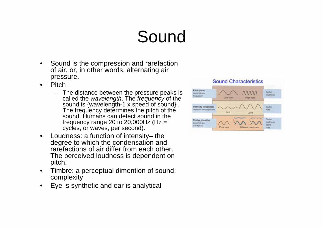
Sound• Sound is the compression and rarefaction
of air, or, in other words, alternating air pressure.
• Pitch– The distance between the pressure peaks is
called the wavelength. The frequency of the sound is {wavelength-1 x speed of sound} . The frequency determines the pitch of the sound. Humans can detect sound in the frequency range 20 to 20,000Hz (Hz = cycles, or waves, per second).
• Loudness: a function of intensity– the degree to which the condensation and rarefactions of air differ from each other. The perceived loudness is dependent on pitch.
• Timbre: a perceptual dimention of sound; complexity
• Eye is synthetic and ear is analytical
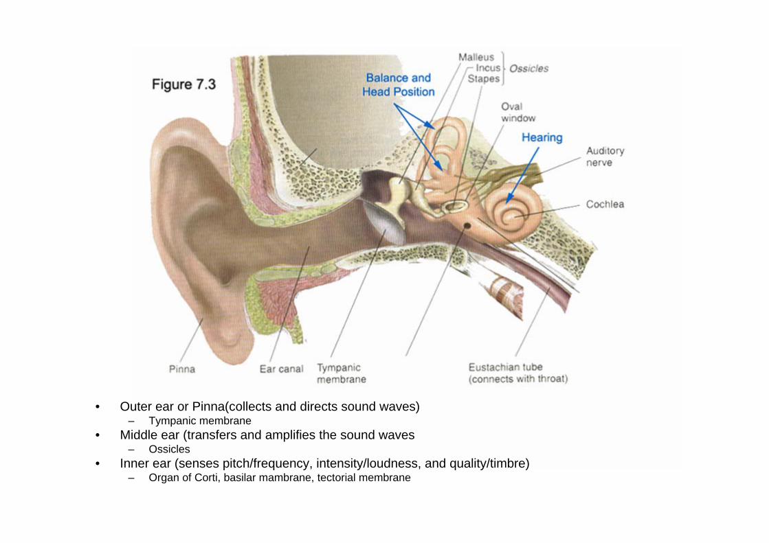
• Outer ear or Pinna(collects and directs sound waves)– Tympanic membrane
• Middle ear (transfers and amplifies the sound waves– Ossicles
• Inner ear (senses pitch/frequency, intensity/loudness, and quality/timbre) – Organ of Corti, basilar mambrane, tectorial membrane
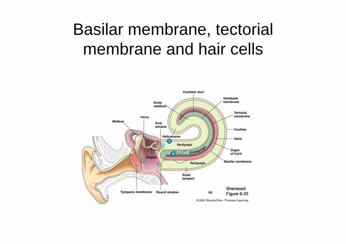
Basilar membrane, tectorialmembrane and hair cells
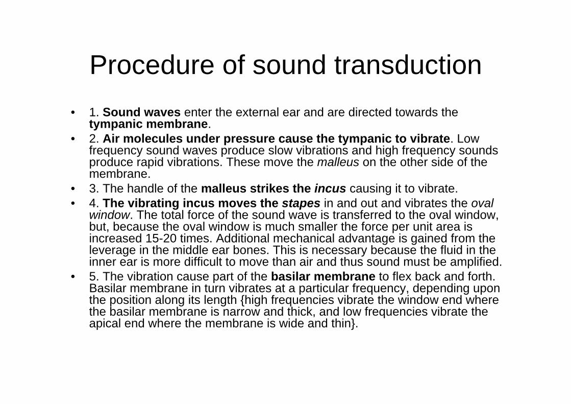
Procedure of sound transduction• 1. Sound waves enter the external ear and are directed towards the
tympanic membrane. • 2. Air molecules under pressure cause the tympanic to vibrate. Low
frequency sound waves produce slow vibrations and high frequency sounds produce rapid vibrations. These move the malleus on the other side of the membrane.
• 3. The handle of the malleus strikes the incus causing it to vibrate. • 4. The vibrating incus moves the stapes in and out and vibrates the oval
window. The total force of the sound wave is transferred to the oval window, but, because the oval window is much smaller the force per unit area is increased 15-20 times. Additional mechanical advantage is gained from the leverage in the middle ear bones. This is necessary because the fluid in the inner ear is more difficult to move than air and thus sound must be amplified.
• 5. The vibration cause part of the basilar membrane to flex back and forth. Basilar membrane in turn vibrates at a particular frequency, depending upon the position along its length {high frequencies vibrate the window end where the basilar membrane is narrow and thick, and low frequencies vibrate the apical end where the membrane is wide and thin}.
• 6. The cilia of the hair cells, which contact the overlying tectorial membrane, bend as the basilar membrane vibrates,
• 7. Opens ion channels and causes the entry of ions into the hair cell and a generator potential develops. If large enough, the generator potential causes transmitter release from the hair cells which excites the afferent nerve. Displacement of the stereocilia in the direction of the tallest stereocilia (called the kinocilium in hair cells of the vestibular system and immature auditory system) is excitatory and in the opposite direction is inhibitory.
• 8. Activation of Ganglion cells• 9. Cochlear nerves to auditory pathway
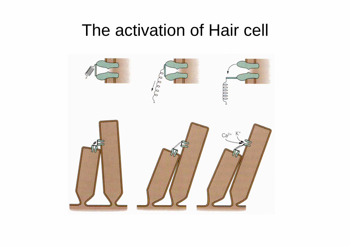
The activation of Hair cell
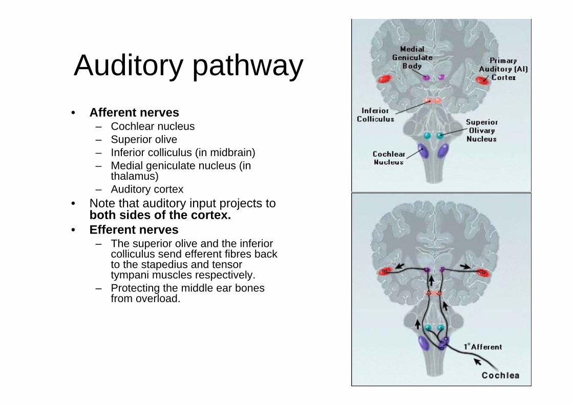
Auditory pathway• Afferent nerves
– Cochlear nucleus– Superior olive– Inferior colliculus (in midbrain)– Medial geniculate nucleus (in
thalamus) – Auditory cortex
• Note that auditory input projects to both sides of the cortex.
• Efferent nerves– The superior olive and the inferior
colliculus send efferent fibres back to the stapedius and tensor tympani muscles respectively.
– Protecting the middle ear bones from overload.
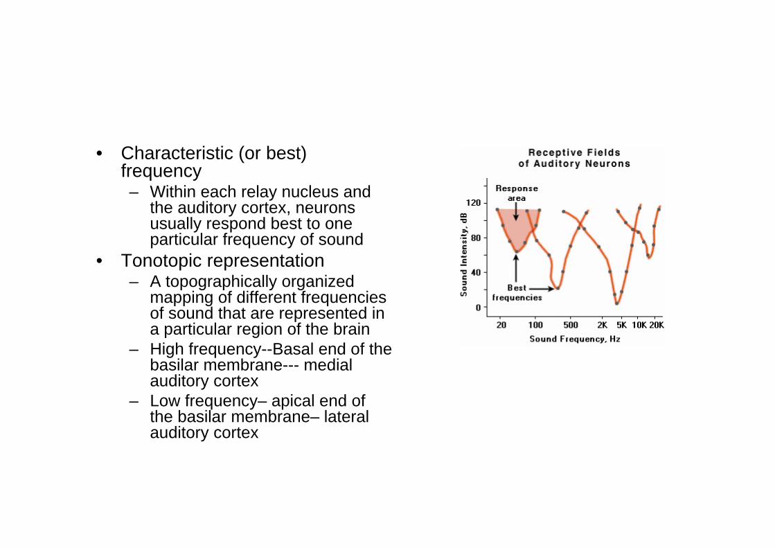
• Characteristic (or best) frequency– Within each relay nucleus and
the auditory cortex, neurons usually respond best to one particular frequency of sound
• Tonotopic representation– A topographically organized
mapping of different frequencies of sound that are represented in a particular region of the brain
– High frequency--Basal end of the basilar membrane--- medial auditory cortex
– Low frequency– apical end of the basilar membrane– lateral auditory cortex
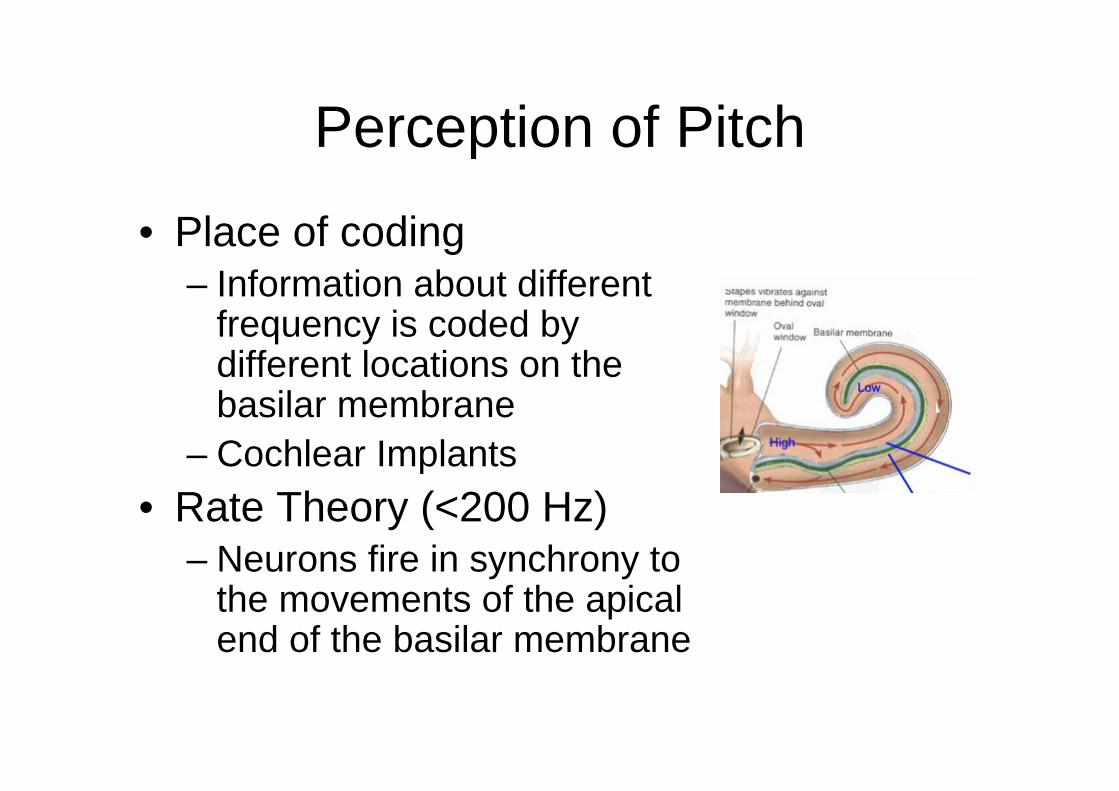
Perception of Pitch
• Place of coding– Information about different
frequency is coded by different locations on the basilar membrane
– Cochlear Implants• Rate Theory (<200 Hz)
– Neurons fire in synchrony to the movements of the apical end of the basilar membrane
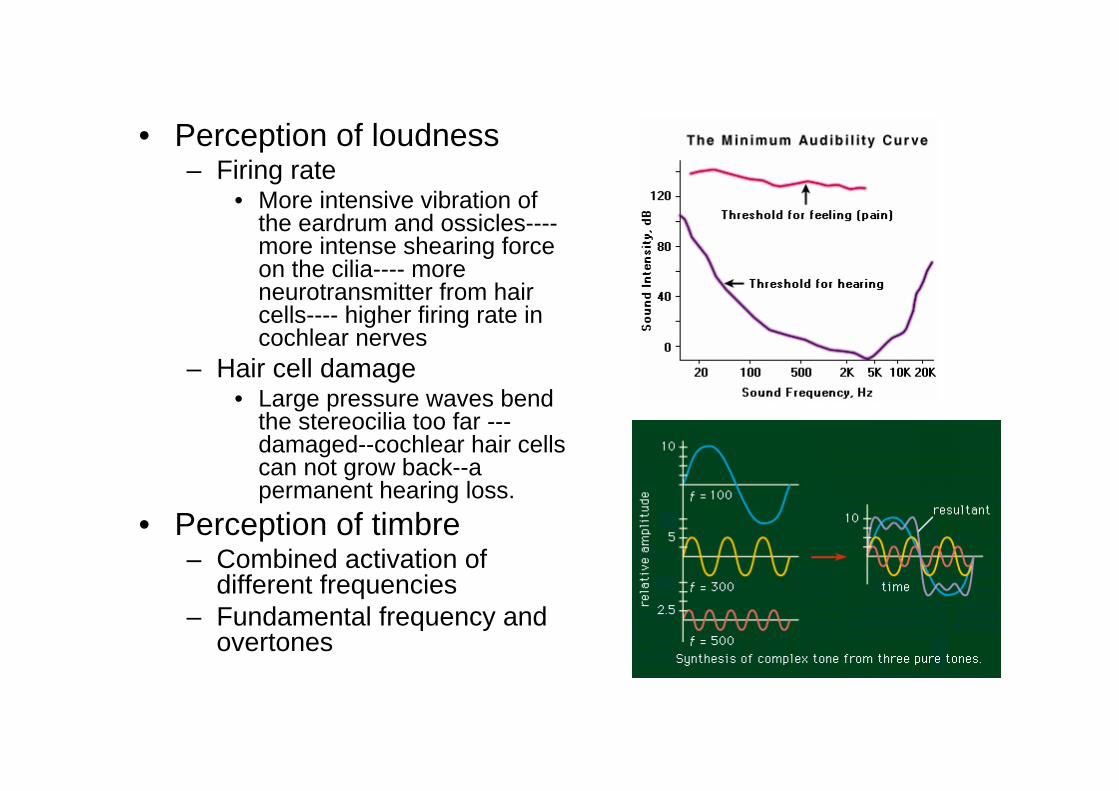
• Perception of loudness– Firing rate
• More intensive vibration of the eardrum and ossicles----more intense shearing force on the cilia---- more neurotransmitter from hair cells---- higher firing rate in cochlear nerves
– Hair cell damage• Large pressure waves bend
the stereocilia too far ---damaged--cochlear hair cells can not grow back--a permanent hearing loss.
• Perception of timbre– Combined activation of
different frequencies– Fundamental frequency and
overtones
Perception of Spatial Location
• By means of arrival time and phase differences
• By means of intensity differences• By means of timbre
– Perception of timbre change at different orientation/ distance
– e.g: Ambulance
• Complex sound: pattern recognition• Auditory Association Cortex
– Ventral (inferior frontal): sound recognition– Dorsal (superior parietal and frontal): sound
location/movement• Music: pitches, timbres, rhythm
– Perception of music involves both primary auditory cortex and subcortical areas.
– Cerebellum and basal ganglia are involved in timing of musical rhythms.
– Increased size of primary auditory cortex and amplitude of the electromagnetic response are found in professional and amateur musicians produced by musical tones
– Amusia: loss of ability to perceive or produce melodic or rhythmic aspect of music
Vestibular System• Components
– 3 Semicircular canals– Vestibular sacs
• Function– (1) it plays the dominant role in the subjective sensation of
motion and spatial orientation of the head– (2) it adjusts muscular activity and body position to maintain
posture– (3) it stabilizes in space the fixation point of the eyes when the
head moves, providing a stable image upon the retina• Pathway
– Afferent: Hair cell– bipolar cell---(vestibular nerve)—medulla—cerebellum, spinal cord, medulla, pons and temporal cortex
– Efferent: from cerebellum and medulla
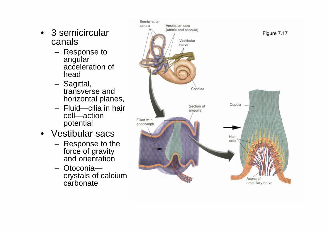
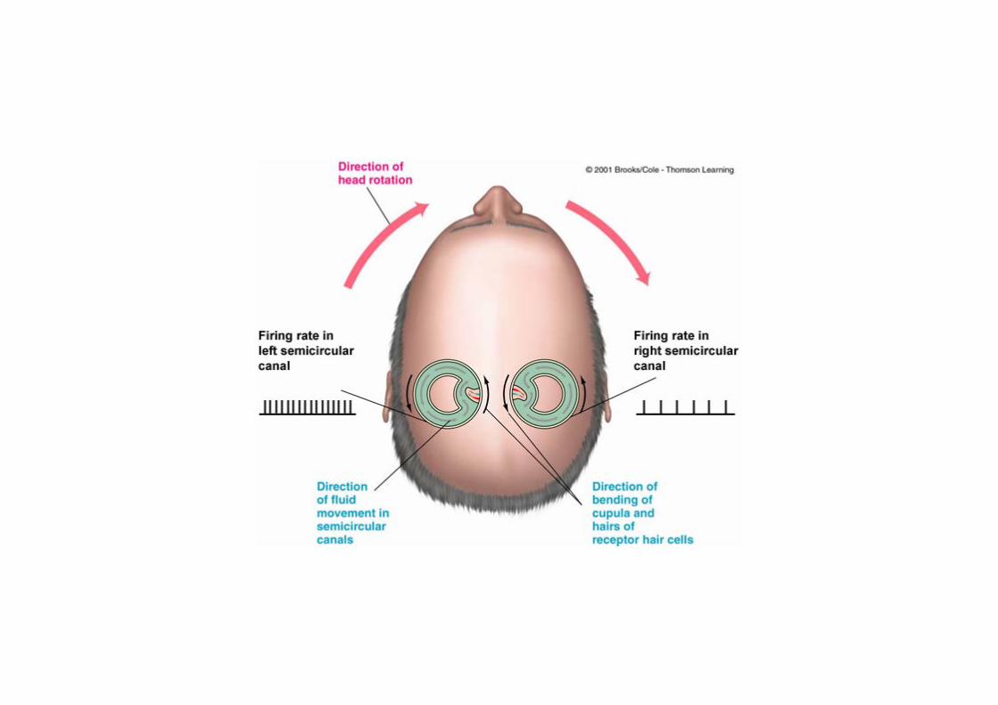
• 3 semicircular canals– Response to
angular acceleration of head
– Sagittal, transverse and horizontal planes,
– Fluid—cilia in hair cell—action potential
• Vestibular sacs– Response to the
force of gravity and orientation
– Otoconia—crystals of calcium carbonate
• Vestibuloocular reflex (VOR)– An induced deviation of the eyes away from the
direction of rotation of the head and results from vestibular connections to the ocular motoneurons
• Nystagmus– Rapid involuntary rhythmic eye movement, with the
eyes moving quickly in one direction (quick phase), and then slowly in the other (slow phase)
• Vertigo– The false sensation that either the person or his world
is rotating due to irritation of vestibular nerve fibers
Somatosenses• Cutaneous sense: touch
– Receive various signals from the skin that form the sense of touch: pressure Vibration heating/cooling stimuli that damage tissue (and producing pain)
• Kinesthesia– provides information about the body position and movement– Kinesthetic signals arise from receptors located within the joints,
tendons and muscles• Organic senses
– a sense modality that arises from receptors located within the inner organs of the body
– providing us with unpleasant sensations such as stomachaches.
Cutaneous Senses• Three different sensations are reported to the brain by receptors localized within skin• Touch involves perception of pressure and vibration of an object on the skin
– Touch receptors (Pacinian corpuscle, Nuffini corpuscle, Merssner’s corpuscle, Merkel’s disk), all mechanoreceptors
– Precisely localized– Adaptation: Moderate & constant stimulus will not produce sensation after it is present for a
period of time. – Stimuli & Motor info: Somatosenses work with the motor system to help us get the info we
need about the objects we come into contact with • Temperature
– Warmth and cold receptors– Receptor activation is relative to the baseline temperature– The receptor lie at different levels of the skin (code are close to the surface of the skin)– Poorly localized
• Pain is associated within skin tissue damage– Detected by Nociceptors, containing both mechanic and chemical receptors– Detected by free nerve endings– Brain reduces pain via endogenous opioids– Poorly localized
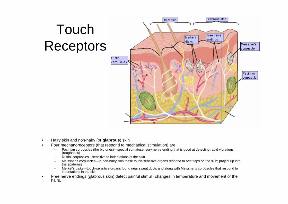
Touch Receptors
• Hairy skin and non-hairy (or glabrous) skin• Four mechanoreceptors (that respond to mechanical stimulation) are:
– Pacinian corpuscles (the big ones)—special somatosensory nerve ending that is good at detecting rapid vibrations (roughness)
– Ruffini corpuscles—sensitive to indentations of the skin – Meissner’s corpuscles—in non-hairy skin these touch-sensitive organs respond to brief taps on the skin; project up into
the epidermis – Merkel’s disks—touch-sensitive organs found near sweat ducts and along with Meissner’s corpuscles that respond to
indentations in the skin • Free nerve endings (glabrous skin) detect painful stimuli, changes in temperature and movement of the
hairs.
Touch related dysfunction
• Tactile agnosia– Apperceptive tactile agnosia
• Can not draw; can not recognize by touch; not able to process info from touch
– Associative tactile agnosia• Able to draw; can not recognized by touch; not connecting to
language/ consciousness
• Tactile apraxia– Difficulty in using normal movement to perceive tactile
information; no damage in tactile perception.
Pain• Pain involves tissue destruction induced by
– Thermal stimuli (e.g. burn hand cooking)– Mechanical force (e.g. slam finger in door)
• Receptors for pain: Nociceptors– Three types: Intense pressure, heat/acids, ATP & ischemia
• Pain receptors are found in – Skin– Sheath around muscles, internal organs– Cornea of the eye– Pulp of the teeth– Not in the brain (no brain pain)!
• Pain receptors are activated by mechanical, chemical stimulation
Perception of Pain• Pain serves a function role for survival
– Persons lacking pain receptors are at great risk• Pain involves three components
– Sensory component: primary and secondary somatosensory cortex– Immediate emotional component: anterior cingulate cortex and insular cortex– Long-term component: prefrontal cortex
• Unique pain responses– No feelings of pain
• Damage to primary somatosensory cortex– Loss of the aversiveness of painful stimuli:
• Feels the pain but doesn’t bother him/her• Damage to anterior cingulate cortex
– Phantom limbs• Sensations that appear to originate in a limb that has been amputated
• Analgesia refers to the reduction of the perception of pain, can be induced externally or internally by
– Hypothsis/ Massage/ Acupuncture/ Opiate/ Placebo/ Electrical stimulation
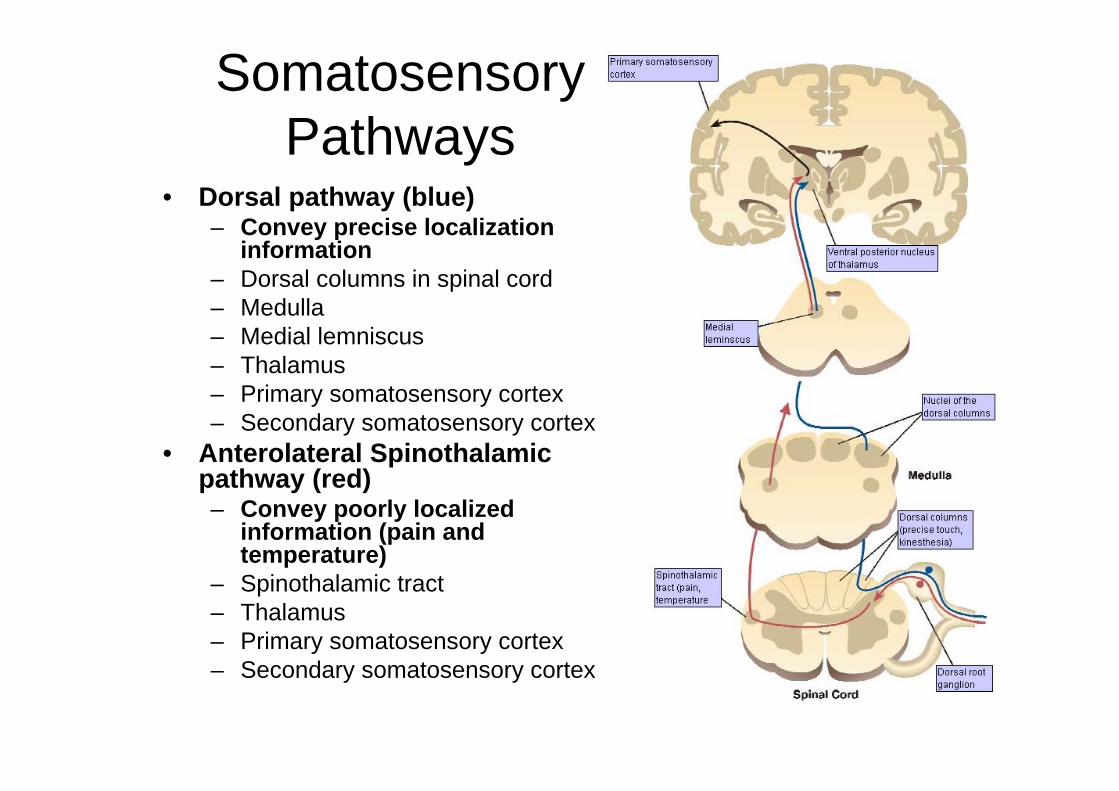
SomatosensoryPathways
• Dorsal pathway (blue)– Convey precise localization
information– Dorsal columns in spinal cord– Medulla– Medial lemniscus– Thalamus– Primary somatosensory cortex– Secondary somatosensory cortex
• Anterolateral Spinothalamicpathway (red)– Convey poorly localized
information (pain and temperature)
– Spinothalamic tract– Thalamus– Primary somatosensory cortex– Secondary somatosensory cortex
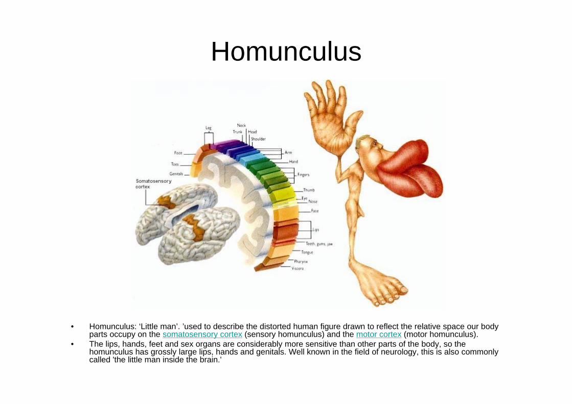
Homunculus
• Homunculus: ‘Little man’. ’used to describe the distorted human figure drawn to reflect the relative space our body parts occupy on the somatosensory cortex (sensory homunculus) and the motor cortex (motor homunculus).
• The lips, hands, feet and sex organs are considerably more sensitive than other parts of the body, so the homunculus has grossly large lips, hands and genitals. Well known in the field of neurology, this is also commonly called 'the little man inside the brain.'
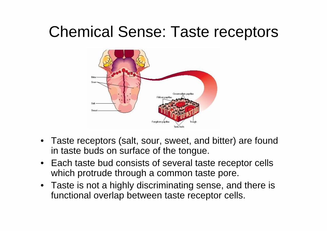
Chemical Sense: Taste receptors
• Taste receptors (salt, sour, sweet, and bitter) are found in taste buds on surface of the tongue.
• Each taste bud consists of several taste receptor cells which protrude through a common taste pore.
• Taste is not a highly discriminating sense, and there is functional overlap between taste receptor cells.
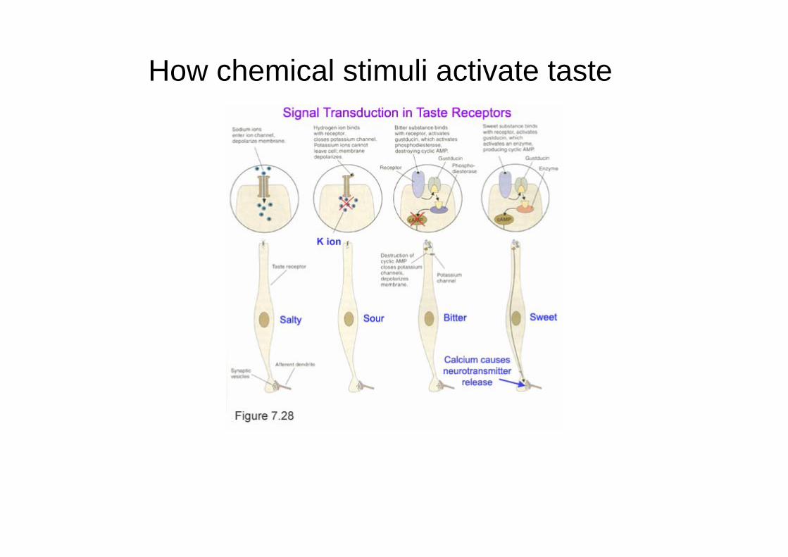
How chemical stimuli activate taste
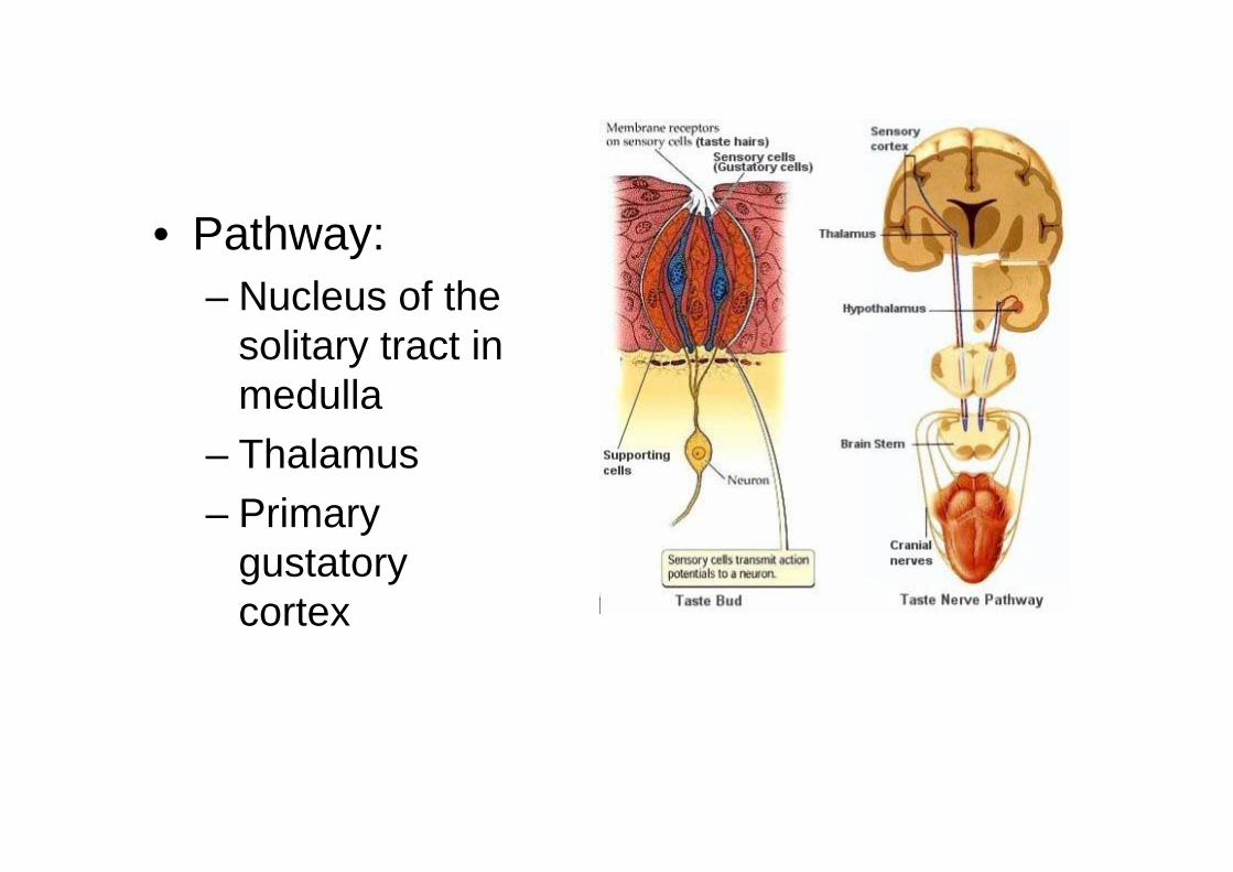
• Pathway: – Nucleus of the
solitary tract in medulla
– Thalamus– Primary
gustatory cortex
Chemical sense: Olfactory System
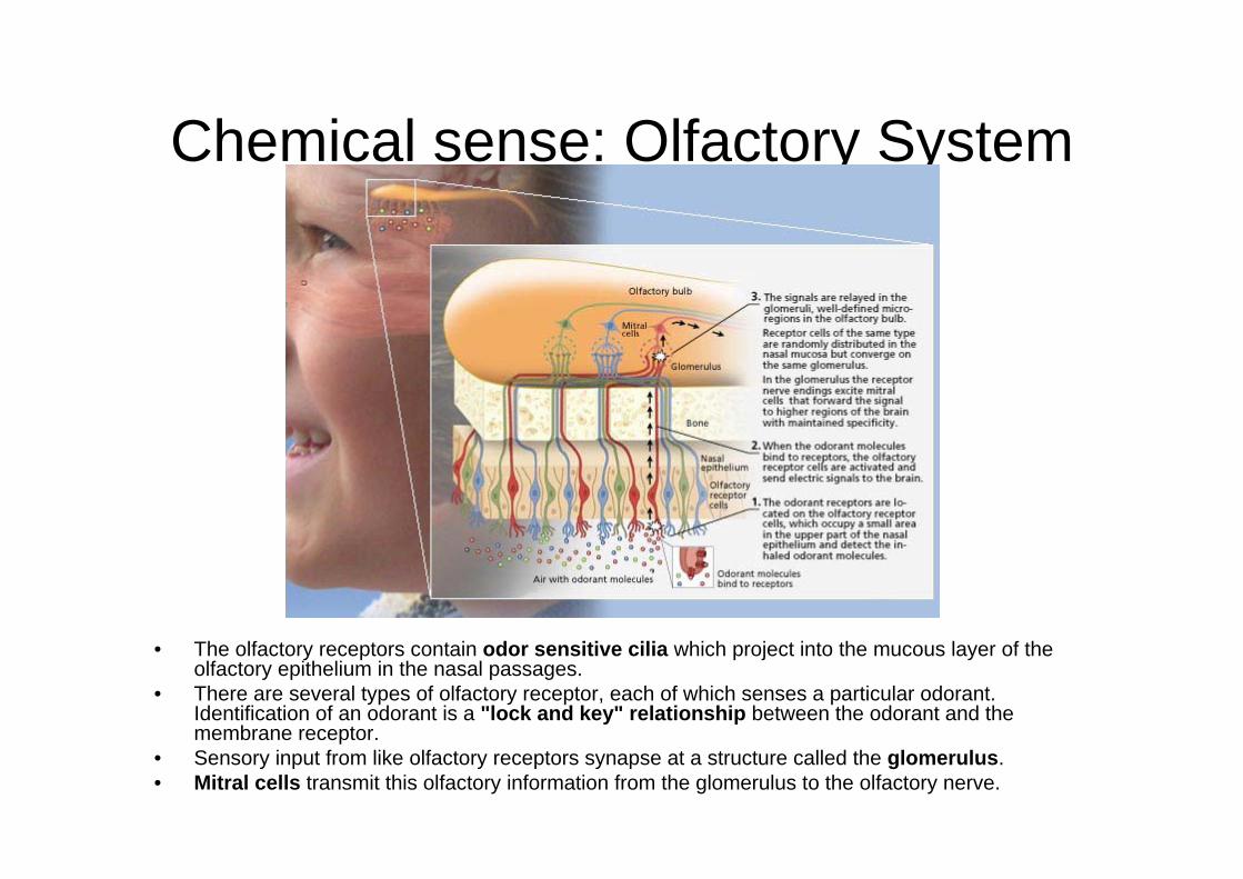
• The olfactory receptors contain odor sensitive cilia which project into the mucous layer of the olfactory epithelium in the nasal passages.
• There are several types of olfactory receptor, each of which senses a particular odorant. Identification of an odorant is a "lock and key" relationship between the odorant and the membrane receptor.
• Sensory input from like olfactory receptors synapse at a structure called the glomerulus. • Mitral cells transmit this olfactory information from the glomerulus to the olfactory nerve.
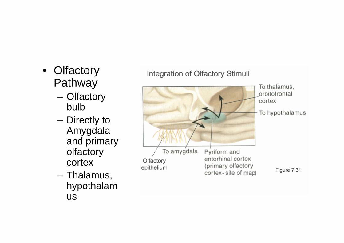
• Olfactory Pathway – Olfactory
bulb– Directly to
Amygdalaand primary olfactory cortex
– Thalamus, hypothalamus
Synesthesia• Some people experience a phenomenon called
synesthesia in which one type of stimulation evokes the sensation of another.
• For example, the hearing of a sound may result in the sensation of the visualization of a color, or a shape may be sensed as a smell.
• Synesthesia is hereditary and it is estimated that it occurs in 1 out of 1000 individuals with variations of type and intensity.
• The most common forms of synesthesia link numbers or letters with colors.

![Neuropsychology and Spina Bifida [Read-Only]spinabifidant.org/wp-content/...Full-Neuropsychology-and-Spina-Bifida.pdf · NEUROPSYCHOLOGY AND THE PSYCHOLOGICAL ASPECTS OF SPINA BIFIDA](https://img.pdfslide.us/doc/110x75/5d59960688c9933d7a8b8165/neuropsychology-and-spina-bifida-read-only-neuropsychology-and-the-psychological.jpg)