Embed Size (px)
Citation preview

1© 2013 David Wild. Published by Elsevier Ltd. The Immunoassay Handbook. Fourth Edition D. Wild (Ed.) http://dx.doi.org/10.1016/B978-0-08-097037-0.00002-6
10002-WILD-9780080970370
To protect the rights of the author(s) and publisher we inform you that this PDF is an uncorrected proof for internal business use only by the author(s), editor(s), reviewer(s), Elsevier and typesetter TNQ Books and Journals Pvt Ltd. It is not allowed to publish this proof online or in print. This proof copy is the copyright property of the publisher and is confi dential until formal publication.
Immunoassays use reagents to generate a signal from minute amounts of target analyte in a sample. Imagine a magnet, securely tied to the end of a fishing line, resting in a stream. Several minute flecks of metal are attracted to the magnet and caught by the person fishing. In an immunoassay, the magnet is replaced by an antibody , which is usually immobilized onto a plastic surface instead of a fishing line. Antibodies are very selective and only bind to their specific targets, even in the pres-ence of a huge range of other materials in a sample. As the analytes are present in miniscule concentrations, it is not enough simply to “catch” them to know how much is there. Another reagent has to be used to gener-ate a signal from the captured material. The level of signal indicates the concentration of the specific analyte under test.
Immunoassays derive their unique specifi city, sensitiv-ity, and fl exibility from three important properties of antibodies:
● Their ability to bind to an extremely wide range of natural and man-made chemicals, biomolecules, cells, and viruses.
● Exceptional specifi city for the substance to which each antibody binds.
● The strength of the binding between an antibody and its target.
Antibodies can be generated by vaccinating animals with the analyte of interest. This process is described as immunization .
Immunometric
Immunoassays
The simplest type of immunoassay to understand is the immunometric design ( Fig. 1 ). An antibody immobilized onto a plastic surface (such as a well in a microtiter® plate) captures the test analyte from the sample, and a different antibody, specifi c for another part of the analyte molecule, is used as the basis of the signal generation system . This antibody is “labeled,” e.g., with a radioactive isotope. After an incubation to allow the antibodies to bind with the analyte, unbound labeled antibody is washed away. The fi nal stage of the assay involves measurement of the level of signal, which is radioactivity in this example. The signal level in this type of assay is proportional to the analyte concentration in the sample.
The labeled component of an immunoassay is sometimes called the tracer . The effi cient removal of unbound tracer by washing is a critical part of the assay, known as the separation . The material (normally plastic) that the capture antibody is irreversibly bound to is known as the solid phase . Because the antibodies form a sandwich around the analyte, immunometric assays are also known as sandwich assays.
In the next example the format is similar, but the immu-noassay has been designed to detect antibodies in a blood sample using the appropriate antigen as the “bait.” This application is useful for detecting previous exposure to a specifi c infectious disease. Proteins that occur on the surface of a virus can be immobilized onto plastic. They
p0010
p0015
u0010
u0015
u0020
p0035
s0010
p0040
p0045
p0050
Immunoassay for Beginners
David Wild ( [email protected] )
c0002 C H A P T E R
1.2
FIGURE 1 Immunometric immunoassay.
f0010

2 The Immunoassay Handbook
10002-WILD-9780080970370
To protect the rights of the author(s) and publisher we inform you that this PDF is an uncorrected proof for internal business use only by the author(s), editor(s), reviewer(s), Elsevier and typesetter TNQ Books and Journals Pvt Ltd. It is not allowed to publish this proof online or in print. This proof copy is the copyright property of the publisher and is confi dential until formal publication.
capture specifi c antibodies for that virus from the sample. As a tracer, a labeled antibody raised in animals against the constant region of human antibody can be used. This is sometimes referred to as the second antibody ( Fig. 2 ).
The next fi gure shows a similar assay with a different type of label used for the tracer ( Fig. 3 ). Instead of a radioactive isotope, an enzyme is chemically attached ( conjugated ) to the labeled antibody. Like antibodies, enzymes are proteins that bind to specifi c targets, but enzymes also catalyze specifi c reactions. The starting material for an enzyme-catalyzed reaction is called a sub-strate . Enzyme labels, with the appropriate substrate, can be used to generate color or create fl uorescent or lumi-nescent end products, which can be readily measured by optical and electronic equipment. Each molecule of enzyme can convert many molecules of substrate, provid-ing a sensitive signal generation system. This format of assay is often known as an enzyme-linked immunosor-bent assay (ELISA).
Competitive Immunoassays
Immunometric assays work well when the target analyte is a large molecule with suffi cient surface area to accommo-date two molecules of antibody. However, many immuno-assays are for small molecules such as drugs, and a different design is needed. This is illustrated in the next fi gure ( Fig. 4 ). Only one antibody is used, and it is present in a limited quantity. The other key reagent, the tracer, is made from the target analyte, labeled with a suitable signal generation material, such as a radioisotope or enzyme. The propor-tion of tracer that binds to the limited antibody sites is indirectly proportional to the concentration of analyte in the sample. This is known as a competitive immunoassay. In this type of assay, the exact amounts of immobilized
antibody and labeled analyte are critical and these assays are sometimes referred to as reagent limited . (In the same context, immunometric (sandwich) assays are described as reagent excess .)
In immunoassays, the analyte that the antibodies bind to is often referred to as the antigen , although the word “antigen” refers to a substance capable of provoking an antibody response. In many competitive immunoassays, the analyte molecules are too small to elicit an antibody response in animals and need to be chemically linked (con-jugated) to a larger molecule, usually a protein, to generate antibodies. Once the antibodies have been generated, it is usually possible to fi nd antibodies from some or all of the animals vaccinated that bind to the analyte alone. In this situation, the analyte is referred to as a hapten . The molecule used to immunize the animals, whether it is the pure analyte or a conjugated version, is called the immunogen .
Homogeneous
Immunoassays
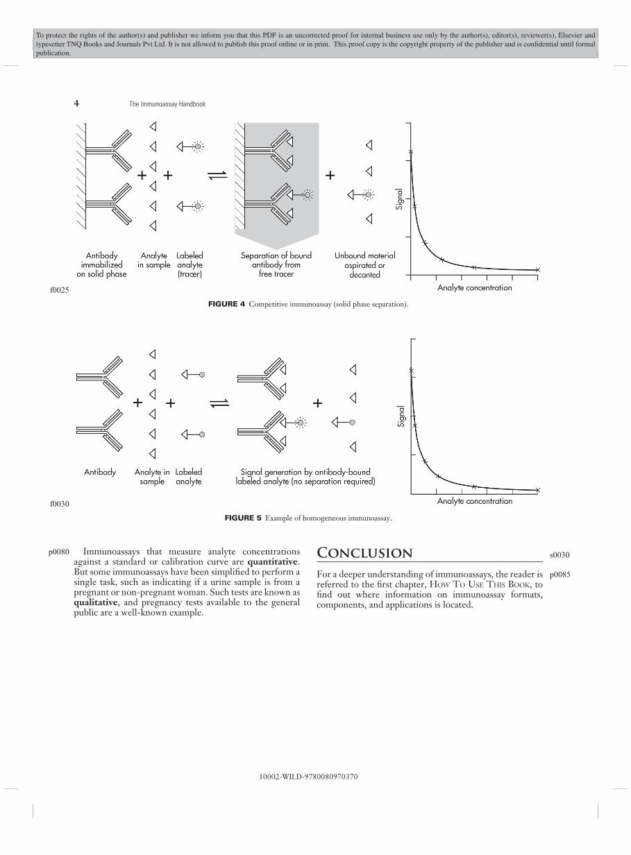
So far, each type of immunoassay described has depended on a separation of unbound tracer before the bound signal is measured. Without a separation (such as a thorough aspiration and wash of the solid phase with buffer prior to signal generation) the level of signal would always be the same, regardless of the concentration of analyte. These assay formats are all examples of heterogeneous immu-noassay. Some assays have been developed that do not require a separation, in which the tracer only generates the signal when it binds to the analyte in an immunometric assay or to the antibody in a competitive assay. They are known as homogeneous immunoassays ( Fig. 5 ).
p0055
s0015
p0060
p0065
s0020
p0070
FIGURE 2 Immunometric assay for antibody testing.
f0015

3CHAPTER 1.2 Immunoassay for Beginners
10002-WILD-9780080970370
To protect the rights of the author(s) and publisher we inform you that this PDF is an uncorrected proof for internal business use only by the author(s), editor(s), reviewer(s), Elsevier and typesetter TNQ Books and Journals Pvt Ltd. It is not allowed to publish this proof online or in print. This proof copy is the copyright property of the publisher and is confi dential until formal publication.
Calibration
In order to estimate the concentration of an analyte from the signal generated, a standard curve is required, which is created by including dilutions of a solution with a known concentration of analyte in the same assay as
the unknown samples. In commercial assay kits, the curve is usually generated by the user from a pre-calibrated set of solutions, known as calibrators , and the curve generated using them is called the calibration curve .
s0025
p0075
FIGURE 3 Enzyme immmunoassay for detection of antibodies (ELISA).
f0020
4 The Immunoassay Handbook
10002-WILD-9780080970370
To protect the rights of the author(s) and publisher we inform you that this PDF is an uncorrected proof for internal business use only by the author(s), editor(s), reviewer(s), Elsevier and typesetter TNQ Books and Journals Pvt Ltd. It is not allowed to publish this proof online or in print. This proof copy is the copyright property of the publisher and is confi dential until formal publication.
Immunoassays that measure analyte concentrations against a standard or calibration curve are quantitative . But some immunoassays have been simplifi ed to perform a single task, such as indicating if a urine sample is from a pregnant or non-pregnant woman. Such tests are known as qualitative , and pregnancy tests available to the general public are a well-known example.
Conclusion
For a deeper understanding of immunoassays, the reader is referred to the fi rst chapter, H OW T O U SE T HIS B OOK , to fi nd out where information on immunoassay formats, components, and applications is located.
p0080 s0030
p0085
FIGURE 4 Competitive immunoassay (solid phase separation).
f0025
FIGURE 5 Example of homogeneous immunoassay.
f0030