Embed Size (px)
Citation preview
18-1
CHA
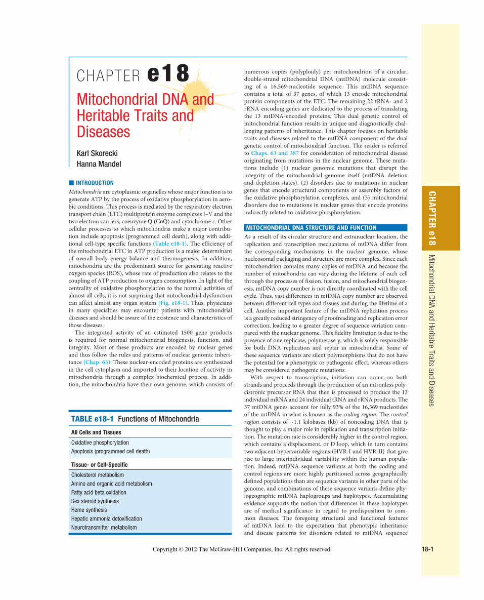
PTER e18
Mitochondrial DNA and Heritable Traits and Diseases
CHAPTER e18
Mitochondrial DNA and Heritable Traits and Diseases Karl Skorecki Hanna Mandel
INTRODUCTION �
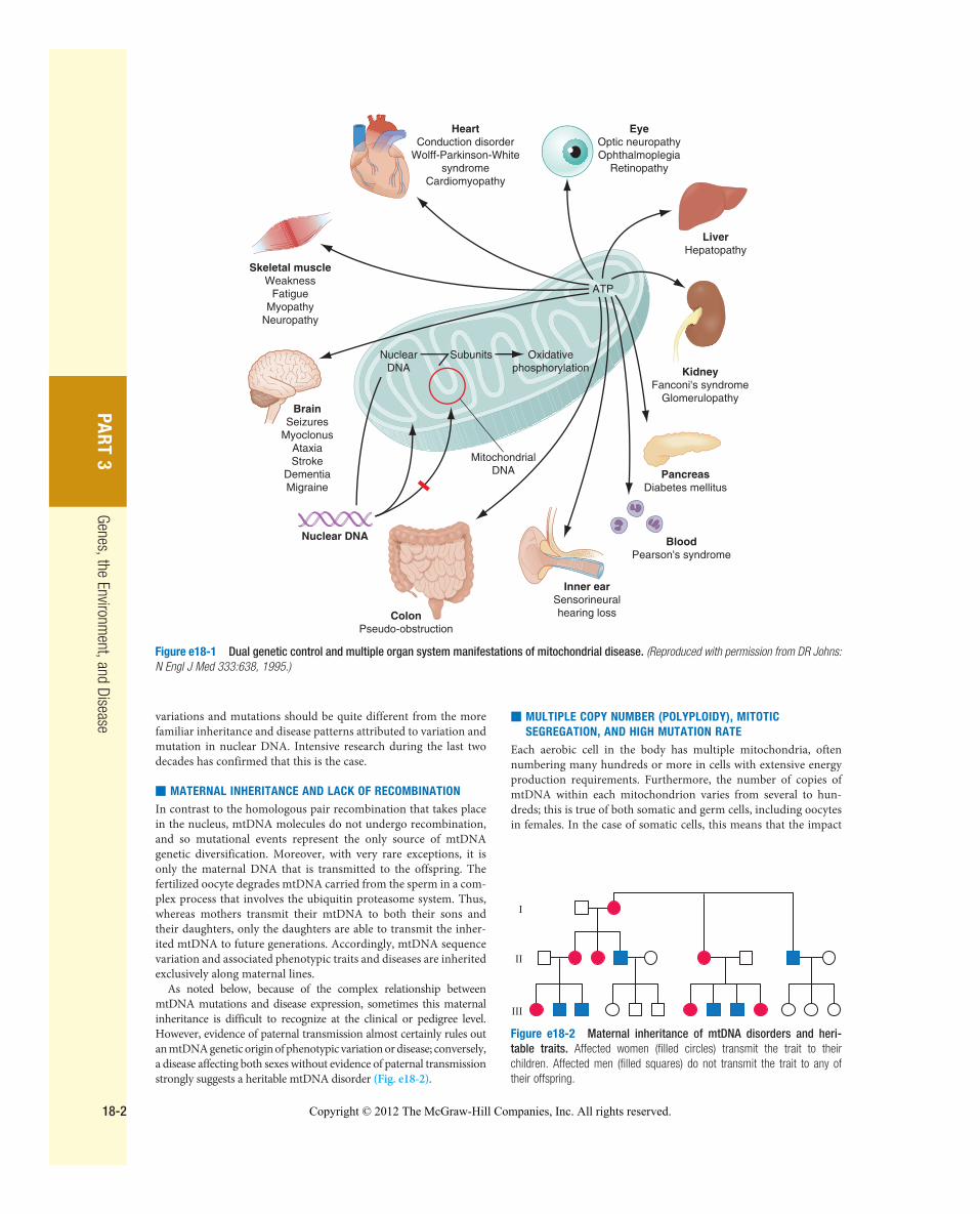
Mitochondria are cytoplasmic organelles whose major function is to generate ATP by the process of oxidative phosphorylation in aero-bic conditions. This process is mediated by the respiratory electron transport chain (ETC) multiprotein enzyme complexes I–V and the two electron carriers, coenzyme Q (CoQ) and cytochrome c. Other cellular processes to which mitochondria make a major contribu-tion include apoptosis (programmed cell death), along with addi-tional cell-type specific functions ( Table e18-1 ) . The efficiency of the mitochondrial ETC in ATP production is a major determinant of overall body energy balance and thermogenesis. In addition, mitochondria are the predominant source for generating reactive oxygen species (ROS), whose rate of production also relates to the coupling of ATP production to oxygen consumption. In light of the centrality of oxidative phosphorylation to the normal activities of almost all cells, it is not surprising that mitochondrial dysfunction can affect almost any organ system ( Fig. e18-1 ) . Thus, physicians in many specialties may encounter patients with mitochondrial diseases and should be aware of the existence and characteristics of those diseases.
The integrated activity of an estimated 1500 gene products is required for normal mitochondrial biogenesis, function, and integrity. Most of these products are encoded by nuclear genes and thus follow the rules and patterns of nuclear genomic inheri-tance ( Chap. 63 ). These nuclear-encoded proteins are synthesized in the cell cytoplasm and imported to their location of activity in mitochondria through a complex biochemical process. In addi-tion, the mitochondria have their own genome, which consists of
numerous copies (polyploidy) per mitochondrion of a circular, double-strand mitochondrial DNA (mtDNA) molecule consist-ing of a 16,569-nucleotide sequence. This mtDNA sequence contains a total of 37 genes, of which 13 encode mitochondrial protein components of the ETC. The remaining 22 tRNA- and 2 rRNA-encoding genes are dedicated to the process of translating the 13 mtDNA-encoded proteins. This dual genetic control of mitochondrial function results in unique and diagnostically chal-lenging patterns of inheritance. This chapter focuses on heritable traits and diseases related to the mtDNA component of the dual genetic control of mitochondrial function. The reader is referred to Chaps. 63 and 387 for consideration of mitochondrial disease originating from mutations in the nuclear genome. These muta-tions include (1) nuclear genomic mutations that disrupt the integrity of the mitochondrial genome itself (mtDNA deletion and depletion states), (2) disorders due to mutations in nuclear genes that encode structural components or assembly factors of the oxidative phosphorylation complexes, and (3) mitochondrial disorders due to mutations in nuclear genes that encode proteins indirectly related to oxidative phosphorylation.
MITOCHONDRIAL DNA STRUCTURE AND FUNCTION As a result of its circular structure and extranuclear location, the replication and transcription mechanisms of mtDNA differ from the corresponding mechanisms in the nuclear genome, whose nucleosomal packaging and structure are more complex. Since each mitochondrion contains many copies of mtDNA and because the number of mitochondria can vary during the lifetime of each cell through the processes of fission, fusion, and mitochondrial biogen-esis, mtDNA copy number is not directly coordinated with the cell cycle. Thus, vast differences in mtDNA copy number are observed between different cell types and tissues and during the lifetime of a cell. Another important feature of the mtDNA replication process is a greatly reduced stringency of proofreading and replication error correction, leading to a greater degree of sequence variation com-pared with the nuclear genome. This fidelity limitation is due to the presence of one replicase, polymerase γ, which is solely responsible for both DNA replication and repair in mitochondria. Some of these sequence variants are silent polymorphisms that do not have the potential for a phenotypic or pathogenic effect, whereas others may be considered pathogenic mutations.
With respect to transcription, initiation can occur on both strands and proceeds through the production of an intronless poly-cistronic precursor RNA that then is processed to produce the 13 individual mRNA and 24 individual tRNA and rRNA products. The 37 mtDNA genes account for fully 93% of the 16,569 nucleotides of the mtDNA in what is known as the coding region . The control region consists of ~1.1 kilobases (kb) of noncoding DNA that is thought to play a major role in replication and transcription initia-tion. The mutation rate is considerably higher in the control region, which contains a displacement, or D loop, which in turn contains two adjacent hypervariable regions (HVR-I and HVR-II) that give rise to large interindividual variability within the human popula-tion. Indeed, mtDNA sequence variants at both the coding and control regions are more highly partitioned across geographically defined populations than are sequence variants in other parts of the genome, and combinations of these sequence variants define phy-logeographic mtDNA haplogroups and haplotypes. Accumulating evidence supports the notion that differences in these haplotypes are of medical significance in regard to predisposition to com-mon diseases. The foregoing structural and functional features of mtDNA lead to the expectation that phenotypic inheritance and disease patterns for disorders related to mtDNA sequence
TABLE e18-1 Functions of Mitochondria
All Cells and Tissues
Oxidative phosphorylation
Apoptosis (programmed cell death)
Tissue- or Cell-Specific
Cholesterol metabolism
Amino and organic acid metabolism
Fatty acid beta oxidation
Sex steroid synthesis
Heme synthesis
Hepatic ammonia detoxification
Neurotransmitter metabolism
Copyright © 2012 The McGraw-Hill Companies, Inc. All rights reserved.
18-2
PART 3
Genes, the Environment, and Disease
variations and mutations should be quite different from the more familiar inheritance and disease patterns attributed to variation and mutation in nuclear DNA. Intensive research during the last two decades has confirmed that this is the case.
MATERNAL INHERITANCE AND LACK OF RECOMBINATION �
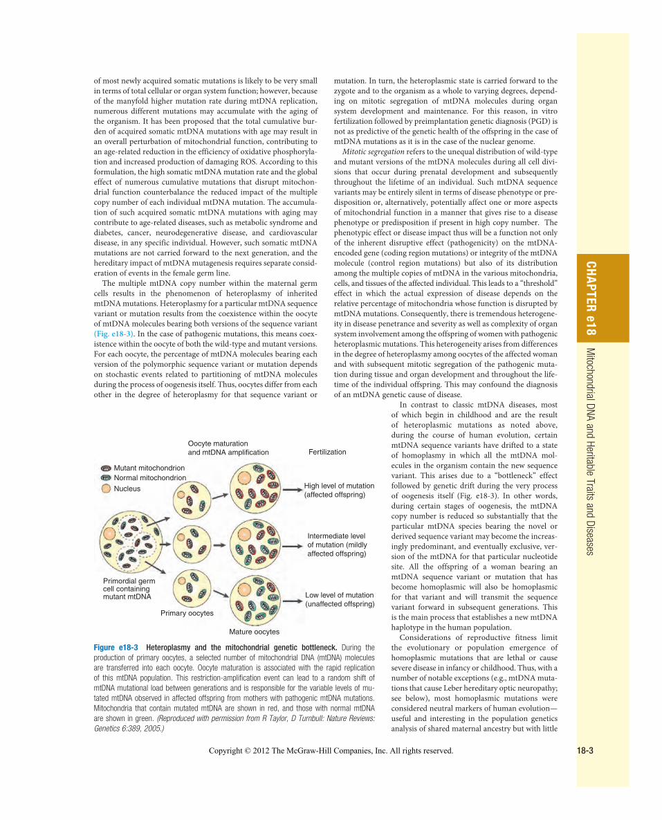
In contrast to the homologous pair recombination that takes place in the nucleus, mtDNA molecules do not undergo recombination, and so mutational events represent the only source of mtDNA genetic diversification. Moreover, with very rare exceptions, it is only the maternal DNA that is transmitted to the offspring. The fertilized oocyte degrades mtDNA carried from the sperm in a com-plex process that involves the ubiquitin proteasome system. Thus, whereas mothers transmit their mtDNA to both their sons and their daughters, only the daughters are able to transmit the inher-ited mtDNA to future generations. Accordingly, mtDNA sequence variation and associated phenotypic traits and diseases are inherited exclusively along maternal lines.
As noted below, because of the complex relationship between mtDNA mutations and disease expression, sometimes this maternal inheritance is difficult to recognize at the clinical or pedigree level. However, evidence of paternal transmission almost certainly rules out an mtDNA genetic origin of phenotypic variation or disease; conversely, a disease affecting both sexes without evidence of paternal transmission strongly suggests a heritable mtDNA disorder ( Fig. e18-2 ) .
MULTIPLE COPY NUMBER (POLYPLOIDY), MITOTIC �SEGREGATION, AND HIGH MUTATION RATE
Each aerobic cell in the body has multiple mitochondria, often numbering many hundreds or more in cells with extensive energy production requirements. Furthermore, the number of copies of mtDNA within each mitochondrion varies from several to hun-dreds; this is true of both somatic and germ cells, including oocytes in females. In the case of somatic cells, this means that the impact
ATP
Oxidativephosphorylation
SubunitsNuclearDNA
Nuclear DNA
BrainSeizures
MyoclonusAtaxiaStroke
DementiaMigraine
Skeletal muscleWeakness
FatigueMyopathy
Neuropathy
HeartConduction disorder
Wolff-Parkinson-Whitesyndrome
Cardiomyopathy
EyeOptic neuropathyOphthalmoplegia
Retinopathy
BloodPearson's syndrome
Inner earSensorineuralhearing lossColon
Pseudo-obstruction
LiverHepatopathy
KidneyFanconi's syndrome
Glomerulopathy
PancreasDiabetes mellitus
MitochondrialDNA
Figure e18-1 Dual genetic control and multiple organ system manifestations of mitochondrial disease. (Reproduced with permission from DR Johns: N Engl J Med 333:638, 1995.)
Figure e18-2 Maternal inheritance of mtDNA disorders and heri-table traits. Affected women (filled circles) transmit the trait to their children. Affected men (filled squares) do not transmit the trait to any of their offspring.
I
II
III
Copyright © 2012 The McGraw-Hill Companies, Inc. All rights reserved.
18-3
CHA
PTER e18
Mitochondrial DNA and Heritable Traits and Diseases
of most newly acquired somatic mutations is likely to be very small in terms of total cellular or organ system function; however, because of the manyfold higher mutation rate during mtDNA replication, numerous different mutations may accumulate with the aging of the organism. It has been proposed that the total cumulative bur-den of acquired somatic mtDNA mutations with age may result in an overall perturbation of mitochondrial function, contributing to an age-related reduction in the efficiency of oxidative phosphoryla-tion and increased production of damaging ROS. According to this formulation, the high somatic mtDNA mutation rate and the global effect of numerous cumulative mutations that disrupt mitochon-drial function counterbalance the reduced impact of the multiple copy number of each individual mtDNA mutation. The accumula-tion of such acquired somatic mtDNA mutations with aging may contribute to age-related diseases, such as metabolic syndrome and diabetes, cancer, neurodegenerative disease, and cardiovascular disease, in any specific individual. However, such somatic mtDNA mutations are not carried forward to the next generation, and the hereditary impact of mtDNA mutagenesis requires separate consid-eration of events in the female germ line.
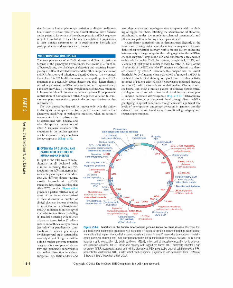
The multiple mtDNA copy number within the maternal germ cells results in the phenomenon of heteroplasmy of inherited mtDNA mutations. Heteroplasmy for a particular mtDNA sequence variant or mutation results from the coexistence within the oocyte of mtDNA molecules bearing both versions of the sequence variant ( Fig. e18-3 ) . In the case of pathogenic mutations, this means coex-istence within the oocyte of both the wild-type and mutant versions. For each oocyte, the percentage of mtDNA molecules bearing each version of the polymorphic sequence variant or mutation depends on stochastic events related to partitioning of mtDNA molecules during the process of oogenesis itself. Thus, oocytes differ from each other in the degree of heteroplasmy for that sequence variant or
mutation. In turn, the heteroplasmic state is carried forward to the zygote and to the organism as a whole to varying degrees, depend-ing on mitotic segregation of mtDNA molecules during organ system development and maintenance. For this reason, in vitro fertilization followed by preimplantation genetic diagnosis (PGD) is not as predictive of the genetic health of the offspring in the case of mtDNA mutations as it is in the case of the nuclear genome.
Mitotic segregation refers to the unequal distribution of wild-type and mutant versions of the mtDNA molecules during all cell divi-sions that occur during prenatal development and subsequently throughout the lifetime of an individual. Such mtDNA sequence variants may be entirely silent in terms of disease phenotype or pre-disposition or, alternatively, potentially affect one or more aspects of mitochondrial function in a manner that gives rise to a disease phenotype or predisposition if present in high copy number. The phenotypic effect or disease impact thus will be a function not only of the inherent disruptive effect (pathogenicity) on the mtDNA-encoded gene (coding region mutations) or integrity of the mtDNA molecule (control region mutations) but also of its distribution among the multiple copies of mtDNA in the various mitochondria, cells, and tissues of the affected individual. This leads to a “threshold” effect in which the actual expression of disease depends on the relative percentage of mitochondria whose function is disrupted by mtDNA mutations. Consequently, there is tremendous heterogene-ity in disease penetrance and severity as well as complexity of organ system involvement among the offspring of women with pathogenic heteroplasmic mutations. This heterogeneity arises from differences in the degree of heteroplasmy among oocytes of the affected woman and with subsequent mitotic segregation of the pathogenic muta-tion during tissue and organ development and throughout the life-time of the individual offspring. This may confound the diagnosis of an mtDNA genetic cause of disease.
In contrast to classic mtDNA diseases, most of which begin in childhood and are the result of heteroplasmic mutations as noted above, during the course of human evolution, certain mtDNA sequence variants have drifted to a state of homoplasmy in which all the mtDNA mol-ecules in the organism contain the new sequence variant. This arises due to a “bottleneck” effect followed by genetic drift during the very process of oogenesis itself ( Fig. e18-3 ). In other words, during certain stages of oogenesis, the mtDNA copy number is reduced so substantially that the particular mtDNA species bearing the novel or derived sequence variant may become the increas-ingly predominant, and eventually exclusive, ver-sion of the mtDNA for that particular nucleotide site. All the offspring of a woman bearing an mtDNA sequence variant or mutation that has become homoplasmic will also be homoplasmic for that variant and will transmit the sequence variant forward in subsequent generations. This is the main process that establishes a new mtDNA haplotype in the human population.
Considerations of reproductive fitness limit the evolutionary or population emergence of homoplasmic mutations that are lethal or cause severe disease in infancy or childhood. Thus, with a number of notable exceptions (e.g., mtDNA muta-tions that cause Leber hereditary optic neuropathy; see below), most homoplasmic mutations were considered neutral markers of human evolution—useful and interesting in the population genetics analysis of shared maternal ancestry but with little
Oocyte maturationand mtDNA amplification Fertilization
Mature oocytes
Primary oocytes
Mutant mitochondrionNormal mitochondrion
Nucleus
Primordial germcell containingmutant mtDNA
High level of mutation(affected offspring)
Intermediate levelof mutation (mildlyaffected offspring)
Low level of mutation(unaffected offspring)
Figure e18-3 Heteroplasmy and the mitochondrial genetic bottleneck. During the production of primary oocytes, a selected number of mitochondrial DNA (mtDNA) molecules are transferred into each oocyte. Oocyte maturation is associated with the rapid replication of this mtDNA population. This restriction-amplification event can lead to a random shift of mtDNA mutational load between generations and is responsible for the variable levels of mu-tated mtDNA observed in affected offspring from mothers with pathogenic mtDNA mutations. Mitochondria that contain mutated mtDNA are shown in red, and those with normal mtDNA are shown in green. (Reproduced with permission from R Taylor, D Turnbull: Nature Reviews: Genetics 6:389, 2005.)
Copyright © 2012 The McGraw-Hill Companies, Inc. All rights reserved.
18-4
PART 3
Genes, the Environment, and Disease
significance in human phenotypic variation or disease predisposi-tion. However, recent research and clinical attention have focused on the potential for certain of these homoplasmic mtDNA sequence variants to contribute to the evolutionary adaptation of populations to their climatic environment or to predispose to heritable late postreproductive and age-associated diseases.
MITOCHONDRIAL DNA DISEASE The true prevalence of mtDNA disease is difficult to estimate because of the phenotypic heterogeneity that occurs as a function of heteroplasmy, the challenge of detecting and assessing hetero-plasmy in different affected tissues, and the other unique features of mtDNA function and inheritance described above. It is estimated that at least 1 in 200 healthy humans harbors a pathogenic mtDNA mutation that potentially causes disease but that heteroplasmic germ-line pathogenic mtDNA mutations affect up to approximately 1 in 5000 individuals. The true overall impact of mtDNA mutation in human health and disease may be much greater if the potential contribution of homoplasmic mtDNA sequence variation to com-mon complex diseases that appear in the postreproductive age also is considered.
The true disease burden will be known only with the ability to distinguish a completely neutral sequence variant from a true phenotype-modifying or pathogenic mutation, when an accurate assessment of heteroplasmy can be determined with fidelity, and when the epistatic interactions of mtDNA sequence variations with mutations in the nuclear genome can be expressed using a systems biology approach ( Chap. e19 ).
OVERVIEW OF CLINICAL AND �PATHOLOGIC FEATURES OF HUMAN MTDNA DISEASE
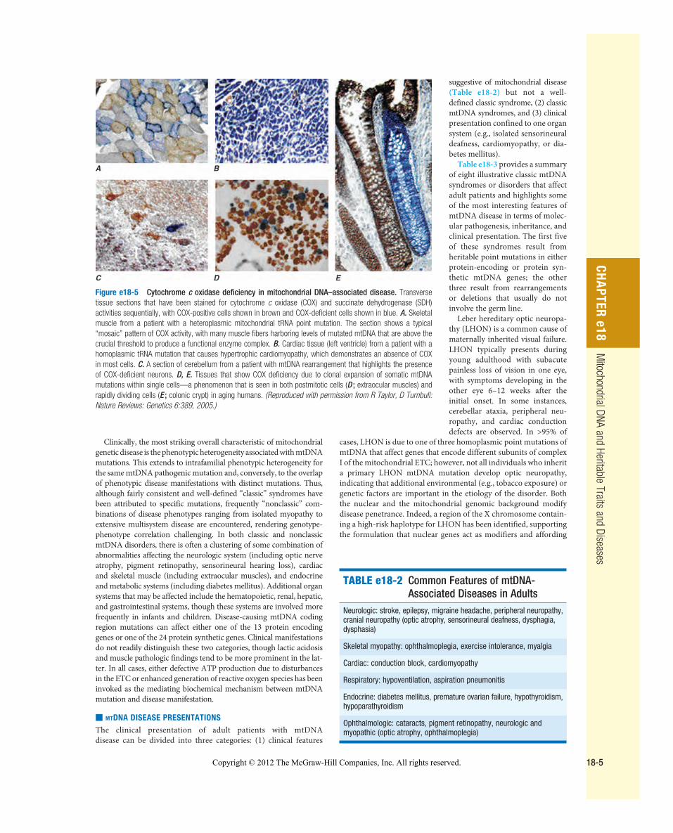
In light of the vital roles of mito-chondria in all nucleated cells, it is not surprising that mtDNA mutations can affect numerous tis-sues with pleiotropic effects. More than 200 different disease-causing, mostly heteroplasmic mtDNA mutations have been described that affect ETC function. Figure e18-4 provides a partial mtDNA map of some of the better characterized of these disorders. A number of clinical clues can increase the index of suspicion for a heteroplasmic mtDNA mutation as an etiology of a heritable trait or disease, including (1) familial clustering with absence of paternal transmission, (2) adher-ence to one of the classic syndromes (see below) or paradigmatic com-binations of disease phenotypes involving several organ systems that normally do not fit together within a single nuclear genomic mutation category, (3) a complex of labora-tory and pathologic abnormalities that reflect disruption in cellular energetics (e.g., lactic acidosis and
neurodegenerative and myodegenerative symptoms with the find-ing of ragged red fibers, reflecting the accumulation of abnormal mitochondria under the muscle sarcolemmal membrane), and (4) a mosaic pattern reflecting a heteroplasmic state.
Heteroplasmy sometimes can be demonstrated elegantly at the tissue level by using histochemical staining for enzymes in the oxi-dative phosphorylation pathway, with a mosaic pattern indicating heterogeneity of the genotype for the coding region for the mtDNA-encoded enzyme. Complex II, CoQ, and cytochrome c are encoded exclusively by nuclear DNA. In contrast, complexes I, III, IV, and V contain at least some subunits encoded by mtDNA. Just 3 of the 13 subunits of the ETC complex IV enzyme, cytochrome c oxidase, are encoded by mtDNA; therefore, this enzyme has the lowest threshold for dysfunction when a threshold of mutated mtDNA is reached. Histochemical staining for cytochrome c oxidase activity in tissues of patients affected with heteroplasmic inherited mtDNA mutations (or with the somatic accumulation of mtDNA mutations; see below) can show a mosaic pattern of reduced histochemical staining in comparison with histochemical staining for the complex II enzyme, succinate dehydrogenase ( Fig. e18-5 ) . Heteroplasmy also can be detected at the genetic level through direct mtDNA genotyping in special conditions, though clinically significant low levels of heteroplasmy can escape detection in genomic samples extracted from whole blood using conventional genotyping and sequencing techniques.
Figure e18-4 Mutations in the human mitochondrial genome known to cause disease. Disorders that are frequently or prominently associated with mutations in a particular gene are shown in boldface. Diseases due to mutations that impair mitochondrial protein synthesis are shown in blue. Diseases due to mutations in protein-coding genes are shown in red. ECM, encephalomyopathy; FBSN, familial bilateral striatal necrosis; LHON, Leber hereditary optic neuropathy; LS, Leigh syndrome; MELAS, mitochondrial encephalomyopathy, lactic acidosis, and strokelike episodes; MERRF, myoclonic epilepsy with ragged red fibers; MILS, maternally inherited Leigh syndrome; NARP, neuropathy, ataxia, and retinitis pigmentosa; PEO, progressive external ophthalmoplegia; PPK, palmoplantar keratoderma; SIDS, sudden infant death syndrome. (Reproduced with permission from S DiMauro, E Schon: N Engl J Med 348: 2656, 2003.)
Parkinsonism,aminoglycoside-induced deafness
MELASmyoglobinuria
Myopathy,PEO
CardiomyopathyECM
Diabetes and deafness
Cardiomyopathy,SIDS, ECM
Cardiomyopathy,PEO, MERRF,
MELAS, deafness
PPK, deafness,MERRF-MELAS
PEO
PEO
Myopathy, lymphomaMyopathy, MELAS
Myopathy, cardiomyopathy, PEO
PEO, LHON, MELAS,myopathy, cardiomyopathy,
diabetes and deafness
Cardiomyopathy
LS, MELAS,multisystem disease
ECM
Myopathy,PEO
LS, ataxia,chorea, myopathy
Cardiomyopathymyoclonus
CardiomyopathyECM
Cardiomyopathy, ECMPEO, myopathy,
sideroblastic anemia
ECM, LHON, myopathy,cardiomyopathy, MELAS
and parkinsonism
LHON, MELAS,diabetes,
LHON and dystonia
LS, MELAS
LHON, myopathy,LHON and dystonia
LHON
LS, ECM,myoglobinuria
NARP, MILS,FBSN
CardiomyopathyLHON
LHON Cyt b
ND6
ND2 ND5
ND4
ND4LND3
COXIII
COXII
COXI
ND1
L1
16SV 12sF
PT
E
H
RGK
DS1
YCNAW
MQI
L2S2
A6A8
Myoglobinuria,motor neuron disease,sideroblastic anemia
Myopathy,multisystem disease,encephalomyopathy
Progressive myoclonus,epilepsy, and optic atrophy
Copyright © 2012 The McGraw-Hill Companies, Inc. All rights reserved.
18-5
CHA
PTER e18
Mitochondrial DNA and Heritable Traits and Diseases
Clinically, the most striking overall characteristic of mitochondrial genetic disease is the phenotypic heterogeneity associated with mtDNA mutations. This extends to intrafamilial phenotypic heterogeneity for the same mtDNA pathogenic mutation and, conversely, to the overlap of phenotypic disease manifestations with distinct mutations. Thus, although fairly consistent and well-defined “classic” syndromes have been attributed to specific mutations, frequently “nonclassic” com-binations of disease phenotypes ranging from isolated myopathy to extensive multisystem disease are encountered, rendering genotype-phenotype correlation challenging. In both classic and nonclassic mtDNA disorders, there is often a clustering of some combination of abnormalities affecting the neurologic system (including optic nerve atrophy, pigment retinopathy, sensorineural hearing loss), cardiac and skeletal muscle (including extraocular muscles), and endocrine and metabolic systems (including diabetes mellitus). Additional organ systems that may be affected include the hematopoietic, renal, hepatic, and gastrointestinal systems, though these systems are involved more frequently in infants and children. Disease-causing mtDNA coding region mutations can affect either one of the 13 protein encoding genes or one of the 24 protein synthetic genes. Clinical manifestations do not readily distinguish these two categories, though lactic acidosis and muscle pathologic findings tend to be more prominent in the lat-ter. In all cases, either defective ATP production due to disturbances in the ETC or enhanced generation of reactive oxygen species has been invoked as the mediating biochemical mechanism between mtDNA mutation and disease manifestation.
� MTDNA DISEASE PRESENTATIONS The clinical presentation of adult patients with mtDNA disease can be divided into three categories: (1) clinical features
suggestive of mitochondrial disease ( Table e18-2 ) but not a well-defined classic syndrome, (2) classic mtDNA syndromes, and (3) clinical presentation confined to one organ system (e.g., isolated sensorineural deafness, cardiomy opathy, or dia-betes mellitus).
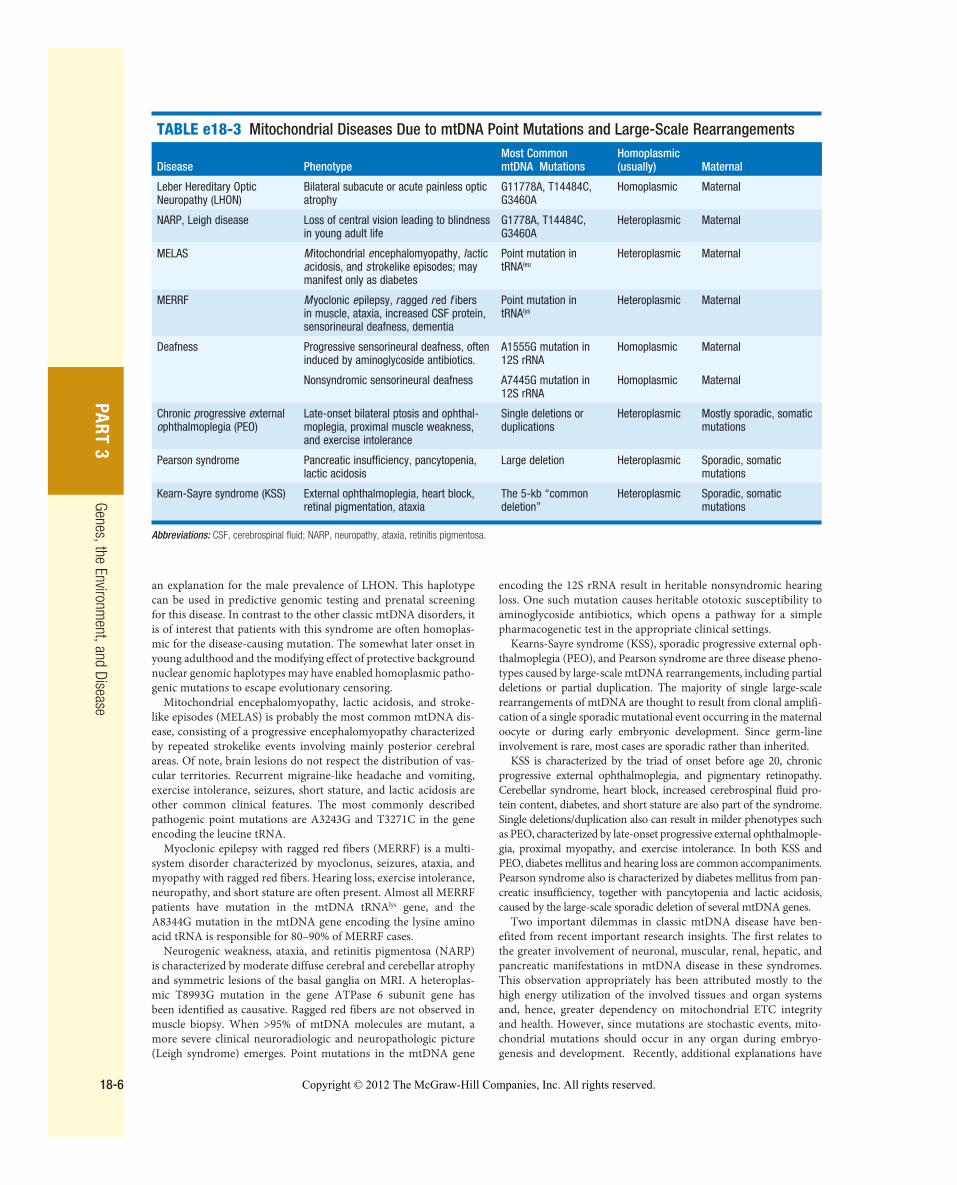
Table e18-3 provides a summary of eight illustrative classic mtDNA syndromes or disorders that affect adult patients and highlights some of the most interesting features of mtDNA disease in terms of molec-ular pathogenesis, inheritance, and clinical presentation. The first five of these syndromes result from heritable point mutations in either protein-encoding or protein syn-thetic mtDNA genes; the other three result from rearrangements or deletions that usually do not involve the germ line.
Leber hereditary optic neuropa-thy (LHON) is a common cause of maternally inherited visual failure. LHON typically presents during young adulthood with subacute painless loss of vision in one eye, with symptoms developing in the other eye 6–12 weeks after the initial onset. In some instances, cerebellar ataxia, peripheral neu-ropathy, and cardiac conduction defects are observed. In >95% of
cases, LHON is due to one of three homoplasmic point mutations of mtDNA that affect genes that encode different subunits of complex I of the mitochondrial ETC; however, not all individuals who inherit a primary LHON mtDNA mutation develop optic neuropathy, indicating that additional environmental (e.g., tobacco exposure) or genetic factors are important in the etiology of the disorder. Both the nuclear and the mitochondrial genomic background modify disease penetrance. Indeed, a region of the X chromosome contain-ing a high-risk haplotype for LHON has been identified, supporting the formulation that nuclear genes act as modifiers and affording
Figure e18-5 Cytochrome c oxidase deficiency in mitochondrial DNA–associated disease. Transverse tissue sections that have been stained for cytochrome c oxidase (COX) and succinate dehydrogenase (SDH) activities sequentially, with COX-positive cells shown in brown and COX-deficient cells shown in blue. A. Skeletal muscle from a patient with a heteroplasmic mitochondrial tRNA point mutation. The section shows a typical “mosaic” pattern of COX activity, with many muscle fibers harboring levels of mutated mtDNA that are above the crucial threshold to produce a functional enzyme complex. B. Cardiac tissue (left ventricle) from a patient with a homoplasmic tRNA mutation that causes hypertrophic cardiomyopathy, which demonstrates an absence of COX in most cells. C. A section of cerebellum from a patient with mtDNA rearrangement that highlights the presence of COX-deficient neurons. D, E. Tissues that show COX deficiency due to clonal expansion of somatic mtDNA mutations within single cells—a phenomenon that is seen in both postmitotic cells ( D ; extraocular muscles) and rapidly dividing cells ( E ; colonic crypt) in aging humans. ( Reproduced with permission from R Taylor, D Turnbull: Nature Reviews: Genetics 6:389, 2005.)
TABLE e18-2 Common Features of mtDNA-Associated Diseases in Adults
Neurologic: stroke, epilepsy, migraine headache, peripheral neuropathy, cranial neuropathy (optic atrophy, sensorineural deafness, dysphagia, dysphasia)
Skeletal myopathy: ophthalmoplegia, exercise intolerance, myalgia
Cardiac: conduction block, cardiomyopathy
Respiratory: hypoventilation, aspiration pneumonitis
Endocrine: diabetes mellitus, premature ovarian failure, hypothyroidism, hypoparathyroidism
Ophthalmologic: cataracts, pigment retinopathy, neurologic and myopathic (optic atrophy, ophthalmoplegia)
Copyright © 2012 The McGraw-Hill Companies, Inc. All rights reserved.
18-6
PART 3
Genes, the Environment, and Disease
an explanation for the male prevalence of LHON. This haplotype can be used in predictive genomic testing and prenatal screening for this disease. In contrast to the other classic mtDNA disorders, it is of interest that patients with this syndrome are often homoplas-mic for the disease-causing mutation. The somewhat later onset in young adulthood and the modifying effect of protective background nuclear genomic haplotypes may have enabled homoplasmic patho-genic mutations to escape evolutionary censoring.
Mitochondrial encephalomyopathy, lactic acidosis, and stroke-like episodes (MELAS) is probably the most common mtDNA dis-ease, consisting of a progressive encephalomyopathy characterized by repeated strokelike events involving mainly posterior cerebral areas. Of note, brain lesions do not respect the distribution of vas-cular territories. Recurrent migraine-like headache and vomiting, exercise intolerance, seizures, short stature, and lactic acidosis are other common clinical features. The most commonly described pathogenic point mutations are A3243G and T3271C in the gene encoding the leucine tRNA.
Myoclonic epilepsy with ragged red fibers (MERRF) is a multi-system disorder characterized by myoclonus, seizures, ataxia, and myopathy with ragged red fibers. Hearing loss, exercise intolerance, neuropathy, and short stature are often present. Almost all MERRF patients have mutation in the mtDNA tRNA lys gene, and the A8344G mutation in the mtDNA gene encoding the lysine amino acid tRNA is responsible for 80–90% of MERRF cases.
Neurogenic weakness, ataxia, and retinitis pigmentosa (NARP) is characterized by moderate diffuse cerebral and cerebellar atrophy and symmetric lesions of the basal ganglia on MRI. A heteroplas-mic T8993G mutation in the gene ATPase 6 subunit gene has been identified as causative. Ragged red fibers are not observed in muscle biopsy. When >95% of mtDNA molecules are mutant, a more severe clinical neuroradiologic and neuropathologic picture (Leigh syndrome) emerges. Point mutations in the mtDNA gene
encoding the 12S rRNA result in heritable nonsyndromic hearing loss. One such mutation causes heritable ototoxic susceptibility to aminoglycoside antibiotics, which opens a pathway for a simple pharmacogenetic test in the appropriate clinical settings.
Kearns-Sayre syndrome (KSS), sporadic progressive external oph-thalmoplegia (PEO), and Pearson syndrome are three disease pheno-types caused by large-scale mtDNA rearrangements, including partial deletions or partial duplication. The majority of single large-scale rearrangements of mtDNA are thought to result from clonal amplifi-cation of a single sporadic mutational event occurring in the maternal oocyte or during early embryonic development. Since germ-line involvement is rare, most cases are sporadic rather than inherited.
KSS is characterized by the triad of onset before age 20, chronic progressive external ophthalmoplegia, and pigmentary retinopathy. Cerebellar syndrome, heart block, increased cerebrospinal fluid pro-tein content, diabetes, and short stature are also part of the syndrome. Single deletions/duplication also can result in milder phenotypes such as PEO, characterized by late-onset progressive external ophthalmople-gia, proximal myopathy, and exercise intolerance. In both KSS and PEO, diabetes mellitus and hearing loss are common accompaniments. Pearson syndrome also is characterized by diabetes mellitus from pan-creatic insufficiency, together with pancytopenia and lactic acidosis, caused by the large-scale sporadic deletion of several mtDNA genes.
Two important dilemmas in classic mtDNA disease have ben-efited from recent important research insights. The first relates to the greater involvement of neuronal, muscular, renal, hepatic, and pancreatic manifestations in mtDNA disease in these syndromes. This observation appropriately has been attributed mostly to the high energy utilization of the involved tissues and organ systems and, hence, greater dependency on mitochondrial ETC integrity and health. However, since mutations are stochastic events, mito-chondrial mutations should occur in any organ during embryo-genesis and development. Recently, additional explanations have
TABLE e18-3 Mitochondrial Diseases Due to mtDNA Point Mutations and Large-Scale Rearrangements
Disease PhenotypeMost Common mtDNA Mutations
Homoplasmic (usually) Maternal
Leber Hereditary Optic Neuropathy (LHON)
Bilateral subacute or acute painless optic atrophy
G11778A, T14484C, G3460A
Homoplasmic Maternal
NARP, Leigh disease Loss of central vision leading to blindness in young adult life
G1778A, T14484C, G3460A
Heteroplasmic Maternal
MELAS M itochondrial encephalomyopathy, lactic acidosis, and s trokelike episodes; may manifest only as diabetes
Point mutation in tRNAleu
Heteroplasmic Maternal
MERRF Myoclonic epilepsy, ragged red f ibers in muscle, ataxia, increased CSF protein, sensorineural deafness, dementia
Point mutation in tRNAlys
Heteroplasmic Maternal
Deafness Progressive sensorineural deafness, often induced by aminoglycoside antibiotics.
A1555G mutation in 12S rRNA
Homoplasmic Maternal
Nonsyndromic sensorineural deafness A7445G mutation in 12S rRNA
Homoplasmic Maternal
Chronic progressive e xternal o phthalmoplegia (PEO)
Late-onset bilateral ptosis and ophthal-moplegia, proximal muscle weakness, and exercise intolerance
Single deletions or duplications
Heteroplasmic Mostly sporadic, somatic mutations
Pearson syndrome Pancreatic insufficiency, pancytopenia, lactic acidosis
Large deletion Heteroplasmic Sporadic, somatic mutations
Kearn-Sayre syndrome (KSS) External ophthalmoplegia, heart block, retinal pigmentation, ataxia
The 5-kb “common deletion”
Heteroplasmic Sporadic, somatic mutations
Abbreviations: CSF, cerebrospinal fluid; NARP, neuropathy, ataxia, retinitis pigmentosa.
Copyright © 2012 The McGraw-Hill Companies, Inc. All rights reserved.
18-7
CHA
PTER e18
Mitochondrial DNA and Heritable Traits and Diseases
been suggested based on studies of the common A3243G transi-tion. The proportion of this mutation in peripheral blood cells was shown to decrease exponentially with age. A selective process acting at the stem cell level with a strong bias against the mutated form would have its greatest effect in reducing the mutant mtDNA only in highly proliferating cells, such as those derived from the hematopoietic system. Tissues and organs with lower cell turnover, such as those involved with mtDNA mutations, would not benefit from this effect and thus would be affected the most.
Another important question of interest arises from the observa-tion that only a subset of mtDNA mutations account for the majority of the familial mtDNA diseases. The random occurrence of muta-tions in the mtDNA sequence should yield a more uniform distribu-tion of disease-causing mutations. However, recent studies utilizing the introduction of one severe and one mild point mutation into the female germ line of experimental animals demonstrated selective elimination during oogenesis of the severe and selective retention of the milder mutations, with the emergence of mitochondrial disease in offspring after multiple generations. Thus, oogenesis itself can act as an “evolutionary” filter for mtDNA disease.
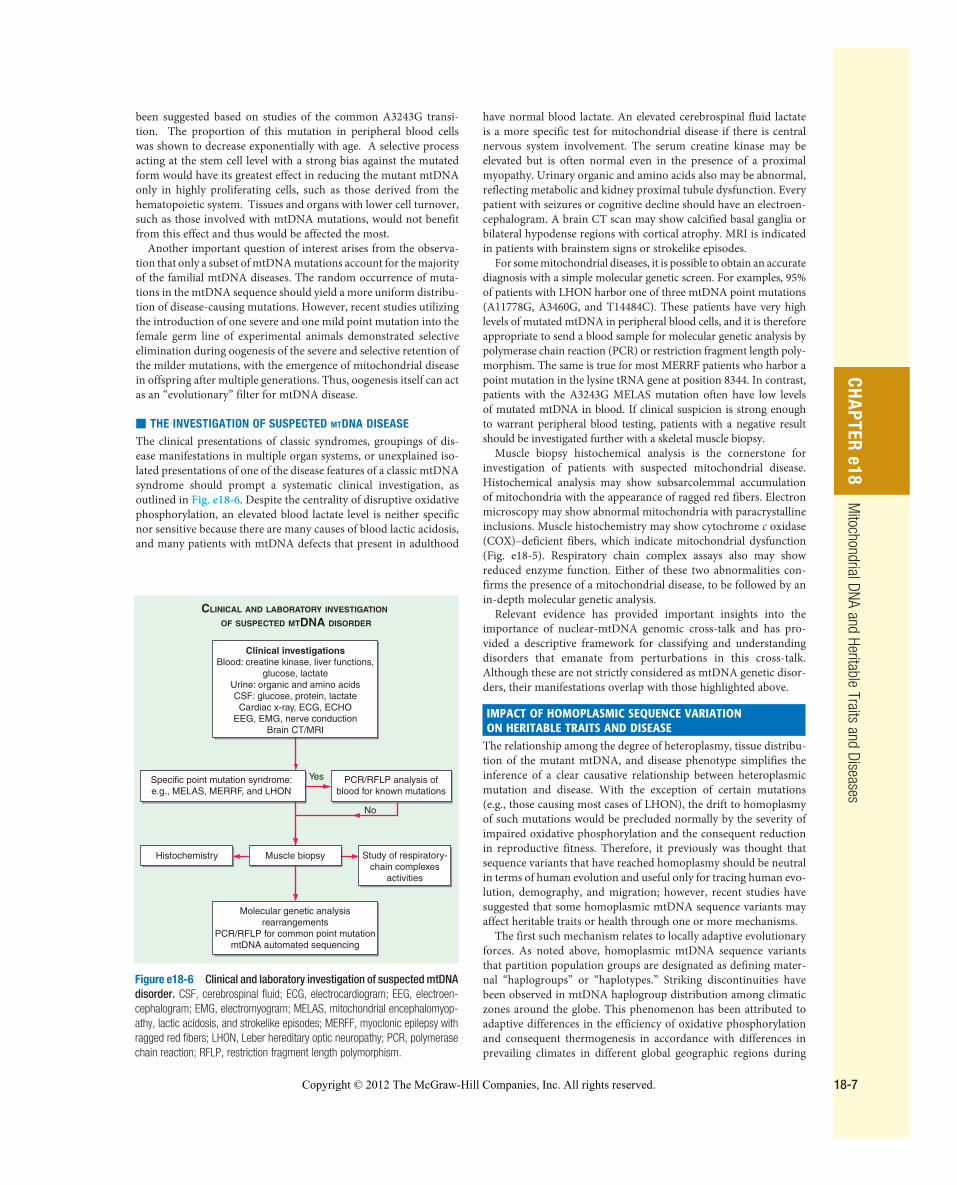
THE INVESTIGATION OF SUSPECTED � MTDNA DISEASE The clinical presentations of classic syndromes, groupings of dis-ease manifestations in multiple organ systems, or unexplained iso-lated presentations of one of the disease features of a classic mtDNA syndrome should prompt a systematic clinical investigation, as outlined in Fig. e18-6 . Despite the centrality of disruptive oxidative phosphorylation, an elevated blood lactate level is neither specific nor sensitive because there are many causes of blood lactic acidosis, and many patients with mtDNA defects that present in adulthood
have normal blood lactate. An elevated cerebrospinal fluid lactate is a more specific test for mitochondrial disease if there is central nervous system involvement. The serum creatine kinase may be elevated but is often normal even in the presence of a proximal myopathy. Urinary organic and amino acids also may be abnormal, reflecting metabolic and kidney proximal tubule dysfunction. Every patient with seizures or cognitive decline should have an electroen-cephalogram. A brain CT scan may show calcified basal ganglia or bilateral hypodense regions with cortical atrophy. MRI is indicated in patients with brainstem signs or strokelike episodes.
For some mitochondrial diseases, it is possible to obtain an accurate diagnosis with a simple molecular genetic screen. For examples, 95% of patients with LHON harbor one of three mtDNA point mutations (A11778G, A3460G, and T14484C). These patients have very high levels of mutated mtDNA in peripheral blood cells, and it is therefore appropriate to send a blood sample for molecular genetic analysis by polymerase chain reaction (PCR) or restriction fragment length poly-morphism. The same is true for most MERRF patients who harbor a point mutation in the lysine tRNA gene at position 8344. In contrast, patients with the A3243G MELAS mutation often have low levels of mutated mtDNA in blood. If clinical suspicion is strong enough to warrant peripheral blood testing, patients with a negative result should be investigated further with a skeletal muscle biopsy.
Muscle biopsy histochemical analysis is the cornerstone for investigation of patients with suspected mitochondrial disease. Histochemical analysis may show subsarcolemmal accumulation of mitochondria with the appearance of ragged red fibers. Electron microscopy may show abnormal mitochondria with paracrystalline inclusions. Muscle histochemistry may show cytochrome c oxidase (COX)–deficient fibers, which indicate mitochondrial dysfunction ( Fig. e18-5 ). Respiratory chain complex assays also may show reduced enzyme function. Either of these two abnormalities con-firms the presence of a mitochondrial disease, to be followed by an in-depth molecular genetic analysis.
Relevant evidence has provided important insights into the importance of nuclear-mtDNA genomic cross-talk and has pro-vided a descriptive framework for classifying and understanding disorders that emanate from perturbations in this cross-talk. Although these are not strictly considered as mtDNA genetic disor-ders, their manifestations overlap with those highlighted above .
IMPACT OF HOMOPLASMIC SEQUENCE VARIATION ON HERITABLE TRAITS AND DISEASE
The relationship among the degree of heteroplasmy, tissue distribu-tion of the mutant mtDNA, and disease phenotype simplifies the inference of a clear causative relationship between heteroplasmic mutation and disease. With the exception of certain mutations (e.g., those causing most cases of LHON), the drift to homoplasmy of such mutations would be precluded normally by the severity of impaired oxidative phosphorylation and the consequent reduction in reproductive fitness. Therefore, it previously was thought that sequence variants that have reached homoplasmy should be neutral in terms of human evolution and useful only for tracing human evo-lution, demography, and migration; however, recent studies have suggested that some homoplasmic mtDNA sequence variants may affect heritable traits or health through one or more mechanisms.
The first such mechanism relates to locally adaptive evolutionary forces. As noted above, homoplasmic mtDNA sequence variants that partition population groups are designated as defining mater-nal “haplogroups” or “haplotypes.” Striking discontinuities have been observed in mtDNA haplogroup distribution among climatic zones around the globe. This phenomenon has been attributed to adaptive differences in the efficiency of oxidative phosphorylation and consequent thermogenesis in accordance with differences in prevailing climates in different global geographic regions during
Figure e18-6 Clinical and laboratory investigation of suspected mtDNA disorder. CSF, cerebrospinal fluid; ECG, electrocardiogram; EEG, electroen-cephalogram; EMG, electromyogram; MELAS, mitochondrial encephalomyop-athy, lactic acidosis, and strokelike episodes; MERFF, myoclonic epilepsy with ragged red fibers; LHON, Leber hereditary optic neuropathy; PCR, polymerase chain reaction; RFLP, restriction fragment length polymorphism.
Clinical investigationsBlood: creatine kinase, liver functions,
glucose, lactateUrine: organic and amino acidsCSF: glucose, protein, lactateCardiac x-ray, ECG, ECHO
EEG, EMG, nerve conductionBrain CT/MRI
PCR/RFLP analysis ofblood for known mutations
Histochemistry Study of respiratory-chain complexes
activities
Yes
No
CLINICAL AND LABORATORY INVESTIGATION OF SUSPECTED MTDNA DISORDER
Molecular genetic analysisrearrangements
PCR/RFLP for common point mutationmtDNA automated sequencing
Specific point mutation syndrome:e.g., MELAS, MERRF, and LHON
Muscle biopsy
Copyright © 2012 The McGraw-Hill Companies, Inc. All rights reserved.
18-8
PART 3
Genes, the Environment, and Disease
much of human evolution. A potential health implication of this finding is the possibility that these mutations might result in del-eterious effects on energy metabolism and caloric balance in the current era of human transglobal migration and ability to modulate residential climate.
A much broader extrapolation of the foregoing mechanism states that many homoplasmic mtDNA mutations affect human health in the postreproductive age only and therefore escaped evolutionary censoring altogether. In the modern era of increased median life span, such mutations are postulated to account for a considerable burden of age-associated common complex disease. During the previous century alone, mean life expectancy rose from ~47 years to ~77 years in many parts of the developing world; therefore, late-onset effects of a subset of homoplasmic mtDNA mutations may contribute significantly to the burden of human illness only in the current era, when a relatively higher percentage of the population is surviving beyond reproductive age. The challenge is to identify those homoplasmic mutations that modify mtDNA function and contribute to late-onset, common complex disease. Indeed, in light of the finding that global populations are more differentiated at the level of mtDNA than they are at the level of the nuclear genome, it is also attractive to postulate that population differences with the predisposition to certain late-onset common complex metabolic diseases may be attributed in part to population-based mtDNA sequence variation. The diseases that have been of particular inter-est are those involving the very organ systems familiar from the known classic heteroplasmic mtDNA syndromes described above.
METABOLIC SYNDROME AND TYPE 2 DIABETES �MELLITUS (T2DM)
Insulin release by pancreatic beta cells is modulated in response to ATP metabolism, and insulin action is perturbed by metabolites of mitochondrial fatty acid oxidation. This has led investigators to consider mtDNA itself as a potential genomic locus for susceptibil-ity to T2DM. A rather clear-cut case is that of a mutation in mtDNA nucleotide 3243 encoding the mitochondrial tRNA for the amino acid leucine. Even a low level of heteroplasmy for a particular point mutation in the mtDNA tRNA gene encoding the leucine tRNA is thought to contribute to the pathogenesis of up to 1% of all cases of T2DM. This and other findings at the biochemical and popula-tion genetics levels have motivated the search for more definitive evidence of the role of homoplasmic variants in the predisposi-tion to metabolic syndrome and T2DM. Such evidence has been obtained with the finding of significant segregation of a homoplas-mic mtDNA tRNA mutation (T to C transition in the nucleotide immediately 5′ to the isoleucine tRNA anticodon) with metabolic syndrome phenotypes in a large Caucasian kindred.
Since the metabolic syndrome is so common and can result from numerous different genetic susceptibility loci and environmen-tal causes and since many nuclear genetic susceptibility loci and environmental risk factors have been identified, special features in this particular reported kindred enabled the accurate distinction of affected from unaffected individuals for purposes of determining the causality of the mtDNA variant. The affected individuals had signs of hypomagnesemia, hypertension, and hypercholesterolemia. This particular mutation in a tRNA-encoding mtDNA gene also highlights the expected difference in the phenotypic impact of mutations in genomic regions encoding tRNAs in the mitochon-drial versus the nuclear genome, since a mutation in the latter would affect too many gene products to be compatible with life.
Beyond the immediate medical and biologic significance, the importance of the findings in this kindred is to highlight the fact that the contribution of common homoplasmic mutations to common complex late-onset human disease syndromes is prob-ably underestimated. A common variant mtDNA sequence variant
(T16189C) has been related to low birth weight, impaired glu-cose tolerance, and metabolic syndrome in specific populations. However, rigorous population-based association studies using case-control designs have not provided definitive evidence for a relationship between mtDNA haplogroups and susceptibility to T2DM or its complications.
NEURODEGENERATIVE DISEASE �
The prominence of neurologic injury in classic mtDNA diseases, together with the presumed role of reactive oxygen species in neu-ronal injury and the late age of onset of neurodegenerative diseases, has led investigators to consider the possibility that homoplasmic variants in mtDNA sequence that define population haplogroups also may modify the susceptibility to neurodegenerative diseases such as Parkinson’s disease and Alzheimer’s disease. Particular configura-tions of mtDNA sequence polymorphisms that define population haplogroups designated in phylogenetics by the labels J, T, U, and K have been reported to be potentially protective against Parkinson’s disease in different populations. In the case of Alzheimer’s disease, some studies have shown haplogroup J to increase risk, with haplo-group D decreasing risk. Mutations in the mtDNA control region do not produce defective polypeptide products but affect both the light and heavy strand promoters, as well as the heavy strand origin of replication, and thus may modulate mtDNA replication and transcription. Mitochondrial DNA control region sequence variants (e.g., T414G) have been identified in Alzheimer’s disease brains in association with a significant reduction in mtDNA copy number and a reduction in specific transcripts. A number of studies have focused on the interaction of mtDNA haplogroup–designating mutations with the well-established Alzheimer’s disease risk alleles at the nuclear apoE4 locus. From those studies it was postulated that the ETC-uncoupling mutations that minimize ROS production are those which confer protection against neuronal injury, but defini-tive proof of this postulate awaits further studies.
OTHER DISEASES AND NONDISEASE HERITABLE TRAITS �
Consideration of the potential contribution of mtDNA muta-tions to numerous heritable traits and common complex dis-eases requires consideration of the common variant–common phenotype model (including disease phenotype) versus the rare variant–common phenotype model, which are also applicable to the nuclear genome. According to the common variant–common phenotype model, DNA sequence variants inherited identically by descent and present in large numbers of individuals within one or more populations may predispose to common phenotypes. In the rare variant–common phenotype model, different mutations within one or more genetic loci involved in a particular molecular pathway may predispose to a common phenotype or disease. In this regard, the entire mtDNA can be considered a single genomic locus. Genomewide association studies have been utilized to try to map common variants responsible for common diseases, using case-control or multiplex family approaches. These approaches have been applied to common variants in mtDNA sequence as well, as noted above for metabolic syndrome and neurodegenerative dis-ease. Additional examples include the variable length of an mtDNA control region polycytosine stretch (16189 variant) as a contribut-ing genomic influence in the onset of age-related cardiomyopathy with T2DM. An association of mtDNA haplogroup T and a poly-morphism at position 13368 with hypertrophic cardiomyopathy has been reported in a European population, and a number of studies have suggested an association between mtDNA mutations and mitochondrial dysfunction in predisposition to heart failure. In the case of age-related cancers as well, the association of a number of heritable homoplasmic mtDNA mutations with certain cancers has been reported, including prostate, kidney, and breast cancer.
Copyright © 2012 The McGraw-Hill Companies, Inc. All rights reserved.
18-9
CHA
PTER e18
Mitochondrial DNA and Heritable Traits and Diseases
The association of mtDNA haplogroups with at least two non-disease heritable traits also has been studied: life expectancy and exercise endurance. Several mtDNA control region mutations, including the C150T mutation that shifts the heavy chain origin of replication, have been reported to accumulate with age in specific tissues, including lymphocytes of centenarians and their twins. The relationship between the C150T mutation and longevity has been replicated in Italian, Finnish, and Japanese populations, suggesting a common ancient origin. The alternative of evolutionary conver-gence of this mutation for longevity seems less likely, as the trait does not confer reproductive advantage. The association of haplogroup J and its subhaplogroups with longevity has been demonstrated in north Italian, north Irish, and Finnish sample sets. At least in the Italian study, this association was shown to be population-specific, since it was not reproduced in sample sets of southern Italian com-munities. Furthermore, in the case of the north Italian communi-ties, an additional interaction of the mtDNA haplogroup designated as J2 with several different mutations adjacent to replication origins, including the aforementioned C150T, has been noted.
The functional importance of one or more of the mutations desig nated as haplogroup J is strengthened further by the finding of the interesting interaction with the mtDNA mutations that cause LHON, as noted previously. Reduced disease predilection suggests that one or more of the ancient sequence variants designated as haplo group J appear to attenuate predisposition to degenerative disease in the face of other risk factors. It has been proposed that the mtDNA haplogroups associated with exceptional longevity favor a relatively uncoupled state of the ETC, with reduced efficiency of production of ATP and ROS and increased thermogenesis. Although this has not been demonstrated biochemically, the notion is strengthened by the finding of a relative paucity of these mtDNA haplogroups among successful endurance athletes, for whom maxi-mum efficiency of oxidative phosphorylation confers an athletic competitive advantage.
It should be noted that not all studies have replicated associa-tions of mtDNA haplogroups with longevity, athletic performance, or other heritable phenotypes. Most of these studies are limited by small sample sizes, possible genotyping inaccuracies, and the possibility of population stratification or ethnic ancestry bias. Since mtDNA haplogroups are so prominently partitioned along phylogeographic lines, it is difficult to rule out the possibility that a haplogroup for which an association has been found is simply a marker for differences in populations with a societal or envi-ronmental difference or with different allele frequencies at other genomic loci that are actually causally related to the heritable trait or disease of interest. The difficulty in generating cellular or animal models to test the functional influence of homoplasmic sequence variants (as a result of mtDNA polyploidy) further compounds the challenge. The most likely formulation is that the risk conferred by different mtDNA haplogroup–defining homoplasmic mutations for common diseases depends on the concomitant nuclear genomic background, together with environmental influences. Progress in minimizing potentially misleading associations in mtDNA heritable trait and disease studies should include ensuring adequate sample size taken from a large sample recruitment base, together with the use of carefully matched controls and population structure determi-nation, along with analysis that takes into account epistatic interac-tions with other genomic loci and environmental factors.
IMPACT OF ACQUIRED SOMATIC MTDNA MUTATION ON HUMAN HEALTH AND DISEASE
Studies on aging humans and animals have shown a potentially important correlation of age with the accumulation of heteroge-neous mtDNA mutations, especially in the organ systems that
undergo the most prominent age-related degenerative tissue phe-notype. Sequencing of PCR-amplified single mtDNA molecules has demonstrated an average of two to three point mutations per molecule in elderly subjects compared with younger ones. Point mutations observed include those responsible for known heritable heteroplasmic mtDNA disorders such as the A3344G and A3243G mutations responsible for the MERRF and MELAS syndromes, respectively. However, the cumulative burden of these acquired somatic point mutations with age was observed to remain well below the threshold expected for phenotypic expression (<2%). Point mutations at other sites not normally involved in inher-ited mtDNA disorders also have been shown to accumulate to much higher levels in some tissues of elderly individuals, with the description of tissue-specific “hot spots” for mtDNA point muta-tions. Along the same lines, an age-associated and tissue-specific accumulation of mtDNA deletions has been observed, including deletions involved in known heritable mtDNA disorders, as well as others. The accumulation of functional mtDNA deletions in a given tissue is expected to be associated with mitochondrial dysfunction, as reflected in an age-associated patchy and reduced cytochrome c oxidase activity on histochemical staining, especially in skeletal and cardiac muscle and brain. A particularly well-studied and poten-tially important example is the accumulation of mtDNA deletions and cytochrome c oxidase deficiency observed in neurons of the substantia nigra in Parkinson’s disease patients.
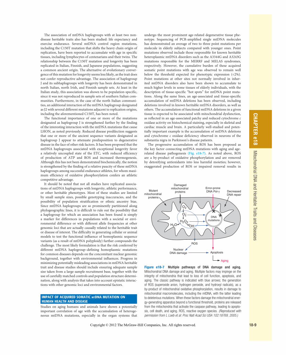
The progressive accumulation of ROS has been proposed as the key factor connecting mtDNA mutations with aging and age-related disease pathogenesis ( Fig. e18-7 ) . As noted above, ROS are a by-product of oxidative phosphorylation and are removed by detoxifying antioxidants into less harmful moieties; however, exaggerated production of ROS or impaired removal results in
Mutantmitochondrial
proteins
Damagedmitochondrial
proteinsError-proneDNA Pol-γ
NuclearDNA damage
ROS
DecreasedDNA repair
Apoptosis
DNAmutations
O2
O2
H2O2H2O OH
Aging
X X−
Figure e18-7 Multiple pathways of DNA damage and aging. Mitochondrial DNA damage and aging. Multiple factors may impinge on the integrity of mitochondria that lead to loss of cell function, apoptosis, and aging. The classic pathway is indicated with blue arrows; the generation of ROS (superoxide anion, hydrogen peroxide, and hydroxyl radicals), as a by-product of mitochondrial oxidative phosphorylation, results in damage to mitochondrial macromolecules, including the mtDNA, with the latter leading to deleterious mutations. When these factors damage the mitochondrial ener-gy-generating apparatus beyond a functional threshold, proteins are released from the mitochondria that activate the caspase pathway, leading to apopto-sis, cell death, and aging. ROS, reactive oxygen species. ( Reproduced with permission from L Loeb et al: Proc Natl Acad Sci USA 102:18769, 2005.)
Copyright © 2012 The McGraw-Hill Companies, Inc. All rights reserved.
18-10
PART 3
Genes, the Environment, and Disease
their accumulation. One of the main targets for ROS-mediated injury is DNA, and mtDNA is particularly vulnerable because of its lack of protective histones and less efficient injury repair systems compared with nuclear DNA. In turn, accumulation of mtDNA mutations results in inefficient oxidative phosphorylation, with the potential for excessive production of ROS, generating a “vicious cycle” of cumulative mtDNA damage. Indeed, measurement of the oxidative stress biomarker 8-hydroxy-2-deoxyguanosine has been used to measure age-dependent increases in mtDNA oxida-tive damage at a rate exceeding that of nuclear DNA. It should be noted that mtDNA mutation potentially can occur in postmitotic cells as well, since mtDNA replication is not synchronized with the cell cycle. Two other proposed links between mtDNA mutation and aging, besides ROS-mediated tissue injury, are the perturbations in efficiency of oxidative phosphorylation with disturbed cellular aerobic function and perturbations in apoptotic pathways whose execution steps involve mitochondrial activity.
Genetic intervention studies in animal models have sought to clarify the potential causative relationship between acquired somatic mtDNA mutation and the aging phenotype, the role of ROS in particular. Replication of the mitochondrial genome is mediated by the activity of the nuclear-encoded polymerase γ gene. A transgenic homozygous mouse knock-in mutation of this gene renders the polymerase enzyme deficient in proofreading and results in a three- to fivefold increase in the mtDNA mutation rate. Such mice develop a premature aging phenotype, which includes subcutaneous lipoatrophy, alopecia, kyphonia, and weight loss with premature death. Although the finding of increases in mtDNA mutation and mitochondrial dysfunction with age has been solidly established, the causative role and specific contribution of mito-chondrial ROS to aging and age-related disease in humans have yet to be proved. Similarly, although many tumors display higher levels of heterogeneous mtDNA mutations, a causal relationship to tumorigenesis has not been proved.
Besides the age-dependent acquired accumulation in somatic cells of heterogeneous point mutations and deletions, a quite dif-ferent effect of nonheritable and acquired mtDNA mutation has been described affecting tissue stem cells. In particular, disease phenotypes attributed to acquired mtDNA mutation have been observed in sporadic and apparently nonfamilial cases involving a single individual or even tissue, usually skeletal muscle. The presentation consists of decreased exercise tolerance and myalgias, sometimes progressing to rhabdomyolysis. As in the case of the sporadic heteroplasmic large-scale deletion classic syndromes of chronic PEO, Pearson syndrome, and KSS, the absence of a mater-nal inheritance pattern, together with the finding of limited tissue distribution, suggests a molecular pathogenic mechanism emanat-ing from mutations arising de novo in muscle stem cells after germ-line differentiation (somatic mutations that are not sporadic and occur in tissue-specific stem cells during fetal development or in the postnatal maintenance or postinjury repair stage). Such mutations would be expected to be propagated only within the progeny of that stem cell and affect a particular tissue within a specific individual, without evidence of heritability.
PROSPECTS FOR PREVENTION AND TREATMENT OF MTDNA DISEASE
GENETIC COUNSELING IN � MTDNA DISORDERS The provision of accurate genetic counseling and reproductive options to families with mtDNA mutations is complicated by the unique genetic features of mtDNA that distinguish it from Mendelian genetics. Although there is no risk of disease transmission from an affected male, the risk of maternal transmission of disease
phenotypes associated with heteroplasmic mutations is a function of differential segregation and copy number of mutant mtDNA during oogenesis and, subsequently, after tissue and organ develop-ment in the offspring. This is rarely predictable with any degree of accuracy. In addition, interactions with the mtDNA haplotype background or nuclear human genome (as in the case of LHON) serve as an additional important determinant of disease penetrance. Environmental interactions are also of importance, such as the strong and consistent association between visual loss in LHON and smoking. A clinical penetrance of 93% was found in men who smoked. Asymptomatic carriers of a LHON mtDNA mutation therefore should be strongly advised not to smoke and to mode-rate their alcohol intake. Although not a cure, these interventions might stave off the devastating clinical manifestations of the LHON mutation. Another example is the familial syndrome of ototoxic susceptibility to aminoglycosides in the case of the mtDNA A1555G mutation of the 12S rRNA encoding gene.
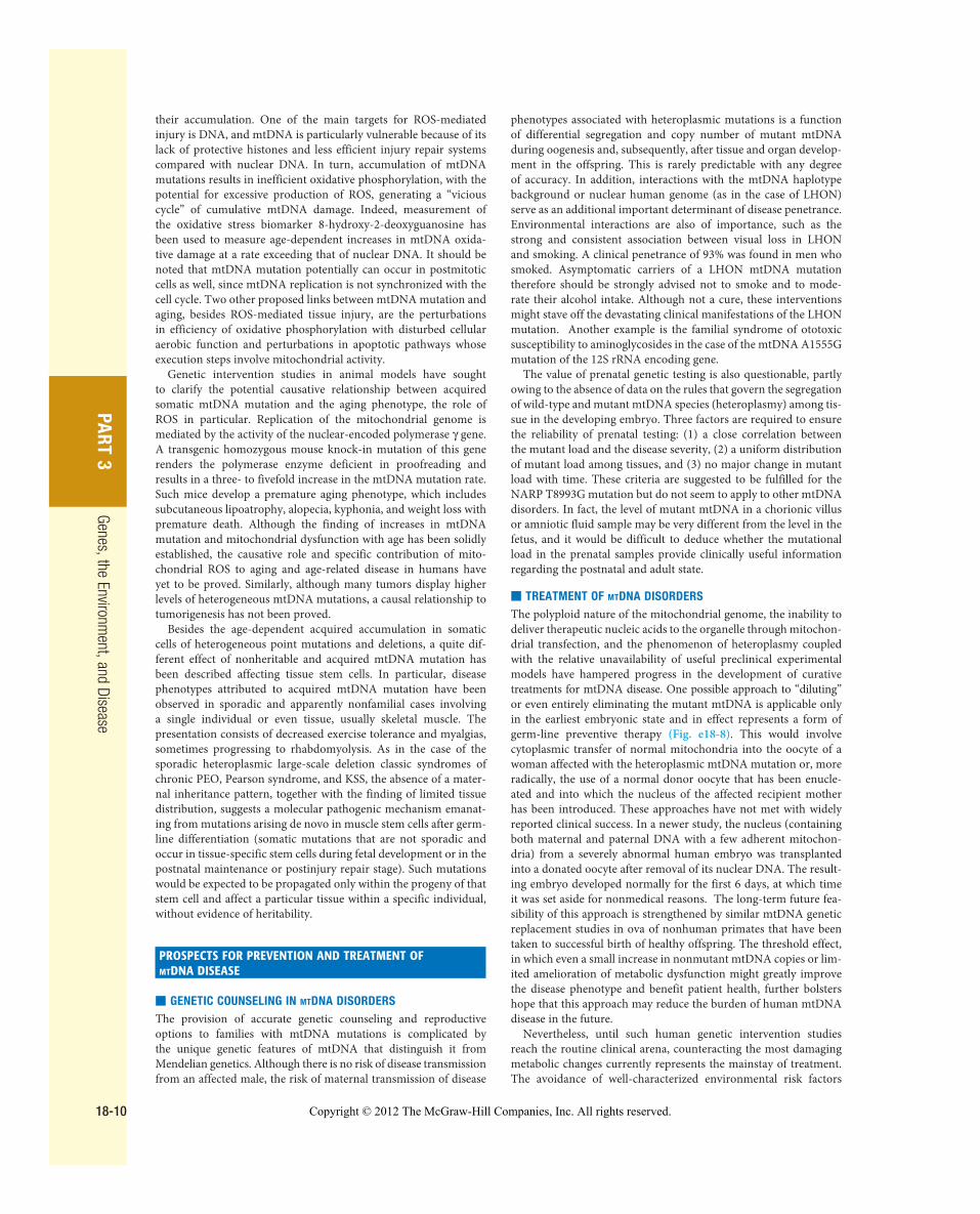
The value of prenatal genetic testing is also questionable, partly owing to the absence of data on the rules that govern the segregation of wild-type and mutant mtDNA species (heteroplasmy) among tis-sue in the developing embryo. Three factors are required to ensure the reliability of prenatal testing: (1) a close correlation between the mutant load and the disease severity, (2) a uniform distribution of mutant load among tissues, and (3) no major change in mutant load with time. These criteria are suggested to be fulfilled for the NARP T8993G mutation but do not seem to apply to other mtDNA disorders. In fact, the level of mutant mtDNA in a chorionic villus or amniotic fluid sample may be very different from the level in the fetus, and it would be difficult to deduce whether the mutational load in the prenatal samples provide clinically useful information regarding the postnatal and adult state.
TREATMENT OF � MTDNA DISORDERS The polyploid nature of the mitochondrial genome, the inability to deliver therapeutic nucleic acids to the organelle through mitochon-drial transfection, and the phenomenon of heteroplasmy coupled with the relative unavailability of useful preclinical experimental models have hampered progress in the development of curative treatments for mtDNA disease. One possible approach to “diluting” or even entirely eliminating the mutant mtDNA is applicable only in the earliest embryonic state and in effect represents a form of germ-line preventive therapy ( Fig. e18-8 ). This would involve cytoplasmic transfer of normal mitochondria into the oocyte of a woman affected with the heteroplasmic mtDNA mutation or, more radically, the use of a normal donor oocyte that has been enucle-ated and into which the nucleus of the affected recipient mother has been introduced. These approaches have not met with widely reported clinical success. In a newer study, the nucleus (containing both maternal and paternal DNA with a few adherent mitochon-dria) from a severely abnormal human embryo was transplanted into a donated oocyte after removal of its nuclear DNA. The result-ing embryo developed normally for the first 6 days, at which time it was set aside for nonmedical reasons. The long-term future fea-sibility of this approach is strengthened by similar mtDNA genetic replacement studies in ova of nonhuman primates that have been taken to successful birth of healthy offspring. The threshold effect, in which even a small increase in nonmutant mtDNA copies or lim-ited amelioration of metabolic dysfunction might greatly improve the disease phenotype and benefit patient health, further bolsters hope that this approach may reduce the burden of human mtDNA disease in the future.
Nevertheless, until such human genetic intervention studies reach the routine clinical arena, counteracting the most damaging metabolic changes currently represents the mainstay of treatment. The avoidance of well-characterized environmental risk factors
Copyright © 2012 The McGraw-Hill Companies, Inc. All rights reserved.
18-11
CHA
PTER e18
Mitochondrial DNA and Heritable Traits and Diseases
as noted above, such as tobacco use in the case of LHON, and the avoidance of aminoglycosides in the case of mtDNA A1555G mutation have very major benefits. Less specific interventions in the case of other disorders involve combined treatment strategies and include dietary intervention and removal of toxic metabolites. Cofactors and vitamin supplements are used widely in the treatment of diseases of mitochondrial oxidative phosphorylation, although there is little evidence, apart from anecdotal reports, to support their use. This includes administration of artificial electron acceptors, including vitamin K 3, vitamin C, and ubiquinone (coenzyme Q 10 ); administration of cofactors (coenzymes), including riboflavin, carnitine, and creatine; and use of oxygen radical scavengers, such as vitamin E, copper, selenium, ubiquinone, and idebenone. Drugs that could interfere with mitochondrial defects, such as the anes-thetic agent propofol, barbiturates, and high doses of valproate, should be avoided. Supplementation with the nitric oxide synthase substrate l-arginine has been advocated as a vasodilator treatment during strokelike episodes.
In the case of homoplasmic mtDNA variants that predispose to late-onset common complex disease, it is more realistic to think of using their identification in a specific patient as a nonmodifiable risk factor, which guides the aggressiveness of medical intervention
for the associated modifiable risk factors for the same disorder. For example, the identification of a haplogroup-defining homoplasmic mtDNA mutation that confers added risk for metabolic syndrome should trigger intensive dietary, lifestyle, and medical intervention to reduce other factors that promote the metabolic syndrome and its complications. In the case of acquired somatic mutations—to the extent that a vicious cycle of ROS production with mtDNA mutation plays a role—effective antioxidant and ROS scavenging therapeutic strategies may prove to be of benefit.
FURTHER READINGS
Bandelt HJ et al: Exaggerated status of “novel” and “pathogenic” mtDNA sequence variants due to inadequate database searches. Hum Mutat 30:191, 2009
Bredenoord AL et al: PGD to reduce reproductive risk: The case of mitochondrial DNA disorders. Hum Reprod 23:2392, 2008
Calvo S et al: Systematic identification of human mitochondrial dis-ease genes through integrative genomics. Nat Genet 38: 576, 2006
Craven L et al: Pronuclear transfer in human embryos to prevent transmission of mitochondrial DNA disease. Nature 465:82, 2010
Normal conception
Mothercarryingmutant
mitochondrialDNA
Mother’soocytes
fertilized withpartner’s
sperm
Develpingembryos
Offspring
Oocyte donation
Implanted embryo isderived from donoroocytes fertilizedwith the partner’s
sperm in vitro
Preimplantationembryo samplingand selection oflow-risk embryosfor implantation
into uterus
Carriermother’s
oocytes arefertilized
in vitro andinjectedinto an
enucleateddonoroocyte
The likelihoodthat adherentmitochondriawill also betransferredmeans that
prenatal testingis also
recommended
Unrelateddonor
unfertilizedoocytes
areenucleated
+ +
Preimplantationgenetic diagnosis
Nuclear transfer intodonated oocytes:
a future possibility?
A B C D
Figure e18-8 Possible approaches for prevention of mtDNA disease. A . No intervention: Offspring’s mutant mitochondrial DNA load will vary greatly. B . Oocyte donation: currently permitted in some constituencies but limited by the availability of oocyte donors. C . Preimplantation genetic diagno-sis: available for some mtDNA diseases (reliable in determining background
nuclear genomic haplotype risk). D . Nuclear transfer: research stage, includ-ing initial studies in nonhuman primates. Red represents mutant mitochon-drial DNA, pink and white represent successively higher proportions of normal mitochondrial DNA, and blue represents genetic material from an unrelated donor. (Adapted with permission from Poulton et al.)
Copyright © 2012 The McGraw-Hill Companies, Inc. All rights reserved.
18-12
PART 3
Genes, the Environment, and Disease
Cree LM et al: A reduction of mitochondrial DNA molecules dur-ing embryogenesis explains the rapid segregation of genotypes. Nat Genet 40:249, 2008
Di Donato S: Multisystem manifestations of mitochondrial disor-ders. J Neurol 256:693, 2009
Dimauro S: Mitochondrial DNA medicine. Biosci Rep 27:5, 2007 Filosto M, Manusco M: Mitochondrial diseases: A nosological
update. Acta Neurol Scand 115:211, 2007 Hudson G et al: Clinical expression of Leber hereditary optic
neuropathy is affected by the mitochondrial DNA-haplogroup background. Am J Hum Genet 81:228, 2007
———: Mutation of OPA1 causes dominant optic atrophy with external ophthalmoplegia, ataxia, deafness and multiple mito-chondrial DNA deletions: A novel disorder of mtDNA main-tenance. Brain 131:329, 2008
Mancuso M et al: Mitochondrial DNA-related disorders: Natural selection shaped regional mtDNA variation in humans. Biosci Rep 27:31, 2007
McKenzie M et al: Mitochondrial disease: Mutations and mecha-nisms. Neurochem Res 29:589, 2004
Neimi AK, Majamaa K: Mitochondrial DNA and ACTN3 geno-types in Finnish elite endurance and sprint athletes. Eur J Hum Genet 13:965, 2005
Poulton J et al: Preventing transmission of maternally inherited mitochondrial DNA diseases. BMJ 338:b94, 2009
Salas A et al: A critical reassessment of the role of mitochondria in tumorigenesis. PLoS Med 2:1158, 2005
Spinazzola A, Zeviani M: Disorders from perturbations of nuclear-mitochondrial intergenomic cross-talk. J Intern Med 265:174, 2009
Suissa S et al: Ancient mtDNA genetic variants modulate mtDNA transcription and replication. PLoS Genet 5:e1000474, 2009
Wallace D: The mitochondrial genome in human adaptive radia-tion and disease: On the road to therapeutics and performance enhancement. Gene 354:169, 2005
Wilson FH et al: A cluster of metabolic defects caused by mutation in a mitochondrial tRNA. Science 306:1190, 2004
Zeviani M, Carelli V: Mitochondrial disorders. Curr Opin Neurol 20:564, 2007
Copyright © 2012 The McGraw-Hill Companies, Inc. All rights reserved.