Embed Size (px)
Citation preview

Chapter , Dr. Enriquez 1
15The Special Senses
Part A

Chapter , Dr. Enriquez 2
Chemical Senses
Chemical senses – gustation (taste) and olfaction (smell)
Their chemoreceptors respond to chemicals in aqueous solution Taste – to substances dissolved in saliva Smell – to substances dissolved in fluids of the nasal
membranes

Chapter , Dr. Enriquez 3
Taste Buds
Most of the 10,000 or so taste buds are found on the tongue
Taste buds are found in papillae of the tongue mucosa
Papillae come in three types: filiform, fungiform, and circumvallate
Fungiform and circumvallate papillae contain taste buds

Chapter , Dr. Enriquez 4
Taste Buds
Figure 15.1

Chapter , Dr. Enriquez 5
Anatomy of a Taste Bud
Each gourd-shaped taste bud consists of three major cell types Supporting cells – insulate the receptor Basal cells – dynamic stem cells Gustatory cells – taste cells

Chapter , Dr. Enriquez 6
Taste Sensations
There are five basic taste sensations Sweet – sugars, saccharin, alcohol, and some amino
acids Salt – metal ions Sour – hydrogen ions Bitter – alkaloids such as quinine and nicotine Umami – elicited by the amino acid glutamate

Chapter , Dr. Enriquez 7
Physiology of Taste
In order to be tasted, a chemical: Must be dissolved in saliva Must contact gustatory hairs
Binding of the food chemical: Depolarizes the taste cell membrane, releasing
neurotransmitter Initiates a generator potential that elicits an action
potential

Chapter , Dr. Enriquez 8
Taste Transduction
The stimulus energy of taste is converted into a nerve impulse by: Na+ influx in salty tastes H+ in sour tastes (by directly entering the cell, by
opening cation channels, or by blockade of K+ channels)
Gustducin in sweet and bitter tastes

Chapter , Dr. Enriquez 9
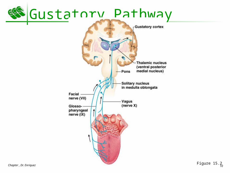
Gustatory Pathway
Cranial Nerves VII and IX carry impulses from taste buds to the solitary nucleus of the medulla
These impulses then travel to the thalamus, and from there fibers branch to the: Gustatory cortex (taste) Hypothalamus and limbic system (appreciation of
taste)
Chapter , Dr. Enriquez 10
Gustatory Pathway
Figure 15.2

Chapter , Dr. Enriquez 11
Influence of Other Sensations on Taste
Taste is 80% smell Thermoreceptors, mechanoreceptors, nociceptors
also influence tastes Temperature and texture enhance or detract from
taste

Chapter , Dr. Enriquez 12
Sense of Smell
The organ of smell is the olfactory epithelium, which covers the superior nasal concha
Olfactory receptor cells are bipolar neurons with radiating olfactory cilia
Olfactory receptors are surrounded and cushioned by supporting cells
Basal cells lie at the base of the epithelium

Chapter , Dr. Enriquez 13
Sense of Smell
Figure 15.3

Chapter , Dr. Enriquez 14
Physiology of Smell
Olfactory receptors respond to several different odor-causing chemicals
When bound to ligand these proteins initiate a G protein mechanism, which uses cAMP as a second messenger
cAMP opens Na+ and Ca2+ channels, causing depolarization of the receptor membrane that then triggers an action potential

Chapter , Dr. Enriquez 15
Olfactory Pathway
Olfactory receptor cells synapse with mitral cells Glomerular mitral cells process odor signals Mitral cells send impulses to:
The olfactory cortex The hypothalamus, amygdala, and limbic system

Chapter , Dr. Enriquez 16
Olfactory Transduction Process
Figure 15.4
Odorant binding protein
Odorant chemical
Na+
Cytoplasm
Inactive Active
Na+ influx causes depolarization
Adenylate cyclase
ATP
cAMP
Depolarization of olfactory receptor cell membrane triggers action potentials in axon of receptor

Chapter , Dr. Enriquez 17
Eye and Associated Structures
70% of all sensory receptors are in the eye Most of the eye is protected by a cushion of fat
and the bony orbit Accessory structures include eyebrows, eyelids,
conjunctiva, lacrimal apparatus, and extrinsic eye muscles

Chapter , Dr. Enriquez 18
Eyebrows
Coarse hairs that overlie the supraorbital margins Functions include:
Shading the eye Preventing perspiration from reaching the eye
Orbicularis muscle – depresses the eyebrows Corrugator muscles – move the eyebrows
medially

Chapter , Dr. Enriquez 19
Palpebrae (Eyelids)
Protect the eye anteriorly Palpebral fissure – separates eyelids Canthi – medial and lateral angles (commissures)

Chapter , Dr. Enriquez 20
Palpebrae (Eyelids)
Lacrimal caruncle – contains glands that secrete a whitish, oily secretion (Sandman’s eye sand)
Tarsal plates of connective tissue support the eyelids internally
Levator palpebrae superioris – gives the upper eyelid mobility

Chapter , Dr. Enriquez 21
Palpebrae (Eyelids)
Eyelashes Project from the free margin of each eyelid Initiate reflex blinking
Lubricating glands associated with the eyelids Meibomian glands and sebaceous glands Ciliary glands lie between the hair follicles

Chapter , Dr. Enriquez 22
Palpebrae (Eyelids)
Figure 15.5b

Chapter , Dr. Enriquez 23
Conjunctiva
Transparent membrane that: Lines the eyelids as the palpebral conjunctiva Covers the whites of the eyes as the ocular
conjunctiva Lubricates and protects the eye

Chapter , Dr. Enriquez 24
Lacrimal Apparatus
Consists of the lacrimal gland and associated ducts
Lacrimal glands secrete tears Tears
Contain mucus, antibodies, and lysozyme Enter the eye via superolateral excretory ducts Exit the eye medially via the lacrimal punctum Drain into the nasolacrimal duct

Chapter , Dr. Enriquez 25
Lacrimal Apparatus
Figure 15.6

Chapter , Dr. Enriquez 26
Extrinsic Eye Muscles
Six straplike extrinsic eye muscles Enable the eye to follow moving objects Maintain the shape of the eyeball
Four rectus muscles originate from the annular ring
Two oblique muscles move the eye in the vertical plane

Chapter , Dr. Enriquez 27
Extrinsic Eye Muscles
Figure 15.7a, b

Chapter , Dr. Enriquez 28
Summary of Cranial Nerves and Muscle Actions
Names, actions, and cranial nerve innervation of the extrinsic eye muscles
Figure 15.7c

Chapter , Dr. Enriquez 29
Structure of the Eyeball
A slightly irregular hollow sphere with anterior and posterior poles
The wall is composed of three tunics – fibrous, vascular, and sensory
The internal cavity is filled with fluids called humors
The lens separates the internal cavity into anterior and posterior segments

Chapter , Dr. Enriquez 30
Structure of the Eyeball
Figure 15.8a

Chapter , Dr. Enriquez 31
Fibrous Tunic
Forms the outermost coat of the eye and is composed of: Opaque sclera (posteriorly) Clear cornea (anteriorly)
The sclera protects the eye and anchors extrinsic muscles
The cornea lets light enter the eye

Chapter , Dr. Enriquez 32
Vascular Tunic (Uvea): Choroid Region
Has three regions: choroid, ciliary body, and iris Choroid region
A dark brown membrane that forms the posterior portion of the uvea
Supplies blood to all eye tunics

Chapter , Dr. Enriquez 33
Vascular Tunic: Ciliary Body
A thickened ring of tissue surrounding the lens Composed of smooth muscle bundles (ciliary
muscles) Anchors the suspensory ligament that holds the
lens in place

Chapter , Dr. Enriquez 34
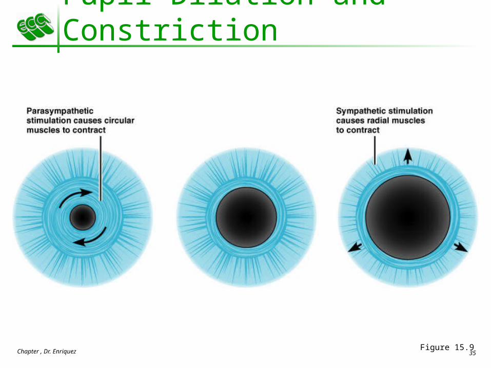
Vascular Tunic: Iris
The colored part of the eye Pupil – central opening of the iris
Regulates the amount of light entering the eye during: Close vision and bright light – pupils constrict Distant vision and dim light – pupils dilate Changes in emotional state – pupils dilate when the subject
matter is appealing or requires problem-solving skills
Chapter , Dr. Enriquez 35
Pupil Dilation and Constriction
Figure 15.9

Chapter , Dr. Enriquez 36
Sensory Tunic: Retina
A delicate two-layered membrane Pigmented layer – the outer layer that absorbs
light and prevents its scattering Neural layer, which contains:
Photoreceptors that transduce light energy Bipolar cells and ganglion cells Amacrine and horizontal cells

Chapter , Dr. Enriquez 37
Sensory Tunic: Retina
Figure 15.10a
Chapter , Dr. Enriquez 38
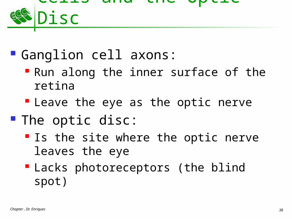
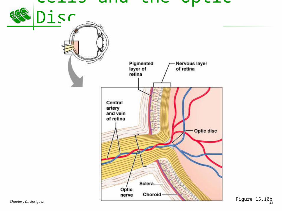
The Retina: Ganglion Cells and the Optic Disc
Ganglion cell axons: Run along the inner surface of the retina Leave the eye as the optic nerve
The optic disc: Is the site where the optic nerve leaves the eye Lacks photoreceptors (the blind spot)
Chapter , Dr. Enriquez 39
The Retina: Ganglion Cells and the Optic Disc
Figure 15.10b

Chapter , Dr. Enriquez 40
The Retina: Photoreceptors
Rods: Respond to dim light Are used for peripheral vision
Cones: Respond to bright light Have high-acuity color vision Are found in the macula lutea Are concentrated in the fovea centralis

Chapter , Dr. Enriquez 41
Blood Supply to the Retina
The neural retina receives its blood supply from two sources The outer third receives its blood from the choroid The inner two-thirds is served by the central artery
and vein Small vessels radiate out from the optic disc and
can be seen with an ophthalmoscope

Chapter , Dr. Enriquez 42
15The Special Senses
Part B

Chapter , Dr. Enriquez 43
Inner Chambers and Fluids
The lens separates the internal eye into anterior and posterior segments
The posterior segment is filled with a clear gel called vitreous humor that: Transmits light Supports the posterior surface of the lens Holds the neural retina firmly against the pigmented
layer Contributes to intraocular pressure

Chapter , Dr. Enriquez 44
Anterior Segment
Composed of two chambers Anterior – between the cornea and the iris Posterior – between the iris and the lens
Aqueous humor A plasmalike fluid that fills the anterior segment Drains via the canal of Schlemm
Supports, nourishes, and removes wastes

Chapter , Dr. Enriquez 45
Anterior Segment
Figure 15.12

Chapter , Dr. Enriquez 46
Lens
A biconvex, transparent, flexible, avascular structure that: Allows precise focusing of light onto the retina Is composed of epithelium and lens fibers
Lens epithelium – anterior cells that differentiate into lens fibers
Lens fibers – cells filled with the transparent protein crystallin
With age, the lens becomes more compact and dense and loses its elasticity
Chapter , Dr. Enriquez 47
Light
Electromagnetic radiation – all energy waves from short gamma rays to long radio waves
Our eyes respond to a small portion of this spectrum called the visible spectrum
Different cones in the retina respond to different wavelengths of the visible spectrum

Chapter , Dr. Enriquez 48
Light
Figure 15.14

Chapter , Dr. Enriquez 49
Refraction and Lenses
When light passes from one transparent medium to another its speed changes and it refracts (bends)
Light passing through a convex lens (as in the eye) is bent so that the rays converge to a focal point
When a convex lens forms an image, the image is upside down and reversed right to left

Chapter , Dr. Enriquez 50
Refraction and Lenses
Figure 15.16

Chapter , Dr. Enriquez 51
Focusing Light on the Retina
Pathway of light entering the eye: cornea, aqueous humor, lens, vitreous humor, and the neural layer of the retina to the photoreceptors
Light is refracted: At the cornea Entering the lens Leaving the lens
The lens curvature and shape allow for fine focusing of an image

Chapter , Dr. Enriquez 52
Focusing for Distant Vision Light from a
distance needs little adjustment for proper focusing
Far point of vision – the distance beyond which the lens does not need to change shape to focus (20 ft.)
Figure 15.17a

Chapter , Dr. Enriquez 53
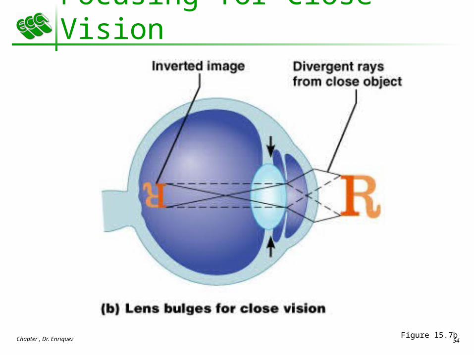
Focusing for Close Vision
Close vision requires: Accommodation – changing the lens shape by ciliary
muscles to increase refractory power Constriction – the pupillary reflex constricts the
pupils to prevent divergent light rays from entering the eye
Convergence – medial rotation of the eyeballs toward the object being viewed
Chapter , Dr. Enriquez 54
Focusing for Close Vision
Figure 15.7b

Chapter , Dr. Enriquez 55
Problems of Refraction
Emmetropic eye – normal eye with light focused properly
Myopic eye (nearsighted) – the focal point is in front of the retina Corrected with a concave lens
Hyperopic eye (farsighted) – the focal point is behind the retina Corrected with a convex lens

Chapter , Dr. Enriquez 56
Problems of Refraction
Figure 15.18

Chapter , Dr. Enriquez 57
Photoreception – process by which the eye detects light energy
Rods and cones contain visual pigments (photopigments) Arranged in a stack of disklike infoldings of the
plasma membrane that change shape as they absorb light
Photoreception: Functional Anatomy of Photoreceptors

Chapter , Dr. Enriquez 58Figure 15.19
Photoreception: Functional Anatomy of Photoreceptors

Chapter , Dr. Enriquez 59
Rods
Functional characteristics Sensitive to dim light and best suited for night vision Absorb all wavelengths of visible light Perceived input is in gray tones only Sum of visual input from many rods feeds into a
single ganglion cell Results in fuzzy and indistinct images

Chapter , Dr. Enriquez 60
Cones
Functional characteristics Need bright light for activation (have low sensitivity) Have pigments that furnish a vividly colored view Each cone synapses with a single ganglion cell Vision is detailed and has high resolution

Chapter , Dr. Enriquez 61
Cones and Rods
Figure 15.10a

Chapter , Dr. Enriquez 62
Chemistry of Visual Pigments
Retinal is a light-absorbing molecule Combines with opsins to form visual pigments Similar to and is synthesized from vitamin A Two isomers: 11-cis and all-trans
Isomerization of retinal initiates electrical impulses in the optic nerve

Chapter , Dr. Enriquez 63
Chemistry of Visual Pigments
Figure 15.20

Chapter , Dr. Enriquez 64
Excitation of Rods
The visual pigment of rods is rhodopsin (opsin + 11-cis retinal)
Light phase Rhodopsin breaks down into all-trans retinal + opsin
(bleaching of the pigment) Dark phase
All-trans retinal converts to 11-cis form 11-cis retinal is also formed from vitamin A 11-cis retinal + opsin regenerate rhodopsin

Chapter , Dr. Enriquez 65
Excitation of Rods
Figure 15.21

Chapter , Dr. Enriquez 66
Excitation of Cones
Visual pigments in cones are similar to rods (retinal + opsins)
There are three types of cones: blue, green, and red
Intermediate colors are perceived by activation of more than one type of cone
Method of excitation is similar to rods
Chapter , Dr. Enriquez 67
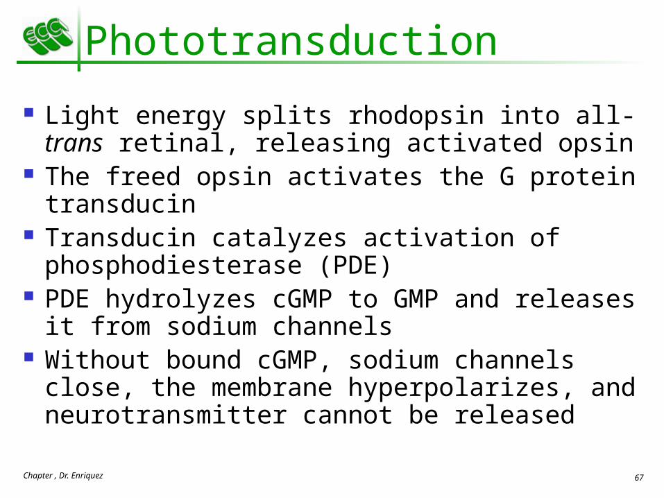
Phototransduction Light energy splits rhodopsin into all-trans
retinal, releasing activated opsin The freed opsin activates the G protein transducin Transducin catalyzes activation of
phosphodiesterase (PDE) PDE hydrolyzes cGMP to GMP and releases it
from sodium channels Without bound cGMP, sodium channels close,
the membrane hyperpolarizes, and neurotransmitter cannot be released

Chapter , Dr. Enriquez 68
Phototransduction
Figure 15.22

Chapter , Dr. Enriquez 69
Adaptation
Adaptation to bright light (going from dark to light) involves: Dramatic decreases in retinal sensitivity – rod
function is lost Switching from the rod to the cone system – visual
acuity is gained Adaptation to dark is the reverse
Cones stop functioning in low light Rhodopsin accumulates in the dark and retinal
sensitivity is restored

Chapter , Dr. Enriquez 70
Visual Pathways Axons of retinal ganglion cells form the optic
nerve Medial fibers of the optic nerve decussate at the
optic chiasm Most fibers of the optic tracts continue to the
lateral geniculate body of the thalamus Other optic tract fibers end in superior colliculi
(initiating visual reflexes) and pretectal nuclei (involved with pupillary reflexes)
Optic radiations travel from the thalamus to the visual cortex

Chapter , Dr. Enriquez 71
Visual Pathways
Figure 15.23

Chapter , Dr. Enriquez 72
Visual Pathways
Some nerve fibers send tracts to the midbrain ending in the superior colliculi
A small subset of visual fibers contain melanopsin (circadian pigment) which: Mediates papillary light reflexes Sets daily biorhythms

Chapter , Dr. Enriquez 73
Depth Perception
Achieved by both eyes viewing the same image from slightly different angles
Three-dimensional vision results from cortical fusion of the slightly different images
If only one eye is used, depth perception is lost and the observer must rely on learned clues to determine depth

Chapter , Dr. Enriquez 74
On-center fields Stimulated by light hitting the center of the field Inhibited by light hitting the periphery of the field
Off-center fields have the opposite effects These responses are due to receptor types in the
“on” and “off” fields
Retinal Processing: Receptive Fields of Ganglion Cells

Chapter , Dr. Enriquez 75Figure 15.24
Retinal Processing: Receptive Fields of Ganglion Cells

Chapter , Dr. Enriquez 76
Thalamic Processing
The lateral geniculate nuclei of the thalamus: Relay information on movement Segregate the retinal axons in preparation for depth
perception Emphasize visual inputs from regions of high cone
density Sharpen the contrast information received by the
retina

Chapter , Dr. Enriquez 77
Cortical Processing
Striate cortex processes Basic dark/bright and contrast information
Prestriate cortices (association areas) processes Form, color, and movement
Visual information then proceeds anteriorly to the: Temporal lobe – processes identification of objects Parietal cortex and postcentral gyrus – processes
spatial location

Chapter , Dr. Enriquez 78
The Ear: Hearing and Balance
The three parts of the ear are the inner, outer, and middle ear
The outer and middle ear are involved with hearing
The inner ear functions in both hearing and equilibrium
Receptors for hearing and balance: Respond to separate stimuli Are activated independently

Chapter , Dr. Enriquez 79
The Ear: Hearing and Balance
Figure 15.25a

Chapter , Dr. Enriquez 80
Outer Ear
The auricle (pinna) is composed of: The helix (rim) The lobule (earlobe)
External auditory canal Short, curved tube filled with ceruminous glands

Chapter , Dr. Enriquez 81
Outer Ear
Tympanic membrane (eardrum) Thin connective tissue membrane that vibrates in
response to sound Transfers sound energy to the middle ear ossicles Boundary between outer and middle ears

Chapter , Dr. Enriquez 82
Middle Ear (Tympanic Cavity)
A small, air-filled, mucosa-lined cavity Flanked laterally by the eardrum Flanked medially by the oval and round windows
Epitympanic recess – superior portion of the middle ear
Pharyngotympanic tube – connects the middle ear to the nasopharynx Equalizes pressure in the middle ear cavity with the
external air pressure

Chapter , Dr. Enriquez 83
Middle Ear (Tympanic Cavity)
Figure 15.25b

Chapter , Dr. Enriquez 84
15The Special Senses
Part C

Chapter , Dr. Enriquez 85
Ear Ossicles
The tympanic cavity contains three small bones: the malleus, incus, and stapes Transmit vibratory motion of the eardrum to the oval
window Dampened by the tensor tympani and stapedius
muscles

Chapter , Dr. Enriquez 86
Ear Ossicles
Figure 15.26

Chapter , Dr. Enriquez 87
Inner Ear
Bony labyrinth Tortuous channels worming their way through the temporal
bone Contains the vestibule, the cochlea, and the semicircular canals Filled with perilymph
Membranous labyrinth Series of membranous sacs within the bony labyrinth Filled with a potassium-rich fluid

Chapter , Dr. Enriquez 88
Inner Ear
Figure 15.27

Chapter , Dr. Enriquez 89
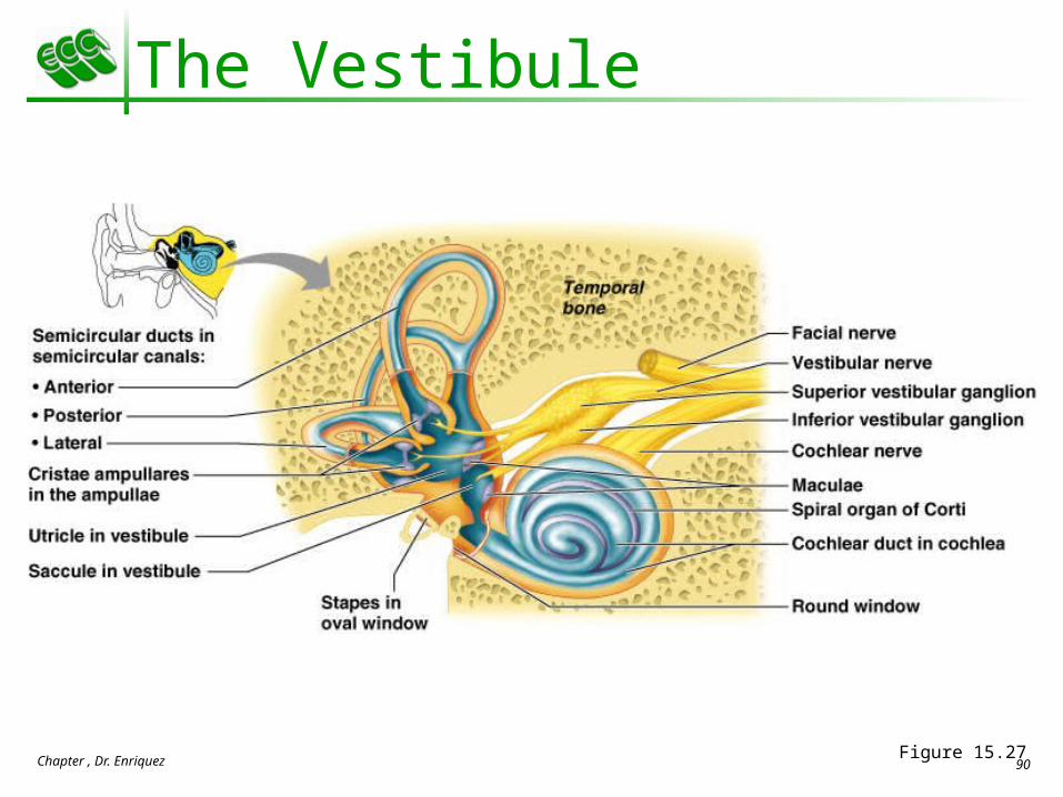
The Vestibule
The central egg-shaped cavity of the bony labyrinth
Suspended in its perilymph are two sacs: the saccule and utricle
The saccule extends into the cochlea The utricle extends into the semicircular canals These sacs:
House equilibrium receptors called maculae Respond to gravity and changes in the position of the
head
Chapter , Dr. Enriquez 90
The Vestibule
Figure 15.27

Chapter , Dr. Enriquez 91
The Semicircular Canals
Three canals that each define two-thirds of a circle and lie in the three planes of space
Membranous semicircular ducts line each canal and communicate with the utricle
The ampulla is the swollen end of each canal and it houses equilibrium receptors in a region called the crista ampullaris
These receptors respond to angular movements of the head

Chapter , Dr. Enriquez 92
The Semicircular Canals
Figure 15.27

Chapter , Dr. Enriquez 93
The Cochlea
A spiral, conical, bony chamber that: Extends from the anterior vestibule Coils around a bony pillar called the modiolus Contains the cochlear duct, which ends at the
cochlear apex Contains the organ of Corti (hearing receptor)

Chapter , Dr. Enriquez 94
The Cochlea
The cochlea is divided into three chambers: Scala vestibuli Scala media Scala tympani

Chapter , Dr. Enriquez 95
The Cochlea
The scala tympani terminates at the round window
The scalas tympani and vestibuli: Are filled with perilymph Are continuous with each other via the helicotrema
The scala media is filled with endolymph

Chapter , Dr. Enriquez 96
The Cochlea
The “floor” of the cochlear duct is composed of: The bony spiral lamina The basilar membrane, which supports the organ of
Corti The cochlear branch of nerve VIII runs from the
organ of Corti to the brain

Chapter , Dr. Enriquez 97
The Cochlea
Figure 15.28

Chapter , Dr. Enriquez 98
Sound and Mechanisms of Hearing
Sound vibrations beat against the eardrum The eardrum pushes against the ossicles, which
presses fluid in the inner ear against the oval and round windows This movement sets up shearing forces that pull on
hair cells Moving hair cells stimulates the cochlear nerve that
sends impulses to the brain
Chapter , Dr. Enriquez 99
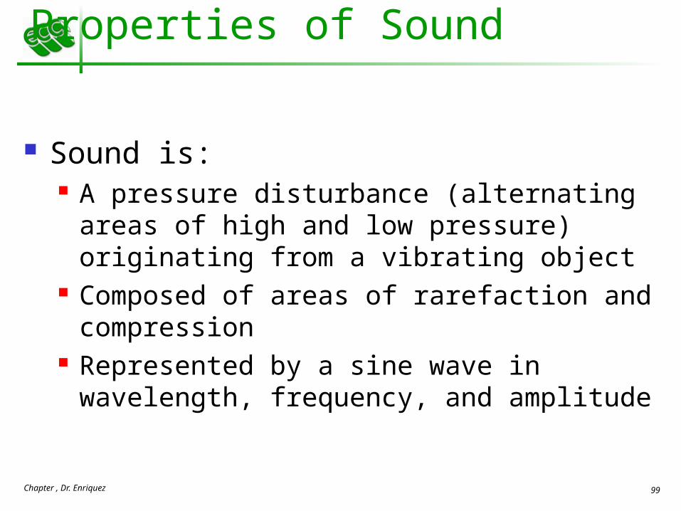
Properties of Sound
Sound is: A pressure disturbance (alternating areas of high and
low pressure) originating from a vibrating object Composed of areas of rarefaction and compression Represented by a sine wave in wavelength,
frequency, and amplitude

Chapter , Dr. Enriquez 100
Properties of Sound
Frequency – the number of waves that pass a given point in a given time
Pitch – perception of different frequencies (we hear from 20–20,000 Hz)

Chapter , Dr. Enriquez 101
Properties of Sound Amplitude – intensity of a sound measured in
decibels (dB) Loudness – subjective interpretation of sound
intensity
Figure 15.29

Chapter , Dr. Enriquez 102
Transmission of Sound to the Inner Ear
The route of sound to the inner ear follows this pathway: Outer ear – pinna, auditory canal, eardrum Middle ear – malleus, incus, and stapes to the oval
window Inner ear – scalas vestibuli and tympani to the
cochlear duct Stimulation of the organ of Corti Generation of impulses in the cochlear nerve
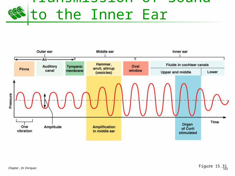
Chapter , Dr. Enriquez 103
Transmission of Sound to the Inner Ear
Figure 15.31

Chapter , Dr. Enriquez 104
Resonance of the Basilar Membrane
Sound waves of low frequency (inaudible): Travel around the helicotrema Do not excite hair cells
Audible sound waves: Penetrate through the cochlear duct Vibrate the basilar membrane Excite specific hair cells according to frequency of the
sound

Chapter , Dr. Enriquez 105
Resonance of the Basilar Membrane
Figure 15.32

Chapter , Dr. Enriquez 106
The Organ of Corti
Is composed of supporting cells and outer and inner hair cells
Afferent fibers of the cochlear nerve attach to the base of hair cells
The stereocilia (hairs): Protrude into the endolymph Touch the tectorial membrane

Chapter , Dr. Enriquez 107
Excitation of Hair Cells in the Organ of Corti
Bending cilia: Opens mechanically gated ion channels Causes a graded potential and the release of a
neurotransmitter (probably glutamate) The neurotransmitter causes cochlear fibers to
transmit impulses to the brain, where sound is perceived

Chapter , Dr. Enriquez 108
Excitation of Hair Cells in the Organ of Corti
Figure 15.28c

Chapter , Dr. Enriquez 109
Auditory Pathway to the Brain
Impulses from the cochlea pass via the spiral ganglion to the cochlear nuclei
From there, impulses are sent to the: Superior olivary nucleus Inferior colliculus (auditory reflex center)
From there, impulses pass to the auditory cortex Auditory pathways decussate so that both
cortices receive input from both ears

Chapter , Dr. Enriquez 110
Simplified Auditory Pathways
Figure 15.34

Chapter , Dr. Enriquez 111
Auditory Processing
Pitch is perceived by: The primary auditory cortex Cochlear nuclei
Loudness is perceived by: Varying thresholds of cochlear cells The number of cells stimulated
Localization is perceived by superior olivary nuclei that determine sound

Chapter , Dr. Enriquez 112
Deafness Conduction deafness – something hampers sound
conduction to the fluids of the inner ear (e.g., impacted earwax, perforated eardrum, osteosclerosis of the ossicles)
Sensorineural deafness – results from damage to the neural structures at any point from the cochlear hair cells to the auditory cortical cells
Tinnitus – ringing or clicking sound in the ears in the absence of auditory stimuli
Meniere’s syndrome – labyrinth disorder that affects the cochlea and the semicircular canals, causing vertigo, nausea, and vomiting

Chapter , Dr. Enriquez 113
Mechanisms of Equilibrium and Orientation
Vestibular apparatus – equilibrium receptors in the semicircular canals and vestibule Maintains our orientation and balance in space Vestibular receptors monitor static equilibrium Semicircular canal receptors monitor dynamic
equilibrium

Chapter , Dr. Enriquez 114
Anatomy of Maculae
Maculae are the sensory receptors for static equilibrium Contain supporting cells and hair cells Each hair cell has stereocilia and kinocilium
embedded in the otolithic membrane Otolithic membrane – jellylike mass studded with
tiny CaCO3 stones called otoliths Utricular hairs respond to horizontal movement Saccular hairs respond to vertical movement

Chapter , Dr. Enriquez 115
Anatomy of Maculae
Figure 15.35

Chapter , Dr. Enriquez 116
Effect of Gravity on Utricular Receptor Cells
Otolithic movement in the direction of the kinocilia: Depolarizes vestibular nerve fibers Increases the number of action potentials generated
Movement in the opposite direction: Hyperpolarizes vestibular nerve fibers Reduces the rate of impulse propagation
From this information, the brain is informed of the changing position of the head

Chapter , Dr. Enriquez 117
Effect of Gravity on Utricular Receptor Cells
Figure 15.36

Chapter , Dr. Enriquez 118
Crista Ampullaris and Dynamic Equilibrium
The crista ampullaris (or crista): Is the receptor for dynamic equilibrium Is located in the ampulla of each semicircular canal Responds to angular movements
Each crista has support cells and hair cells that extend into a gel-like mass called the cupula
Dendrites of vestibular nerve fibers encircle the base of the hair cells

Chapter , Dr. Enriquez 119
Crista Ampullaris and Dynamic Equilibrium
Figure 15.37b

Chapter , Dr. Enriquez 120
Activating Crista Ampullaris Receptors
Cristae respond to changes in velocity of rotatory movements of the head
Directional bending of hair cells in the cristae causes: Depolarizations, and rapid impulses reach the brain at
a faster rate Hyperpolarizations, and fewer impulses reach the
brain The result is that the brain is informed of
rotational movements of the head

Chapter , Dr. Enriquez 121
Rotary Head Movement
Figure 15.37d

Chapter , Dr. Enriquez 122
Balance and Orientation Pathways
There are three modes of input for balance and orientation Vestibular receptors Visual receptors Somatic receptors
These receptors allow our body to respond reflexively
Figure 15.38

Chapter , Dr. Enriquez 123
Developmental Aspects
All special senses are functional at birth Chemical senses – few problems occur until the
fourth decade, when these senses begin to decline
Vision – optic vesicles protrude from the diencephalon during the fourth week of development These vesicles indent to form optic cups and their
stalks form optic nerves Later, the lens forms from ectoderm
Chapter , Dr. Enriquez 124
Developmental Aspects
Vision is not fully functional at birth Babies are hyperopic, see only gray tones, and
eye movements are uncoordinated Depth perception and color vision is well
developed by age five and emmetropic eyes are developed by year six
With age the lens loses clarity, dilator muscles are less efficient, and visual acuity is drastically decreased by age 70

Chapter , Dr. Enriquez 125
Developmental Aspects Ear development begins in the three-week
embryo Inner ears develop from otic placodes, which
invaginate into the otic pit and otic vesicle The otic vesicle becomes the membranous
labyrinth, and the surrounding mesenchyme becomes the bony labyrinth
Middle ear structures develop from the pharyngeal pouches
The branchial groove develops into outer ear structures