Embed Size (px)
Citation preview
1
Chapter 12
Nervous Tissue
2
INTRODUCTION
• The nervous system, along with
the endocrine system, helps to
keep controlled conditions within
limits that maintain health and
helps to maintain homeostasis
• The nervous system is
responsible for all our behaviors,
memories, and movements
• The branch of medical science
that deals with the normal
functioning and disorders of the
nervous system is called
neurology
3
Nervous System
• Three basic functions:
– sensing changes with sensory receptors
• fullness of stomach or sun on your face
– interpreting and remembering those changes
– reacting to those changes with effectors
• muscular contractions
• glandular secretions
4
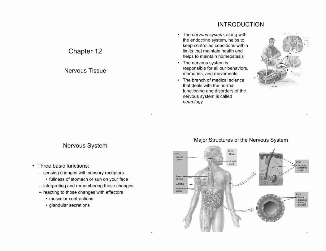
Major Structures of the Nervous System
5
Structures of the Nervous System - Overview
• Twelve pairs of cranial nerves emerge from the base of the
brain through foramina of the skull.
– A nerve is a bundle of hundreds or thousands of axons, each serving
a specific region of the body.
• The spinal cord connects to the brain through the foramen
magnum of the skull and is encircled by the bones of the
vertebral column.
– Thirty-one pairs of spinal nerves emerge from the spinal cord.
• Ganglia, located outside the brain and spinal cord, are small
masses of nervous tissue, containing primarily cell bodies of
neurons.
• Sensory receptors are either parts of neurons or specialized
cells that monitor changes in the internal or external
environment.
6
Functions of the Nervous Systems
• The sensory function of the nervous system is to
sense changes in the internal and external
environment through sensory receptors.
– Sensory (afferent) neurons serve this function.
• The integrative function is to analyze the sensory
information, store some aspects, and make
decisions regarding appropriate behaviors.
– Association or interneurons serve this function.
• The motor function is to respond to stimuli by
initiating action.
– Motor (efferent) neurons serve this function.
7
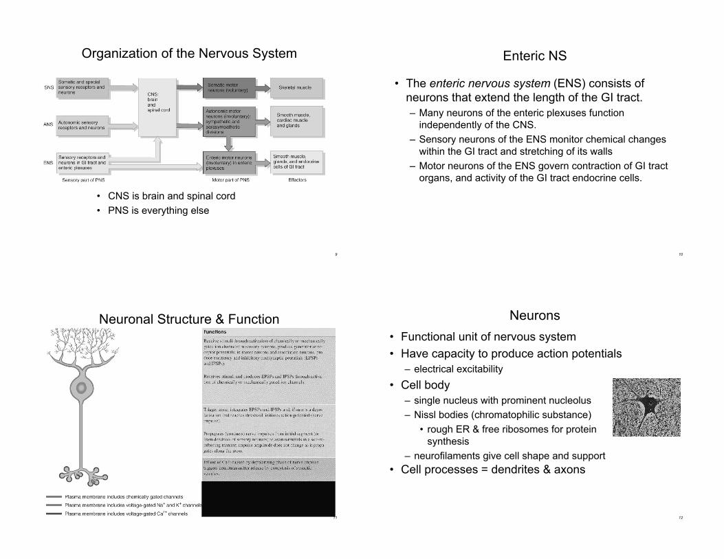
Nervous System Divisions
• Central nervous system (CNS)
– consists of the brain and spinal cord
• Peripheral nervous system (PNS)
– consists of cranial and spinal nerves that contain both
sensory and motor fibers
– connects CNS to muscles, glands & all sensory receptors
8
Subdivisions of the PNS
• Somatic (voluntary) nervous system (SNS)
– neurons from cutaneous and special sensory receptors to
the CNS
– motor neurons to skeletal muscle tissue
• Autonomic (involuntary) nervous systems (ANS)
– sensory neurons from visceral organs to CNS
– motor neurons to smooth & cardiac muscle and glands
• sympathetic division (speeds up heart rate)
• parasympathetic division (slow down heart rate)
• Enteric nervous system (ENS)– involuntary sensory & motor neurons control GI tract
– neurons function independently of ANS & CNS
9
Organization of the Nervous System
• CNS is brain and spinal cord
• PNS is everything else
10
Enteric NS
• The enteric nervous system (ENS) consists of
neurons that extend the length of the GI tract.
– Many neurons of the enteric plexuses function
independently of the CNS.
– Sensory neurons of the ENS monitor chemical changes
within the GI tract and stretching of its walls
– Motor neurons of the ENS govern contraction of GI tract
organs, and activity of the GI tract endocrine cells.
11
Neuronal Structure & Function
12
Neurons
• Functional unit of nervous system
• Have capacity to produce action potentials
– electrical excitability
• Cell body
– single nucleus with prominent nucleolus
– Nissl bodies (chromatophilic substance)
• rough ER & free ribosomes for protein
synthesis
– neurofilaments give cell shape and support
• Cell processes = dendrites & axons
13
Nucleus with
Nucleolus
Parts of a Neuron
Axons or
Dendrites
Cell body
Neuroglial cells
14
Dendrites
• Conducts impulses
towards the cell body
• Typically short, highly
branched &
unmyelinated
• Surfaces specialized
for contact with other
neurons
15
Axons• Conduct impulses away
from cell body
• To another neuron or to
an effector (muscle or
gland)
• Long, thin cylindrical
process of cell
• Arises at axon hillock
• Impulses arise from initial
segment (trigger zone)
• Swollen tips called
synaptic end bulbs
contain vesicles filled with
neurotransmitters
16
Axonal Transport
• Cell body is location for most protein synthesis
– neurotransmitters & repair proteins
• Axonal transport system moves substances
– slow axonal flow
• movement in one direction only -- away from cell body
• movement at 1-5 mm per day
– fast axonal flow
• moves organelles & materials along surface of microtubules
• at 200-400 mm per day
• transports in either direction
• Fast axonal transport route by which toxins or pathogens
reach neuron cell bodies
– tetanus (Clostridium tetani bacteria)
– disrupts motor neurons causing painful muscle spasms
17
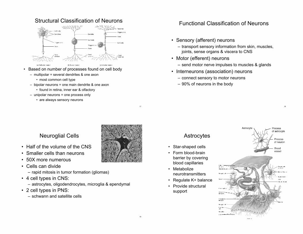
Structural Classification of Neurons
• Based on number of processes found on cell body
– multipolar = several dendrites & one axon
• most common cell type
– bipolar neurons = one main dendrite & one axon
• found in retina, inner ear & olfactory
– unipolar neurons = one process only
• are always sensory neurons
18
Functional Classification of Neurons
• Sensory (afferent) neurons
– transport sensory information from skin, muscles,
joints, sense organs & viscera to CNS
• Motor (efferent) neurons
– send motor nerve impulses to muscles & glands
• Interneurons (association) neurons
– connect sensory to motor neurons
– 90% of neurons in the body
19
• Half of the volume of the CNS
• Smaller cells than neurons
• 50X more numerous
• Cells can divide– rapid mitosis in tumor formation (gliomas)
• 4 cell types in CNS:– astrocytes, oligodendrocytes, microglia & ependymal
• 2 cell types in PNS:– schwann and satellite cells
Neuroglial Cells
20
Astrocytes
• Star-shaped cells
• Form blood-brain
barrier by covering
blood capillaries
• Metabolize
neurotransmitters
• Regulate K+ balance
• Provide structural
support
21
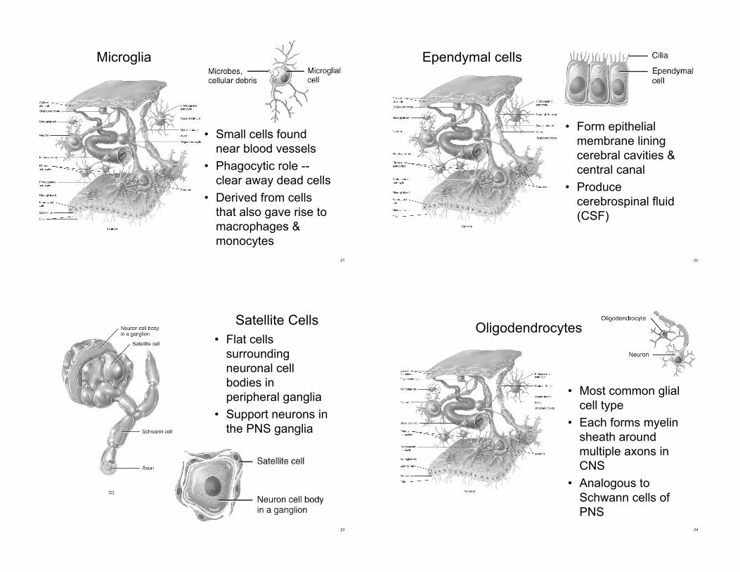
Microglia
• Small cells found
near blood vessels
• Phagocytic role --
clear away dead cells
• Derived from cells
that also gave rise to
macrophages &
monocytes
22
Ependymal cells
• Form epithelial
membrane lining
cerebral cavities &
central canal
• Produce
cerebrospinal fluid
(CSF)
23
Satellite Cells
• Flat cells
surrounding
neuronal cell
bodies in
peripheral ganglia
• Support neurons in
the PNS ganglia
24
Oligodendrocytes
• Most common glial
cell type
• Each forms myelin
sheath around
multiple axons in
CNS
• Analogous to
Schwann cells of
PNS
25
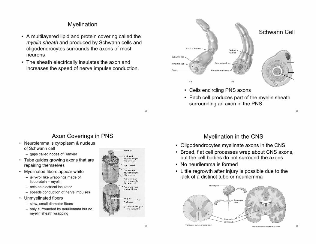
Myelination
• A multilayered lipid and protein covering called the
myelin sheath and produced by Schwann cells and
oligodendrocytes surrounds the axons of most
neurons
• The sheath electrically insulates the axon and
increases the speed of nerve impulse conduction.
26
Schwann Cell
• Cells encircling PNS axons
• Each cell produces part of the myelin sheath
surrounding an axon in the PNS
27
Axon Coverings in PNS• Neurolemma is cytoplasm & nucleus
of Schwann cell
– gaps called nodes of Ranvier
• Tube guides growing axons that are
repairing themselves
• Myelinated fibers appear white
– jelly-roll like wrappings made of
lipoprotein = myelin
– acts as electrical insulator
– speeds conduction of nerve impulses
• Unmyelinated fibers
– slow, small diameter fibers
– only surrounded by neurilemma but no
myelin sheath wrapping
28
Myelination in the CNS
• Oligodendrocytes myelinate axons in the CNS
• Broad, flat cell processes wrap about CNS axons,but the cell bodies do not surround the axons
• No neurilemma is formed
• Little regrowth after injury is possible due to thelack of a distinct tube or neurilemma
29
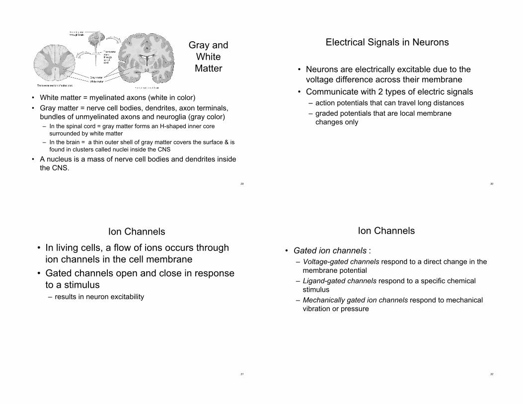
Gray and
White
Matter
• White matter = myelinated axons (white in color)
• Gray matter = nerve cell bodies, dendrites, axon terminals,
bundles of unmyelinated axons and neuroglia (gray color)
– In the spinal cord = gray matter forms an H-shaped inner core
surrounded by white matter
– In the brain = a thin outer shell of gray matter covers the surface & is
found in clusters called nuclei inside the CNS
• A nucleus is a mass of nerve cell bodies and dendrites inside
the CNS.
30
Electrical Signals in Neurons
• Neurons are electrically excitable due to the
voltage difference across their membrane
• Communicate with 2 types of electric signals
– action potentials that can travel long distances
– graded potentials that are local membrane
changes only
31
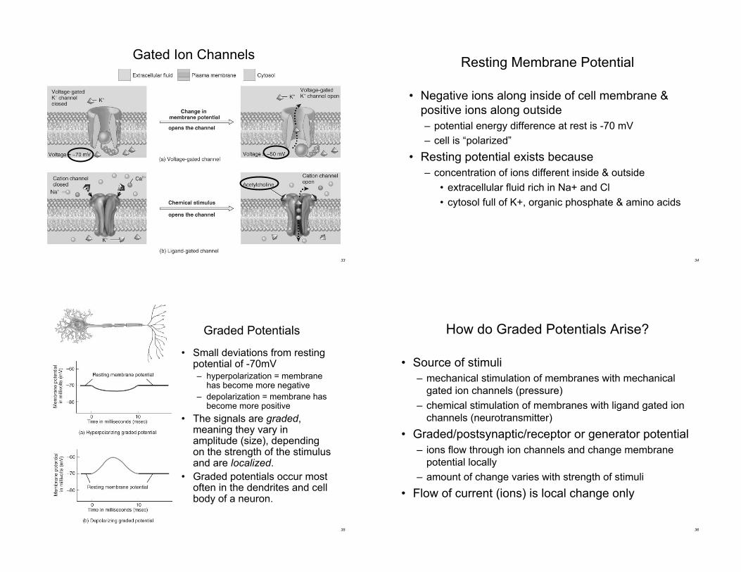
Ion Channels
• In living cells, a flow of ions occurs through
ion channels in the cell membrane
• Gated channels open and close in response
to a stimulus– results in neuron excitability
32
Ion Channels
• Gated ion channels :
– Voltage-gated channels respond to a direct change in the
membrane potential
– Ligand-gated channels respond to a specific chemical
stimulus
– Mechanically gated ion channels respond to mechanical
vibration or pressure
33
Gated Ion Channels
34
Resting Membrane Potential
• Negative ions along inside of cell membrane &
positive ions along outside
– potential energy difference at rest is -70 mV
– cell is “polarized”
• Resting potential exists because
– concentration of ions different inside & outside
• extracellular fluid rich in Na+ and Cl
• cytosol full of K+, organic phosphate & amino acids
35
Graded Potentials
• Small deviations from restingpotential of -70mV– hyperpolarization = membrane
has become more negative
– depolarization = membrane hasbecome more positive
• The signals are graded,meaning they vary inamplitude (size), dependingon the strength of the stimulusand are localized.
• Graded potentials occur mostoften in the dendrites and cellbody of a neuron.
36
How do Graded Potentials Arise?
• Source of stimuli
– mechanical stimulation of membranes with mechanical
gated ion channels (pressure)
– chemical stimulation of membranes with ligand gated ion
channels (neurotransmitter)
• Graded/postsynaptic/receptor or generator potential
– ions flow through ion channels and change membrane
potential locally
– amount of change varies with strength of stimuli
• Flow of current (ions) is local change only
37
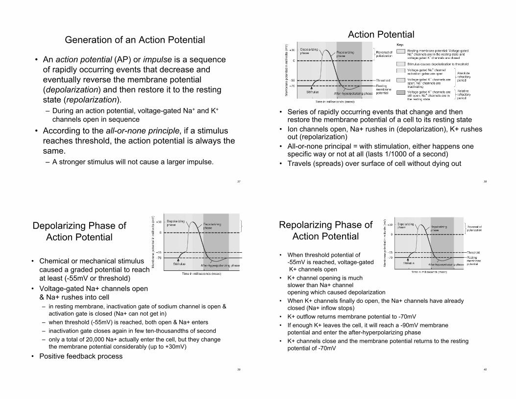
Generation of an Action Potential
• An action potential (AP) or impulse is a sequence
of rapidly occurring events that decrease and
eventually reverse the membrane potential
(depolarization) and then restore it to the resting
state (repolarization).
– During an action potential, voltage-gated Na+ and K+
channels open in sequence
• According to the all-or-none principle, if a stimulus
reaches threshold, the action potential is always the
same.
– A stronger stimulus will not cause a larger impulse.
38
Action Potential
• Series of rapidly occurring events that change and thenrestore the membrane potential of a cell to its resting state
• Ion channels open, Na+ rushes in (depolarization), K+ rushesout (repolarization)
• All-or-none principal = with stimulation, either happens onespecific way or not at all (lasts 1/1000 of a second)
• Travels (spreads) over surface of cell without dying out
39
Depolarizing Phase of
Action Potential
• Chemical or mechanical stimulus
caused a graded potential to reach
at least (-55mV or threshold)
• Voltage-gated Na+ channels open
& Na+ rushes into cell
– in resting membrane, inactivation gate of sodium channel is open &
activation gate is closed (Na+ can not get in)
– when threshold (-55mV) is reached, both open & Na+ enters
– inactivation gate closes again in few ten-thousandths of second
– only a total of 20,000 Na+ actually enter the cell, but they change
the membrane potential considerably (up to +30mV)
• Positive feedback process
40
Repolarizing Phase of
Action Potential
• When threshold potential of
-55mV is reached, voltage-gated
K+ channels open
• K+ channel opening is much
slower than Na+ channel
opening which caused depolarization
• When K+ channels finally do open, the Na+ channels have already
closed (Na+ inflow stops)
• K+ outflow returns membrane potential to -70mV
• If enough K+ leaves the cell, it will reach a -90mV membrane
potential and enter the after-hyperpolarizing phase
• K+ channels close and the membrane potential returns to the resting
potential of -70mV
41
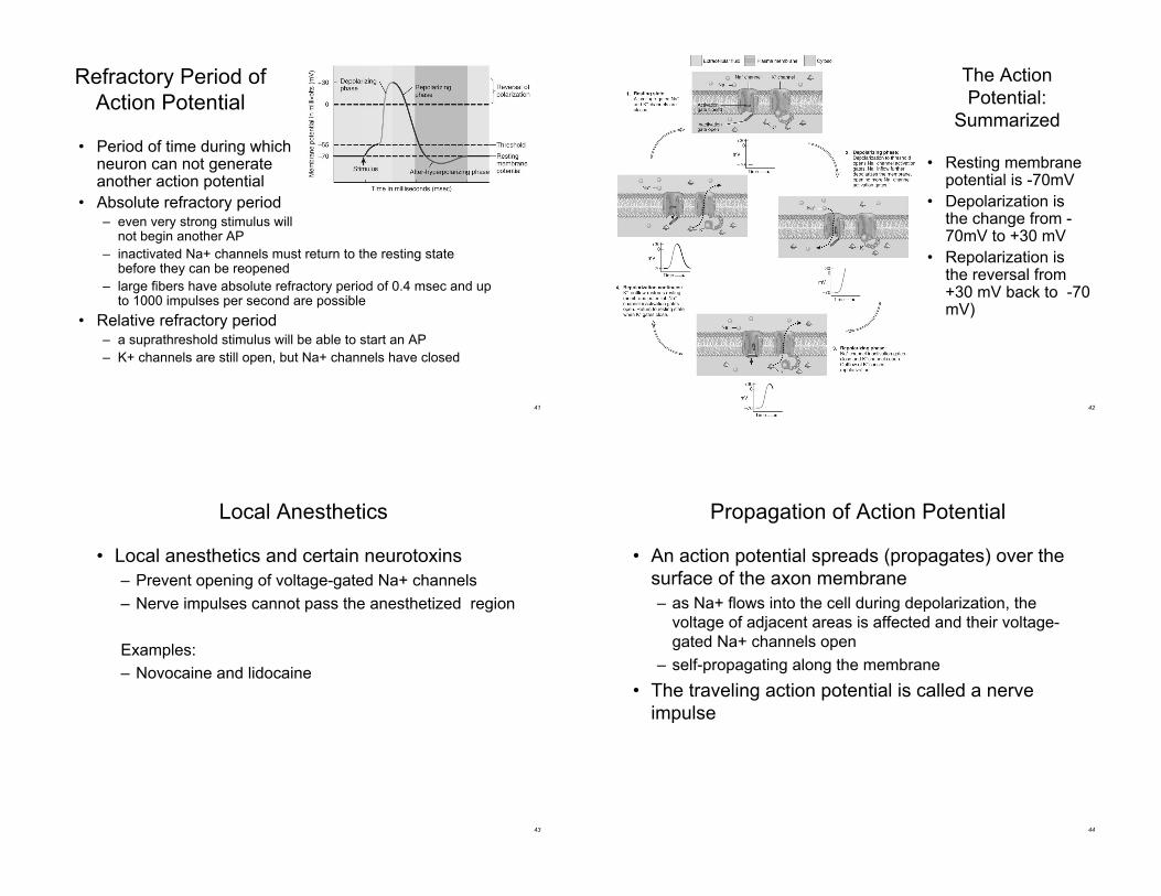
Refractory Period of
Action Potential
• Period of time during whichneuron can not generateanother action potential
• Absolute refractory period– even very strong stimulus will
not begin another AP
– inactivated Na+ channels must return to the resting statebefore they can be reopened
– large fibers have absolute refractory period of 0.4 msec and upto 1000 impulses per second are possible
• Relative refractory period– a suprathreshold stimulus will be able to start an AP
– K+ channels are still open, but Na+ channels have closed
42
The Action
Potential:
Summarized
• Resting membranepotential is -70mV
• Depolarization isthe change from -70mV to +30 mV
• Repolarization isthe reversal from+30 mV back to -70mV)
43
Local Anesthetics
• Local anesthetics and certain neurotoxins
– Prevent opening of voltage-gated Na+ channels
– Nerve impulses cannot pass the anesthetized region
Examples:
– Novocaine and lidocaine
44
Propagation of Action Potential
• An action potential spreads (propagates) over the
surface of the axon membrane
– as Na+ flows into the cell during depolarization, the
voltage of adjacent areas is affected and their voltage-
gated Na+ channels open
– self-propagating along the membrane
• The traveling action potential is called a nerve
impulse
45
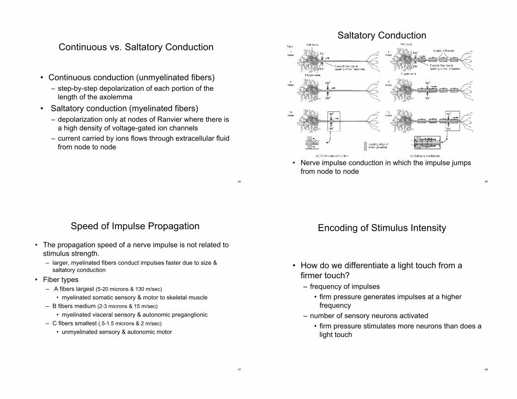
Continuous vs. Saltatory Conduction
• Continuous conduction (unmyelinated fibers)
– step-by-step depolarization of each portion of the
length of the axolemma
• Saltatory conduction (myelinated fibers)
– depolarization only at nodes of Ranvier where there is
a high density of voltage-gated ion channels
– current carried by ions flows through extracellular fluid
from node to node
46
Saltatory Conduction
• Nerve impulse conduction in which the impulse jumps
from node to node
47
Speed of Impulse Propagation
• The propagation speed of a nerve impulse is not related to
stimulus strength.
– larger, myelinated fibers conduct impulses faster due to size &
saltatory conduction
• Fiber types
– A fibers largest (5-20 microns & 130 m/sec)
• myelinated somatic sensory & motor to skeletal muscle
– B fibers medium (2-3 microns & 15 m/sec)
• myelinated visceral sensory & autonomic preganglionic
– C fibers smallest (.5-1.5 microns & 2 m/sec)
• unmyelinated sensory & autonomic motor
48
Encoding of Stimulus Intensity
• How do we differentiate a light touch from a
firmer touch?
– frequency of impulses
• firm pressure generates impulses at a higher
frequency
– number of sensory neurons activated
• firm pressure stimulates more neurons than does a
light touch
49
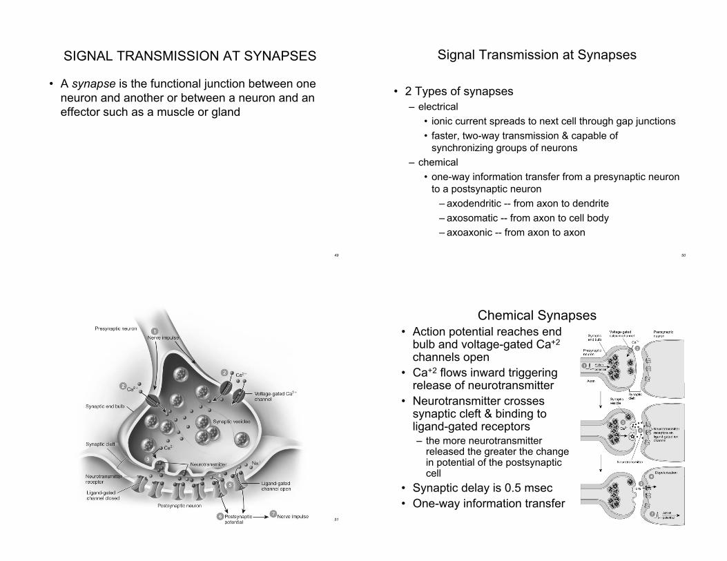
SIGNAL TRANSMISSION AT SYNAPSES
• A synapse is the functional junction between one
neuron and another or between a neuron and an
effector such as a muscle or gland
50
Signal Transmission at Synapses
• 2 Types of synapses
– electrical
• ionic current spreads to next cell through gap junctions
• faster, two-way transmission & capable of
synchronizing groups of neurons
– chemical
• one-way information transfer from a presynaptic neuron
to a postsynaptic neuron
– axodendritic -- from axon to dendrite
– axosomatic -- from axon to cell body
– axoaxonic -- from axon to axon
51 52
Chemical Synapses• Action potential reaches end
bulb and voltage-gated Ca+2
channels open
• Ca+2 flows inward triggeringrelease of neurotransmitter
• Neurotransmitter crossessynaptic cleft & binding toligand-gated receptors– the more neurotransmitter
released the greater the changein potential of the postsynapticcell
• Synaptic delay is 0.5 msec
• One-way information transfer
53
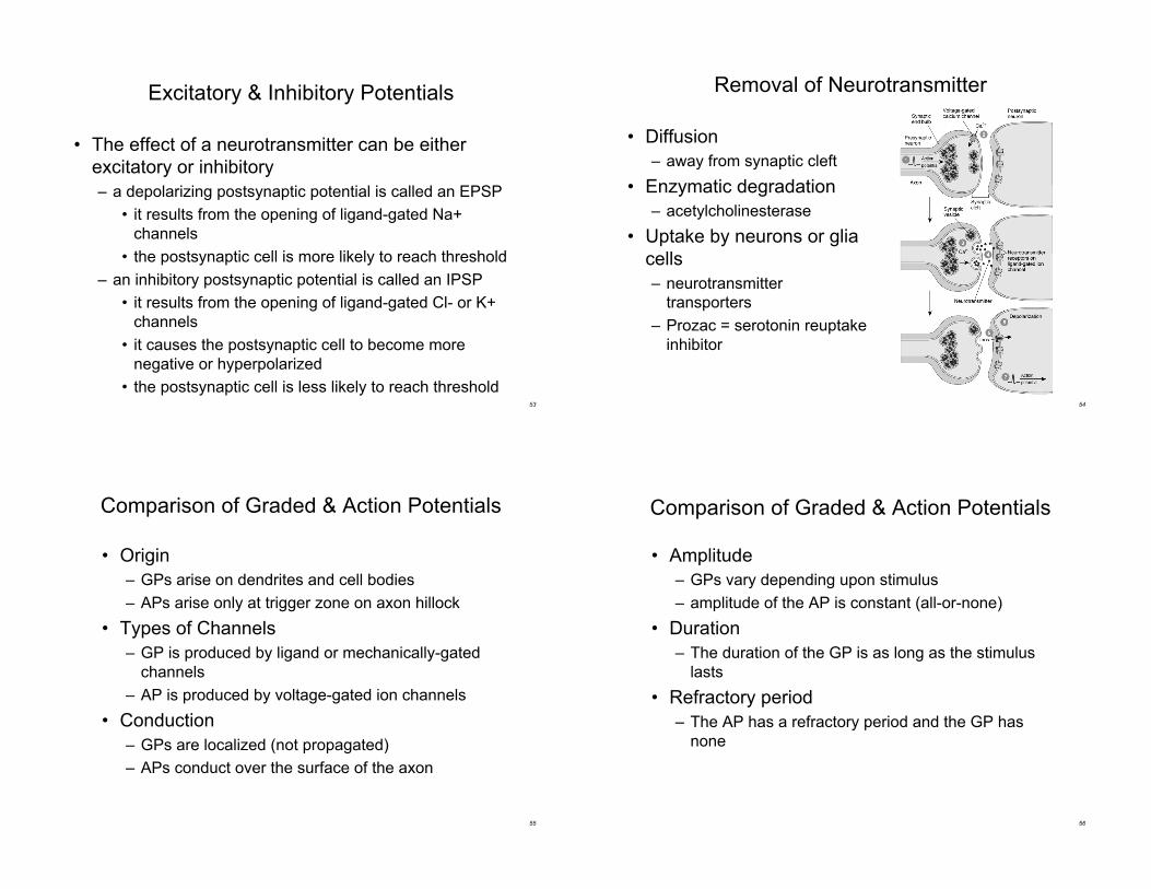
Excitatory & Inhibitory Potentials
• The effect of a neurotransmitter can be either
excitatory or inhibitory
– a depolarizing postsynaptic potential is called an EPSP
• it results from the opening of ligand-gated Na+
channels
• the postsynaptic cell is more likely to reach threshold
– an inhibitory postsynaptic potential is called an IPSP
• it results from the opening of ligand-gated Cl- or K+
channels
• it causes the postsynaptic cell to become more
negative or hyperpolarized
• the postsynaptic cell is less likely to reach threshold54
Removal of Neurotransmitter
• Diffusion
– away from synaptic cleft
• Enzymatic degradation
– acetylcholinesterase
• Uptake by neurons or glia
cells
– neurotransmitter
transporters
– Prozac = serotonin reuptake
inhibitor
55
• Origin
– GPs arise on dendrites and cell bodies
– APs arise only at trigger zone on axon hillock
• Types of Channels
– GP is produced by ligand or mechanically-gated
channels
– AP is produced by voltage-gated ion channels
• Conduction
– GPs are localized (not propagated)
– APs conduct over the surface of the axon
Comparison of Graded & Action Potentials
56
Comparison of Graded & Action Potentials
• Amplitude
– GPs vary depending upon stimulus
– amplitude of the AP is constant (all-or-none)
• Duration
– The duration of the GP is as long as the stimulus
lasts
• Refractory period
– The AP has a refractory period and the GP has
none
57
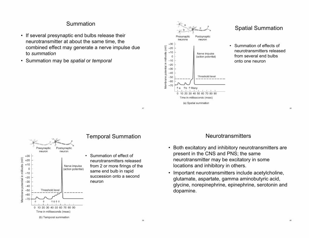
Summation
• If several presynaptic end bulbs release their
neurotransmitter at about the same time, the
combined effect may generate a nerve impulse due
to summation
• Summation may be spatial or temporal
58
• Summation of effects of
neurotransmitters released
from several end bulbs
onto one neuron
Spatial Summation
59
Temporal Summation
• Summation of effect of
neurotransmitters released
from 2 or more firings of the
same end bulb in rapid
succession onto a second
neuron
60
Neurotransmitters
• Both excitatory and inhibitory neurotransmitters are
present in the CNS and PNS; the same
neurotransmitter may be excitatory in some
locations and inhibitory in others.
• Important neurotransmitters include acetylcholine,
glutamate, aspartate, gamma aminobutyric acid,
glycine, norepinephrine, epinephrine, serotonin and
dopamine.
61
Neurotransmitter Effects
• Neurotransmitter effects can be modified
– synthesis can be stimulated or inhibited
– release can be blocked or enhanced
– removal can be stimulated or blocked
– receptor site can be blocked or activated
• Agonist
– anything that enhances a transmitters effects
• Antagonist
– anything that blocks the action of a neurotranmitter
62
Small-Molecule Neurotransmitters
• Acetylcholine (ACh)
– released by many PNS neurons & some CNS
– excitatory on NMJ but inhibitory at others
– inactivated by acetylcholinesterase
• Amino Acids
– glutamate released by nearly all excitatory neurons in
the brain
– GABA is inhibitory neurotransmitter for 1/3 of all brain
synapses (Valium is a GABA agonist -- enhancing its
inhibitory effect)
63
• Biogenic Amines
– modified amino acids (tyrosine)
• norepinephrine -- regulates mood, dreaming,
awakening from deep sleep
• dopamine -- regulating skeletal muscle tone
• serotonin -- control of mood, temperature regulation
& induction of sleep
Small-Molecule Neurotransmitters
64
• ATP and other purines (ADP, AMP & adenosine)
– excitatory in both CNS & PNS
• Gases (nitric oxide or NO)
– formed from amino acid arginine by an enzyme
– formed on demand and acts immediately
• diffuses out of cell that produced it to affect
neighboring cells
• may play a role in memory & learning
– first recognized as vasodilator that helps lower blood
pressure
Small-Molecule Neurotransmitters
65
Neuropeptides
• 3-40 amino acids linked by peptide bonds
• Substance P -- enhances our perception of pain
• Pain relief
– enkephalins -- pain-relieving effect by blocking the
release of substance P
– acupuncture may produce loss of pain sensation
because of release of opioid-like substances such as
endorphins or dynorphins
66
Strychnine Poisoning
• In spinal cord, an inhibitory neurotransmitter
(glycine) is normally released onto motor neurons
preventing excessive muscle contraction
• Strychnine binds to and blocks glycine receptors in
the spinal cord
• Massive tetanic contractions of all skeletal muscles
are produced
– when the diaphragm contracts & remains contracted,
breathing can not occur
67
Regeneration & Repair
• Plasticity maintained throughout life
– sprouting of new dendrites
– synthesis of new proteins
– changes in synaptic contacts with other neurons
• Limited ability for regeneration (repair)
– PNS can repair damaged axons
– CNS no repairs are possible
68
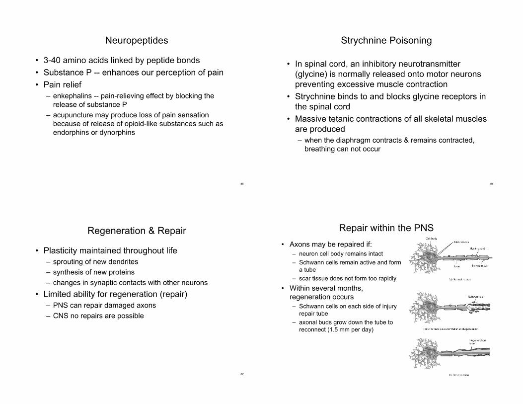
• Axons may be repaired if:
– neuron cell body remains intact
– Schwann cells remain active and form
a tube
– scar tissue does not form too rapidly
• Within several months,
regeneration occurs
– Schwann cells on each side of injury
repair tube
– axonal buds grow down the tube to
reconnect (1.5 mm per day)
Repair within the PNS

69
Neurogenesis in the CNS
• Formation of new neurons from stem cells was
not thought to occur in humans
– 1992 -- a growth factor was found that stimulates adult
mice brain cells to multiply
– 1998 -- new neurons found to form within adult human
hippocampus (area important for learning)
• There is a lack of neurogenesis in other regions
of the brain and spinal cord
70
Multiple Sclerosis (MS)
• Autoimmune disorder causing destruction of
myelin sheaths in CNS
– sheaths becomes scars or plaques
– 1/2 million people in the United States
– appears between ages 20 and 40
– females twice as often as males
• Symptoms include muscular weakness,
abnormal sensations or double vision
• Remissions & relapses result in progressive,
cumulative loss of function
71
• The second most common neurological
disorder
– affects 1% of population
• Characterized by short, recurrent attacks
initiated by electrical discharges in the brain
– lights, noise, or smells may be sensed
– skeletal muscles may contract involuntarily
– loss of consciousness
• Epilepsy has many causes, including;
– brain damage at birth, metabolic disturbances,
infections, toxins, vascular disturbances, head
injuries, and tumors
Epilepsy