Embed Size (px)
Citation preview

Chapter 2. Anatomy of the uterine cervix and the transformation zone 1313
chapter 2.
Anatomy of the uterine cervix and the transformation zone
The cervix is a fibromuscular organ that links the uterine cavity to the vagina. Although it is described as being cylindrical in shape, the an-terior and posterior walls are more often ordinarily apposed. The cer-vix is approximately 4 cm in length and 3 cm in diameter. The cervix of a parous woman is considerably larger than that of a nulliparous woman, and the cervix of a woman of reproductive age is considerably larger than that of a postmenopausal woman. The cervix occupies both an internal and an external position. Its lower half, or intravaginal part, lies at the upper end of the vagina, and its upper half lies above the va-gina, in the pelvic/abdominal cavity (Fig. 2.1). The two parts are approx-imately equal in size. The cervix lies between the bladder anteriorly and the bowel posteriorly. Laterally, the ureters are in close proximity, as are
CH
AP
TER
1
the uterine arteries superiorly and laterally.
The cervix has several different linings. The endocervical canal is lined with glandular epithelium, and the ectocervix is lined with squamous epithelium. The squamous epithelium
meets the glandular epithelium at the squamocolumnar junction (SCJ). The SCJ is dynamic and moves dur-ing early adolescence and during a first pregnancy. The original SCJ originates in the endocervical canal, but as the cervix everts during these
Fig. 2.1. Line drawing of normal female genital tract anatomy: sagittal sec-tion. In this drawing, the uterus is anteverted.
Uterus
Posterior fornix
Rectum
Sacrum
Vagina
Cervix
Bladder
Anterior fornix
Pubic bone
Urethra
CH
AP
TER
2

14
times, the SCJ comes to lie on the ectocervix and becomes the new SCJ. In colposcopy terminology, the SCJ is this new SCJ. The epithelium between these two SCJs is the TZ or transition zone, and its position is also variable. It may be small or large and usually becomes more ectocer-vical during a woman’s reproductive years, returning to an endocervical position after menopause.
When the uterus is anteverted, the cervix enters the vaginal vault through a slightly posterior approach, whereby at speculum examination the cervical os is directed towards the posterior vaginal wall (Fig. 2.1). When the speculum is opened, the cervix tends to be brought more cen-trally into view and into line with the longitudinal axis of the vagina. Most women have an anteverted uter-us. When the uterus is retroverted, the cervix tends to enter the vagina slightly more anteriorly, and in this case the cervix may be more difficult to locate at first speculum exposure. When the speculum is positioned properly and opened, the cervix tends to become positioned centrally and in a plane perpendicular to the longitudinal axis of the vagina.
The external os of the cervix will nearly always be visible to the naked eye at speculum examination. The visible external lining of the cervix derives from the vaginal (squamous) epithelium. The endocervical or glandular epithelium is not usually visible to the naked eye at speculum examination. At the upper end of the endocervical canal, the endocervical epithelium becomes the endome-trial lining of the uterine cavity. The lower half, or intravaginal part, of the cervix lies at the top of the vagina, surrounded by the vaginal fornices. These are the lateral, anterior, and posterior fornices and are where the vaginal epithelium sweeps into the cervix circumferentially. Squa-mous cervical cancer accounts for
the majority of cervical cancer and originates in the TZ. Glandular cer-vical cancer originates in either the TZ or the glandular epithelium above the TZ.
2.1 Tissue constituents of the cervix
2.1.1 Stroma
The stroma of the cervix is com-posed of dense, fibromuscular tissue through which vascular, lymphatic, and nerve supplies to the cervix pass and form a complex plexus.
The arterial supply of the cervix is derived from the internal iliac arter-ies through the cervical and vaginal branches of the uterine arteries. The cervical branches of the uterine ar-teries descend in the lateral aspects of the cervix at the 3 o’clock and 9 o’clock positions. The veins of the cervix run parallel to the arteries and drain into the hypogastric venous plexus. The lymphatic vessels from the cervix drain into the common ili-ac, external iliac, internal iliac, obtu-rator, and parametrial nodes.
The nerve supply to the cervix is derived from the hypogastric plex-us. The endocervix has extensive sensory nerve endings, whereas there are very few in the ectocervix. Hence, procedures such as biopsy, thermal coagulation, and cryother-apy are relatively well tolerated in most women, although there is good evidence that local anaesthesia ef-fectively prevents the discomfort of
these procedures. Also, the cervix of a parous woman tends to have slight-ly less sensory appreciation, which may be due to damage to nerve end-ings during childbirth. Because sym-pathetic and parasympathetic fibres are also abundant in the endocervix, dilatation and/or curettage of the en-docervix may occasionally lead to a vasovagal reaction.
The cervix is covered by both stratified, non-keratinizing squa-mous epithelium and columnar epi-thelium. As mentioned above, these two types of epithelium meet at the SCJ.
2.1.2 Squamous epithelium
Usually, most of the ectocervix and the entire length of the vagina is lined with squamous epithelium, which is uniform, stratified, and non-keratiniz-ing. Because mature squamous epi-thelium contains glycogen, it readily takes up Lugol’s iodine (and is there-fore Schiller test-negative). When epithelium does not take up Lugol’s iodine, it is Schiller test-positive. Cer-vical squamous epithelium is smooth and looks slightly pink to the naked eye in its non-pregnant state. During pregnancy it becomes progressively more vascular and develops a bluish hue.
The lowest level of cells in the squamous epithelium (Fig. 2.2) is a single layer of round basal cells with large dark-staining nuclei and little cytoplasm, attached to the base-ment membrane. The basement
Fig. 2.2. Squamous epithelium of the vagina and ectocervix.
Superficial cell layer
Intermediate cell layer
Parabasal layer
Basal cell layer
Stromal papilla
StromaBasement membrane

Chapter 2. Anatomy of the uterine cervix and the transformation zone 15
membrane separates the epithelium from the underlying stroma. The epithelial–stromal junction is usual-ly straight. Sometimes it is slightly undulating, with short projections of stroma, which occur at regular inter-vals. These stromal projections are called papillae, and the parts of the epithelium between the papillae are called rete pegs.
The basal cells divide and mature to form the next few layers of cells, called parabasal cells, which also have relatively large dark-staining nuclei and greenish-blue basophilic cytoplasm. Further differentiation and maturation of these cells leads to the intermediate layers of polyg-onal cells with abundant cytoplasm and small, round nuclei. These cells form a basket-weave pattern. With further maturation, the superficial layers of large and markedly flat-tened cells with small, dense, pyk-notic nuclei and transparent cyto-plasm are formed. Overall, from the basal layer to the superficial layer, these cells undergo an increase in size and a reduction in nuclear size.
The cells in the intermediate and superficial layers contain abundant glycogen in their cytoplasm, which stains mahogany brown or black after the application of Lugol’s iodine and magenta with periodic acid–Schiff stain in histological sections. Gly-cogenation of the intermediate and superficial layers is a sign of normal maturation and development of the squamous epithelium. Abnormal or altered maturation is characterized by a lack of glycogen production.
The maturation of the squamous epithelium of the cervix is dependent on estrogen, and if estrogen is lack-ing, full maturation and glycogena-tion do not take place. Hence, after menopause, the cells do not mature beyond the parabasal layer and do not accumulate as multiple layers of flat cells. Consequently, the epitheli-um becomes thin and atrophic. On
visual examination, it appears pale, sometimes with subepithelial pete-chial haemorrhagic spots, because it is easily prone to trauma.
2.1.3 Columnar epithelium
The endocervical canal is lined with columnar epithelium (often referred to as glandular epithelium). It is com-posed of a single layer of tall cells with dark-staining nuclei close to the basement membrane (Fig. 2.3). Because of its single layer of cells, it is much shorter in height than the stratified squamous epithelium of the cervix. On visual examination, it appears reddish, because the thin single-cell layer allows penetration of the stromal vascularity. At its dis-tal or upper limit, it merges with the endometrial epithelium in the lowest part of the body of the uterus. At its proximal or lower limit, it meets with the squamous epithelium at the SCJ. It covers a variable extent of the ec-tocervix, depending on the woman’s age and reproductive, hormonal, and menopausal status.
The columnar epithelium does not form a flattened surface in the endocervical canal but is thrown into multiple longitudinal folds pro-truding into the lumen of the canal, giving rise to papillary projections. It forms several invaginations into the substance of the cervical stroma, re-sulting in the formation of endocer-vical crypts (sometimes referred to as endocervical glands) (Fig. 2.4). The crypts may traverse as far as 5–6 mm from the surface of the cer-vix. This complex architecture, con-sisting of mucosal folds and crypts, gives the columnar epithelium a grainy or grape-like appearance on visual inspection.
A localized overgrowth of the en-docervical columnar epithelium may occasionally be visible as a reddish mass protruding through the exter-nal os on visual examination of the cervix. This is called a cervical polyp (Figs. 2.5 and 2.6). It usually begins as a localized enlargement of a sin-gle columnar papilla and appears as a mass as it enlarges. It is composed of a core of endocervical stroma
Fig. 2.4. Endocervical crypts lined with columnar epithelium.
Crypt opening
Columnar cells
Crypt
Fig. 2.3. Columnar epithelium of the endocervical canal.
Basement membrane
Columnar cells
Stroma
CH
AP
TER
2

1616
lined with columnar epithelium with underlying crypts. Occasionally, mul-tiple polyps may arise from the co-lumnar epithelium. The polyp shown in Fig. 2.5 is lined with columnar ep-ithelium, and it is protected from the metaplastic influence of the vaginal environment by endocervical mucus. The polyp shown in Fig. 2.6 has un-dergone a degree of metaplasia so that it is partially covered by squa-mous epithelium.
Glycogenation and mitoses are absent in the columnar epithelium. Because of the lack of intracellular cytoplasmic glycogen, the columnar epithelium does not change colour after the application of Lugol’s iodine or remains slightly discoloured with a thin film of iodine solution.
2.1.4 Squamocolumnar junction (SCJ)
The SCJ (Fig. 2.7) sometimes ap-pears as a sharp line with a step,
because of the difference in the height of the squamous and colum-nar epithelium. The location of the SCJ in relation to the external os is variable over a woman’s lifetime and depends on factors such as age, hormonal status, birth trauma, use of oral contraceptives, and pregnancy.
The SCJ that is visible during childhood, during perimenarche, af-ter puberty, and in early reproductive
life is referred to as the original SCJ, because this represents the junction between the columnar epithelium and the original squamous epitheli-um laid down during embryogenesis in intrauterine life. During childhood and around the menarche, the orig-inal SCJ is located at, or very close to, the external os (Fig. 2.8).
After puberty and during the re-productive period, the female genital organs develop under the influence of estrogen. Thus, the cervix swells and enlarges and the endocervical canal elongates. This leads to ever-sion of the columnar epithelium in the lower part of the endocervical ca-nal out onto the ectocervix (Fig. 2.9). This condition is called ectropion or ectopy, which is visible as a strikingly reddish-looking ectocervix on visual inspection (Fig. 2.10). It is sometimes called an erosion or ulcer, which are misnomers. Thus, the original SCJ is located on the ectocervix, far away from the external os (Fig. 2.9). An ec-tropion may begin or become much more pronounced during pregnancy.
The buffer action of the mu-cus covering the columnar cells is interfered with when the everted columnar epithelium of an ectropi-on is exposed to the acidic vaginal environment. This leads to the de-struction and eventual replacement of the columnar epithelium by the
Fig. 2.6. A cervical polyp that has undergone a degree of metaplasia so that it is partially covered by squamous epithelium.
Fig. 2.7. The squamocolumnar junction (SCJ).
Squamous epithelium
SCJ
Columnar epithelium
Fig. 2.8. Before puberty, the squa-mocolumnar junction is positioned above and very close to the external os.
Fig. 2.9. The squamocolumnar junc-tion after eversion of the columnar epithelium out onto the ectocervix, which occurs most commonly during adolescence and early pregnancy.
Fig. 2.5. A cervical polyp in the endocervical canal. It is protected from the vaginal environment by abundant mucus in the canal. It is lined with columnar epithelium.
Chapter 2. Anatomy of the uterine cervix and the transformation zone 1717
newly formed metaplastic squamous epithelium (metaplasia refers to the change or replacement of one type of epithelium by another).
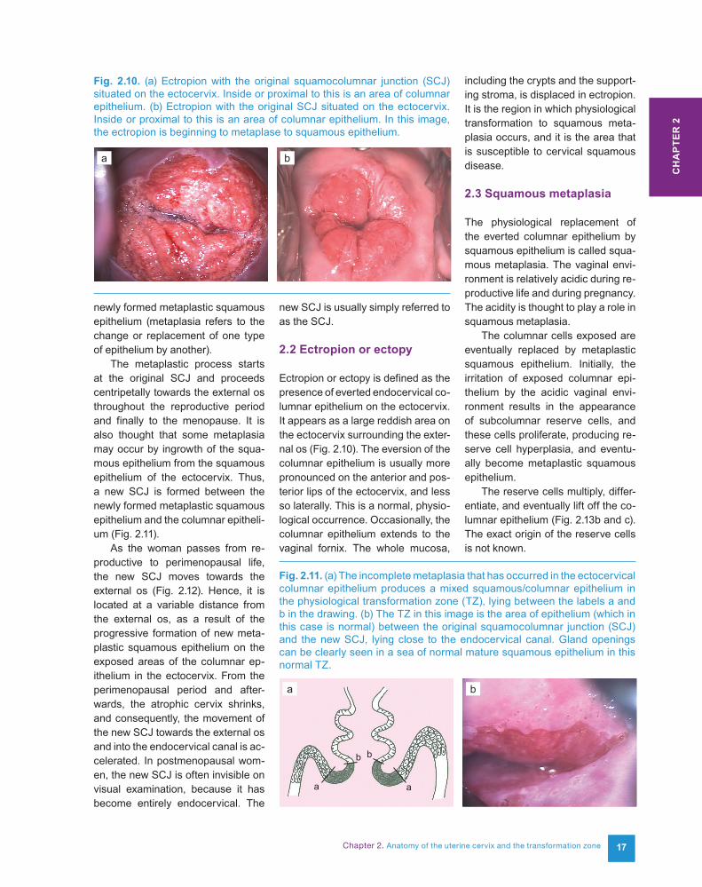
The metaplastic process starts at the original SCJ and proceeds centripetally towards the external os throughout the reproductive period and finally to the menopause. It is also thought that some metaplasia may occur by ingrowth of the squa-mous epithelium from the squamous epithelium of the ectocervix. Thus, a new SCJ is formed between the newly formed metaplastic squamous epithelium and the columnar epitheli-um (Fig. 2.11).
As the woman passes from re-productive to perimenopausal life, the new SCJ moves towards the external os (Fig. 2.12). Hence, it is located at a variable distance from the external os, as a result of the progressive formation of new meta-plastic squamous epithelium on the exposed areas of the columnar ep-ithelium in the ectocervix. From the perimenopausal period and after-wards, the atrophic cervix shrinks, and consequently, the movement of the new SCJ towards the external os and into the endocervical canal is ac-celerated. In postmenopausal wom-en, the new SCJ is often invisible on visual examination, because it has become entirely endocervical. The
new SCJ is usually simply referred to as the SCJ.
2.2 Ectropion or ectopy
Ectropion or ectopy is defined as the presence of everted endocervical co-lumnar epithelium on the ectocervix. It appears as a large reddish area on the ectocervix surrounding the exter-nal os (Fig. 2.10). The eversion of the columnar epithelium is usually more pronounced on the anterior and pos-terior lips of the ectocervix, and less so laterally. This is a normal, physio-logical occurrence. Occasionally, the columnar epithelium extends to the vaginal fornix. The whole mucosa,
including the crypts and the support-ing stroma, is displaced in ectropion. It is the region in which physiological transformation to squamous meta-plasia occurs, and it is the area that is susceptible to cervical squamous disease.
2.3 Squamous metaplasia
The physiological replacement of the everted columnar epithelium by squamous epithelium is called squa-mous metaplasia. The vaginal envi-ronment is relatively acidic during re-productive life and during pregnancy. The acidity is thought to play a role in squamous metaplasia.
The columnar cells exposed are eventually replaced by metaplastic squamous epithelium. Initially, the irritation of exposed columnar epi-thelium by the acidic vaginal envi-ronment results in the appearance of subcolumnar reserve cells, and these cells proliferate, producing re-serve cell hyperplasia, and eventu-ally become metaplastic squamous epithelium.
The reserve cells multiply, differ-entiate, and eventually lift off the co-lumnar epithelium (Fig. 2.13b and c). The exact origin of the reserve cells is not known.
Fig. 2.10. (a) Ectropion with the original squamocolumnar junction (SCJ) situated on the ectocervix. Inside or proximal to this is an area of columnar epithelium. (b) Ectropion with the original SCJ situated on the ectocervix. Inside or proximal to this is an area of columnar epithelium. In this image, the ectropion is beginning to metaplase to squamous epithelium.
ba
Fig. 2.11. (a) The incomplete metaplasia that has occurred in the ectocervical columnar epithelium produces a mixed squamous/columnar epithelium in the physiological transformation zone (TZ), lying between the labels a and b in the drawing. (b) The TZ in this image is the area of epithelium (which in this case is normal) between the original squamocolumnar junction (SCJ) and the new SCJ, lying close to the endocervical canal. Gland openings can be clearly seen in a sea of normal mature squamous epithelium in this normal TZ.
ba
aa
b b
CH
AP
TER
2

18
The first sign of squamous meta-plasia is the appearance and prolif-eration of reserve cells (Fig. 2.13a and b). This is initially seen as a sin-gle layer of small, round cells with dark-staining nuclei, situated very close to the nuclei of columnar cells, which further proliferate to produce reserve cell hyperplasia (Fig. 2.13b). Morphologically, the reserve cells have a similar appearance to the basal cells of the original squamous epithelium, with round nuclei and little cytoplasm. As the metaplastic
process progresses, the reserve cells proliferate and differentiate to form a thin, multicellular epithelium of immature squamous cells with no evidence of stratification (Fig. 2.13c). The term “immature squamous metaplastic epithelium” is used when there is little or no stratification in this thin, newly formed metaplas-tic epithelium. Immature squamous metaplastic epithelium does not produce glycogen and, hence, does not stain brown or black with Lugol’s iodine. Groups of mucin-containing
columnar cells may be seen em-bedded in the immature squamous metaplastic epithelium at this stage.
Numerous continuous and/or isolated fields or foci of immature squamous metaplasia may arise at the same time. It has been pro-posed that the basement membrane of the original columnar epithelium dissolves and is reformed between the proliferating and differentiating reserve cells and the cervical stro-ma. Squamous metaplasia usual-ly begins at the original SCJ at the distal limit of the ectopy, but it may also occur in the columnar epitheli-um close to this junction or as islands scattered in the exposed columnar epithelium.
As the process continues, the im-mature metaplastic squamous cells differentiate into mature stratified metaplastic epithelium (Fig. 2.13d). For all practical purposes, this re-sembles original stratified squamous epithelium. Some residual columnar cells or vacuoles of mucus are seen in the mature squamous metaplastic epithelium, which contains glyco-gen from the intermediate cell layer onward. Thus, the more mature the metaplasia is, the more it will stain brown or black after the application of Lugol’s iodine (Fig. 2.14).
Inclusion cysts, also called nabothian follicles or nabothian cysts, may be observed in the TZ in mature metaplastic squamous epithelium (Fig. 2.15). Nabothian cysts are re-tention cysts that develop as a result of the occlusion of an endocervical crypt opening or outlet by the over-lying metaplastic squamous epitheli-um. The buried columnar epithelium continues to secrete mucus, which eventually fills and distends the cyst. The columnar epithelium in the wall of the cyst is flattened and ultimate-ly destroyed by the pressure of the mucus in it. The outlets of the crypts of columnar epithelium, not yet cov-ered by the metaplastic epithelium,
Fig. 2.12. Development of the transformation zone from fetal life to postmen-opausal life.

Chapter 2. Anatomy of the uterine cervix and the transformation zone 19
remain as persistent crypt openings (Fig. 2.11b). The farthest extent of the metaplastic epithelium onto the ectocervix can be best judged by the location of the crypt opening farthest away from the SCJ. Fig. 2.16 is a dia-grammatic representation of normal TZ tissue components.
Squamous metaplasia is an ir-reversible process; the transformed epithelium (now squamous in char-acter) cannot revert to columnar ep-ithelium. The metaplastic process in the cervix is sometimes referred to as indirect metaplasia, because the columnar cells do not transform into squamous cells but are replaced by the proliferating subcolumnar cuboi-dal reserve cells. Squamous meta-plasia may progress at varying rates in different areas of the same cervix, and hence many areas of widely dif-fering maturity may be seen in the metaplastic squamous epithelium with or without islands of columnar
epithelium. The metaplastic epitheli-um adjacent to the SCJ is composed of immature metaplasia, and the ma-ture metaplastic epithelium is found near the original SCJ.
Further development of the newly formed immature metaplastic epithe-lium may take two directions. In the vast majority of women, it develops into mature squamous metaplastic
epithelium, which is similar to the normal glycogen-containing original squamous epithelium for all practical purposes. In a very small minority of women, an atypical, dysplastic ep-ithelium may develop. Certain on-cogenic HPV types may infect the immature basal squamous meta-plastic cells and, rarely, turn them into precancerous cells. The uncon-trolled proliferation and expansion of these atypical cells may lead to the formation of an abnormal dysplastic epithelium, which may regress to normal, persist as dysplasia, or pro-gress to invasive cancer after sever-al years, depending on whether the HPV infection is allowed to become a transforming infection (see Chap-ter 4).
2.4 Transformation zone (TZ)
The TZ is that area of epithelium that lies between the native and un-affected columnar epithelium of the endocervical canal and the native squamous epithelium deriving from the vaginal and ectocervical squa-mous epithelium (Fig. 2.17).
The eversion of endocervical epithelium from inside the endocer-vical canal onto the outside of the cervix, i.e. the ectocervix, takes place at variable times and rates, but
Fig. 2.13. Development of squamous metaplastic epithelium. (a) The arrows indicate the subcolumnar reserve cells. (b) The reserve cells proliferate. (c) The reserve cells further proliferate and differentiate. (d) Mature squa-mous epithelium, indistinguishable from native squamous epithelium.
a b
c d
(40×) (20×)
(10×) (10×)
Immature squamous metaplasia
Immature squamous metaplastic epithelium
Mature squamous metaplastic epithelium
Original squamous epithelium
Fig. 2.14. Uptake of Lugol’s iodine in the cervix of a premenopausal woman.
Fig. 2.15. A nabothian follicle is seen at the 7 o’clock position in this image of a normal transformation zone. There is a little light reflection seen at its tip.
CH
AP
TER
2

20
generally speaking it occurs during adolescence and during a first preg-nancy (Fig. 2.12). As a result, the co-lumnar epithelium on the ectocervix is exposed to the relatively acidic vaginal environment and undergoes squamous metaplasia, producing the physiological TZ, as described above. This process, when com-plete, probably results in protection from HPV infection, but during this process the dynamic TZ is suscep-tible to HPV infection, whereby it may infect the basal layers of the epithelium in the TZ and, in a small proportion of cases, initiate the de-velopment of CIN (Fig. 2.18). Why this dysplastic epithelium develops in some women and not in others is currently uncertain, but it is associ-ated with oncogenic HPV types in 99% of cases. Most people will be infected with oncogenic HPV types early on in their normal sexual life, but the great majority will clear the infection without consequence (see
Chapter 4). When the TZ becomes abnormal or atypical, it is called the atypical TZ. The components of the TZ are also depicted in Fig. 2.16.
The TZ varies in its size and its precise position on the cervix, and it may lie partially or completely in the endocervical canal. Where it is and how visible it is determine its type
(see Annex 1). In most women of re-productive age, the TZ is of type 1, and the magnified and light-illuminat-ed view afforded by colposcopy will usually present the colposcopist with a clear view of all the components of the TZ as well as the native squa-mous epithelium and the native and untransformed columnar epithelium of the endocervical canal (Fig. 2.16). Occasionally (in about 4% of cases), the examination will reveal a so-called original or congenital TZ, as depicted in Fig. 2.19.
2.4.1 Congenital transforma-tion zone
During early embryonic life, the cuboidal epithelium of the vaginal tube is replaced by the squamous epithelium, which begins at the cau-dal end of the dorsal urogenital si-nus. This process is completed well before birth, and the entire length of the vagina and the ectocervix is nor-mally covered by squamous epitheli-um. This process proceeds very rap-idly along the lateral walls, and later in the anterior and posterior vaginal walls. If the epithelialization pro-ceeds normally, the original SCJ will be located at the external os at birth. If this process is arrested for some reason, or incomplete, the original
Fig. 2.16. Epithelial components of the transformation zone.
Columnar epithelium
Papillae
Crypts
Squamous metaplasia
Crypt opening
Nabothian follicles
Fig. 2.17. (a) Schematic diagram of the normal transformation zone. (b) Schematic diagram of the abnormal or atypical transformation zone harbouring dysplasia. SCJ, squamocolumnar junction.
Original squamous epithelium
Original SCJ
Mature squamous metaplasia
New SCJ
Immature squamous metaplasia
Acetowhite area
indicating dysplasia
a b
Fig. 2.18. Effect of infection with oncogenic HPV on immature squamous epithelium.
Immature squamous metaplasia
Transforming infection with oncogenic HPV
Mature squamous epithelium Dysplastic epithelium
Transient or no infection with oncogenic HPV
Columnar epithelium

Chapter 2. Anatomy of the uterine cervix and the transformation zone 21
SCJ will be located distal to the ex-ternal os or may rarely be located on the vaginal walls, particularly in-volving the anterior and posterior
fornices. The cuboidal epithelium re-maining here will undergo squamous metaplasia. This late conversion to squamous epithelium in the anterior
and posterior vaginal walls, as well as the ectocervix, results in the for-mation of the congenital TZ. Thus, it is a variant of intrauterine squamous metaplasia in which differentiation of the squamous epithelium is not fully completed because of an interfer-ence with normal maturation. Exces-sive maturation is seen on the sur-face (as evidenced by keratinization), with delayed, incomplete maturation in deeper layers. Clinically, it may be seen as an extensive whitish-grey, hyperkeratotic area extending from the anterior and posterior lips of the cervix to the vaginal fornices. Grad-ual maturation of the epithelium may occur over several years. This type of TZ is seen in fewer than 5% of women and is a variant of the normal TZ.
Fig. 2.19. The typical congenital or original transformation zone (TZ).
Upper limit of TZ
Columnar epithelium
Outer limit of TZ
Squamous epithelium
• The cervix enters the vagina anteriorly or posteriorly depending on its version.
• The position of the original squamocolumnar junction moves during periods of relatively high circulating estrogen levels.
• The exposed everted columnar epithelium will metaplase over time to become mature glycogen-laden squamous epithelium, which stains iodine-positive (Schiller test-negative).
• The transformation zone is where squamous cervical cancer originates.
• The transformation zone is an area of partially squamous, partially columnar, and partially metaplastic epithelium, which lies between the original and new squamocolumnar junctions.
Key points
CH
AP
TER
2