Embed Size (px)
Citation preview
Sylvia S. Mader
Copyright © The McGraw Hill Companies Inc. Permission required for reproduction or displayPowerPoint® Lecture Slides are prepared by Dr. Isaac Barjis, Biology Instructor
BIOLOGY10th Edition
The Cell Cycle and Cellular Reproduction
Chapter 9: pp. 150 - 168
Insert figure 9.1 here
1
Copyright © The McGraw-Hill Companies, Inc. Permission required for reproduction or display.
G1(growth)G0
G2(growth and finalpreparations for
division)
S(growth and DNA
replication)
M
Cytokinesis
Telo
phas
eA
naph
ase
Met
apha
se
Late
pro
phas
e
Prophase
Interphase
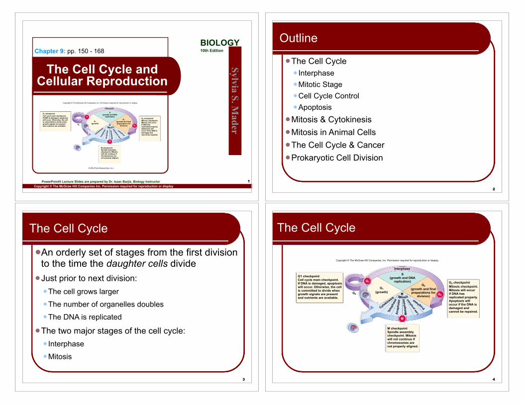
G1 checkpointCell cycle main checkpoint.If DNA is damaged, apoptosiswill occur. Otherwise, the cellis committed to divide whengrowth signals are presentand nutrients are available.
M checkpointSpindle assemblycheckpoint. Mitosiswill not continue ifchromosomes arenot properly aligned.
G2 checkpointMitosis checkpoint.Mitosis will occurif DNA hasreplicated properly.Apoptosis willoccur if the DNA isdamaged andcannot be repaired.
.
M
G2
G1
© SPL/Photo Researchers, Inc.;
2
Outline
!The Cell Cycle! Interphase!Mitotic Stage!Cell Cycle Control!Apoptosis
!Mitosis & Cytokinesis!Mitosis in Animal Cells!The Cell Cycle & Cancer!Prokaryotic Cell Division
3
The Cell Cycle
!An orderly set of stages from the first division to the time the daughter cells divide
!Just prior to next division:!The cell grows larger!The number of organelles doubles!The DNA is replicated
!The two major stages of the cell cycle:! Interphase!Mitosis
4
The Cell Cycle
Copyright © The McGraw-Hill Companies, Inc. Permission required for reproduction or display.
G1(growth)G0
G2 checkpointMitosis checkpoint.Mitosis will occurif DNA hasreplicated properly.Apoptosis willoccur if the DNA isdamaged andcannot be repaired.
S(growth and DNA
replication)
M
Cytokinesis
Telo
phas
eA
naph
ase
Met
apha
se
Late
pro
phas
e
Prophase
Interphase
G1 checkpointCell cycle main checkpoint.If DNA is damaged, apoptosiswill occur. Otherwise, the cellis committed to divide whengrowth signals are presentand nutrients are available.
M checkpointSpindle assemblycheckpoint. Mitosiswill not continue ifchromosomes arenot properly aligned.
M
G2
G1G2
(growth and finalpreparations for
division)
Fig. 9.1
G1(growth)G0
G2(growth and finalpreparations for
division)
S(growth and DNA
replication)
M
Cytokinesis
Telo
phas
eA
naph
ase
Met
apha
se
Late
pro
phas
e
Prophase
Interphase
G1 checkpointCell cycle main checkpoint.If DNA is damaged, apoptosiswill occur. Otherwise, the cellis committed to divide whengrowth signals are presentand nutrients are available.
M checkpointSpindle assemblycheckpoint. Mitosiswill not continue ifchromosomes arenot properly aligned.
G2 checkpointMitosis checkpoint.Mitosis will occurif DNA hasreplicated properly.Apoptosis willoccur if the DNA isdamaged andcannot be repaired.
M
G2
G1
Copyright © The McGraw-Hill Companies, Inc. Permission required for reproduction or display.
9
Interphase
! Most of the cell cycle is spent in interphase! Cell performs its usual functions! Time spent in interphase varies by cell type! Nerve and muscle cells do not complete the
cell cycle (remain in the G0 stage)
10
Interphase
! Interphase consists of: G1, S and G2 phases!G1 Phase:
! Recovery from previous division! Cell doubles its organelles! Cell grows in size! Accumulates raw materials for DNA synthesis (DNA replication)
!S Phase:! DNA replication ! Proteins associated with DNA are synthesized ! Chromosomes enter with 1 chromatid each! Chromosomes leave with 2 identical chromatids each
!G2 Phase:! Between DNA replication and onset of mitosis! Cell synthesizes proteins necessary for division
11
Mitotic (M) Stage
!Includes:!Mitosis (karyokinesis)
!Nuclear division
!Daughter chromosomes distributed to two daughter nuclei
!Cytokinesis!Cytoplasm division
!Results in two genetically identical daughter cells
12
Cell Cycle Control
!Cell cycle controlled by internal and external signals!A signal is a molecule that either stimulates or
inhibits a metabolic event. !External signals
! Growth factors! Received at the plasma membrane! Cause completion of cell cycle
! Internal signals! Family of proteins called cyclins! Increase and decrease as cell cycle continues ! Without them cycle stops at G1, M or G2 (checkpoints)! Allows time for any damage to be repaired
13
Apoptosis
!Apoptosis is programmed cell death!It involves a sequence of cellular events:
!fragmenting of the nucleus, !blistering of the plasma membrane!engulfing of cell fragments.
!Apoptosis is caused by enzymes called caspases.
!Mitosis and apoptosis are opposing forces!Mitosis increases cell number!Apoptosis decreases cell number
14
Apoptosis
!Cells harbor caspases in check by inhibitors!Can be unleashed by internal or external signals
!Signal protein P53!Stops cycle at G1 when DNA damaged
!Initiates DNA attempt at repair!If successful, cycle continues to mitosis!If not, apoptosis is initiated
15
Apoptosis
Copyright © The McGraw-Hill Companies, Inc. Permission required for reproduction or display.
apoptotic cell
cellfragment
DNAfragment
Cell roundsup, and nucleuscollapses.
Chromatincondenses, andnucleus fragments.
Plasma membraneblisters, and blebsform.
Cell fragmentscontain DNAfragments.
blebs
Courtesy Douglas R. Green/LaJolla Institute for Allergy and Immunology
17
Mitosis: Preparation
!DNA is in very long threads!Chromosomes!Stretched out and intertangled between divisions!DNA is associated with histone proteins!Collectively called chromatin
!Before mitosis begins:!Chromatin condenses (coils) into distinctly visible
chromosomes!Each species has a characteristic chromosome number
! Humans 46! Corn 20! Goldfish 94
19
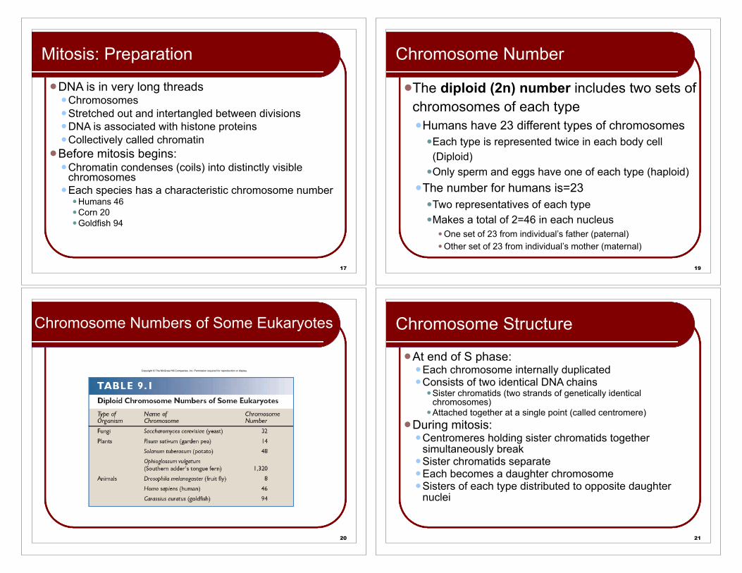
Chromosome Number
!The diploid (2n) number includes two sets of chromosomes of each type!Humans have 23 different types of chromosomes
!Each type is represented twice in each body cell (Diploid)
!Only sperm and eggs have one of each type (haploid)!The number for humans is=23
!Two representatives of each type!Makes a total of 2=46 in each nucleus
! One set of 23 from individual’s father (paternal)! Other set of 23 from individual’s mother (maternal)
20
Chromosome Numbers of Some Eukaryotes
Copyright © The McGraw-Hill Companies, Inc. Permission required for reproduction or display.
21
Chromosome Structure
!At end of S phase:!Each chromosome internally duplicated!Consists of two identical DNA chains
! Sister chromatids (two strands of genetically identical chromosomes)
! Attached together at a single point (called centromere)!During mitosis:
!Centromeres holding sister chromatids together simultaneously break
!Sister chromatids separate !Each becomes a daughter chromosome!Sisters of each type distributed to opposite daughter
nuclei
22
Duplicated ChromosomeCopyright © The McGraw-Hill Companies, Inc. Permission required for reproduction or display.
centromere
sister chromatids
one chromatida. b.
kinetochore
9,850© Andrew Syred/Photo Researchers, Inc.
23
Mitosis in Animal Cells
!Just outside nucleus is the centrosome!This is the microtubule organizing center!Organizes the mitotic spindle
! Contains many fibers! Each composed of a bundle of microtubules
! In animals, contains two barrel-shaped centrioles! Oriented at right angles to each other within centrosome! Each with 9 triplets of microtubules arranged in a cylinder
!Centrosome was also replicated in S-phase, so now two centrosomes
24
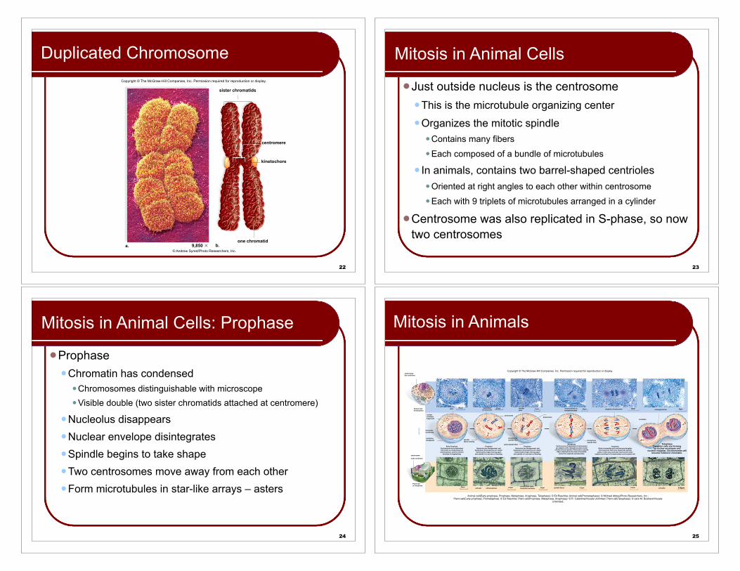
Mitosis in Animal Cells: Prophase
!Prophase!Chromatin has condensed
! Chromosomes distinguishable with microscope! Visible double (two sister chromatids attached at centromere)
!Nucleolus disappears!Nuclear envelope disintegrates!Spindle begins to take shape!Two centrosomes move away from each other!Form microtubules in star-like arrays – asters
25
Mitosis in Animals
Copyright © The McGraw-Hill Companies, Inc. Permission required for reproduction or display.
Animal cell(Early prophase, Prophase, Metaphase, Anaphase, Telophase): © Ed Reschke; Animal cell(Prometaphase): © Michael Abbey/Photo Researchers, Inc.;Plant cell(Early prophase, Prometaphse): © Ed Reschke; Plant cell(Prophase, Metaphase, Anaphase): © R. Calentine/Visuals Unlimited; Plant cell(Telophase): © Jack M. Bostrack/Visuals
Unlimited;
Plant Cellat Interphase
centromere
aster
kinetochore
polar spindle fiber
chromosomescell wall25!m
centrosome
lacks centrioles
MIT
OSI
S
centrosomehas centrioles
Animal Cellat Interphase
nuclearenvelopefragments
chromatincondenses
nucleolusdisappears
Early ProphaseCentrosomes have duplicated.Chromatin is condensing into
chromosomes, and the nuclearenvelope is fragmenting.
ProphaseNucleolus has disappeared, and
duplicated chromosomes are visible.Centrosomes begin moving apart,
and spindle is in process of forming.
ProphaseNucleolus has disappeared, and
duplicated chromosomes are visible.Centrosomes begin moving apart,
and spindle is in process of forming.
20 !m duplicatedchromosome
20 !m
spindlefibers forming
spindlepole
9 !m
kinetochorespindle fiber
cleavage furrow
spindle fibers
20!m 16!m
kinetochorespindle fiber
AnaphaseSister chromatids part and become daughterchromosomes that move toward the spindle
poles. In this way, each pole receives the samenumber and kinds of chromosomes as the parent cell.
MetaphaseCentromeres of duplicated chromosomesare aligned at the metaphase plate (center
of fully formed spindle). Kinetochore spindlefibers attached to the sister chromatids
come from opposite spindle poles.
chromosomes atmetaphase plate
6.2!m6.2!m20!m6.2!mspindle pole lackscentrioles and aster
TelophaseDaughter cells are formingas nuclear envelopes and
nucleoli reappear. Chromosomes willbecome indistinct chromatin.
daughter chromosome 20!m
nucleolus
cell plate 6.6!m
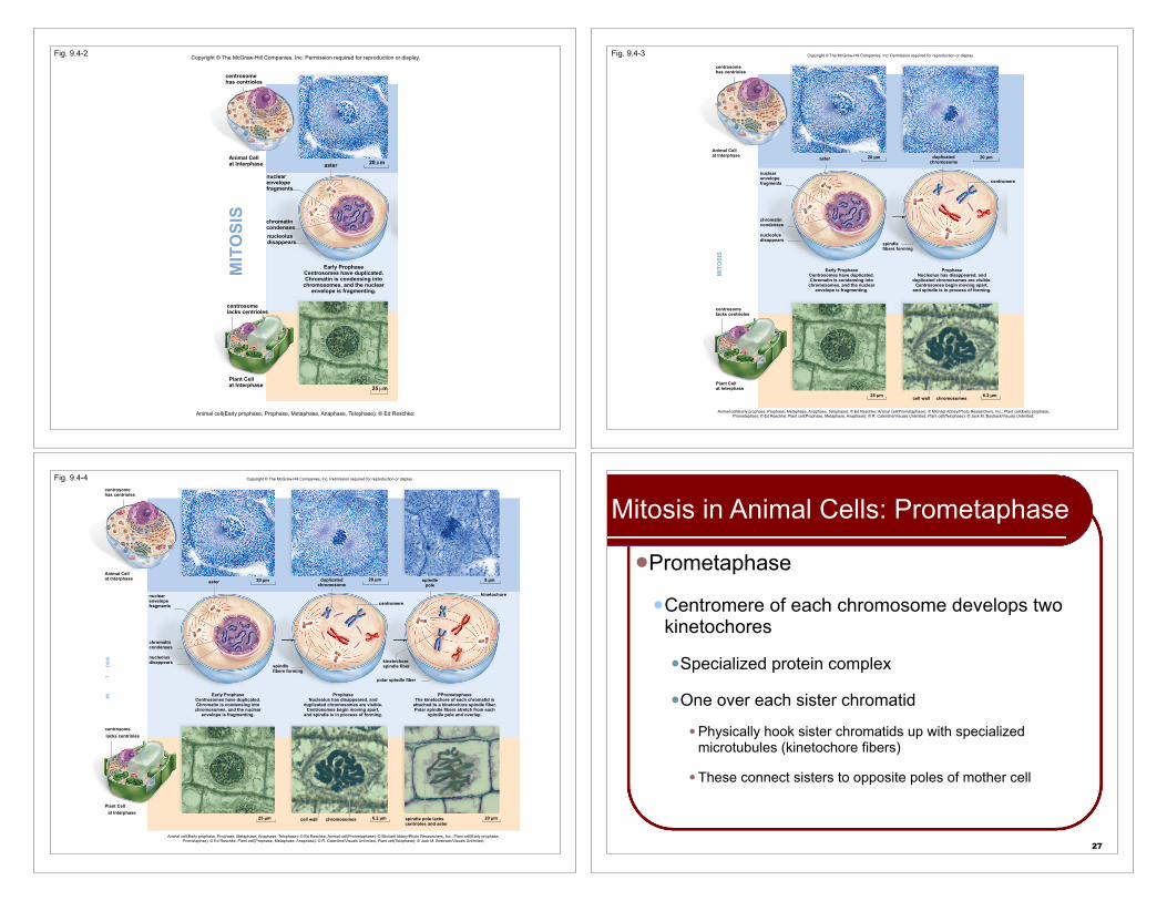
Fig. 9.4-2
Animal Cellat Interphase
Plant Cellat Interphase
Early ProphaseCentrosomes have duplicated.Chromatin is condensing into
chromosomes, and the nuclearenvelope is fragmenting.
nuclearenvelopefragments
chromatincondensesnucleolusdisappears
aster 20 m
25 m
centrosomehas centrioles
centrosomelacks centrioles
MIT
OSI
S
Animal cell(Early prophase, Prophase, Metaphase, Anaphase, Telophase): © Ed Reschke;
Copyright © The McGraw-Hill Companies, Inc. Permission required for reproduction or display. Fig. 9.4-3
Animal Cellat Interphase
Plant Cellat Interphase
Early ProphaseCentrosomes have duplicated.Chromatin is condensing into
chromosomes, and the nuclearenvelope is fragmenting.
ProphaseNucleolus has disappeared, and
duplicated chromosomes are visible.Centrosomes begin moving apart,
and spindle is in process of forming.
nuclearenvelopefragments
chromatincondenses
spindlefibers forming
duplicatedchromosome
centromere
aster
cell wall
centrosomehas centrioles
centrosomelacks centrioles
MIT
OSI
S
nucleolusdisappears
20 !m20 !m
25 !m 6.2 !mchromosomes
Copyright © The McGraw-Hill Companies, Inc. Permission required for reproduction or display.
Animal cell(Early prophase, Prophase, Metaphase, Anaphase, Telophase): © Ed Reschke; Animal cell(Prometaphase): © Michael Abbey/Photo Researchers, Inc.; Plant cell(Early prophase, Prometaphse): © Ed Reschke; Plant cell(Prophase, Metaphase, Anaphase): © R. Calentine/Visuals Unlimited; Plant cell(Telophase): © Jack M. Bostrack/Visuals Unlimited;
Fig. 9.4-4
Plant Cellat Interphase
centromere
spindlepole
aster
kinetochore
kinetochorespindle fiber
polar spindle fiber
chromosomescell wall
centrosomelacks centrioles
spindle pole lackscentrioles and aster
MI
TO
SIS
centrosomehas centrioles
Animal Cellat Interphase
nuclearenvelopefragments
chromatincondenses
nucleolusdisappears
Early ProphaseCentrosomes have duplicated.Chromatin is condensing into
chromosomes, and the nuclearenvelope is fragmenting.
spindlefibers forming
ProphaseNucleolus has disappeared, and
duplicated chromosomes are visible.Centrosomes begin moving apart,
and spindle is in process of forming.
PPrometaphaseThe kinetochore of each chromatid is
attached to a kinetochore spindle fiber.Polar spindle fibers stretch from each
spindle pole and overlap.
25 !m 6.2 !m 20 !m
20 !m duplicatedchromosome
20 !m 9 !m
Copyright © The McGraw-Hill Companies, Inc. Permission required for reproduction or display.
Animal cell(Early prophase, Prophase, Metaphase, Anaphase, Telophase): © Ed Reschke; Animal cell(Prometaphase): © Michael Abbey/Photo Researchers, Inc.; Plant cell(Early prophase, Prometaphse): © Ed Reschke; Plant cell(Prophase, Metaphase, Anaphase): © R. Calentine/Visuals Unlimited; Plant cell(Telophase): © Jack M. Bostrack/Visuals Unlimited;
27
Mitosis in Animal Cells: Prometaphase
!Prometaphase
!Centromere of each chromosome develops two kinetochores
!Specialized protein complex
!One over each sister chromatid
! Physically hook sister chromatids up with specialized microtubules (kinetochore fibers)
! These connect sisters to opposite poles of mother cell
28
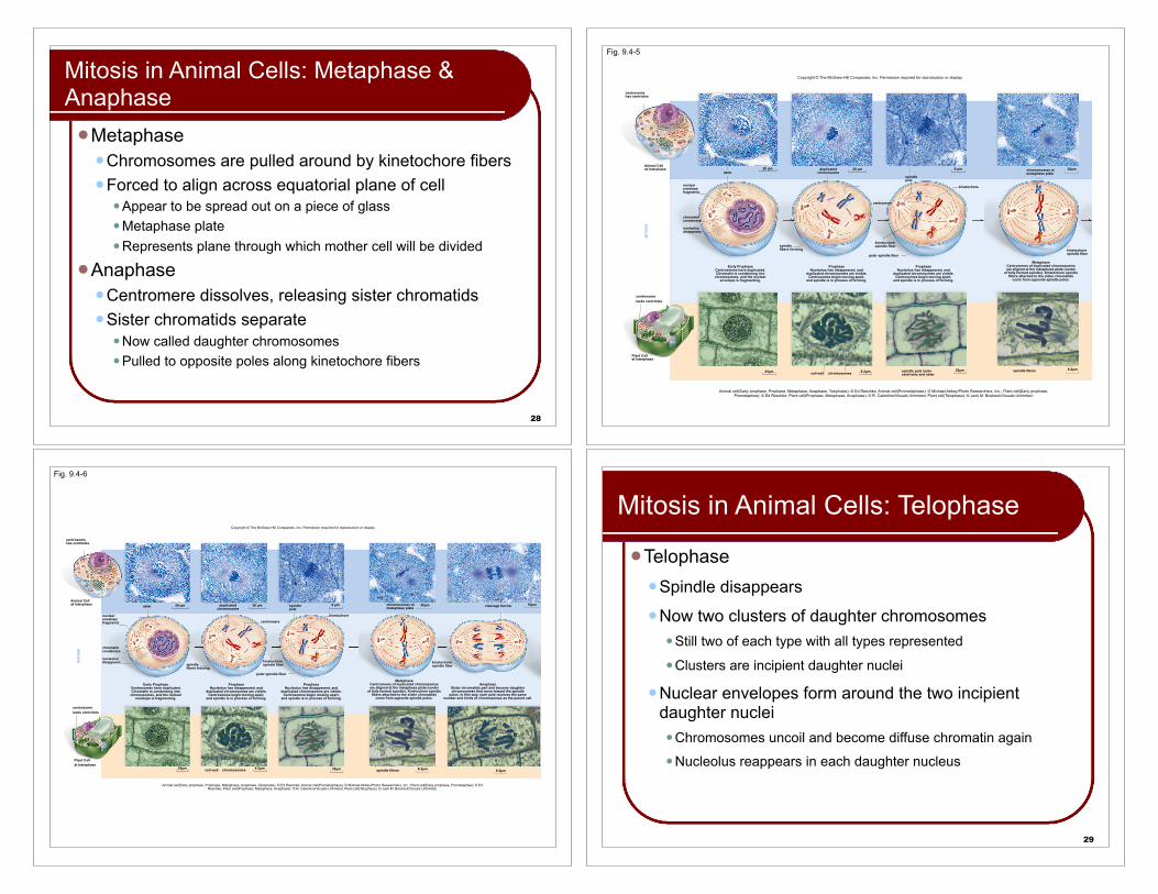
Mitosis in Animal Cells: Metaphase & Anaphase!Metaphase
!Chromosomes are pulled around by kinetochore fibers!Forced to align across equatorial plane of cell
! Appear to be spread out on a piece of glass! Metaphase plate! Represents plane through which mother cell will be divided
!Anaphase!Centromere dissolves, releasing sister chromatids!Sister chromatids separate
! Now called daughter chromosomes! Pulled to opposite poles along kinetochore fibers
Fig. 9.4-5
Copyright © The McGraw-Hill Companies, Inc. Permission required for reproduction or display.
Animal cell(Early prophase, Prophase, Metaphase, Anaphase, Telophase): © Ed Reschke; Animal cell(Prometaphase): © Michael Abbey/Photo Researchers, Inc.; Plant cell(Early prophase, Prometaphse): © Ed Reschke; Plant cell(Prophase, Metaphase, Anaphase): © R. Calentine/Visuals Unlimited; Plant cell(Telophase): © Jack M. Bostrack/Visuals Unlimited;
Plant Cellat Interphase
centrosomelacks centrioles
MIT
OSI
S
centrosomehas centrioles
Animal Cellat Interphase
aster
25!m
nuclearenvelopefragments
chromatincondenses
nucleolusdisappears
Early ProphaseCentrosomes have duplicated.Chromatin is condensing into
chromosomes, and the nuclearenvelope is fragmenting.
20 !m
chromosomescell wall
ProphaseNucleolus has disappeared, and
duplicated chromosomes are visible.Centrosomes begin moving apart,
and spindle is in process of forming.
duplicatedchromosome
20 !m
spindlefibers forming
6.2!m
centromere
kinetochore
polar spindle fiber
ProphaseNucleolus has disappeared, and
duplicated chromosomes are visible.Centrosomes begin moving apart,
and spindle is in process of forming.
spindlepole
9 !m
kinetochorespindle fiber
20!mspindle pole lackscentrioles and aster
spindle fibers
20!m
MetaphaseCentromeres of duplicated chromosomesare aligned at the metaphase plate (center
of fully formed spindle). Kinetochore spindlefibers attached to the sister chromatids
come from opposite spindle poles.
chromosomes atmetaphase plate
6.2!m
kinetochorespindle fiber
Fig. 9.4-6
Plant Cellat Interphase
centromere
aster
kinetochore
polar spindle fiber
chromosomescell wall25!m
centrosomelacks centrioles
MIT
OSI
S
centrosomehas centrioles
Animal Cellat Interphase
nuclearenvelopefragments
chromatincondenses
nucleolusdisappears
Early ProphaseCentrosomes have duplicated.Chromatin is condensing into
chromosomes, and the nuclearenvelope is fragmenting.
ProphaseNucleolus has disappeared, and
duplicated chromosomes are visible.Centrosomes begin moving apart,
and spindle is in process of forming.
ProphaseNucleolus has disappeared, and
duplicated chromosomes are visible.Centrosomes begin moving apart,
and spindle is in process of forming.
20 !m duplicatedchromosome
20 !m
spindlefibers forming
spindlepole
9 !m
kinetochorespindle fiber
cleavage furrow
spindle fibers
20!m 16!m
kinetochorespindle fiber
AnaphaseSister chromatids part and become daughterchromosomes that move toward the spindle
poles. In this way, each pole receives the samenumber and kinds of chromosomes as the parent cell.
MetaphaseCentromeres of duplicated chromosomesare aligned at the metaphase plate (center
of fully formed spindle). Kinetochore spindlefibers attached to the sister chromatids
come from opposite spindle poles.
chromosomes atmetaphase plate
6.2!m6.2!m20!m
Copyright © The McGraw-Hill Companies, Inc. Permission required for reproduction or display.
6.2!m
Animal cell(Early prophase, Prophase, Metaphase, Anaphase, Telophase): © Ed Reschke; Animal cell(Prometaphase): © Michael Abbey/Photo Researchers, Inc.; Plant cell(Early prophase, Prometaphse): © Ed Reschke; Plant cell(Prophase, Metaphase, Anaphase): © R. Calentine/Visuals Unlimited; Plant cell(Telophase): © Jack M. Bostrack/Visuals Unlimited;
29
Mitosis in Animal Cells: Telophase
!Telophase!Spindle disappears!Now two clusters of daughter chromosomes
! Still two of each type with all types represented! Clusters are incipient daughter nuclei
!Nuclear envelopes form around the two incipient daughter nuclei! Chromosomes uncoil and become diffuse chromatin again! Nucleolus reappears in each daughter nucleus
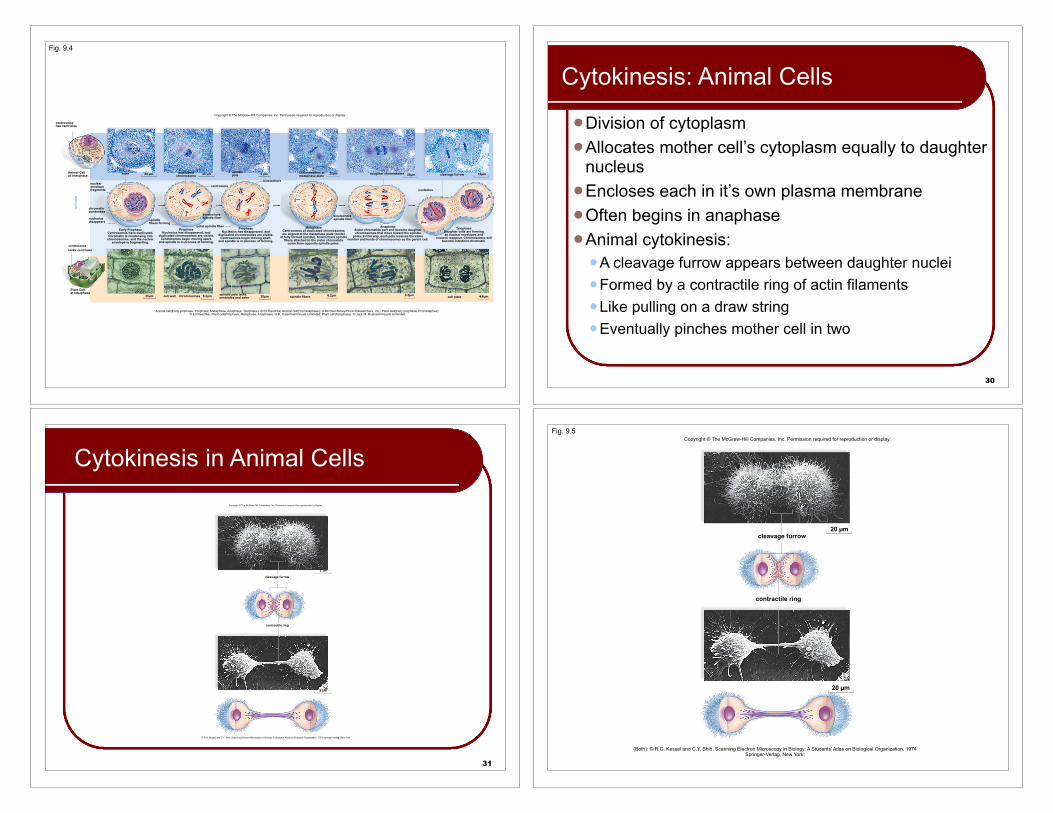
Fig. 9.4
Copyright © The McGraw-Hill Companies, Inc. Permission required for reproduction or display.
Animal cell(Early prophase, Prophase, Metaphase, Anaphase, Telophase): © Ed Reschke; Animal cell(Prometaphase): © Michael Abbey/Photo Researchers, Inc.; Plant cell(Early prophase, Prometaphse): © Ed Reschke; Plant cell(Prophase, Metaphase, Anaphase): © R. Calentine/Visuals Unlimited; Plant cell(Telophase): © Jack M. Bostrack/Visuals Unlimited;
Plant Cellat Interphase
centromere
aster
kinetochore
polar spindle fiber
chromosomescell wall25!m
centrosomelacks centrioles
MIT
OSI
S
centrosomehas centrioles
Animal Cellat Interphase
nuclearenvelopefragments
chromatincondenses
nucleolusdisappears
Early ProphaseCentrosomes have duplicated.Chromatin is condensing into
chromosomes, and the nuclearenvelope is fragmenting.
ProphaseNucleolus has disappeared, and
duplicated chromosomes are visible.Centrosomes begin moving apart,
and spindle is in process of forming.
ProphaseNucleolus has disappeared, and
duplicated chromosomes are visible.Centrosomes begin moving apart,
and spindle is in process of forming.
20 !m duplicatedchromosome 20 !m
spindlefibers forming
spindlepole 9 !m
kinetochorespindle fiber
cleavage furrow
spindle fibers
20!m 16!m
kinetochorespindle fiber
AnaphaseSister chromatids part and become daughterchromosomes that move toward the spindle
poles. In this way, each pole receives the samenumber and kinds of chromosomes as the parent cell.
MetaphaseCentromeres of duplicated chromosomesare aligned at the metaphase plate (center
of fully formed spindle). Kinetochore spindlefibers attached to the sister chromatids
come from opposite spindle poles.
chromosomes atmetaphase plate
6.2!m6.2!m20!m6.2!m spindle pole lackscentrioles and aster
TelophaseDaughter cells are formingas nuclear envelopes and
nucleoli reappear. Chromosomes willbecome indistinct chromatin.
daughter chromosome 20!m
nucleolus
cell plate 6.6!m
30
Cytokinesis: Animal Cells
!Division of cytoplasm!Allocates mother cell’s cytoplasm equally to daughter
nucleus!Encloses each in it’s own plasma membrane!Often begins in anaphase!Animal cytokinesis:
!A cleavage furrow appears between daughter nuclei!Formed by a contractile ring of actin filaments!Like pulling on a draw string!Eventually pinches mother cell in two
31
Cytokinesis in Animal CellsCopyright © The McGraw-Hill Companies, Inc. Permission required for reproduction or display.
2 m
2 m
contractile ring
cleavage furrow
© R.G. Kessel and C.Y. Shih, Scanning Electron Microscopy in Biology: A Students' Atlas on Biological Organization, 1974 Springer-Verlag, New York
Fig. 9.5
contractile ring
cleavage furrow20 !m
20 !m
Copyright © The McGraw-Hill Companies, Inc. Permission required for reproduction or display.
(Both): © R.G. Kessel and C.Y. Shih, Scanning Electron Microscopy in Biology: A Students' Atlas on Biological Organization, 1974 Springer-Verlag, New York;
32
Cytokinesis: Plant Cells
!Rigid cell walls outside plasma membrane do not permit furrowing
!Begins with formation of a cell plate!Many small membrane-bounded vesicles!Eventually fuse into one thin vesicle extending across the
mother cell!The membranes of the cell plate become the plasma
membrane between the daughter cells! Contents of vesicles become the middle lamella between the two
daughter cells! Daughter cells later secrete primary cell walls on opposite sides of
middle lamella
33
Cytokinesis in Plant CellsCopyright © The McGraw-Hill Companies, Inc. Permission required for reproduction or display.
nuclei
cell wall
Vesicles containing cell wall components fusing to form cell plate
cell plate forming
microtubules
cell plate forming
© Katherine Esau; 9.8d: © Biophoto Associates/Photo Researchers, Inc.
34
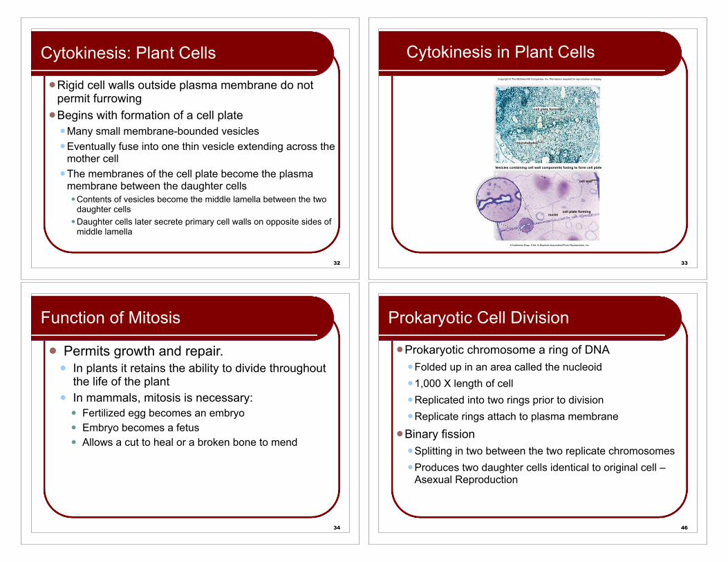
Function of Mitosis
! Permits growth and repair.! In plants it retains the ability to divide throughout
the life of the plant ! In mammals, mitosis is necessary:
! Fertilized egg becomes an embryo ! Embryo becomes a fetus! Allows a cut to heal or a broken bone to mend
46
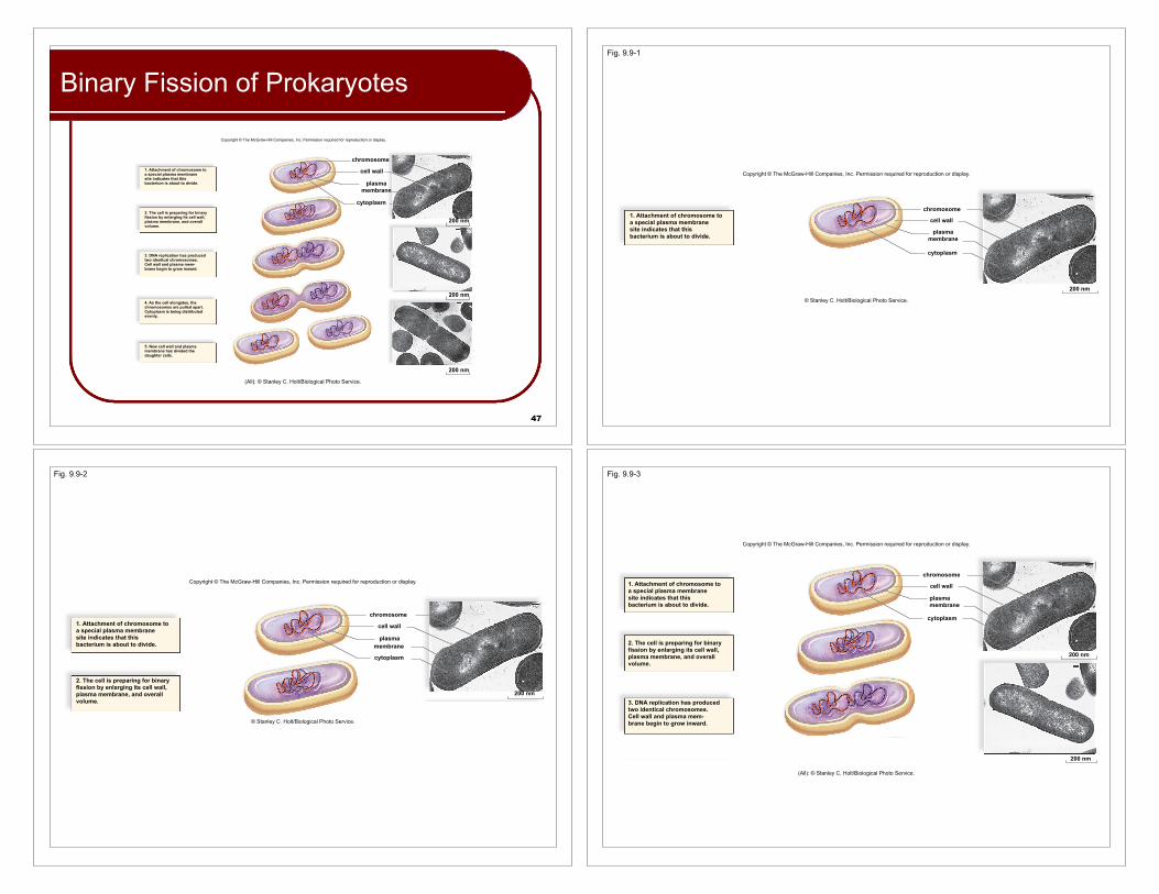
Prokaryotic Cell Division
!Prokaryotic chromosome a ring of DNA!Folded up in an area called the nucleoid!1,000 X length of cell!Replicated into two rings prior to division!Replicate rings attach to plasma membrane
!Binary fission!Splitting in two between the two replicate chromosomes!Produces two daughter cells identical to original cell –
Asexual Reproduction
47
Binary Fission of Prokaryotes
Copyright © The McGraw-Hill Companies, Inc. Permission required for reproduction or display.
5. New cell wall and plasmamembrane has divided thedaughter cells.
1. Attachment of chromosome toa special plasma membranesite indicates that thisbacterium is about to divide.
2. The cell is preparing for binaryfission by enlarging its cell wall,plasma membrane, and overallvolume.
3. DNA replication has producedtwo identical chromosomes.Cell wall and plasma mem-brane begin to grow inward.
4. As the cell elongates, thechromosomes are pulled apart.Cytoplasm is being distributedevenly..
(All): © Stanley C. Holt/Biological Photo Service.
200 nm
200 nm
200 nm
chromosome
cell wall
plasmamembrane
cytoplasm
Fig. 9.9-1
1. Attachment of chromosome toa special plasma membranesite indicates that thisbacterium is about to divide.
chromosome
cell wall
plasmamembrane
200 nm
cytoplasm
Copyright © The McGraw-Hill Companies, Inc. Permission required for reproduction or display.
© Stanley C. Holt/Biological Photo Service.
Fig. 9.9-2
1. Attachment of chromosome toa special plasma membranesite indicates that thisbacterium is about to divide.
chromosome
cell wall
plasmamembrane
cytoplasm
2. The cell is preparing for binaryfission by enlarging its cell wall,plasma membrane, and overallvolume.
200 nm
Copyright © The McGraw-Hill Companies, Inc. Permission required for reproduction or display.
© Stanley C. Holt/Biological Photo Service.
Fig. 9.9-3
3. DNA replication has producedtwo identical chromosomes.Cell wall and plasma mem-brane begin to grow inward.
chromosome
cell wall
plasmamembrane
cytoplasm
200 nm
200 nm
1. Attachment of chromosome toa special plasma membranesite indicates that thisbacterium is about to divide.
2. The cell is preparing for binaryfission by enlarging its cell wall,plasma membrane, and overallvolume.
Copyright © The McGraw-Hill Companies, Inc. Permission required for reproduction or display.
(All): © Stanley C. Holt/Biological Photo Service.
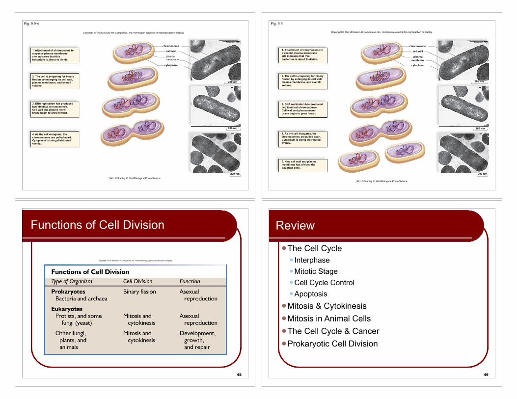
Fig. 9.9-4
4. As the cell elongates, thechromosomes are pulled apart.Cytoplasm is being distributedevenly..
chromosome
cell wall
plasmamembrane
cytoplasm
200 nm
200 nm
200 nm
1. Attachment of chromosome toa special plasma membranesite indicates that thisbacterium is about to divide.
2. The cell is preparing for binaryfission by enlarging its cell wall,plasma membrane, and overallvolume.
3. DNA replication has producedtwo identical chromosomes.Cell wall and plasma mem-brane begin to grow inward.
Copyright © The McGraw-Hill Companies, Inc. Permission required for reproduction or display.
(All): © Stanley C. Holt/Biological Photo Service.
Fig. 9.9
5. New cell wall and plasmamembrane has divided thedaughter cells.
chromosome
cell wall
plasmamembrane
cytoplasm
200 nm
200 nm
200 nm
1. Attachment of chromosome toa special plasma membranesite indicates that thisbacterium is about to divide.
2. The cell is preparing for binaryfission by enlarging its cell wall,plasma membrane, and overallvolume.
3. DNA replication has producedtwo identical chromosomes.Cell wall and plasma mem-brane begin to grow inward.
4. As the cell elongates, thechromosomes are pulled apart.Cytoplasm is being distributedevenly..
Copyright © The McGraw-Hill Companies, Inc. Permission required for reproduction or display.
(All): © Stanley C. Holt/Biological Photo Service.
48
Functions of Cell Division
Copyright © The McGraw-Hill Companies, Inc. Permission required for reproduction or display.
49
Review
!The Cell Cycle! Interphase!Mitotic Stage!Cell Cycle Control!Apoptosis
!Mitosis & Cytokinesis!Mitosis in Animal Cells!The Cell Cycle & Cancer!Prokaryotic Cell Division
Sylvia S. Mader
Copyright © The McGraw Hill Companies Inc. Permission required for reproduction or displayPowerPoint® Lecture Slides are prepared by Dr. Isaac Barjis, Biology Instructor
BIOLOGY10th Edition
The Cell Cycle and Cellular Reproduction
Chapter 9: pp. 150 - 168
Insert figure 9.1 here
50
Copyright © The McGraw-Hill Companies, Inc. Permission required for reproduction or display.
G1(growth)G0
G2(growth and finalpreparations for
division)
S(growth and DNA
replication)
M
Cytokinesis
Telo
phas
eA
naph
ase
Met
apha
se
Late
pro
phas
e
Prophase
Interphase
G1 checkpointCell cycle main checkpoint.If DNA is damaged, apoptosiswill occur. Otherwise, the cellis committed to divide whengrowth signals are presentand nutrients are available.
M checkpointSpindle assemblycheckpoint. Mitosiswill not continue ifchromosomes arenot properly aligned.
G2 checkpointMitosis checkpoint.Mitosis will occurif DNA hasreplicated properly.Apoptosis willoccur if the DNA isdamaged andcannot be repaired.
.
M
G2
G1
© SPL/Photo Researchers, Inc.;