Embed Size (px)
Citation preview
CrackCast Show Notes – Abdominal Aortic Aneurysm – June 2017 www.canadiem.org/crackcast
Chapter 88 – PE and DVT
Episode Overview:
1. List 8 DDx for DVT
2. Describe management of superficial thrombophlebitis + isolated calf thrombosis
3. How is the d-dimer test used in the diagnosis of DVT?
4. List 8 causes of an elevated D-dimer
5. What are the Wells criteria for DVT? Describe how to use this score.
6. Describe management of suspected DVT
7. How is a proximal lower limb DVT managed?
8. What are the common causes of upper limb DVT?
9. How are upper limb DVTs managed?
10. List classic risk factors for PE x 10
11. What are the types of PE?
12. List 4 ECG + 2 CXR findings consistent with PE
13. What are the Wells criteria for PE? Describe how to use this score.
14. What is the PERC rule? How is it used?
15. Which imaging tests can be used to diagnose PE? List advantages and
disadvantages of each.
16. List indications for thrombolysis in PE, what is the risk of ICH?
17. What are the absolute and relative contraindications?
18. List markers of poor prognosis in patients with PE.
Wisecracks
1. What is phlegmasia cerulea dolens? How is it managed? 2. Which patients should have an IVC filter? 3. What about PE/DVT in pregnancy? 4. What is the cause of hypoxia in patients with PE? What causes chest pain? What
causes hypotension? 5. What is Paget-Schroetter Syndrome?
Rosen's in Perspective
● This chapter is all about VTE - venous thromboembolism - as it manifests in PE’s and
DVT’s.
● VTE - formation depends on excess fibrin formation (thrombin working on fibrinogen)
● Factors that enhance fibrinogen synthesis and catalysis to fibrin are:
○ Systemic inflammation
○ Vascular trauma (includes immune related trauma)
○ Inherited thrombophilias
○ Hemoglobinopathies
○ Cancer
○ Pragnancy
○ Sluggish blood flow
CrackCast Show Notes – Abdominal Aortic Aneurysm – June 2017 www.canadiem.org/crackcast
● The triad:
○ Venous injury
○ Slow blood flow
○ Hypercoagulability
● Each year of life “independently increases the likelihood of imbalanced clot
formation”
Now lets jump into each of these diseases….
1) List 8 DDx for DVT
First off, DVT…
● This is a spectrum: isolated calf vein thrombosis ← to → limb threatening illiofemoral
clot
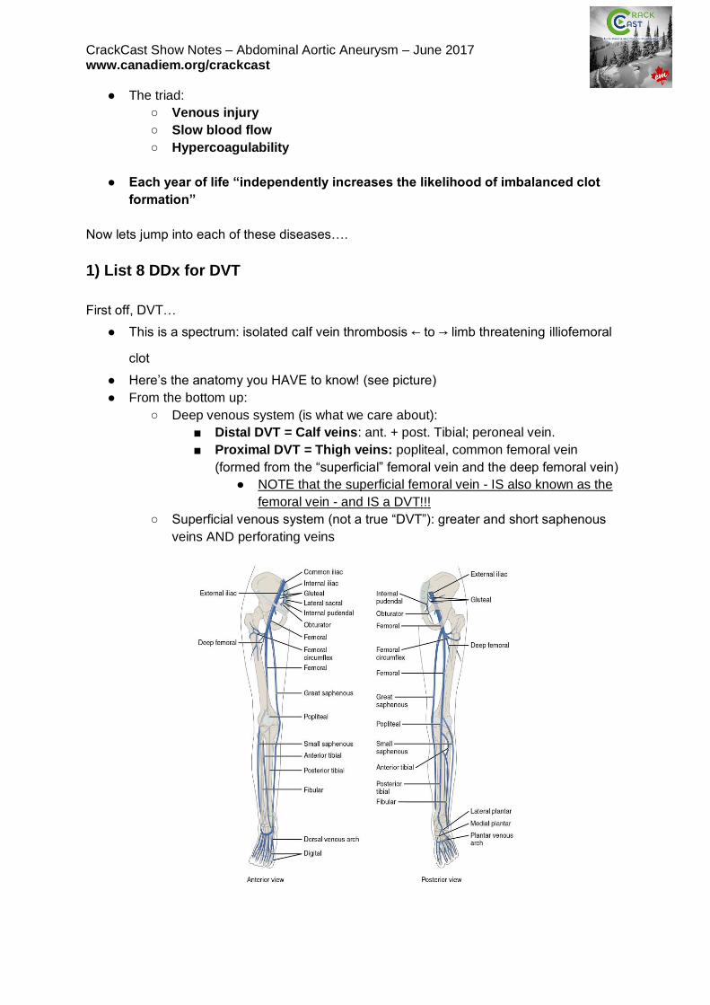
● Here’s the anatomy you HAVE to know! (see picture)
● From the bottom up:
○ Deep venous system (is what we care about):
■ Distal DVT = Calf veins: ant. + post. Tibial; peroneal vein.
■ Proximal DVT = Thigh veins: popliteal, common femoral vein
(formed from the “superficial” femoral vein and the deep femoral vein)
● NOTE that the superficial femoral vein - IS also known as the
femoral vein - and IS a DVT!!!
○ Superficial venous system (not a true “DVT”): greater and short saphenous
veins AND perforating veins
CrackCast Show Notes – Abdominal Aortic Aneurysm – June 2017 www.canadiem.org/crackcast DDx: (Box 88-1)
1) Fracture (stress / occult / pathologic)
2) Popliteal cyst / rupture of
3) Cellulitis
4) Superficial thrombophlebitis
5) Vasculitis
6) Proximal venous compression (tumour, gravid uterus)
7) CHF related bilateral leg swelling
8) Hypoalbuminemia
9) Lymphedema
10) Muscle strain / calf strain
11) Hematoma
12) Chronic venous insufficiency
2) Describe management of superficial thrombophlebitis + isolated calf
thrombosis
SLT:
● These patients have a clot in the greater saphenous vein that may extend above the
knee.
○ This can propagate proximally
● Treatment:
○ NSAIDs for symptoms
○ Heat
○ Graded compression stockings
○ **mandatory repeat ultrasound in 2-5 days**
■ If it has migrated proximally most people at least treat these patients
with LMWH or fondaparinux for 10 days, followed by repeat ultrasound
ICT:
● Controversial!
● 25% risk of proximal propagation proximally
● High risk symptomatic patients - can consider repeat duplex U/S in 2-5 days OR
anticoagulation based on bleeding / clotting risk
● For healthy ambulatory patients - Rosen’s recommends:
○ 325 mg ASA daily and repeat U/S at 2-5 days to look for clot propagation
3) How is the d-dimer test used in the diagnosis of DVT?
So, you’re wondering if this is a DVT….good for you!
The symptoms of DVT can be very subtle and nonspecific (cramping, sensation of fullness in
the calf)
● Unilateral swelling, edema, erythema, warmth, tenderness to palpation of the venous
system, dilation of the superficial collateral veins, palpable venous cord
● Homan’s sign is insensitive and nonspecific = useless! Do not rely on it!
CrackCast Show Notes – Abdominal Aortic Aneurysm – June 2017 www.canadiem.org/crackcast ****the lack of objective swelling is an unreliable gestalt tool to exclude the diagnosis of
DVT!****
● When the DVT is at the “charley horse” stage - non specific mild symptoms - it is best
for us to catch it because we can reduce the morbidity and mortality of VTE
Use of D-dimer:
● (See question 5!)
● People with a LOW pre-test probability can be risk stratified as “unlikely DVT” with a
NORMAL quantitative D-Dimer.
4) List 8 causes of an elevated D-dimer
D-dimer (protein breakdown product of cross-linked fibrin breakdown = i.e. Clot has formed
somewhere in the body in the last 72 hrs)
● D-dimer concentration is proportional to the size of the clot (ie. may be falsely low
with chronic clots because they are mature)
● There are over 75 different D-dimer assays!
● Most common assay uses cut off of 500 NG/ML
○ Sensitivity = 88-97% (for calf and proximal DVT)
■ A “negative” May not exclude the disease in someone who is at high
risk for the disease! (This is especially true for qualitative D-dimer
tools)
1. Endothelial damage
a. Aging
b. Recent surgery
c. Infection
d. New indwelling catheters
e. Inflammation
f. StrOke
g. MI
2. Venous stasis
a. Prolonged bed rest / limb casting
3. Hypercoagulable state:
a. Active malignancy
b. Pregnancy
5) What are the Wells criteria for DVT? Describe how to use this score.
● Dx of DVT starts with us using our clinical skills to calculate the pre-test probability
● Usually we use “the clinical gestalt of an experienced provider” OR a clinical decision
tool
○ According to current literature, neither expert consensus of clinical
gestalt NOR clinical prediction rules are superior to one another.
CrackCast Show Notes – Abdominal Aortic Aneurysm – June 2017 www.canadiem.org/crackcast
○ You could lump patients into two groups of people:
■ Low - mod - high risk as groups (3)
■ Unlikely - likely (2)
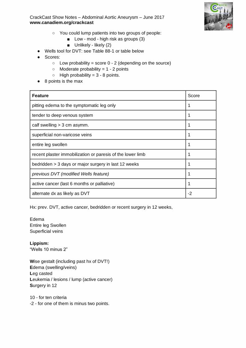
● Wells tool for DVT: see Table 88-1 or table below
● Scores:
○ Low probability = score 0 - 2 (depending on the source)
○ Moderate probability = 1 - 2 points
○ High probability = 3 - 8 points.
● 8 points is the max
Feature Score
pitting edema to the symptomatic leg only 1
tender to deep venous system 1
calf swelling > 3 cm asymm. 1
superficial non-varicose veins 1
entire leg swollen 1
recent plaster immobilization or paresis of the lower limb 1
bedridden > 3 days or major surgery in last 12 weeks 1
previous DVT (modified Wells feature) 1
active cancer (last 6 months or palliative) 1
alternate dx as likely as DVT -2
Hx: prev. DVT, active cancer, bedridden or recent surgery in 12 weeks,
Edema
Entire leg Swollen
Superficial veins
Lippism:
“Wells 10 minus 2”
Wise gestalt (including past hx of DVT!)
Edema (swelling/veins)
Leg casted
Leukemia / lesions / lump (active cancer)
Surgery in 12
10 - for ten criteria
-2 - for one of them is minus two points.
CrackCast Show Notes – Abdominal Aortic Aneurysm – June 2017 www.canadiem.org/crackcast
● Several criticisms of the original well’s score:
○ Prospective studies showing a 12% risk of DVT in the low risk group (not the
advertised 3% risk). See PMID: 16027451
○ A Meta analysis showing poor performance in the old, multiple comorbidities,
and prior hx of DVT groups. PMID: 16027455
The modified wells score:
● Includes the question of “previous DVT” = score of 1 (included in table above)
● Also breaks people into:
○ DVT likely =. Score of 2 or greater
○ DVT unlikely =. Score of 1 or less
After my exam writing is done, I’m probably not going to use Wells score…..
My approach:
DVT and no other clear DDx? → risk stratify as unlikely / likely DVT →
● if unlikely and a negative D-dimer STOP testing
● IF unlikely and a positive d-dimer get an ultrasound
● If likely DVT = jump right to ultrasound (ideally a single whole-leg ultrasound)
6) Describe management of suspected DVT
Radiographic eval:
● U/S = 95% sens & spec. For proximal DVT
○ Using the standard 3 point compression tests
○ The standard scanning protocol misses the calf veins and superficial veins
(which can progress to a DVT)
● For low risk groups with a negative 3-point U/S after a +ve d-dimer you can stop
testing
● For moderate-high risk groups, a single negative 3 point venous U/S is NOT
sufficient to exclude a DVT, they should have a follow-up U/S in 2-7 days to exclude
a DVT
● If you perform a single FULL leg doppler U/S (whole leg including superficial veins) -
you can rule out a DVT in any of the risk groups (high/med/low)
If you’re trying to diagnose an iliac or pelvic vein DVT - you need to use CT-
venography
7) How is a proximal lower limb DVT managed?
● Start an appropriate LMWH (Enoxaparin 1 mg/kg SC q12hrs) OR
○ Alternatives: (Check out Uptodate for a great discussion)
■ Fondaparinux
■ Apixaban
■ Rivaroxaban
● OR heparin infusion after a loading bolus
● Ideally transition to oral anticoagulation at least 3 months (some up to 12 months)
CrackCast Show Notes – Abdominal Aortic Aneurysm – June 2017 www.canadiem.org/crackcast
● Encourage ambulation as much as possible
○ Bedrest promotes DVT extension, risk of embolization, and post-DVT
syndrome
8) What are the common causes of upper limb DVT?
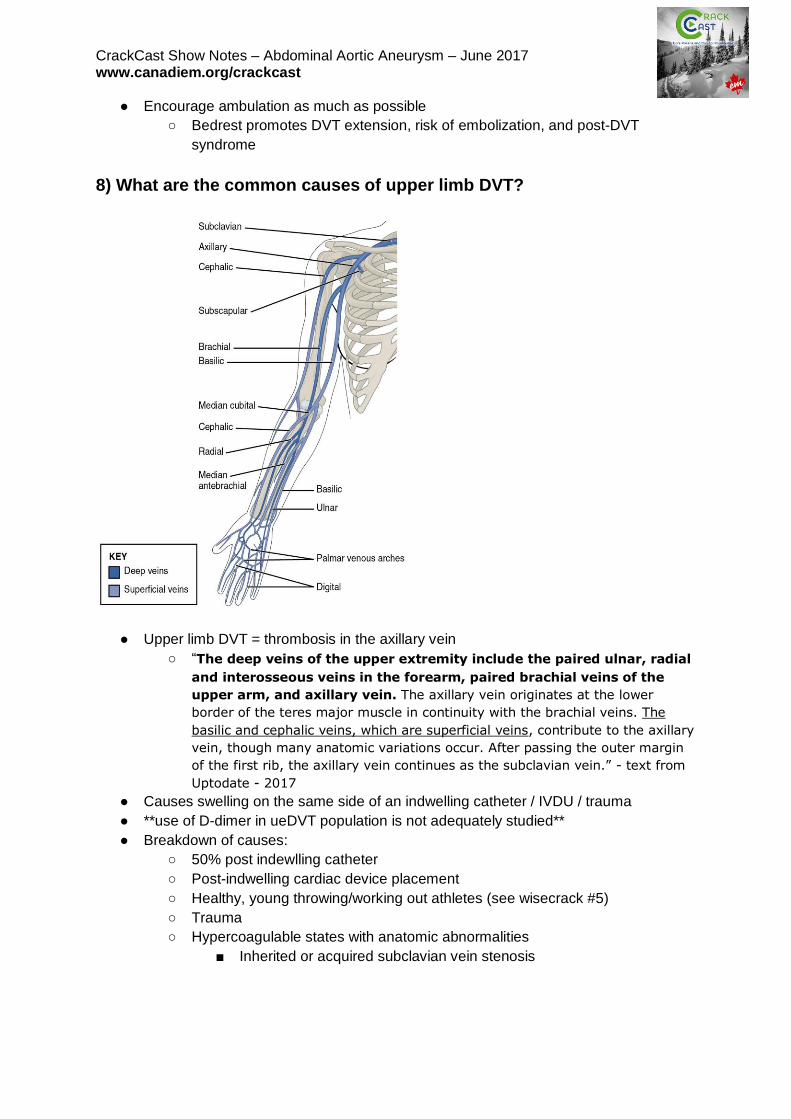
● Upper limb DVT = thrombosis in the axillary vein
○ “The deep veins of the upper extremity include the paired ulnar, radial
and interosseous veins in the forearm, paired brachial veins of the
upper arm, and axillary vein. The axillary vein originates at the lower
border of the teres major muscle in continuity with the brachial veins. The
basilic and cephalic veins, which are superficial veins, contribute to the axillary
vein, though many anatomic variations occur. After passing the outer margin
of the first rib, the axillary vein continues as the subclavian vein.” - text from
Uptodate - 2017
● Causes swelling on the same side of an indwelling catheter / IVDU / trauma
● **use of D-dimer in ueDVT population is not adequately studied**
● Breakdown of causes:
○ 50% post indewlling catheter
○ Post-indwelling cardiac device placement
○ Healthy, young throwing/working out athletes (see wisecrack #5)
○ Trauma
○ Hypercoagulable states with anatomic abnormalities
■ Inherited or acquired subclavian vein stenosis
CrackCast Show Notes – Abdominal Aortic Aneurysm – June 2017 www.canadiem.org/crackcast
9) How are upper limb DVTs managed?
At similar risk of PE, so is treated like a lower extremity DVT
● May or may not have to remove the catheter (depending on why it’s there)
● Because it can cause a PE - anyone with a u.e. DVT proximal to the elbow require
definitive treatment
○ Optimal dosing and duration is debated
■ Usually at least 3 months of anticoagulation (do your risk analysis for
everyone though!)
● Infusion phlebitis ) isolated brachial vein thrombosis - post recent IV infusion may be
treated like a superficial thrombophlebitis of the lower leg, but good evidence is
lacking.
Remember, that we not only treat DVT’s to prevent PE’s, but ALSO because DVT’s
damage the valves of veins - which can lead to venous insufficiency (see prev.
chapter). This can become a chronic disabling disease!
● Pain, varicosities, ulcers, skin changes, swelling - all with high morbidity!
Now, we’re moving on to PE’s
10) List classic risk factors for PE x 10
Every PE has its birth as a DVT somewhere in the body, which migrates to the lungs.
See Table 88-2 for a detailed list.
These are the ones that are highly associated with PE in the ED population:
● Hypercoagulable states:
○ Inherited thrombophilia
○ Active cancer
○ Estrogen
○ Prior PE / DVT
● Endothelial damage
○ Surgery or Trauma within the last 4 weeks requiring hospitalization/GA
● Venous stasis:
○ Surgery or Trauma within the last 4 weeks requiring hospitalization/GA
● Clinical signs/symptoms:
○ Dyspnea
○ Hemoptysis
○ Pulse > 100
○ O2 Sat < 95%
○ Unilateral leg / arm swelling
Notice that smoking is NOT on that list! Smoking does not seem to increase the risk for PE
compared with another patient who is a non-smoker with the same clinical presentation.
CrackCast Show Notes – Abdominal Aortic Aneurysm – June 2017 www.canadiem.org/crackcast Remember, just like in cardiac disease these are epidemiologic/population risk factors and
the abscence of them in a patient who has symptoms of a PE cannot be used to exclude the
diagnosis!
Many symptoms are nonspecific in PE, and the suddenness of the onset of symptoms
neither increases nor decreases the risk of PE.
Temp > 38.6 is more suggestive of pneumonia than PE.
***as many as 50% of people diagnosed with a PE have no identifiable risk factors for
thrombosis!***
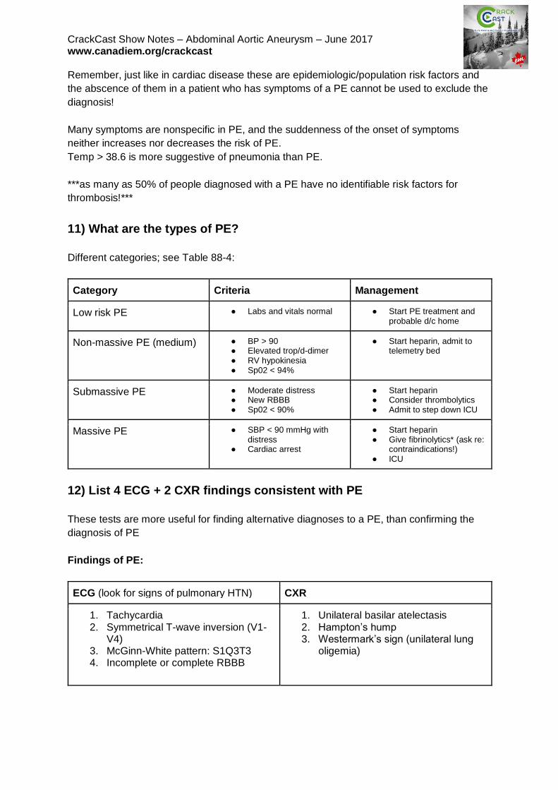
11) What are the types of PE?
Different categories; see Table 88-4:
Category Criteria Management
Low risk PE ● Labs and vitals normal
● Start PE treatment and probable d/c home
Non-massive PE (medium) ● BP > 90 ● Elevated trop/d-dimer ● RV hypokinesia ● Sp02 < 94%
● Start heparin, admit to telemetry bed
Submassive PE ● Moderate distress ● New RBBB ● Sp02 < 90%
● Start heparin ● Consider thrombolytics ● Admit to step down ICU
Massive PE ● SBP < 90 mmHg with distress
● Cardiac arrest
● Start heparin ● Give fibrinolytics* (ask re:
contraindications!) ● ICU
12) List 4 ECG + 2 CXR findings consistent with PE
These tests are more useful for finding alternative diagnoses to a PE, than confirming the
diagnosis of PE
Findings of PE:
ECG (look for signs of pulmonary HTN) CXR
1. Tachycardia 2. Symmetrical T-wave inversion (V1-
V4) 3. McGinn-White pattern: S1Q3T3 4. Incomplete or complete RBBB
1. Unilateral basilar atelectasis 2. Hampton’s hump 3. Westermark’s sign (unilateral lung
oligemia)
CrackCast Show Notes – Abdominal Aortic Aneurysm – June 2017 www.canadiem.org/crackcast
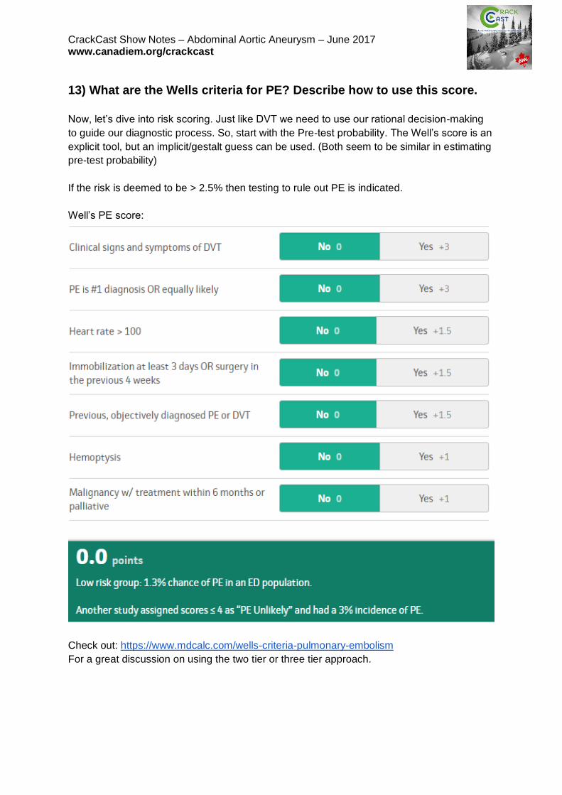
13) What are the Wells criteria for PE? Describe how to use this score.
Now, let’s dive into risk scoring. Just like DVT we need to use our rational decision-making
to guide our diagnostic process. So, start with the Pre-test probability. The Well’s score is an
explicit tool, but an implicit/gestalt guess can be used. (Both seem to be similar in estimating
pre-test probability)
If the risk is deemed to be > 2.5% then testing to rule out PE is indicated.
Well’s PE score:
Check out: https://www.mdcalc.com/wells-criteria-pulmonary-embolism
For a great discussion on using the two tier or three tier approach.
CrackCast Show Notes – Abdominal Aortic Aneurysm – June 2017 www.canadiem.org/crackcast For people with a risk > 2.5%, Rosen’s suggests we divide pts. Into two groups (this is the
recommended two tier approach):
1) Non-high (<40%) risk
a) Order D-dimer
i) If -ve = NO PE
ii) If +ve = Get a CT-PE scan
2) High (>40%) risk
a) Get a CT scan
i) If -ve: consider further testing with doppler U/S for DVTs.
ii) Review the scan with your radiologist!
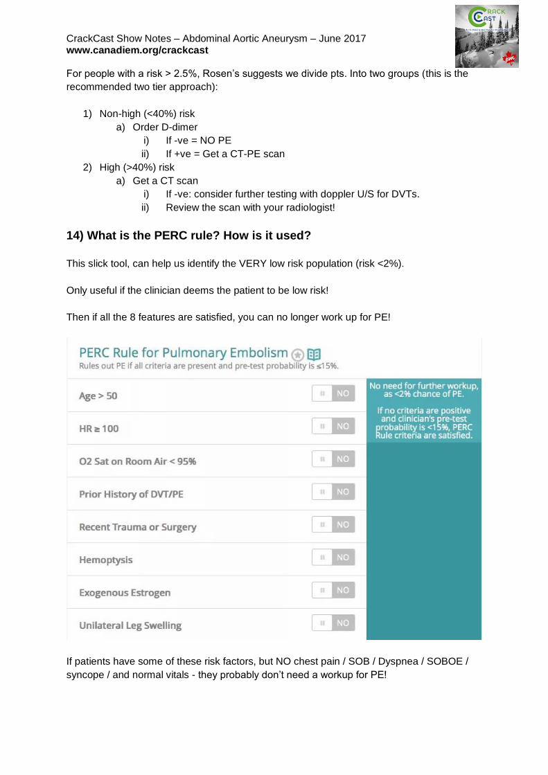
14) What is the PERC rule? How is it used?
This slick tool, can help us identify the VERY low risk population (risk <2%).
Only useful if the clinician deems the patient to be low risk!
Then if all the 8 features are satisfied, you can no longer work up for PE!
If patients have some of these risk factors, but NO chest pain / SOB / Dyspnea / SOBOE /
syncope / and normal vitals - they probably don’t need a workup for PE!
CrackCast Show Notes – Abdominal Aortic Aneurysm – June 2017 www.canadiem.org/crackcast
15) Which imaging tests can be used to diagnose PE? List advantages
and disadvantages of each.
CT-PE protocol
● +’ves: finds alternate diagnoses, rapid and fast, no iodine contrast used. Highly
sensitive and specific.
● –‘ves: ionizing radiation
V/Q scanning
● +’ves: less radiation
● –‘ves: not as sensitive, does not find as many alternative diagnoses, must be
stratified based on high-intermediate-low-normal probability.
○ For example, someone with an intermediate probability scan still requires a
CT-PE scan.
16) List indications for thrombolysis in PE, what is the risk of ICH?
● tPa: use is controversial
○ Usually reserved for massive PE (diagnosed on CT-scan):
■ Systolic BP < 90 mmHg for > 15 mins
● Or a reduction of ~> 60 mmHg from baseline
○ Case by case use:
■ Pt. in cardiac arrest
■ Extensive clot burden
■ RV dysfunction
■ Hypoxemia
■ Clot in transit
○ Alteplase: 15 mg bolus, then 2 hr infusion of 85 mg. (then start heparin)
Risk of ICH: 1-2%
17) What are the absolute and relative contraindications?
Absolute or major contraindications to systemic thrombolytic therapy in acute PE include an
intracranial neoplasm, recent (ie, <2 months) intracranial or spinal surgery or trauma, history
of a hemorrhagic stroke, active bleeding or bleeding diathesis, or nonhemorrhagic stroke
within the previous three months.
Relative contraindications include severe uncontrolled hypertension (ie, systolic blood
pressure >200 mmHg or diastolic blood pressure >110 mmHg), nonhemorrhagic stroke older
than three months, surgery within the previous 10 days, pregnancy.
From:
Antithrombotic therapy for VTE disease: Antithrombotic Therapy and Prevention of
Thrombosis, 9th ed: American College of Chest Physicians Evidence-Based Clinical Practice
Guidelines. https://www.ncbi.nlm.nih.gov/pubmed?term=22315268
CrackCast Show Notes – Abdominal Aortic Aneurysm – June 2017 www.canadiem.org/crackcast Those patients with severe hypotension and a contraindication to fibrinolysis should
be referred to see a vascular surgeon for embolectomy.
18) List markers of poor prognosis in patients with PE.
Rosen’s states that 8% of patients with a PE in the ED will die within 30 days.
● Pulseless electrical activity (fully occluded pulmonary artery)
● Arterial hypotension
● Worsening respiratory distress
● Clot in transit
● Syncope or seizure in the ED
● Hypoxemia
● Evolving RBBB
● Multiple comorbidities
● RV dusyunfction (on Echo, troponin, or BNP)
Wisecracks
1) What is phlegmasia cerulea dolens? How is it managed?
● Massive iliofemoral DVT:
○ Need aggressive management:
■ usually thrombolysis and/or thrombectomy.
■ Intravenous (IV) UFH is usually the anticoagulant of choice
■ Seek professional decision on whether to pursue more aggressive
therapy (thrombolysis vs. thrombectomy)
○ May progress to phlegmasia alba dolens or venous gangrene
2) Which patients should have an IVC filter?
● Used in patients with acute proximal DVT and PE who:
○ have an absolute contraindication to anticoagulant therapy (eg, recent
surgery, hemorrhagic stroke, active bleeding)
○ Also often considered in patients with recurrent embolism despite adequate
anticoagulation
3) What about PE/DVT in pregnancy?
● DVT / PE
○ Managed with adjusted doses of LMWH for at least 3-6 months (at least 4-6
weeks post-partum)
■ Not recommended:
● Heparin or Warfarin
■ Do NOT use:
● Fondaparinux
● DOAC’s
CrackCast Show Notes – Abdominal Aortic Aneurysm – June 2017 www.canadiem.org/crackcast
4) What is the cause of hypoxia in patients with PE? What causes chest
pain? What causes hypotension?
Hypoxia: due to V/Q mismatch. Areas of the lung that are ventilated (dead space) not being
perfused.
Chest pain: thought due to a focal area of lung tissue necrosis (caused by intensive
inflammatory processes)
Hypotension: impaired LV filling
5) What is Paget-Schroetter Syndrome?
“Effort thrombosis, or Paget-Schroetter Syndrome, refers to axillary-subclavian vein
thrombosis associated with strenuous and repetitive activity of the upper extremities.
Anatomical abnormalities at the thoracic outlet and repetitive trauma to the
endothelium of the subclavian vein are key factors in its initiation and progression.
The role of hereditary and acquired thrombophilias is unclear. The pathogenesis of effort
thrombosis is thus distinct from other venous thromboembolic disorders. Doppler
ultrasonography is the preferred initial test, while contrast venography remains the gold
standard for diagnosis. Computed tomographic venography and magnetic resonance
venography are comparable to conventional venography and are being increasingly used.
Conservative management with anticoagulation alone is inadequate and leads to significant
residual disability. An aggressive multimodal treatment strategy consisting of catheter-
directed thrombolysis, with or without early thoracic outlet decompression, is essential for
optimizing outcomes. Despite excellent insights into its pathogenesis and advances in
treatment, a significant number of patients with effort thrombosis continue to be treated
suboptimally. Hence, there is an urgent need for increasing physician awareness about risk
factors, etiology and the management of this unique and relatively infrequent disorder.”
From: https://www.ncbi.nlm.nih.gov/pubmed?term=21079709
Look for Urschel’s sign!