Embed Size (px)
Citation preview
Copyright 2009 John Wiley & Sons, Inc.
Chapter 8
The Skeletal System:
The Appendicular Skeleton
Copyright 2009 John Wiley & Sons, Inc.
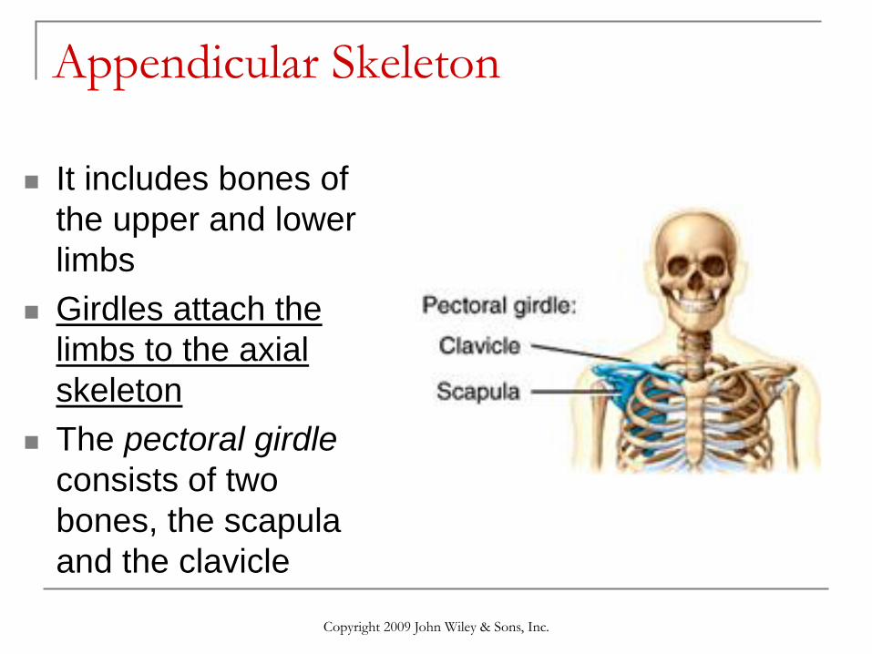
Appendicular Skeleton
It includes bones of
the upper and lower
limbs
Girdles attach the
limbs to the axial
skeleton
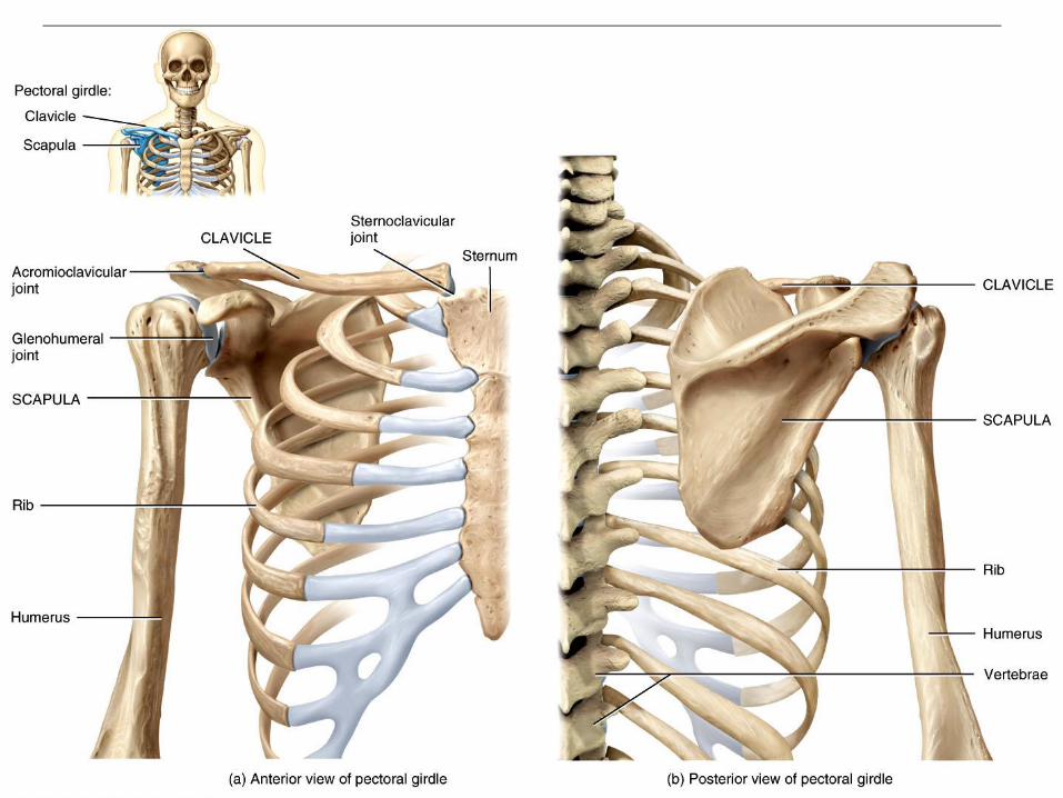
The pectoral girdle
consists of two
bones, the scapula
and the clavicle
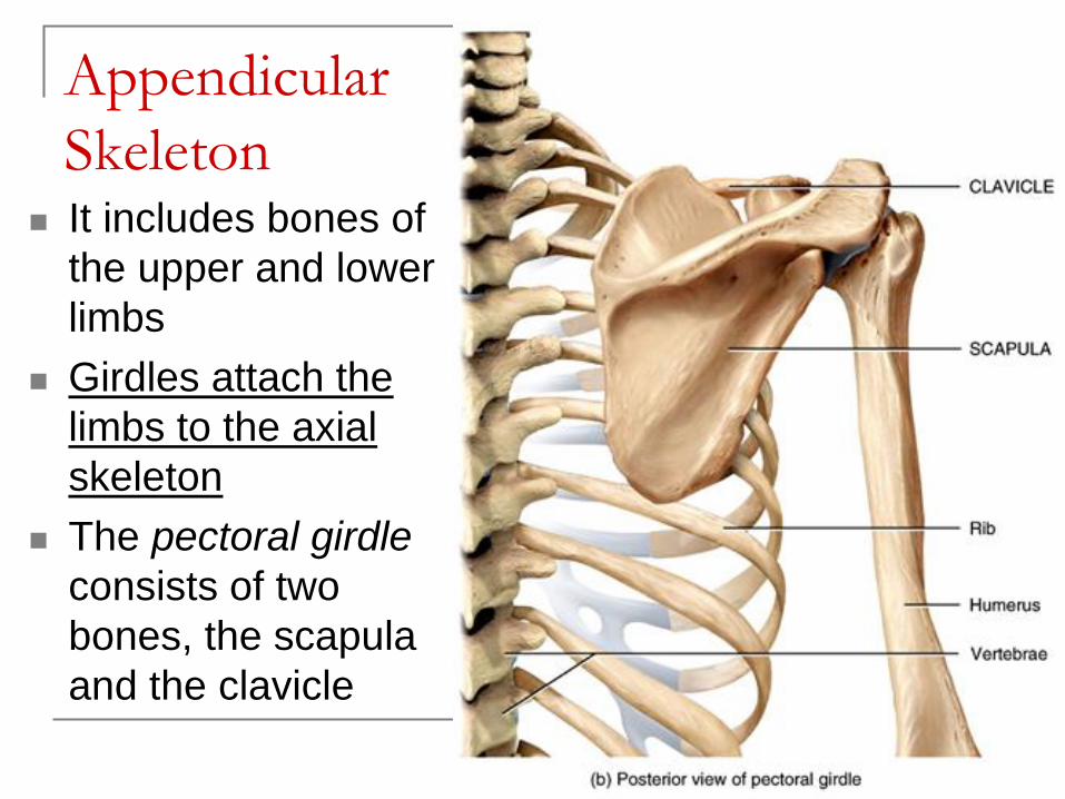
Appendicular
Skeleton It includes bones of
the upper and lower
limbs
Girdles attach the
limbs to the axial
skeleton
The pectoral girdle
consists of two
bones, the scapula
and the clavicle
Copyright 2009 John Wiley & Sons, Inc.
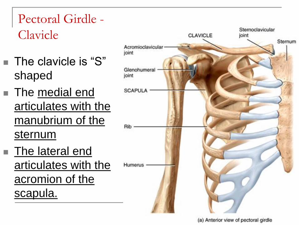
Pectoral Girdle -
Clavicle
The clavicle is “S”
shaped
The medial end
articulates with the
manubrium of the
sternum
The lateral end
articulates with the
acromion of the
scapula.
Copyright 2009 John Wiley & Sons, Inc.
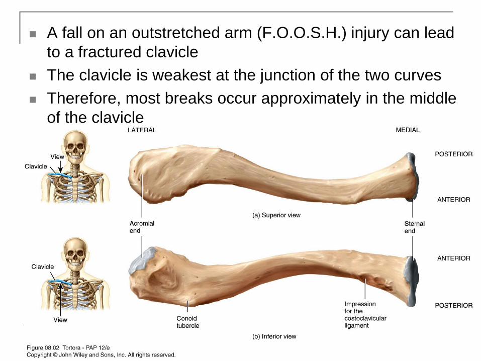
A fall on an outstretched arm (F.O.O.S.H.) injury can lead
to a fractured clavicle
The clavicle is weakest at the junction of the two curves
Therefore, most breaks occur approximately in the middle
of the clavicle
Copyright 2009 John Wiley & Sons, Inc.
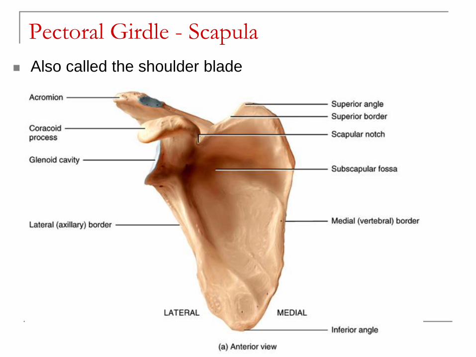
Pectoral Girdle - Scapula
Also called the shoulder blade
Copyright 2009 John Wiley & Sons, Inc.
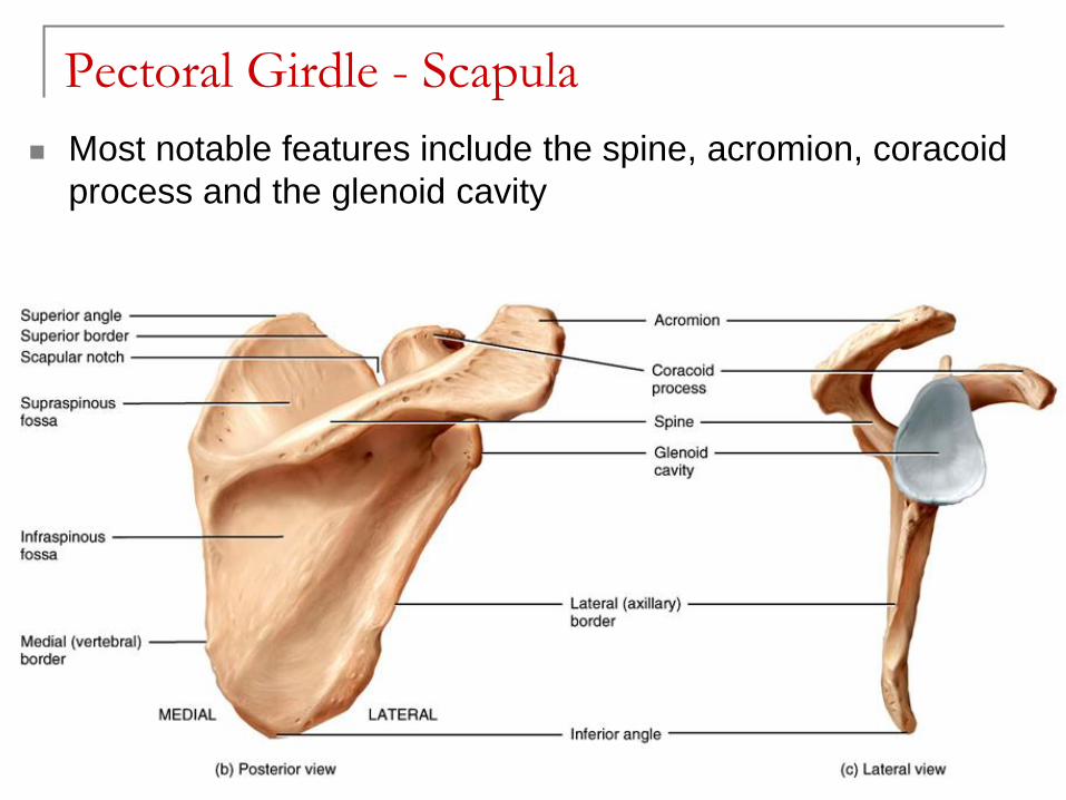
Pectoral Girdle - Scapula
Most notable features include the spine, acromion, coracoid
process and the glenoid cavity
Copyright 2009 John Wiley & Sons, Inc.
Features on the Scapula
Spine - a large process on the posterior of the scapula that ends laterally as the acromion
Acromion - the flattened lateral portion of the spine of the scapula
Coracoid process - a protruding projection on the anterior surface just inferior to the lateral aspect of the clavicle
Glenoid cavity - shallow concavity that articulates with the head of the humerus
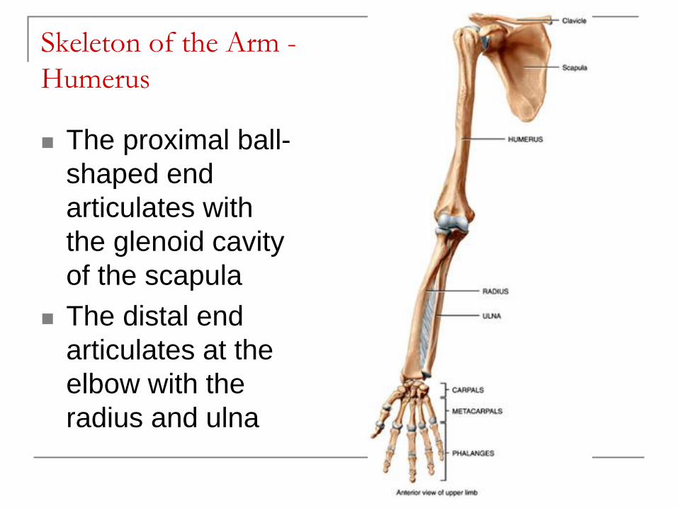
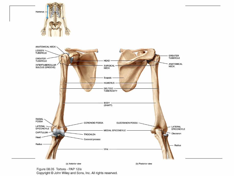
Skeleton of the Arm -
Humerus
The proximal ball-
shaped end
articulates with
the glenoid cavity
of the scapula
The distal end
articulates at the
elbow with the
radius and ulna
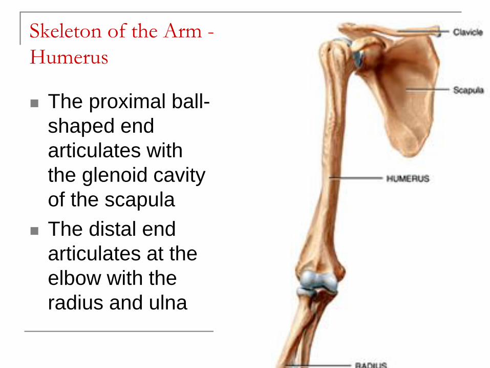
Skeleton of the Arm -
Humerus
The proximal ball-
shaped end
articulates with
the glenoid cavity
of the scapula
The distal end
articulates at the
elbow with the
radius and ulna
Copyright 2009 John Wiley & Sons, Inc.
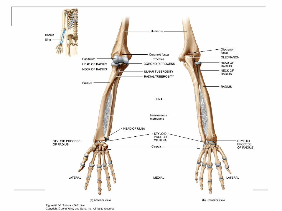
Humerus - Surface Features
Coronoid fossa - anterior depression that
receives the coronoid process of the ulna
during forearm flexion
Olecranon fossa - posterior depression that
receives the olecranon of the ulna during
forearm extension
The medial and lateral epicondyles are bony
projections to which the forearm muscles
attach
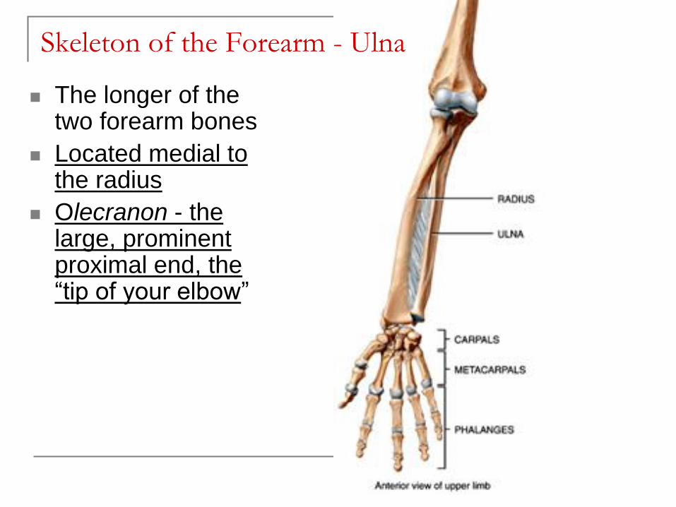
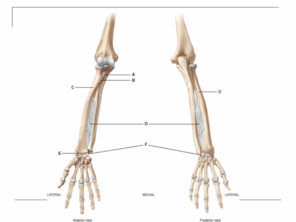
Skeleton of the Forearm - Ulna
The longer of the two forearm bones
Located medial to the radius
Olecranon - the large, prominent proximal end, the “tip of your elbow”
Copyright 2009 John Wiley & Sons, Inc.
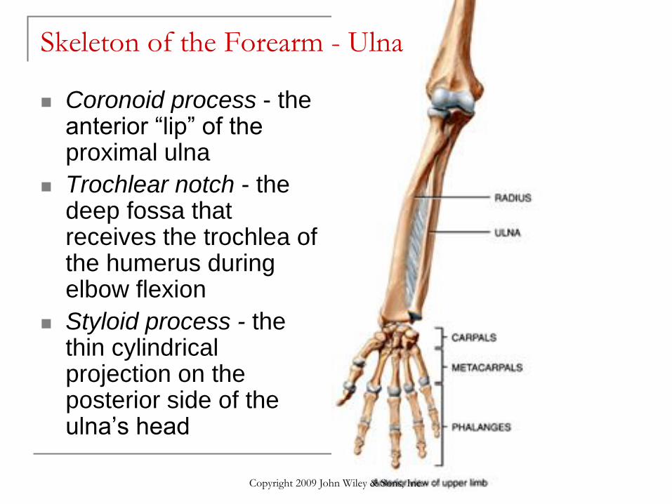
Skeleton of the Forearm - Ulna
Coronoid process - the anterior “lip” of the proximal ulna
Trochlear notch - the deep fossa that receives the trochlea of the humerus during elbow flexion
Styloid process - the thin cylindrical projection on the posterior side of the ulna’s head
Copyright 2009 John Wiley & Sons, Inc.
Copyright 2009 John Wiley & Sons, Inc.
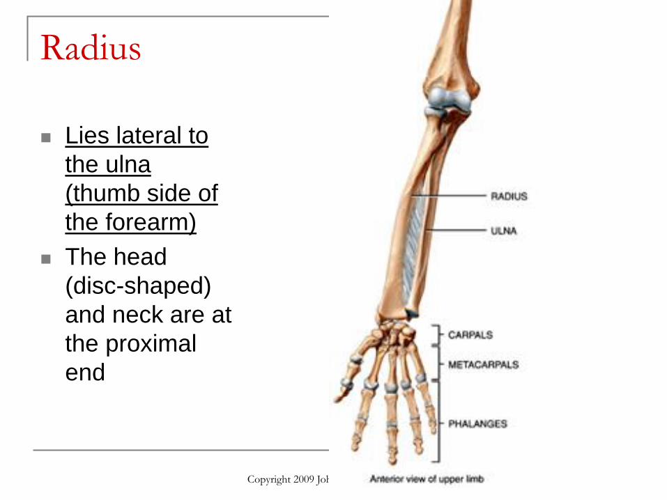
Radius
Lies lateral to
the ulna
(thumb side of
the forearm)
The head
(disc-shaped)
and neck are at
the proximal
end
Copyright 2009 John Wiley & Sons, Inc.
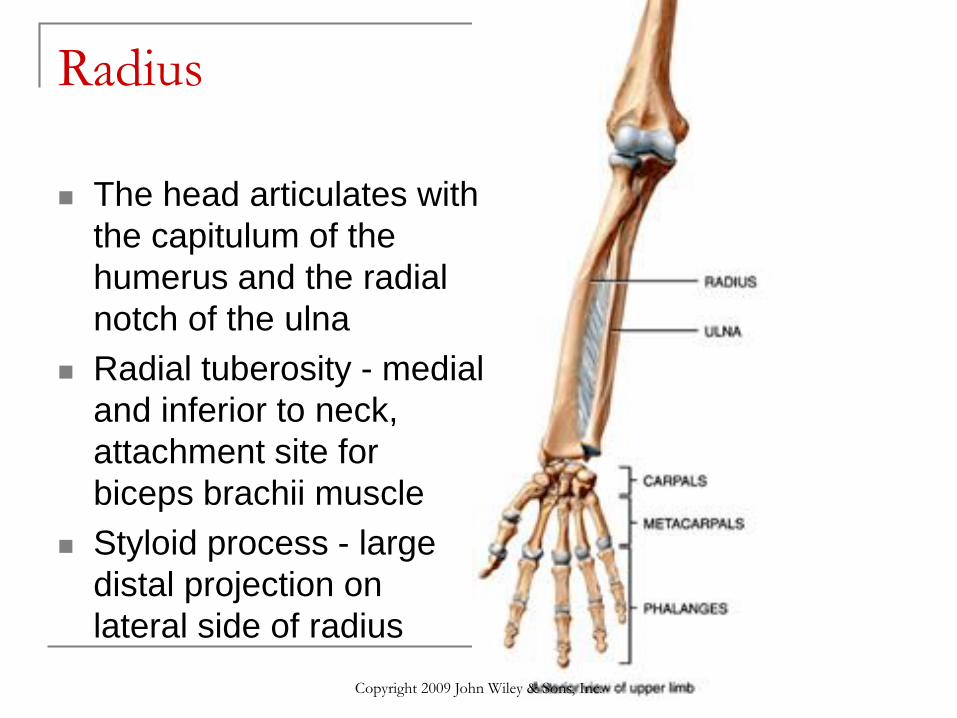
Radius
The head articulates with
the capitulum of the
humerus and the radial
notch of the ulna
Radial tuberosity - medial
and inferior to neck,
attachment site for
biceps brachii muscle
Styloid process - large
distal projection on
lateral side of radius
Copyright 2009 John Wiley & Sons, Inc.
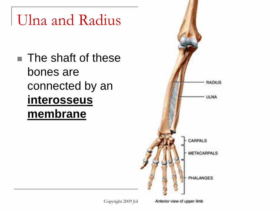
Ulna and Radius
The shaft of these
bones are
connected by an
interosseus
membrane
Copyright 2009 John Wiley & Sons, Inc.
Copyright 2009 John Wiley & Sons, Inc.
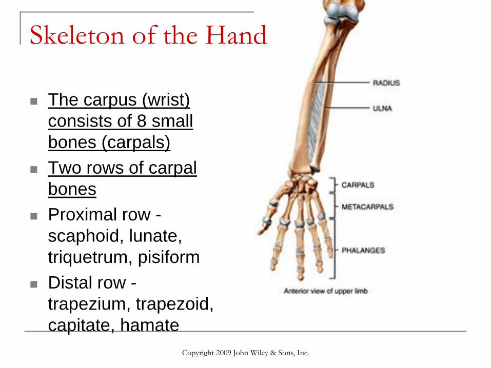
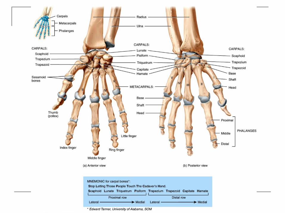
Skeleton of the Hand
The carpus (wrist)
consists of 8 small
bones (carpals)
Two rows of carpal
bones
Proximal row -
scaphoid, lunate,
triquetrum, pisiform
Distal row -
trapezium, trapezoid,
capitate, hamate

Copyright 2009 John Wiley & Sons, Inc.
Skeleton of the Hand
Scaphoid - most
commonly fractured
Carpal tunnel -
space between
carpal bones and
flexor retinaculum
Copyright 2009 John Wiley & Sons, Inc.
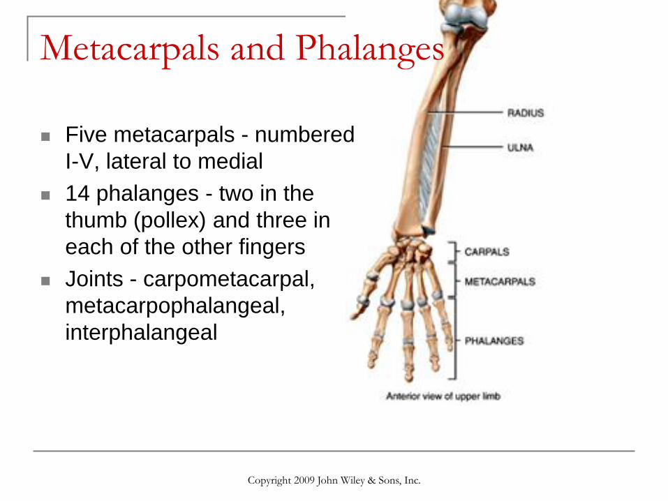
Metacarpals and Phalanges
Five metacarpals - numbered
I-V, lateral to medial
14 phalanges - two in the
thumb (pollex) and three in
each of the other fingers
Joints - carpometacarpal,
metacarpophalangeal,
interphalangeal
Copyright 2009 John Wiley & Sons, Inc.
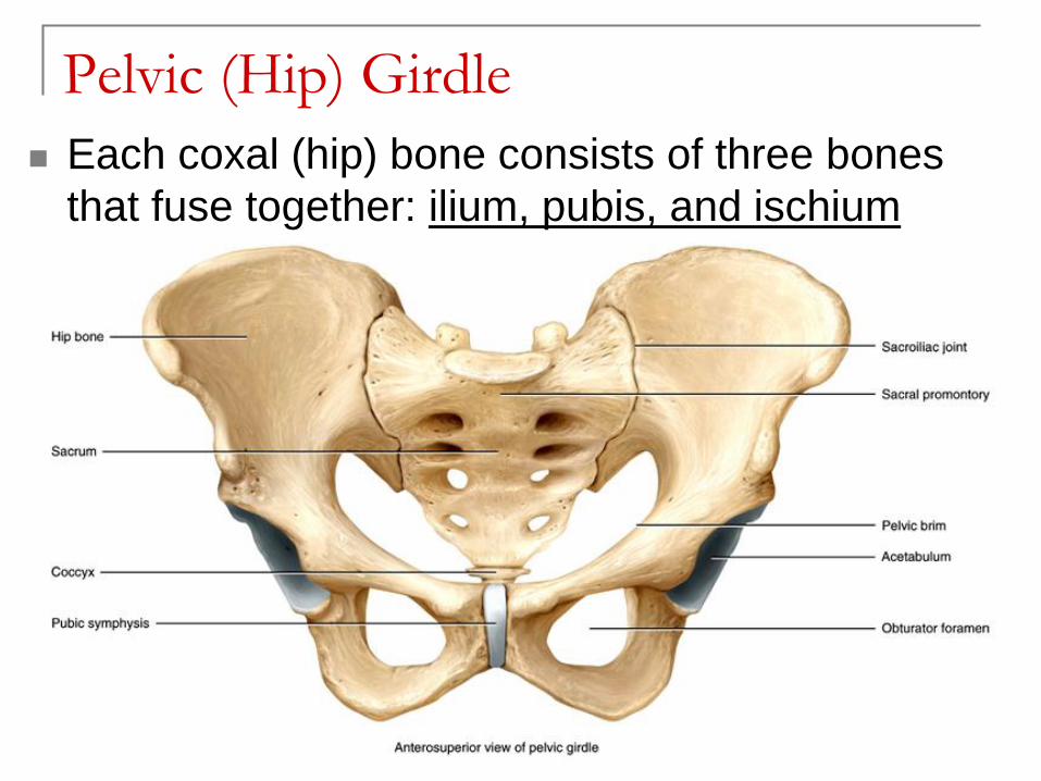
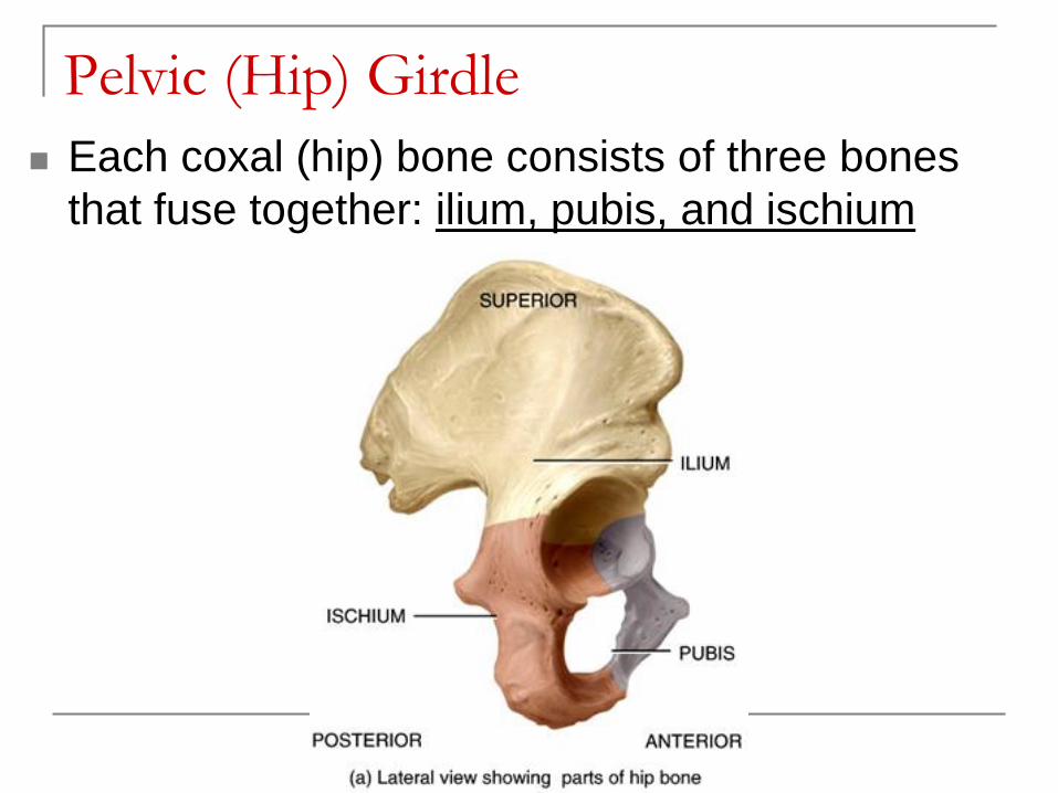
Pelvic (Hip) Girdle
Each coxal (hip) bone consists of three bones
that fuse together: ilium, pubis, and ischium
Copyright 2009 John Wiley & Sons, Inc.
Pelvic (Hip) Girdle
Each coxal (hip) bone consists of three bones
that fuse together: ilium, pubis, and ischium
Copyright 2009 John Wiley & Sons, Inc.
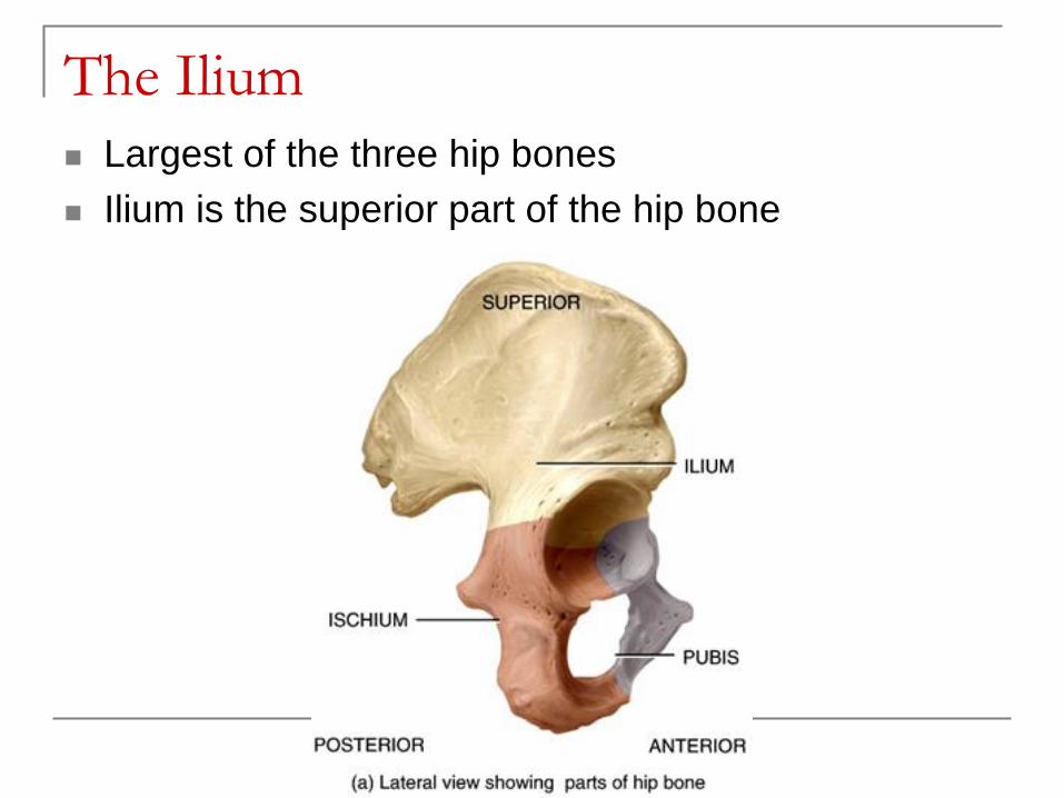
The Ilium
Largest of the three hip bones
Ilium is the superior part of the hip bone
Copyright 2009 John Wiley & Sons, Inc.
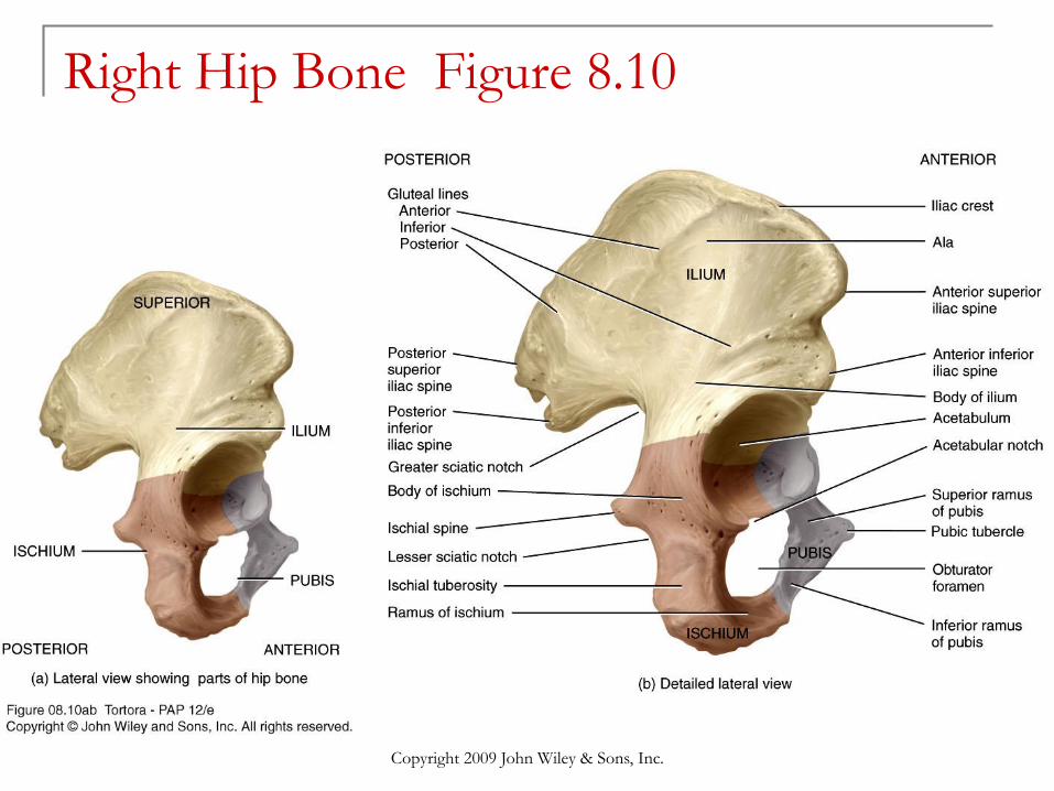
Right Hip Bone Figure 8.10
Copyright 2009 John Wiley & Sons, Inc.
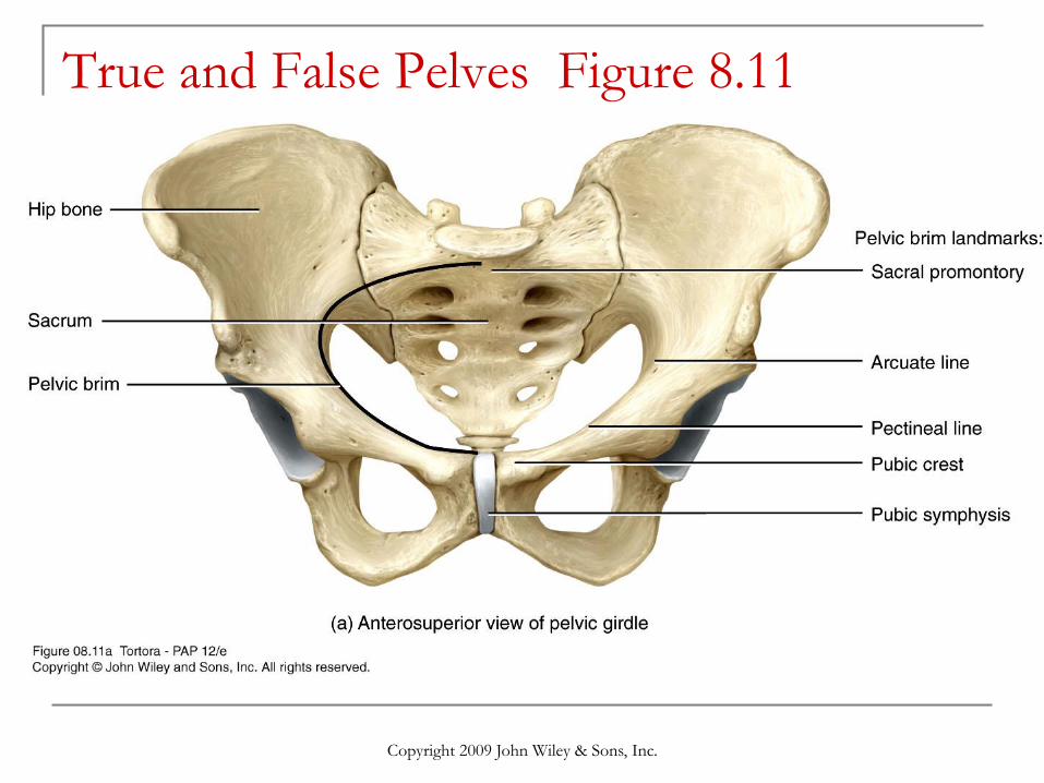
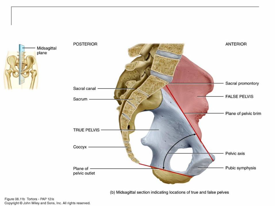
True and False Pelves Figure 8.11
Copyright 2009 John Wiley & Sons, Inc.
Copyright 2009 John Wiley & Sons, Inc.
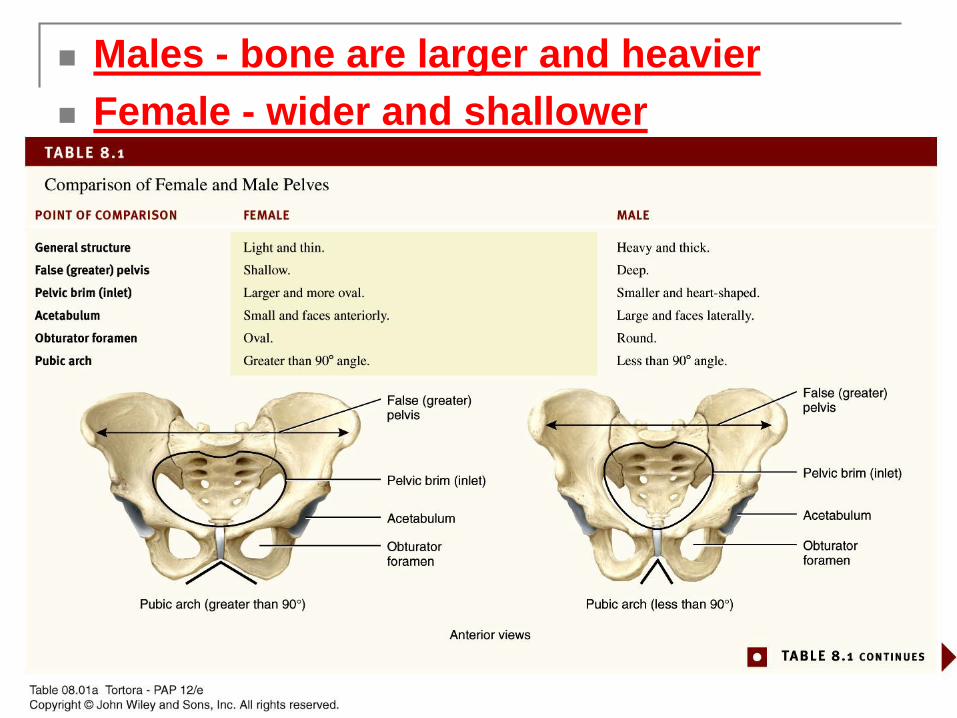
Males - bone are larger and heavier
Female - wider and shallower
Copyright 2009 John Wiley & Sons, Inc.
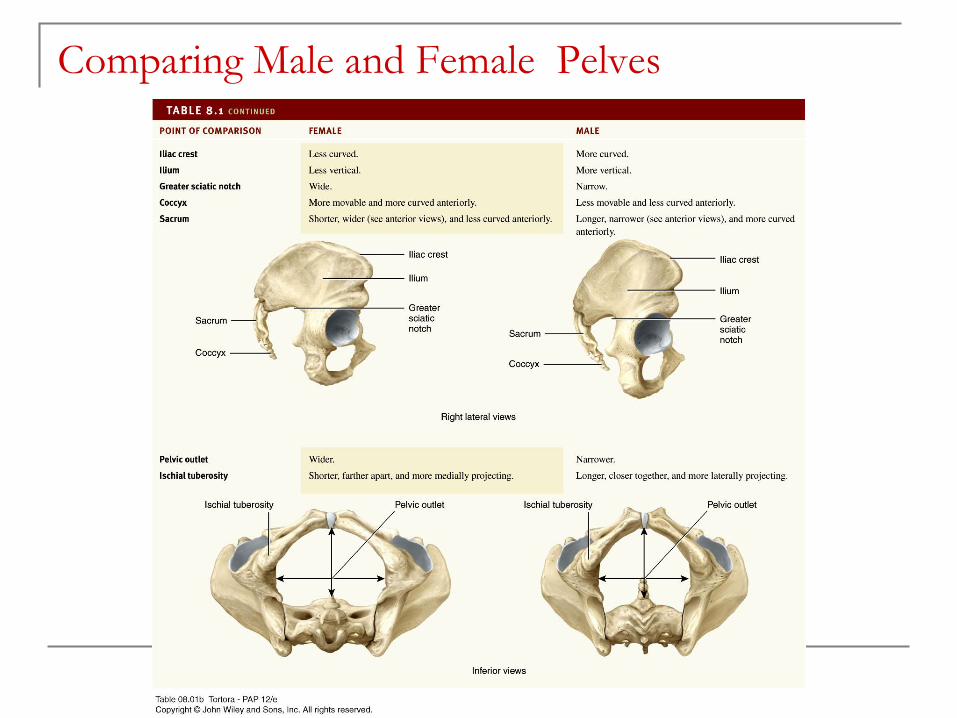
Comparing Male and Female Pelves
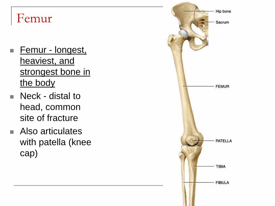
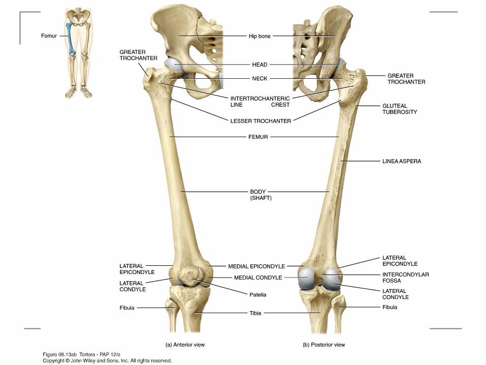
Femur
Femur - longest,
heaviest, and
strongest bone in
the body
Neck - distal to
head, common
site of fracture
Also articulates
with patella (knee
cap)
Copyright 2009 John Wiley & Sons, Inc.
Copyright 2009 John Wiley & Sons, Inc.
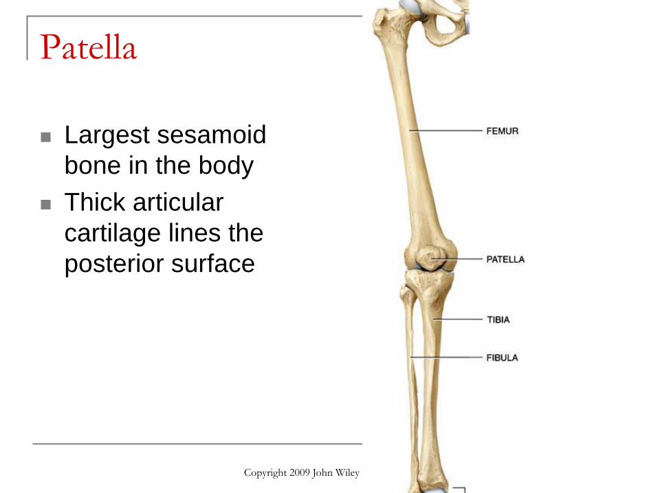
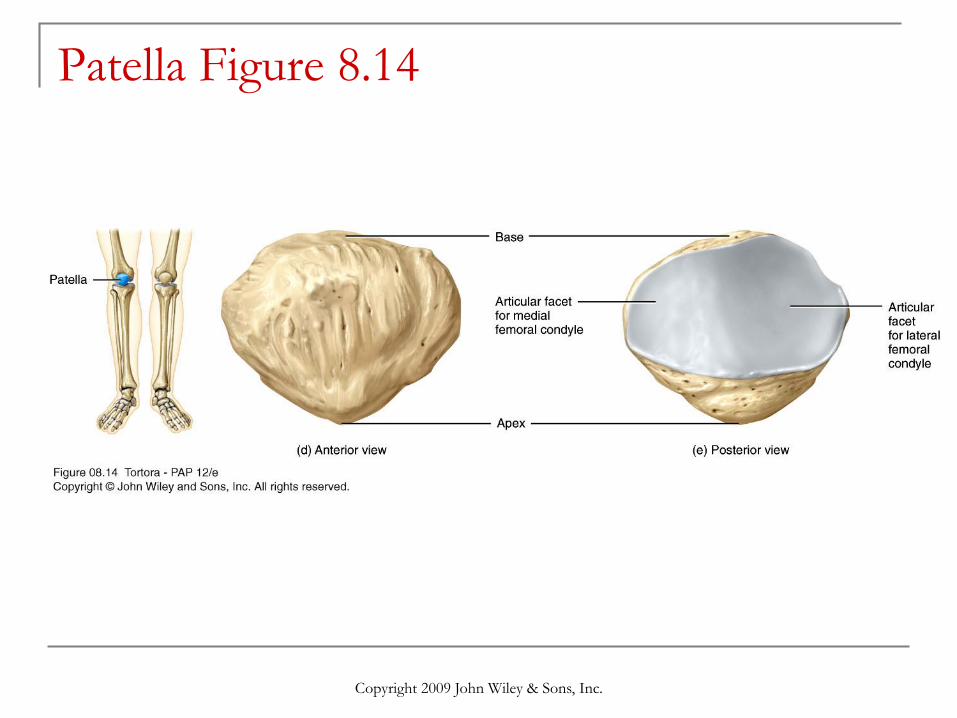
Patella
Largest sesamoid
bone in the body
Thick articular
cartilage lines the
posterior surface
Copyright 2009 John Wiley & Sons, Inc.
Patella Figure 8.14
Copyright 2009 John Wiley & Sons, Inc.
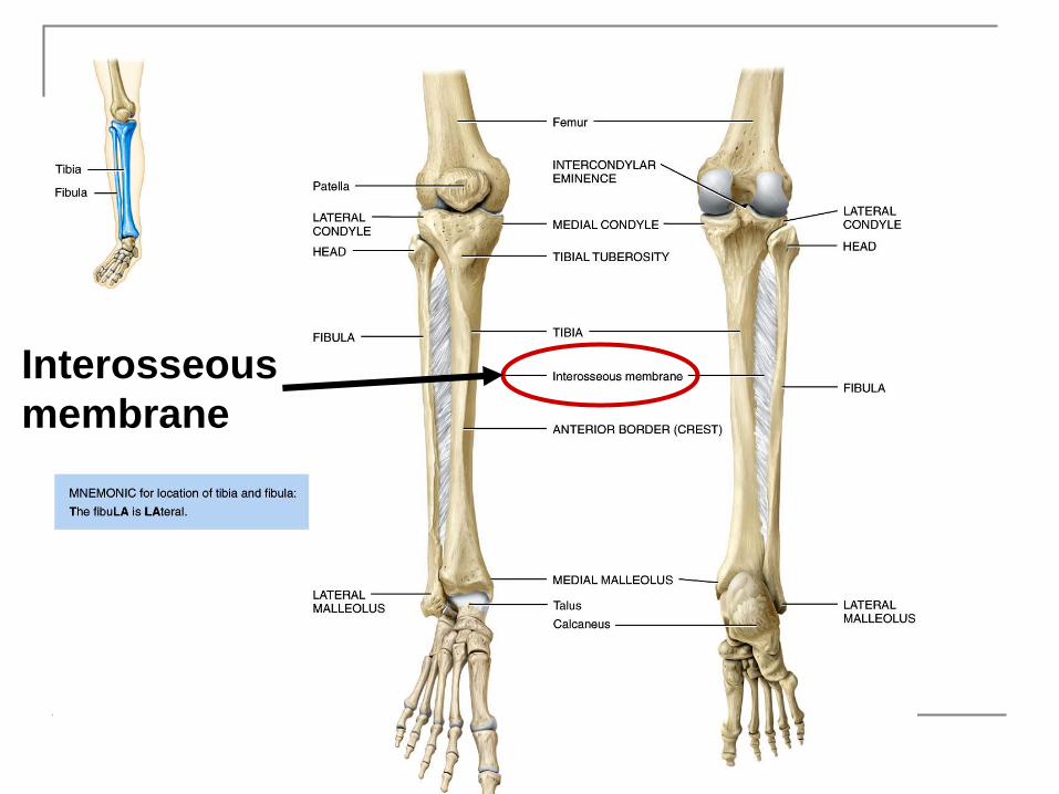
Interosseous
membrane
Copyright 2009 John Wiley & Sons, Inc.
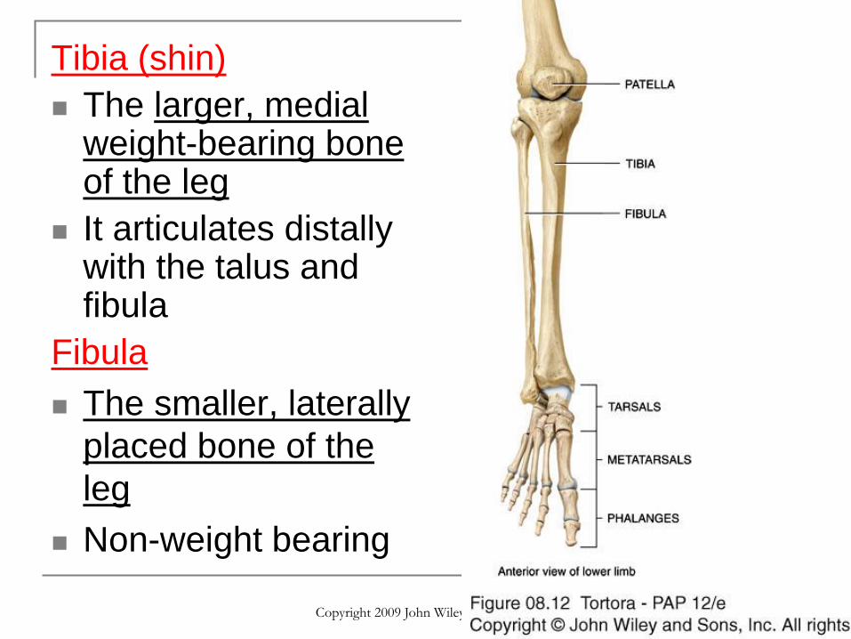
Tibia (shin)
The larger, medial weight-bearing bone of the leg
It articulates distally with the talus and fibula
Fibula
The smaller, laterally
placed bone of the
leg
Non-weight bearing
Copyright 2009 John Wiley & Sons, Inc.
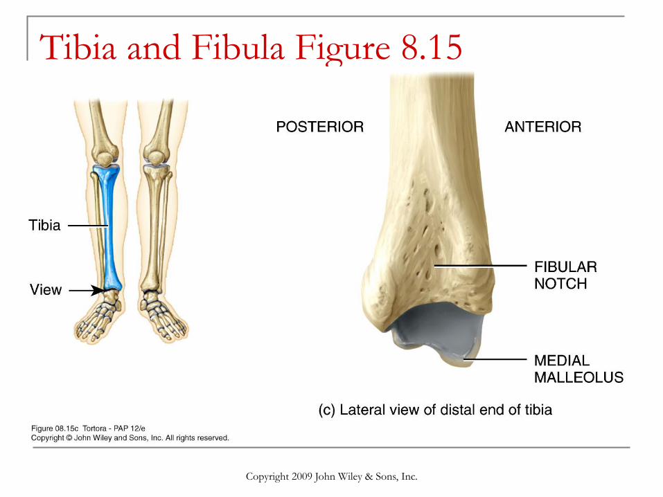
Tibia and Fibula Figure 8.15
Copyright 2009 John Wiley & Sons, Inc.
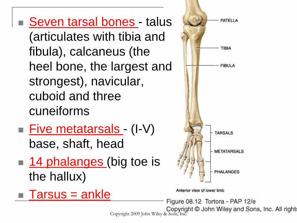
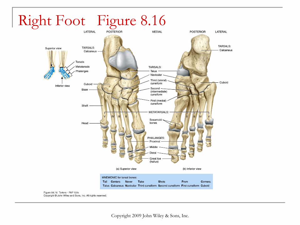
Seven tarsal bones - talus
(articulates with tibia and
fibula), calcaneus (the
heel bone, the largest and
strongest), navicular,
cuboid and three
cuneiforms
Five metatarsals - (I-V)
base, shaft, head
14 phalanges (big toe is
the hallux)
Tarsus = ankle
Copyright 2009 John Wiley & Sons, Inc.
Right Foot Figure 8.16
Copyright 2009 John Wiley & Sons, Inc.
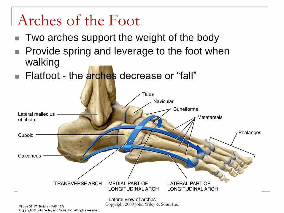
Arches of the Foot Two arches support the weight of the body
Provide spring and leverage to the foot when walking
Flatfoot - the arches decrease or “fall”
Copyright 2009 John Wiley & Sons, Inc.
Bone Review
Pairs are students are going to get one bone.
You must complete the following:
Identify the bone.
Name two structures on the bone (processes,
notches, etc.).
Name one other bone it articulates with.
Explain its function.
Copyright 2009, John Wiley & Sons, Inc.
Online Quiz Review
Website: http://highered.mcgraw-
hill.com/sites/0072351136/student_view0/chapter6/chapter_quiz.ht
ml
Or Google “anatomy and physiology quiz” and look for
the above website. Be sure to go to the Chapter 6 quiz.
Copy and answer the following questions.
Chapter 6
1,4,5,8,9,10,11,14,23,24,32,34,37,38,39 (15 total)
Copyright 2009 John Wiley & Sons, Inc.