Embed Size (px)
Citation preview
Chapter 8Chapter 8The Nervous SystemThe Nervous System
Marisol Boatwright, MHA, Marisol Boatwright, MHA, CMA (AAMA)CMA (AAMA)
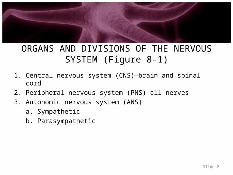
ORGANS AND DIVISIONS OF THE NERVOUS SYSTEM (Figure 8-1)
1. Central nervous system (CNS)—brain and spinal cord2. Peripheral nervous system (PNS)—all nerves3. Autonomic nervous system (ANS)
a. Sympatheticb. Parasympathetic
Slide 2
Slide 3
CELLS OF THE NERVOUS SYSTEM
• Neurons– Consist of three main parts—dendrites; cell body of neuron; and axon
(Figure 8-2)• Dendrites conduct impulses to cell body of neuron• Axons conduct impulses away from cell body of neuron
– Neurons classified according to function• Sensory (afferent) neurons: conduct impulses to the spinal cord
and brain• Motor (efferent) neurons: conduct impulses away from brain and
spinal cord to muscles and glands• Interneurons: conduct impulses from sensory neurons to motor
neurons Slide 4
CELLS OF THE NERVOUS SYSTEM
• Glia (neuroglia)– Support cells, bringing the cells of nervous tissue together structurally
and functionally– Three main types of glial cells of the CNS (Figure 8-3)
• Astrocytes—star-shaped cells that anchor small blood vessels to neurons• Microglia—small cells that move in inflamed brain tissue carrying on
phagocytosis• Oligodendrocytes—form myelin sheaths on axons in the CNS
– Schwann cells form myelin sheaths on axons of the PNS (Figure 8-2)
Slide 5
NERVES AND TRACTS(Figure 8-4)
• Nerve—bundle of peripheral axons– Tract—bundle of central axons– White matter—tissue composed primarily of myelinated axons (nerves
or tracts)– Gray matter—tissue composed primarily of cell bodies and
unmyelinated fibers• Nerve coverings—fibrous connective tissue
1. Endoneurium—surrounds individual fibers within a nerve2. Perineurium—surrounds a group (fascicle) of nerve fibers3. Epineurium—surrounds the entire nerve
Slide 6
REFLEX ARCS• Nerve impulses are conducted from receptors to effectors
over neuron pathways or reflex arcs; conduction by a reflex arc results in a reflex (that is, contraction by a muscle or secretion by a gland)
• The simplest reflex arcs are two-neuron arcs—consisting of sensory neurons synapsing in the spinal cord with motor neurons
• Three-neuron arcs consist of sensory neurons synapsing in the spinal cord with interneurons that synapse with motor neurons (Figure 8-5)
Slide 7
NERVE IMPULSES• Definition—self-propagating wave of electrical disturbance that travels
along the surface of a neuron membrane• Mechanism
– A stimulus triggers the opening of Na+ channels in the plasma membrane of the neuron
– Inward movement of positive sodium ions leaves a slight excess of negative ions outside at a stimulated point; marks the beginning of a nerve impulse
Slide 8
THE SYNAPSE• Definition—chemical compounds released from axon
terminals (of a presynaptic neuron) into a synaptic cleft• Neurotransmitters bind to specific receptor molecules in the
membrane of a postsynaptic neuron, opening ion channels and thereby stimulating impulse conduction by the membrane
• Names of neurotransmitters—acetylcholine, catecholamines (norepinephrine, dopamine, and serotonin), and other compounds
Slide 9
CENTRAL NERVOUS SYSTEM(CNS)
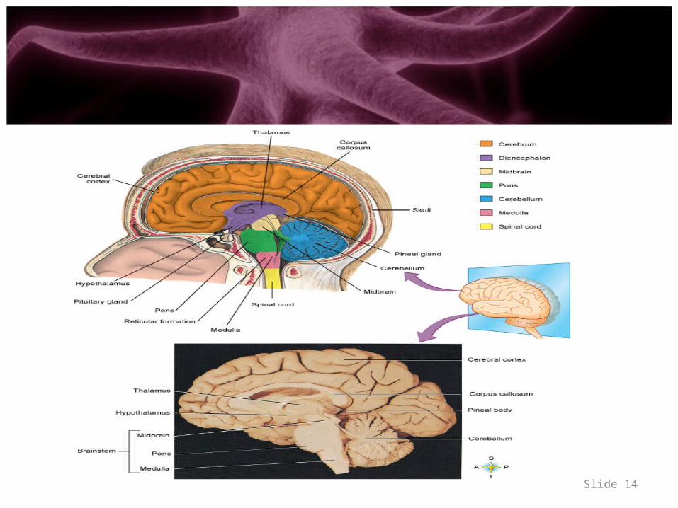
• Divisions of the brain (Figure 8-9 and Table 8-1)– Brainstem
• Consists of three parts of brain; named in ascending order: the medulla oblongata, pons, and midbrain
• Structure—white matter with bits of gray matter scattered through it• Function—gray matter in the brainstem functions as reflex centers (e.g.,
for heartbeat, respirations, and blood vessel diameter)– Sensory tracts in the brainstem conduct impulses to the higher parts of the
brain– Motor tracts conduct from the higher parts of the brain to the spinal cord
Slide 10
CENTRAL NERVOUS SYSTEM• Divisions of the brain (cont.)
– Diencephalon• Structure and function of the hypothalamus
– Consists mainly of the posterior pituitary gland, pituitary stalk, and gray matter
– Acts as the major center for controlling the ANS; therefore, it helps control the functioning of most internal organs
– Controls hormone secretion by anterior and posterior pituitary glands; therefore, it indirectly helps control hormone secretion by most other endocrine glands
– Contains centers for controlling body temperature, appetite, wakefulness, and pleasure
• Structure and function of the thalamus– Dumbbell-shaped mass of gray matter in each cerebral hemisphere– Relays sensory impulses to cerebral cortex sensory areas– In some way produces the emotions of pleasantness or unpleasantness
associated with sensations Slide 11
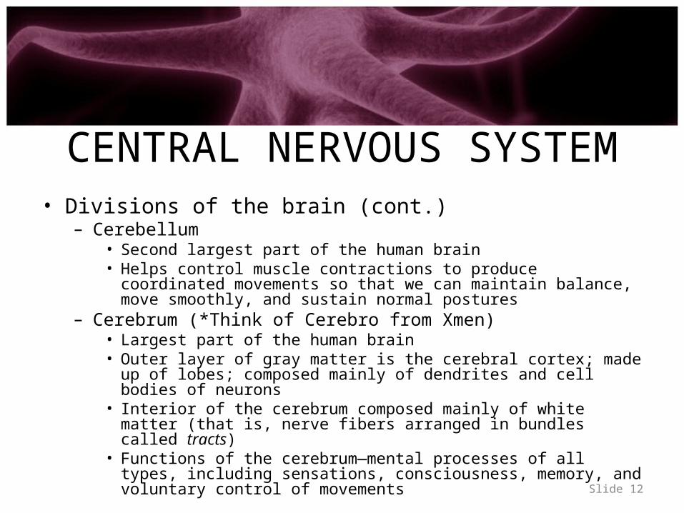
CENTRAL NERVOUS SYSTEM• Divisions of the brain (cont.)
– Cerebellum• Second largest part of the human brain• Helps control muscle contractions to produce coordinated movements so
that we can maintain balance, move smoothly, and sustain normal postures
– Cerebrum (*Think of Cerebro from Xmen)• Largest part of the human brain• Outer layer of gray matter is the cerebral cortex; made up of lobes;
composed mainly of dendrites and cell bodies of neurons• Interior of the cerebrum composed mainly of white matter (that is, nerve
fibers arranged in bundles called tracts)• Functions of the cerebrum—mental processes of all types, including
sensations, consciousness, memory, and voluntary control of movementsSlide 12
Slide 13
Slide 14
CENTRAL NERVOUS SYSTEM• Spinal cord (Figure 8-11)
– Outer part is composed of white matter made up of many bundles of axons called tracts; interior composed of gray matter made up mainly of neuron dendrites and cell bodies
– Functions as the center for all spinal cord reflexes; sensory tracts conduct impulses to the brain, and motor tracts conduct impulses from the brain
Slide 15
CENTRAL NERVOUS SYSTEM• Coverings and fluid spaces of the brain and spinal cord
– Coverings:• Cranial bones and vertebrae• Cerebral and spinal meninges—the dura mater, the pia mater, and
the arachnoid mater (Figure 8-13)– Fluid spaces—subarachnoid spaces of meninges, central canal inside
cord, and ventricles in brain (Figure 8-14)
Slide 16
PERIPHERAL NERVOUS SYSTEM(PNS)
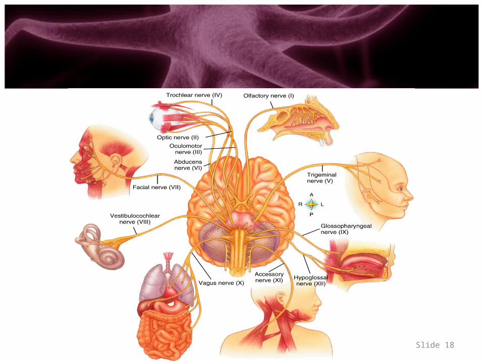
• Cranial nerves (Figure 8-16 and Table 8-2)– Twelve pairsTwelve pairs—attached to undersurface of the brain– Connect brain with the neck and structures in the thorax and
abdomen• Spinal nerves
– Structure—contain dendrites of sensory neurons and axons of motor neurons
– Functions—conduct impulses necessary for sensations and voluntary movements
Slide 17
Slide 18
AUTONOMIC NERVOUS SYSTEM(ANS)
• Autonomic nervous system—motor neurons that conduct impulses from the central nervous system to cardiac muscle, smooth muscle, and glandular epithelial tissue; regulates the body’s automatic or involuntary functions (Figure 8-18)
• Autonomic neurons—preganglionic autonomic neurons conduct from spinal cord or brainstem to an autonomic ganglion; postganglionic neurons conduct from autonomic ganglia to cardiac muscle, smooth muscle, and glandular epithelial tissue
Slide 19
AUTONOMIC NERVOUS SYSTEM
• Autonomic or visceral effectors—tissues to which autonomic neurons conduct impulses (that is, cardiac and smooth muscle and glandular epithelial tissue)
• Composed of two divisions—the sympathetic system and the parasympathetic system
Slide 20
AUTONOMIC NERVOUS SYSTEM• Sympathetic nervous system
– Structure• Dendrites and cell bodies of sympathetic preganglionic neurons are located
in the gray matter of the thoracic and upper lumbar segments of the spinal cord
• Axons leave the spinal cord in the anterior roots of spinal nerves, extend to sympathetic or collateral ganglia, and synapse with several postganglionic neurons whose axons extend to spinal or autonomic nerves to terminate in visceral effectors
• A chain of sympathetic ganglia is in front of and at each side of the spinal column
– Functions• Serves as the emergency or stress system, controlling visceral effectors
during strenuous exercise and strong emotions (anger, fear, hate, or anxiety)
• Group of changes induced by sympathetic control is called the fight-or-flight response
Slide 21
AUTONOMIC NERVOUS SYSTEM
• Parasympathetic nervous system– Structure
• Parasympathetic preganglionic neurons have dendrites and cell bodies in the gray matter of the brainstem and the sacral segments of the spinal cord
• Parasympathetic preganglionic neurons terminate in parasympathetic ganglia located in the head and the thoracic and abdominal cavities close to visceral effectors
• Each parasympathetic preganglionic neuron synapses with postganglionic neurons to only one effector
– Function—dominates control of many visceral effectors under normal, everyday conditions
Slide 22
AUTONOMIC NERVOUS SYSTEM
• Autonomic neurotransmitters– Cholinergic fibers—preganglionic axons of parasympathetic and
sympathetic systems and parasympathetic postganglionic axons release acetylcholine
– Adrenergic fibers—axons of sympathetic postganglionic neurons release norepinephrine (noradrenaline)
• Autonomic nervous system as a whole– Regulates the body’s automatic functions in ways that maintain or
quickly restore homeostasis– Many visceral effectors are doubly innervated (that is, they receive
fibers from parasympathetic and sympathetic divisions and are influenced in opposite ways by the two divisions)
Slide 23