Embed Size (px)
DESCRIPTION
CHAPTER 8 The Cellular Basis of Reproduction and Inheritance. Modules 8.1 – 8.3. How to Make a Sea Star — With and Without Sex. The life cycle of a multicellular organism includes development reproduction This sea star embryo (morula) shows one stage in the development of a fertilized egg - PowerPoint PPT Presentation
Citation preview
BIOLOGYCONCEPTS & CONNECTIONS
Fourth Edition
Copyright © 2003 Pearson Education, Inc. publishing as Benjamin Cummings
Neil A. Campbell • Jane B. Reece • Lawrence G. Mitchell • Martha R. Taylor
From PowerPoint® Lectures for Biology: Concepts & Connections
CHAPTER 8The Cellular Basis of
Reproduction and Inheritance
Modules 8.1 – 8.3
Copyright © 2003 Pearson Education, Inc. publishing as Benjamin Cummings
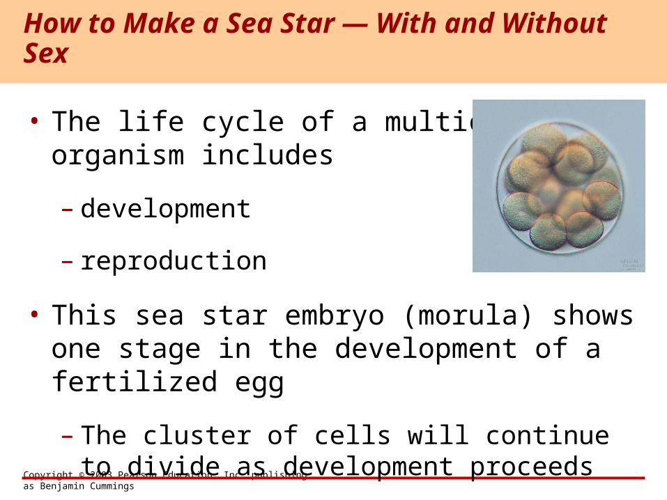
• The life cycle of a multicellular organism includes
– development
– reproduction
• This sea star embryo (morula) shows one stage in the development of a fertilized egg
– The cluster of cells will continue to divide as development proceeds
How to Make a Sea Star — With and Without Sex
Copyright © 2003 Pearson Education, Inc. publishing as Benjamin Cummings
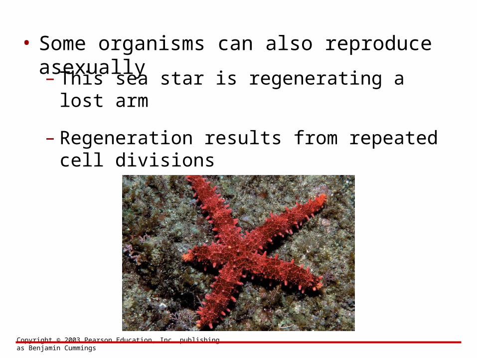
• Some organisms can also reproduce asexually– This sea star is regenerating a lost arm
– Regeneration results from repeated cell divisions
Copyright © 2003 Pearson Education, Inc. publishing as Benjamin Cummings
• Cell division is at the heart of the reproduction of cells and organisms
• Organisms can reproduce sexually or asexually
CONNECTIONS BETWEEN CELL DIVISION AND REPRODUCTION
Copyright © 2003 Pearson Education, Inc. publishing as Benjamin Cummings
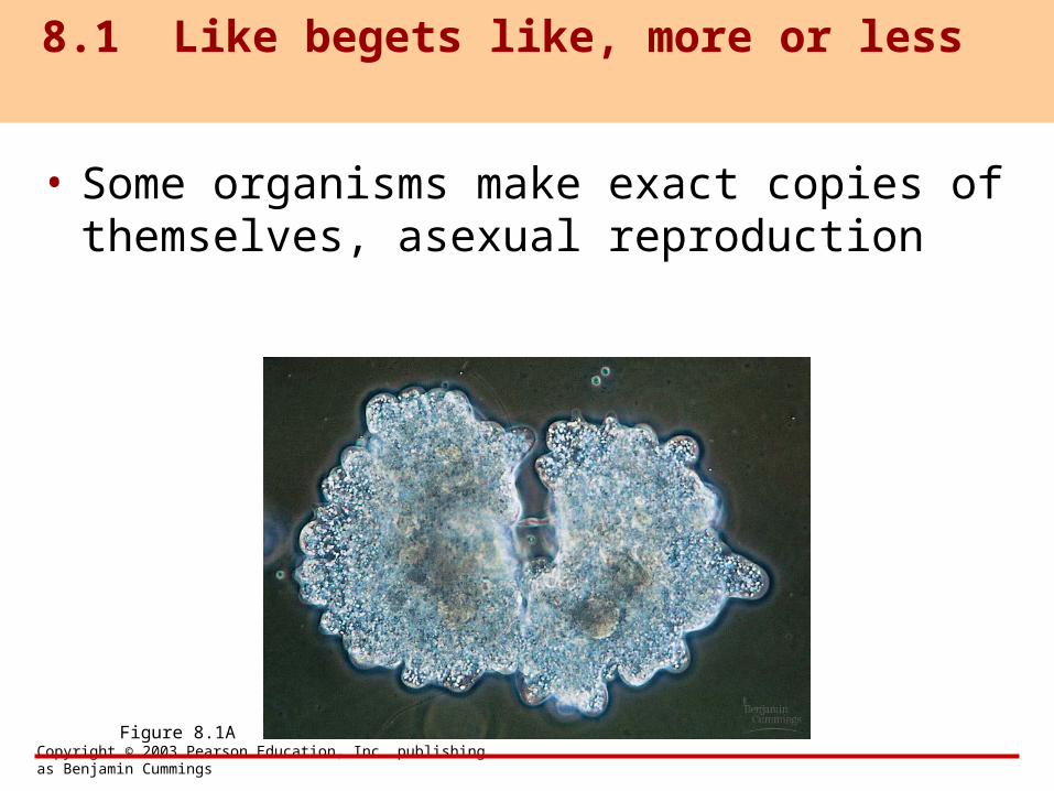
• Some organisms make exact copies of themselves, asexual reproduction
8.1 Like begets like, more or less
Figure 8.1A
Copyright © 2003 Pearson Education, Inc. publishing as Benjamin Cummings
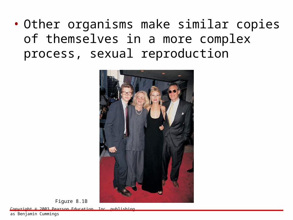
• Other organisms make similar copies of themselves in a more complex process, sexual reproduction
Figure 8.1B
Copyright © 2003 Pearson Education, Inc. publishing as Benjamin Cummings
• All cells come from cells
• Cellular reproduction is called cell division
– Cell division allows an embryo to develop into an adult
– It also ensures the continuity of life from one generation to the next
8.2 Cells arise only from preexisting cells
Copyright © 2003 Pearson Education, Inc. publishing as Benjamin Cummings
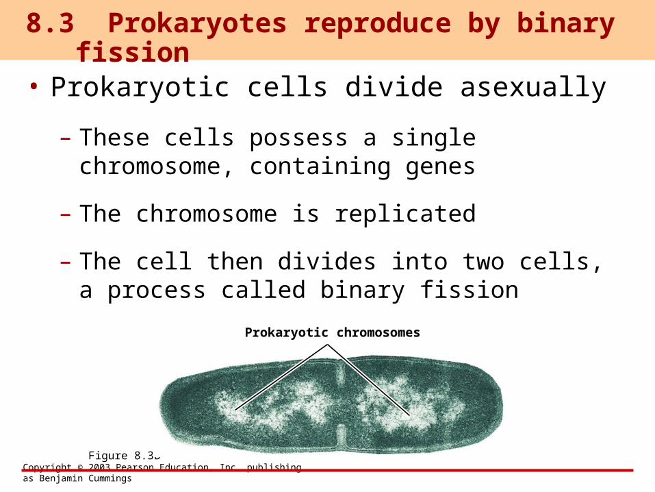
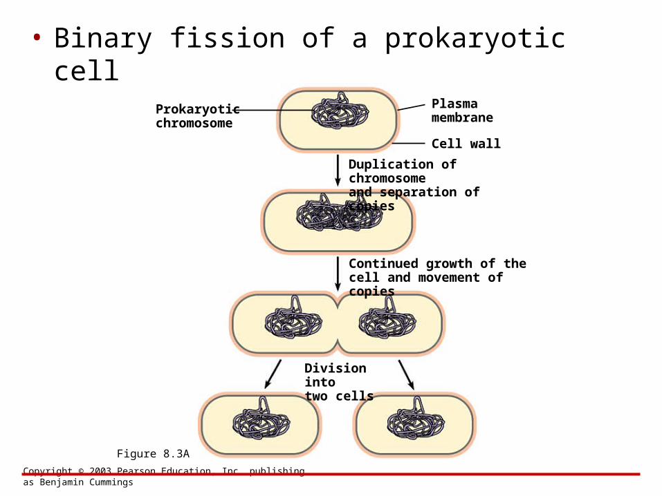
• Prokaryotic cells divide asexually
– These cells possess a single chromosome, containing genes
– The chromosome is replicated
– The cell then divides into two cells, a process called binary fission
8.3 Prokaryotes reproduce by binary fission
Figure 8.3B
Prokaryotic chromosomes
Copyright © 2003 Pearson Education, Inc. publishing as Benjamin Cummings
Figure 8.3A
• Binary fission of a prokaryotic cell
Prokaryoticchromosome
Plasmamembrane
Cell wall
Duplication of chromosomeand separation of copies
Continued growth of the cell and movement of copies
Division intotwo cells

BIOLOGYCONCEPTS & CONNECTIONS
Fourth Edition
Copyright © 2003 Pearson Education, Inc. publishing as Benjamin Cummings
Neil A. Campbell • Jane B. Reece • Lawrence G. Mitchell • Martha R. Taylor
From PowerPoint® Lectures for Biology: Concepts & Connections
CHAPTER 8The Cellular Basis of
Reproduction and Inheritance
Modules 8.4 – 8.11
Copyright © 2003 Pearson Education, Inc. publishing as Benjamin Cummings
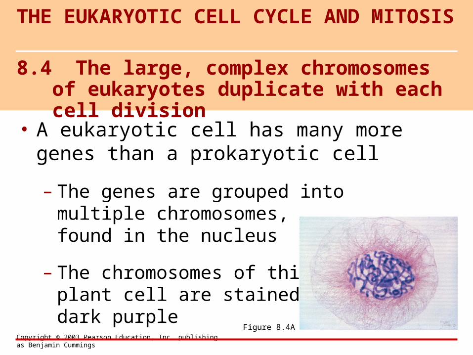
• A eukaryotic cell has many more genes than a prokaryotic cell
– The genes are grouped into multiple chromosomes, found in the nucleus
– The chromosomes of this plant cell are stained dark purple
8.4 The large, complex chromosomes of eukaryotes duplicate with each cell division
THE EUKARYOTIC CELL CYCLE AND MITOSIS
Figure 8.4A
Copyright © 2003 Pearson Education, Inc. publishing as Benjamin Cummings
• Chromosomes contain a very long DNA molecule with thousands of genes
– Individual chromosomes are only visibleduring cell division
– They are packaged as chromatin
Copyright © 2003 Pearson Education, Inc. publishing as Benjamin Cummings
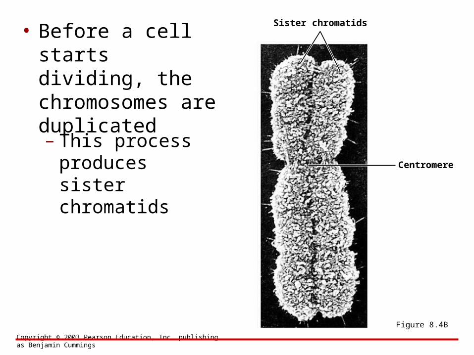
• Before a cell starts dividing, the chromosomes are duplicated
– This process produces sister chromatids
Centromere
Sister chromatids
Figure 8.4B
Copyright © 2003 Pearson Education, Inc. publishing as Benjamin Cummings
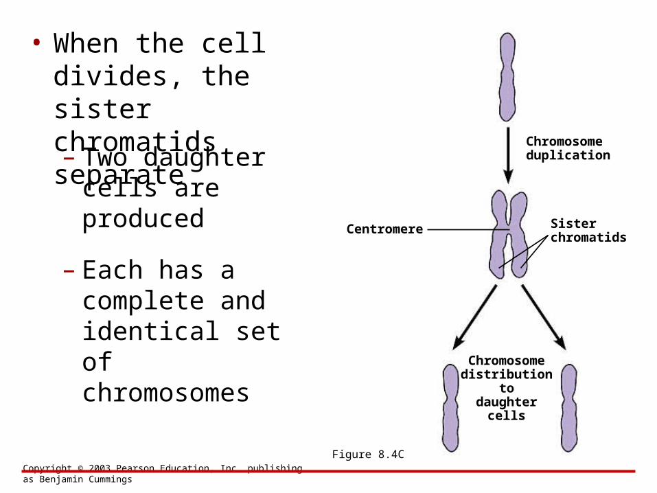
• When the cell divides, the sister chromatids separate – Two daughter
cells are produced
– Each has a complete and identical set of chromosomes
Centromere Sister chromatids
Figure 8.4C
Chromosomeduplication
Chromosomedistribution
todaughter
cells
Copyright © 2003 Pearson Education, Inc. publishing as Benjamin Cummings
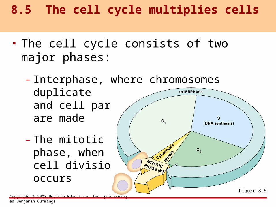
• The cell cycle consists of two major phases:
– Interphase, where chromosomes duplicate
and cell parts are made
– The mitotic phase, when cell division occurs
8.5 The cell cycle multiplies cells
Figure 8.5
Copyright © 2003 Pearson Education, Inc. publishing as Benjamin Cummings
• Eukaryotic cell division consists of two stages:
– Mitosis
– Cytokinesis
8.6 Cell division is a continuum of dynamic changes
Copyright © 2003 Pearson Education, Inc. publishing as Benjamin Cummings
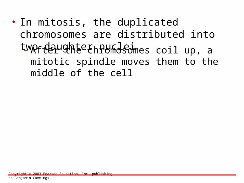
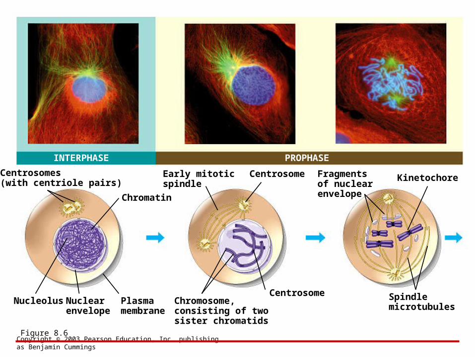
• In mitosis, the duplicated chromosomes are distributed into two daughter nuclei
– After the chromosomes coil up, a mitotic spindle moves them to the middle of the cell
Copyright © 2003 Pearson Education, Inc. publishing as Benjamin Cummings
INTERPHASE PROPHASE
Centrosomes(with centriole pairs)
Chromatin
Nucleolus Nuclearenvelope
Plasmamembrane
Early mitoticspindle
Centrosome
CentrosomeChromosome,consisting of twosister chromatids
Fragmentsof nuclearenvelope
Kinetochore
Spindlemicrotubules
Figure 8.6
Copyright © 2003 Pearson Education, Inc. publishing as Benjamin Cummings
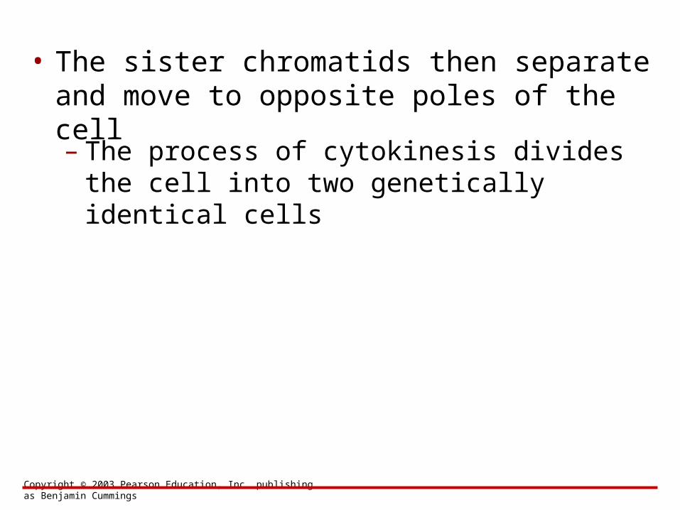
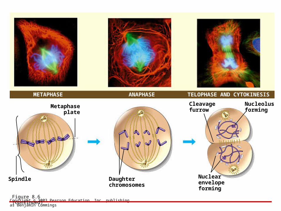
• The sister chromatids then separate and move to opposite poles of the cell
– The process of cytokinesis divides the cell into two genetically identical cells
Copyright © 2003 Pearson Education, Inc. publishing as Benjamin Cummings
METAPHASE TELOPHASE AND CYTOKINESIS
Metaphaseplate
Spindle Daughterchromosomes
Cleavagefurrow
Nucleolusforming
Nuclearenvelopeforming
ANAPHASE
Figure 8.6 (continued)
Copyright © 2003 Pearson Education, Inc. publishing as Benjamin Cummings
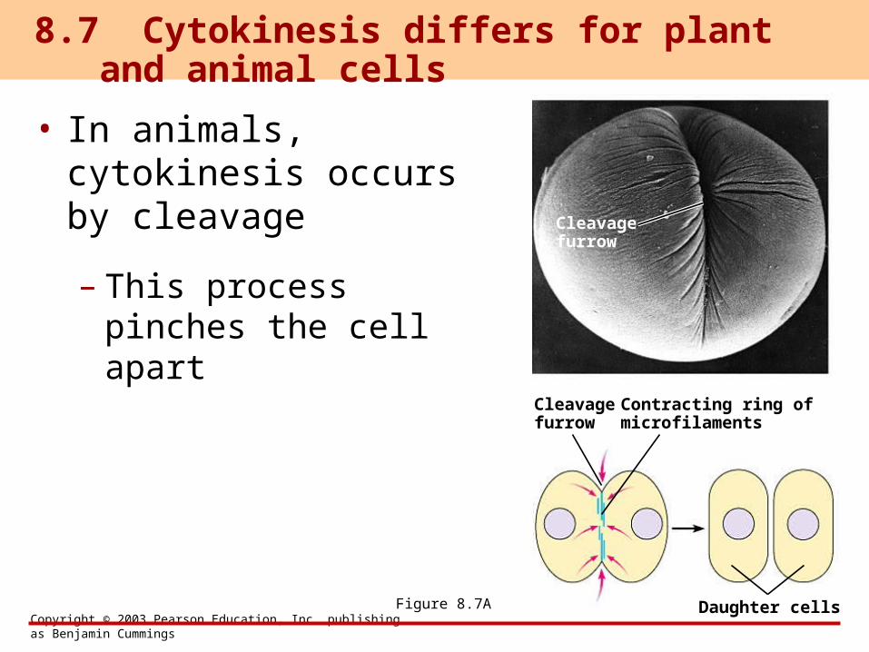
• In animals, cytokinesis occurs by cleavage
– This process pinches the cell apart
8.7 Cytokinesis differs for plant and animal cells
Figure 8.7A
Cleavagefurrow
Cleavagefurrow
Contracting ring ofmicrofilaments
Daughter cells
Copyright © 2003 Pearson Education, Inc. publishing as Benjamin Cummings
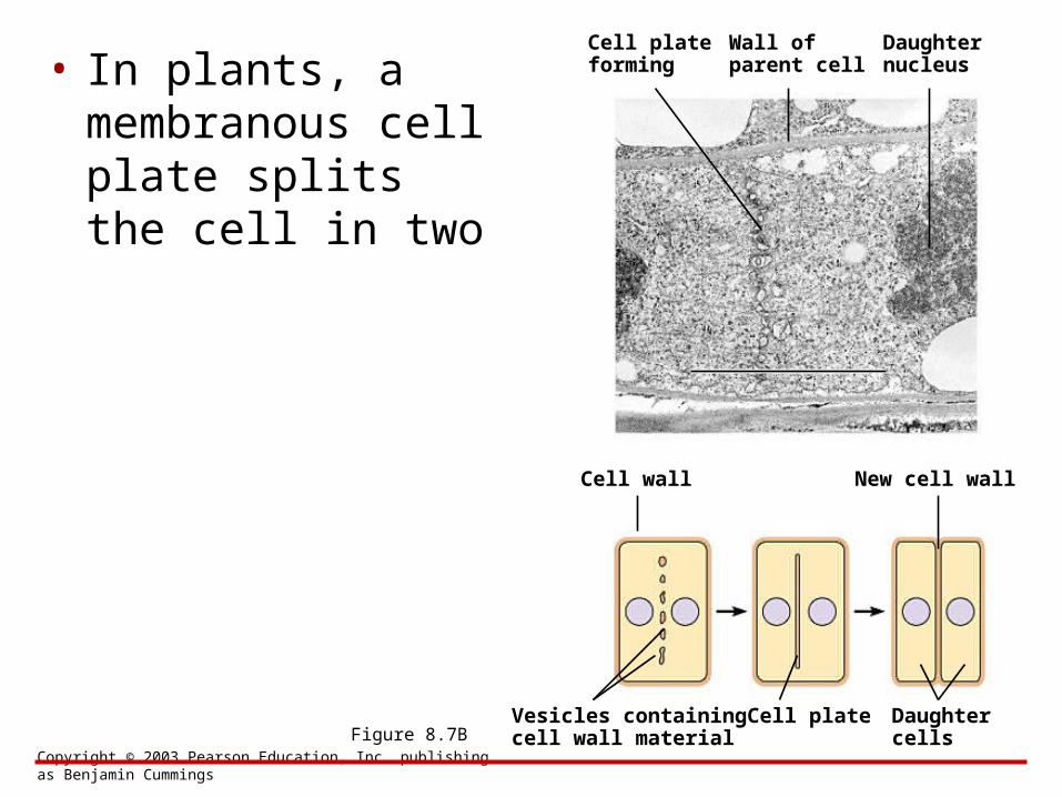
• In plants, a membranous cell plate splits the cell in two
Vesicles containingcell wall material
Cell plateforming
Figure 8.7BCell plate Daughter
cells
Wall ofparent cell
Daughternucleus
Cell wall New cell wall
Copyright © 2003 Pearson Education, Inc. publishing as Benjamin Cummings
• Most animal cells divide only when stimulated, and others not at all
• In laboratory cultures, most normal cells divide only when attached to a surface
– They are anchorage dependent
8.8 Anchorage, cell density, and chemical growth factors affect cell division
Copyright © 2003 Pearson Education, Inc. publishing as Benjamin Cummings
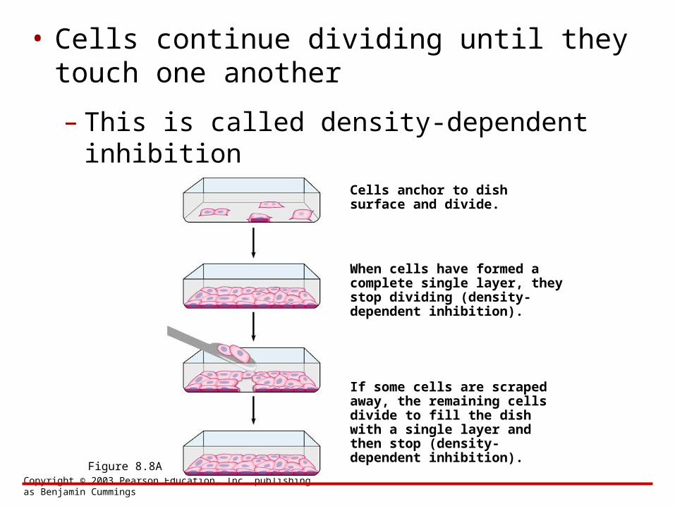
• Cells continue dividing until they touch one another
– This is called density-dependent inhibition
Cells anchor to dish surface and divide.
Figure 8.8A
When cells have formed a complete single layer, they stop dividing (density-dependent inhibition).
If some cells are scraped away, the remaining cells divide to fill the dish with a single layer and then stop (density-dependent inhibition).
Copyright © 2003 Pearson Education, Inc. publishing as Benjamin Cummings
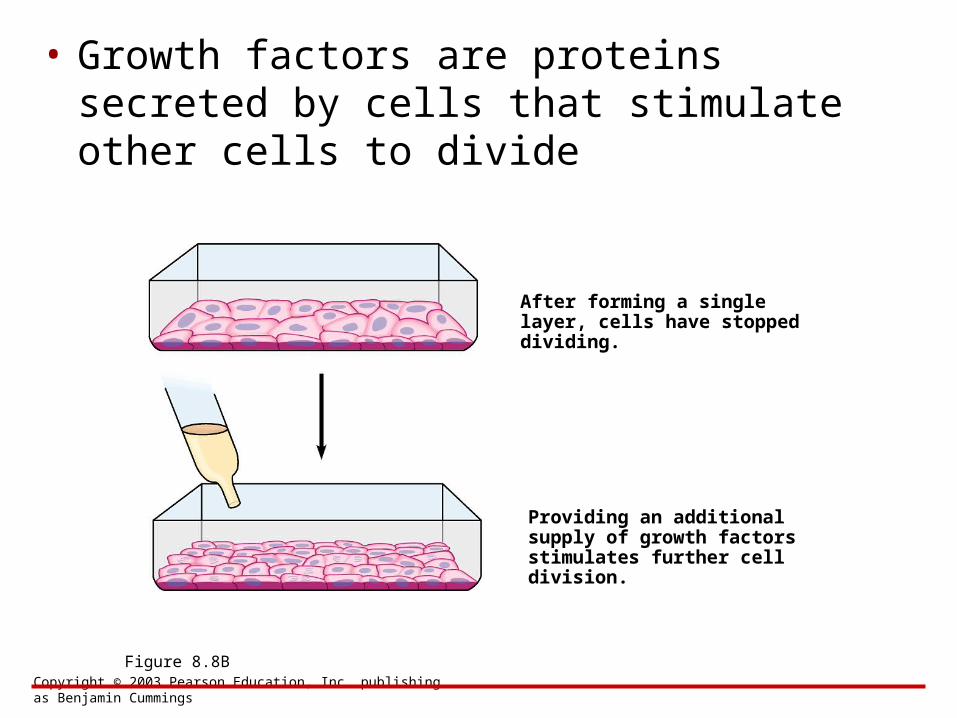
• Growth factors are proteins secreted by cells that stimulate other cells to divide
After forming a single layer, cells have stopped dividing.
Figure 8.8B
Providing an additional supply of growth factors stimulates further cell division.
Copyright © 2003 Pearson Education, Inc. publishing as Benjamin Cummings
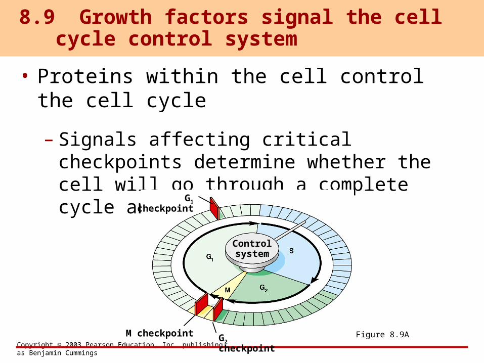
• Proteins within the cell control the cell cycle
– Signals affecting critical checkpoints determine whether the cell will go through a complete cycle and divide
8.9 Growth factors signal the cell cycle control system
G1 checkpoint
M checkpoint G2 checkpoint
Controlsystem
Figure 8.9A
Copyright © 2003 Pearson Education, Inc. publishing as Benjamin Cummings
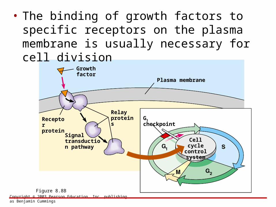
• The binding of growth factors to specific receptors on the plasma membrane is usually necessary for cell division
Growth factor
Figure 8.8B
Cell cyclecontrolsystem
Plasma membrane
Receptorprotein
Signal transduction pathway
G1 checkpointRelayproteins
Copyright © 2003 Pearson Education, Inc. publishing as Benjamin Cummings
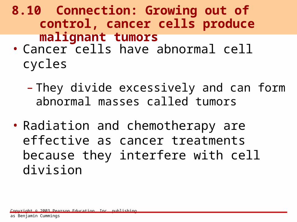
• Cancer cells have abnormal cell cycles
– They divide excessively and can form abnormal masses called tumors
• Radiation and chemotherapy are effective as cancer treatments because they interfere with cell division
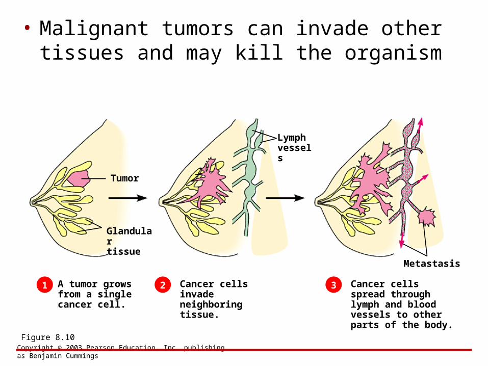
8.10 Connection: Growing out of control, cancer cells produce malignant tumors
Copyright © 2003 Pearson Education, Inc. publishing as Benjamin Cummings
• Malignant tumors can invade other tissues and may kill the organism
Tumor
Figure 8.10
Glandulartissue
1 2 3A tumor grows from a single cancer cell.
Cancer cells invade neighboring tissue.
Lymphvessels
Cancer cells spread through lymph and blood vessels to other parts of the body.
Metastasis
Copyright © 2003 Pearson Education, Inc. publishing as Benjamin Cummings
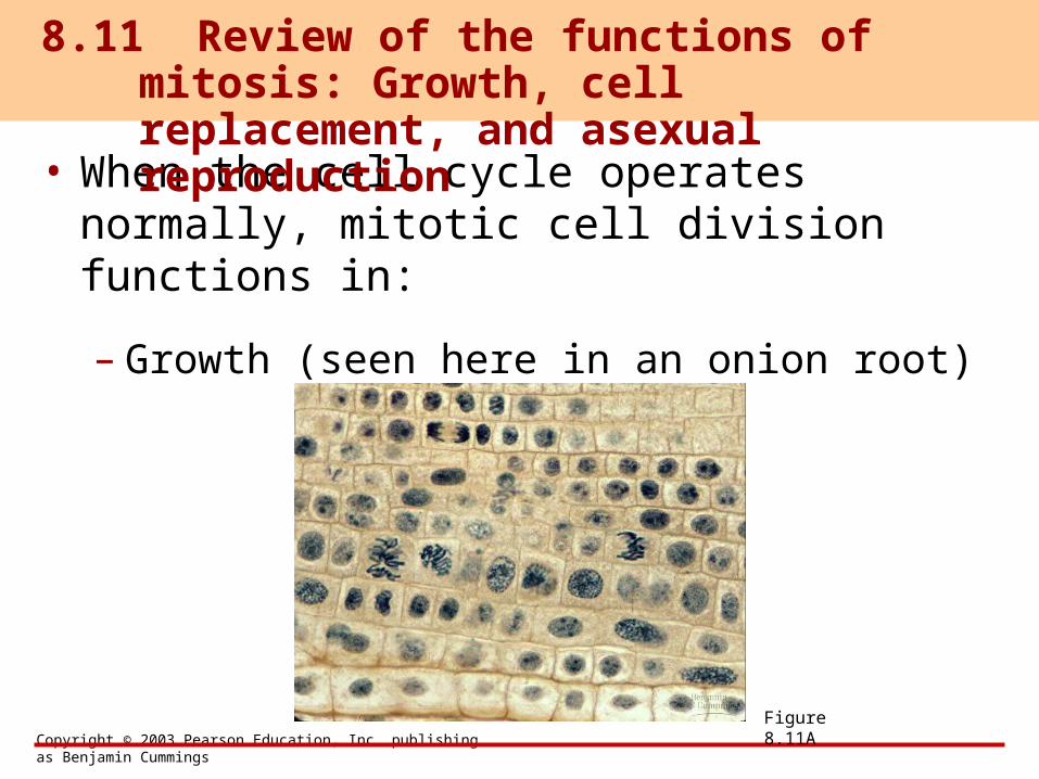
• When the cell cycle operates normally, mitotic cell division functions in:
– Growth (seen here in an onion root)
8.11 Review of the functions of mitosis: Growth, cell replacement, and asexual reproduction
Figure 8.11A
Copyright © 2003 Pearson Education, Inc. publishing as Benjamin Cummings
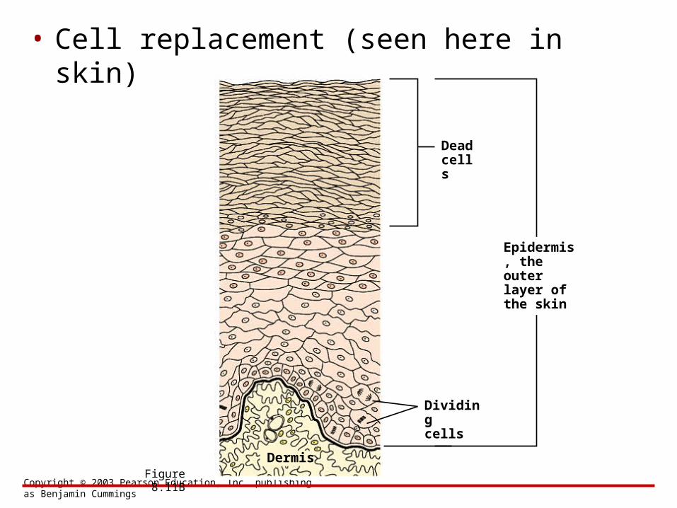
• Cell replacement (seen here in skin)
Deadcells
Figure 8.11B
Dividingcells
Epidermis, the outer layer of the skin
Dermis

Copyright © 2003 Pearson Education, Inc. publishing as Benjamin Cummings
• Asexual reproduction (seen here in a hydra)
Figure 8.11C

BIOLOGYCONCEPTS & CONNECTIONS
Fourth Edition
Copyright © 2003 Pearson Education, Inc. publishing as Benjamin Cummings
Neil A. Campbell • Jane B. Reece • Lawrence G. Mitchell • Martha R. Taylor
From PowerPoint® Lectures for Biology: Concepts & Connections
CHAPTER 8The Cellular Basis of
Reproduction and Inheritance
Modules 8.12 – 8.18
Copyright © 2003 Pearson Education, Inc. publishing as Benjamin Cummings
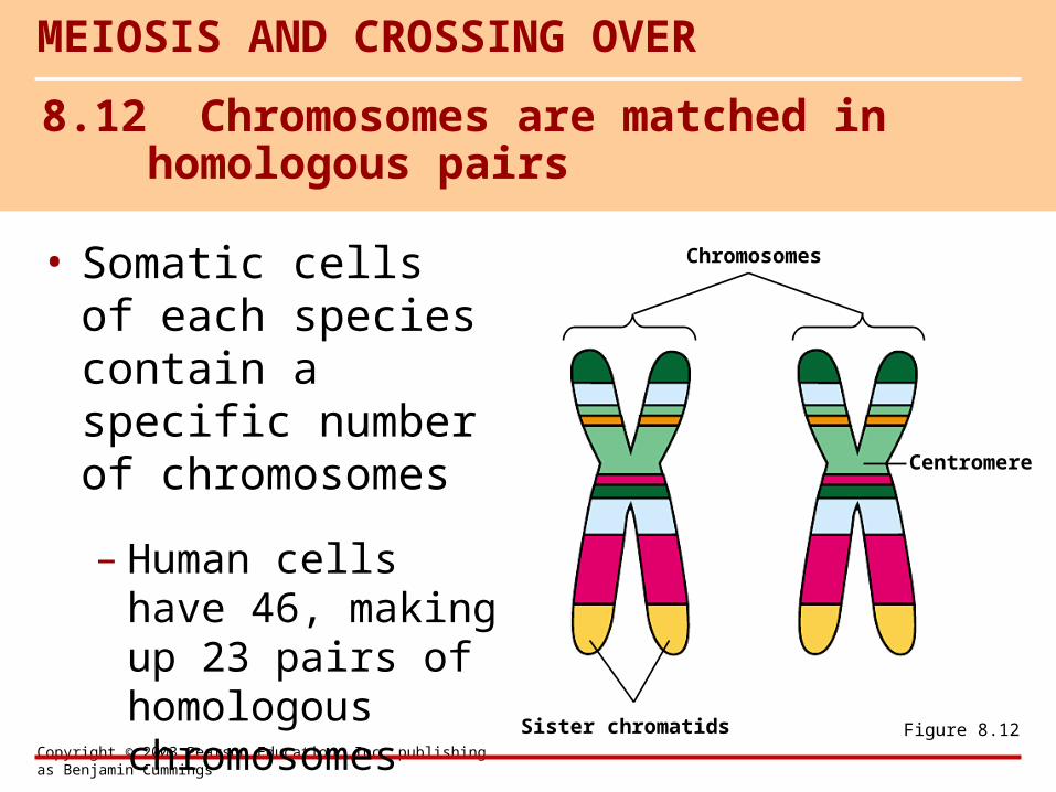
• Somatic cells of each species contain a specific number of chromosomes
– Human cells have 46, making up 23 pairs of homologous chromosomes
MEIOSIS AND CROSSING OVER
8.12 Chromosomes are matched in homologous pairs
Chromosomes
Centromere
Sister chromatids Figure 8.12
Copyright © 2003 Pearson Education, Inc. publishing as Benjamin Cummings
• Cells with two sets of chromosomes are said to be diploid
• Gametes are haploid, with only one set of chromosomes
8.13 Gametes have a single set of chromosomes
Copyright © 2003 Pearson Education, Inc. publishing as Benjamin Cummings
• At fertilization, a sperm fuses with an egg, forming a diploid zygote
– Repeated mitotic divisions lead to the development of a mature adult
– The adult makes haploid gametes by meiosis
– All of these processes make up the sexual life cycle of organisms
Copyright © 2003 Pearson Education, Inc. publishing as Benjamin Cummings
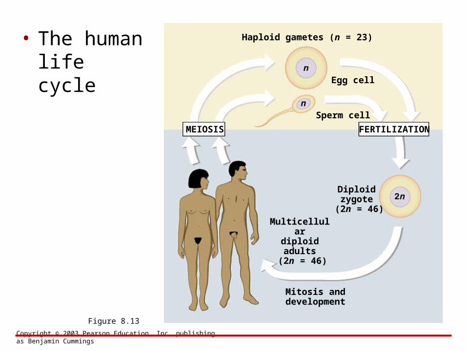
• The human life cycle
Figure 8.13
MEIOSIS FERTILIZATION
Haploid gametes (n = 23)
Egg cell
Sperm cell
Diploidzygote
(2n = 46)Multicellular
diploid adults (2n = 46)
Mitosis anddevelopment
Copyright © 2003 Pearson Education, Inc. publishing as Benjamin Cummings
• Meiosis, like mitosis, is preceded by chromosome duplication
– However, in meiosis the cell divides twice to form four daughter cells
8.14 Meiosis reduces the chromosome number from diploid to haploid
Copyright © 2003 Pearson Education, Inc. publishing as Benjamin Cummings
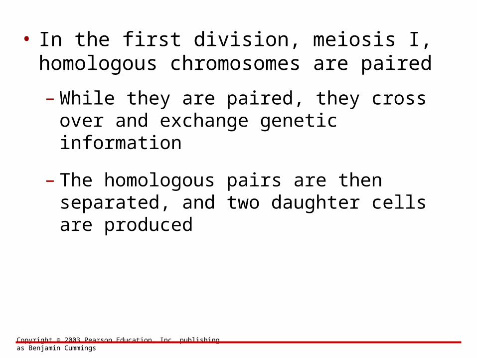
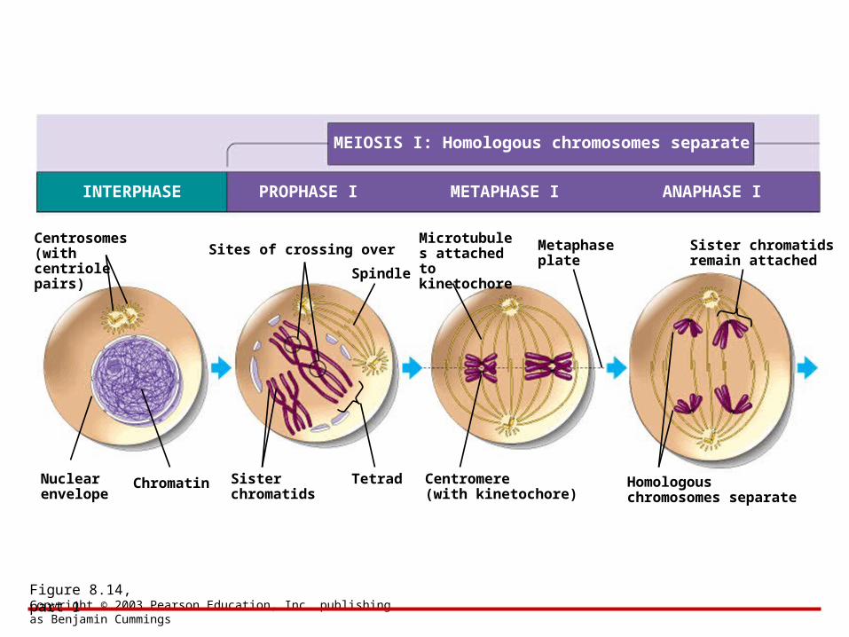
• In the first division, meiosis I, homologous chromosomes are paired
– While they are paired, they cross over and exchange genetic information
– The homologous pairs are then separated, and two daughter cells are produced
Copyright © 2003 Pearson Education, Inc. publishing as Benjamin Cummings
Figure 8.14, part 1
MEIOSIS I: Homologous chromosomes separate
INTERPHASE PROPHASE I METAPHASE I ANAPHASE I
Centrosomes(withcentriolepairs)
Nuclearenvelope
Chromatin
Sites of crossing over
Spindle
Sisterchromatids
Tetrad
Microtubules attached tokinetochore
Metaphaseplate
Centromere(with kinetochore)
Sister chromatidsremain attached
Homologouschromosomes separate
Copyright © 2003 Pearson Education, Inc. publishing as Benjamin Cummings
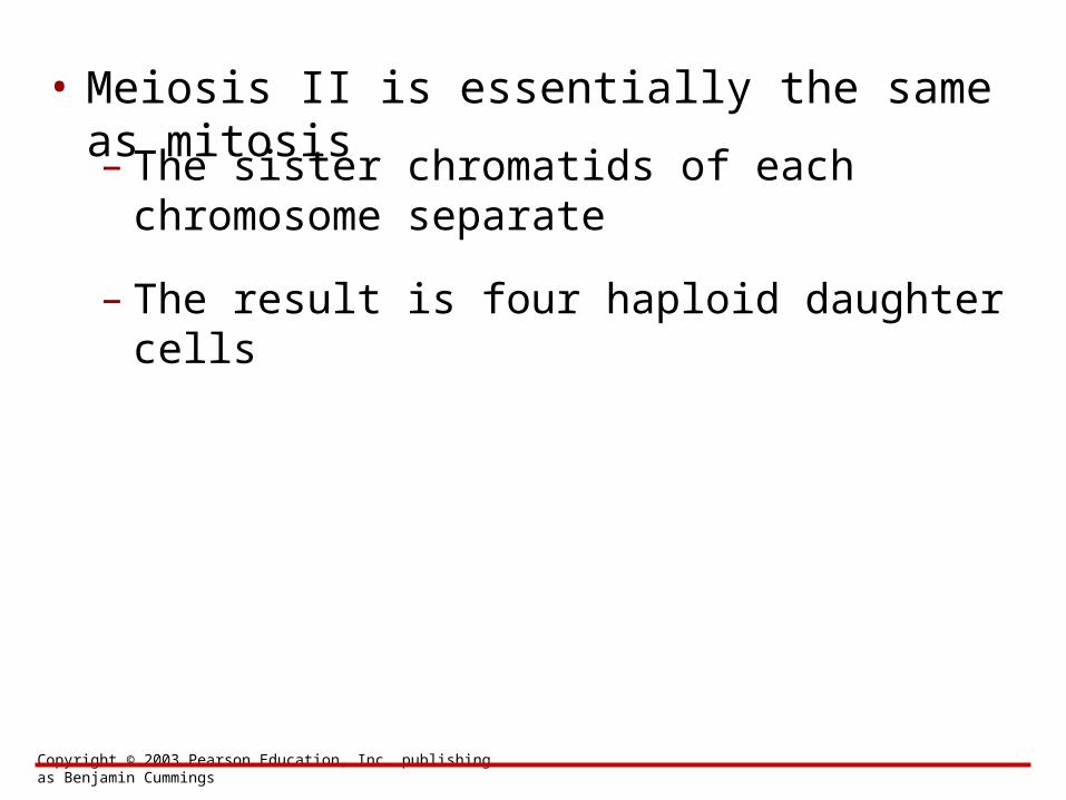
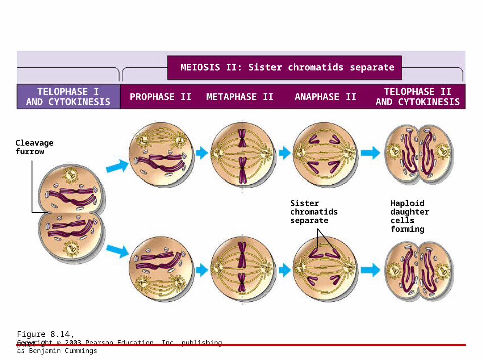
• Meiosis II is essentially the same as mitosis– The sister chromatids of each
chromosome separate
– The result is four haploid daughter cells
Copyright © 2003 Pearson Education, Inc. publishing as Benjamin Cummings
Figure 8.14, part 2
MEIOSIS II: Sister chromatids separate
TELOPHASE IAND CYTOKINESIS PROPHASE II METAPHASE II ANAPHASE II
Cleavagefurrow
Sister chromatidsseparate
TELOPHASE IIAND CYTOKINESIS
Haploiddaughter cellsforming
Copyright © 2003 Pearson Education, Inc. publishing as Benjamin Cummings
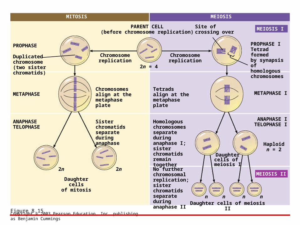
• For both processes, chromosomes replicate only once, during interphase
8.15 Review: A comparison of mitosis and meiosis
Copyright © 2003 Pearson Education, Inc. publishing as Benjamin Cummings
Figure 8.15
MITOSIS MEIOSIS
PARENT CELL(before chromosome replication)
Site ofcrossing over
MEIOSIS I
PROPHASE ITetrad formedby synapsis of homologous chromosomes
PROPHASE
Duplicatedchromosome(two sister chromatids)
METAPHASE
Chromosomereplication
Chromosomereplication
2n = 4
ANAPHASETELOPHASE
Chromosomes align at the metaphase plate
Tetradsalign at themetaphase plate
METAPHASE I
ANAPHASE ITELOPHASE I
Sister chromatidsseparate duringanaphase
Homologouschromosomesseparateduringanaphase I;sisterchromatids remain together
No further chromosomal replication; sister chromatids separate during anaphase II
2n 2n
Daughter cellsof mitosis
Daughter cells of meiosis II
MEIOSIS II
Daughtercells of
meiosis I
Haploidn = 2
n n n n
Copyright © 2003 Pearson Education, Inc. publishing as Benjamin Cummings
• Each chromosome of a homologous pair comes from a different parent
– Each chromosome thus differs at many points from the other member of the pair
8.16 Independent orientation of chromosomes in meiosis and random fertilization lead to varied offspring
Copyright © 2003 Pearson Education, Inc. publishing as Benjamin Cummings
• The large number of possible arrangements of chromosome pairs at metaphase I of meiosis leads to many different combinations of chromosomes in gametes• Random fertilization also increases variation in offspring
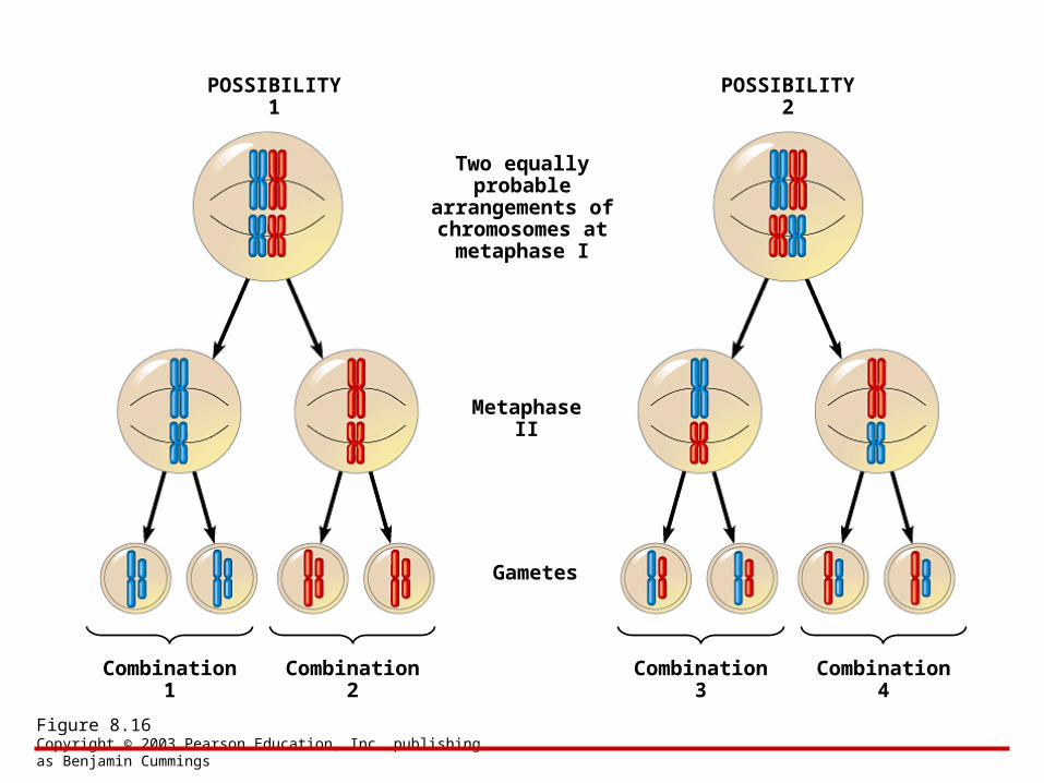
Copyright © 2003 Pearson Education, Inc. publishing as Benjamin Cummings
Figure 8.16
POSSIBILITY 1 POSSIBILITY 2
Two equally probable
arrangements of chromosomes at
metaphase I
Metaphase II
Gametes
Combination 1 Combination 2 Combination 3 Combination 4
Copyright © 2003 Pearson Education, Inc. publishing as Benjamin Cummings
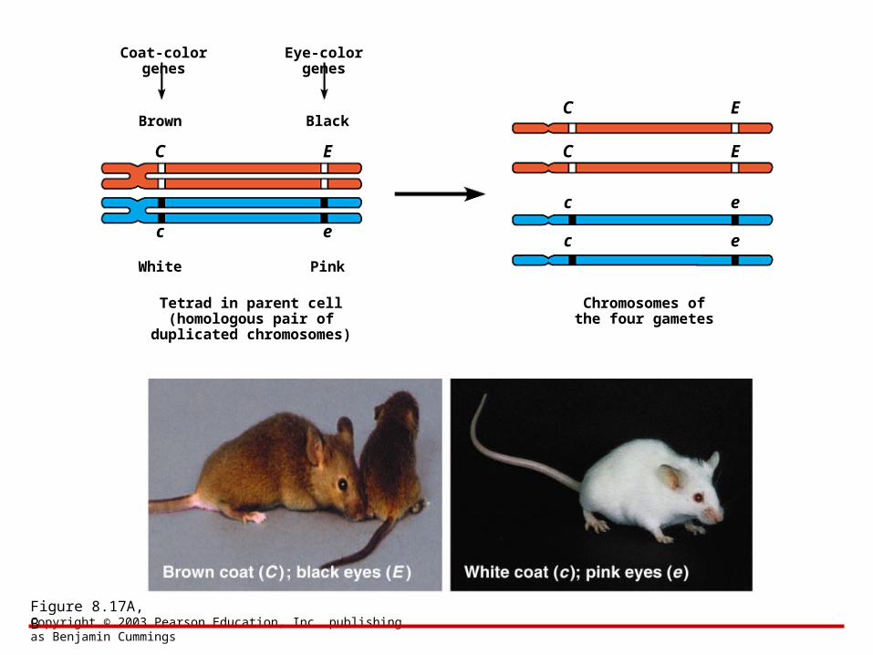
• The differences between homologous chromosomes are based on the fact that they can carry different versions of a gene at corresponding loci
8.17 Homologous chromosomes carry different versions of genes
Copyright © 2003 Pearson Education, Inc. publishing as Benjamin Cummings
Figure 8.17A, B
Coat-color genes Eye-color genes
Brown Black
C E
c e
White Pink
C E
c e
C E
c e
Tetrad in parent cell(homologous pair of
duplicated chromosomes)
Chromosomes ofthe four gametes
Copyright © 2003 Pearson Education, Inc. publishing as Benjamin Cummings
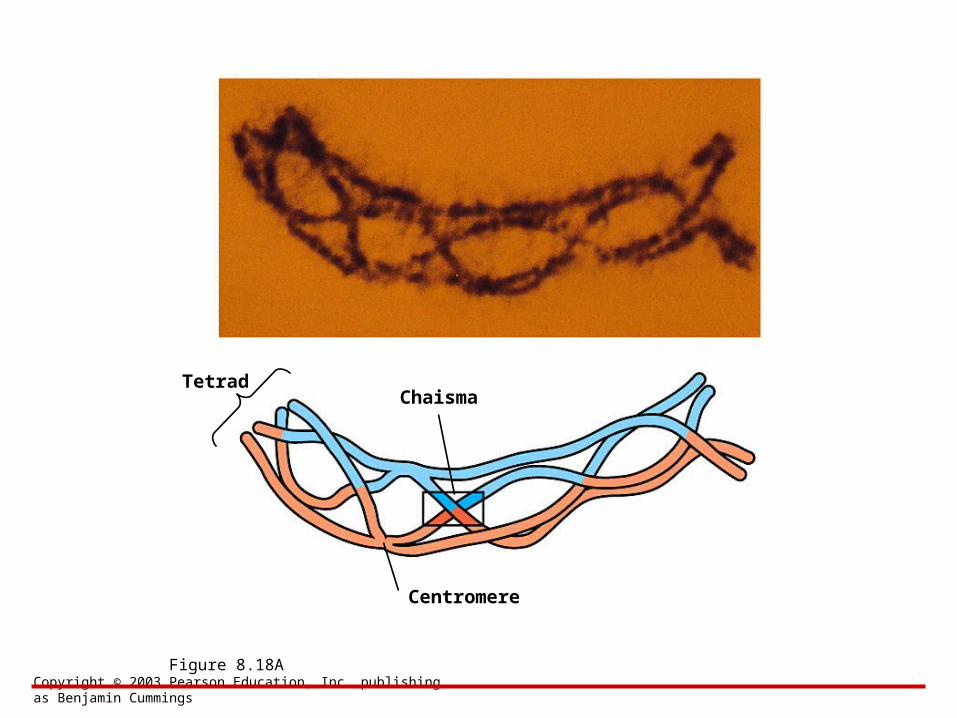
• Crossing over is the exchange of corresponding segments between two homologous chromosomes
• Genetic recombination results from crossing over during prophase I of meiosis
– This increases variation further
8.18 Crossing over further increases genetic variability
Copyright © 2003 Pearson Education, Inc. publishing as Benjamin Cummings
Figure 8.18A
TetradChaisma
Centromere
Copyright © 2003 Pearson Education, Inc. publishing as Benjamin Cummings
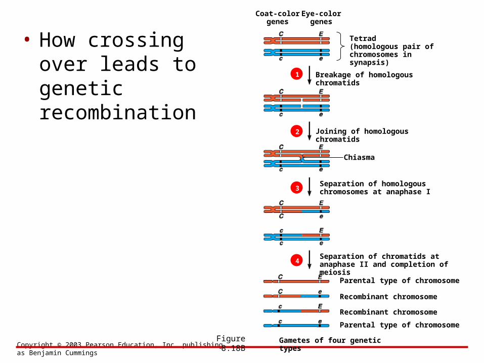
• How crossing over leads to genetic recombination
Figure 8.18B
Tetrad(homologous pair ofchromosomes in synapsis)
Breakage of homologous chromatids
Joining of homologous chromatids
Chiasma
Separation of homologouschromosomes at anaphase I
Separation of chromatids atanaphase II and completion of meiosis
Parental type of chromosome
Recombinant chromosome
Recombinant chromosome
Parental type of chromosome
Gametes of four genetic types
1
2
3
4
Coat-colorgenes
Eye-colorgenes
BIOLOGYCONCEPTS & CONNECTIONS
Fourth Edition
Copyright © 2003 Pearson Education, Inc. publishing as Benjamin Cummings
Neil A. Campbell • Jane B. Reece • Lawrence G. Mitchell • Martha R. Taylor
From PowerPoint® Lectures for Biology: Concepts & Connections
CHAPTER 8The Cellular Basis of
Reproduction and Inheritance
Modules 8.19 – 8.23
Copyright © 2003 Pearson Education, Inc. publishing as Benjamin Cummings
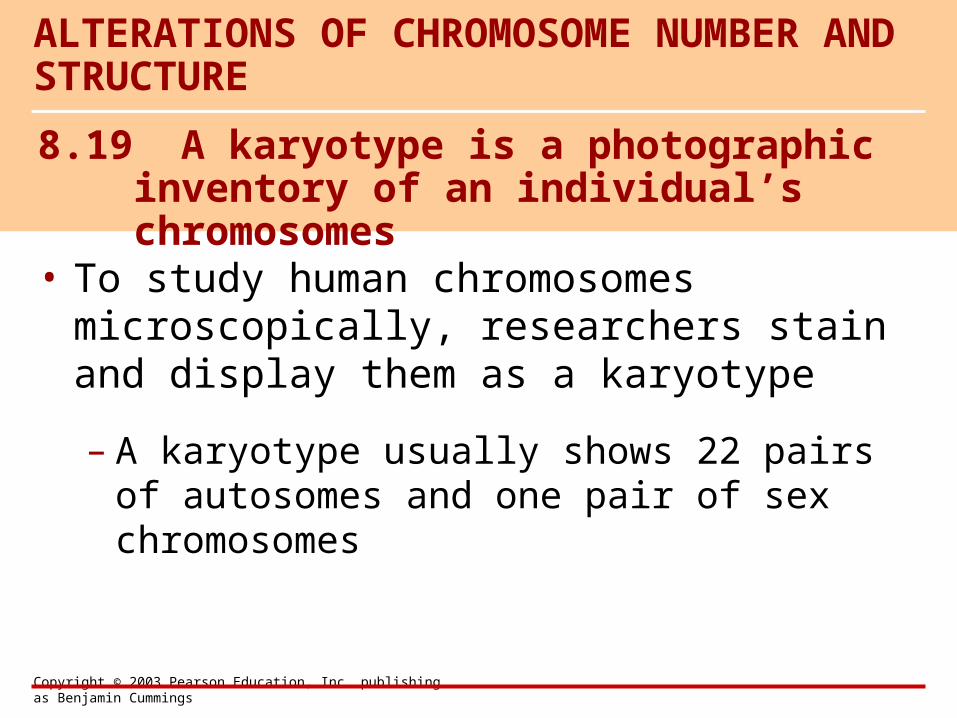
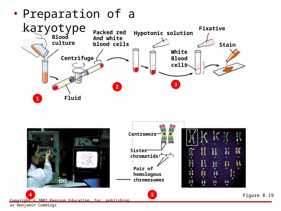
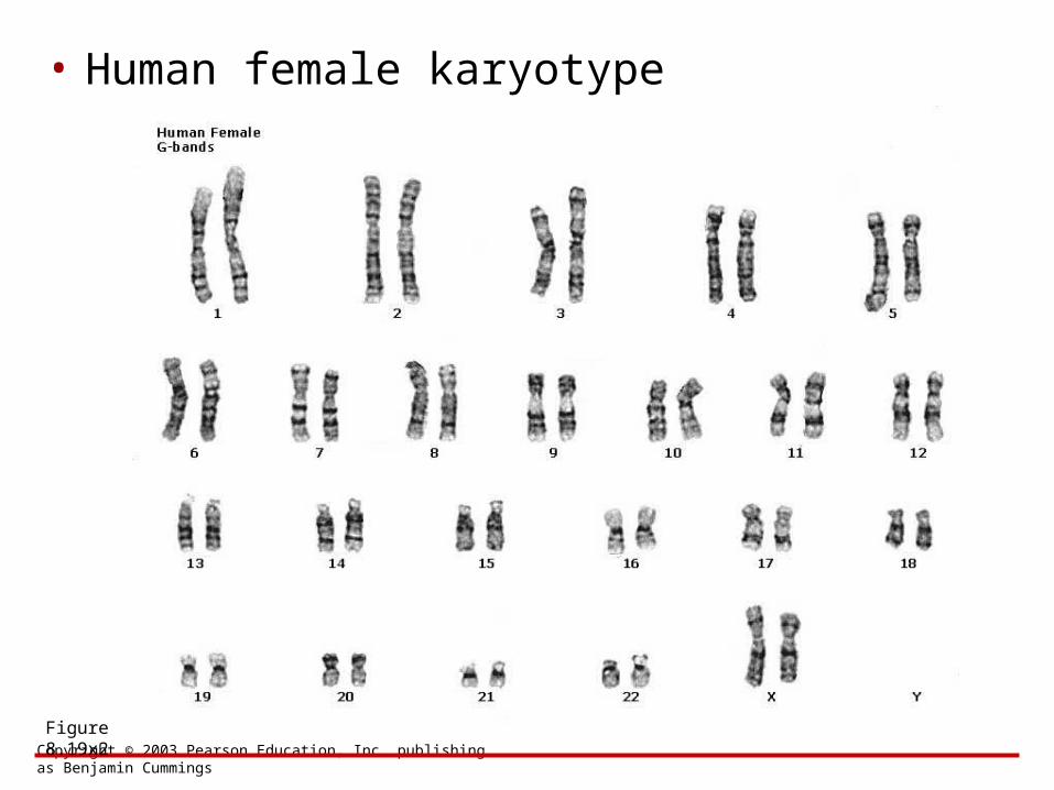
• To study human chromosomes microscopically, researchers stain and display them as a karyotype
– A karyotype usually shows 22 pairs of autosomes and one pair of sex chromosomes
ALTERATIONS OF CHROMOSOME NUMBER AND STRUCTURE
8.19 A karyotype is a photographic inventory of an individual’s chromosomes
Copyright © 2003 Pearson Education, Inc. publishing as Benjamin Cummings
• Preparation of a karyotype
Figure 8.19
Blood culture
1
Centrifuge
Packed redAnd white blood cells
Fluid
2
Hypotonic solution
3
Fixative
WhiteBloodcells
Stain
4 5
Centromere
Sisterchromatids
Pair of homologouschromosomes
Copyright © 2003 Pearson Education, Inc. publishing as Benjamin Cummings
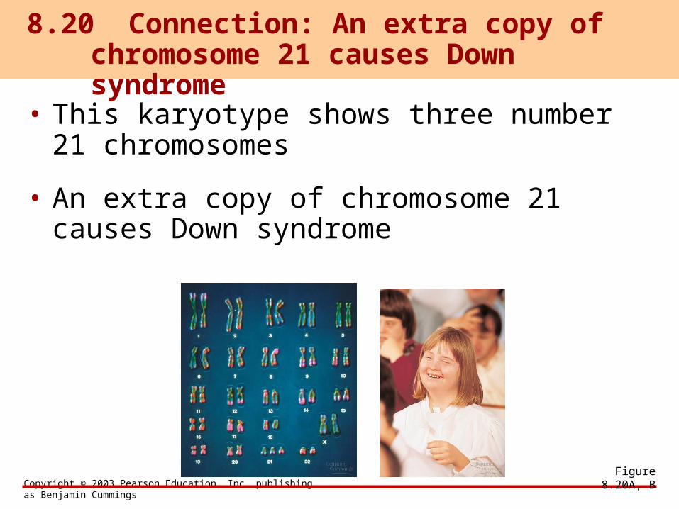
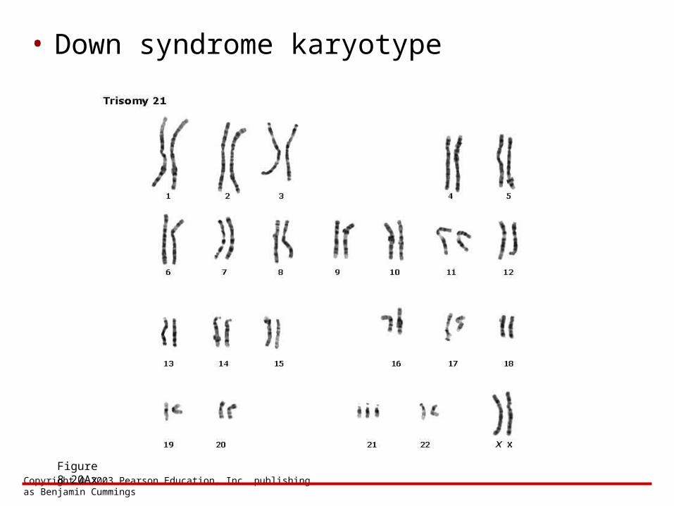
• This karyotype shows three number 21 chromosomes
• An extra copy of chromosome 21 causes Down syndrome
8.20 Connection: An extra copy of chromosome 21 causes Down syndrome
Figure 8.20A, B
Copyright © 2003 Pearson Education, Inc. publishing as Benjamin Cummings
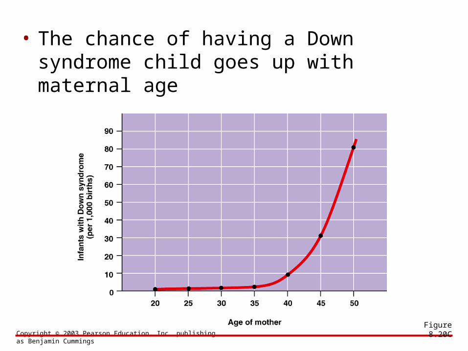
• The chance of having a Down syndrome child goes up with maternal age
Figure 8.20C
Copyright © 2003 Pearson Education, Inc. publishing as Benjamin Cummings
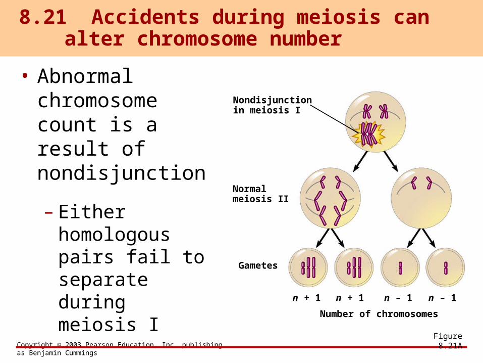
• Abnormal chromosome count is a result of nondisjunction
– Either homologous pairs fail to separate during meiosis I
8.21 Accidents during meiosis can alter chromosome number
Figure 8.21A
Nondisjunctionin meiosis I
Normalmeiosis II
Gametes
n + 1 n + 1 n – 1 n – 1
Number of chromosomes
Copyright © 2003 Pearson Education, Inc. publishing as Benjamin Cummings
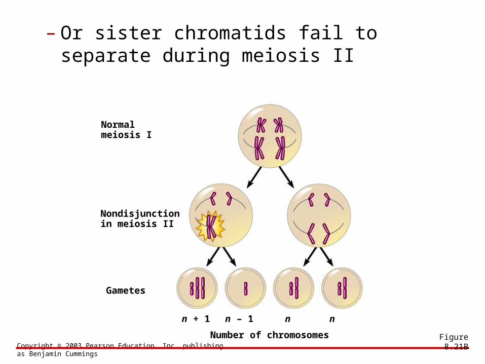
– Or sister chromatids fail to separate during meiosis II
Figure 8.21B
Normalmeiosis I
Nondisjunctionin meiosis II
Gametes
n + 1 n – 1 n n
Number of chromosomes
Copyright © 2003 Pearson Education, Inc. publishing as Benjamin Cummings
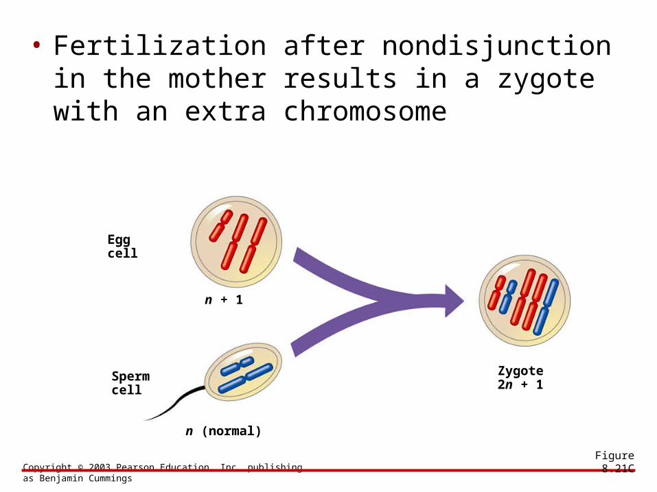
• Fertilization after nondisjunction in the mother results in a zygote with an extra chromosome
Figure 8.21C
Eggcell
Spermcell
n + 1
n (normal)
Zygote2n + 1
Copyright © 2003 Pearson Education, Inc. publishing as Benjamin Cummings
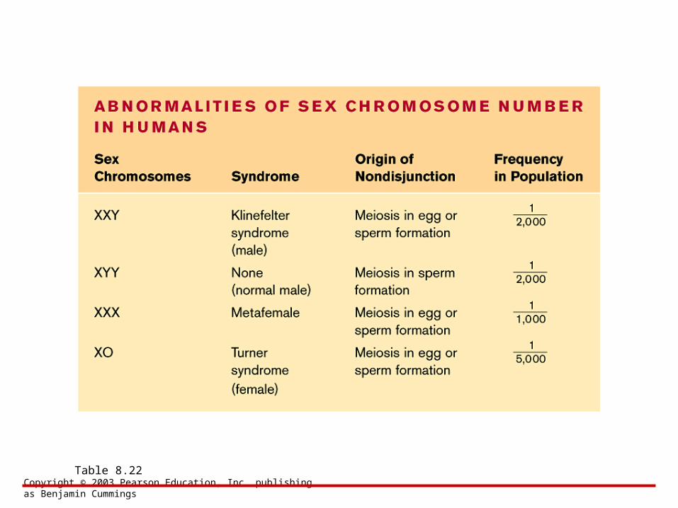
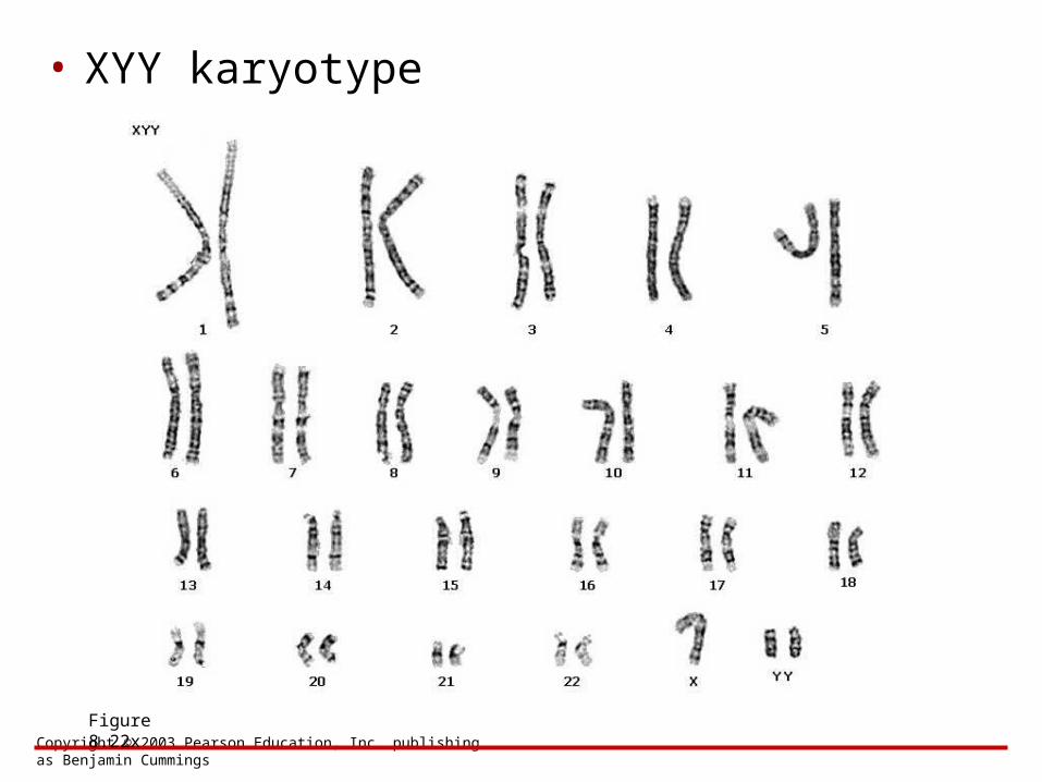
• Nondisjunction can also produce gametes with extra or missing sex chromosomes
– Unusual numbers of sex chromosomes upset the genetic balance less than an unusual number of autosomes
8.22 Connection: Abnormal numbers of sex chromosomes do not usually affect survival
Copyright © 2003 Pearson Education, Inc. publishing as Benjamin Cummings
Table 8.22
Copyright © 2003 Pearson Education, Inc. publishing as Benjamin Cummings
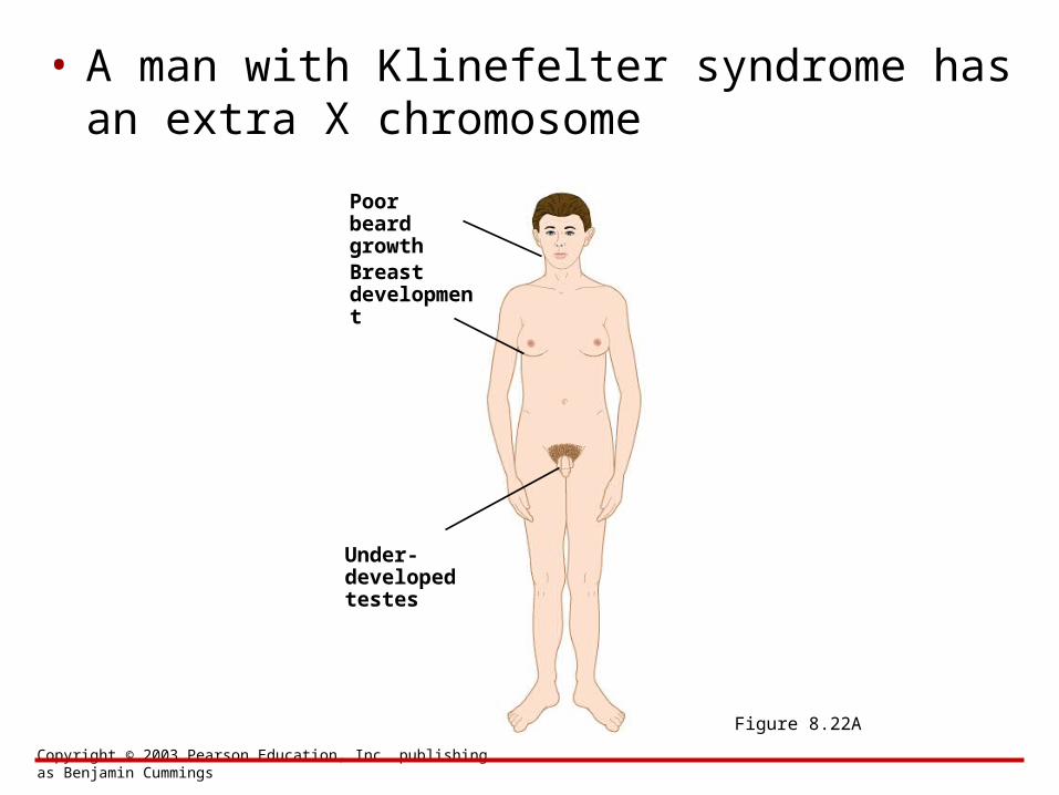
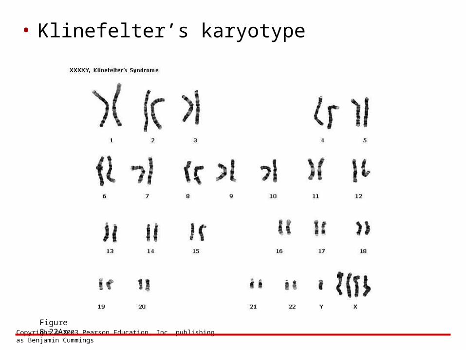
• A man with Klinefelter syndrome has an extra X chromosome
Figure 8.22A
Poor beardgrowth
Under-developedtestes
Breastdevelopment
Copyright © 2003 Pearson Education, Inc. publishing as Benjamin Cummings
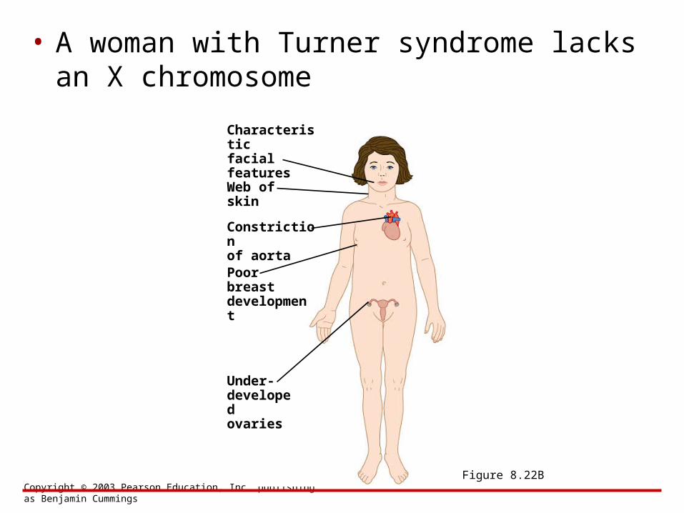
• A woman with Turner syndrome lacks an X chromosome
Figure 8.22B
Characteristicfacialfeatures
Web ofskin
Constrictionof aorta
Poorbreastdevelopment
Under-developedovaries
Copyright © 2003 Pearson Education, Inc. publishing as Benjamin Cummings
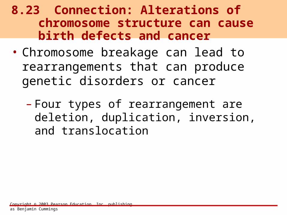
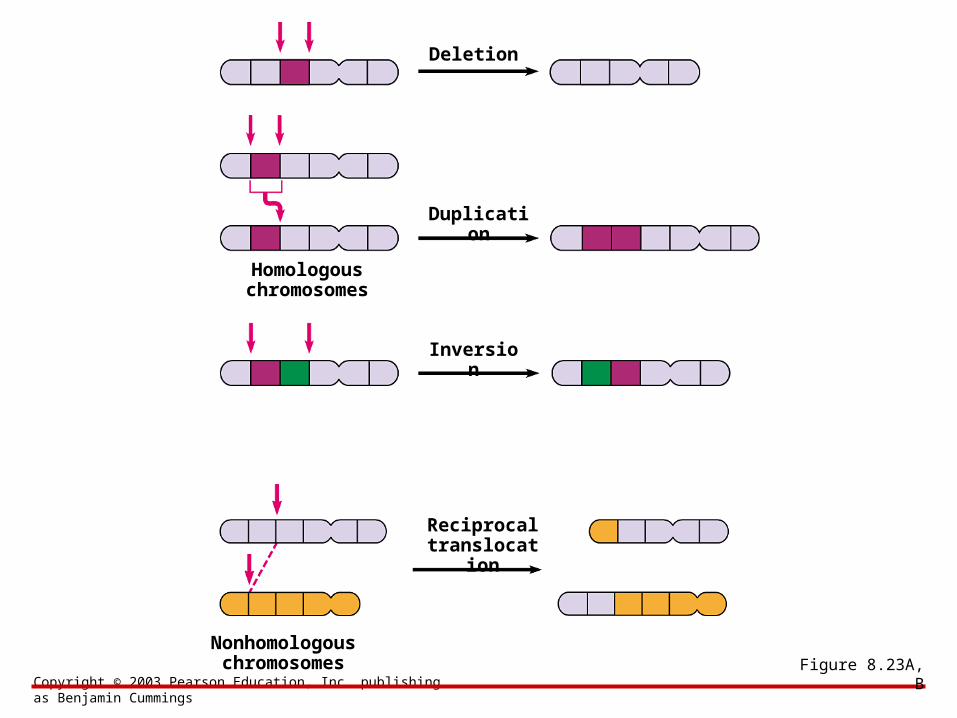
• Chromosome breakage can lead to rearrangements that can produce genetic disorders or cancer
– Four types of rearrangement are deletion, duplication, inversion, and translocation
8.23 Connection: Alterations of chromosome structure can cause birth defects and cancer
Copyright © 2003 Pearson Education, Inc. publishing as Benjamin Cummings
Figure 8.23A, B
Deletion
Duplication
Inversion
Homologouschromosomes
Reciprocaltranslocatio
n
Nonhomologouschromosomes
Copyright © 2003 Pearson Education, Inc. publishing as Benjamin Cummings
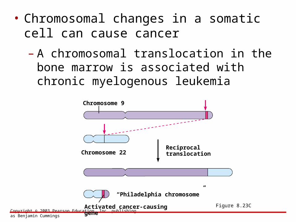
• Chromosomal changes in a somatic cell can cause cancer
Figure 8.23C
Chromosome 9
– A chromosomal translocation in the bone marrow is associated with chronic myelogenous leukemia
Chromosome 22Reciprocaltranslocation
“Philadelphia chromosome”
Activated cancer-causing gene
BIOLOGYCONCEPTS & CONNECTIONS
Fourth Edition
Copyright © 2003 Pearson Education, Inc. publishing as Benjamin Cummings
Neil A. Campbell • Jane B. Reece • Lawrence G. Mitchell • Martha R. Taylor
From PowerPoint® Lectures for Biology: Concepts & Connections
CHAPTER 8Extra Photographs
Copyright © 2003 Pearson Education, Inc. publishing as Benjamin Cummings
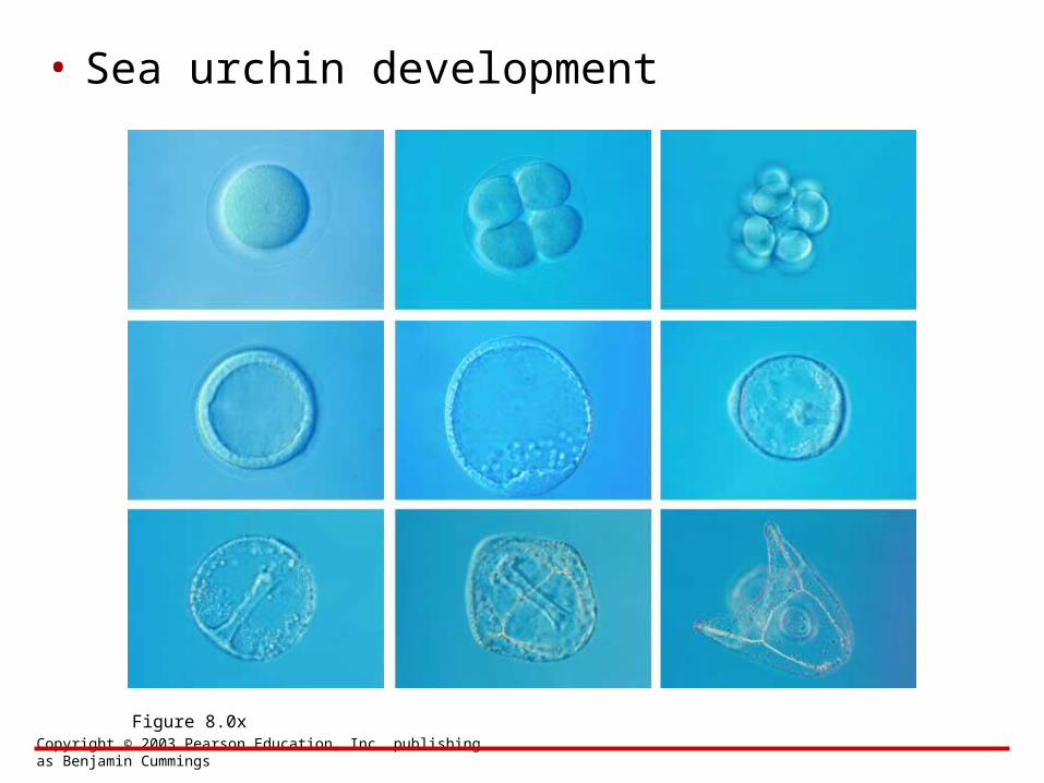
• Sea urchin development
Figure 8.0x
Copyright © 2003 Pearson Education, Inc. publishing as Benjamin Cummings
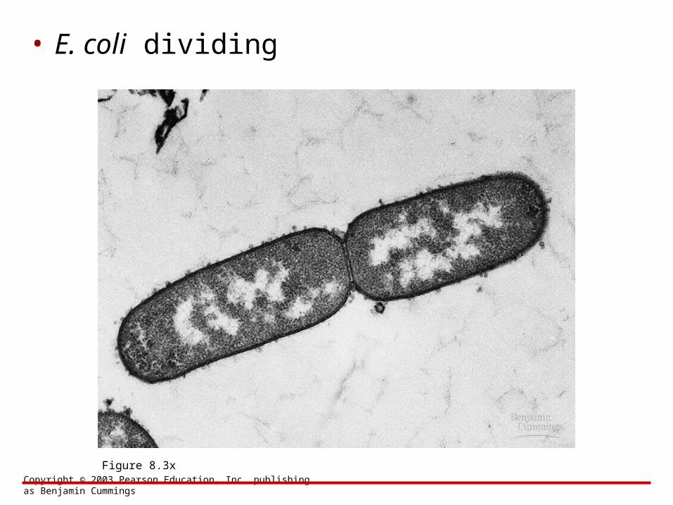
• E. coli dividing
Figure 8.3x
Copyright © 2003 Pearson Education, Inc. publishing as Benjamin Cummings
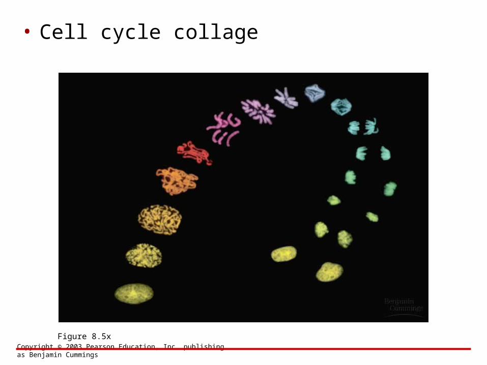
• Cell cycle collage
Figure 8.5x
Copyright © 2003 Pearson Education, Inc. publishing as Benjamin Cummings
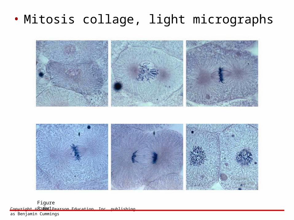
• Mitosis collage, light micrographs
Figure 8.6x1
Copyright © 2003 Pearson Education, Inc. publishing as Benjamin Cummings
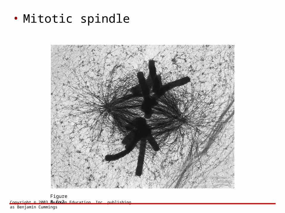
• Mitotic spindle
Figure 8.6x2
Copyright © 2003 Pearson Education, Inc. publishing as Benjamin Cummings
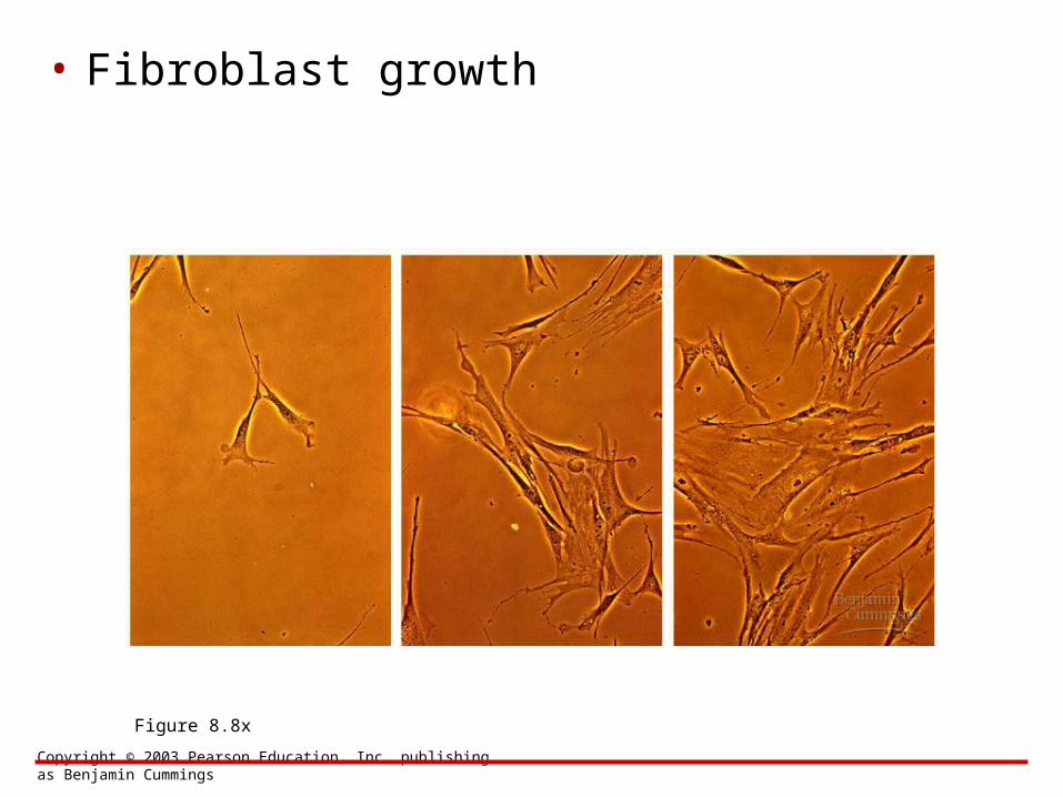
• Fibroblast growth
Figure 8.8x
Copyright © 2003 Pearson Education, Inc. publishing as Benjamin Cummings
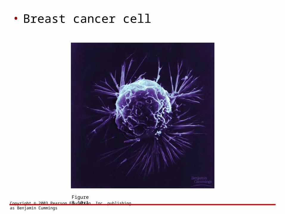
• Breast cancer cell
Figure 8.10x1
Copyright © 2003 Pearson Education, Inc. publishing as Benjamin Cummings
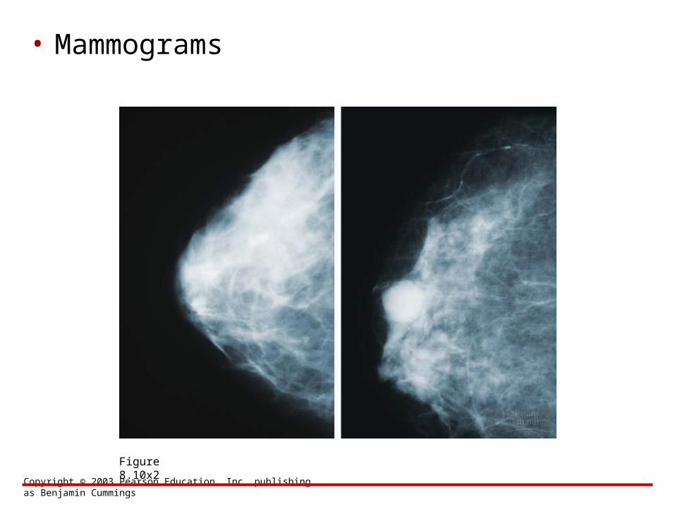
• Mammograms
Figure 8.10x2
Copyright © 2003 Pearson Education, Inc. publishing as Benjamin Cummings
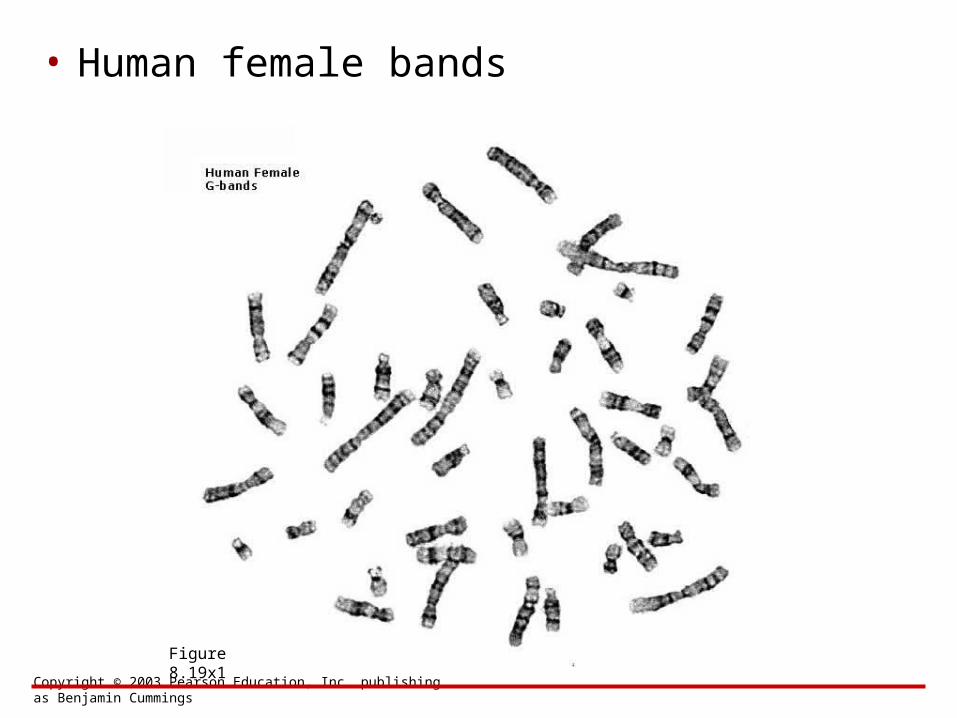
• Human female bands
Figure 8.19x1
Copyright © 2003 Pearson Education, Inc. publishing as Benjamin Cummings
• Human female karyotype
Figure 8.19x2
Copyright © 2003 Pearson Education, Inc. publishing as Benjamin Cummings
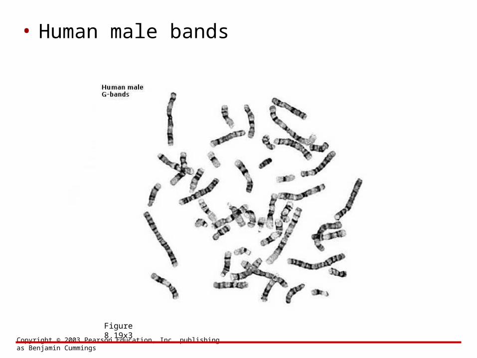
• Human male bands
Figure 8.19x3

Copyright © 2003 Pearson Education, Inc. publishing as Benjamin Cummings
• Human male karyotype
Figure 8.19x4
Copyright © 2003 Pearson Education, Inc. publishing as Benjamin Cummings
• Down syndrome karyotype
Figure 8.20Ax
Copyright © 2003 Pearson Education, Inc. publishing as Benjamin Cummings
• Klinefelter’s karyotype
Figure 8.22Ax
Copyright © 2003 Pearson Education, Inc. publishing as Benjamin Cummings
• XYY karyotype
Figure 8.22x
Copyright © 2003 Pearson Education, Inc. publishing as Benjamin Cummings
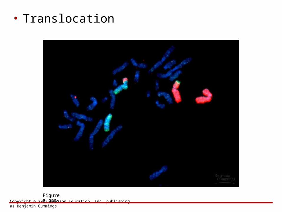
• Translocation
Figure 8.23Bx