Embed Size (px)
Citation preview

··
Introduction
The generation and characterization of mutants isan essential component of any study on structure–function relationships. Knowledge of the three-dimensional structure of a protein, RNA species, or DNA regulatory element (e.g. a promoter) canprovide clues to the way in which they function butproof that the correct mechanism has been elucid-ated requires the analysis of mutants that haveamino acid or nucleotide changes at key residues(see Box 8.2).
Classically, mutants are generated by treating the test organism with chemical or physical agentsthat modify DNA (mutagens). This method of muta-genesis has been extremely successful, as witnessedby the growth of molecular biology and functionalgenomics, but suffers from a number of disadvant-ages. First, any gene in the organism can be mutatedand the frequency with which mutants occur in thegene of interest can be very low. This means thatselection strategies have to be developed. Second,even when mutants with the desired phenotype areisolated, there is no guarantee that the mutation hasoccurred in the gene of interest. Third, prior to thedevelopment of gene-cloning and sequencing tech-niques, there was no way of knowing where in thegene the mutation had occurred and whether itarose by a single base change, an insertion of DNA,or a deletion.
As techniques in molecular biology have devel-oped, so that the isolation and study of a single geneis not just possible but routine, so mutagenesis hasalso been refined. Instead of crudely mutagenizingmany cells or organisms and then analyzing manythousands or millions of offspring to isolate a desiredmutant, it is now possible to change specifically anygiven base in a cloned DNA sequence. This techniqueis known as site-directed mutagenesis. It has become a basic tool of gene manipulation, for it simplifiesDNA manipulations that in the past required a great
deal of ingenuity and hard work, e.g. the creation orelimination of cleavage sites for restriction endonu-cleases. The importance of site-directed mutagenesisgoes beyond gene structure–function relationshipsfor the technique enables mutant proteins with novelproperties of value to be created (protein engineering).Such mutant proteins may have only minor changesbut it is not uncommon for entire domains to bedeleted or new domains added.
Primer extension (the single-primer method)is a simple method for site-directed mutation
The first method of site-directed mutagenesis to be developed was the single-primer method (Gillamet al. 1980, Zoller & Smith 1983). As originallydescribed the method involves in vitro DNA synthesiswith a chemically synthesized oligonucleotide (7–20nucleotides long) that carries a base mismatch withthe complementary sequence. As shown in Fig. 8.1,the method requires that the DNA to be mutated is available in single-stranded form, and cloning the gene in M13-based vectors makes this easy.However, DNA cloned in a plasmid and obtained in duplex form can also be converted to a partiallysingle-stranded molecule that is suitable (Dalbadie-McFarland et al. 1982).
The synthetic oligonucleotide primes DNA syn-thesis and is itself incorporated into the resulting heteroduplex molecule. After transformation of the host E. coli, this heteroduplex gives rise to homo-duplexes whose sequences are either that of the original wild-type DNA or that containing the mutatedbase. The frequency with which mutated clones arise,compared with wild-type clones, may be low. Inorder to pick out mutants, the clones can be screenedby nucleic acid hybridization with 32P-labeled oli-gonucleotide as probe. Under suitable conditions ofstringency, i.e. temperature and cation concentra-tion, a positive signal will be obtained only withmutant clones. This allows ready detection of the
CHAPTER 8
Changing genes: site-directed mutagenesis andprotein engineering
POGC08 12/8/05 8:47 AM Page 141

··
142 CHAPTER 8
desired mutant (Wallace et al. 1981, Traboni et al. 1983). It is prudent to check the sequence of themutant directly by DNA sequencing, in order tocheck that the procedure has not introduced otheradventitious changes. This was a particular neces-sity with early versions of the technique which made use of E. coli DNA polymerase. The more recent use of the high-fidelity DNA polymerases has minimizedthe problem of extraneous mutations as well asshortening the time for copying the second strand.Also, these polymerases do not “strand-displace” the oligomer, a process which would eliminate theoriginal mutant oligonucleotide.
A variation of the procedure (Fig. 8.2) outlinedabove involves oligonucleotides containing insertedor deleted sequences. As long as stable hybrids areformed with single-stranded wild-type DNA, prim-
ing of in vitro DNA synthesis can occur, ultimatelygiving rise to clones corresponding to the inserted or deleted sequence (Wallace et al. 1980, Norranderet al. 1983).
The single-primer method has a number ofdeficiencies
The efficiency with which the single-primer methodyields mutants is dependent upon several factors.The double-stranded heteroduplex molecules thatare generated will be contaminated both by any single-stranded non-mutant template DNA that hasremained uncopied and by partially double-strandedmolecules. The presence of these species considerablyreduces the proportion of mutant progeny. They can be removed by sucrose gradient centrifugation
AGTACGA
Single-strandedM13 recombinant
Transform E. coli
(1) DNA polymerase + 4dNTPs(2) T4 DNA ligase + ATP
T
CA G G
C
T
AGTACGA*
*
T C A G G C T
Chemically synthesizedoligonucleotide
T
CA G G
C
T
AGTACGA*
*
Mutant
T
CA G G
C
T
AGTACGA*
Wild-type
T
CA G G
C
T
AGTACGA*
Screen plaques with 32P-labeledoligonucleotide as hybridizationprobeIsolate mutant
Anneal
Fig. 8.1Oligonucleotide-directed mutagenesis.Asterisks indicatemismatched bases.Originally the Klenowfragment of DNApolymerase was used,but now this has beenlargely replaced withT7 polymerase.
POGC08 12/8/05 8:47 AM Page 142

··
Changing genes: site-directed mutagenesis and protein engineering 143
or by agarose gel electrophoresis, but this is time-consuming and inconvenient.
Following transformation and in vivo DNA synthesis, segregation of the two strands of the heteroduplex molecule can occur, yielding a mixedpopulation of mutant and non-mutant progeny.Mutant progeny have to be purified away fromparental molecules, and this process is complicatedby the cell’s mismatch repair system. In theory, themismatch repair system should yield equal numbersof mutant and non-mutant progeny, but in practicemutants are counterselected. The major reason forthis low yield of mutant progeny is that the methyl-directed mismatch repair system of E. coli favors therepair of non-methylated DNA. In the cell, newlysynthesized DNA strands that have not yet beenmethylated are preferentially repaired at the positionof the mismatch, thereby eliminating a mutation. Ina similar way, the non-methylated in vitro-generatedmutant strand is repaired by the cell so that themajority of progeny are wild type (Kramer et al.1984). The problems associated with the mismatchrepair system can be overcome by using host strainscarrying the mutL, mutS, or mutH mutations, whichprevent the methyl-directed repair of mismatches.
A heteroduplex molecule with one mutant andone non-mutant strand must inevitably give rise to both mutant and non-mutant progeny upon replication. It would be desirable to suppress thegrowth of non-mutants, and various strategies havebeen developed with this in mind (Kramer, B. 1984,Carter et al. 1985, Kunkel 1985, Sayers & Eckstein1991).
Another disadvantage of all of the primer exten-sion methods is that they require a single-strandedtemplate. In contrast, with PCR-based mutagenesis
(see below) the template can be single-stranded ordouble-stranded, circular or linear. In comparisonwith single-stranded DNAs, double-stranded DNAsare much easier to prepare. Also, gene inserts are ingeneral more stable with double-stranded DNAs.
The issues raised above account for the fact thatmost of the mutagenesis kits that are available com-mercially make use of multiple primers and double-stranded templates. For example, in the GeneEditorTM
system (Fig. 8.3), two primers are used. One of theseprimers encodes the mutation to be inserted into the target gene. The second encodes a mutation thatenhances the antibiotic resistance properties of theampicillin-resistance determinant on the vector byconferring resistance to ceftazidime as well. Afterextending the two primers to yield an intact circularDNA molecule, the mutated plasmid is transformedinto E. coli and selection made for the enhancedantibiotic resistance. Plasmids encoding the enhancedantibiotic resistance also should carry the mutatedtarget gene. In a variant of this procedure, the vectorhas two antibiotic resistance determinants (ampicillinand tetracycline) but one of these (AmpR) carries amutation. Again, two primers are used: one carryingthe mutation to be introduced to the target gene and the other restores ampicillin resistance. Afterthe in vitro mutagenesis steps, the plasmid is trans-formed into E. coli and selection made for ampicillinresistance.
Methods have been developed that simplifythe process of making all possible amino acidsubstitutions at a selected site
Using site-directed mutagenesis it is possible tochange two or three adjacent nucleotides so that
Multiple pointmutations
Mutant oligonucleotidewith multiple (four)single base pairmismatches
Insertionmutagenesis
Mutant oligonucleotidecarrying a sequence tobe inserted sandwichedbetween two regionswith sequencescomplementary to siteson either sides of thetarget site in thetemplate
Deletionmutagenesis
Mutant oligonucleotidespanning the region tobe deleted, binding totwo separate sites, oneon either side of thetarget
Fig. 8.2Oligonucleotide-directed mutagenesisused for multiple pointmutation, insertionmutagenesis, anddeletion mutagenesis.
POGC08 12/8/05 8:47 AM Page 143

··
144 CHAPTER 8
every possible amino acid substitution is made at a site of interest. This generates a requirement for 19 different mutagenic oligonucleotides assumingonly one codon will be used for each substitution. An alternative way of changing one amino acid to all the alternatives is cassette mutagenesis. Thisinvolves replacing a fragment of the gene with different fragments containing the desired codonchanges. It is a simple method for which the efficiencyof mutagenesis is close to 100%. However, if it isdesired to change the amino acids at two sites to allthe possible alternatives then 400 different oligos or fragments would be required and the practicalityof the method becomes questionable. One solution to this problem is to use doped oligonucleotides (Fig. 8.4). Many different variations of this techniquehave been developed and the interested reader shouldconsult the review of Neylon (2004).
The PCR can be used for site-directedmutagenesis
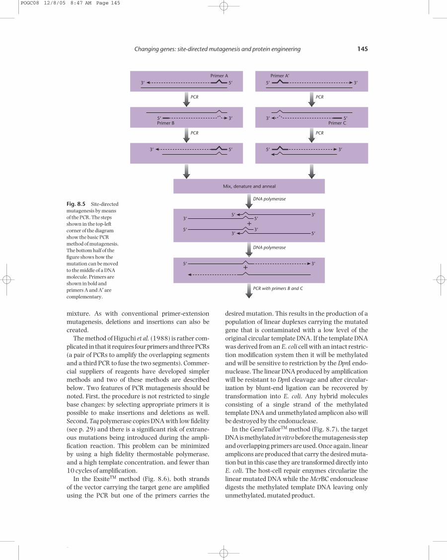
Early work on the development of the PCR method ofDNA amplification showed its potential for mutage-nesis (Scharf et al. 1986). Single bases mismatchedbetween the amplification primer and the templatebecome incorporated into the template sequence as a result of amplification (Fig. 8.5). Higuchi et al.(1988) have described a variation of the basic methodwhich enables a mutation in a PCR-produced DNAfragment to be introduced anywhere along its length.Two primary PCR reactions produce two overlappingDNA fragments, both bearing the same mutation inthe overlap region. The overlap in sequence allowsthe fragments to hybridize (Fig. 8.5). One of the twopossible hybrids is extended by DNA polymerase to produce a duplex fragment. The other hybrid has recessed 5′ ends and, since it is not a substrate for the polymerase, is effectively lost from the reaction
Insert
AmpR
Insert
AmpR
Mutant
AmpR + newresistance
+
+
1. Alkaline denature dsDNA template, anneal the mutagenic oligonucleotide and selection oligonucleotide.
2. Synthesize the mutant strand with T4 DNA polymerase and T4 DNA ligase.
3. Transform competent cells with the mutagenesis reaction. Grow overnight with the antibiotic selection mix.
4. Isolate plasmid DNA and transform into competent cells. Select mutants on media containing ampicillin and the antibiotic selection mix.
Fig. 8.3 The GeneEditorTM system for generating a highfrequency of mutations using site-directed mutagenesis.
G T A CSphI
TCGAXhoI
T A GTMet
A CGSer
T CG TTVal
C AAG GCAla
C CGG AAGlu
C TTA GTMet
T CAG GAC CT
GluA CTIle
T GAG AAGlu
C TTC AGArg * **
N CN G
I II
* **
I IIN CN G
Fig. 8.4 Mutagenesis by means of doped oligonucleotides. During synthesis of the upper strand of the oligonucleotide, a mixtureof all four nucleotides is used at the positions indicated by the letter N. When the lower strand is synthesized, inosine (I) is insertedat the positions shown. The double-stranded oligonucleotide is inserted into the relevant position of the vector.
POGC08 12/8/05 8:47 AM Page 144
··
Changing genes: site-directed mutagenesis and protein engineering 145
mixture. As with conventional primer-extensionmutagenesis, deletions and insertions can also becreated.
The method of Higuchi et al. (1988) is rather com-plicated in that it requires four primers and three PCRs(a pair of PCRs to amplify the overlapping segmentsand a third PCR to fuse the two segments). Commer-cial suppliers of reagents have developed simplermethods and two of these methods are describedbelow. Two features of PCR mutagenesis should benoted. First, the procedure is not restricted to singlebase changes: by selecting appropriate primers it ispossible to make insertions and deletions as well.Second, Taq polymerase copies DNA with low fidelity(see p. 29) and there is a significant risk of extrane-ous mutations being introduced during the ampli-fication reaction. This problem can be minimized by using a high fidelity thermostable polymerase,and a high template concentration, and fewer than10 cycles of amplification.
In the ExsiteTM method (Fig. 8.6), both strands of the vector carrying the target gene are amplifiedusing the PCR but one of the primers carries the
desired mutation. This results in the production of apopulation of linear duplexes carrying the mutatedgene that is contaminated with a low level of theoriginal circular template DNA. If the template DNAwas derived from an E. coli cell with an intact restric-tion modification system then it will be methylatedand will be sensitive to restriction by the DpnI endo-nuclease. The linear DNA produced by amplificationwill be resistant to DpnI cleavage and after circular-ization by blunt-end ligation can be recovered bytransformation into E. coli. Any hybrid moleculesconsisting of a single strand of the methylated template DNA and unmethylated amplicon also willbe destroyed by the endonuclease.
In the GeneTailorTM method (Fig. 8.7), the targetDNA is methylated in vitro before the mutagenesis stepand overlapping primers are used. Once again, linearamplicons are produced that carry the desired muta-tion but in this case they are transformed directly intoE. coli. The host-cell repair enzymes circularize thelinear mutated DNA while the McrBC endonucleasedigests the methylated template DNA leaving onlyunmethylated, mutated product.
DNA polymerase
PCR with primers B and C
DNA polymerase
PCR
PCR
3‘ 5‘
Primer A
Primer B3‘5‘
3‘ 5‘
PCR
PCR
5‘ 3‘
Primer A‘
Primer C5‘3‘
5‘ 3‘
5‘ 3‘3‘ 5‘
3‘ 5‘3‘5‘
+
5‘ 3‘+
Mix, denature and anneal
Fig. 8.5 Site-directedmutagenesis by means of the PCR. The stepsshown in the top-leftcorner of the diagramshow the basic PCRmethod of mutagenesis.The bottom half of thefigure shows how themutation can be movedto the middle of a DNAmolecule. Primers areshown in bold andprimers A and A′ arecomplementary.
POGC08 12/8/05 8:47 AM Page 145

··
146 CHAPTER 8
Methods are available to enable mutations to be introduced randomly throughout atarget gene
The methods described above enable defined muta-tions to be introduced at defined locations within a gene and are of particular value in determiningstructure–activity relationships. However, if theobjective of a study is to select mutants with alteredand/or improved characteristics then a better ap-proach is to mutate the gene at random and then positively select those with the desired properties.Methods for the random mutagenesis of clonedgenes are described in this section and the next whileselection methods are described later (p. 148).
It is well known that the polymerase chain reac-tion is error prone and that there is a high probabilityof base changes in amplicons. However, even the relatively low fidelity Taq polymerase is too accurateto be of value in generating mutant libraries. Never-theless, increases in error rates can be obtained in anumber of ways. One of the commonest ways ofachieving this is to introduce a small amount of Mn2+,in place of the normal Mg2+, and to include an excessof dGTP and dTTP relative to the other two nucleotidetriphosphates. With this protocol it is possible toachieve error rates of one nucleotide per kilobase(Caldwell & Joyce 1994, Cirino et al. 2003). Evenhigher rates of mutagenesis can be achieved by usingnucleoside triphosphate analogs (Zaccolo et al. 1996).
The methodologies for error-prone PCR all involveeither a misincorporation process in which the poly-merase adds an incorrect base to the growing daugh-ter strand or a lack of proofreading ability on the partof the polymerase. It might be expected that theygenerate a completely random set of mutants but inreality the mutant libraries produced are heavilybiased. There are three sources of bias. First, theinherent characteristics of the DNA polymerase usedmean that some types of errors are more commonthan others (Cirino et al. 2003). The second source ofbias arises because of the nature of the genetic code.For example, a single point mutation in a valinecodon can change it to one encoding phenylalanine,leucine, isoleucine, alanine, aspartate, or glycine but
Add PCR primers
PCR
T4 DNA ligase
Add DpnI endonuclease
Transformcompetent
cells
Mutatedinsert
Mutation
Insert
Mismatch
Fig. 8.6 (left) The ExsiteTM method for generating mutantsusing the PCR. The parental plasmid (shown in blue) carryingthe target gene is derived from a restriction-proficient strain of E. coli and so is methylated. This makes it sensitive to the DpnIendonuclease and hence it can be eliminated selectively fromthe final PCR mixture.
POGC08 12/8/05 8:47 AM Page 146

··
Changing genes: site-directed mutagenesis and protein engineering 147
two or three adjacent point mutations are requiredto change it to one encoding all the other aminoacids. The final source of bias arises from the processof amplification. A mutant that is generated early inthe amplification process will be over-represented inthe final library compared to one that arises in laterrounds of amplification.
Error-prone PCR protocols are effective as a meansof randomly changing one amino acid into anotherin the final protein. However, sometimes it might bedesirable to explore the effect of randomly deleting orinserting amino acids and this is possible using therandom insertion/deletion (RID) process devised byMurakami et al. (2002, 2003). The method is basedon ligating an insertion or deletion cassette at nearlyrandom locations within the gene.
Altered proteins can be produced by insertingunusual amino acids during protein synthesis
All the mutation methods described above result inthe replacement of one or more amino acid residues
in a protein with other natural amino acids, e.g. thereplacement of a phenylalanine residue with tyrosine,tryptophan, histidine, etc. The ability to incorporateunnatural amino acids into proteins in vivo wouldpermit the production of large quantities of proteinswith novel properties. For example, the replacementof methionine with selenomethionine facilitates thedetermination of the three-dimensional structure of proteins (Hendrickson et al. 1990). While it is possible to “force” bacteria to incorporate unnaturalamino acids into proteins (for review, see Link et al.2003) a better method is to engineer the transla-tional apparatus. This is achieved by generating an aminoacyl-tRNA synthetase and tRNA pair thatfunction independently of the synthetases and tRNAsendogenous to E. coli (Wang et al. 2001a, Santoro et al. 2003). Such a pair are said to be orthogonaland satisfy a number of criteria:
• The tRNA is not a substrate for any of the endogen-ous E. coli synthetases but functions efficiently inprotein translation.
• The orthogonal synthetase efficiently aminoacy-lates the orthogonal tRNA whose anticodon hasbeen modified to recognize an amber (UAG) or opal(UGA) stop codon.
• The synthetase does not aminoacylate any of theendogenous E. coli tRNAs.
Archaebacteria appear to be an especially good sourceof orthogonal pairs for use in E. coli.
Modifying the anticodon on the tRNA such that itrecognizes amber and opal codons is relatively easy.However, the synthetase also needs to be modifiedsuch that it charges the cognate tRNA with unusualamino acids more efficiently than the normal aminoacid. To do this a library of synthetase mutants is gen-erated and subjected to positive selection based onsuppression of an amber codon located in a plasmid-borne gene encoding chloramphenicol acetyltrans-ferase (Wang et al. 2001a). Using this approach thetyrosyl-tRNA synthetase of Methanococcus jannaschiiwas modified to permit the site-specific incorporationinto proteins of phenylalanine and tyrosine deriva-tives such as O-allyltyrosine, p-acetyl-phenylalanine,and p-benzoyl-phenylalanine. These modified aminoacids can be used as sites for chemical modification ofthe protein in vitro after purification, e.g. the attach-ment of fluorescent labels (Chin et al. 2003, Link et al.2003).
There have been two significant developments of the above technique. In the first of these, Zhang
Methylatedplasmid
Mutatedplasmid
Transformation
Mutagenesis
Methylation Methylate plasmid DNAwith DNA methylase.
Amplify the plasmid in amutagenesis reaction withtwo overlapping primers, oneof which contains the targetmutation. The product islinear, double-stranded DNAcontaining the mutation.
Transform the mutagenesismixture into wild type E. coli.The host cell circularizes thelinear mutated DNA, andMcrBC endonuclease in thehost cell digests themethylated template DNA,leaving only unmethylated,mutated product.
Fig. 8.7 The GeneTailorTM method for generating mutantsusing the PCR.
POGC08 12/8/05 8:47 AM Page 147

··
148 CHAPTER 8
et al. (2003) have shown that chemical modificationof proteins can occur in vivo as well as in vitro. Forexample, m-acetylphenylalanine was substituted for Lys7 of the cytoplasmic domain of protein Z andfor Arg200 of the outer membrane protein LamB. Onaddition of a membrane-permeable dye (fluoresceinhydrazide) to intact cells, these modified proteins wereselectively labeled. In the case of cells expressing the modified LamB derivative, labeling was possible with a range of fluorescein derivatives that are notmembrane permeable. The second development isthe ability to charge the orthogonal tRNA with glyco-sylated amino acids. For example, Zhang et al. (2004)were able to synthesize in E. coli a myoglobin deriva-tive containing β–N-acetylglucosamine (GlcNAc) ata defined position. This GlcNAc moiety was recog-nized by a saccharide-binding protein and could bemodified by a galactosyltransferase.
Phage display can be used to facilitate theselection of mutant peptides
In phage display, a segment of foreign DNA is insertedinto either a phagemid or an infectious filamentous
phage genome and expressed as a fusion product witha phage coat protein. It is a very powerful techniquefor selecting and engineering polypeptides with novelfunctions. The technique was developed first for theE. coli phage M13 (Parmley & Smith 1988), but hassince been extended to other phages such as T4 andλ (Ren & Black 1998, Santini et al. 1998).
The M13 phage particle consists of a single-stranded DNA molecule surrounded by a coat con-sisting of several thousand copies of the major coat protein, P8. At one end of the particle are fivecopies each of the two minor coat proteins P9 and P7 and at the other end five copies each of P3 and P6.In early examples of phage display, a random DNA cassette (see above) was inserted into either the P3 or the P8 gene at the junction between the signalsequence and the native peptide. E. coli transfectedwith the recombinant DNA molecules secretedphage particles that displayed on their surface theamino acids encoded by the foreign DNA. Particularphage displaying peptide motifs with, for example,antibody-binding properties were isolated by affinitychromatography (Fig. 8.8). Several rounds of affin-ity chromatography and phage propagation can be
Phagevector
f1ori
Randompeptide
DNA
Promoter Signal M13 gene III
Test binding to antibodyor receptor
Infect male E. coli
Phageparticle
Gene IIIprotein
Randomprotein
DNA
Fig. 8.8 The principleof phage display ofrandom peptides.
POGC08 12/8/05 8:47 AM Page 148
··
Changing genes: site-directed mutagenesis and protein engineering 149
used to further enrich for phage with the desiredbinding characteristics. In this way, millions of random peptides have been screened for their abilityto bind to an anti-peptide antibody or to streptavidin(Cwirla et al. 1990, Devlin et al. 1990, Scott & Smith1990), and variants of human growth hormone withimproved affinity and receptor specificity have beenisolated (Lowman et al. 1991).
One disadvantage of the original method of phagedisplay is that polypeptide inserts greater than 10residues compromise coat-protein function and socannot be efficiently displayed. This problem can be solved by the use of phagemid display (Bass et al.1990). In this system, the starting-point is a plasmidcarrying a single copy of the P3 or P8 gene from M13 plus the M13 ori sequence (i.e. a phagemid, see p. 75). As before, the random DNA sequence isinserted into the P3 or P8 gene downstream from thesignal peptide-cleavage site and the construct trans-formed into E. coli. Phage particles displaying theamino acid sequences encoded by the DNA insert are obtained by superinfecting the transformed cellswith helper phage. The resulting phage particles are phenotypically mixed and their surfaces are amosaic of normal coat protein and fusion protein.
Specialized phagemid display vectors have beendeveloped for particular purposes. For example,phagemids have been constructed that have an amber(chain-terminating) codon immediately downstream
from the foreign DNA insert and upstream from thebody of P3 or P8. When the recombinant phagemidis transformed into non-suppressing strains of E. coli,the protein encoded by the foreign DNA terminatesat the amber codon and is secreted into the medium.However, if the phagemid is transformed into cellscarrying an amber suppressor, the entire fusion pro-tein is synthesized and displayed on the surface of thesecreted phage particles (Winter et al. 1994). Otherstudies ( Jespers et al. 1995, Fuh & Sidhu 2000, Fuhet al. 2000) have shown that proteins can be dis-played as fusions to the carboxy terminus of P3, P6,and P8. Although amino-terminal display formats arelikely to dominate established applications, carboxy-terminal display permits constructs that are unsuitedto amino-terminal display.
For a detailed review of phage and phagemid dis-play, the reader should consult Sidhu (2000) andSidhu et al. (2000).
Cell-surface display is a more versatilealternative to phage display
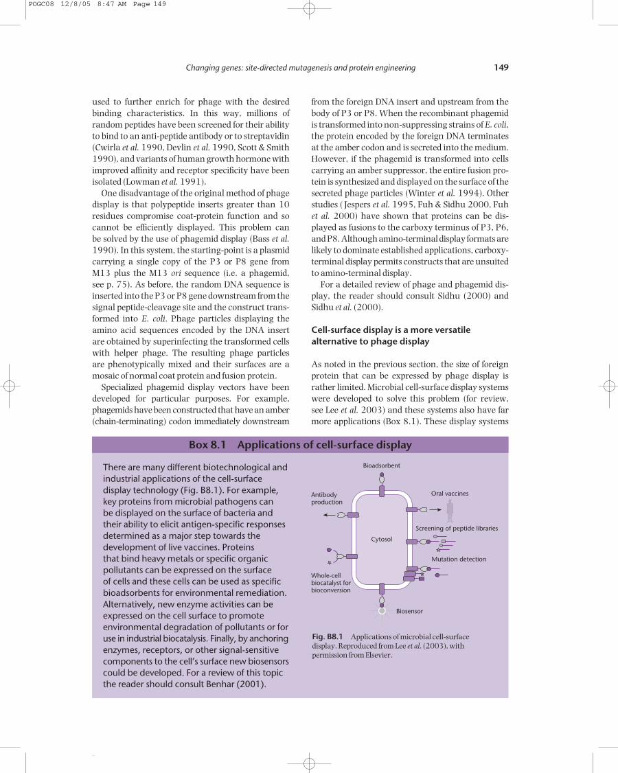
As noted in the previous section, the size of foreignprotein that can be expressed by phage display israther limited. Microbial cell-surface display systemswere developed to solve this problem (for review, see Lee et al. 2003) and these systems also have farmore applications (Box 8.1). These display systems
There are many different biotechnological andindustrial applications of the cell-surfacedisplay technology (Fig. B8.1). For example,key proteins from microbial pathogens can be displayed on the surface of bacteria andtheir ability to elicit antigen-specific responsesdetermined as a major step towards thedevelopment of live vaccines. Proteins that bind heavy metals or specific organicpollutants can be expressed on the surface of cells and these cells can be used as specificbioadsorbents for environmental remediation.Alternatively, new enzyme activities can beexpressed on the cell surface to promoteenvironmental degradation of pollutants or foruse in industrial biocatalysis. Finally, by anchoringenzymes, receptors, or other signal-sensitivecomponents to the cell’s surface new biosensorscould be developed. For a review of this topicthe reader should consult Benhar (2001).
Box 8.1 Applications of cell-surface display
Antibodyproduction
Whole-cellbiocatalyst forbioconversion
Mutation detection
Screening of peptide libraries
Bioadsorbent
Cytosol
Biosensor
Oral vaccines
Fig. B8.1 Applications of microbial cell-surface display. Reproduced from Lee et al. (2003), withpermission from Elsevier.
POGC08 12/8/05 8:47 AM Page 149
··
150 CHAPTER 8
involve expressing a heterologous peptide or pro-tein of interest (the passenger or target protein) as a fusion protein with various cell-surface proteins(carrier proteins). Depending on the properties of the passenger and carrier proteins, the passengerprotein is expressed as an N-terminal, a C-terminalor a sandwich fusion.
For a cell-surface protein to be a successful car-rier it should satisfy four requirements. First, itshould have an efficient signal peptide to permit the fusion protein to pass through the inner membrane.Second, it should have a strong anchoring structureto keep fusion proteins on the cell surface withoutdetachment. Third, it should be compatible with the passenger protein such that the fusion is notunstable. Finally, it should be resistant to attack byproteases present in the periplasmic space or thegrowth medium. In Gram-negative bacteria such as E. coli many different proteins have been sub-jugated as carriers. Basically, these proteins fall intotwo classes: outer membrane proteins (e.g. the adhesinprotein, peptidiglycan-associated lipoprotein, and theOmpC and TraT proteins) and protein components ofappendages such as pili and flagella. Where outermembrane proteins are used as the carrier it is im-portant to know which part of them is exposed on theouter surface of the cell since this needs to be the siteof insertion of the passenger protein.
The passenger protein to be displayed is selectedby the required application but its properties influ-ence the translocation process and the effectivenessof the display procedure. For example, the formationof disulfide bridges at the periplasmic side of the outermembrane can affect the efficiency of translocation.Also, the presence of many charged or hydrophobicresidues can result in inefficient secretion. Thus, ifdisplay technology is used to screen variants producedby random mutagenesis, there may be negative orpositive selection for those mutants that affect theefficiency of translocation.
Protein engineering
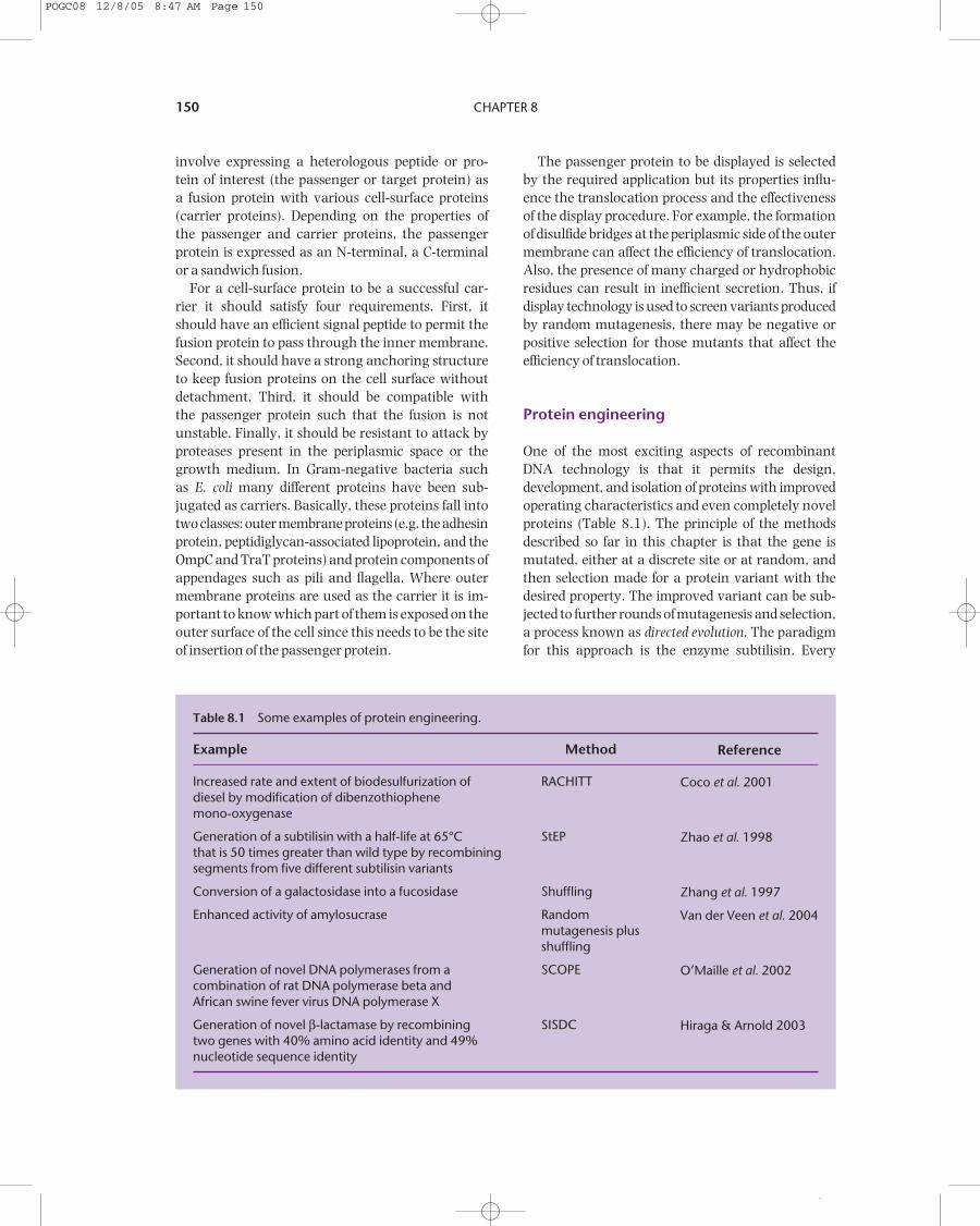
One of the most exciting aspects of recombinantDNA technology is that it permits the design, development, and isolation of proteins with improved operating characteristics and even completely novelproteins (Table 8.1). The principle of the methodsdescribed so far in this chapter is that the gene ismutated, either at a discrete site or at random, andthen selection made for a protein variant with thedesired property. The improved variant can be sub-jected to further rounds of mutagenesis and selection,a process known as directed evolution. The paradigmfor this approach is the enzyme subtilisin. Every
Table 8.1 Some examples of protein engineering.
Example
Increased rate and extent of biodesulfurization of diesel by modification of dibenzothiophene mono-oxygenase
Generation of a subtilisin with a half-life at 65°C that is 50 times greater than wild type by recombining segments from five different subtilisin variants
Conversion of a galactosidase into a fucosidase
Enhanced activity of amylosucrase
Generation of novel DNA polymerases from a combination of rat DNA polymerase beta and African swine fever virus DNA polymerase X
Generation of novel β-lactamase by recombining two genes with 40% amino acid identity and 49% nucleotide sequence identity
Method
RACHITT
StEP
Shuffling
Randommutagenesis plusshuffling
SCOPE
SISDC
Reference
Coco et al. 2001
Zhao et al. 1998
Zhang et al. 1997
Van der Veen et al. 2004
O’Maille et al. 2002
Hiraga & Arnold 2003
POGC08 12/8/05 8:47 AM Page 150

··
Changing genes: site-directed mutagenesis and protein engineering 151
property of this serine protease has been alteredincluding its rate of catalysis, substrate specificity,pH-rate profile, and stability to oxidative, thermal,and alkaline inactivation (for review, see Bryan2000). Variants also have been produced that favor
aminolysis (synthesis) over hydrolysis in aqueoussolvents (see Box 8.2).
An alternative approach to directed evolution isgene shuffling. The principle of this method is thatmany protein variants with desirable characteristics
Oxidation-resistant variants of a1-antitrypsin (AAT)
Cumulative damage to lung tissue is thoughtto be responsible for the development of emphysema, an irreversible diseasecharacterized by loss of lung elasticity. Theprimary defense against elastase damage isAAT, a glycosylated serum protein of 394amino acids. The function of AAT is knownbecause its genetic deficiency leads to apremature breakdown of connective tissue. In healthy individuals there is an associationbetween AAT and neutrophil elastase followedby cleavage of AAT between methionineresidue 358 and serine residue 359 (see Fig. B8.2). After cleavage, there isnegligible dissociation of the complex.
Smokers are more prone to emphysema,because smoking results in an increasedconcentration of leucocytes in the lung and consequently increased exposure toneutrophil elastase. In addition, leucocytesliberate oxygen free radicals and these canoxidize methionine-358 to methioninesulfoxide. Since methionine sulfoxide is muchbulkier than methionine, it does not fit into the active site of elastase. Hence oxidized AATis a poor inhibitor. By means of site-directedmutagenesis, an oxidation-resistant mutant of AAT has been constructed by replacingmethionine-358 with valine (Courtney et al.
1985). In a laboratory model of inflammation,the modified AAT was an effective inhibitor ofelastase and was not inactivated by oxidation.Clinically, this could be important, sinceintravenous replacement therapy with plasmaconcentrates of AAT is used with patients witha genetic deficiency in AAT production.
Improving the performance of subtilisin
Proof of the power of gene manipulationcoupled with the techniques of in vitro(random and site-directed) mutagenesis as a means of generating improved enzymes isprovided by the work done on subtilisin overthe past 15 years (for review, see Bryan 2000).Every property of this serine protease has beenaltered, including its rate of catalysis, substratespecificity, pH-rate profile, and stability tooxidative, thermal, and alkaline inactivation. Inthe process, well over 50% of the 275 aminoacids of subtilisin have been changed. At somepositions in the molecule, the effects of replacingthe usual amino acid with all the other 19natural amino acids have been evaluated.
Many of the changes described above weremade to improve the ability of subtilisin tohydrolyze protein when incorporated intodetergents. However, serine proteases can beused to synthesize peptides and this approachhas a number of advantages over conventionalmethods (Abrahmsen et al. 1991). A problem
Box 8.2 Improving enzymes
394
Met 358
394
Ser 359
Met 358
Fig. B8.2 The cleavage of α1-antitrypsin onbinding to neutrophil elastase.
continued
POGC08 12/8/05 8:47 AM Page 151

··
152 CHAPTER 8
already exist in nature and novel combinations ofthese variants may have even more desirable prop-erties (Fig. 8.9). There are three sources of variantsfor gene shuffling. First, different polymorphisms ofthe gene of interest might exist naturally in a singleorganism or might have been created by random invitro mutagenesis (as described on p. 146). Second,the same protein with the same activity may be foundin other organisms but the gene and protein sequenceswill be different. Third, the protein of interest mightbelong to a protein family where the different mem-bers have different but related activities.
A good example of gene shuffling is work done on subtilisin by Ness et al. (1999). They started withthe genes for 26 members of the subtilisin family and created a library of chimeric proteases. Whenthis library was screened for four distinct enzymeproperties, variants were found that were signific-antly improved over any of the parental enzymes foreach individual property. Similarly, Lehmann et al.(2000) started with a family of mesophilic phytaseswhose amino acid sequence had been determined.Using these data they constructed a “consensus”
with the use of subtilisin for peptide synthesisis that hydrolysis is strongly favored overaminolysis, unless the reaction is undertaken in organic solvents. Solvents, in turn, reducethe half-life of subtilisin. Using site-directedmutagenesis, a number of variants of subtilisinhave been isolated with greatly enhancedsolvent stability (Wong et al. 1990, Zhong et al. 1991). Changes introduced included the minimization of surface changes to reducesolvation energy, the enhancement of internalpolar and hydrophobic interactions, and theintroduction of conformational restrictions to reduce the tendency of the protein todenature. Designing these changes requiresan extensive knowledge of the enzyme’sstructure and function. Chen and Arnold(1991, 1993) have provided an alternativesolution. They utilized random mutagenesiscombined with screening for enhancedproteolysis in the presence of solvent(dimethyl formamide) and substrate (casein).
The engineering of subtilisin has now goneone step further, in that it has been modified
such that aminolysis (synthesis) is favored overhydrolysis, even in aqueous solvents. This wasachieved by changing a serine residue in theactive site to cysteine (Abrahmsen et al. 1991).The reasons for this enhancement derivemainly from the increased affinity andreactivity of the acyl intermediate for theamino nucleophile (Fig. B8.3). Theseengineered “peptide ligases” are in turn being used to synthesize novel glycopeptides.A glycosyl amino acid is used in peptidesynthesis to form a glycosyl peptide ester,which will react with another C-protectedpeptide in the presence of the peptide ligaseto form a larger glycosyl peptide.
Box 8.2 continued
RCOOR’ + E OH ERCOO
H2O
R–NH2
Hydrolysis
Aminolysis
RCOOR’ + E OH
RCOOH + E OH
Fig. B8.3 The aminolysis (synthetic) and hydrolysisreactions mediated by an acylated protease.
Ancestral species
Species 1
Species 2
Species 3
Species 4
Evolution
DNA shufflingin vitro
Hybrid genes
Fig. 8.9 Schematic representation of gene shuffling.
POGC08 12/8/05 8:47 AM Page 152

··
Changing genes: site-directed mutagenesis and protein engineering 153
phytase sequence and found that an enzyme withthis sequence was much more thermostable thanany of the parent enzymes.
A number of different methods of geneshuffling have been developed
In the original method of gene shuffling (Stemmer1993, 2004), one starts by purifying the differentgenes that will provide the source of variation. These genes are digested with DNase to generate thefragments that will be recombined. The fragmentsfrom the different sources are mixed together andsubjected to repeated rounds of melting, annealing,and extension (Fig. 8.10). Eventually a full-lengthgene should be synthesized and this can be amplifiedby the PCR and cloned. The smaller the fragmentsthat are produced in the initial step the greater thenumber of single site variations that can be incorp-orated in the final product. However, the smaller thefragments the greater the number of cycles needed to reassemble a complete gene.
An alternative method is the staggered extensionprocess (StEP, Zhao et al. 1998). This also relies onrepeated cycles of melting, annealing, and extensionto build the variant genes. However, in the StEP pro-cess one starts with a mixture of full-length genes,denatures them, and then primes the synthesis ofcomplementary strands (Fig. 8.11). After a shortperiod of primer extension, the DNA is subjected to around of melting, annealing, and extension. Some ofthe extended primers will anneal to templates with adifferent base sequence and on further extension willgenerate chimeras. The more cycles of extension,melting, and annealing the greater the variabilitythat can be produced.
RACHITT (random chimeragenesis on transienttemplates) is conceptually similar to the originalDNA-shuffling method but is designed to producechimeras with a much larger number of crossovers(Coco et al. 2001, Coco 2003). In this method thegene fragments are generated from one strand of all but one of the parental DNAs (Fig. 8.12). Thesefragments then are reassembled on the full-length
Extend DNA
Denature and anneal
Extend DNA
Repeat until full-length strands generated
Fragment with DNase
Denature, mix and anneal
Fig. 8.10 The originalmethod of gene shuffling. After fragmentation of the two homologous genes, thecycles of denaturation,annealing, and extension arecontinued until full-lengthgenes can be detected by gelelectrophoresis.
POGC08 12/8/05 8:47 AM Page 153

··
154 CHAPTER 8
opposite strand of the remaining parent (the transienttemplate). The fragments are cut back to removemismatched sections, extended, and then ligated togenerate full-length genes. Finally, the template strandis destroyed to leave only the ligated gene fragmentsto be converted to double-stranded DNA.
Each of the methods described above has itsadvantages and disadvantages and all of them rely
to a greater or lesser extent on the annealing of mismatched DNA sequences. Thus there is always achance that the parental molecules will be recreatedpreferentially or that the degree of variation gener-ated will not be as great as expected. However, methods for “forcing” the generation of recombinantshave been developed (for review, see Neylon 2004).
Chimeric proteins can be produced in theabsence of gene homology
The gene-shuffling methods described above havean absolute requirement for significant homologybetween the parental sequences. However, there maybe a wish to create hybrids between proteins withfunctional similarities but whose sequence homologyis less than 50%. Achieving this requires methods forcombining non-homologous sequences and the firstone to be developed (Ostermeier et al. 1999) wasITCHY (incremental truncation for the creation ofhybrid enzymes). This method is based on the directligation of libraries of fragments generated by thetruncation of two template sequences, each templatebeing truncated from opposite ends (Fig. 8.13). Thisligation procedure removes any need for homologyat the point of crossover but the downside is that theDNA fragments may be reconnected in a way that is not at all analogous to their position in the tem-plate gene.
In the original ITCHY process the incrementaltruncation was performed using timed exonucleasedigestions. In practice, these digestions are difficultto control. An improved process was developed wherethe initial templates are generated with phospho-rothioate linkages incorporated at random along the length of the gene (Lutz et al. 2001a). Completeexonuclease digestion then generates fragments withlengths determined by the position of the nuclease-resistant phosphorothioate linkage. This method isknown as thio-ITCHY and is much simpler to perform.One drawback of ITCHY libraries is that they containonly one crossover per gene. However, by combin-ing ITCHY libraries with DNA-shuffling methods, a process known as SCRATCHY, it is possible to generate additional variation (Lutz et al. 2001b).
A major problem with methods such as ITCHY isthat they generate large numbers of non-functionalsequences due to mutations, insertions, and dele-tions. Furthermore, when one examines the three-dimensional structure of proteins it is clear that theyare organized into domains and motifs. Therefore, a more attractive way of generating chimeric
Denature andanneal primer
DNA extensionfor short period
Denature andanneal
DNA extensionfor short period
Multiple cycles ofDNA extension for short
period, denaturationand annealing
or or
Primer
Fig. 8.11 The StEP method for generating hybrid proteins.In the example shown, a hybrid gene will be constructed fromtwo homologous genes (shown in purple and black). Cloningof the hybrid gene will result in the production of a hybridprotein. For clarity, only one strand of each gene is shownafter the initial denaturation step.
POGC08 12/8/05 8:47 AM Page 154

··
Convert to double strands
DNase
Mix and anneal
Exonuclease+ DNA polymerase+ DNA ligase
Endonuclease V
Singlestrandsfromhomologousgenes
Uracil
Synthesizecomplementarystrand with dUTPinstead of dTTP
Fig. 8.12 The RACHITT method for creating hybrid proteins.
POGC08 12/8/05 8:47 AM Page 155

··
156 CHAPTER 8
proteins might be to recombine these domains and motifs in novel ways. Two general methods of doing this have been developed (O’Maille et al.2002, Hiraga & Arnold 2003) and these are SCOPE (structure-based combinatorial protein engineering)and SISDC (sequence-independent site-directedchimeragenesis).
Suggested reading
Brannigan J.A. & Wilkinson A.J. (2002) Protein engineering 20 years on. Nature Reviews MolecularCell Biology 3, 964–70.
A short but excellent review of the development and phar-maceutical applications of protein engineering.
Collins C.H., Yokobayashi Y., Umeno D. & Arnold F.H.(2003) Engineering proteins that bind, move, makeand break DNA. Current Opinion in Biotechnology 14,371–8.
Another short but excellent review that focuses on what can be achieved with protein engineering rather than on themethods themselves.
Link A.J., Mock M.L. & Tirrell D.A. (2003) Non-canonicalamino acids in protein engineering. Current Opinionin Biotechnology 14, 603–9.
Lu Y. (2005) Design and engineering of metallo-proteins containing unnatural amino acids as non-native metal-containing cofactors. Current Opinion inChemical Biology 9, 118–26.
These two papers provide short reviews of the novelchemistries that are possible once unusual amino acids areintroduced to proteins.
Lutz S. & Patrick W.M. (2004) Novel methods fordirected evolution of enzymes: quality not quantity.Current Opinion in Biotechnology 15, 291–7.
Neylon C. (2004) Chemical and biochemical strategiesfor the randomisation of protein encoding DNAsequences: library construction methods for directedevolution. Nucleic Acids Research 32, 1448–59.
Each of the methods for generating gene libraries is reviewedin these papers with particular attention being given to thepracticality of the methods and the characteristics of thelibraries that are produced.
Roodveldt C., Aharoni A. & Tawfik D.S. (2005) Directedevolution of proteins for heterologous expression and stability. Current Opinion in Structural Biology 15,50–6.
A short review of the application of protein engineering foroverproduction of commercial proteins.
Endonucleasecleavage site
Endonucleasecleavage
Exonuclease IIIdigestion
Digestion withmung beannucelase
Ligation
Gen
e 1 Gene2
Fig. 8.13 The ITCHY method for creating hybrids of tworelated proteins. In the figure, the two related proteins areencoded by genes 1 (shown in purple) and 2 (shown in gray).The end result is a hybrid gene comprising the 5′ end of gene 1and the 3′ end of gene 2.
POGC08 12/8/05 8:47 AM Page 156