Embed Size (px)
Citation preview
Lecture Presentations by
Nicole Tunbridge and
Kathleen Fitzpatrick
Chapter 8
Cell Membranes
© 2018 Pearson Education Ltd.
Life at the Edge
The plasma membrane is the boundary that
separates the living cell from its surroundings
The plasma membrane exhibits selective
permeability, allowing some substances to cross it
more easily than others
Transport proteins are often responsible for
controlling passage across cellular membranes
© 2018 Pearson Education Ltd.
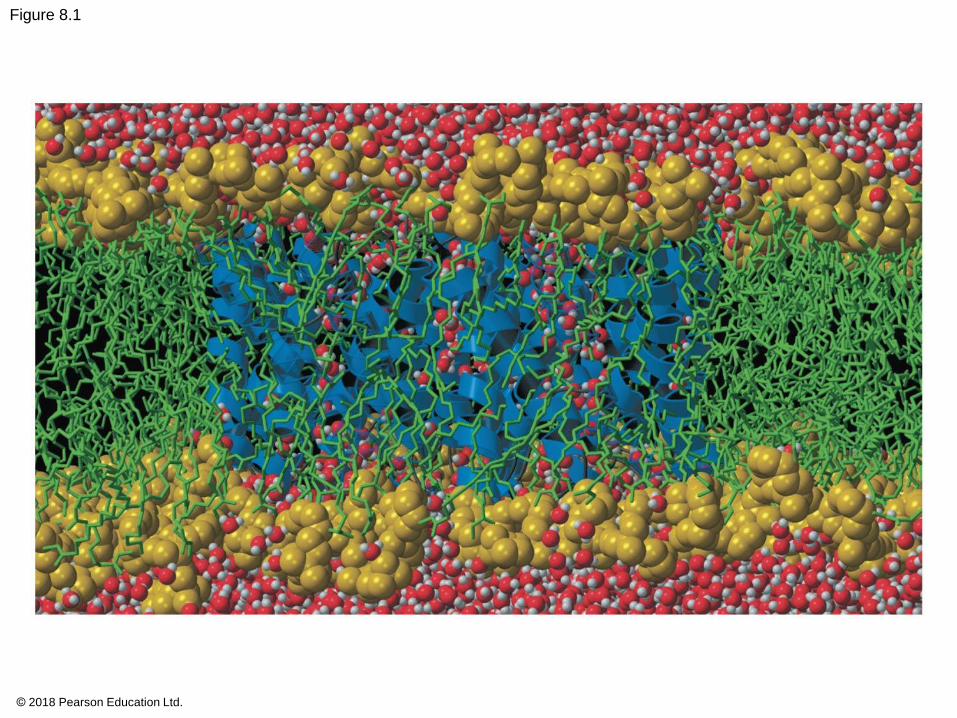
Figure 8.1
© 2018 Pearson Education Ltd.
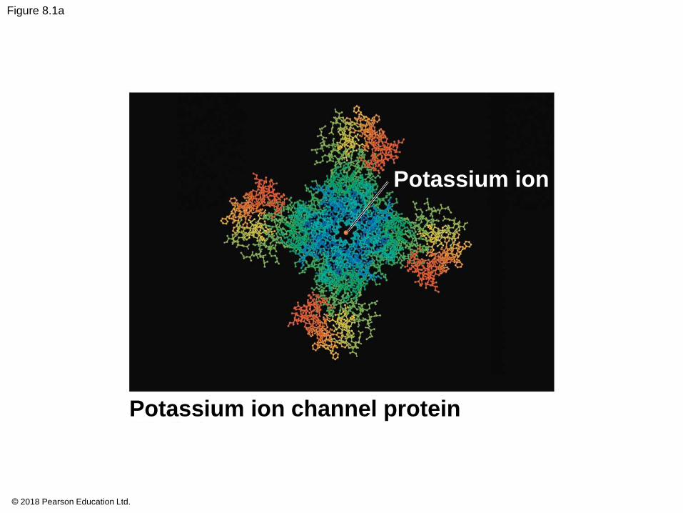
Figure 8.1a
Potassium ion
Potassium ion channel protein
© 2018 Pearson Education Ltd.
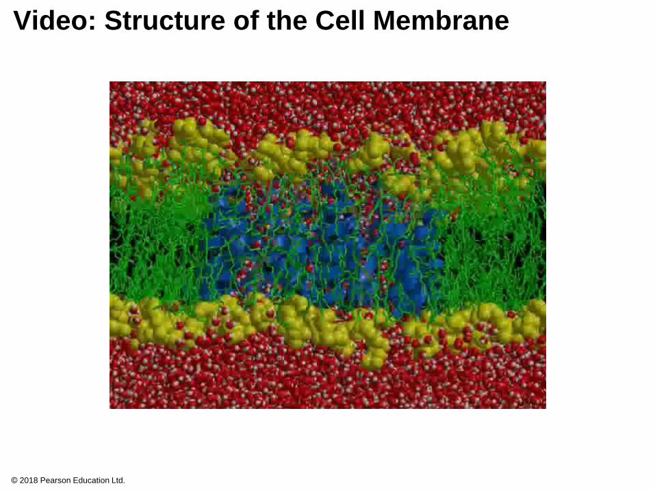
Video: Structure of the Cell Membrane
© 2018 Pearson Education Ltd.
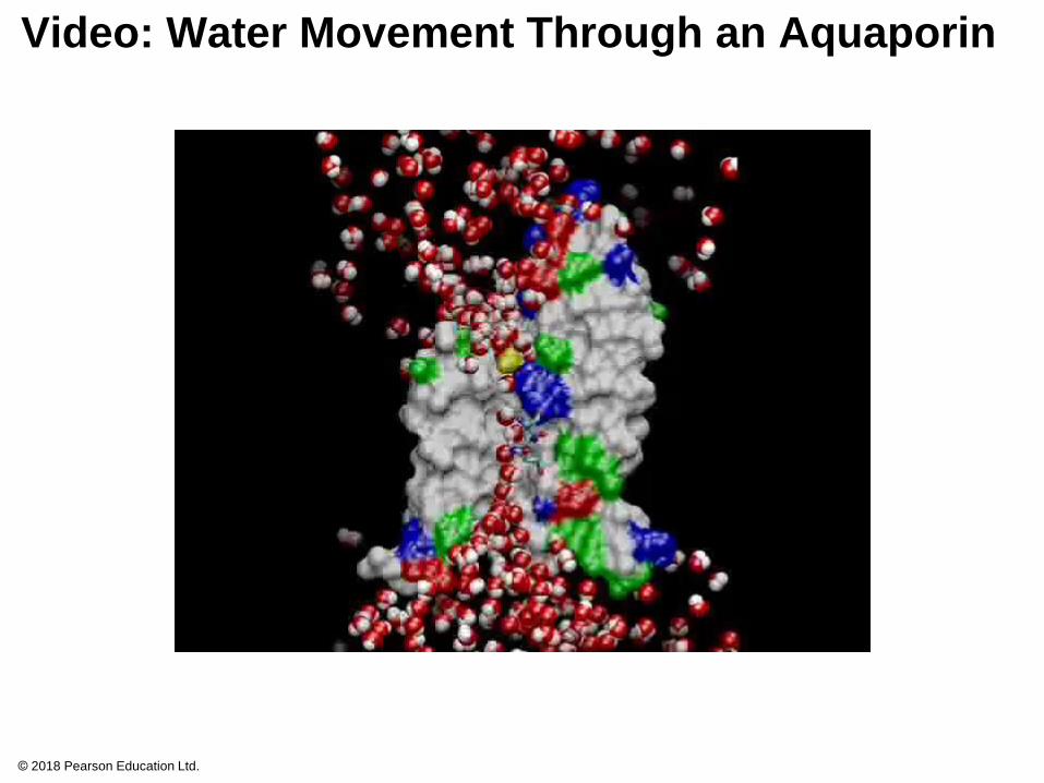
Video: Water Movement Through an Aquaporin
© 2018 Pearson Education Ltd.
Concept 8.1: Cellular membranes are fluid
mosaics of lipids and proteins
Phospholipids are the most abundant lipid in the
plasma membrane
Phospholipids are amphipathic molecules,
containing hydrophobic (“water-fearing”) and
hydrophilic (“water-loving”) regions
The hydrophobic tails of the phospholipids are
sheltered inside the membrane, while the hydrophilic
heads are exposed to water on either side
© 2018 Pearson Education Ltd.
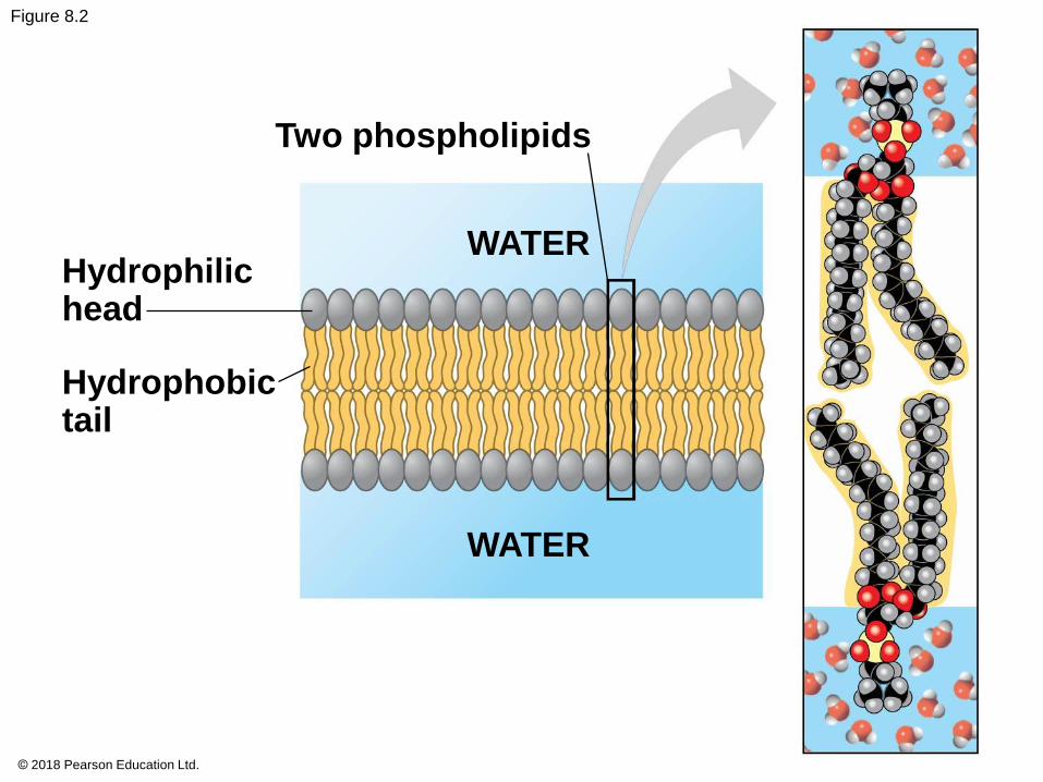
Figure 8.2
Two phospholipids
WATERHydrophilichead
Hydrophobictail
WATER
© 2018 Pearson Education Ltd.
In the fluid mosaic model, the membrane is a
mosaic of protein molecules bobbing in a fluid
bilayer of phospholipids
Proteins are not randomly distributed in the
membrane
© 2018 Pearson Education Ltd.
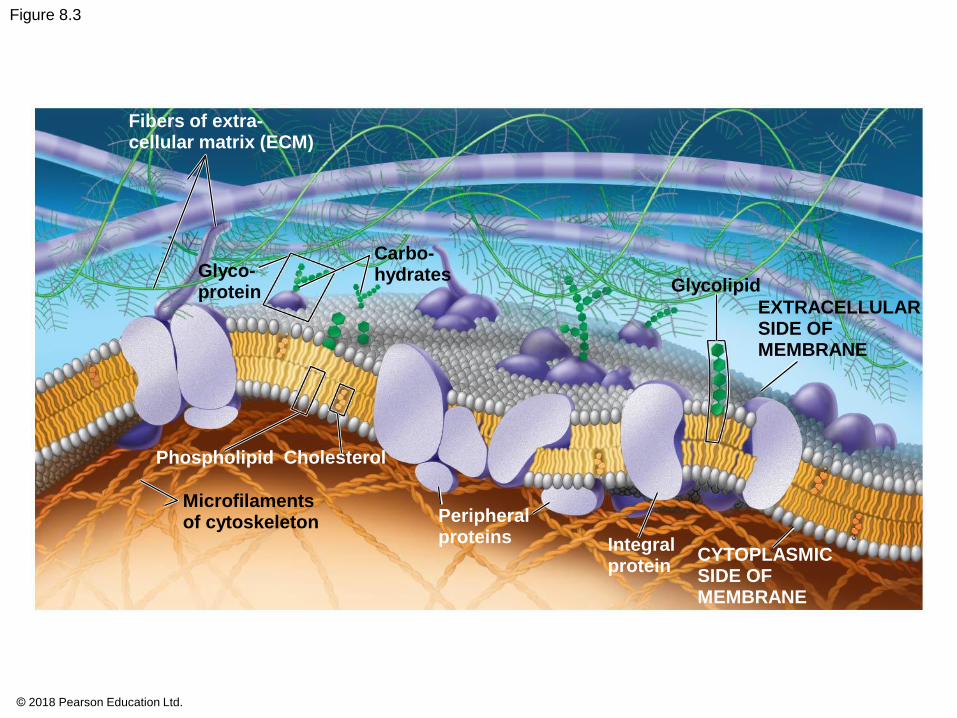
Figure 8.3
Fibers of extra-cellular matrix (ECM)
Glyco-protein
Carbo-hydrates
Glycolipid
EXTRACELLULARSIDE OFMEMBRANE
Phospholipid Cholesterol
Microfilamentsof cytoskeleton Peripheral
proteins Integralprotein
CYTOPLASMICSIDE OFMEMBRANE
© 2018 Pearson Education Ltd.
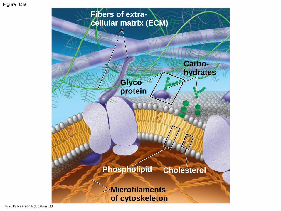
Figure 8.3a
Fibers of extra-cellular matrix (ECM)
Carbo-hydrates
Glyco-protein
Phospholipid Cholesterol
Microfilamentsof cytoskeleton
© 2018 Pearson Education Ltd.

Figure 8.3b
Glycolipid
EXTRACELLULARSIDE OFMEMBRANE
Peripheralproteins
Integralprotein CYTOPLASMIC
SIDE OFMEMBRANE
© 2018 Pearson Education Ltd.
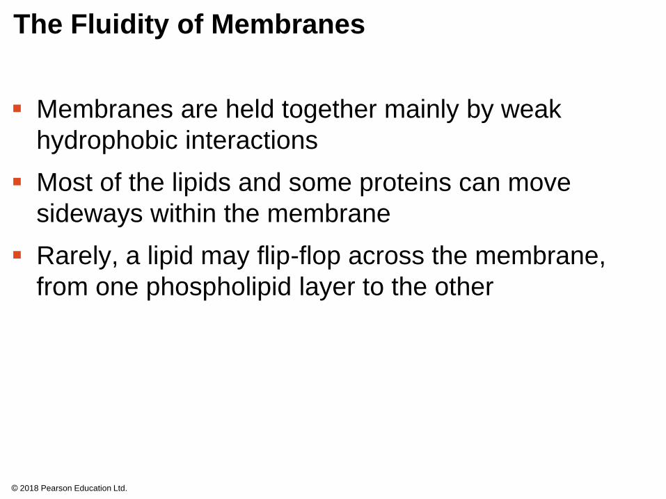
The Fluidity of Membranes
Membranes are held together mainly by weak
hydrophobic interactions
Most of the lipids and some proteins can move
sideways within the membrane
Rarely, a lipid may flip-flop across the membrane,
from one phospholipid layer to the other
© 2018 Pearson Education Ltd.
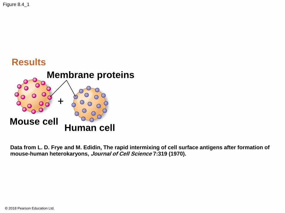
Figure 8.4_1
Results
Membrane proteins
Mouse cellHuman cell
Data from L. D. Frye and M. Edidin, The rapid intermixing of cell surface antigens after formation ofmouse-human heterokaryons, Journal of Cell Science 7:319 (1970).
© 2018 Pearson Education Ltd.
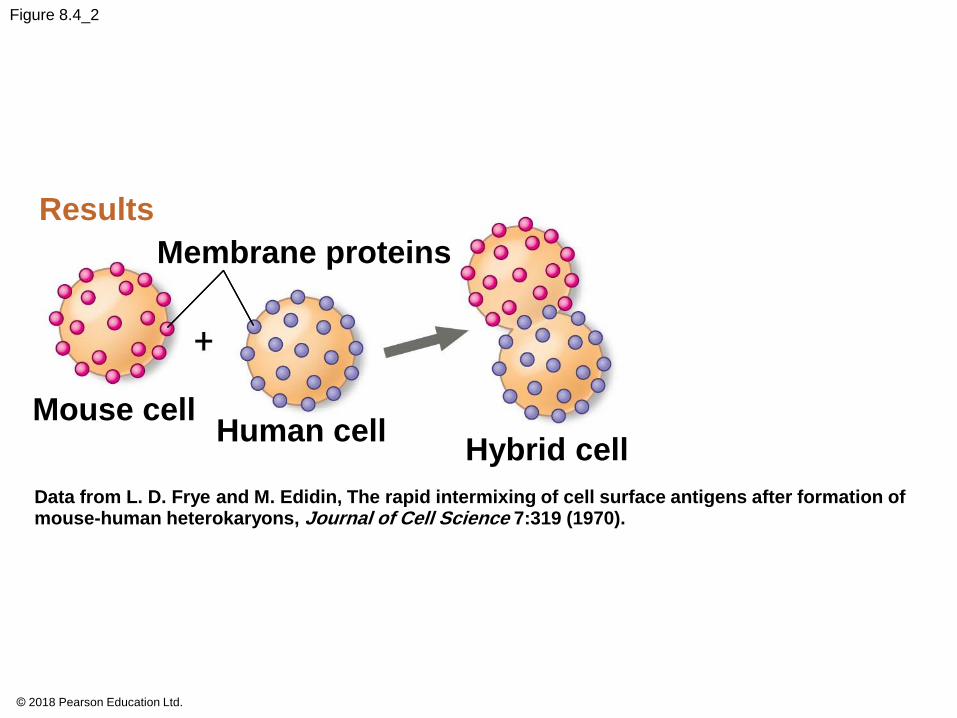
Figure 8.4_2
Results
Membrane proteins
Mouse cellHuman cell
Hybrid cell
Data from L. D. Frye and M. Edidin, The rapid intermixing of cell surface antigens after formation ofmouse-human heterokaryons, Journal of Cell Science 7:319 (1970).
© 2018 Pearson Education Ltd.

Figure 8.4_3
Results
Membrane proteins
Mouse cellHuman cell
Hybrid cell
Mixed proteins
after 1 hour
Data from L. D. Frye and M. Edidin, The rapid intermixing of cell surface antigens after formation ofmouse-human heterokaryons, Journal of Cell Science 7:319 (1970).
© 2018 Pearson Education Ltd.
As temperatures cool, membranes switch from a
fluid state to a solid state
The temperature at which a membrane solidifies
depends on the types of lipids
Membranes rich in unsaturated fatty acids are more
fluid than those rich in saturated fatty acids
Membranes must be fluid to work properly;
membranes are usually about as fluid as salad oil
© 2018 Pearson Education Ltd.
The steroid cholesterol has different effects on the
membrane fluidity of animal cells at different
temperatures
At warm temperatures (such as 37ºC), cholesterol
restrains movement of phospholipids
At cool temperatures, it maintains fluidity by
preventing tight packing
Though cholesterol is present in plants, they use
related steroid lipids to buffer membrane fluidity
© 2018 Pearson Education Ltd.
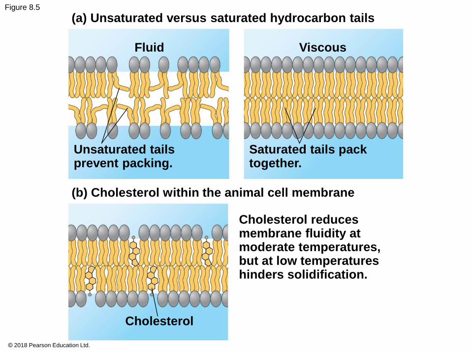
Figure 8.5
(a) Unsaturated versus saturated hydrocarbon tails
Fluid Viscous
Unsaturated tailsprevent packing.
Saturated tails packtogether.
(b) Cholesterol within the animal cell membrane
Cholesterol reducesmembrane fluidity atmoderate temperatures, but at low temperatureshinders solidification.
Cholesterol
© 2018 Pearson Education Ltd.
Evolution of Differences in Membrane Lipid
Composition
Variations in lipid composition of cell membranes of
many species appear to be adaptations to specific
environmental conditions
Ability to change the lipid compositions in response
to temperature changes has evolved in organisms
that live where temperatures vary
© 2018 Pearson Education Ltd.
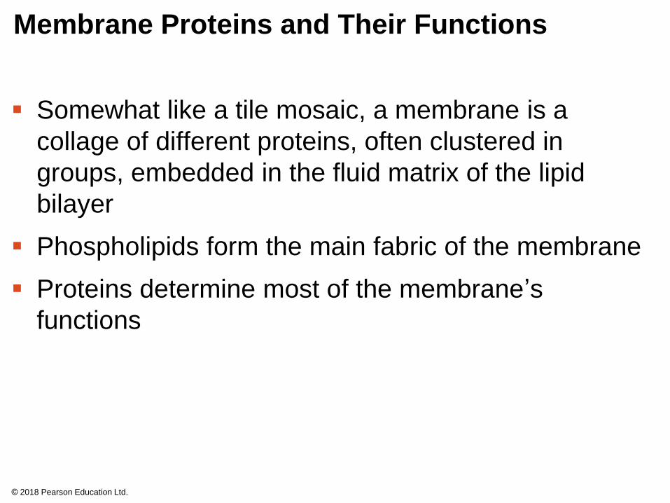
Membrane Proteins and Their Functions
Somewhat like a tile mosaic, a membrane is a
collage of different proteins, often clustered in
groups, embedded in the fluid matrix of the lipid
bilayer
Phospholipids form the main fabric of the membrane
Proteins determine most of the membrane’s
functions
© 2018 Pearson Education Ltd.
Figure 8.UN01
© 2018 Pearson Education Ltd.
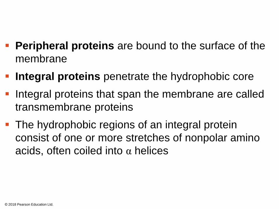
Peripheral proteins are bound to the surface of the
membrane
Integral proteins penetrate the hydrophobic core
Integral proteins that span the membrane are called
transmembrane proteins
The hydrophobic regions of an integral protein
consist of one or more stretches of nonpolar amino
acids, often coiled into α helices
© 2018 Pearson Education Ltd.
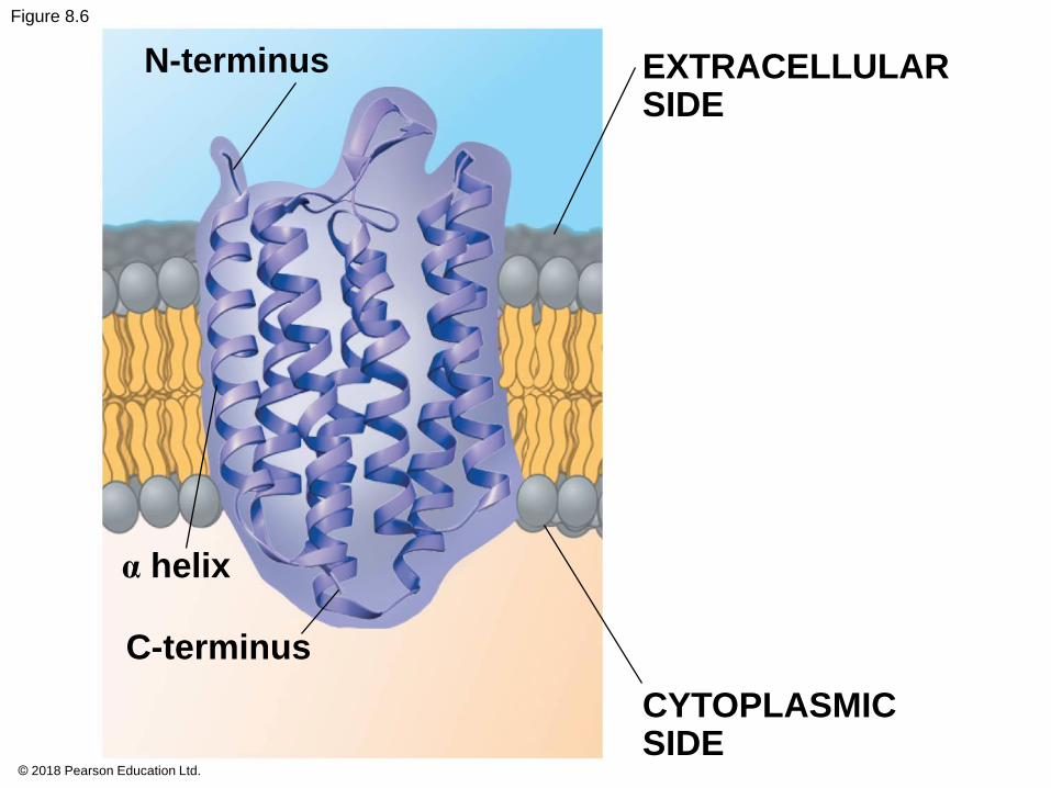
Figure 8.6
N-terminus EXTRACELLULARSIDE
α helix
C-terminus
CYTOPLASMICSIDE
© 2018 Pearson Education Ltd.
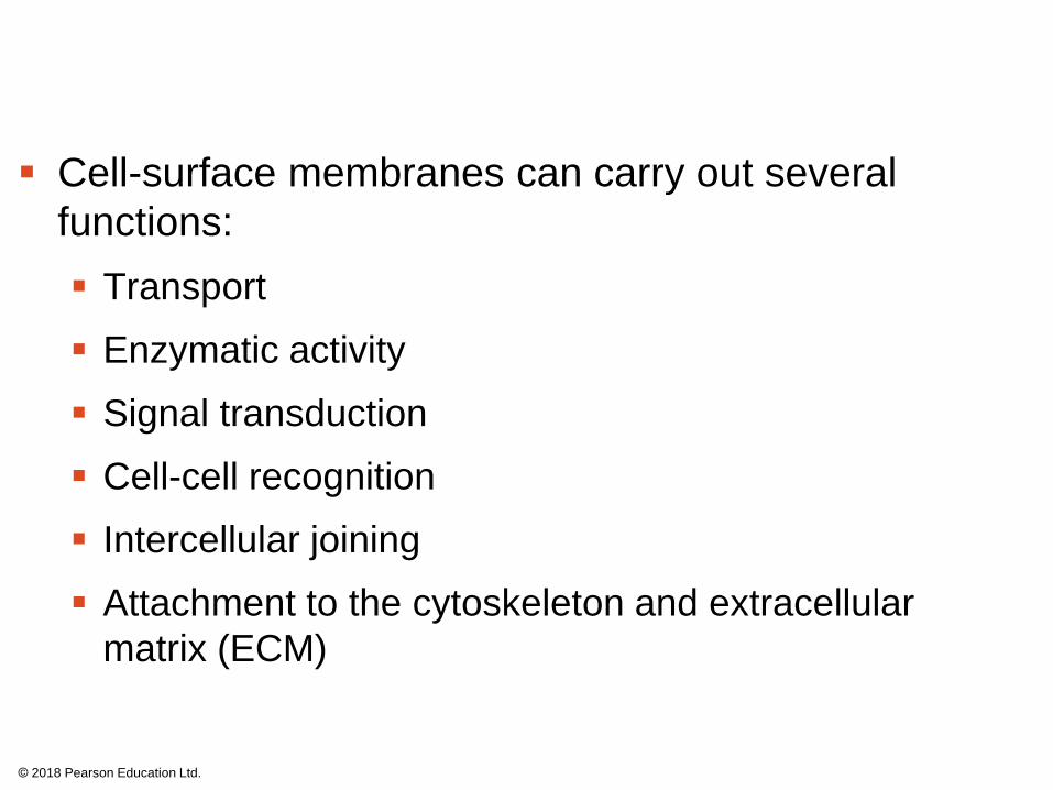
Cell-surface membranes can carry out several
functions:
Transport
Enzymatic activity
Signal transduction
Cell-cell recognition
Intercellular joining
Attachment to the cytoskeleton and extracellular
matrix (ECM)
© 2018 Pearson Education Ltd.
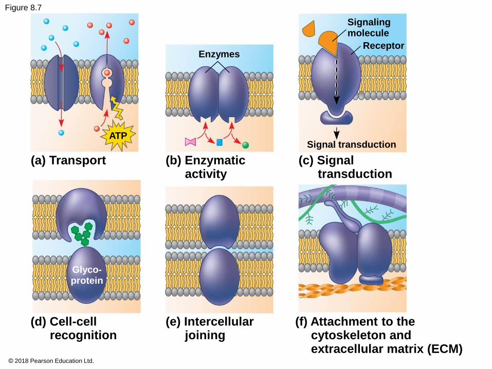
Figure 8.7
Enzymes
Signalingmolecule
Receptor
ATPSignal transduction
(a) Transport (b) Enzymaticactivity
(c) Signaltransduction
Glyco-protein
(d) Cell-cellrecognition
(e) Intercellularjoining
(f) Attachment to thecytoskeleton andextracellular matrix (ECM)
© 2018 Pearson Education Ltd.
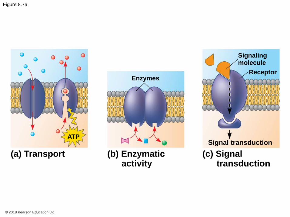
Figure 8.7a
Enzymes
Signalingmolecule
Receptor
ATPSignal transduction
(a) Transport (b) Enzymaticactivity
(c) Signaltransduction
© 2018 Pearson Education Ltd.
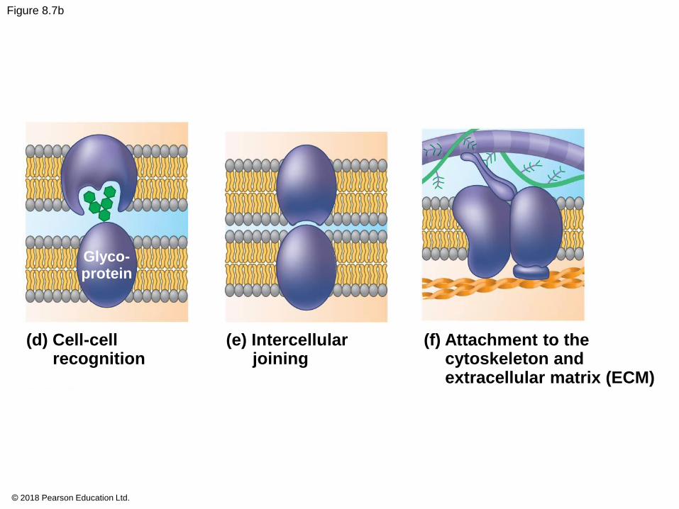
Figure 8.7b
Glyco-protein
(d) Cell-cellrecognition
(e) Intercellularjoining
(f) Attachment to thecytoskeleton andextracellular matrix (ECM)
© 2018 Pearson Education Ltd.
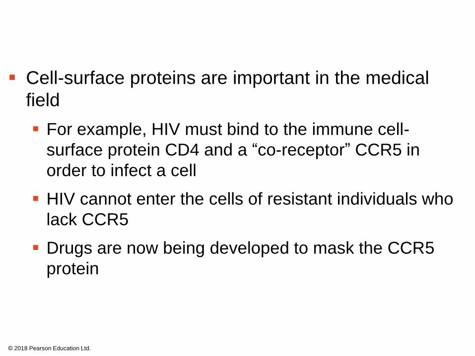
Cell-surface proteins are important in the medical
field
For example, HIV must bind to the immune cell-
surface protein CD4 and a “co-receptor” CCR5 in
order to infect a cell
HIV cannot enter the cells of resistant individuals who
lack CCR5
Drugs are now being developed to mask the CCR5
protein
© 2018 Pearson Education Ltd.
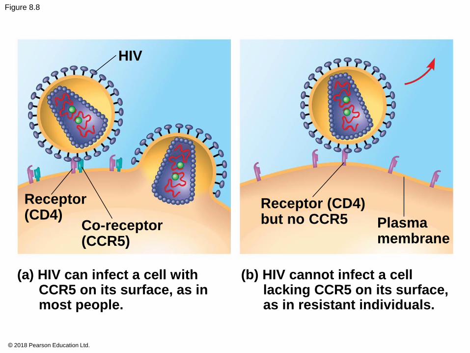
Figure 8.8
HIV
Receptor(CD4)
Co-receptor(CCR5)
(a) HIV can infect a cell withCCR5 on its surface, as inmost people.
Receptor (CD4)but no CCR5 Plasma
membrane
(b) HIV cannot infect a celllacking CCR5 on its surface,as in resistant individuals.
© 2018 Pearson Education Ltd.
The Role of Membrane Carbohydrates in Cell-
Cell Recognition
Cells recognize each other by binding to molecules,
often containing carbohydrates, on the extracellular
surface of the plasma membrane
Membrane carbohydrates may be covalently bonded
to lipids (forming glycolipids) or, more commonly, to
proteins (forming glycoproteins)
Carbohydrates on the extracellular side of the
plasma membrane vary among species, individuals,
and even cell types in an individual
© 2018 Pearson Education Ltd.
Synthesis and Sidedness of Membranes
Membranes have distinct inside and outside faces
The asymmetrical distribution of proteins, lipids, and
associated carbohydrates in the plasma membrane
is determined when the membrane is built by the ER
and Golgi apparatus
© 2018 Pearson Education Ltd.
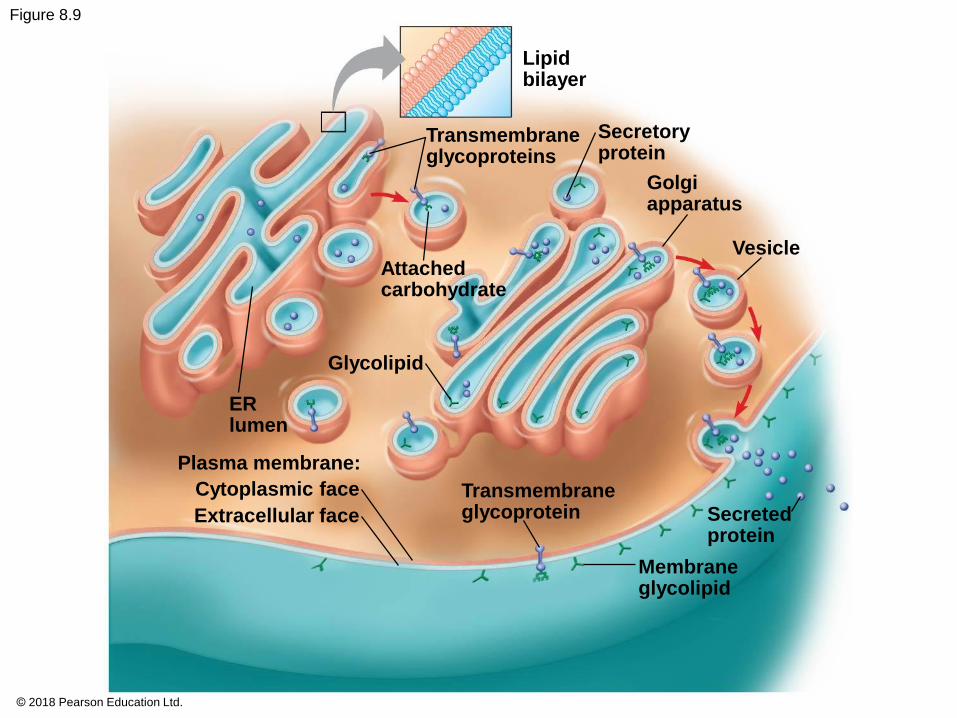
Figure 8.9
Lipidbilayer
Transmembraneglycoproteins
Secretoryprotein
Golgiapparatus
Attachedcarbohydrate
Glycolipid
ERlumen
Plasma membrane:
Cytoplasmic face
Extracellular face
Transmembraneglycoprotein
Vesicle
Secretedprotein
Membraneglycolipid
© 2018 Pearson Education Ltd.
Concept 8.2: Membrane structure results in
selective permeability
A cell must exchange materials with its
surroundings, a process controlled by the plasma
membrane
Plasma membranes are selectively permeable,
regulating the cell’s molecular traffic
© 2018 Pearson Education Ltd.
The Permeability of the Lipid Bilayer
Hydrophobic (nonpolar) molecules, such as
hydrocarbons, can dissolve in the lipid bilayer and
pass through the membrane rapidly
Hydrophilic molecules including ions and polar
molecules do not cross the membrane easily
Proteins built into the membrane play key roles in
regulating transport
© 2018 Pearson Education Ltd.
Transport Proteins
Transport proteins allow passage of hydrophilic
substances across the membrane
Some transport proteins, called channel proteins,
have a hydrophilic channel that certain molecules or
ions can use as a tunnel
Channel proteins called aquaporins greatly facilitate
the passage of water molecules
© 2018 Pearson Education Ltd.
Other transport proteins, called carrier proteins, bind
to molecules and change shape to shuttle them
across the membrane
A transport protein is specific for the substance
it moves
© 2018 Pearson Education Ltd.
Concept 8.3: Passive transport is diffusion of a
substance across a membrane with no energy
investment
Diffusion is the tendency for molecules to spread
out evenly into the available space
Although each molecule moves randomly, diffusion
of a population of molecules may be directional
At dynamic equilibrium, as many molecules cross
the membrane in one direction as in the other
© 2018 Pearson Education Ltd.
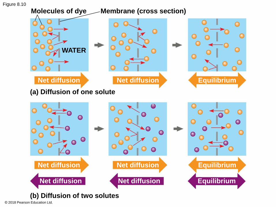
Figure 8.10
Molecules of dye Membrane (cross section)
WATER
Net diffusion
(a) Diffusion of one solute
Net diffusion Equilibrium
Net diffusion
Net diffusion
Net diffusion
Net diffusion
Equilibrium
Equilibrium
(b) Diffusion of two solutes© 2018 Pearson Education Ltd.
Animation: Diffusion
© 2018 Pearson Education Ltd.
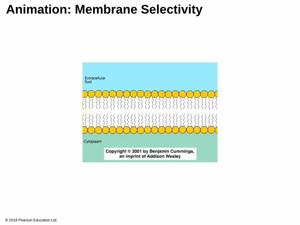
Animation: Membrane Selectivity
© 2018 Pearson Education Ltd.
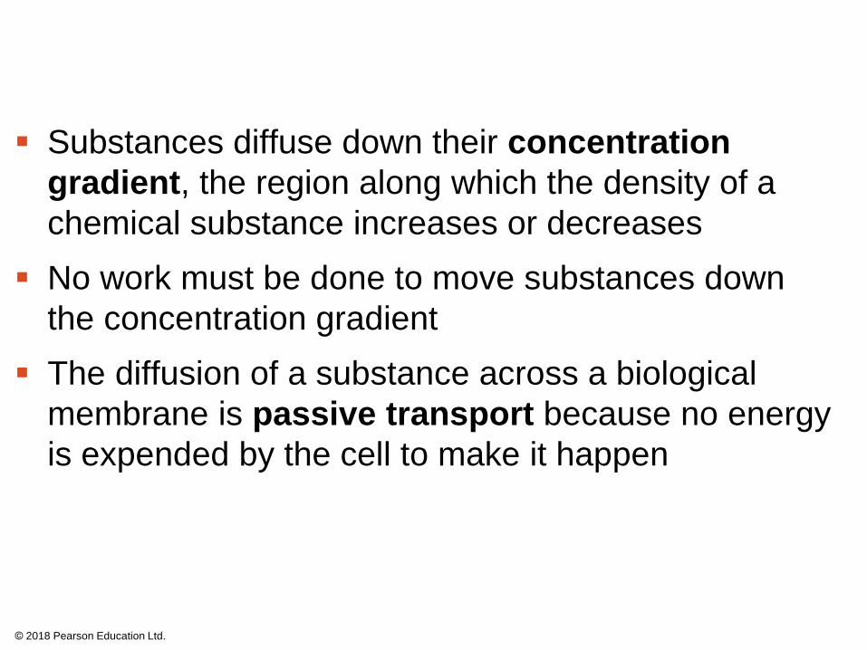
Substances diffuse down their concentration
gradient, the region along which the density of a
chemical substance increases or decreases
No work must be done to move substances down
the concentration gradient
The diffusion of a substance across a biological
membrane is passive transport because no energy
is expended by the cell to make it happen
© 2018 Pearson Education Ltd.
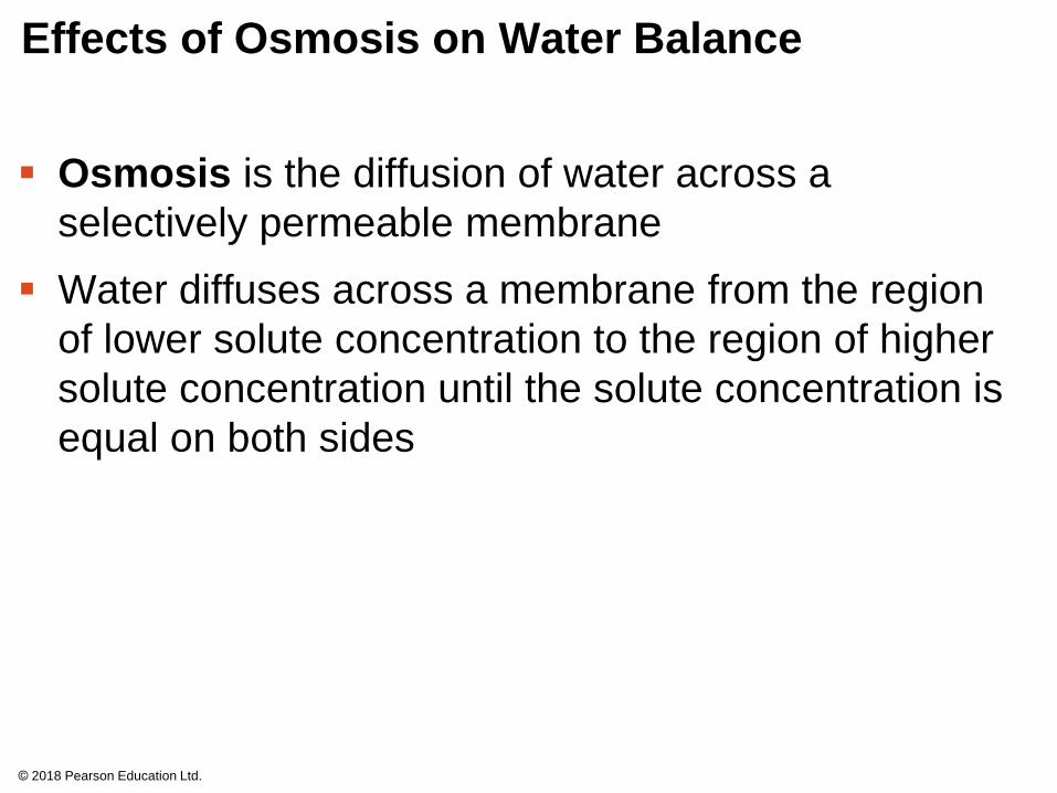
Effects of Osmosis on Water Balance
Osmosis is the diffusion of water across a
selectively permeable membrane
Water diffuses across a membrane from the region
of lower solute concentration to the region of higher
solute concentration until the solute concentration is
equal on both sides
© 2018 Pearson Education Ltd.
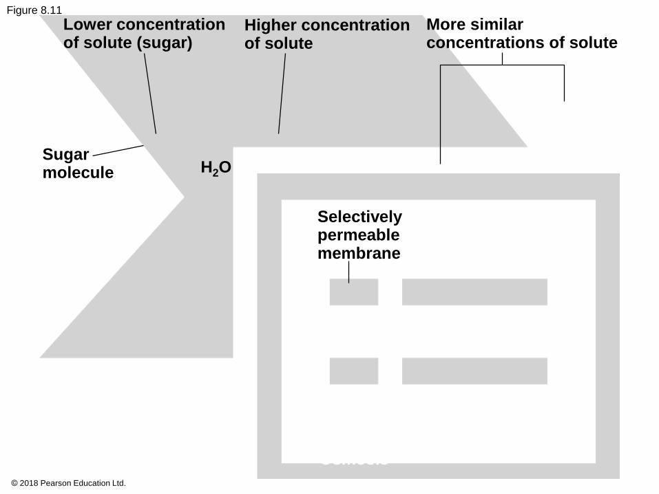
Figure 8.11
Lower concentrationof solute (sugar)
Higher concentrationof solute
More similarconcentrations of solute
Sugarmolecule H2O
Selectivelypermeablemembrane
Osmosis
© 2018 Pearson Education Ltd.
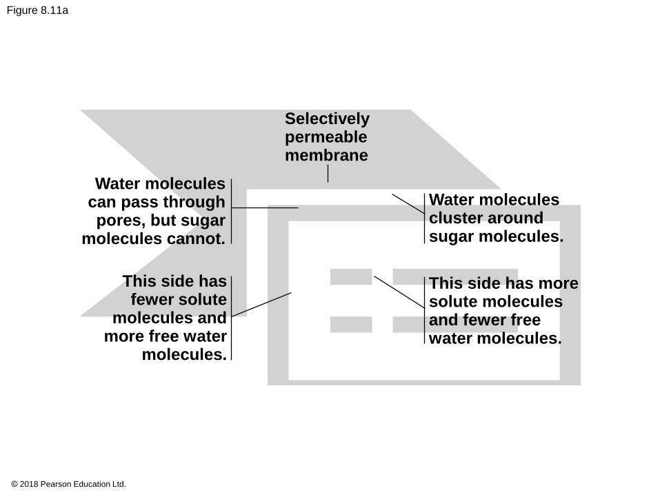
Figure 8.11a
Selectivelypermeablemembrane
Water moleculescan pass throughpores, but sugar
molecules cannot.
This side hasfewer solute
molecules andmore free water
molecules.
Water moleculescluster aroundsugar molecules.
This side has moresolute moleculesand fewer freewater molecules.
Osmosis
© 2018 Pearson Education Ltd.
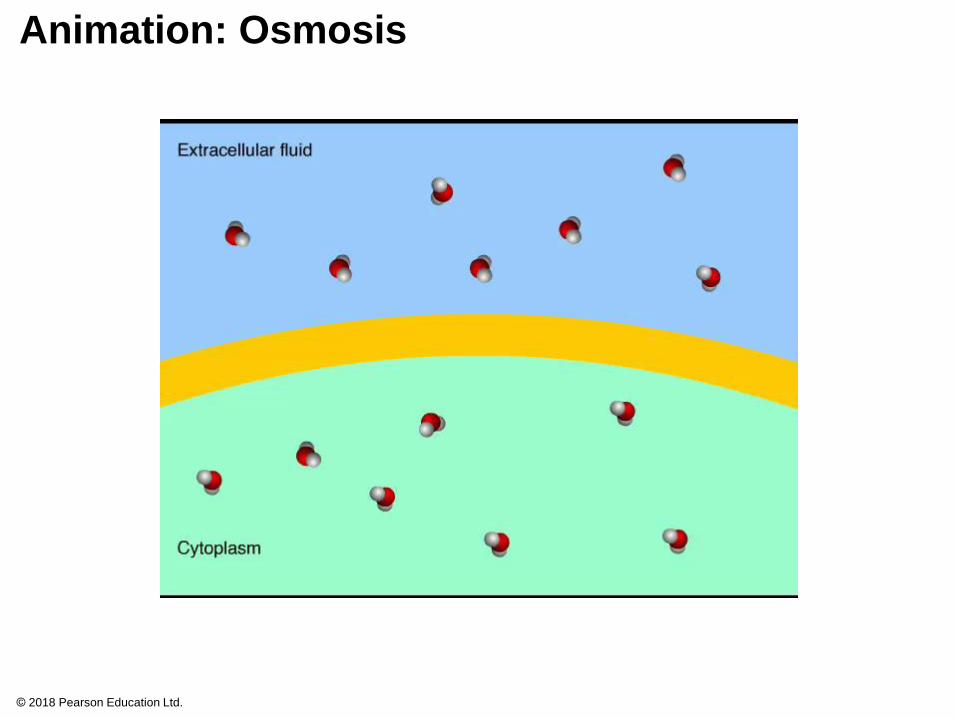
Animation: Osmosis
© 2018 Pearson Education Ltd.
Water Balance of Cells Without Cell Walls
Tonicity is the ability of a surrounding solution to
cause a cell to gain or lose water
The tonicity of a solution depends on its
concentration of solutes that cannot cross the
membrane relative to that inside the cell
© 2018 Pearson Education Ltd.
Isotonic solution: Solute concentration is the same
as that inside the cell; no net water movement
across the plasma membrane
Hypertonic solution: Solute concentration is greater
than that inside the cell; cell loses water
Hypotonic solution: Solute concentration is less
than that inside the cell; cell gains water
Cells without cell walls will shrivel in hypertonic
solution and lyse (burst) in a hypotonic solution
© 2018 Pearson Education Ltd.
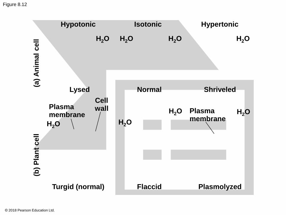
Figure 8.12
Hypotonic
H2O H2O
Isotonic
H2O
Hypertonic
H2O(a
) A
nim
al
cell
Lysed
Plasmamembrane
H2O
Cellwall
H2O
Normal Shriveled
H2O Plasmamembrane
H2O
(b)
Pla
nt
cell
Turgid (normal) Flaccid Plasmolyzed
© 2018 Pearson Education Ltd.
Hypertonic or hypotonic environments create
osmotic problems for organisms that have cells
without rigid walls
Osmoregulation, the control of solute
concentrations and water balance, is a necessary
adaptation for life in such environments
For example, the unicellular eukaryote Paramecium,
which is hypertonic to its pond water environment, has
a contractile vacuole that acts as a pump
© 2018 Pearson Education Ltd.
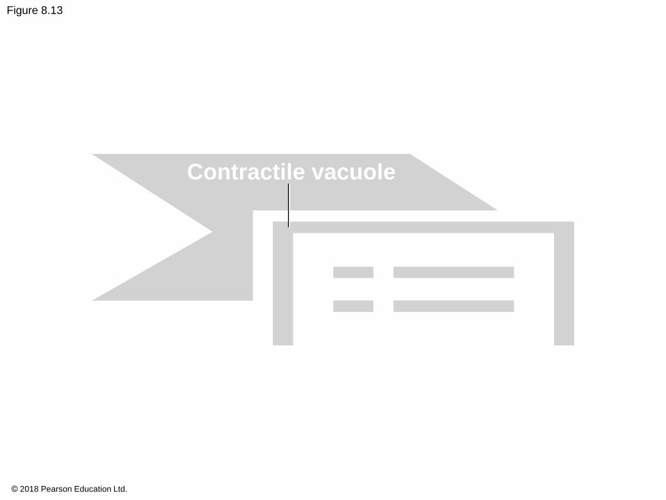
Figure 8.13
Contractile vacuole50 µm
© 2018 Pearson Education Ltd.
Video: Chlamydomonas
© 2018 Pearson Education Ltd.
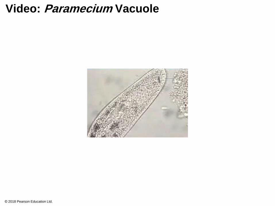
Video: Paramecium Vacuole
© 2018 Pearson Education Ltd.
Bacteria and archaea that live in hypersaline
(excessively salty) environments have cellular
mechanisms to balance internal and external solute
concentrations
© 2018 Pearson Education Ltd.
Water Balance of Cells with Cell Walls
Cell walls help maintain water balance
A plant cell in a hypotonic solution swells until the
wall opposes uptake; the cell is now turgid (firm)
If a plant cell and its surroundings are isotonic, there
is no net movement of water into the cell; the cell
becomes flaccid (limp)
© 2018 Pearson Education Ltd.
In a hypertonic environment, plant cells lose water
The membrane pulls away from the cell wall,
causing the plant to wilt, a potentially lethal effect
called plasmolysis
© 2018 Pearson Education Ltd.
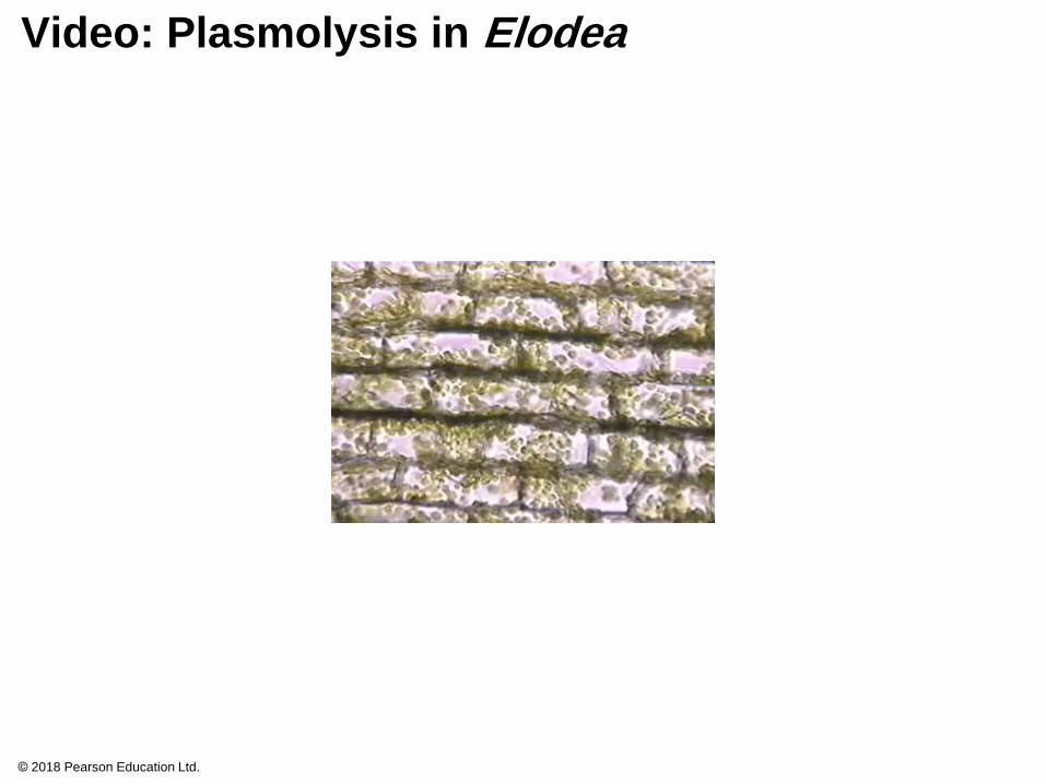
Video: Plasmolysis in Elodea
© 2018 Pearson Education Ltd.

Video: Turgid Elodea
© 2018 Pearson Education Ltd.
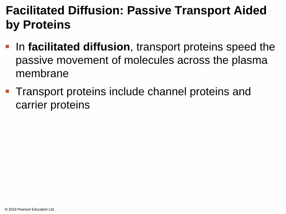
Facilitated Diffusion: Passive Transport Aided
by Proteins
In facilitated diffusion, transport proteins speed the
passive movement of molecules across the plasma
membrane
Transport proteins include channel proteins and
carrier proteins
© 2018 Pearson Education Ltd.
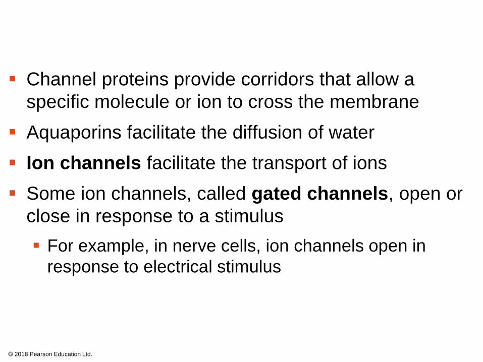
Channel proteins provide corridors that allow a
specific molecule or ion to cross the membrane
Aquaporins facilitate the diffusion of water
Ion channels facilitate the transport of ions
Some ion channels, called gated channels, open or
close in response to a stimulus
For example, in nerve cells, ion channels open in
response to electrical stimulus
© 2018 Pearson Education Ltd.
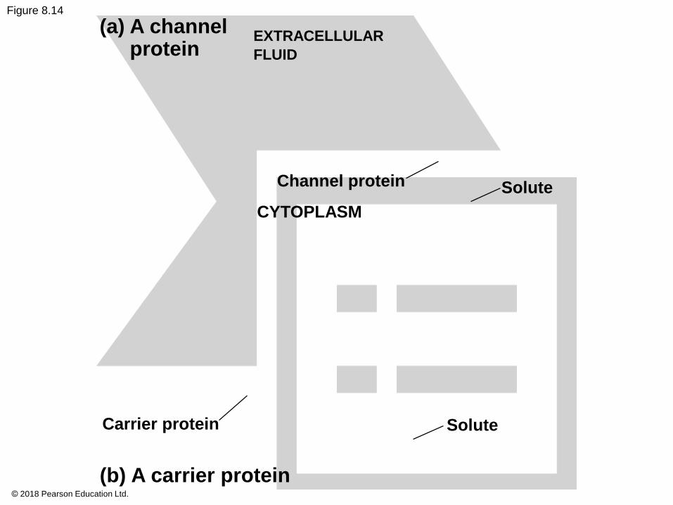
Figure 8.14
(a) A channelprotein
EXTRACELLULAR
FLUID
Channel protein
CYTOPLASM
Solute
Carrier protein Solute
(b) A carrier protein© 2018 Pearson Education Ltd.
Carrier proteins undergo a subtle change in shape
that translocates the solute-binding site across the
membrane
This change in shape can be triggered by the
binding and release of the transported molecule
© 2018 Pearson Education Ltd.
Concept 8.4: Active transport uses energy to
move solutes against their gradients
Facilitated diffusion is still passive because the
solute moves down its concentration gradient, and
the transport requires no energy
Some transport proteins, however, can move solutes
against their concentration gradients
© 2018 Pearson Education Ltd.
The Need for Energy in Active Transport
Active transport requires energy, usually in the
form of ATP hydrolysis, to move substances against
their concentration gradients
All proteins involved in active transport are carrier
proteins
© 2018 Pearson Education Ltd.
Active transport allows cells to maintain
concentration gradients that differ from their
surroundings
For example, an animal cell has a much higher
potassium (K+) and a much lower sodium (Na+)
concentration compared to its surroundings
This is controlled by the sodium-potassium pump, a
transport protein that is energized by transfer of a
phosphate group from the hydrolysis of ATP
© 2018 Pearson Education Ltd.
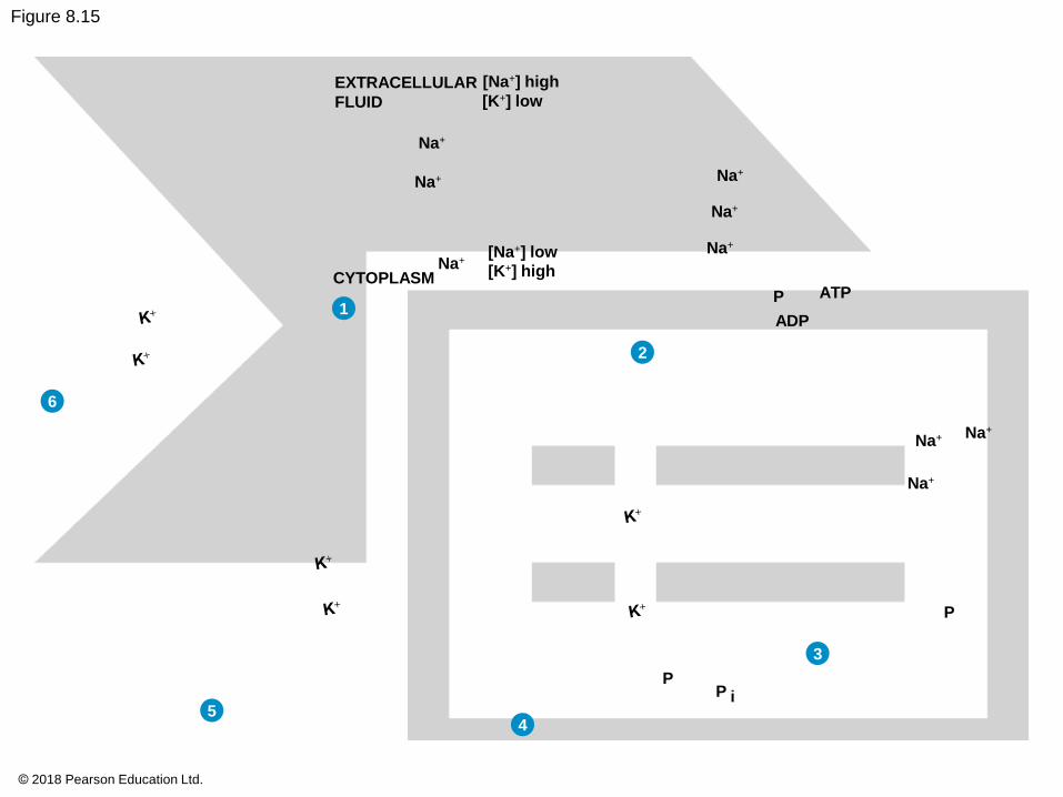
Figure 8.15
EXTRACELLULAR
FLUID
P
ADP
ATPCYTOPLASM
Na+
Na+ Na+
Na+
Na+Na+
Na+
Na+
Na+
P
P
P i
[K+] low
[Na+] low
[K+] high
[Na+] high
6
1
2
3
45
© 2018 Pearson Education Ltd.
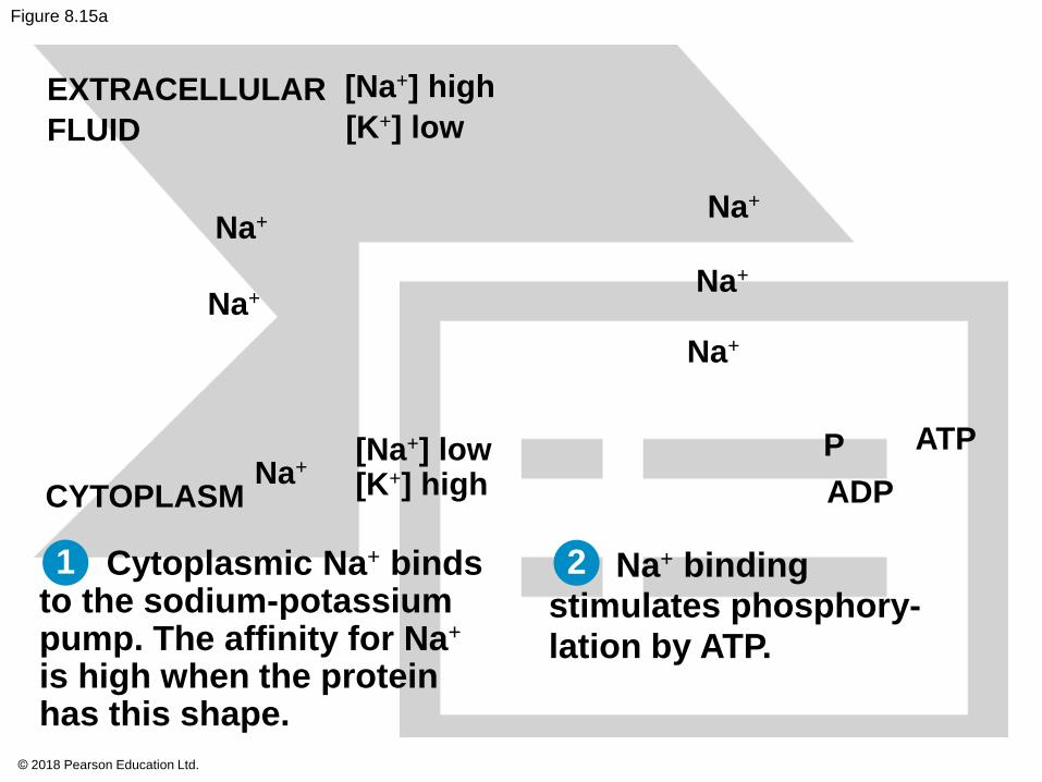
Figure 8.15a
Cytoplasmic Na+ bindsto the sodium-potassiumpump. The affinity for Na+
is high when the proteinhas this shape.
Na+ bindingstimulates phosphory-lation by ATP.
ATPP
ADP
EXTRACELLULAR
FLUID
CYTOPLASM
[K+] low
Na+
Na+
Na+
Na+
Na+
[Na+] low[K+] highNa+
[Na+] high
1 2
© 2018 Pearson Education Ltd.
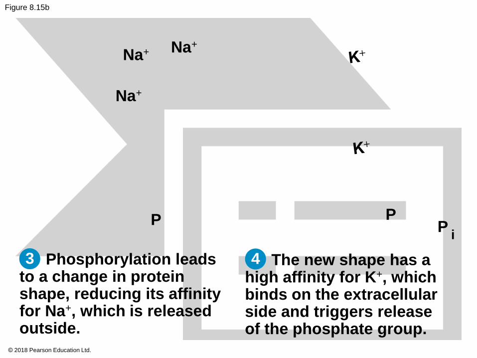
Figure 8.15b
Na+
Na+
Na+
P
Phosphorylation leadsto a change in proteinshape, reducing its affinityfor Na+, which is releasedoutside.
PP
The new shape has ahigh affinity for K+, whichbinds on the extracellularside and triggers releaseof the phosphate group.
i
43
© 2018 Pearson Education Ltd.
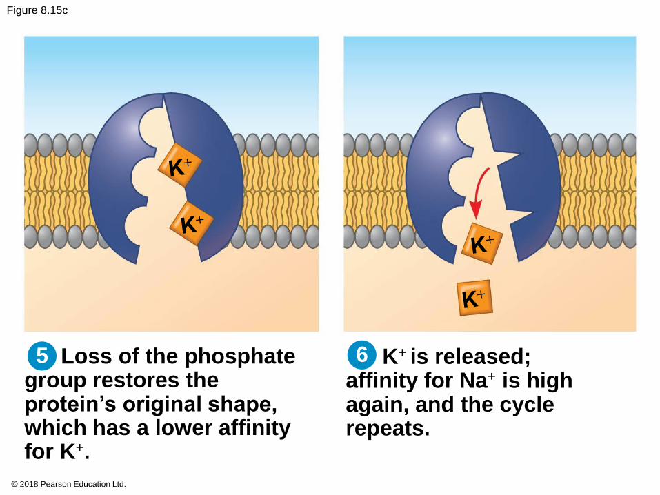
Figure 8.15c
Loss of the phosphategroup restores theprotein’s original shape,which has a lower affinityfor K+.
K+ is released;affinity for Na+ is highagain, and the cyclerepeats.
5 6
© 2018 Pearson Education Ltd.
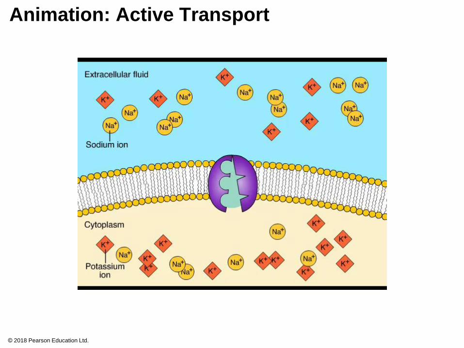
Animation: Active Transport
© 2018 Pearson Education Ltd.
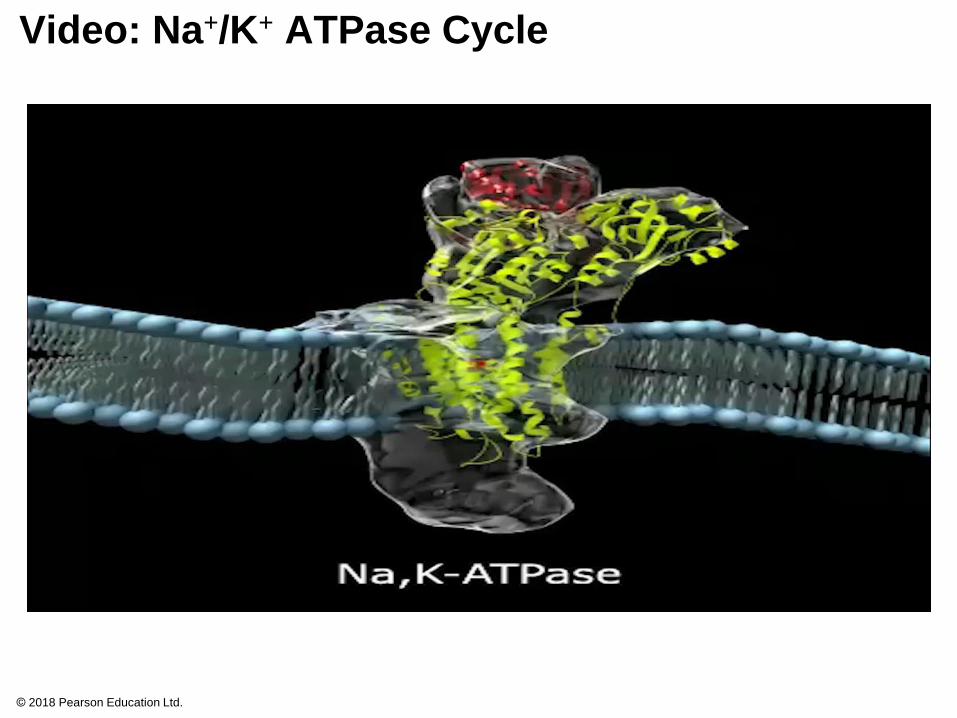
Video: Na+/K+ ATPase Cycle
© 2018 Pearson Education Ltd.
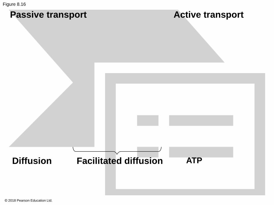
Figure 8.16
Passive transport Active transport
Diffusion Facilitated diffusion ATP
© 2018 Pearson Education Ltd.
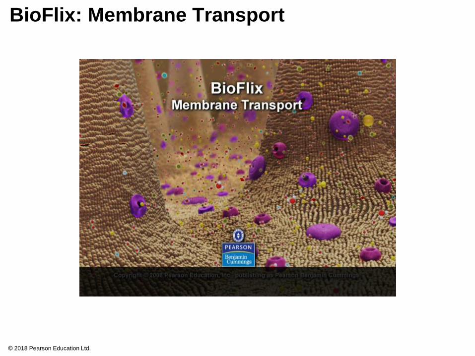
BioFlix: Membrane Transport
© 2018 Pearson Education Ltd.
How Ion Pumps Maintain Membrane Potential
Membrane potential is the voltage across a
membrane
Voltage is created by differences in the distribution
of positive and negative ions across a membrane
The cytoplasmic side of the membrane is negative in
charge relative to the extracellular side
© 2018 Pearson Education Ltd.
Two combined forces, collectively called the
electrochemical gradient, drive the diffusion of ions
across a membrane
A chemical force (the ion’s concentration gradient)
An electrical force (the effect of the membrane
potential on the ion’s movement)
© 2018 Pearson Education Ltd.
An electrogenic pump is a transport protein that
generates voltage across a membrane
The sodium-potassium pump is the major
electrogenic pump of animal cells
The main electrogenic pump of plants, fungi, and
bacteria is a proton pump, which actively transports
hydrogen ions (H+) out of the cell
Electrogenic pumps help store energy that can be
used for cellular work
© 2018 Pearson Education Ltd.
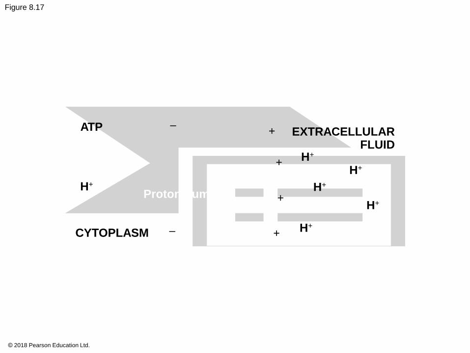
Figure 8.17
ATP –+
H+
Proton pump
–
EXTRACELLULARFLUID
H+
+H+
H+
+H+
H+CYTOPLASM +
© 2018 Pearson Education Ltd.
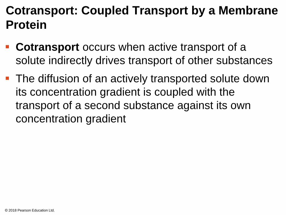
Cotransport: Coupled Transport by a Membrane
Protein
Cotransport occurs when active transport of a
solute indirectly drives transport of other substances
The diffusion of an actively transported solute down
its concentration gradient is coupled with the
transport of a second substance against its own
concentration gradient
© 2018 Pearson Education Ltd.
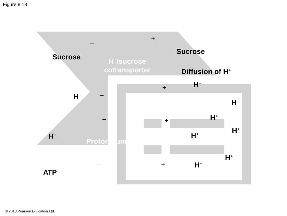
Figure 8.18
–
SucroseH+/sucrose
cotransporter
–
–
H+
Proton pump
–
+
Sucrose
Diffusion of H+
+ H+
H+
+
H+
H+
H+
H+
H+
ATP+ H+
© 2018 Pearson Education Ltd.
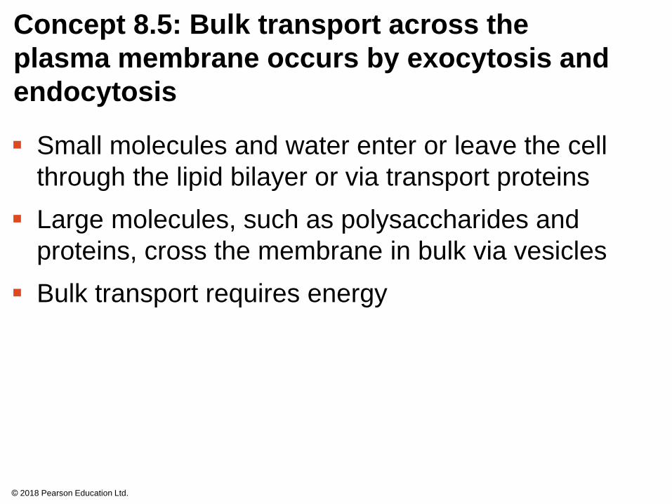
Concept 8.5: Bulk transport across the
plasma membrane occurs by exocytosis and
endocytosis
Small molecules and water enter or leave the cell
through the lipid bilayer or via transport proteins
Large molecules, such as polysaccharides and
proteins, cross the membrane in bulk via vesicles
Bulk transport requires energy
© 2018 Pearson Education Ltd.
Animation: Exocytosis and Endocytosis Introduction
© 2018 Pearson Education Ltd.
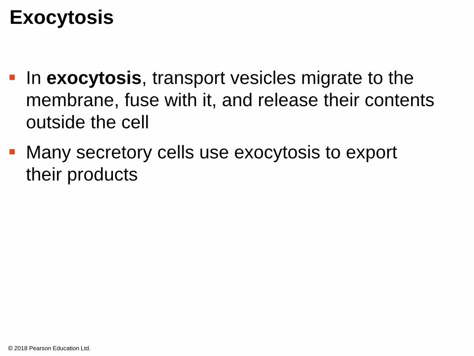
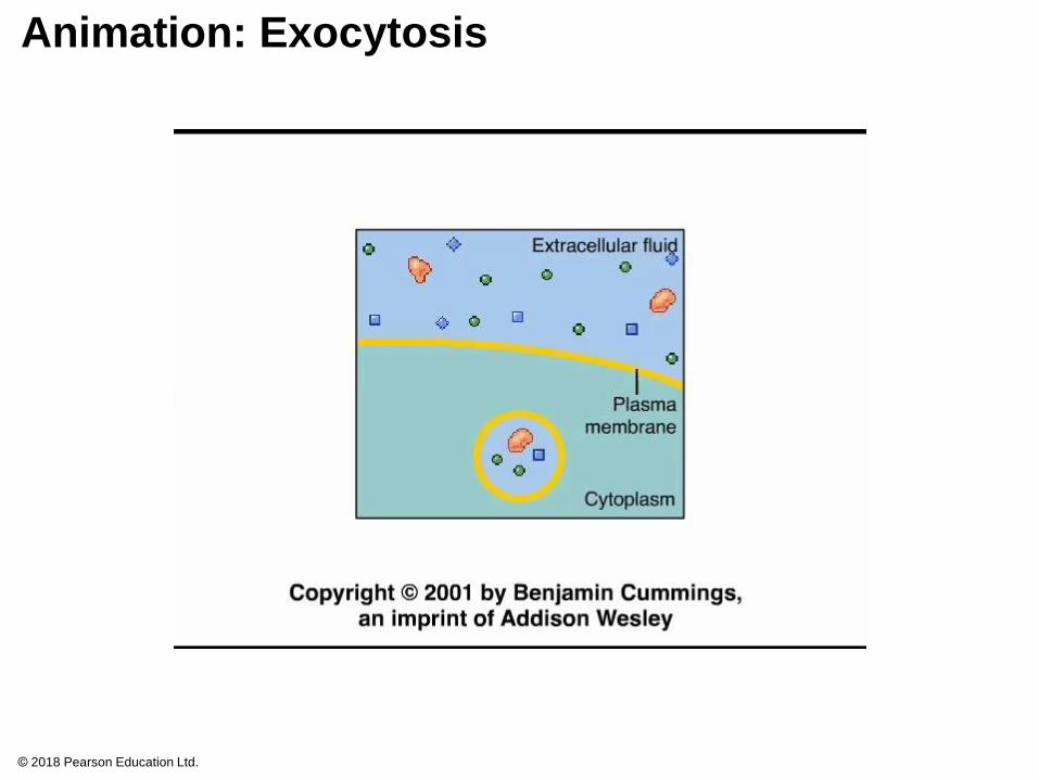
Exocytosis
In exocytosis, transport vesicles migrate to the
membrane, fuse with it, and release their contents
outside the cell
Many secretory cells use exocytosis to export
their products
© 2018 Pearson Education Ltd.
Animation: Exocytosis
© 2018 Pearson Education Ltd.
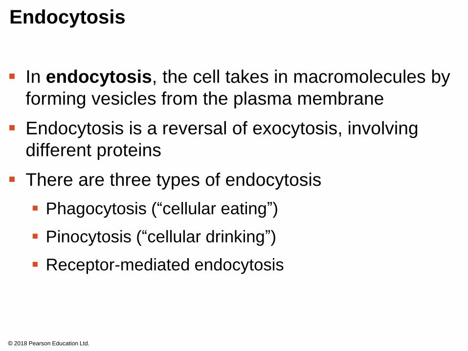
Endocytosis
In endocytosis, the cell takes in macromolecules by
forming vesicles from the plasma membrane
Endocytosis is a reversal of exocytosis, involving
different proteins
There are three types of endocytosis
Phagocytosis (“cellular eating”)
Pinocytosis (“cellular drinking”)
Receptor-mediated endocytosis
© 2018 Pearson Education Ltd.

Figure 8.19
Phagocytosis
EXTRACELLULARFLUID Solutes
Pseudopodium
PinocytosisReceptor-Mediated
Endocytosis
Solutes
Plasmamembrane
Coatprotein
Receptor
Foodorotherparticle
Coatedpit
Foodvacuole
CYTOPLASM
Coatedvesicle
Coated vesicle withspecific solutes(purple) bound toreceptors (red)
© 2018 Pearson Education Ltd.
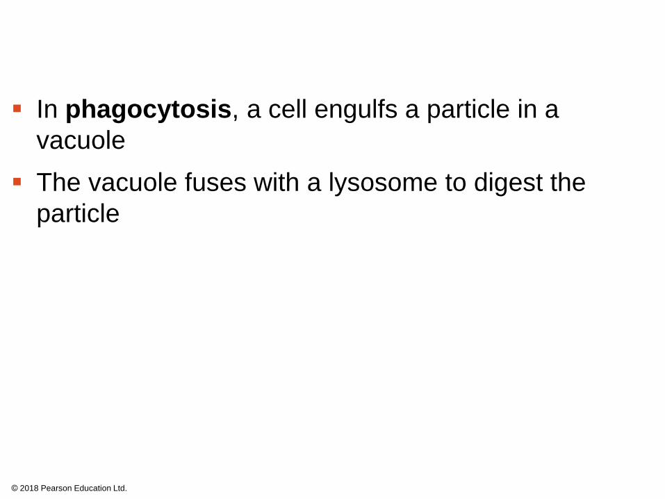
In phagocytosis, a cell engulfs a particle in a
vacuole
The vacuole fuses with a lysosome to digest the
particle
© 2018 Pearson Education Ltd.
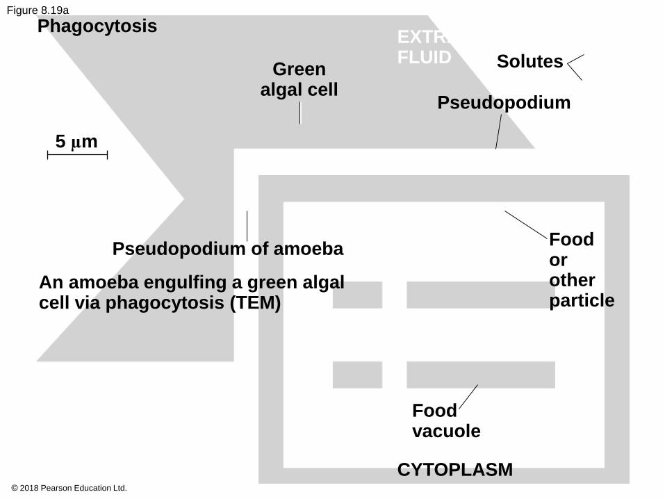
Figure 8.19a
Phagocytosis
Greenalgal cell
5 µm
EXTRACELLULARFLUID Solutes
Pseudopodium
Pseudopodium of amoeba
An amoeba engulfing a green algalcell via phagocytosis (TEM)
Foodorotherparticle
Foodvacuole
CYTOPLASM© 2018 Pearson Education Ltd.
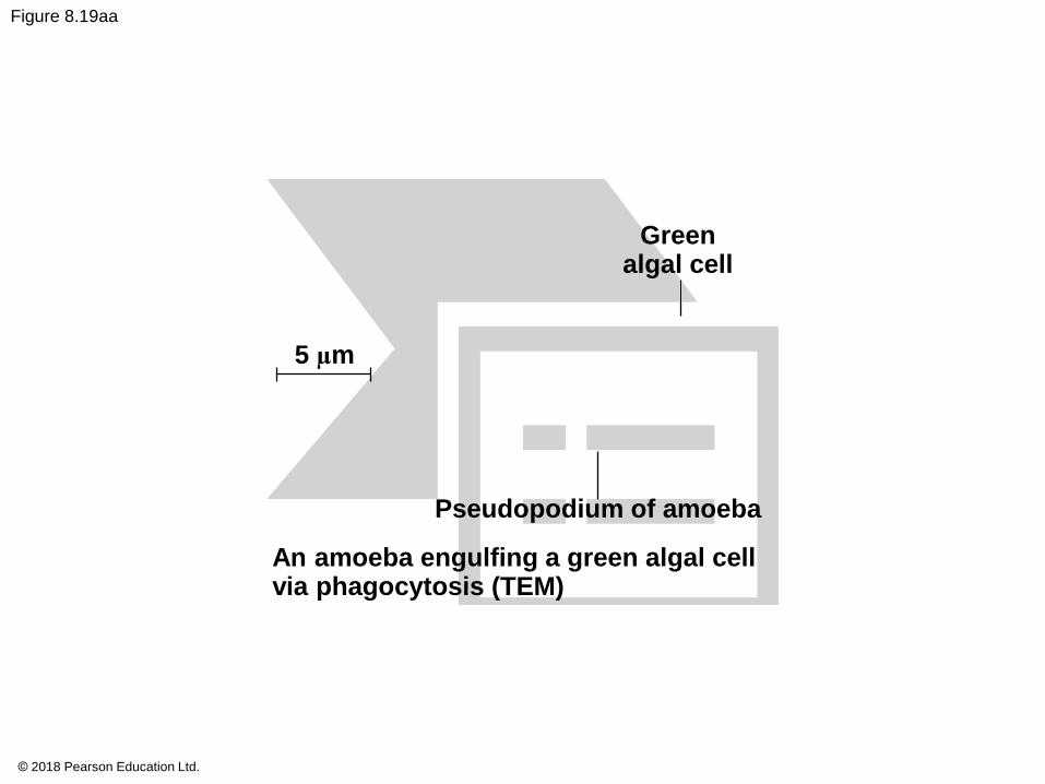
Figure 8.19aa
Greenalgal cell
5 µm
Pseudopodium of amoeba
An amoeba engulfing a green algal cellvia phagocytosis (TEM)
© 2018 Pearson Education Ltd.
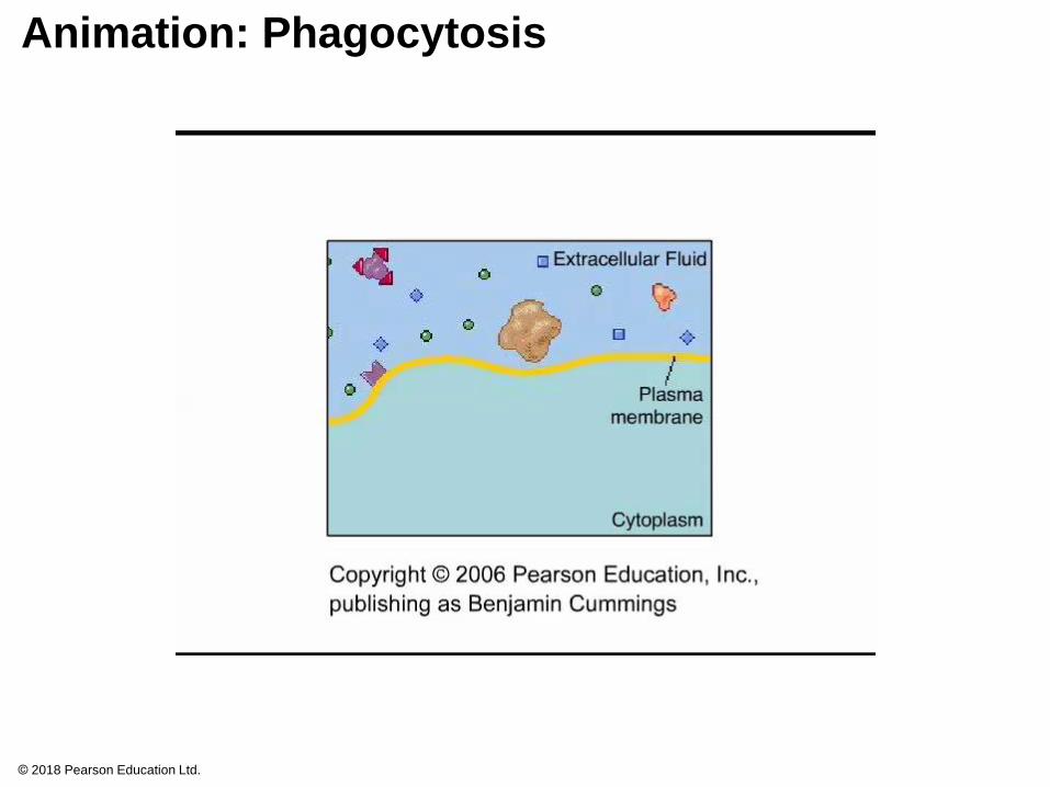
Animation: Phagocytosis
© 2018 Pearson Education Ltd.
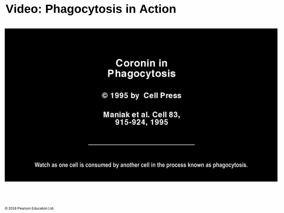
Video: Phagocytosis in Action
© 2018 Pearson Education Ltd.
In pinocytosis, molecules dissolved in droplets are
taken up when extracellular fluid is “gulped” into tiny
vesicles
© 2018 Pearson Education Ltd.

Figure 8.19b
Pinocytosis
Solutes
Plasmamembrane
0.2
5 µ
m Coatprotein
Coatedpit
Pinocytotic vesiclesforming (TEMs)
Coatedvesicle
© 2018 Pearson Education Ltd.
Figure 8.19ba
Pinocytotic vesicles forming (TEMs)
0.2
5 µ
m
© 2018 Pearson Education Ltd.
Animation: Pinocytosis
© 2018 Pearson Education Ltd.
In receptor-mediated endocytosis, binding of
specific solutes to receptors triggers vesicle
formation
Receptor proteins, receptors, and other molecules
from the extracellular fluid are transported in the
vesicles
Emptied receptors are recycled to the plasma
membrane
© 2018 Pearson Education Ltd.
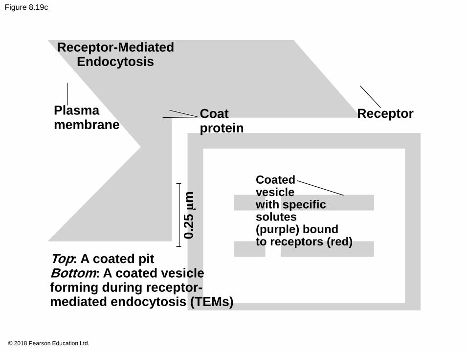
Figure 8.19c
Receptor-MediatedEndocytosis
Plasmamembrane
Coatprotein
Receptor
Coatedvesiclewith specificsolutes(purple) boundto receptors (red)
Top: A coated pitBottom: A coated vesicleforming during receptor-mediated endocytosis (TEMs)
0.2
5 µ
m
© 2018 Pearson Education Ltd.
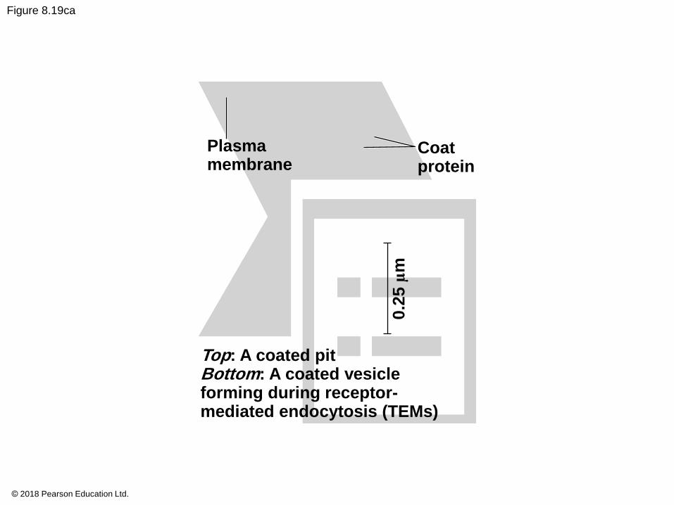
Figure 8.19ca
Plasmamembrane
Coatprotein
Top: A coated pitBottom: A coated vesicleforming during receptor-mediated endocytosis (TEMs)
0.2
5 µ
m
© 2018 Pearson Education Ltd.
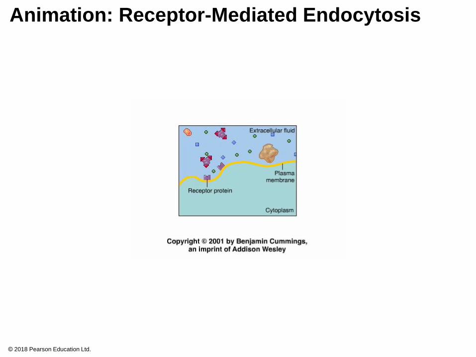
Animation: Receptor-Mediated Endocytosis
© 2018 Pearson Education Ltd.
Human cells use receptor-mediated endocytosis to
take in cholesterol, which is carried in particles
called low-density lipoproteins (LDLs)
Individuals with the disease familial
hypercholesterolemia have missing or defective LDL
receptor proteins
© 2018 Pearson Education Ltd.
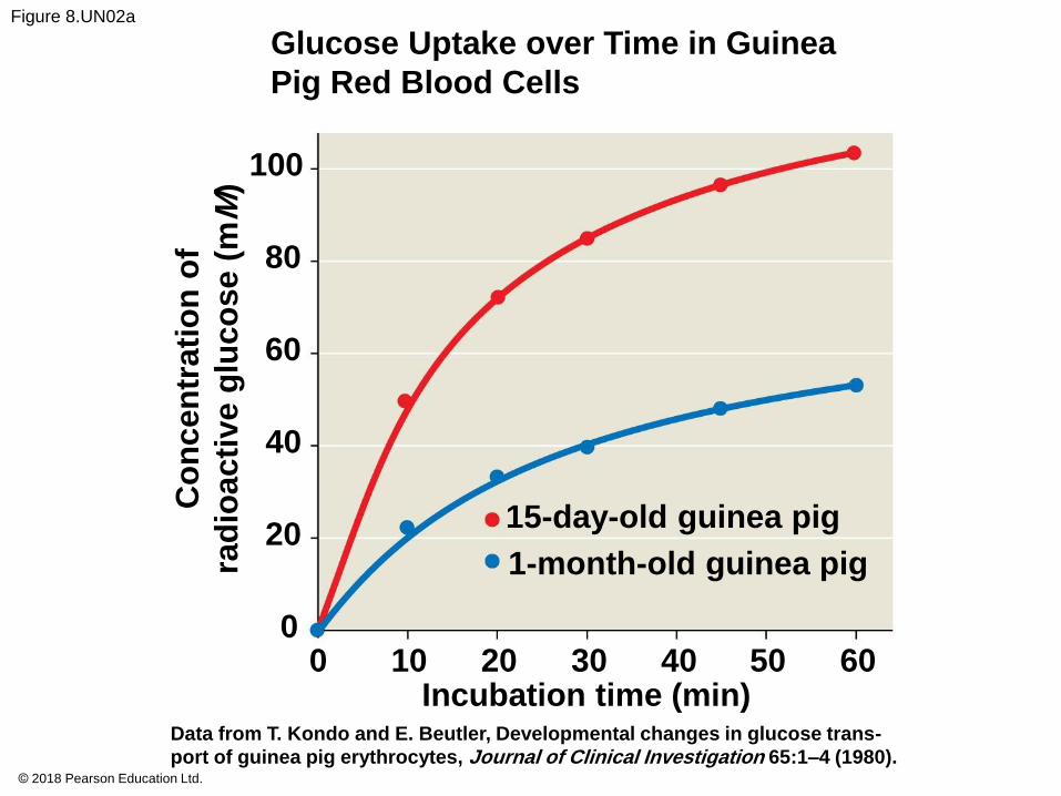
Figure 8.UN02a
Glucose Uptake over Time in Guinea
Pig Red Blood Cells
100
80
60
40
20
0
15-day-old guinea pig
1-month-old guinea pig
0 10 20 30 40 50Incubation time (min)
60
Co
nc
en
tra
tio
n o
f
rad
ioa
cti
ve
glu
co
se
(m
M)
Data from T. Kondo and E. Beutler, Developmental changes in glucose trans-
port of guinea pig erythrocytes, Journal of Clinical Investigation 65:1–4 (1980).© 2018 Pearson Education Ltd.
Figure 8.UN02b
© 2018 Pearson Education Ltd.
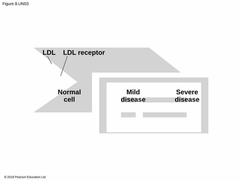
Figure 8.UN03
LDL LDL receptor
Normalcell
Milddisease
Severedisease
© 2018 Pearson Education Ltd.
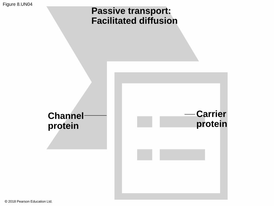
Figure 8.UN04
Passive transport:Facilitated diffusion
Channelprotein
Carrierprotein
© 2018 Pearson Education Ltd.
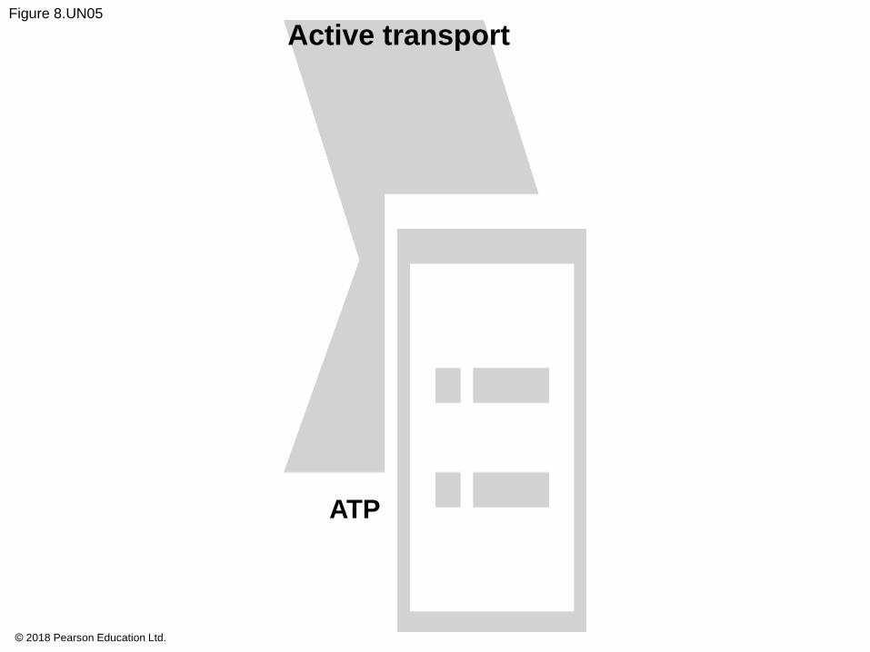
Figure 8.UN05
Active transport
ATP
© 2018 Pearson Education Ltd.
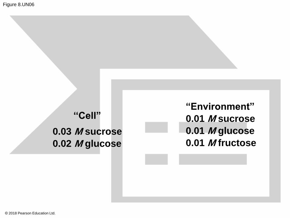
Figure 8.UN06
‘‘Cell”
0.03 M sucrose
0.02 M glucose
“Environment”
0.01 M sucrose
0.01 M glucose
0.01 M fructose
© 2018 Pearson Education Ltd.
Figure 8.UN07
© 2018 Pearson Education Ltd.