Embed Size (px)
Citation preview

Chapter 8 - Carbohydrates
Introduction
Carbohdrates (saccharides) are the most abundant biomolecule. Although
chemically simpler {( C H2O)n} than amino acids and nucleic acids, their
derivatives do contain N and S. As recently as the 1960's they were thought to be
primarily energy sources and to have structural roles (< 1960's), they are now
known to have important roles in recognition processes (cell - cell, protein -
protein)
Monosaccharides
Monosaccharides, or simple sugars, are polyhydroxy aldehydes (aldose) and
ketones (ketoses). Aldoses are shown in Figure 8-1.

Note the presence of 1 chiral carbon for 3-carbon aldoses, 2 for 4-carbon aldoses,
or n-2 chiral carbons for n-carbon aldoses. The D- form has the OH on the
furthest chiral carbon from the aldehyde group on the right in the Fischer
projection, and predominates in nature (whereas L-amino acids predominate).
Recall stereochemistry (enantiomers, diastereomers, epimers). You should be able
to draw D-glucose, D-galactose, D-fructose (a ketose, see below), D-ribose.
Ketoses (Figure 8-2) with n carbons have n-3 chiral carbons; know D-
fructose only. Note that glucose and fructose are structural isomers as compared to
glucose and galactose, for example, which are stereoisomers.
OHR +OR
CH
R OH'CR
H
O'
OHR +OR
CR
R OH'CR
O
R'
cyclic forms: Recall from organic chemistry that alcohols and aldehydes
react to form hemiacetals
Alcohol + Aldehyde W Hemiacetal
and that alcohols and ketones react to form hemiketals:
Alcohol + Ketone W Hemiketal
Since carbohydrates have the alcohol and carbonyl functionalities in the same
molecule, they can react to form intramolecular, or cylic hemiacetals and cyclic
hemiketals (Figure 8-3)
The cyclic forms of carbohydrates are typically represented as Haworth
Projections, and are called pyranoses or furanoses based on the parent compounds,
pyran and furan.
Conventions: Left in Fisher, up in Haworth, terminal CH2OH group bonded
to C5 is up for D isomer. The anomeric carbon is derived from the carbonyl group
and has two oxygens bonded to it. I will always draw Haworth structures so that
the anomeric carbon, regardess whether is the number 1 carbon or not, will always
be the furthest to the right.
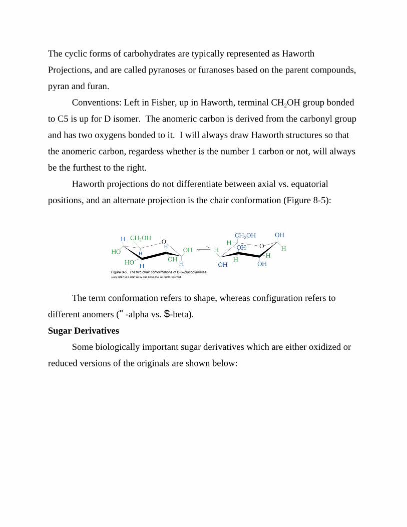
Haworth projections do not differentiate between axial vs. equatorial
positions, and an alternate projection is the chair conformation (Figure 8-5):
The term conformation refers to shape, whereas configuration refers to
different anomers ("-alpha vs. $-beta).
Sugar Derivatives
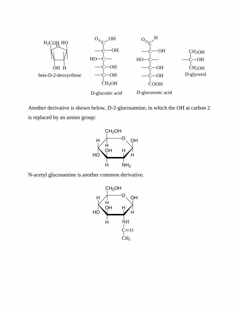
Some biologically important sugar derivatives which are either oxidized or
reduced versions of the originals are shown below:
H
HOO
OH
H2COH
beta-D-2-deoxyribose
C
C
C
C
C
CH2
HO
OH
OH
OHO
OH
OH
D-gluconic acid
C
C
C
C
C
C
HO
OH
OH
HO
OH
D-glucuronic acid
OOH
CH2
C OH
CH2
OH
OHD-glycerol
OCH2OH
H
NH2
HOH
H
HO
H OH
H
OCH2OH
H
NH
HOH
H
HO
H OH
H
C
CH3
O
Another derivative is shown below, D-2-glucosamine, in which the OH at carbon 2
is replaced by an amino group:
N-acetyl glucosamine is another common derivative.
OCH2OH
H
OH
HOH
H
HO
H H
OH+
OCH2OH
H
OH
HOH
H
HO
H H
OH
OCH2OH
H
OH
HOH
H
HO
H H OCH2OH
H
OH
HOH
H
H H
OHO
- H2O
alpha (1 - 4)glycosidic linkage
Maltose
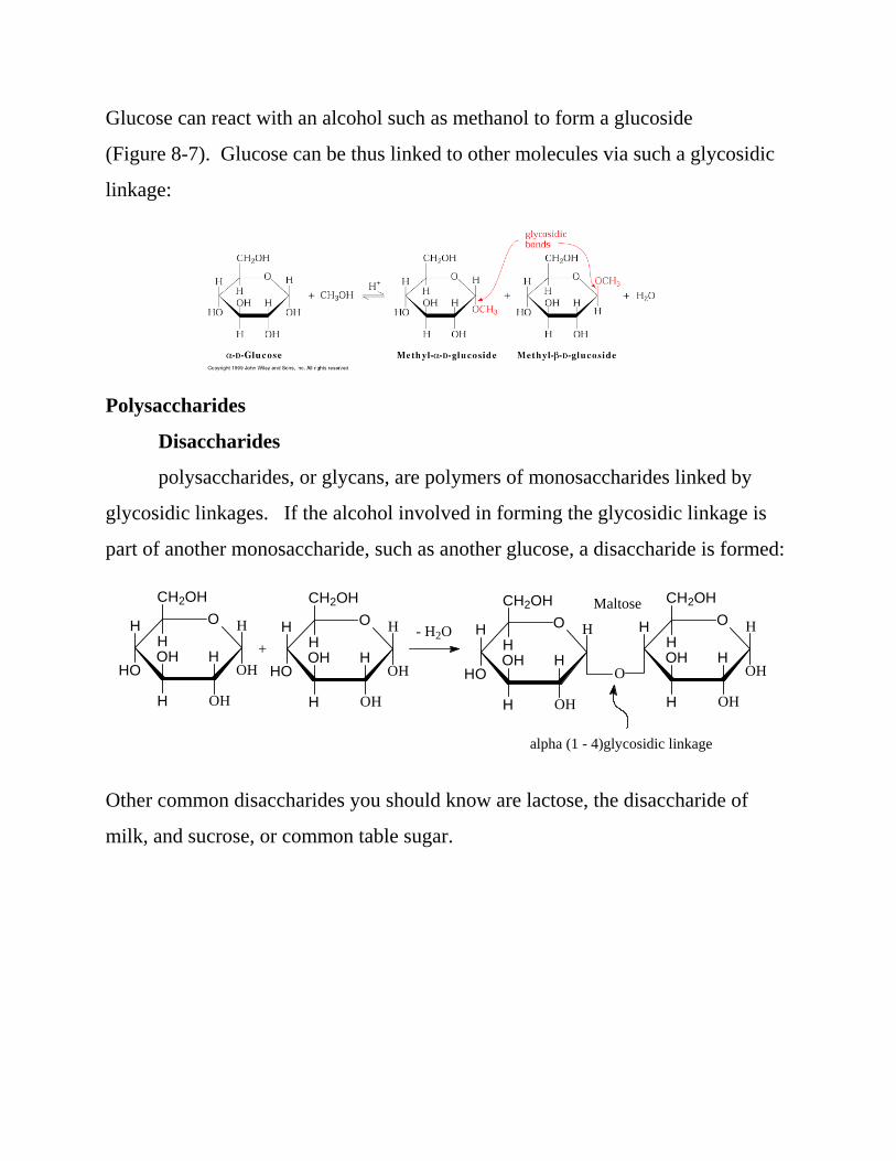
Glucose can react with an alcohol such as methanol to form a glucoside
(Figure 8-7). Glucose can be thus linked to other molecules via such a glycosidic
linkage:
Polysaccharides
Disaccharides
polysaccharides, or glycans, are polymers of monosaccharides linked by
glycosidic linkages. If the alcohol involved in forming the glycosidic linkage is
part of another monosaccharide, such as another glucose, a disaccharide is formed:
Other common disaccharides you should know are lactose, the disaccharide of
milk, and sucrose, or common table sugar.

OCH2OH
H
OH
HOH
H
OHO
OCH2OH
H
OH
HOH
H
H OH
H
beta (1 - 4) glycosidic linkage
LactoseO
CH2OH
H
OH
HOH
H
HO
H H
O
O
HOH
OH H
CH2OHH
HOCH2
Sucrose
alpha, beta (1 - 2)glycosidic linkage
OCH2OH
H
NH2
HOH
H
HO
H H OCH2OH
H
NH2
HOH
H
H H
OHO
OCH2OH
H
NH2
HOH
H
HO
H HC
OHCH2OH
H
NH2
HOH
H
H
O
H
O
Cu++
Cu+ (Cu)2O(red ppt)
OCH2OH
H
OH
HOH
H
HO
H HC
OHCH2OH
H
OH
HOH
H
H
OO2
-
Note that the preferred way to draw sucrose is as shown above, and not as
shown in the text on page 215, which breaks a lot of the rules I’ve given you.
Note also that maltose and lactose have the anomeric carbon of one residue
(on the left) involved in glycosidic linkage, whereas the other anomeric carbon (on
the right) is not involved in a glycosidic linkage; in other words, it is “free.” The
hemiacetal forms of the glucose residue (free anomeric carbon) exist in equilibrium
with the open chain forms which possess a free carbonyl group. These carbonyl
groups can be further oxidized, and maltose and lactose are called reducing sugars
because they reduce the oxidizing agent:
The left residue of both maltose and lactose is the non-reducing end, whereas the
right residue is the reducing end. There is thus a directionality to these
disaccharides and larger oligo- and polysaccharides in that the nonreducing end is
on the left, the reducing end on the right (recall for proteins the amino terminus is
drawn on the left, the carboxy terminus on the right).
Polysaccharides (glycans)
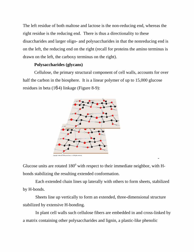
Cellulose, the primary structural component of cell walls, accounts for over
half the carbon in the biosphere. It is a linear polymer of up to 15,000 glucose
residues in beta (164) linkage (Figure 8-9):
-
Glucose units are rotated 1800 with respect to their immediate neighbor, with H-
bonds stabilizing the resulting extended conformation.
Each extended chain lines up laterally with others to form sheets, stabilized
by H-bonds.
Sheets line up vertically to form an extended, three-dimensional structure
stabilized by extensive H-bonding.
In plant cell walls such cellulose fibers are embedded in and cross-linked by
a matrix containing other polysaccharides and lignin, a plastic-like phenolic

polymer.
Chitin, the primary structural component of the exoskeletons of
invertebrates, is similar in composition to cellulose, except that N-acetyl
glucosamine replaces glucose.
Starch and glycogen, in contrast to cellulose and chitin, are storage
polysaccharides. Starch is a mixture of amylose + amylopectin. Amylose is a
linear polymer of glucose in alpha (1 - 4) linkage. The result of the alpha linkage is
that amylose adopts an irregular aggregating helically coiled conformation (Figure
8-10)
OCH2OH
H
OH
HOH
H
HO
H H OCH2OH
H
OH
HOH
H
H H
O O
OH
OH
HOH
H
H H OCH2OH
H
OH
HOH
H
H H
OHO
OCH2
H
OH
HOH
H
H H
O
OHAlpha (1 - 6) linkage
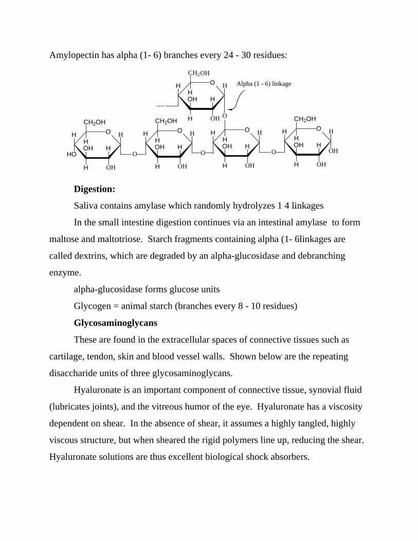
Amylopectin has alpha (1- 6) branches every 24 - 30 residues:
Digestion:
Saliva contains amylase which randomly hydrolyzes 1 4 linkages
In the small intestine digestion continues via an intestinal amylase to form
maltose and maltotriose. Starch fragments containing alpha (1- 6linkages are
called dextrins, which are degraded by an alpha-glucosidase and debranching
enzyme.
alpha-glucosidase forms glucose units
Glycogen = animal starch (branches every 8 - 10 residues)
Glycosaminoglycans
These are found in the extracellular spaces of connective tissues such as
cartilage, tendon, skin and blood vessel walls. Shown below are the repeating
disaccharide units of three glycosaminoglycans.
Hyaluronate is an important component of connective tissue, synovial fluid
(lubricates joints), and the vitreous humor of the eye. Hyaluronate has a viscosity
dependent on shear. In the absence of shear, it assumes a highly tangled, highly
viscous structure, but when sheared the rigid polymers line up, reducing the shear.
Hyaluronate solutions are thus excellent biological shock absorbers.

SO3-O
OH
C
O
HOH
H
H H OCH2
H
NH
HOH
H
H H
O
O2-
SO3- OSO3
-
Heparin
OC
H
OH
HOH
H
H
H O
OCH2OH
H
NH
H
H
O
H
O
COCH3
-O3SO2-
Chondroitin-4-Sulfate
OC
H
OH
HOH
H
H
H O
OCH2OH
H
NH
H
H
O
H
O
COCH3
-O3SO2-
Hyaluronate
Heparin, in contrast to the other glycosaminoglycans, is found in
intracellular granules of the mast cells in arterial walls. Heparin inhibits clotting;
thus after injury, release of heparin is thought to prevent runaway clot formation.
Chondroitin-4-sulfate and chondroitin-6-sulfate are found in connective
tissue. Supplements of Chondroitin and glucosamine are taken as dietary
supplements to foster healthy joints.
Glycoproteins
These are proteins with carbohydrate attached via N - or O - linkages.
N-linkage - involves the side chain of Asn residues:

OCH2OH
H
NH
HOH
H
HO
H NH
H
CCH3
O
C
O
CH2 CH
NH
C O
OCH2OH
H
NH2
HOH
H
OH
H
OCH2OH
H
NH
H
H
HO
H
CCH3
O
O
CH
NH
C O
H2CO
O-linkage - involves the side chain of Ser or Thr residues (Ser shown):
Carbohydrate content ranges from < 1% to > 90%. Glycoproteins include
enzymes, transport proteins, receptors, hormones and structural proteins.
Since carbo is attached by enzymes, thus is not under direct genetic control,
composition and structure varies (microheterogeneity).
Proteoglycans
These are protein/glycosaminoglycan mixes (covalent and noncovalent) that
occur in the extracellular matrix (Figure 8-14): A hyaluronic “backbone” has
attached to it other glycosaminoglycans, to which are attached up to 100 associated
core proteins.
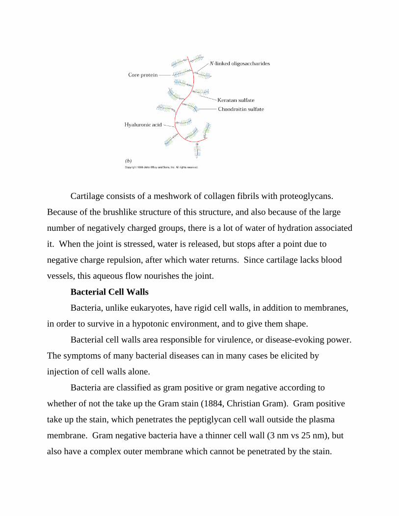
Cartilage consists of a meshwork of collagen fibrils with proteoglycans.
Because of the brushlike structure of this structure, and also because of the large
number of negatively charged groups, there is a lot of water of hydration associated
it. When the joint is stressed, water is released, but stops after a point due to
negative charge repulsion, after which water returns. Since cartilage lacks blood
vessels, this aqueous flow nourishes the joint.
Bacterial Cell Walls
Bacteria, unlike eukaryotes, have rigid cell walls, in addition to membranes,
in order to survive in a hypotonic environment, and to give them shape.
Bacterial cell walls area responsible for virulence, or disease-evoking power.
The symptoms of many bacterial diseases can in many cases be elicited by
injection of cell walls alone.
Bacteria are classified as gram positive or gram negative according to
whether of not the take up the Gram stain (1884, Christian Gram). Gram positive
take up the stain, which penetrates the peptiglycan cell wall outside the plasma
membrane. Gram negative bacteria have a thinner cell wall (3 nm vs 25 nm), but
also have a complex outer membrane which cannot be penetrated by the stain.

Gram negative bacteria are also more resistance to antibiotics because it is harder
for them to penetrate the outer membrane (Figure 8-15).
Lysozyme (tears) cleaves the glycosidic linkages between carbohydrate
residues making up the peptidoglycan of the cell wall.
Penicillin prevents the cross-linking of peptidoglycans by peptides. Its
structure resembles a D-amino acid in the cross-link (thus is a “silver bullet”)
Note: Almost all secreted and membrane -associated proteins of eukaryotic cells
are glycosylated, which are covalently attached to proteins via either N - (Asn) or )
O - (Ser or Thr) glycosidic linkages
Oligosaccharides mediate recognition Events
Because there are OH functional groups at each carbon of glucose, for
example, there are 6 x 6 = 36 different ways two glucose molecules can combine,
whereas there are only 2 possible ways 2 amino acids can combine. Additionally,
polysaccharides can be branched structures. Thus there is the potential for
tremendous structural diversity, hence biological information, in carbohydrates.
It is thus not surprising that all cells are coated with carbohydrate in the form
of glycoconjugates - carbohydrate linked covalently to proteins or membrane
lipids. The carbohydrates form a fuzzy layer up to 1400 angstroms thick in some
cells (Figure 8-20)
Additional evidence that cell-surface carbohydrates are involved in
recognition events is that lectins frequently appear on cell surfaces. Lectins are
proteins that bind carbohydrates.
Cell-surface carbohydrates are known to be immunochemical markers, such
as the ABO blood group antigens. These are the oligosaccharide components of
surface glycolipids. A group individuals have A antigens on their cell surfaces and
B antibodies; B group individuals have B antigens and A antibodies in their blood;
AB individuals have both A and B antigens and no antibodies (thus AB individuals
can receive blood from any donor). Type O individuals have no antigens (thus are
universal donors) and both A and B antibodies (actually, they possess a “core”
oligosaccharide which is non-antigenic).
Other protein-carbohydrate interactions include:
N - linked oligosaccharides with mannose-6-phosphate resides tag a protein
for processing or degradation by lysosomes.
Removal of sialic acid from circulating glycoproteins results in their
selective clearance from the blood.
Normal cells stop growing when they touch each other (contact inhibition).
Cancer cells have lost this function, thus form malignant tumors.
Many viruses, bacteria and eukaryotic parasites invade target cells by first
binding to cell-surface cargohydrates.
Problems: 1, 6, 8, 9

![Chapter 24: Carbohydrates · 2020. 2. 27. · Chapter 24: Carbohydrates [Sections: 24.1–24.10] Carbohydrates definition • naturally occuring compounds derived from carbon, oxygen](https://img.pdfslide.us/doc/110x75/60fa974869a56a2caa24abeb/chapter-24-carbohydrates-2020-2-27-chapter-24-carbohydrates-sections-241a2410.jpg)