Embed Size (px)
Citation preview
1
Chapter 7
The Skeletal System:
Bone Tissue
2
INTRODUCTION
• Bone is made up of several different tissues
working together: bone tissue, cartilage, dense
connective tissue, epithelium, blood forming
tissues, adipose tissue, and nervous tissue
• Each individual bone is an organ
• Dynamic and ever-changing throughout life
• The bones, along with their cartilages, make up the
skeletal system
3
Functions of Bone
• Supporting & protecting soft tissues
• Attachment site for muscles making
movement possible
• Storage of the minerals, calcium &
phosphate -- mineral homeostasis
• Blood cell production occurs in red bone
marrow (hemopoiesis)
• Energy storage in yellow bone marrow
4
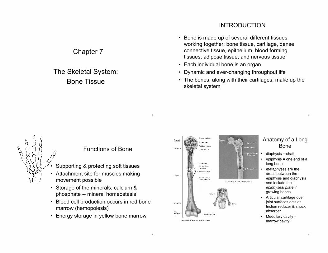
Anatomy of a Long
Bone
• diaphysis = shaft
• epiphysis = one end of a
long bone
• metaphyses are the
areas between the
epiphysis and diaphysis
and include the
epiphyseal plate in
growing bones.
• Articular cartilage over
joint surfaces acts as
friction reducer & shock
absorber
• Medullary cavity =
marrow cavity
5
Anatomy of a Long
Bone
• Endosteum = liningof marrow cavity
• Periosteum = toughmembrane coveringbone but not thecartilage
– fibrous layer =dense irregularCT
– osteogenic layer =bone cells & bloodvessels thatnourish or helpwith repairs
6
Histology of
Bone• A type of
connective tissue
as seen by widely
spaced cells
separated by
matrix
• Matrix of 25%
water, 25%
collagen fibers &
50% crystalized
mineral salts
• 4 types of cells in
bone tissue
7
Histology of Bone Tissue
• Bone (osseous) tissue consists of widely separated cells
surrounded by large amounts of matrix.
• The matrix of bone contains inorganic salts, primarily
hydroxyapatite and some calcium carbonate, and collagen
fibers.
• These and a few other salts are deposited in a framework of
collagen fibers, by a process called calcification.
– The process of calcification occurs only in the presence
of collagen fibers.
– Mineral salts confer hardness on bone while collagen
fibers give bone its great tensile strength.
8
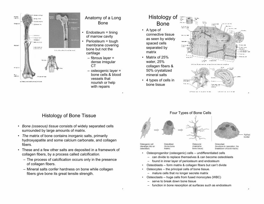
Four Types of Bone Cells
• Osteoprogenitor (osteogenic) cells -- undifferentiated cells
– can divide to replace themselves & can become osteoblasts
– found in inner layer of periosteum and endosteum
• Osteoblasts -- form matrix & collagen fibers but can’t divide
• Osteocytes -- the principal cells of bone tissue.
– mature cells that no longer secrete matrix
• Osteoclasts -- huge cells from fused monocytes (WBC)
– serve to break down bone tissue
– function in bone resorption at surfaces such as endosteum
9
Matrix of Bone
• Inorganic mineral salts provide bone’s hardness
– hydroxyapatite (calcium phosphate) & calcium carbonate
• Organic collagen fibers provide bone’s flexibility
– their tensile strength resists being stretched or torn
• Remove minerals with acid & rubbery structure
results
• Denature collagen by heating and bones become
brittle
• Bone is not completely solid since it has small spaces
for vessels and red bone marrow
– spongy bone has many such spaces
– compact bone has very few such spaces
10
Compact Bone
• Compact bone is arranged in units called osteons
or Haversian systems
• Osteons contain blood vessels, lymphatic vessels,
nerves, and osteocytes along with the calcified
matrix.
• Osteons are aligned in the same direction along
lines of stress. These lines can slowly change as
the stresses on the bone changes.
11
Compact or
Dense Bone
• Looks like solid hard layer of bone
• Makes up the shaft of long bones and theexternal layer of all bones
• Resists stresses produced by weight andmovement
12
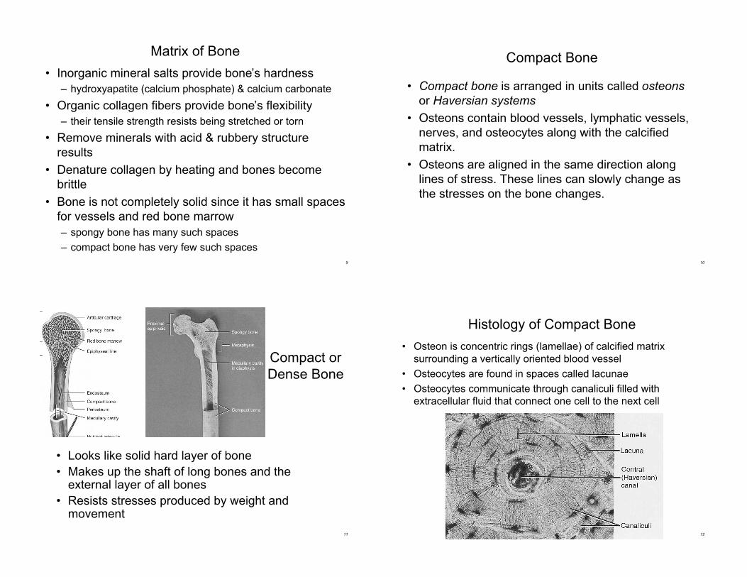
Histology of Compact Bone
• Osteon is concentric rings (lamellae) of calcified matrix
surrounding a vertically oriented blood vessel
• Osteocytes are found in spaces called lacunae
• Osteocytes communicate through canaliculi filled with
extracellular fluid that connect one cell to the next cell
13
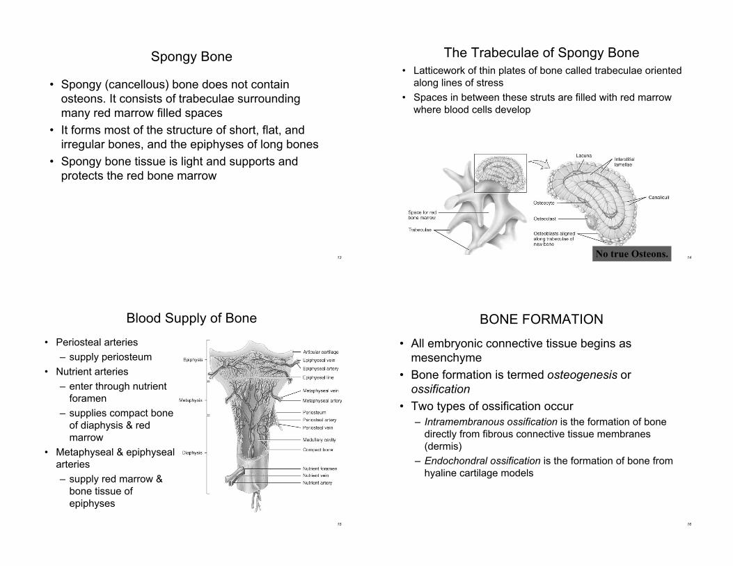
Spongy Bone
• Spongy (cancellous) bone does not contain
osteons. It consists of trabeculae surrounding
many red marrow filled spaces
• It forms most of the structure of short, flat, and
irregular bones, and the epiphyses of long bones
• Spongy bone tissue is light and supports and
protects the red bone marrow
14
The Trabeculae of Spongy Bone
• Latticework of thin plates of bone called trabeculae oriented
along lines of stress
• Spaces in between these struts are filled with red marrow
where blood cells develop
No true Osteons.
15
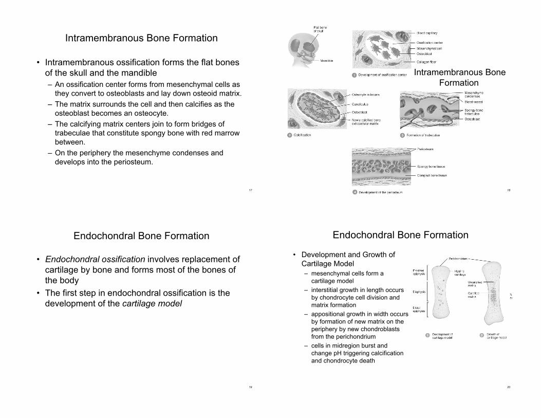
Blood Supply of Bone
• Periosteal arteries
– supply periosteum
• Nutrient arteries
– enter through nutrient
foramen
– supplies compact bone
of diaphysis & red
marrow
• Metaphyseal & epiphyseal
arteries
– supply red marrow &
bone tissue of
epiphyses
16
BONE FORMATION
• All embryonic connective tissue begins as
mesenchyme
• Bone formation is termed osteogenesis or
ossification
• Two types of ossification occur
– Intramembranous ossification is the formation of bone
directly from fibrous connective tissue membranes
(dermis)
– Endochondral ossification is the formation of bone from
hyaline cartilage models
17
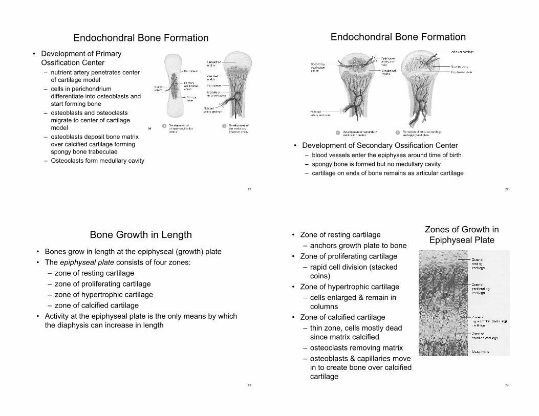
Intramembranous Bone Formation
• Intramembranous ossification forms the flat bones
of the skull and the mandible
– An ossification center forms from mesenchymal cells as
they convert to osteoblasts and lay down osteoid matrix.
– The matrix surrounds the cell and then calcifies as the
osteoblast becomes an osteocyte.
– The calcifying matrix centers join to form bridges of
trabeculae that constitute spongy bone with red marrow
between.
– On the periphery the mesenchyme condenses and
develops into the periosteum.
18
Intramembranous Bone
Formation
19
Endochondral Bone Formation
• Endochondral ossification involves replacement of
cartilage by bone and forms most of the bones of
the body
• The first step in endochondral ossification is the
development of the cartilage model
20
Endochondral Bone Formation
• Development and Growth of
Cartilage Model
– mesenchymal cells form a
cartilage model
– interstitial growth in length occurs
by chondrocyte cell division and
matrix formation
– appositional growth in width occurs
by formation of new matrix on the
periphery by new chondroblasts
from the perichondrium
– cells in midregion burst and
change pH triggering calcification
and chondrocyte death
21
Endochondral Bone Formation
• Development of Primary
Ossification Center
– nutrient artery penetrates center
of cartilage model
– cells in perichondrium
differentiate into osteoblasts and
start forming bone
– osteoblasts and osteoclasts
migrate to center of cartilage
model
– osteoblasts deposit bone matrix
over calcified cartilage forming
spongy bone trabeculae
– Osteoclasts form medullary cavity
22
Endochondral Bone Formation
• Development of Secondary Ossification Center
– blood vessels enter the epiphyses around time of birth
– spongy bone is formed but no medullary cavity
– cartilage on ends of bone remains as articular cartilage
23
Bone Growth in Length
• Bones grow in length at the epiphyseal (growth) plate
• The epiphyseal plate consists of four zones:
– zone of resting cartilage
– zone of proliferating cartilage
– zone of hypertrophic cartilage
– zone of calcified cartilage
• Activity at the epiphyseal plate is the only means by which
the diaphysis can increase in length
24
Zones of Growth in
Epiphyseal Plate• Zone of resting cartilage
– anchors growth plate to bone
• Zone of proliferating cartilage
– rapid cell division (stacked
coins)
• Zone of hypertrophic cartilage
– cells enlarged & remain in
columns
• Zone of calcified cartilage
– thin zone, cells mostly dead
since matrix calcified
– osteoclasts removing matrix
– osteoblasts & capillaries move
in to create bone over calcified
cartilage
25
Bone Growth in Length
• Between ages 18 to 25, epiphyseal
plates close
– cartilage cells stop dividing and
bone replaces the cartilage
(epiphyseal line)
• Growth in length stops by age 25
26
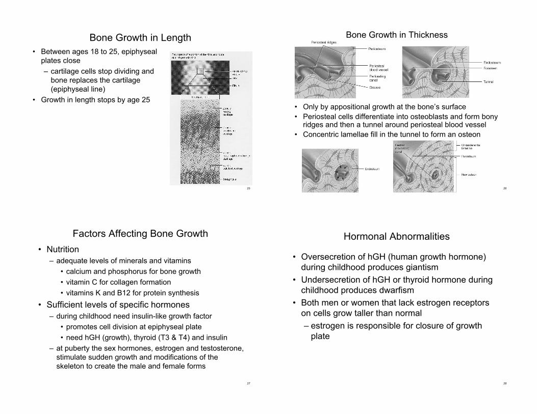
Bone Growth in Thickness
• Only by appositional growth at the bone’s surface
• Periosteal cells differentiate into osteoblasts and form bonyridges and then a tunnel around periosteal blood vessel
• Concentric lamellae fill in the tunnel to form an osteon
27
Factors Affecting Bone Growth
• Nutrition
– adequate levels of minerals and vitamins
• calcium and phosphorus for bone growth
• vitamin C for collagen formation
• vitamins K and B12 for protein synthesis
• Sufficient levels of specific hormones
– during childhood need insulin-like growth factor
• promotes cell division at epiphyseal plate
• need hGH (growth), thyroid (T3 & T4) and insulin
– at puberty the sex hormones, estrogen and testosterone,
stimulate sudden growth and modifications of the
skeleton to create the male and female forms
28
Hormonal Abnormalities
• Oversecretion of hGH (human growth hormone)
during childhood produces giantism
• Undersecretion of hGH or thyroid hormone during
childhood produces dwarfism
• Both men or women that lack estrogen receptors
on cells grow taller than normal
– estrogen is responsible for closure of growth
plate
29
Bone Remodeling
• Remodeling is the ongoing replacement of old bone
tissue by new bone tissue
– Old bone is constantly destroyed by osteoclasts, whereas
new bone is constructed by osteoblasts
– In orthodontics teeth are moved by braces. This places
stress on bone in the sockets causing osteoclasts and
osteablasts to remodel the sockets so that the teeth can
be properly aligned
– Several hormones and calcitriol control bone growth and
bone remodeling
30
Bone Remodeling
• Ongoing since osteoclasts carve out small tunnels
and osteoblasts rebuild osteons.
– osteoclasts form leak-proof seal around cell edges
– secrete enzymes and acids beneath themselves
– release calcium and phosphorus into interstitial fluid
– osteoblasts take over bone rebuilding
• Continual redistribution of bone matrix along lines
of mechanical stress
– distal femur is fully remodeled every 4 months
31
Fracture & Repair of Bone
• A fracture is any break in a bone
• Healing is faster in bone than in cartilage due
to lack of blood vessels in cartilage
• Healing of bone is still slow process due to
vessel damage
• Clinical treatment
– closed reduction = restore pieces to normal
position by manipulation
– open reduction = realignment during surgery
32
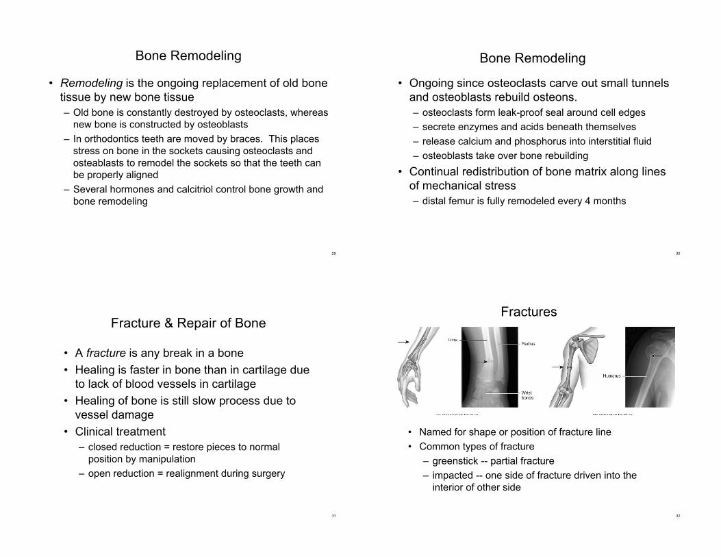
Fractures
• Named for shape or position of fracture line
• Common types of fracture
– greenstick -- partial fracture
– impacted -- one side of fracture driven into the
interior of other side
33
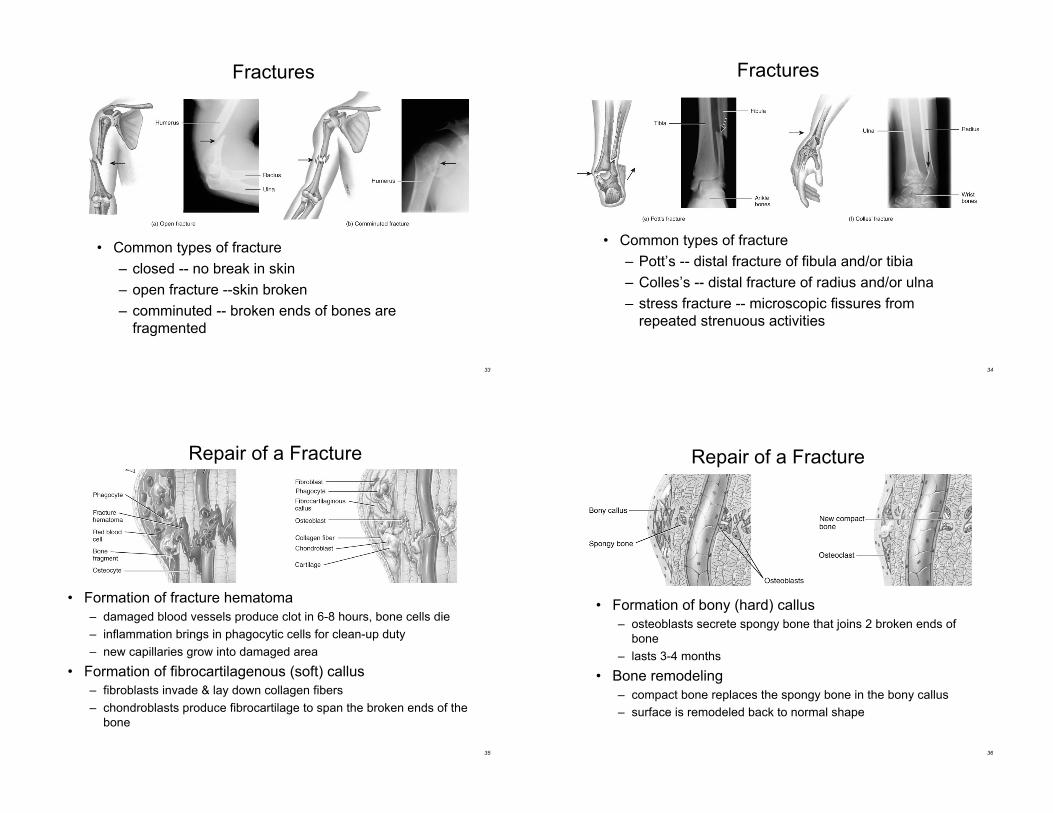
Fractures
• Common types of fracture
– closed -- no break in skin
– open fracture --skin broken
– comminuted -- broken ends of bones are
fragmented
34
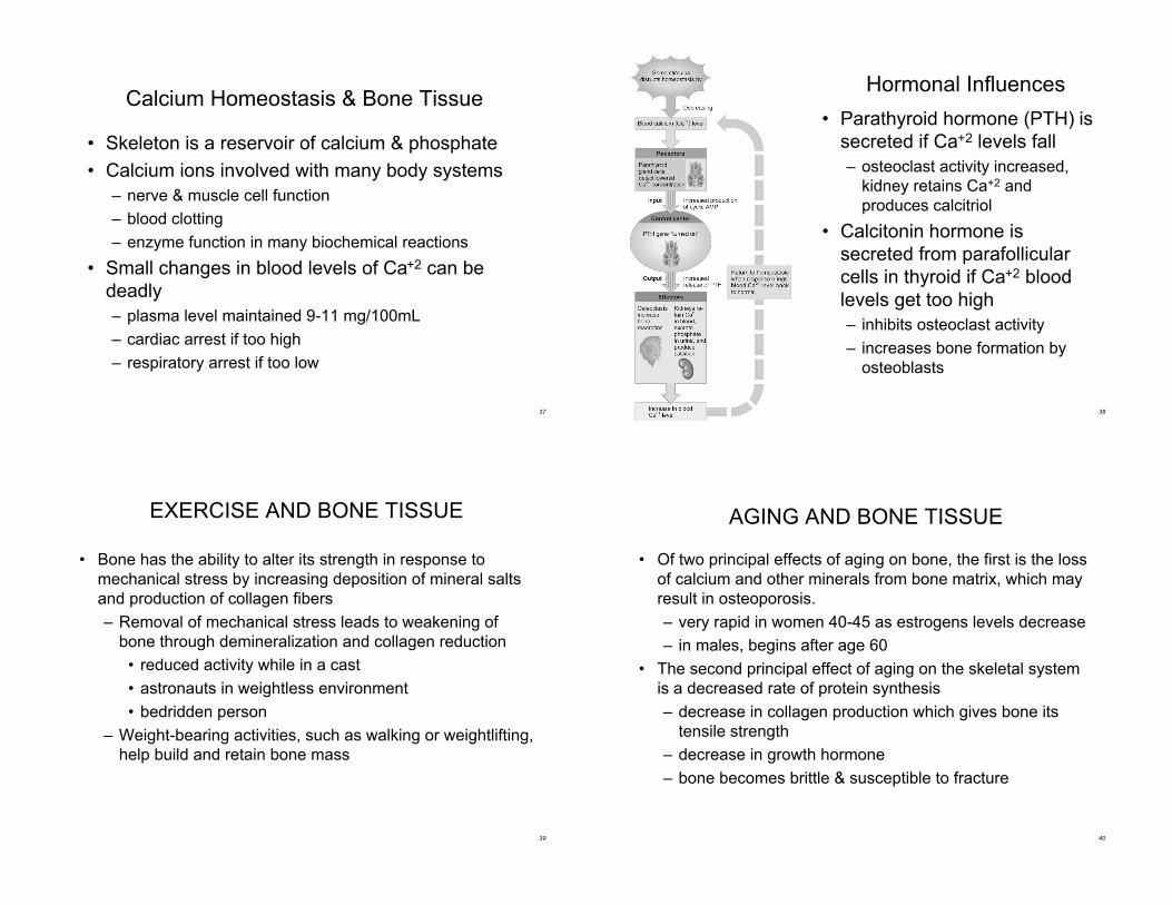
Fractures
• Common types of fracture
– Pott’s -- distal fracture of fibula and/or tibia
– Colles’s -- distal fracture of radius and/or ulna
– stress fracture -- microscopic fissures from
repeated strenuous activities
35
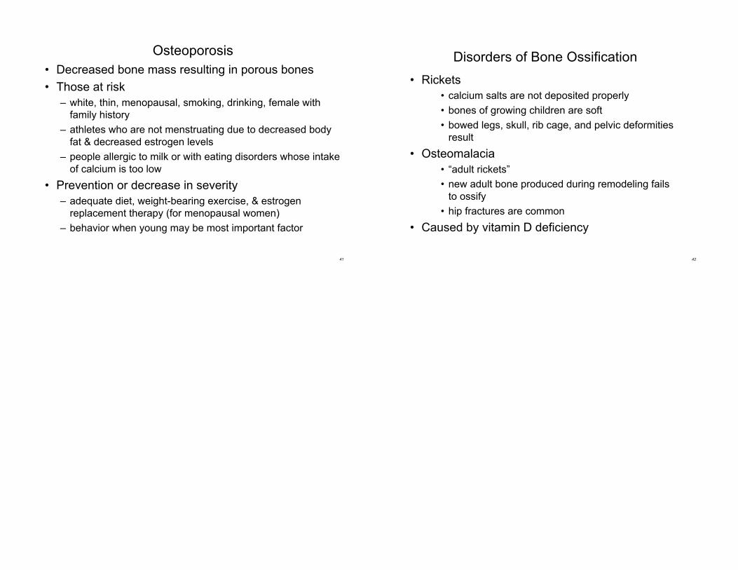
Repair of a Fracture
• Formation of fracture hematoma
– damaged blood vessels produce clot in 6-8 hours, bone cells die
– inflammation brings in phagocytic cells for clean-up duty
– new capillaries grow into damaged area
• Formation of fibrocartilagenous (soft) callus
– fibroblasts invade & lay down collagen fibers
– chondroblasts produce fibrocartilage to span the broken ends of the
bone
36
Repair of a Fracture
• Formation of bony (hard) callus
– osteoblasts secrete spongy bone that joins 2 broken ends of
bone
– lasts 3-4 months
• Bone remodeling
– compact bone replaces the spongy bone in the bony callus
– surface is remodeled back to normal shape
37
Calcium Homeostasis & Bone Tissue
• Skeleton is a reservoir of calcium & phosphate
• Calcium ions involved with many body systems
– nerve & muscle cell function
– blood clotting
– enzyme function in many biochemical reactions
• Small changes in blood levels of Ca+2 can be
deadly
– plasma level maintained 9-11 mg/100mL
– cardiac arrest if too high
– respiratory arrest if too low
38
Hormonal Influences
• Parathyroid hormone (PTH) is
secreted if Ca+2 levels fall
– osteoclast activity increased,
kidney retains Ca+2 and
produces calcitriol
• Calcitonin hormone is
secreted from parafollicular
cells in thyroid if Ca+2 blood
levels get too high
– inhibits osteoclast activity
– increases bone formation by
osteoblasts
39
EXERCISE AND BONE TISSUE
• Bone has the ability to alter its strength in response to
mechanical stress by increasing deposition of mineral salts
and production of collagen fibers
– Removal of mechanical stress leads to weakening of
bone through demineralization and collagen reduction
• reduced activity while in a cast
• astronauts in weightless environment
• bedridden person
– Weight-bearing activities, such as walking or weightlifting,
help build and retain bone mass
40
AGING AND BONE TISSUE
• Of two principal effects of aging on bone, the first is the loss
of calcium and other minerals from bone matrix, which may
result in osteoporosis.
– very rapid in women 40-45 as estrogens levels decrease
– in males, begins after age 60
• The second principal effect of aging on the skeletal system
is a decreased rate of protein synthesis
– decrease in collagen production which gives bone its
tensile strength
– decrease in growth hormone
– bone becomes brittle & susceptible to fracture
41
Osteoporosis
• Decreased bone mass resulting in porous bones
• Those at risk
– white, thin, menopausal, smoking, drinking, female with
family history
– athletes who are not menstruating due to decreased body
fat & decreased estrogen levels
– people allergic to milk or with eating disorders whose intake
of calcium is too low
• Prevention or decrease in severity
– adequate diet, weight-bearing exercise, & estrogen
replacement therapy (for menopausal women)
– behavior when young may be most important factor
42
Disorders of Bone Ossification
• Rickets
• calcium salts are not deposited properly
• bones of growing children are soft
• bowed legs, skull, rib cage, and pelvic deformities
result
• Osteomalacia
• “adult rickets”
• new adult bone produced during remodeling fails
to ossify
• hip fractures are common
• Caused by vitamin D deficiency