Embed Size (px)
DESCRIPTION
Chapter 7 The Peripheral Nervous System: Efferent Division. Outline. Autonomic nervous system Somatic nervous System Neuromuscular Junction. Outline. Autonomic nervous system Anatomy Pre and post fibers, sympathetic ganglia chain, collateral ganglia, terminal ganglia Neurotransmitters - PowerPoint PPT Presentation
Citation preview
Chapter 7The Peripheral Nervous System:
Efferent Division
Outline
• Autonomic nervous system
• Somatic nervous System
• Neuromuscular Junction
Outline
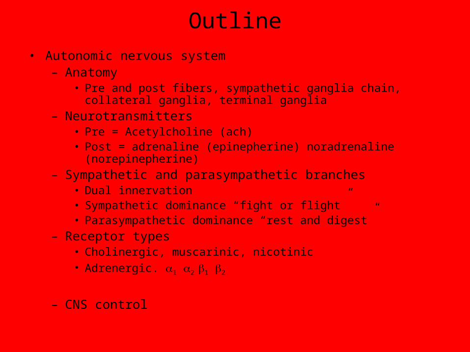
• Autonomic nervous system– Anatomy
• Pre and post fibers, sympathetic ganglia chain, collateral ganglia, terminal ganglia
– Neurotransmitters• Pre = Acetylcholine (ach)• Post = adrenaline (epinepherine) noradrenaline (norepinepherine)
– Sympathetic and parasympathetic branches• Dual innervation• Sympathetic dominance “fight or flight”• Parasympathetic dominance “rest and digest”
– Receptor types• Cholinergic, muscarinic, nicotinic• Adrenergic. 1 2 1 2
– CNS control
PNS: Efferent Division
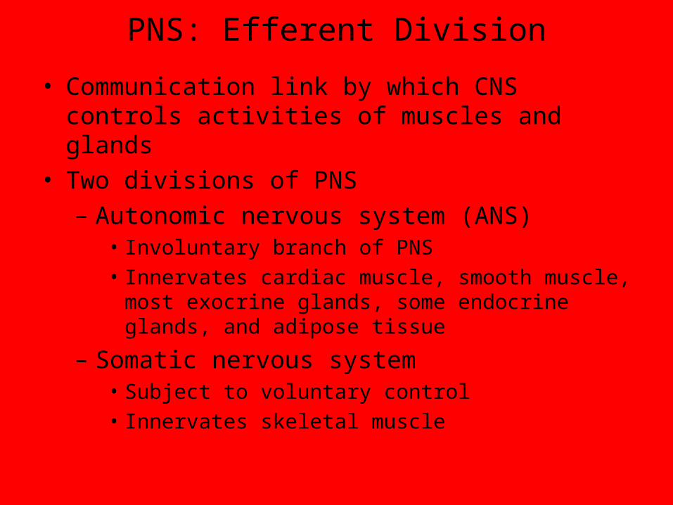
• Communication link by which CNS controls activities of muscles and glands
• Two divisions of PNS
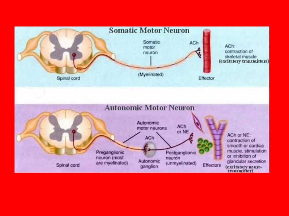
– Autonomic nervous system (ANS)• Involuntary branch of PNS
• Innervates cardiac muscle, smooth muscle, most exocrine glands, some endocrine glands, and adipose tissue
– Somatic nervous system• Subject to voluntary control
• Innervates skeletal muscle
ANS
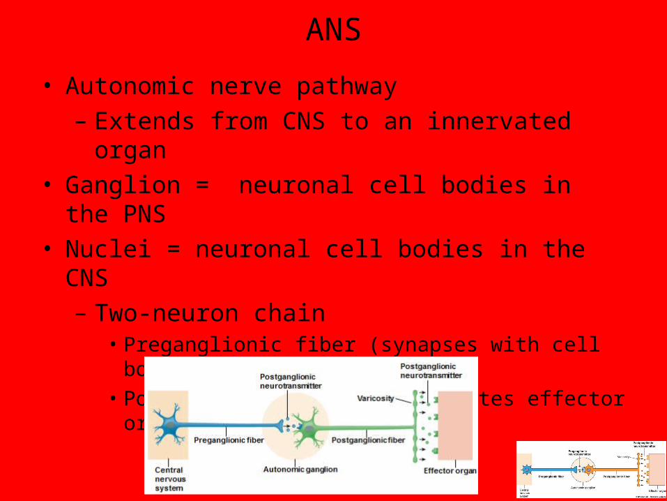
• Autonomic nerve pathway
– Extends from CNS to an innervated organ
• Ganglion = neuronal cell bodies in the PNS
• Nuclei = neuronal cell bodies in the CNS
– Two-neuron chain• Preganglionic fiber (synapses with cell body of second
neuron)
• Postganglionic fiber (innervates effector organ)
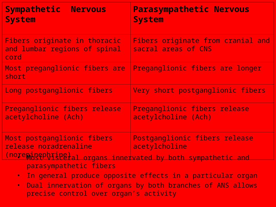
Sympathetic Nervous System Parasympathetic Nervous System
Fibers originate in thoracic and lumbar regions of spinal cord
Fibers originate from cranial and sacral areas of CNS
Most preganglionic fibers are short Preganglionic fibers are longer
Long postganglionic fibers Very short postganglionic fibers
Preganglionic fibers release acetylcholine (Ach)
Preganglionic fibers release acetylcholine (Ach)
Most postganglionic fibers release noradrenaline (norepinephrine)
Postganglionic fibers release acetylcholine
• Most visceral organs innervated by both sympathetic and parasympathetic fibers
• In general produce opposite effects in a particular organ
• Dual innervation of organs by both branches of ANS allows precise control over organ’s activity

Sympathetic preganglionic fiber
Autonomiceffectors
Parasympathetic preganglionic fiberParasympathetic postganglionic fiber
Epinephrine
NorepinephrineAcetylcholine
Sympathetic postganglionic fiber
Muscarinicreceptor
ACh
Terminalganglion
Nicotinicreceptor
Sympatheticganglion chain
AdrenalMedulla
BloodE
Muscarinicreceptor
Collateralganglion
Nicotinicreceptor
Terminalganglion
AChACh
β1 receptor
β2 receptor
NE
E
E
α receptor
NE
E
ACh
NE
Adiposetissue
Mostendocrineglandsand someendocrineglands
Smoothmuscle
Cardiacmuscle
KEY
AChNE
EFig. 7-2, p. 241
Nicotinicreceptor
Nicotinicreceptor
ANS
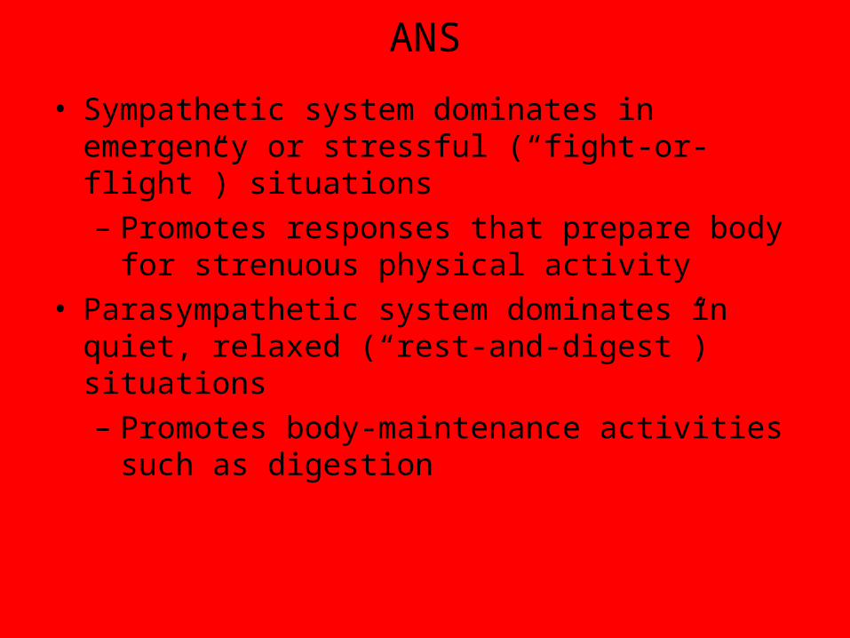
• Sympathetic system dominates in emergency or stressful (“fight-or-flight”) situations
– Promotes responses that prepare body for strenuous physical activity
• Parasympathetic system dominates in quiet, relaxed (“rest-and-digest”) situations
– Promotes body-maintenance activities such as digestion
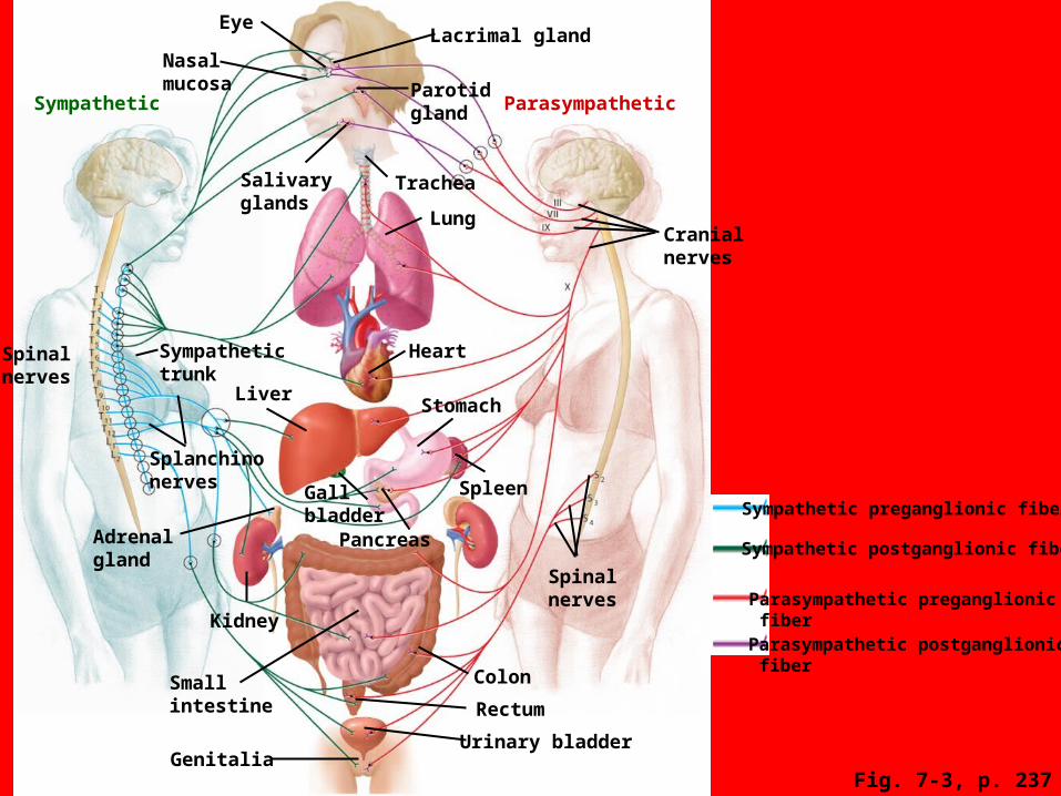
Eye
Nasalmucosa
Sympathetic
Spinal nerves
Sympathetictrunk
Splanchinonerves
Liver
Gallbladder
PancreasAdrenal gland
Kidney
Smallintestine
Colon
Rectum
Urinary bladderGenitalia
Lung
Heart
Spinalnerves
Cranialnerves
Salivaryglands
ParasympatheticParotidgland
Trachea
Lacrimal gland
Stomach
SpleenSympathetic preganglionic fiber
Parasympathetic postganglionic fiber
Parasympathetic preganglionic fiber
Sympathetic postganglionic fiber
Fig. 7-3, p. 237
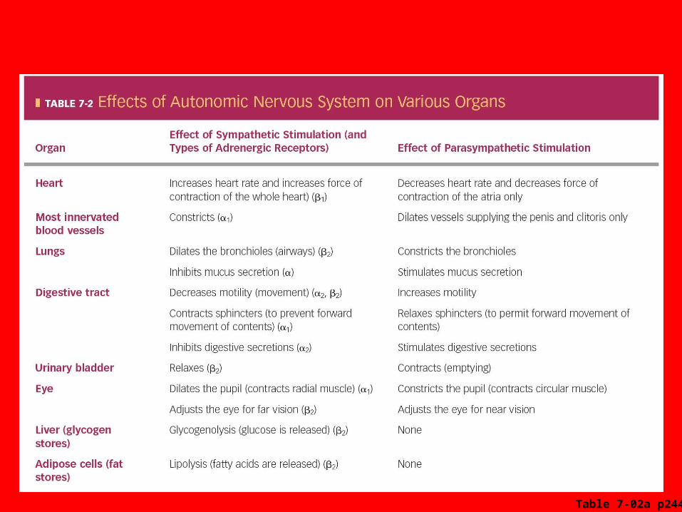
Table 7-02a p244
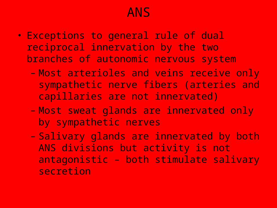
ANS
• Exceptions to general rule of dual reciprocal innervation by the two branches of autonomic nervous system
– Most arterioles and veins receive only sympathetic nerve fibers (arteries and capillaries are not innervated)
– Most sweat glands are innervated only by sympathetic nerves
– Salivary glands are innervated by both ANS divisions but activity is not antagonistic – both stimulate salivary secretion

ANS
• Adrenal medulla is a modified part of sympathetic nervous system
– Modified sympathetic ganglion that does not give rise to postganglionic fibers
– Stimulation of preganglionic fiber prompts secretion of hormones into blood
• About 20% of hormone release is norepinephrine
• About 80% of hormone released is epinephrine (adrenaline)
• Broadcast vs. localized
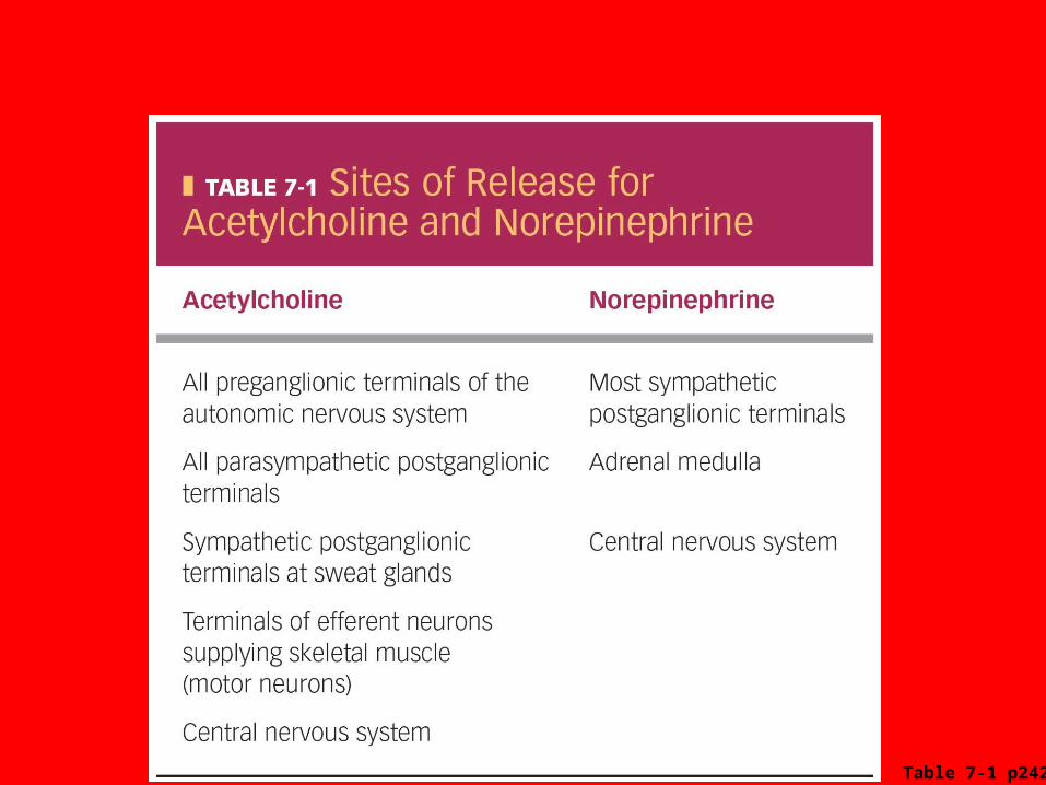
Table 7-1 p242
Sympathetic preganglionic fiber
Autonomiceffectors
Parasympathetic preganglionic fiberParasympathetic postganglionic fiber
Epinephrine
NorepinephrineAcetylcholine
Sympathetic postganglionic fiber
Muscarinicreceptor
ACh
Terminalganglion
Nicotinicreceptor
Sympatheticganglion chain
AdrenalMedulla
BloodE
Muscarinicreceptor
Collateralganglion
Nicotinicreceptor
Terminalganglion
AChACh
β1 receptor
β2 receptor
NE
E
E
α receptor
NE
E
ACh
NE
Adiposetissue
Mostendocrineglandsand someendocrineglands
Smoothmuscle
Cardiacmuscle
KEY
AChNE
EFig. 7-2, p. 241
Nicotinicreceptor
Nicotinicreceptor
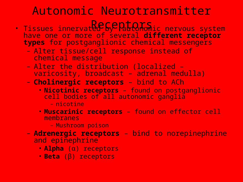
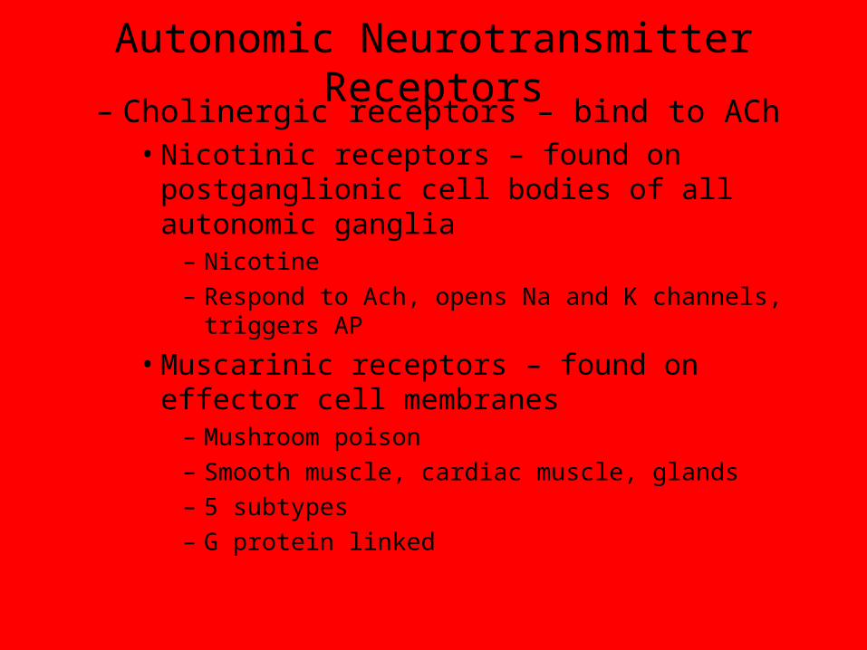
Autonomic Neurotransmitter Receptors
• Tissues innervated by autonomic nervous system have one or more of several different receptor types for postganglionic chemical messengers– Alter tissue/cell response instead of chemical message– Alter the distribution (localized – varicosity, broadcast –
adrenal medulla)– Cholinergic receptors – bind to ACh
• Nicotinic receptors – found on postganglionic cell bodies of all autonomic ganglia
– nicotine• Muscarinic receptors – found on effector cell membranes
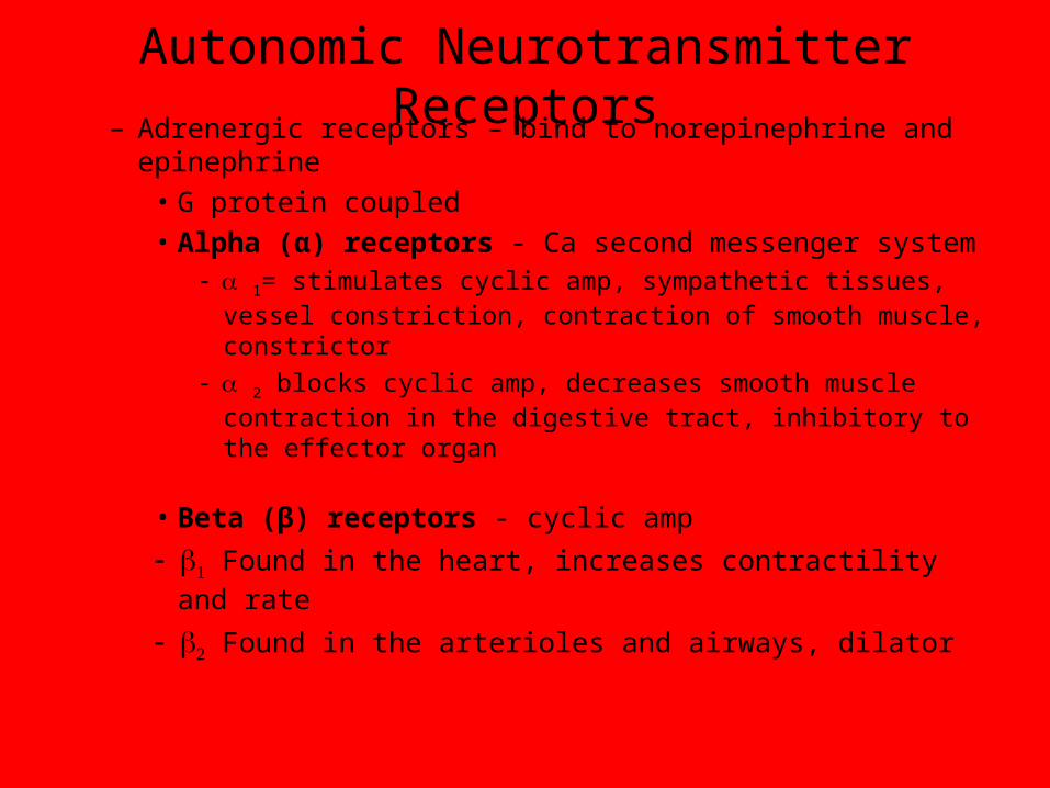
– Mushroom poison– Adrenergic receptors – bind to norepinephrine and
epinephrine• Alpha (α) receptors• Beta (β) receptors
Autonomic Neurotransmitter Receptors
– Cholinergic receptors – bind to ACh• Nicotinic receptors – found on postganglionic cell
bodies of all autonomic ganglia– Nicotine– Respond to Ach, opens Na and K channels, triggers AP
• Muscarinic receptors – found on effector cell membranes
– Mushroom poison– Smooth muscle, cardiac muscle, glands– 5 subtypes– G protein linked
Autonomic Neurotransmitter Receptors
– Adrenergic receptors – bind to norepinephrine and epinephrine
• G protein coupled
• Alpha (α) receptors - Ca second messenger system 1= stimulates cyclic amp, sympathetic tissues, vessel
constriction, contraction of smooth muscle, constrictor
2 blocks cyclic amp, decreases smooth muscle contraction in the digestive tract, inhibitory to the effector organ
• Beta (β) receptors - cyclic amp
Found in the heart, increases contractility and rate
Found in the arterioles and airways, dilator
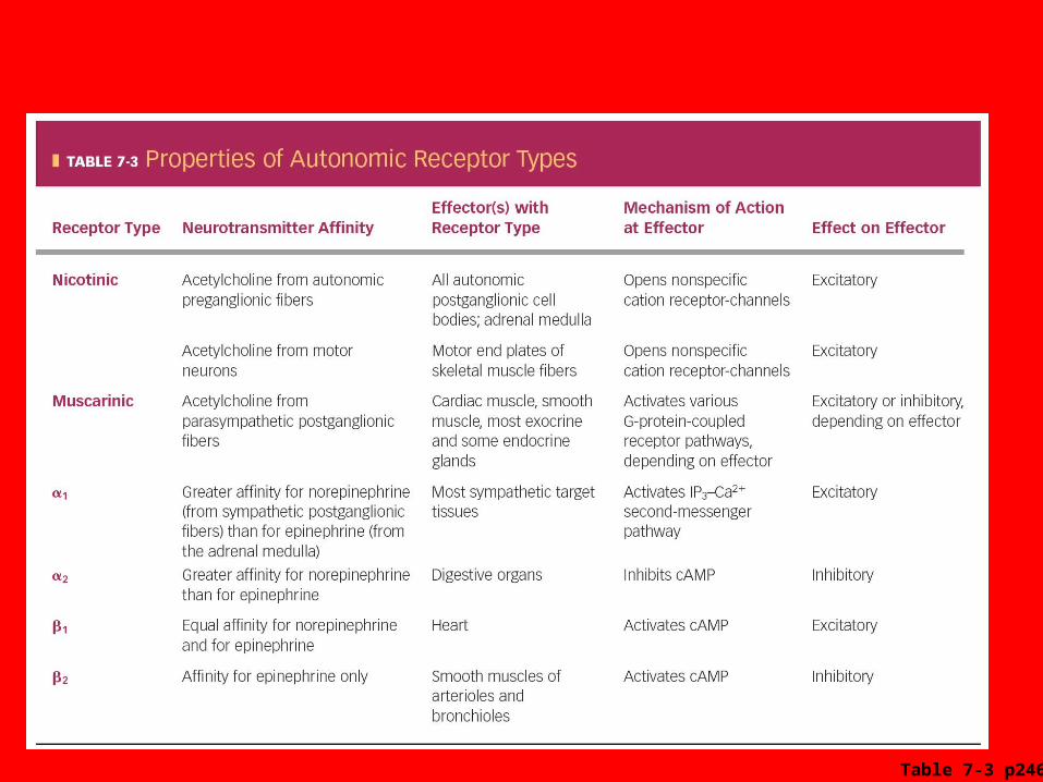
Table 7-3 p246
Autonomic Agonists and Antagonists
• Agonists
– Bind to same receptor as neurotransmitter
– Elicit an effect that mimics that of neurotransmitter
• Antagonists
– Bind with receptor
– Block neurotransmitter’s response
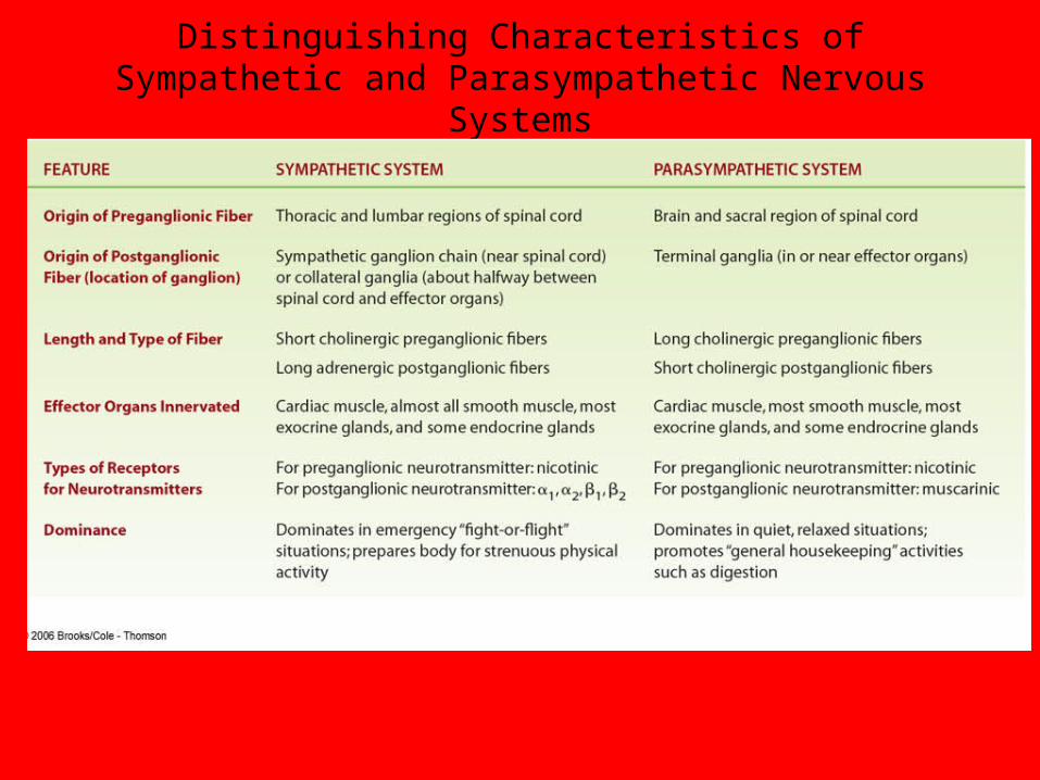
Distinguishing Characteristics of Sympathetic and Parasympathetic Nervous Systems
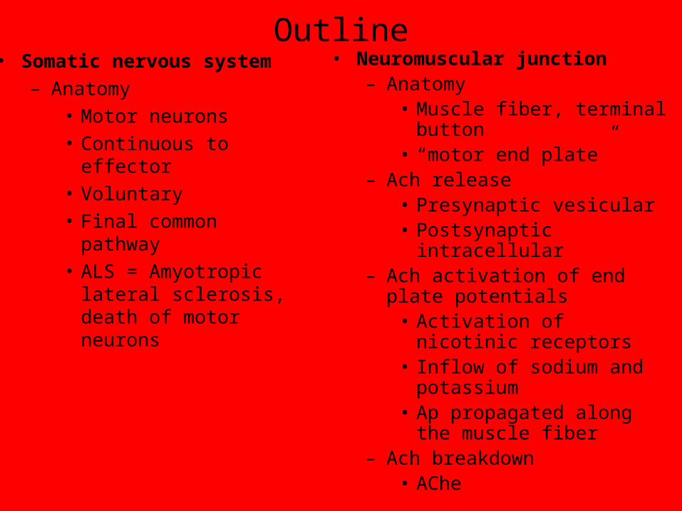
Outline• Somatic nervous system
– Anatomy• Motor neurons• Continuous to effector• Voluntary• Final common pathway• ALS = Amyotropic lateral
sclerosis, death of motor neurons
• Neuromuscular junction– Anatomy
• Muscle fiber, terminal button• “motor end plate”
– Ach release• Presynaptic vesicular• Postsynaptic intracellular
– Ach activation of end plate potentials
• Activation of nicotinic receptors
• Inflow of sodium and potassium
• Ap propagated along the muscle fiber
– Ach breakdown• AChe
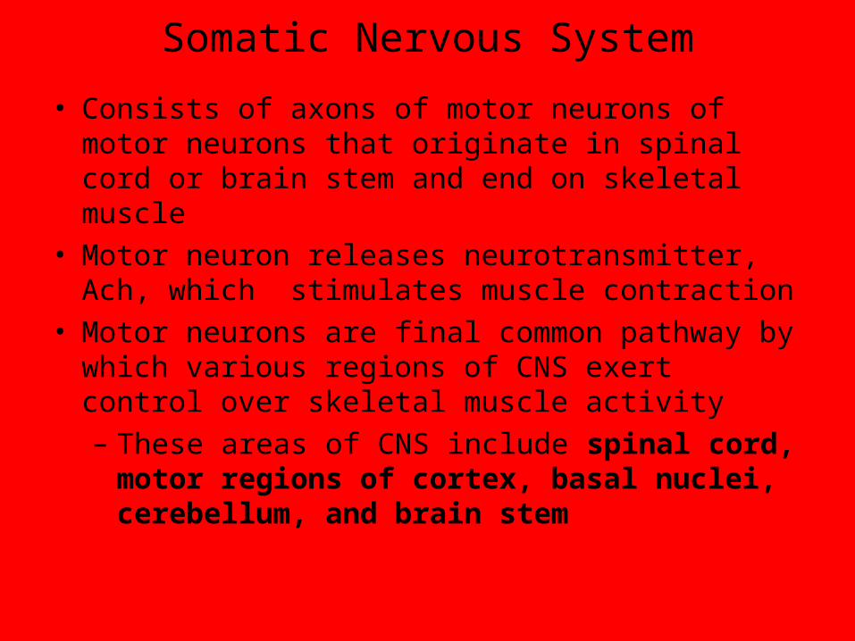
Somatic Nervous System
• Consists of axons of motor neurons of motor neurons that originate in spinal cord or brain stem and end on skeletal muscle
• Motor neuron releases neurotransmitter, Ach, which stimulates muscle contraction
• Motor neurons are final common pathway by which various regions of CNS exert control over skeletal muscle activity
– These areas of CNS include spinal cord, motor regions of cortex, basal nuclei, cerebellum, and brain stem
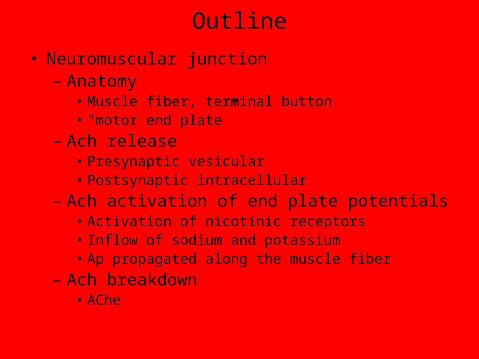
Outline
• Neuromuscular junction– Anatomy
• Muscle fiber, terminal button• “motor end plate”
– Ach release• Presynaptic vesicular• Postsynaptic intracellular
– Ach activation of end plate potentials• Activation of nicotinic receptors• Inflow of sodium and potassium• Ap propagated along the muscle fiber
– Ach breakdown• AChe
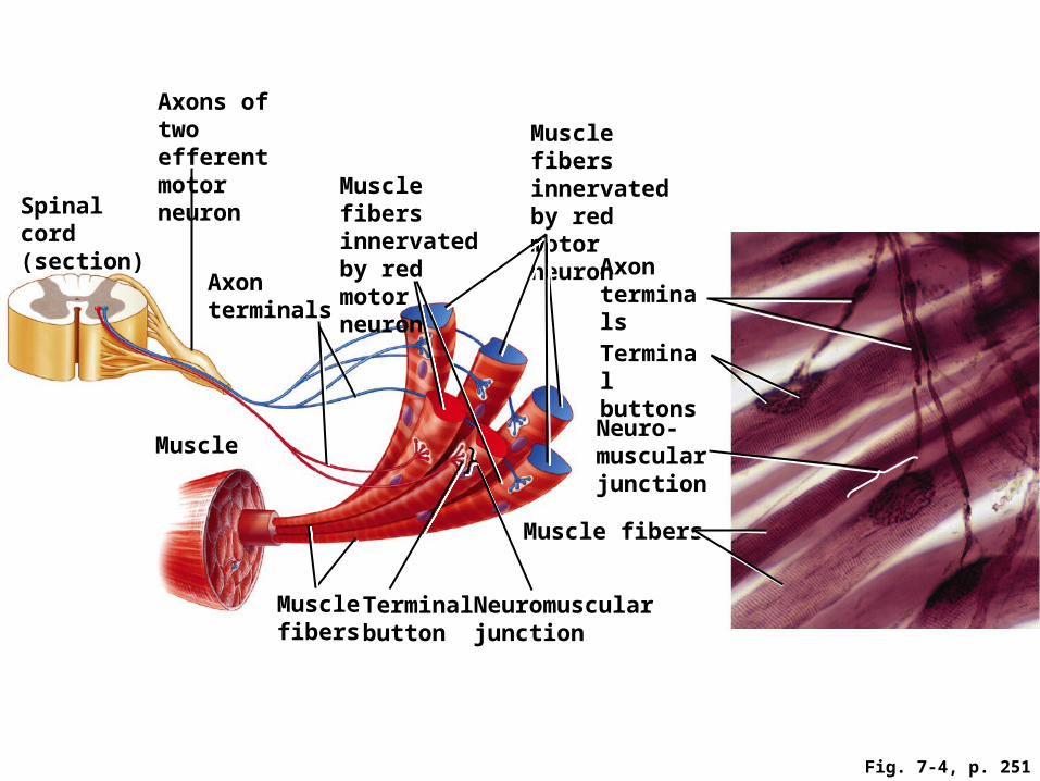
Axon terminals
Neuromuscularjunction
Spinal cord (section)
Muscle
Axonterminals
Axons of two efferentmotor neuron
Musclefibers
Terminalbutton
Terminal buttons
Neuro-muscularjunction
Muscle fibers
Fig. 7-4, p. 251
Muscle fibers innervated by red motor neuron
Muscle fibers innervated by red motor neuron
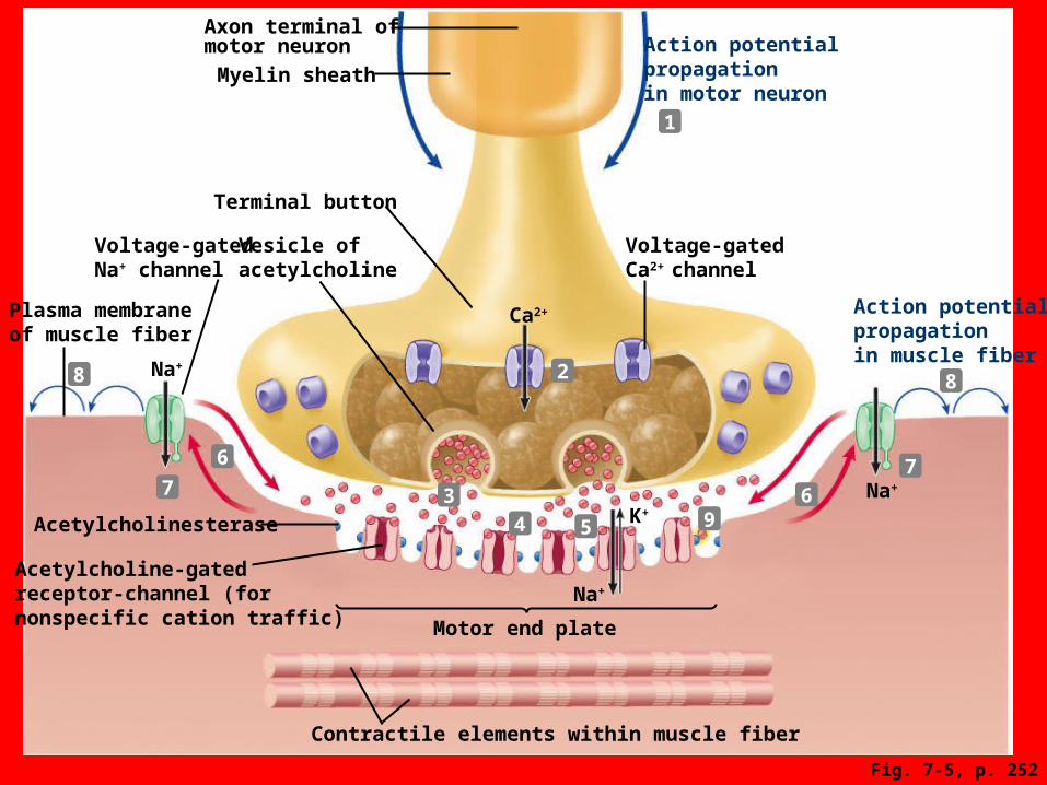
Contractile elements within muscle fiber
Acetylcholine-gatedreceptor-channel (fornonspecific cation traffic)
Action potentialpropagationin muscle fiber
Voltage-gatedCa2+ channel
Action potentialpropagationin motor neuron
Na+
Na+
Na+
Plasma membraneof muscle fiber
Voltage-gatedNa+ channel
Terminal button
Motor end plate
Vesicle ofacetylcholine
Ca2+
Myelin sheath
Axon terminal ofmotor neuron
K+Acetylcholinesterase
Fig. 7-5, p. 252
1
2
34
6
7
88
7
6
95
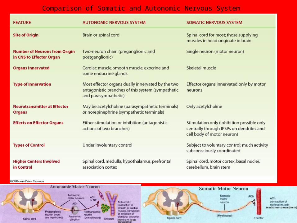
Comparison of Somatic and Autonomic Nervous System
Neuromuscular Junction
• Acetylcholinesterase – Inactivates ACh– Ends end-plate potential and the action potential
and resultant contraction• Neuromuscular junction is vulnerable to chemical
agents and diseases– Black widow spider venom causes explosive
release of ACh– Botulism toxin blocks release of ACh – Curare blocks action of ACh at receptor sites– Organophosphates prevent inactivation of ACh– Myasthenia gravis inactivates ACh receptor sites
Neuromuscular conditions and compounds
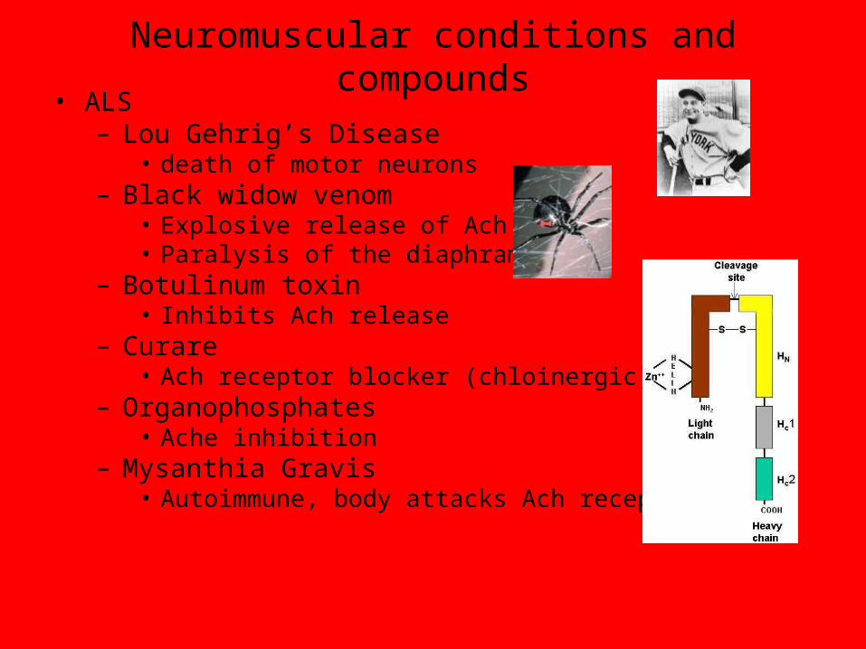
• ALS– Lou Gehrig’s Disease
• death of motor neurons– Black widow venom
• Explosive release of Ach• Paralysis of the diaphram
– Botulinum toxin• Inhibits Ach release
– Curare• Ach receptor blocker (chloinergic)
– Organophosphates• Ache inhibition
– Mysanthia Gravis• Autoimmune, body attacks Ach receptors