Embed Size (px)
Citation preview

Chapter 7 - The Nervous System
I. ORGANIZATION OF THE NERVOUS SYSTEM
- master control & communication. Control behavior of other tissues(increase or decrease activity) in order to maintain homeostasis.
*along with endocrine system, which acts slower but has longereffects.
- monitor changes, take info to CNS via an afferent pathway, CNS makesa decision, send out an efferent command to an effector.
*sensory input, integration, and motor output
*transduction: take stimulus, turn it into an impulse (AP).
*propagation: move the impulse down a neuron
*synapse: special connection with another cell, which allows AP topass to another organ (effectors: muscle, gland or other neuron).
*innervation: tissue is synapsed with nervous tissue.

A. Structural Classification
1. Central Nervous System (CNS) : Integration center. Decisions,emotions, consciousness, thought. Interpretation & commands.Subdivided into:
-brain
- spinal cord
2. Peripheral Nervous System (PNS) : Nerves and sensory apparatus,along with connections (synapses). Subdivided into:
- spinal nerves : leave spinal cord
-cranial nerves : leave brain
B. Functional Classification
- PNS only. CNS is just broken down into brain & spinal cord, but theywork on very similar principles.
1. Sensory (Afferent) Division (towards the CNS; respond to stimuli, andtake info to CNS). Although there are classification of sensory receptors,they all will work in a similar manner.
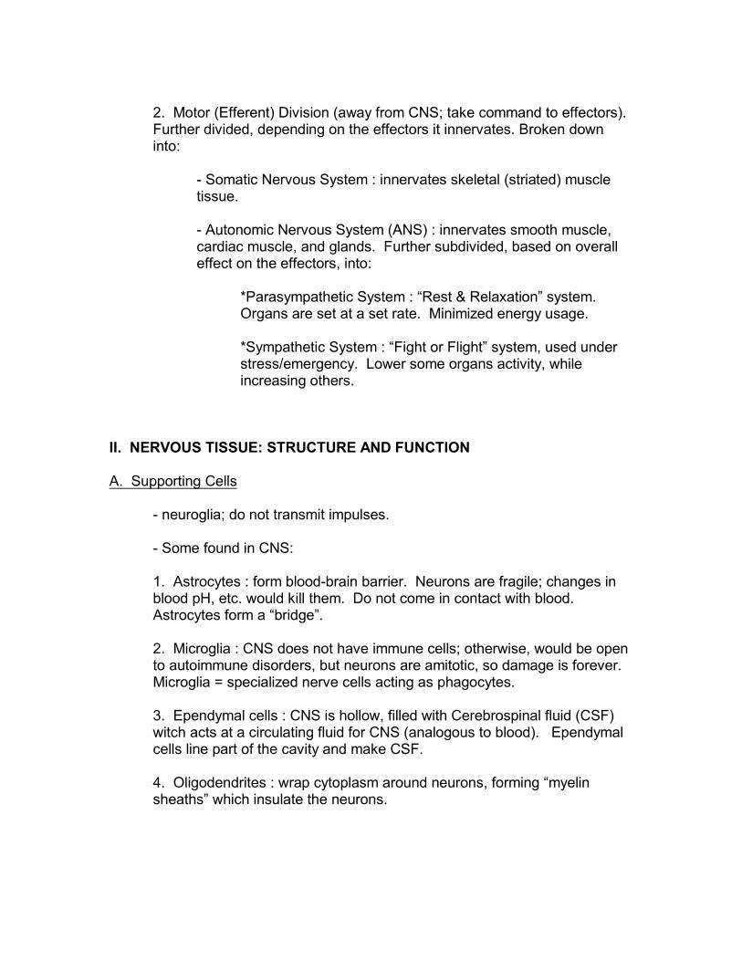
2. Motor (Efferent) Division (away from CNS; take command to effectors). Further divided, depending on the effectors it innervates. Broken downinto:
- Somatic Nervous System : innervates skeletal (striated) muscletissue.
- Autonomic Nervous System (ANS) : innervates smooth muscle,cardiac muscle, and glands. Further subdivided, based on overalleffect on the effectors, into:
*Parasympathetic System : “Rest & Relaxation” system. Organs are set at a set rate. Minimized energy usage.
*Sympathetic System : “Fight or Flight” system, used understress/emergency. Lower some organs activity, whileincreasing others.
II. NERVOUS TISSUE: STRUCTURE AND FUNCTION
A. Supporting Cells
- neuroglia; do not transmit impulses.
- Some found in CNS:
1. Astrocytes : form blood-brain barrier. Neurons are fragile; changes inblood pH, etc. would kill them. Do not come in contact with blood. Astrocytes form a “bridge”.
2. Microglia : CNS does not have immune cells; otherwise, would be opento autoimmune disorders, but neurons are amitotic, so damage is forever. Microglia = specialized nerve cells acting as phagocytes.
3. Ependymal cells : CNS is hollow, filled with Cerebrospinal fluid (CSF)witch acts at a circulating fluid for CNS (analogous to blood). Ependymalcells line part of the cavity and make CSF.
4. Oligodendrites : wrap cytoplasm around neurons, forming “myelinsheaths” which insulate the neurons.
- Some found in PNS:
1. Schwann cells: analogous to Oligodendrites found in CNS.
2. Satellite cells : protective cushioning.
B. Neurons
1. Anatomy :
- body (perikaryon)
- neurilemma : the PM of neurons. Special, because they cantransmit an impulse (AP). That is, they are excitable (they canconduct electricity).
- processes, which include:
i. axon : transmit impulse. Often have myelin sheaths, whichact to insulate the neurons and speed up the transmitting ofimpulses.
ii. dendrite : receive stimulus, carry info to body.
iii. axons end in an axon terminal, which contains vesiclesfilled with neurotransmitters (chemical that will allow theneuron to pas the impulse (information) to the next tissue,whether it is another neuron, muscle or gland).
- synapse : specialized connection between cells. Specialized fortransferring information (AP or impulse) from 1 cell to the next. Includes the end bulb of the neuron and the membrane of the“receiving” cell.
*vesicles with neurotransmitters : sacs with a chemical thatcan cause an impulse (AP) on the receiving cell
*cleft or gap : a short gap between the neuron and thereceiving cell.

2. Classification
i. Sensory (Afferent) :
- Neurons cell bodies are found in a ganglion.
- Structurally, they are unipolar (“one pole” or “one process” , withthe body hanging off).
- Synapse with a receptor cell at one end and an Interneuron on theother end. Type of sensors:
*cutaneous : many types. Simplest are the free nerveendings (which detect pain). Also: Chemoreceptors(chemicals), Pressoreceptors (“hair cells” that detect touchor movement), Baroreceptors (pressure), Heat detectors.
*proprioreceptors: detect stretch of muscles and tendons, soyou never over-stretch them.
*special senses: complex organs that make up what youthink of as your “senses”: hearing, vision, taste, smell, etc.
ii. Association Neurons (Interneurons) :
- Found in the CNS. Usually short, multiple branches.
- They do integration.
- They synapse with each other and a motor neuron (next on list).

- Structurally, they are “multipolar” (“many processes”), where allthe processes are short and branching ... difficult to tell an axonfrom a dendrite.
iii. Motor (Efferent) Neurons :
- carry the information (AP) to an effector organ/tissue (muscle orgland).
- Structurally, they are also multipolar, although one process is verylong (the axon). This is what takes the info to the effector.
3. Physiology
a) Nerve Impulses - occur on the neurilemma of the axon. Carry &transfer information. They are electrical current.
Putting electrical current in context of what the system does:

There are 5 aspects to them:
i. Resting Membrane Potential: you must first have a polarity, before you cangenerate electrical current.
* At rest, the neurilemma has a positive charge on the outside(extracellular fluid) due to a high [sodium].
* The inside has a negative charge, due to the presence of proteins (whichtend to be negatively charged). -70mV.
This polarity (difference in charges) is called the Resting Membrane Potential.
ii. You must detect a stimulus: Depolarization

* The neurilemma has special channels, called gates, that allow sodiuminto the cell if they are opened. If they are opened, the neurilemmadepolarizes ... that is, changes the polarity.
* Then, potassium (K+) gates open, and K+ flows outward, into theextracellular fluid. This repolarizes the neurilemma.
Excitable = these channels can open, if they are stimulated in the correctway. If they are stimulated enough, the neurilemma depolarizes so muchthat it has an electrical impulse (the AP - the next step!).
*neurons, muscles & glands have these channels, and are thereforeexcitable. However, the PM of muscles and glands have a different name(see later chapters).
* what is the stimulus?
For the receptors, it is the presence of chemicals, heat, pressure,etc. (that is...whatever it is they are detecting).
iii. If the stimulus is strong enough that a signal must go to the brain, alerting it: Electrical Current (Propagation of an Action Potential (AP))
* If the depolarization is strong enough, enough sodium will enter the cellto cause a “switch” in the polarity. This point is called “THRESHOLD”
* If this happens, it will open the sodium gate next to it. It will thendepolarize, and the signal will switch.

* which will cause the same thing to happen at the next gate down theline!!!!
ACTION POTENTIAL: a series of “switches in polarity” down the cell!
** This is a signal for something to occur at the end of the cell!
(all excitable tissues can do this!)
*Propagation of the AP: Now, we must move the AP down the axon towherever it need to go!
- But that is not a problem, because an AP on one spot causes anAP on the next spot of the neurilemma, as it opens gates on theneighboring section, which allows Na+ in, etc.

- This will continue all the way down the axon....this is the electricalcurrent! Therefore, APs are called “All or Nothing”, because if youhave enough stimulus to have one AP, you’ll have them all the waydown the axon.
- myelin sheaths, formed by the Schwann Cells in the PNS, insulatethe neurons and speed up this propagation of APs. This is called“Saltatory Conduction” (“Jumping Conduction”).
*myelin is white .... therefore anything myelinated is “whitematter”, like your nerves, and parts of your brain and spinalcord. Non-myelinated tissues are grey .... like the greymatter of your brain.
Iv.. Also...you must have a way to deaden the neuron so you can turn it off, ormake sure the signal doesn’t go the wrong way!
Hyperpolarization (“more polarized” = turning it off) : anything I do to theneurilemma that would make it “more polarized” will make it harder to depolarizein the first place, basically “deadening” the neuron.
- If I interfere with the sodium channels so neurotransmitters can’topen them, or if I increase K+ permeability.
- this is called “inhibition”. Some chemicals in your body are“inhibitory”, and act to turn off your neurons.
*NOTE: Anything that lowers Na+ permeability or increasesK+ permeability can deaden your neurons! Anaesthetics,toxins, pain relievers, alcohol, snake venom, etc.
* ALSO: anything that changes the [ ] of these electrolytes inthe body’s fluids will interfere with the electrical system! Twitching, spasms, sweating are all signs of electrolyteimbalance.
v. Transmit the signal to another tissue: The Synapse & Neurotransmitter
* something crosses from 1 cell to the next and controls it’sbehavior (turn it on or off). It is a chemical called aneurotransmitter, which will bind to the sodium gates and opensthem for a moment.
- This is how the synapse passes the info from one nervouscell to another.

- Therefore, it is an “electrochemical event”!
* excitatory neurotransmitter: causes and AP on the next cell.
Acetycholine (ACh) is the most common
*inhibitory neurotransmitter: causes and AP on the next cell.
** Some tissues (cardiac and smooth muscle) are “self-generating”... that is, the gates open by themselves, in a timed manner, so thetissue doesn’t have to have a stimulus to depolarize and have anAP (your heart will beat for awhile even outside of your body).
** this will be important in the Heart and Digestivechapters, for example.
* ALSO: notice that integration within the CNS is the multipleinputs from several inputs at the same time.
** The response is a “decision” based on severalinputs.
** This is what some people mean when they sayyour CNS (brain) is like a computer...however, this isan over-simplification.
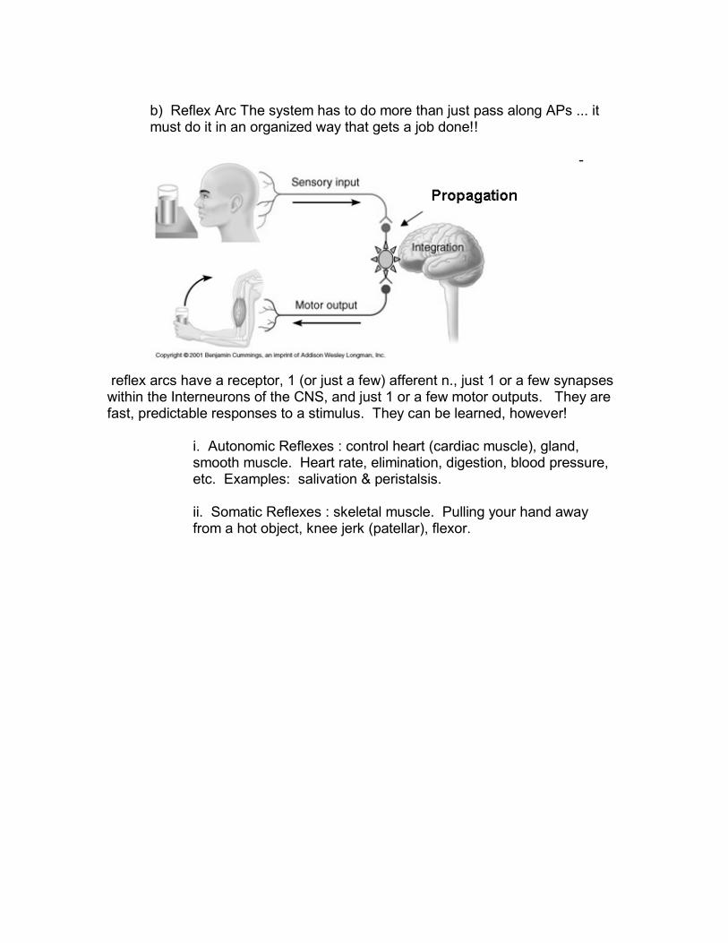
b) Reflex Arc The system has to do more than just pass along APs ... itmust do it in an organized way that gets a job done!!
-
reflex arcs have a receptor, 1 (or just a few) afferent n., just 1 or a few synapseswithin the Interneurons of the CNS, and just 1 or a few motor outputs. They arefast, predictable responses to a stimulus. They can be learned, however!
i. Autonomic Reflexes : control heart (cardiac muscle), gland,smooth muscle. Heart rate, elimination, digestion, blood pressure,etc. Examples: salivation & peristalsis.
ii. Somatic Reflexes : skeletal muscle. Pulling your hand awayfrom a hot object, knee jerk (patellar), flexor.

III PERIPHERAL NERVOUS SYSTEM : NOTE: in book, found at the end of thechapter.
- the nerves and ganglia found outside the CNS, including synapses.
A. Structure of a Nerve
- nerve = bundle of neurons, plus blood vessels. Carry APs to/from theCNS. Wrapped in connective tissue for protection & insulation. Endoneurium around the whole nerve, perineruium around a fascicle(“bundle”) within the nerve, and an endoneurium around an individualneuron.
* ALS : gradual destruction of myelin sheaths. Genetic component.Patient loses control of skeletal muscle. Death often comes fromsuffocation, as they can’t control pharynx or diaphragm.
- spinal nerves leave/enter the spinal cord, cranial nerves leave/enter thebrain.
1. Mixed Nerves : both afferent & efferent fibers. All spinal nerves aremixed.
2. Sensory (Afferent) Nerves : some cranial nerves.
3. Motor (Efferent) Nerves : some cranial nerves.
B. Cranial Nerves
- 12 nerves entering/leaving brain. Some mixed, some afferent, someefferent.
Olfactory Optic Oculomotor Trochlear Trigeminal Abducens Facial Vestibulocochlear Glossopharyngeal Vagus Accessory Hypoglossal
C. Spinal Nerves and Nerve Plexuses
- 31 pairs. All = mixed. Very short, branch almost immediately into rami,which then branch into plexi (plexus = singular, “web”).Ventral and DorsalRami. Cervical, Brachial, lumbar and Sacral plexus. Know the names ofthe important nerves leaving each plexus see on table 7.2.
D. Autonomic Nervous System
- “involuntary” system.
- 2 sensory neurons: pre-ganglionic and post-ganglionic.
1. Comparison of the Somatic and Autonomic Nervous Systems :
- somatic nerves innervate skeletal muscle; they are all the same. Nosubdivisions. Main neurotransmitter = ACh.
- autonomic nerves control “automatic” responses. Severalneurotransmitters: ACh and the “adrenalines” (epinephrine,norepinephrine) can be used, depending on whether the CNS wants tospeed them up (excitatory) or slow them down (inhibition). So ... all theseorgans have 2 autonomic innervations: parasympathetic (“resting state”)and sympathetic (“emergency state”).
2. Anatomy of the Sympathetic Division : “Fight or Flight” system; alsocalled the “thoracolumbar” because this is where the nerves originate fromspinal cord.
- Ganglia are close to the spinal cord (= short pre-ganglionic), forming thesympathetic trunk.
- Epinephrine & Norepinephrine control the effectors.
3. Anatomy of the Parasympathetic Division : “Rest and Relax” system;also called the “craniosacral” because this is where the nerves originatefrom spinal cord.
- Vagus nerve is an important member of this group. Fibers originate inthe medulla oblongata (see later) of the brain, it innervates almost lallviscerl organs, setting a “vagal tone” (resting rate).
- Ganglia are far from the spinal cord (= long pre-ganglionic), no trunkformed.
- ACh controls the effectors.

4. Autonomic Functioning : sometime a given neurotransmitter will speedup an organ (excite), whereas the same neurotransmitter will slow downanother (inhibit).
- Example: the parasympathetic system slows down the heart rate, butspeeds up peristalsis in the gut. The sympathetic innervation speeds upheart rate, but slows down the gut.
On This Diagram: Dark arrows “speed up”, lighter arrows “slow down”
- see table 7.3 for more examples. However, student should be able tofigure it out, if they remember that the parasympathetic system isconserving ATP for the muscles and other important organs for fighting orrunning ... everything else is inhibited.
- the effects of many drugs can be explained as their effects on these 2systems. Some make you drowsy; others make you nervous, sweat,weight loss, etc. These drugs mimic these neurotransmitters, often sittingon the same receptors that would be normally used by theneurotransmitters themselves.

IV. CENTRAL NERVOUS SYSTEM : NOTE: Different organization than thebook
- receives incoming info, interprets and integrates, and sends motoroutput.
- Interneurons = grey matter.
- Terminology:
*tracts = bundlses of neurons, like nerves in the PNS.
*nuclei = collections of bodies, like ganglia in PNS.
- NOTE: there are lots of other neurotransmitters besides thosementioned. Many are chemicals that affect certain regions of the CNS indifferent ways. These include dopamine, serotonin, nitrous oxide, opiates,among many others.
A. Spinal Cord
- dense cord of neural tissue, protected by the vertebral column, blood-brain barrier, and the meninges (protective sheets .... see later). Found inthe dorsal cavity. Interneurons. White matter outside, grey inner core =integration (non-myelinated interneurons).
- center = hollow central canal, filled with CSF (circulating fluid, made byEpendymal cells).
B. Gray Matter of the Spinal Cords, White Matter Columns and Spinal Roots
1. Grey matter: center = grey matter = integration. Surrounded by whitematter = speedy transmission of impulses up & down (no integration; justtake sensory info up to brain, carry commands or motor output down). PNS nerves enter/leave the spinal cord via roots, the PNS neuronssynapse with interneurons in the grey.

2. Roots: although PNS nerves are mixed, they split up as they enter thespinal cord: motors leave dorsally (dorsal root, which has a motor rootganglion), the sensory enter ventrally (no ganglion; recall the discussion ofpre-ganglionic and post-ganglionic neurons earlier).
* polio myelitis: virus that can attack the dorsal root, causingparalysis. If attacks upper part of spinal cord, may affect thesplenic nerve, patient can’t breath (use the diaphragm muscle). Iron lungs.
3. White Matter of the Spinal Cord : myelinated, in bundles (“tracts”) goingup & down through structures called “columns”. Sensory tracts go up,motor tracts go down. Many motor tracts are posterior columns, which iswhy damage to back of spinal cord can cause paralysis but patient stillhas “feeling”.
*spastic paralysis: transection of spinal cord leads to paralysis. Neurons = amitotic, so it is permanent. In spastic paralysis, muscleis not lost, as spinal reflexes still exist, so muscles still receivesstimulation.
*atrophy: permanent loss of muscle via fibrosis, if spinal reflexesare lost.
*Paraplegia: thoracic transection; 2 limbs (legs) lost
*Quadriplegia: cervical transection; 4 limbs lost.
C. Functional Anatomy of the Brain
- brain is an enlargement of CNS at head. About 3 pounds.
- central canal also enlarges into 4 ventricles, filled with CSF.
- Functions:
1. accept sensory input, interpret meaning, make a decision, and sendmotor output.
2. Coordinate muscular activities. Communicate and coordinate with thespinal cord.
3. Make hormones that control visceral functioning, and set a constant“tone” for those visceral organs that need it (lungs, heart, gut, etc.).

- Tissues basically the same as spinal cord, but major difference = greyoutside, white inside.
- “Ridges” give more surface area for functioning.
- 4 major regions, each with individual structures and surrounding theventricles.

Each region has it’s own function:
1. Cerebrum with Cerebral Hemispheres : Accept sensory info, makedecision, send motor output.
- grey matter cortex, with white matter structures inside.
- cortex:
*2 separate hemispheres, separated by the longitudinal fissure.
*Each has lobes, fissures, sulci and gyri. Notice the lobes arenamed for the bones that overlay and protect them.
*Certain spots in grey matter have functions (visual cortex, auditorycortex, gustatory cortex, primary motor cortex, etc.).
*Functional distribution:
i. everything anterior to the central sulcus is motor output(including personality and cognition)
ii. everything posterior is sensory input, memory (for themost part) is on the sides.
- White matter tracts tie the individual structures together to integrate.
*Association” areas of the cortex tie sensory and motor cortices.
*Some structures provide communicate/coordination betweenhemispheres (corpus callosum (white matter “bridge”)
*Some structures provide communication/coordination with the nextsection of the brain (cerebral nuclei connect with the diencephalon).
**Parkinson’s disorder: problem at basal nuclei withdeficiency of neurotransmitter dopamine. Tremors,interspersed with catatonia.

2. Diencephalon : many parts, most dealing with hormone secretion ortransferring information:
- thalamus : surrounds 3 ventricle. Ependymal cells make CSF. Realy uprd
to sensory cortex.
- hypothalamus : has the pituitary gland, which secretes many hormonesinvolved in visceral homeostasis. Also, important part of the limbicsystem, which is the emotinal brain.
- pineal gland: melatonin secretion, controls wake/sleep cycle.
3. Brain Stem : controls visceral “tone” by containing several nuclei(respiratory, cardic, etc.). Contains the Reticular Activatin System, whichgives you consciousness (damaging the reticular formation leads tocoma). # major structures:
- Midbrain : relay sensory up and motor down. Also, involved in hearingand vision reflexes (corpora quadrigemina).
- Pons : relay with spinal cord and respiratory reflexes.
- Medulla Oblongata : many nuclei controling vital visceral activities, heartrate, breathing, blood pressure, swallowing, vomiting, etc. Many fibers ofthe vagus nerve originate here..
4. Cerebellum : coordinate muscular activities, bu monitoring info fromproprioceptors, and motor output by the cerebrum.

D. Protection of the Central Nervous System
Meninges
Meningitis and Encephalitis
Cerebrospinal Fluid
Hydrocephalus
The Blood-Brain Barrier
E. Brain Dysfunctions
Traumatic Brain Injuries
Degenerative Brain Diseases
Cerebrovascular Accidents
Alzheimer's Disease