Embed Size (px)
Citation preview
MCAT-3200184 book November 12, 2015 14:52 MHID: 1-25-958837-8 ISBN: 1-25-958837-2
CHAPTER 7
The Nature ofMolecules andIntermolecular
Interactions
Read This Chapter to Learn About➤ The Chemical Bond
➤ Molecular Shape
➤ Molecular Orbitals
➤ Noncovalent Bonds
THE CHEMICAL BONDWithout chemical bonds, molecules would not exist. Therefore, rationalizing chemical
phenomena must begin with an understanding of bonding.
Types of BondsNobel laureate Roald Hoffmann described molecules as “persistent groupings of
atoms.” Such assemblies are held together by chemical bonds, which conventionally
fall into one of two categories: ionic and covalent. Both bonds involve electrostatic
forces: ionic bonds are present between a positively-charged cation and a negatively
charged anion (as in sodium chloride), whereas covalent bonds arise from the
mutual attraction of two positively charged nuclei to negatively-charged electron
203

MCAT-3200184 book November 12, 2015 14:52 MHID: 1-25-958837-8 ISBN: 1-25-958837-2
204UNIT II:ChemicalFoundations ofBiological Systems
density shared between them (as in diatomic nitrogen). However, covalent and ionic
bonds are just two ends of a bonding continuum.
When the electronegativity difference between the atoms is quite small, the elec-
tron density is equitably distributed along the internuclear axis, and it reaches a max-
imum at the midpoint of the bond. Such a bond is said to be purely covalent. On the
other hand, when the two nuclei have widely divergent electronegativities, the elec-
tron density is not “shared” at all: two ionic species are formed and the electron density
approaches zero along the internuclear axis at the edge of the ionic radius.
However, organic chemistry rarely operates at the boundaries of purely covalent
or purely ionic bonding. Instead, most examples lie along a continuum between these
two extremes (see Figure 7-1). For example, polar covalent bonds result from an un-
even sharing of electron density, a situation that sets up a permanent dipole along
the bond axis. The O H and C F bonds are examples of polar covalent bonds. Such
bonds are stronger than you would expect, because the covalent attraction is aug-
mented by the coulombic forces set up by the dipole.
Conversely, there are many examples of essentially ionic compounds that exhibit
covalent character. In other words, even though there are practically two ionic species
bound together, electron density is still shared between them. Almost all carbon-metal
bonds fall into this category. For example, methyllithium (H3CLi) can be thought of as a
methyl anion (H3C−) with a lithium counterion (Li+). Even though this is not a strictly
accurate representation (i.e, there is indeed shared electron density), it still allows you
to make sound predictions about its chemical behavior.
Purely
covalent
Polar
covalent
Ionic with
covalent character
Purely
ionic
N N H3CO H H3C Lid2 d1
Li F1 2
Difference in electronegativity (DEN )
FIGURE 7-1 Types of bonding in organic molecules.
Lewis Structures and Resonance FormsExamining electronegativity trends can allow you to make predictions about bond
polarity, but you also need to understand the larger bonding picture: how many bonds
are formed with each atom? The Lewis dot diagram represents a surprisingly simple
device for representing global molecular bonding on a primary level. These diagrams
are built up by considering the valence electrons brought to the table by each atom
(conveniently remembered by counting from the left on the periodic table) and then
forming bonds by intuitively combining unpaired electrons. For example, methane
(CH4) and formaldehyde (H2CO) are constructed as shown in Figure 7-2.

MCAT-3200184 book November 12, 2015 14:52 MHID: 1-25-958837-8 ISBN: 1-25-958837-2
205CHAPTER 7:
The Nature ofMolecules andIntermolecular
Interactions
C
O
H
HH
H
C
H
H
C
H
HH
H
O
C
H
HH
H
OC
H
H
C
H
H
Step 1: draw atoms
with valence electrons
Step 2: form bonds
by combining
unpaired electrons
Step 3: draw shared
pairs as single lines,
lone pairs as dots
Methane
Formaldehyde
FIGURE 7-2 Lewis structures for methane and formaldehyde.
Notice that there are two types of electron pairs in the molecules in Figure 7-2:
(1) shared pairs (or bonds), which are represented by lines (each line representing two
shared electrons), and (2) lone pairs, which are depicted using two dots. When calcu-
lating formal charges—which, incidentally, should always be done—assign to a given
atom all of its lone pair electrons and half of each shared pair; then compare the sum
to the number of valence electrons normally carried. For example, consider the amide
species (see Figure 7-3). The nitrogen atom is surrounded by two lone pairs (nitrogen
“owns” all four) and two shared pairs (nitrogen “owns” only one in each pair), giving a
total of six electrons assigned to nitrogen. Compared to the five valence electrons nor-
mally carried by nitrogen, this represents an excess of one electron; therefore, a formal
charge of −1 is given to the nitrogen atom.
NHH
FIGURE 7-3 Lewis structure for the amide species.
Occasionally, the Lewis structures don’t coalesce right away into such tidy pack-
ages. Therefore, you must often select the most reasonable Lewis representation from
a collection of candidates. These are known as resonance forms, and while all reason-
able candidates tell you something about the nature of the molecule they represent,
some resonance structures are more significant contributors than others. In making
such an assessment, the following guidelines are helpful:
A. Octet Rules
1. Big octet rule : no row 2 element can accommodate more than 8 electrons.
NH
H
C
H
NH
H
C
H O
is ok is in violation
O O
O

MCAT-3200184 book November 12, 2015 14:52 MHID: 1-25-958837-8 ISBN: 1-25-958837-2
206UNIT II:ChemicalFoundations ofBiological Systems
2. Little octet rule : all things being equal, each atom should have an octet.
C
H
H
C
H
H
O is better than O
B. Rules of Charge Separation
1. All things being equal, structures should have minimal charge separation.
C
H
H
C
H
H
is better thanO O
2. Any charge separation should be guided by electronegativity trends.
C
H
H
C
H
H
is better than OO
Of all these, only the big octet rule is inviolable. Structures that break the latter
three rules are less desirable—those that break the first one are unreasonable and
unsupportable. Keep in mind that Lewis structures are gross simplifications of a more
complex reality, and sometimes no one representation is adequate to describe the total
bonding within a molecule. Even when a structure satisfies all the rules—as with the
first depiction of formaldehyde—other structures (resonance forms) may need to be
considered to predict the properties of a molecule. The concept of resonance is con-
sidered later in this chapter.
The Condensed Formula and Line NotationAnother useful principle for constructing a structural representation from a molecu-
lar formula involves the idea of valency, or the number of bonds typically formed by
a given element. For example, carbon normally has a valency of four; nitrogen, three;
oxygen, two; hydrogen and the halogens, one. It is valency that underlies the hidden
code of the so-called condensed formula, as illustrated in Figure 7-4 for the compound
3-hydroxypentanal. A condensed formula can appear to be ambiguous about struc-
ture, but in fact it is rich in structural information as long as you are aware of a few
simple rules:
1. A condensed formula is read from left to right.
2. Each carbon atom in the formula is connected to the next carbon in the formula.
3. Everything to the right of a carbon (but before the next) is connected to that carbon.
4. Parentheses are used to indicate whole groups attached to a carbon.
5. Normal valencies must be satisfied (C = 4; N = 3; O = 2; H = 1; etc.).

MCAT-3200184 book November 12, 2015 14:52 MHID: 1-25-958837-8 ISBN: 1-25-958837-2
207CHAPTER 7:
The Nature ofMolecules andIntermolecular
Interactions
As the example in Figure 7-4 shows, the first step in converting a condensed for-
mula into a structural formula is to lay out the carbon backbone—in this case a five-
carbon chain. You then connect all the indicated substituents: three hydrogens to the
first carbon, two hydrogens to the second, and so on, recognizing that the hydroxy (OH)
moiety is treated as an entire group attached to the third carbon. The tricky part is the
expansion of the CHO group. It is tempting to imagine a structure in which the carbon
is attached to the hydrogen, which in turn is attached to the oxygen (lower left depic-
tion). However, this would violate three valency guidelines: oxygen has only one bond,
carbon only two, and hydrogen one too many. A proper reading of the condensed for-
mula would be, “hydrogen belongs to the fifth carbon, and so does oxygen.” With a
bit of thought, the only reasonable arrangement would be the one shown in the lower
right, which includes a carbon-oxygen double bond.
A depiction that fulfills valencyTempting, but incorrect!
The condensed formula The implied carbon backbone A partial expansion of the structure
CH
H
H
H
C
H
C
H
H
C
H
H
O
CHOCH3CH2CH(OH)CH2CHO CH3CH2CH(OH)CH2CHO
CH
H
H
H
C
H
C
H
H
C
H
H
O
C H O H H
H
C C
H
H
C
H
C
H
H
C
H
H
O O
FIGURE 7-4 Converting a condensed formula to a full formula.
A condensed formula is one type of shortcut for depicting structure—its claim
to fame is that it can be produced using a keyboard even when graphical software is
unavailable. However, an even more important and widespread shortcut is line nota-
tion, which is universally used by organic chemists when they sketch compounds, its
advantage being that the salient features of complex structures can be quickly and
conveniently depicted. Figure 7-5 shows 3-hydroxypentanal in line notation. The
assumptions underlying the simplification are these:
1. Every unlabeled vertex or terminus is a carbon atom.
2. Any unused valency of carbon is filled by hydrogen.
H H
H
C C
H
H
C
H
C
H
H
C
H
H
O O OH
O
In line notationIn full formula notation
FIGURE 7-5 Two valid depictions of 3-hydroxypentanal.
MCAT-3200184 book November 12, 2015 14:52 MHID: 1-25-958837-8 ISBN: 1-25-958837-2
208UNIT II:ChemicalFoundations ofBiological Systems
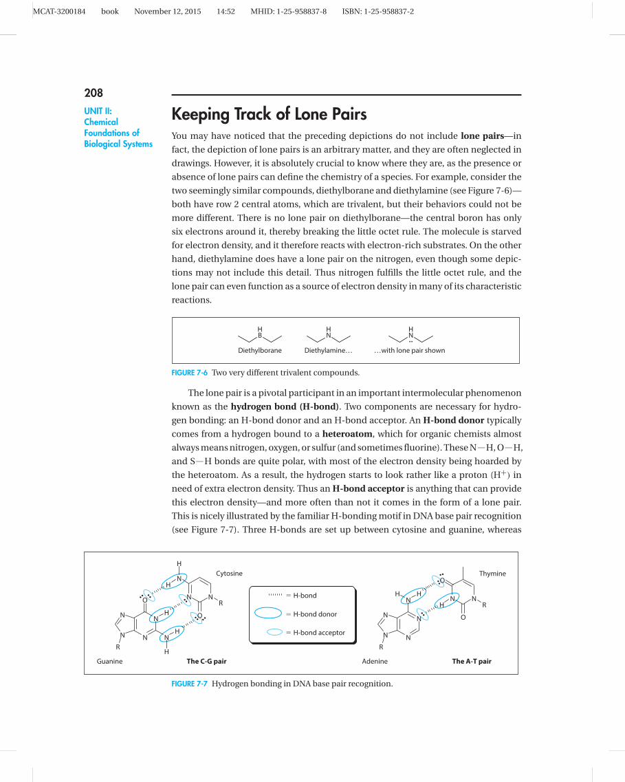
Keeping Track of Lone PairsYou may have noticed that the preceding depictions do not include lone pairs—in
fact, the depiction of lone pairs is an arbitrary matter, and they are often neglected in
drawings. However, it is absolutely crucial to know where they are, as the presence or
absence of lone pairs can define the chemistry of a species. For example, consider the
two seemingly similar compounds, diethylborane and diethylamine (see Figure 7-6)—
both have row 2 central atoms, which are trivalent, but their behaviors could not be
more different. There is no lone pair on diethylborane—the central boron has only
six electrons around it, thereby breaking the little octet rule. The molecule is starved
for electron density, and it therefore reacts with electron-rich substrates. On the other
hand, diethylamine does have a lone pair on the nitrogen, even though some depic-
tions may not include this detail. Thus nitrogen fulfills the little octet rule, and the
lone pair can even function as a source of electron density in many of its characteristic
reactions.
HN
HN
HB
…with lone pair shownDiethylborane Diethylamine…
FIGURE 7-6 Two very different trivalent compounds.
The lone pair is a pivotal participant in an important intermolecular phenomenon
known as the hydrogen bond (H-bond). Two components are necessary for hydro-
gen bonding: an H-bond donor and an H-bond acceptor. An H-bond donor typically
comes from a hydrogen bound to a heteroatom, which for organic chemists almost
always means nitrogen, oxygen, or sulfur (and sometimes fluorine). These N H, O H,
and S H bonds are quite polar, with most of the electron density being hoarded by
the heteroatom. As a result, the hydrogen starts to look rather like a proton (H+) in
need of extra electron density. Thus an H-bond acceptor is anything that can provide
this electron density—and more often than not it comes in the form of a lone pair.
This is nicely illustrated by the familiar H-bonding motif in DNA base pair recognition
(see Figure 7-7). Three H-bonds are set up between cytosine and guanine, whereas
N N
O
ON
NN
N
N
R
R
N
NN
N
O
N
R
N N
N
O
R
H
H
H
H
H
H H
H
..
....
.. ..
..
..
..
5 H-bond
Cytosine
Guanine The C-G pair Adenine
Thymine
The A-T pair
5 H-bond donor
5 H-bond acceptor
FIGURE 7-7 Hydrogen bonding in DNA base pair recognition.

MCAT-3200184 book November 12, 2015 14:52 MHID: 1-25-958837-8 ISBN: 1-25-958837-2
209CHAPTER 7:
The Nature ofMolecules andIntermolecular
Interactions
two are enjoyed by thymine and adenine. In each case, the H-bond donors are N H
bonds, while the H-bond acceptors are lone pairs on nitrogen or oxygen. Inspection
reveals that the degree of H-bonding would be far less with a G-T pair or an A-C pair.
MOLECULAR SHAPEWith an understanding of connectivity among atoms within molecules, it is now
appropriate to consider the three-dimensional arrangement of these atoms, since
molecular shape is one of the chief factors determining the functionality and reactivity
of a given molecule.
Geometry of Atoms Within MoleculesThere are three frequently encountered geometries in organic chemistry: digonal (or
linear), trigonal, and tetrahedral (see Figure 7-8). Each atom within a molecule is
almost always characterized by one of these geometries, the chief hallmark of which
is the associated bond angle: 180◦ for linear arrays, 120◦ for trigonal planar centers,
and 109.5◦ for tetrahedral arrangements. Deviation from these ideal bond angles does
occur, but significant deformation usually has a destabilizing effect known as bond
angle strain.
Geometry Arrangement Bond angle Hybridization of x
Digonal a x
x
x
a
a
dc
c
b 1808 sp
sp2
sp3
1208
109.58
b
b
Trigonal
Tetrahedral
FIGURE 7-8 Common central atom geometries in organic chemistry.
There are at least three ways to conceptualize molecular geometry. One classi-
cal approach is through the hybridization of atomic orbitals. If orbitals are derived
from wave functions, then these functions can be combined mathematically to obtain
hybrid descriptions. Thus if you combine an s orbital, which is spherically symmet-
rical, with a single p orbital, which has directionality along a single axis, it stands to
reason that the outcome (two equivalent sp orbitals) should also have directionality
along one dimension. Likewise, the combination of an s orbital with two p orbitals
gives a result (three equivalent sp2 orbitals) that defines a plane (see Figure 7-9).
Another framework is conceptually more straightforward. Known as valence shell
electron pair repulsion (VSEPR) theory, this approach asks the question of how

MCAT-3200184 book November 12, 2015 14:52 MHID: 1-25-958837-8 ISBN: 1-25-958837-2
210UNIT II:ChemicalFoundations ofBiological Systems
1 5
1 5
51
One dimension (x) represented
Two dimensions (xy) represented
3 sp2 orbitals
2 sp orbitals
4 sp3 orbitals
All three dimensions represented
pzpypxs
py
pxs
pxs
1
11
FIGURE 7-9 Central atom geometry as described by orbital hybridization.
negatively charged electron clouds can most effectively stay out of each other’s way.
If there are only two electron clouds, then the happiest arrangement is to be diamet-
rically opposed to each other; for three clouds, a trigonal planar array; and for four
clouds, a tetrahedral arrangement provides the maximum distance among them.
Interestingly, these considerations predict exactly the same outcomes as hybrid
orbital theory, although they are fundamentally different approaches. Strictly speak-
ing, neither are theoretically accurate as compared to a strict quantum mechanical
analysis—a third method not addressed here—however, they are useful predictive
models nonetheless.
Thus to predict the geometry about a central atom, you must ask only how many
things surround it—where “things” are understood to be either atoms or lone pairs.
Figure 7-10 offers an illustration of this method, and experimental evidence backs up
the predictions (e.g., the C C C bond angle is practically 120◦). A generalization can
also be derived from this example: since carbon is almost always tetravalent, then if
only single bonds are attached to a carbon, it must have four other atoms surrounding
it; if a double bond is attached to that carbon, then only three atoms can surround
H3C CH3
O This carbon has three "things"
around it [two carbons and
one oxygen] => sp2
This oxygen has three "things"
around it [one carbon and two
lone pairs (not shown)] => sp2
Each of these carbons has four
"things" around it [one carbon
and three hydrogens] => sp3
FIGURE 7-10 Prediction of geometry for acetone.
MCAT-3200184 book November 12, 2015 14:52 MHID: 1-25-958837-8 ISBN: 1-25-958837-2
211CHAPTER 7:
The Nature ofMolecules andIntermolecular
Interactions
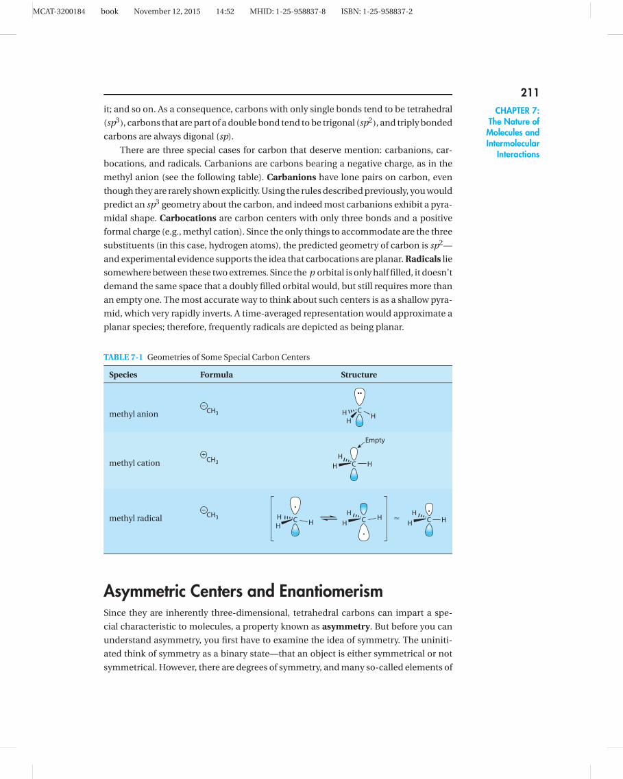
it; and so on. As a consequence, carbons with only single bonds tend to be tetrahedral
(sp3), carbons that are part of a double bond tend to be trigonal (sp2), and triply bonded
carbons are always digonal (sp).
There are three special cases for carbon that deserve mention: carbanions, car-
bocations, and radicals. Carbanions are carbons bearing a negative charge, as in the
methyl anion (see the following table). Carbanions have lone pairs on carbon, even
though they are rarely shown explicitly. Using the rules described previously, you would
predict an sp3 geometry about the carbon, and indeed most carbanions exhibit a pyra-
midal shape. Carbocations are carbon centers with only three bonds and a positive
formal charge (e.g., methyl cation). Since the only things to accommodate are the three
substituents (in this case, hydrogen atoms), the predicted geometry of carbon is sp2—
and experimental evidence supports the idea that carbocations are planar. Radicals lie
somewhere between these two extremes. Since the p orbital is only half filled, it doesn’t
demand the same space that a doubly filled orbital would, but still requires more than
an empty one. The most accurate way to think about such centers is as a shallow pyra-
mid, which very rapidly inverts. A time-averaged representation would approximate a
planar species; therefore, frequently radicals are depicted as being planar.
TABLE 7-1 Geometries of Some Special Carbon Centers
Species Formula Structure
CHH
Hmethyl anion CH3
C HH
H
Empty
methyl cation CH3
<C HH
H
..
H
.
C HH
HCH
Hmethyl radical CH3
Asymmetric Centers and EnantiomerismSince they are inherently three-dimensional, tetrahedral carbons can impart a spe-
cial characteristic to molecules, a property known as asymmetry. But before you can
understand asymmetry, you first have to examine the idea of symmetry. The uniniti-
ated think of symmetry as a binary state—that an object is either symmetrical or not
symmetrical. However, there are degrees of symmetry, and many so-called elements of

MCAT-3200184 book November 12, 2015 14:52 MHID: 1-25-958837-8 ISBN: 1-25-958837-2
212UNIT II:ChemicalFoundations ofBiological Systems
symmetry. This is a topic best treated in the domain of mathematics, but we shall very
briefly scratch the surface here to gain some underpinnings for practical application.
First, consider methane (CH4)—a molecule that is unassuming, yet full of symmetry.
Figure 7-11 shows three types of symmetry elements (there are others not shown here)
belonging to methane. The sigma (σ) plane is an imaginary mirror that slices through
the molecule—reflection through the plane results in an image identical to the starting
depiction. Methane actually has six such planes: one with H1 and H2 in the plane; one
with H1and H3; one with H1 and H4; one with H2 and H3; one with H2 and H4; and one
with H3 and H4. There are also two types of rotational axes. A C2 axis has two “clicks” in
a 360◦ rotation—180◦ each turn; methane has six of these, much like the sigma planes.
A C3 axis has three “clicks” in a full rotation—120◦ each turn; methane has four of these,
one coinciding with each of the C H bonds.
CH2
H2H3H3H4
H4
C
120°
H4H3
C
H1 and H2 lie in the plane
H2
H3 reflects onto H4
The sigma (�) plane� plane
H1
H4H3
C
H2H1
H3H4
C
H1H2
180°
H1 H1
The C2 axis of rotation The C3 axis of rotation
FIGURE 7-11 Symmetry elements of methane.
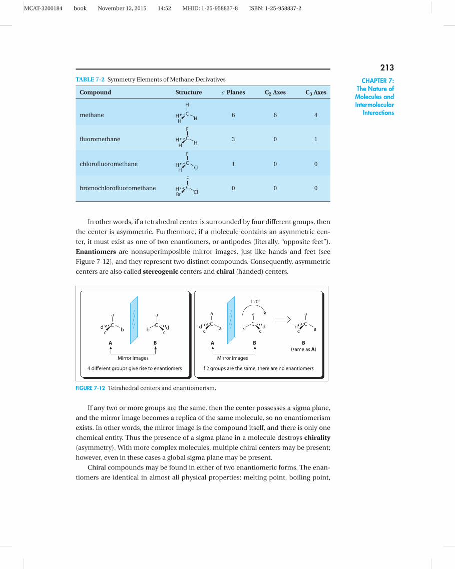
As you add substituents to methane, however, symmetry starts to drop off rapidly.
For example, the addition of a single substituent, such as fluorine (see Table 7-2)
reduces the number of sigma planes by half (each sigma plane must include the flu-
orine), slashes the number of C3 axes to one (the axis which coincides with the C F
bond), and eliminates the C2 axes altogether. A second substituent results in an even
more impoverished symmetry environment, such that chlorofluoromethane has only
a single sigma plane. After a third substituent is introduced (i.e., bromochlorofluo-
romethane), there are no symmetry elements left. Therefore, such a molecule is said
to be asymmetric.
MCAT-3200184 book November 12, 2015 14:52 MHID: 1-25-958837-8 ISBN: 1-25-958837-2
213CHAPTER 7:
The Nature ofMolecules andIntermolecular
Interactions
TABLE 7-2 Symmetry Elements of Methane Derivatives
Compound Structure σ Planes C2 Axes C3 Axes
methaneH
HH
H
C 6 6 4
fluoromethane
F
CH
HH
3 0 1
chlorofluoromethane
F
CCl
HH
1 0 0
bromochlorofluoromethane
F
CCl
HBr
0 0 0
In other words, if a tetrahedral center is surrounded by four different groups, then
the center is asymmetric. Furthermore, if a molecule contains an asymmetric cen-
ter, it must exist as one of two enantiomers, or antipodes (literally, “opposite feet”).
Enantiomers are nonsuperimposible mirror images, just like hands and feet (see
Figure 7-12), and they represent two distinct compounds. Consequently, asymmetric
centers are also called stereogenic centers and chiral (handed) centers.
a
Cbd
c
a
Cb d
c
a
Cad
c
a
Ca d
c
A B
120°
a
Cad
c
B
(same as A)
If 2 groups are the same, there are no enantiomers4 different groups give rise to enantiomers
Mirror images
A B
Mirror images
FIGURE 7-12 Tetrahedral centers and enantiomerism.
If any two or more groups are the same, then the center possesses a sigma plane,
and the mirror image becomes a replica of the same molecule, so no enantiomerism
exists. In other words, the mirror image is the compound itself, and there is only one
chemical entity. Thus the presence of a sigma plane in a molecule destroys chirality
(asymmetry). With more complex molecules, multiple chiral centers may be present;
however, even in these cases a global sigma plane may be present.
Chiral compounds may be found in either of two enantiomeric forms. The enan-
tiomers are identical in almost all physical properties: melting point, boiling point,

MCAT-3200184 book November 12, 2015 14:52 MHID: 1-25-958837-8 ISBN: 1-25-958837-2
214UNIT II:ChemicalFoundations ofBiological Systems
Plane of incomingpolarized light
Plane of outgoingpolarized light
Sample containing 100%
Incomingplane Outgoing
plane
Sample containing 100%
Incomingplane
� �
Outgoingplane
Levorotatory Dextrorotatory
Sample containingchiral compound
F
C HBr
Cl
F
CHBr
Cl
FIGURE 7-13 The phenomenon of optical rotation.
dielectric constant, and so on, with one notable exception. A solution of a chiral com-
pound interacts with light in such a way that the plane of polarized light is rotated upon
passing through a sample of the compound, a phenomenon known as optical rotation
(see Figure 7-13, top). The other enantiomer rotates light to the same degree, but in the
opposite direction (see Figure 7-13, bottom). Enantiomers that rotate light clockwise
(i.e., to the right) are known as dextrorotatory isomers, and those that rotate light in
a counterclockwise direction (i.e., to the left) are known as levorotatory isomers. It is
important to understand that there is no straightforward way to correlate structure to
behavior. In other words, you cannot tell just by looking at a molecule whether it would
be dextrorotatory or levorotatory. By the same token, optical rotation tells you nothing
about a molecule except that it is chiral.
A sample that rotates plane-polarized light is said to be optically active. The degree
to which the sample rotates light is called the optical rotation (in degrees). If a sample
is optically inactive (i.e., it does not rotate plane-polarized light), it could mean one
of two things: either (1) the sample does not contain chiral molecules, or (2) the sam-
ple contains exactly equal quantities (50/50) of two enantiomers. The latter situation,
known as a racemic mixture, produces no optical activity, because for each levorota-
tory molecule there is a dextrorotatory counterpart—equal numbers pulling in oppo-
site directions maintain the status quo.
MCAT-3200184 book November 12, 2015 14:52 MHID: 1-25-958837-8 ISBN: 1-25-958837-2
215CHAPTER 7:
The Nature ofMolecules andIntermolecular
Interactions
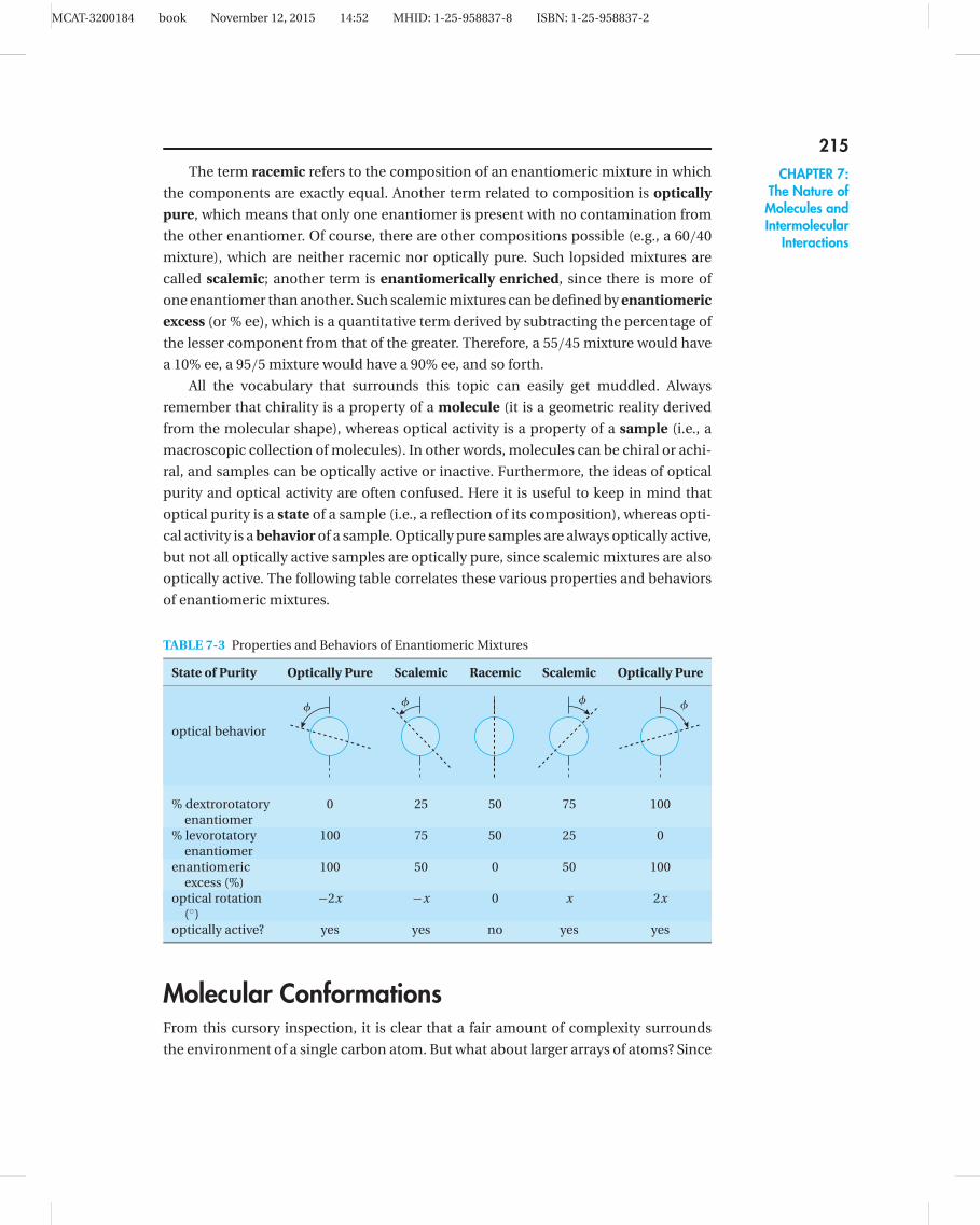
The term racemic refers to the composition of an enantiomeric mixture in which
the components are exactly equal. Another term related to composition is optically
pure, which means that only one enantiomer is present with no contamination from
the other enantiomer. Of course, there are other compositions possible (e.g., a 60/40
mixture), which are neither racemic nor optically pure. Such lopsided mixtures are
called scalemic; another term is enantiomerically enriched, since there is more of
one enantiomer than another. Such scalemic mixtures can be defined by enantiomeric
excess (or % ee), which is a quantitative term derived by subtracting the percentage of
the lesser component from that of the greater. Therefore, a 55/45 mixture would have
a 10% ee, a 95/5 mixture would have a 90% ee, and so forth.
All the vocabulary that surrounds this topic can easily get muddled. Always
remember that chirality is a property of a molecule (it is a geometric reality derived
from the molecular shape), whereas optical activity is a property of a sample (i.e., a
macroscopic collection of molecules). In other words, molecules can be chiral or achi-
ral, and samples can be optically active or inactive. Furthermore, the ideas of optical
purity and optical activity are often confused. Here it is useful to keep in mind that
optical purity is a state of a sample (i.e., a reflection of its composition), whereas opti-
cal activity is a behavior of a sample. Optically pure samples are always optically active,
but not all optically active samples are optically pure, since scalemic mixtures are also
optically active. The following table correlates these various properties and behaviors
of enantiomeric mixtures.
TABLE 7-3 Properties and Behaviors of Enantiomeric Mixtures
State of Purity Optically Pure Scalemic Racemic Scalemic Optically Pure
� � � �
optical behavior
% dextrorotatory 0 25 50 75 100enantiomer
% levorotatory 100 75 50 25 0enantiomer
enantiomeric 100 50 0 50 100excess (%)
optical rotation −2x −x 0 x 2x(◦)
optically active? yes yes no yes yes
Molecular ConformationsFrom this cursory inspection, it is clear that a fair amount of complexity surrounds
the environment of a single carbon atom. But what about larger arrays of atoms? Since

MCAT-3200184 book November 12, 2015 14:52 MHID: 1-25-958837-8 ISBN: 1-25-958837-2
216UNIT II:ChemicalFoundations ofBiological Systems
most molecules have considerable flexibility, it is important to understand what kinds
of shapes are most stable and the energetics involved in their interconversion. Before
doing this, however, it is necessary to come to terms with two additional methods of
depiction for molecular structure: the sawhorse (dash-wedge) projection, and the
Newman projection.
Consider a two-carbon array with three substituents on each carbon. Figure 7-14
shows such an array in two different depictions. The sawhorse projection views the
molecule from the side. Substituents that come out of the plane toward us are
depicted with wedges; those that go away from you are shown with dashes. If there
is neither dash nor wedge, a plain line (and attached substituent) is assumed to lie in
the plane of the paper. “Plain bonds lie in the plane,” is a good mnemonic device in
this regard.
a
b ca
f
b
d
c
e
12
(Observer)
(Observer)
f
d e
Sawhorse Newman
a
b ca
b c
12
f
d
e
f
e
d
Staggeredconformation
Eclipsedconformation
FIGURE 7-14 Sawhorse and Newman projections.
A Newman projection simply looks at the molecule from a different angle, namely
down a carbon-carbon bond (indicated by the observer’s eye). This is a much more
straightforward way of showing the spatial relationship of substituents. Just remember
that the small dot represents the front carbon, while the large circle represents the back
carbon. Thus in the projections shown, the small dot is carbon-2 and the large circle is
carbon-1; in other words, a C2→C1 Newman projection has been drawn. However, it
could have just as easily been drawn as a C1→C2 variant, in which the small dot would
be carbon-1.
Since there can be rotation about carbon-carbon bonds, this molecule can adopt
a variety of conformations. In general, these conformations fall into one of two cate-
gories: staggered and eclipsed. Note that in the sawhorse depictions, the plain bonds
(i.e., neither dash nor wedge) in the staggered conformation describe a “Z” or zigzag
pattern, whereas in the eclipsed conformation they form a “U.” This is a quick and easy
way to distinguish one from another.
In staggered conformations, any two substituents are characterized by one of two
relationships. Substituents are said to be gauche with respect to each other if they are

MCAT-3200184 book November 12, 2015 14:52 MHID: 1-25-958837-8 ISBN: 1-25-958837-2
217CHAPTER 7:
The Nature ofMolecules andIntermolecular
Interactions
side-by-side. In the illustration, there are six gauche relationships: a-e, e-c, c-f, f-b, b-d,
and d-a. The other relationship is antiperiplanar, in which case the two substituents
are as far away as possible from each other. In the same depiction, there are three
antiperiplanar relationships: a-f, e-b, and d-c. In eclipsed conformations, there is only
one type of relationship between substituents to care about, namely eclipsed. In the
eclipsed Newman projection, there are three pairs of eclipsed substituents: a-f, c-d, and
b-e. Don’t be confused by the double duty of the term eclipsed—there are staggered
and eclipsed conformations, which describe the global molecular attitude, and there
are gauche, antiperiplanar, and eclipsed relationships between substituents. There are
only gauche and antiperiplanar relationships in staggered conformations, and there
are only eclipsed relationships in eclipsed conformations.
It should come as no surprise that eclipsed conformations are of higher energy
than staggered conformations. This is due to the steric interactions that result from
the very close proximity of substituents in the eclipsed relationships. By the same
token, staggered conformations that place large groups in a gauche arrangement are
of higher energy than those that have those groups antiperiplanar with respect to each
other. Using these basic principles, you can construct a diagram showing the rela-
tive energies of a conformational ensemble, as illustrated in Figure 7-15 for butane
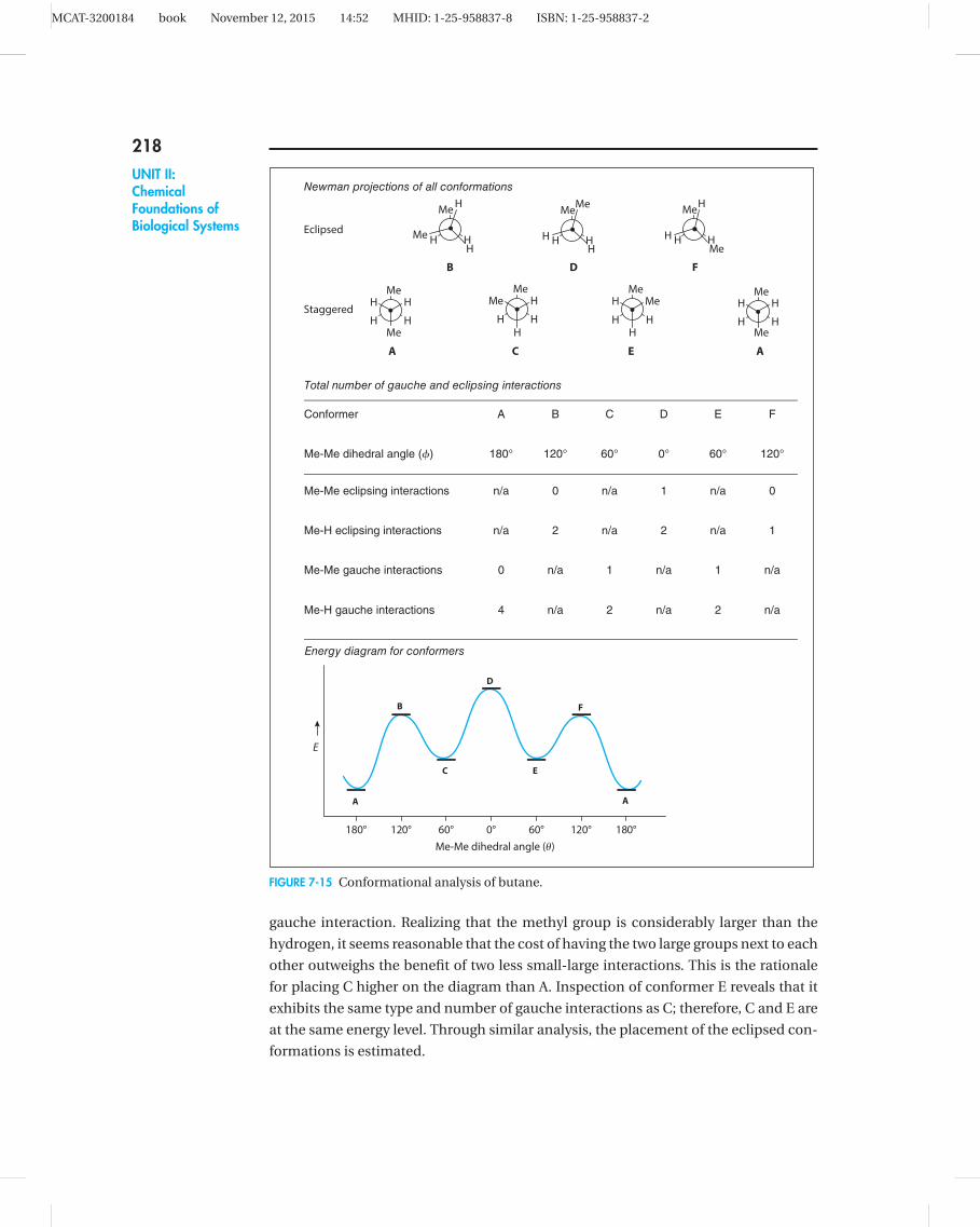
(CH3CH2CH2CH3). This is a process known as conformational analysis.
To begin the conformational analysis, all the possible conformations for the
molecule, both eclipsed and staggered, must be drawn. This might seem more difficult
than it really is—first, just jot down any conformation and then methodically convert
it to the remaining possibilities. For example, in Figure 7-15 the first conformation (A)
is chosen arbitrarily. Conformation A is converted to C by rotating the front carbon
(small dot) clockwise—notice that the back carbon does not change. This maneuver
has the net effect of bringing the front methyl (CH3) group from the 6 o’clock position
to the 10 o’clock position. To get from A to C, the molecule must pass through the high-
energy eclipsed conformer B. The remaining conformations are derived by continuing
to rotate the front carbon in a clockwise fashion until you arrive back at A.
The next step is to assess all the group interactions in each conformer. For the sake
of simplicity, neglect H H interactions, since the hydrogen atom is so small. Thus
in conformer A, observe four methyl-hydrogen gauche interactions (in other words,
each methyl group has a hydrogen to either side—each flanking hydrogen counts as an
interaction). In conformer C, there are only two methyl-hydrogen gauche interactions,
but there is also a methyl-methyl gauche interaction. These interactions are used to
estimate the relative placement of the conformers on the energy diagram.
The diagram can be constructed easily through a three-step approach. First, keep
in mind that all eclipsed conformations will be higher than the staggered conforma-
tions, so there are two separate collections of conformers (i.e., staggered and eclipsed).
Then estimate the relative placement of the conformers within each set. For example,
in comparing the energetics of the staggered conformers A and C, you could say two
methyl hydrogen gauche interactions (2 vs. 4) have been traded for one methyl-methyl
MCAT-3200184 book November 12, 2015 14:52 MHID: 1-25-958837-8 ISBN: 1-25-958837-2
218UNIT II:ChemicalFoundations ofBiological Systems
Me
H HMe
H H
A
B
Me
H HH
Me H
C
D
Me
H HH
H Me
E
F
Me
H HMe
H H
A
Eclipsed
Newman projections of all conformations
Staggered
Me
H H
H
Me
H
Me
H H
Me
HH
Me
H H
H
HMe
Total number of gauche and eclipsing interactions
Conformer A B C D E F
Me-Me dihedral angle (�) 180° 120° 60° 0° 60° 120°
Me-Me eclipsing interactions n/a 0 n/a 1 n/a 0
Me-H eclipsing interactions n/a 2 n/a 2 n/a 1
Me-Me gauche interactions 0 n/a 1 n/a 1 n/a
Me-H gauche interactions 4 n/a 2 n/a 2 n/a
Energy diagram for conformers
180° 120° 60° 0° 60° 120° 180°
E
A
B
C
D
E
F
A
Me-Me dihedral angle (�)
FIGURE 7-15 Conformational analysis of butane.
gauche interaction. Realizing that the methyl group is considerably larger than the
hydrogen, it seems reasonable that the cost of having the two large groups next to each
other outweighs the benefit of two less small-large interactions. This is the rationale
for placing C higher on the diagram than A. Inspection of conformer E reveals that it
exhibits the same type and number of gauche interactions as C; therefore, C and E are
at the same energy level. Through similar analysis, the placement of the eclipsed con-
formations is estimated.

MCAT-3200184 book November 12, 2015 14:52 MHID: 1-25-958837-8 ISBN: 1-25-958837-2
219CHAPTER 7:
The Nature ofMolecules andIntermolecular
Interactions
Conformations of CycloalkanesThere are special conformational issues for cyclic molecules. As a general rule,
cycloalkanes are less flexible than open-chain analogs, and they can adopt far fewer
conformations. However, both types have predictable low-energy conformations. For
example, consider hexane (see Figure 7-16): inasmuch as there is free rotation about all
the carbon-carbon bonds, you could envision the six conformational option described
in butane for each of the internal C C bonds (i.e., C2-C3, C3-C4, and C4-C5). The most
stable conformation is the one that has all antiperiplanar relationships, as shown in
Figure 7-16. Similarly, cyclohexane can and does adopt many conformations, but the
most stable arrangement is the so-called chair conformation.
1 3 5642
1 3
56 4
2
CyclohexaneHexane
FIGURE 7-16 Stable conformations of hexane and cyclohexane.
Although it may not be evident, there are two alternate chair forms that are
related by the chair flip. It is important to understand that the flip is not a rotation of
the molecule, but a reconformation. As the top depiction in Figure 7-17 shows, carbon
1 flips from pointing downward to pointing upward, carbon 4 does the reverse, and the
entire molecule reconforms in place. As a side note, always keep in mind that the chair
projection is a side view of the ring, and the lower bond (i.e., C2-C3) is assumed to be
in front.
When substituents are added to the cyclohexane, the chair flip takes on special
significance. To understand this, first come to terms with some characteristics of the
substituents on a cyclohexane ring. Examination of Figure 7-17 (lower depiction) re-
veals that substituents can adopt one of two attitudes: axial (shown as triangles in the
left chair) or equatorial (shown as squares)—every chair conformation has six of each
1 3
56 4
2
1
3
56
42
13
56 4
2
1
3
56
42
FIGURE 7-17 The chair flip of cyclohexane.

MCAT-3200184 book November 12, 2015 14:52 MHID: 1-25-958837-8 ISBN: 1-25-958837-2
220UNIT II:ChemicalFoundations ofBiological Systems
type. Note that a chair flip interchanges axial and equatorial substituents, so that all of
the triangles become equatorial on the right side. It is a good idea to construct a model
and physically induce a ring flip to see how this works.
It is equally important to understand what does not change during a ring flip. Any
ring has two faces—as these depictions are drawn, they can be called the top and bot-
tom faces. Any substituent points toward either the top or the bottom face. Again,
each chair cyclohexane has six of each type—the six white substituents are all pointing
toward the top face, and the six black ones are pointing toward the bottom face. Note
that there are three axial and three equatorial substituents pointing up, and three of
each pointing down. Also note that a ring flip does not change whether a substituent
points up or down. So in other words the triangle substituent on carbon 4 always points
up, although it may convert from axial to equatorial. As a general rule, the bulkiest sub-
stituent prefers the equatorial position.
Other ring sizes have different lowest energy conformations, and a brief survey
is worthwhile. For example, the most stable conformation of cyclopentane (see Fig-
ure 7-18) is the so-called envelope, which (like the chair) has two forms that equili-
brate through the flipping of the envelope flap. Cyclobutane adopts a so-called
puckered conformation, which again has two forms that equilibrate through a ring
flip. Notice that the same rules for cyclohexanes hold true for these cycloalkanes—
namely, that there are two types of substituent attitudes (here, called pseudoaxial and
pseudoequatorial) that interconvert upon ring flips; also, the substituents point toward
a certain face, and that face remains constant throughout conformational changes.
Finally, cyclopropanes have practically no conformational flexibility—there is really
only one possibility, and all substituents have equivalent attitudes. However, of course,
there are still two faces to the molecule, so substituents can point toward the top face
or the bottom face.
Cyclopentane
Cyclobutane
Cyclopropane
FIGURE 7-18 Stable conformations of other cycloalkanes.

MCAT-3200184 book November 12, 2015 14:52 MHID: 1-25-958837-8 ISBN: 1-25-958837-2
221CHAPTER 7:
The Nature ofMolecules andIntermolecular
Interactions
So what factors govern the adoption of a given stable conformation? The main
determinant is the minimization of ring strain. In cyclic molecules, there are essentially
two sources of strain: bond angle strain and torsional strain. Bond angle strain derives
from a compression of the ideal sp3 bond angle (109.5◦) to accommodate a cyclic array.
Not surprisingly, this is worst for the three-membered ring, and is almost nil for the
five- and six-membered rings. The other source of strain is less obvious. Torsional
strain derives from the torque on individual bonds from eclipsed substituents trying
to get out of each other’s way. Again, this is most pronounced in cyclopropane (com-
pare Figure 7-18), and all the other cycloalkanes twist in ways to minimize or eliminate
this strain. For example, inspection of a molecular model of chair cyclohexane will
reveal that all carbon-carbon bonds have a perfectly staggered conformation. Inter-
estingly, the five-membered ring is not so lucky. Even in the envelope conformation,
substituents tend toward eclipsing each other. The following table summarizes the
individual components, but a good overall take-home message is that ring strain
decreases according to the trend: 3 > 4 � 5 > 6. This has implications in reactivity
of cyclic molecules.
TABLE 7-4 Ring Strain in Cycloalkanes
Cycloalkane Torsional Strain Bond Angle Strain
cyclopropane a lot a lotcyclobutane some a lotcyclopentane a little practically nonecyclohexane none none
Reconciling Visual MeaningComing to terms with the three-dimensionality of organic chemistry is often a chal-
lenge for students, yet this is possibly the most important transferable cognitive skill
developed by the study of the subject. The novice is confronted with a jumble of
alternative depictional devices that appear to be interchangeable. However, a struc-
tural drawing is a way of communicating, and specific information is carried in these
depictions. For example, if you are presented with a simple line drawing (see the follow-
ing table), you can quickly see the landscape of the molecule—what the regiochem-
istry is (that is, where the atoms are) and which functional groups are present—but you
are told nothing about the stereochemistry (that is, the three-dimensional arrange-
ment of the atoms). On the other hand, the Fischer projection was developed specifi-
cally for quickly conveying the absolute stereochemistry (configuration) of a molecule.
You must carefully choose the right depiction for the information you wish to convey,
and you must also be able to fully interpret the messages given by specific structures.
One particularly thorny depictional issue centers around relative and absolute
stereochemistry. For example, you can draw a structure for cis-5-methylcyclohex-
2-enol, which is unequivocal and easily distinguishable from the corresponding

MCAT-3200184 book November 12, 2015 14:52 MHID: 1-25-958837-8 ISBN: 1-25-958837-2
222UNIT II:ChemicalFoundations ofBiological Systems
TABLE 7-5 Summary of Structural Depictions
Type of Depiction Example Best for Depicting
line structure
OH
constitution and connectivity
sawhorse
OH
relative and absolute stereochemistry
Newman
Me
HO H
Et
H Me conformation
Chair
OH
H
H
Me
conformation
Fischer
Me
Et
HO H
Me Hconfiguration (absolute stereochemistry)
trans-isomer (see Figure 7-19). However, it has been arbitrarily chosen to draw the sub-
stituents with two wedges—a structure with two dashes would have been equally valid.
In this example, the only intent was to show that the two substituents are on the same
side of the molecule, that is, their relative stereochemistry. Whether carbon-1 is an R
or S center is unknown.
Unfortunately, the same kind of depiction has been used to show absolute stereo-
chemistry. For example, if you were to draw specifically the 1S, 5S enantiomer of cis-5-
methylcyclohex-2-enol, both substituents would be attached with wedges (see the fol-
lowing table)—but here their absolute placement in space are depicted, not just their
positions relative to each other. Conversely, the R, R enantiomer would be drawn with
dashes. Note that when the configuration is included in the name, cis/trans designation
OH
Me
OH
Me
cis-5-methylcyclohex-2-enol trans-5-methylcyclohex-2-enol
FIGURE 7-19 Relative stereoisomers.

MCAT-3200184 book November 12, 2015 14:52 MHID: 1-25-958837-8 ISBN: 1-25-958837-2
223CHAPTER 7:
The Nature ofMolecules andIntermolecular
Interactions
is unnecessary. Converting a name to a structure is relatively easy—if R/S information
is given, simply represent it accurately in the structure; if only relative (cis/trans) stereo-
chemical information is given, you have a couple of choices for the dashes and wedges;
if no stereochemical information is provided, then you can draw only a simple line
structure. However, the reverse operation is trickier: properly interpreting a sawhorse
structure requires context. A good rule of thumb is that any sawhorse structure con-
taining chiral centers is assumed to be a racemic mixture unless somehow identified as
a single enantiomer. This context can be in the form of optical rotation data or explicit
statements such as “single enantiomer” or “optically pure.”
TABLE 7-6 Relative Versus Absolute Stereochemistry
Structural Depiction Corresponding Name cis/trans
OH
Me
(Optically pure)
(1S, 5S)-5-methylcyclohex-2-enol cis
(Optically pure)
OH
Me(1R, 5R)-5-methylcyclohex-2-enol cis
(Optically pure)
OH
Me(1S, 5R)-5-methylcyclohex-2-enol trans
(Optically pure)
OH
Me (1R, 5S)-5-methylcyclohex-2-enol trans
Occasionally, you need to be deliberately ambiguous about the stereochemistry
of a compound—for example, if the stereochemical arrangement has not been
determined or if you know that there is a mixture of stereoisomers. For this there is a
device known colloquially as the “squiggly line,” which indicates the orientation of the
substituent can be up, down, or both. In cyclic structures, the use of the squiggly line
has the same effect as using plain line structure (see Figure 7-20, left). However, for
alkenes there is really no other alternative to show a mixture of cis and trans isomers
than to employ this handy depictional device (see Figure 7-20, right).

MCAT-3200184 book November 12, 2015 14:52 MHID: 1-25-958837-8 ISBN: 1-25-958837-2
224UNIT II:ChemicalFoundations ofBiological Systems
OH
Me
OH
Me
cis- and/or trans-
5-Methylcyclohex-2-enol
cis- and/or trans-
2-butene
FIGURE 7-20 Deliberately ambiguous stereochemistry.
With this in mind, your choice of depiction must be carefully chosen to reflect
what you know about a particular molecule or collection of molecules, and it must be
suited to the task of representing this information. In addition, a given structural rep-
resentation has specific meaning which you must properly interpret. As an organizing
principle, it is very useful to think of structure (and representation) as layers of detail
(see Figure 7-21), the lowest level of detail being constitutional (how the atoms are
connected), then stereochemical, and ultimately conformational (the particular shape
a molecule adopts)—not unlike the primary, secondary, and tertiary structure of
proteins.
MeCl
ClCl
Cl Cl
Me
Me
Me
Me MeCl
Cl
MeCl Cl
Me
Me
H HHH
1-chloromethyl-3-methylcyclopentane
1-chloro-4-methylcyclohexane 1-chloro-3-methylcyclohexane
CONSTITUTIONAL
ISOMERS
Regioisomers
STEREOISOMERS
trans-1-chloro-3-methylcyclohexane cis-1-chloro-3-methylcyclohexane
Relative
Absolute
[Optically pure]
(1S,3R)-1-chloro-3-methylcyclohexane
[Optically pure]
(1R,3S)-1-chloro-3-methylcyclohexane
CONFORMATIONAL
ISOMERS
Increasing level of
detail
Diequatorial chair conformation Diaxial chair conformation
FIGURE 7-21 Levels of detail in structural representation.
MCAT-3200184 book November 12, 2015 14:52 MHID: 1-25-958837-8 ISBN: 1-25-958837-2
225CHAPTER 7:
The Nature ofMolecules andIntermolecular
Interactions
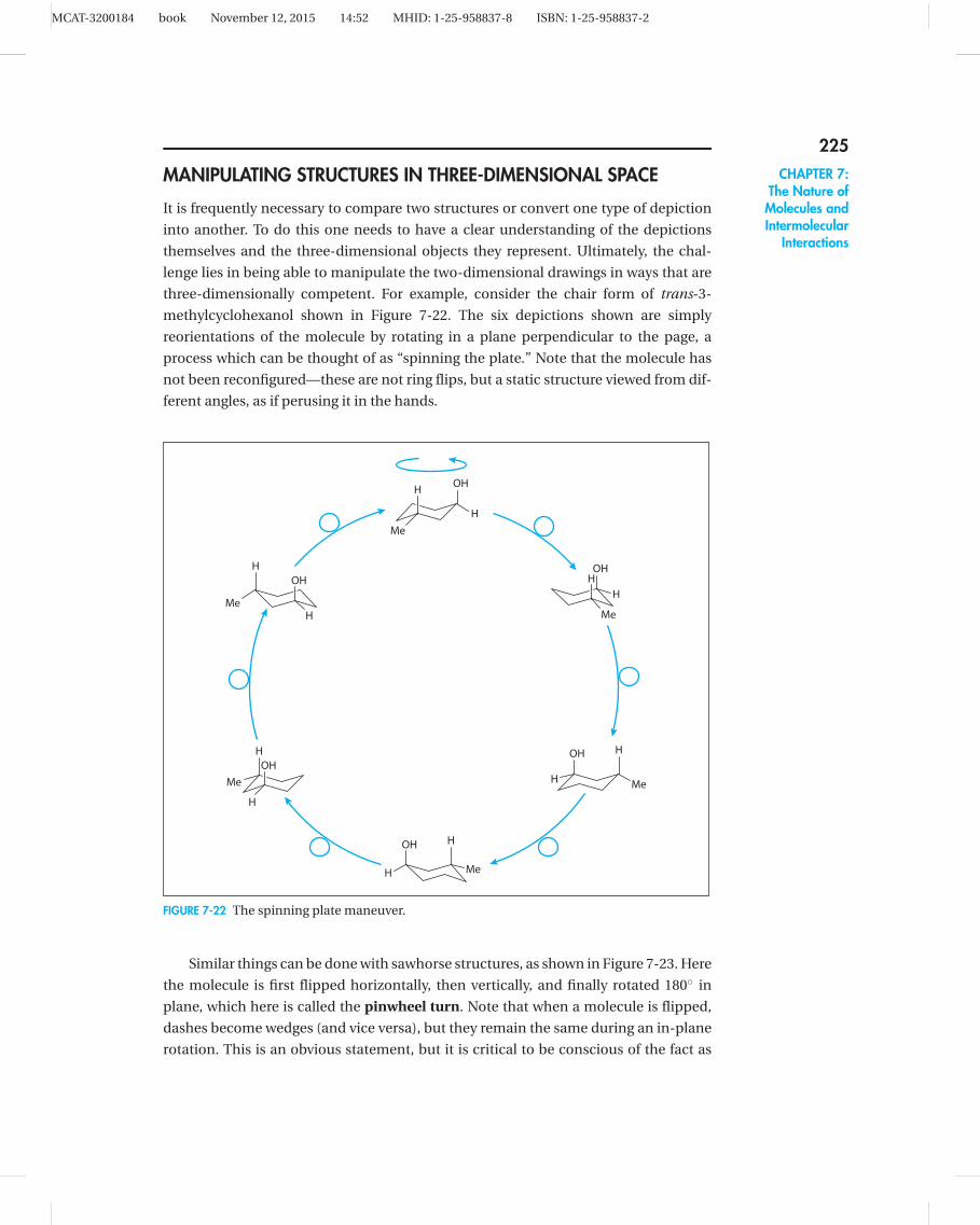
MANIPULATING STRUCTURES IN THREE-DIMENSIONAL SPACE
It is frequently necessary to compare two structures or convert one type of depiction
into another. To do this one needs to have a clear understanding of the depictions
themselves and the three-dimensional objects they represent. Ultimately, the chal-
lenge lies in being able to manipulate the two-dimensional drawings in ways that are
three-dimensionally competent. For example, consider the chair form of trans-3-
methylcyclohexanol shown in Figure 7-22. The six depictions shown are simply
reorientations of the molecule by rotating in a plane perpendicular to the page, a
process which can be thought of as “spinning the plate.” Note that the molecule has
not been reconfigured—these are not ring flips, but a static structure viewed from dif-
ferent angles, as if perusing it in the hands.
OH
H
OH
H
OH
H
OH
H
OH
H
OH
H
H
Me
H
Me
H
Me
H
Me
H
Me
H
Me
FIGURE 7-22 The spinning plate maneuver.
Similar things can be done with sawhorse structures, as shown in Figure 7-23. Here
the molecule is first flipped horizontally, then vertically, and finally rotated 180◦ in
plane, which here is called the pinwheel turn. Note that when a molecule is flipped,
dashes become wedges (and vice versa), but they remain the same during an in-plane
rotation. This is an obvious statement, but it is critical to be conscious of the fact as

MCAT-3200184 book November 12, 2015 14:52 MHID: 1-25-958837-8 ISBN: 1-25-958837-2
226UNIT II:ChemicalFoundations ofBiological Systems
Cl
OH
Cl
OH Cl
OH Cl
OH
horizontal
flip flip
vertical pinwheel
turn
FIGURE 7-23 Some manipulations of sawhorse structures.
you manipulate structures later on. You can also perform these maneuvers on chair
cyclohexanes as well.
When converting specialized depictions—like Newman or Fischer projections—to
more conventional drawings (e.g., sawhorse structures), bear in mind that this is ulti-
mately a change in point of view. So for a Newman projection, you sight the molecule
down a carbon-carbon bond, but in a sawhorse you view the same bond side-on. An
easy way to think about the conversion is to imagine that the Newman projection is
hinged at the back carbon and you push the structure into the page much like you
would shut an open door (see Figure 7-24); thus the bond that points directly toward
you in structure A (the C2 C3 bond) lies in the plane in structure B. You can use the
same technique with Fischer projections, as long as you remember that the stylized
representation (C) really implies the dashes and wedges shown in structure D. Note
that two-centered Fischer projections are always eclipsed.
Me
Cl H
Me
H OHMe
MeH
OH
Cl
H
Me
Me
OHH
ClH
Me
Me
OHH
ClHMe
Me OH
Cl
H
H
shut the
door
shut the
door
A B
C D E
FIGURE 7-24 Shutting the door.
RAPID COMPARISON OF DEPICTIONS
Now imagine that you were asked to evaluate the two representations A and C,
and decide whether they are enantiomers, diastereomers, or the same thing. First,
review the difference between enantiomers and diastereomers. Both fall under the
broader umbrella term of stereoisomers, which refers to different spatial orientation of

MCAT-3200184 book November 12, 2015 14:52 MHID: 1-25-958837-8 ISBN: 1-25-958837-2
227CHAPTER 7:
The Nature ofMolecules andIntermolecular
Interactions
D-ribose L-ribose
(An enantiomer of D-ribose)
D-xylose
(A diastereomer of D-ribose)
OH
OH OH
OHO
OH
OH OH
OHO
OH
OH OH
OHO
FIGURE 7-25 Enantiomers versus diastereomers.
substituents. As an illustration, consider the naturally occurring sugar D-ribose
(see Figure 7-25), which has three chiral centers bearing hydroxy groups. Each group
could be oriented in one of two ways—a dash or a wedge; so in essence it is a binary
outcome. Since there are three centers, then the total number of unique permutations
is 23 = 8. D-ribose represents one such permutation, which here is called the “down-
up-down” isomer (referring to the orientation of the hydroxy groups). The enantiomer
of D-ribose is L-ribose (the D and L designations are peculiar to carbohydrate nomen-
clature), and you will notice that each and every chiral center is inverted—this is true
for any set of enantiomers. So L-ribose accounts for one stereoisomer of D-ribose, but
there are six others—all of them diastereomers of D-ribose. Figure 7-25 shows only one
of the diastereomers, namely D-xylose, which has a down-down-down arrangement of
hydroxy substituents. Therefore, you can define a diastereomer as a stereoisomer in
which one or more, but not all, chiral centers have been inverted.
A common temptation for the novice is to assign R and S designations to each of
the chiral centers and make the comparison in an algorithmic way. Indeed, you would
find that the configuration for D-ribose is (2R, 3R, 4R) and for L-ribose is (2S, 3S, 4S),
thus confirming the designation of enantiomer. However, this method is time consum-
ing and error-prone. For each chiral center, you must assign the CIP priorities, orient
the center correctly, and decide whether a→b→c is clockwise or counterclockwise.
This means that to compare ribose with another structure, 36 discrete operations must
be made, and if an error occurs in any one, the whole comparison breaks down.
Instead, you should become skilled in analyzing these problems from a three-
dimensional standpoint. In other words, manipulate the structures in your mind’s eye
so you can compare them visually.
With this backdrop, return to the question of comparing Newman projection A to
Fischer projection C in Figure 7-24. The first order of business is to convert them to
sawhorse representations (B and E, respectively). Then the question is how to com-
pare the two very different sawhorses. To make a reliable comparison, you must first
reorient the structures so they share elements of commonality. It is generally easier to
simply take one as a reference and reorient the other—for example, to arbitrarily take
structure B as a reference and manipulate E to adopt the elements of commonality (see
Figure 7-26). So what are these elements? Examining structure B, you find that (1) it is

MCAT-3200184 book November 12, 2015 14:52 MHID: 1-25-958837-8 ISBN: 1-25-958837-2
228UNIT II:ChemicalFoundations ofBiological Systems
a staggered conformation, (2) C1, C2, C3, and C4 are all in the plane, (3) the hydroxy
group is on the right hand and pointed toward the top of the page, and (4) the chloro
substituent is on the left hand and pointed toward the bottom of the page (never mind
for the moment whether they are dashes or wedges).
Me
Cl H
Me
H OH
Me
Me
OHH
ClH
see
Fig. 7-24
A
C
Me Me Me
Me Me MeOH
OHCl Cl
Cl OHH
H
H
H H
H
E F G
see
Fig. 7-24
rotate 908
rotate C3
about C2-C3
in-plane
flip
vertically
Me
MeH
Me
MeH
OH
Cl Cl
HOH
H
B H
FIGURE 7-26 Comparing two representations.
Now set about the task of reorienting E to adopt these four elements, starting with
an in-plane rotation to get structure F, followed by a vertical flip to obtain structure G.
Notice that the dashes and wedges remain the same during the rotation, but change
during the vertical flip. The motivation for this maneuver was to place the hydroxy
group on the right side and pointing toward the top of the page. Notice, however, that
the conformation is eclipsed (as indeed all Fischer projections must be). To make a
comparison, you must then rotate about the C2 C3 bond 180◦ to arrive at structure
H. Unlike the previous operations, this is a reconformation of the molecule—but the
identity of the compound remains the same. Now you are in a position to evaluate the
two structures (B and H). Notice that every chiral center in H is the opposite of that in
B; therefore, the two must be enantiomers.
Similar operations can be carried out on cyclic molecules. Again, it is generally eas-
ier to convert all representations into dash-wedge drawings before making manipula-
tions and comparisons. Figure 7-27 demonstrates two mnemonics for re-envisioning
chair cyclohexane depictions. Imagine that the back carbon-carbon bond (highlighted
in blue) is a hinge and that the cyclohexane ring is a flat surface, like a table or iron-
ing board. You can push the molecule up on the hinge, much like a foldaway iron-
ing board, in which case the hydroxy group becomes a dash at the 3 o’clock position
and the methyl group is a wedge at the 10 o’clock position. Alternatively, you can let
the surface fall suspended by the hinge, as if letting down a drop-leaf table. Here the
hydroxy group would still be at 3 o’clock, but as a wedge, and the methyl group would
be a dash at 8 o’clock.
MCAT-3200184 book November 12, 2015 14:52 MHID: 1-25-958837-8 ISBN: 1-25-958837-2
229CHAPTER 7:
The Nature ofMolecules andIntermolecular
Interactions
OH
OH
OH
H
H
Me
OH
H
H
Me
Me
Me
drop-leaf
table
foldaway
ironing board
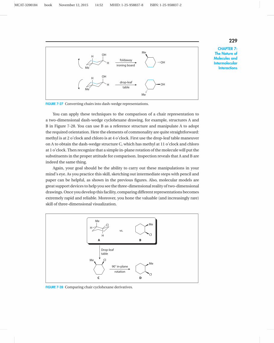
FIGURE 7-27 Converting chairs into dash-wedge representations.
You can apply these techniques to the comparison of a chair representation to
a two-dimensional dash-wedge cyclohexane drawing, for example, structures A and
B in Figure 7-28. You can use B as a reference structure and manipulate A to adopt
the required orientation. Here the elements of commonality are quite straightforward:
methyl is at 2 o’clock and chloro is at 4 o’clock. First use the drop-leaf table maneuver
on A to obtain the dash-wedge structure C, which has methyl at 11 o’clock and chloro
at 1 o’clock. Then recognize that a simple in-plane rotation of the molecule will put the
substituents in the proper attitude for comparison. Inspection reveals that A and B are
indeed the same thing.
Again, your goal should be the ability to carry out these manipulations in your
mind’s eye. As you practice this skill, sketching out intermediate steps with pencil and
paper can be helpful, as shown in the previous figures. Also, molecular models are
great support devices to help you see the three-dimensional reality of two-dimensional
drawings. Once you develop this facility, comparing different representations becomes
extremely rapid and reliable. Moreover, you hone the valuable (and increasingly rare)
skill of three-dimensional visualization.
H
Me
HCl Me
Cl
vs.
A B
C D
Me
Cl
Me Cl
Drop-leaf
table
908 in-plane
rotation
FIGURE 7-28 Comparing chair cyclohexane derivatives.

MCAT-3200184 book November 12, 2015 14:52 MHID: 1-25-958837-8 ISBN: 1-25-958837-2
230UNIT II:ChemicalFoundations ofBiological Systems
MOLECULAR ORBITALSThe concept of hybridization was discussed earlier in this chapter and is a useful way
to predict molecular shape. To understand the electronic behavior of molecules, how-
ever, it is necessary to introduce molecular orbital (MO) theory. While in fact there
are many manifestations of molecular orbital theory, the basic premise is that when
atoms are close enough to each other to form bonds, their orbitals combine in ways
that produce a new molecular orbital outcome.
AlkenesMO theory is very similar to the concept of hybridization, in which s and p orbitals
combine to form new hybrid orbitals. For example, the unhybridized carbon atom
has a 2s orbital and three identical 2 p orbitals in its valence shell. To accommodate
three things (atoms or lone pairs) around carbon, two of the 2 p orbitals and the 2s
orbital combine to form three identical sp2orbitals, leaving one 2 p orbital untouched
(see Figure 7-29).
sp2 hybridized
carbon atom
hybridization
2pz2px 2py
2s
4 orbitals total 4 orbitals total
2pz
3 3 sp2
Unhybridized
carbon atom
FIGURE 7-29 Atomic orbital hybridization.
While useful, the idea of hybridization is a simplification. It’s tempting to imagine
(and in fact some textbooks propose) that these hybrid orbitals interact in predictable
ways to make new molecules. In reality, molecular orbitals are governed by complex
quantum chemical considerations that can be predicted fully accurately only by
sophisticated computer calculations. However, that is not to say that molecular
orbitals are devoid of conceptual power. To explore some of these principles, exam-
ine the molecular orbital scenario for ethene (see Figure 7-30).
Ethene is constituted from two carbon atoms and four hydrogen atoms. Each car-
bon atom has four atomic orbitals (think of them as being sp2 hybridized), and each
of the four hydrogen atoms has a single 1s orbital, for a total of 12 atomic orbitals.
These atomic orbitals combine to form 12 new and unique molecular orbitals, all of
different energies. Hund’s rule and the Pauli exclusion principle apply to molecules
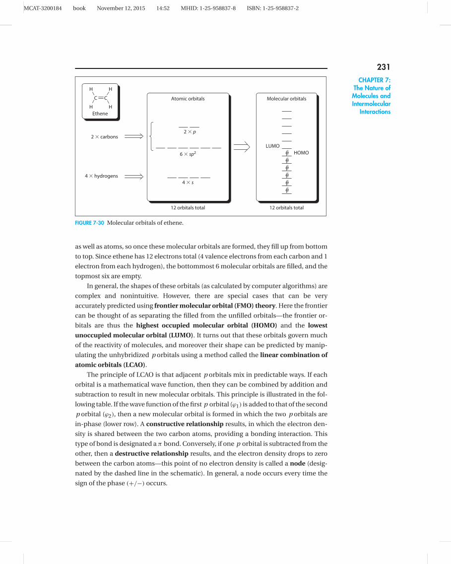
MCAT-3200184 book November 12, 2015 14:52 MHID: 1-25-958837-8 ISBN: 1-25-958837-2
231CHAPTER 7:
The Nature ofMolecules andIntermolecular
Interactions
2 3 carbons
6 3 sp2
4 3 hydrogens
4 3 s
2 3 p
LUMO
HOMO
Molecular orbitals
12 orbitals total 12 orbitals total
C C
H
H
H
H
Ethene
Atomic orbitals
FIGURE 7-30 Molecular orbitals of ethene.
as well as atoms, so once these molecular orbitals are formed, they fill up from bottom
to top. Since ethene has 12 electrons total (4 valence electrons from each carbon and 1
electron from each hydrogen), the bottommost 6 molecular orbitals are filled, and the
topmost six are empty.
In general, the shapes of these orbitals (as calculated by computer algorithms) are
complex and nonintuitive. However, there are special cases that can be very
accurately predicted using frontier molecular orbital (FMO) theory. Here the frontier
can be thought of as separating the filled from the unfilled orbitals—the frontier or-
bitals are thus the highest occupied molecular orbital (HOMO) and the lowest
unoccupied molecular orbital (LUMO). It turns out that these orbitals govern much
of the reactivity of molecules, and moreover their shape can be predicted by manip-
ulating the unhybridized p orbitals using a method called the linear combination of
atomic orbitals (LCAO).
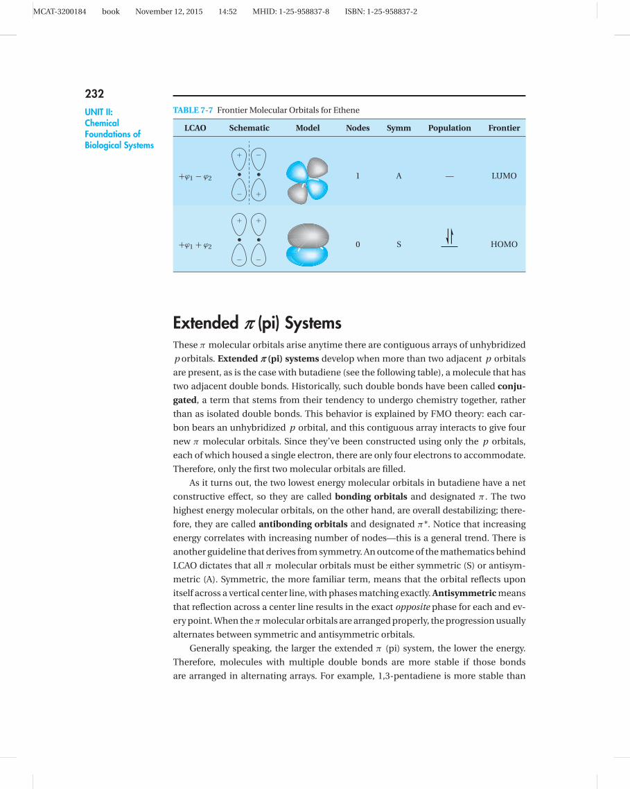
The principle of LCAO is that adjacent p orbitals mix in predictable ways. If each
orbital is a mathematical wave function, then they can be combined by addition and
subtraction to result in new molecular orbitals. This principle is illustrated in the fol-
lowing table. If the wave function of the first p orbital (ϕ1) is added to that of the second
p orbital (ϕ2), then a new molecular orbital is formed in which the two p orbitals are
in-phase (lower row). A constructive relationship results, in which the electron den-
sity is shared between the two carbon atoms, providing a bonding interaction. This
type of bond is designated a π bond. Conversely, if one p orbital is subtracted from the
other, then a destructive relationship results, and the electron density drops to zero
between the carbon atoms—this point of no electron density is called a node (desig-
nated by the dashed line in the schematic). In general, a node occurs every time the
sign of the phase (+/−) occurs.
MCAT-3200184 book November 12, 2015 14:52 MHID: 1-25-958837-8 ISBN: 1-25-958837-2
232UNIT II:ChemicalFoundations ofBiological Systems
TABLE 7-7 Frontier Molecular Orbitals for Ethene
LCAO Schematic Model Nodes Symm Population Frontier
+ϕ1 − ϕ2
1
2
2
1
1 A — LUMO
+ϕ1 + ϕ2
1
2
1
2
0 S HOMO
Extended o (pi) SystemsThese π molecular orbitals arise anytime there are contiguous arrays of unhybridized
p orbitals. Extended o (pi) systems develop when more than two adjacent p orbitals
are present, as is the case with butadiene (see the following table), a molecule that has
two adjacent double bonds. Historically, such double bonds have been called conju-
gated, a term that stems from their tendency to undergo chemistry together, rather
than as isolated double bonds. This behavior is explained by FMO theory: each car-
bon bears an unhybridized p orbital, and this contiguous array interacts to give four
new π molecular orbitals. Since they’ve been constructed using only the p orbitals,
each of which housed a single electron, there are only four electrons to accommodate.
Therefore, only the first two molecular orbitals are filled.
As it turns out, the two lowest energy molecular orbitals in butadiene have a net
constructive effect, so they are called bonding orbitals and designated π . The two
highest energy molecular orbitals, on the other hand, are overall destabilizing; there-
fore, they are called antibonding orbitals and designated π*. Notice that increasing
energy correlates with increasing number of nodes—this is a general trend. There is
another guideline that derives from symmetry. An outcome of the mathematics behind
LCAO dictates that all π molecular orbitals must be either symmetric (S) or antisym-
metric (A). Symmetric, the more familiar term, means that the orbital reflects upon
itself across a vertical center line, with phases matching exactly. Antisymmetric means
that reflection across a center line results in the exact opposite phase for each and ev-
ery point. When the π molecular orbitals are arranged properly, the progression usually
alternates between symmetric and antisymmetric orbitals.
Generally speaking, the larger the extended π (pi) system, the lower the energy.
Therefore, molecules with multiple double bonds are more stable if those bonds
are arranged in alternating arrays. For example, 1,3-pentadiene is more stable than

MCAT-3200184 book November 12, 2015 14:52 MHID: 1-25-958837-8 ISBN: 1-25-958837-2
233CHAPTER 7:
The Nature ofMolecules andIntermolecular
Interactions
TABLE 7-8 Π Molecular Orbitals for Butadiene
Butadiene
LCAO Schematic Model Nodes Symm Population Frontier
+ϕ1 − ϕ2 + ϕ3 − ϕ4
1
2
2
1
1
2
2
1
3 A —
+ϕ1 − ϕ2 − ϕ3 + ϕ4
1
2
2
1
2
1
1
2
2 S — LUMO
+ϕ1 + ϕ2 − ϕ3 − ϕ4
1
2
1
2
2
1
2
1
1 A HOMO
+ϕ1 + ϕ2 + ϕ3 + ϕ4
1
2
1
2
1
2
1
2
0 S
1,4-pentadiene, and 2-cyclohexanone is more stable than 3-cyclohexanone (see Fig-
ure 7-31). This is a phenomenon known as conjugation. Since you know that the
origin of the effect can be described by MO theory, it comes as no surprise that con-
jugated double bonds often behave as a collective, whereas nonconjugated double
bonds behave as two isolated species.
O O
1,3-pentadiene
(conjugated)
1,4-pentadiene
(nonconjugated)
2-cyclohexanone
(conjugated)
3-cyclohexanone
(nonconjugated)
MORE STABLEMORE STABLE
FIGURE 7-31 Conjugated vs. nonconjugated double bonds.
True conjugation arises from a mixing of adjacent p orbitals. A similar (but weaker)
effect can arise from the interaction of p orbitals with adjacent bonds. For example,
the methyl cation is not a very stable species, largely owing to the empty p orbital on

MCAT-3200184 book November 12, 2015 14:52 MHID: 1-25-958837-8 ISBN: 1-25-958837-2
234UNIT II:ChemicalFoundations ofBiological Systems
carbon and the resulting violation of the octet rule. However, the ethyl cation is a bit
more stable, because the C H sigma bond on the methyl group can spill a bit of elec-
tron density into the empty p orbital, thereby stabilizing the cationic center (see Fig-
ure 7-32). This is an effect known as hyperconjugation, and while the sharing of
electron density is not nearly as effective here as in true conjugation, it is still responsi-
ble for the following stability trend in carbocations and radicals: methyl < primary �secondary < tertiary.
HH
H
H
H
H
Empty p orbital Empty p orbital
C-H sigma bond(formed from C sp3
and H s orbitals)
Electron density spillover
Methyl cation Ethyl cation
HH
FIGURE 7-32 Hyperconjugation in the ethyl cation.
Aromatic SystemsIn comparison with linear arrays, when p orbitals are arranged in a circle without
interruption, as in benzene (see Figure 7-33), unexpected stabilization can emerge. As
an outcome of the mathematics of LCAO (which is not discussed here), there are usu-
ally multiple sets of degenerate orbitals (i.e., orbitals of the same energy level). While it
is not intuitively straightforward to construct molecular orbital diagrams themselves,
there is a handy mnemonic device known as Frost’s circle, which allows you to sketch
out the relative energy levels of such molecules.
FIGURE 7-33 Contiguous p orbitals in benzene.
Frost’s circle is really quite simple—it starts by drawing a regular polygon of the
appropriate size with one point down. A circle is then drawn around the polygon, and
each point of contact represents a molecular orbital energy level. For example, benzene
is a six-membered ring, so its array of molecular orbitals starts with a single lowest-
energy MO, followed by two sets of two MOs of the same energy, and then a single
highest-energy MO (see Figure 7-34). Since benzene has 6π electrons, only the three
lowest-energy orbitals are filled. This electronic arrangement accounts for the fact that
benzene is particularly stable, a phenomenon known as aromaticity.
Aromaticity arises only from molecules possessing a cyclic, contiguous, and
coplanar array of p orbitals. But this arrangement can also result in antiaromaticity,

MCAT-3200184 book November 12, 2015 14:52 MHID: 1-25-958837-8 ISBN: 1-25-958837-2
235CHAPTER 7:
The Nature ofMolecules andIntermolecular
Interactions
whereby molecules are less stable than expected. The difference between aromaticity
and antiaromaticity lies in the number of π-electrons that must be accommodated.
This can be predicted using Hückel’s rule, which states that systems having 4nπ elec-
trons, (i.e., 4, 8, 12 electrons) tend to be antiaromatic, and those having (4n+ 2)π elec-
trons (i.e., 2, 6, 10 electrons) tend to be aromatic.
Antibonding
Bonding
Nonbonding0
FIGURE 7-34 Frost’s circle for determining aromatic MO energy levels.
Figure 7-35 demonstrates the molecular orbital basis of Hückel’s rule using
cyclopentadienyl ions. The molecular orbital energy levels are drawn using Frost’s cir-
cle, and then populated with the appropriate number of p electrons. The cyclopen-
tadienyl cation has only 4 electrons in the π (pi) system, whereas the corresponding
anion has 6 (the negative charge represents a lone pair of electrons on carbon). Using
Hückel’s rule, you would predict the anion to be stable (aromatic) and the cation to be
unstable (antiaromatic), which is indeed the experimentally observed result. However,
the MO picture provides more insight into why the cation is so unstable—the molecule
has 2 unpaired electrons. Finally, cyclopentadiene itself (far right), is considered
nonaromatic. Since to be aromatic or antiaromatic, there must be a cyclic, contiguous,
coplanar array of p orbitals, cyclopentadiene is out of the running because there is no
p orbital at the methylene (CH2) center, which interrupts the π (pi) system. Therefore,
Cyclopentadienyl
cation
Cyclopentadienyl
anion
Cyclopentadiene
4e 6e 4e
Antiaromatic Aromatic Nonaromatic
� MOs
� electrons
Aromaticity
FIGURE 7-35 The molecular orbital origin of aromaticity.

MCAT-3200184 book November 12, 2015 14:52 MHID: 1-25-958837-8 ISBN: 1-25-958837-2
236UNIT II:ChemicalFoundations ofBiological Systems
its MO diagram is analogous to that of butadiene (see the earlier table “� Molecular
Orbitals for Butadiene” on page 233).
MO Description of ResonanceMO theory also provides a more thorough understanding of resonance. For example,
the acetaldehyde anion (see Figure 7-36) is stabilized by resonance delocalization,
whereby the negative charge is distributed between the carbon and the oxygen. Res-
onance forms do not “equilibrate”—that is, the molecule does not oscillate between
forms A and B—rather, the two resonance structures are an attempt to represent a
more complete truth than any single Lewis structure can. However, if you apply simple
geometrical rules to each form, you would predict an sp3 hybridization for the anionic
carbon in form A, but form B clearly indicates an sp2 geometry. How do you accurately
predict the geometry of the molecule?
O O ;
BA
O O
FIGURE 7-36 Resonance stabilization of the acetaldehyde anion.
Molecular orbital theory shows that an extended π system can be established by
housing the negative charge in a p orbital. Thus the three-orbital array gives rise to a
set of molecular orbitals in which the electron density is distributed throughout the
molecule and the overall energy is minimized. For this reason, you expect both the
anionic carbon and the oxygen to exhibit sp2 hybridization, since this is the only way
that all three atoms have unhybridized p orbitals. This is at variance with the pre-
dictions for the methyl anion (see earlier table “Geometries of Some Special Carbon
Centers” on page 211), which was based on minimizing steric interactions; here the
energy benefit of an extended π-system compensates for the steric cost. Therefore, the
acetaldehyde anion is a completely planar molecule.
A similar anomaly can be seen with cations, as well. Thus the dimethylamino-
methyl cation (see Figure 7-37) can be shown as two resonance forms (A and B), in
which the nitrogen lone pair participates in stabilizing the adjacent positive charge
on carbon. However, you are faced with a dilemma similar to the acetaldehyde anion:
form A would predict an sp3 hybridization for nitrogen, whereas form B appears to
have sp2 geometry. The only way for form B to have meaning is if a π bond is estab-
lished between the 2 p orbitals on carbon and nitrogen (inset). Therefore, the species
is planar.
Aside from the geometrical considerations, these two examples show the
action of two specific types of functional groups. The carbonyl group (C O) is an

MCAT-3200184 book November 12, 2015 14:52 MHID: 1-25-958837-8 ISBN: 1-25-958837-2
237CHAPTER 7:
The Nature ofMolecules andIntermolecular
Interactions
NCH2
N
CH2
A B
. .N CH2
FIGURE 7-37 Resonance stabilization of the dimethylaminomethyl cation.
electron-withdrawing group (designated EW), while the amino group (NR2) is an
electron-donating group (ED). This idea is important for a variety of chemical reac-
tivities, but the easiest way to keep the categories straight is to examine whether the
group would best stabilize a cation or an anion. Generally speaking, electron density
can be pushed into EW groups, thus stabilizing a negative charge, whereas electrons
can be pushed from ED groups, which stabilizes adjacent positive charges. For this rea-
son, ED groups generally have lone pairs.
There are two components of electron-withdrawing and electron-donating behav-
ior: resonance and induction. As the term suggests, the resonance effect can be repre-
sented by electron-pushing arrows to give different resonance forms. This effect may
be weak or strong, but it can exert influence over very large distances through extended
π-systems. The inductive effect stems primarily from electronegativity; it can be quite
strong, but its influence is local—the magnitude drops off sharply as distance from
the functional group increases. The following table summarizes these effects for some
common functional groups. In most cases the inductive and resonance effects work
in concert, while in some instances they are at odds. The amino and methoxy sub-
stituents are almost always classified as ED groups, even though the central atoms
are electronegative—the activity of the lone pairs dominates their behavior. The halo-
gens are more subtle (e.g., chlorine). Generally speaking, they are grouped with the EW
substituents.
TABLE 7-9 Some Common ED and EW Groups
Functional Group Formula Resonance Effect Inductive Effect
Electron-withdrawing (EW) groupsacetyl COCH3
carbomethoxy CO2CH3
chloro Clcyano CNnitro NO2
phenylsulfonyl SO2Ph
Electron-donating (ED) groupsamino NH2
methoxy OCH3
methyl CH3← more EW | more ED → ← more EW | more ED →

MCAT-3200184 book November 12, 2015 14:52 MHID: 1-25-958837-8 ISBN: 1-25-958837-2
238UNIT II:ChemicalFoundations ofBiological Systems
NONCOVALENT BONDSIn addition to the fixed covalent bonds that hold molecules together, there are a host
of so-called intermolecular forces that play a significant role in the properties of
molecules.
Types of Intermolecular ForcesThe magnitude of intermolecular forces spans a wide range, as depicted in Figure 7-38.
Unlike covalent bonds, which are permanent and relatively easy to study thermody-
namically, these noncovalent interactions are often fugitive, making unequivocal
determination of their energies very difficult. Consequently, published values vary
widely and are dependent upon the method of measurement (or calculation) and the
types of systems studied. For the purposes of this overview, examples will be limited to
organic molecules in solution.
0.01 0.1 1
kcal/mol
Covalent
Ion-dipole
H-bond
Dipole-dipole
pi-pi stacking
Dispersion
10 100 1000
FIGURE 7-38 Comparison of intermolecular interaction energies.
ION-DIPOLE INTERACTIONS
Ion-dipole interactions are largely responsible for the dissolution of ionic compounds
in water. For example, lithium chloride has a lattice enthalpy of 206 kcal/mol and a
melting point of 605 ◦C, yet it dissolves rather easily in water. Remarkably, relatively
weak ion-dipole forces (see Figure 7-39) overcome the strong ionic bonds holding the
salt together. Obviously, this is an outcome of the sheer numbers of interactions in
play: although small individually, these forces are quite significant in sum. Two effects
are noteworthy in this interaction: the ions themselves are stabilized by the multiple
interactions, and the water (or other solvent) is organized around the ions. This well-
defined organization of solvent around a solute is often called the solvent shell.
Not surprisingly, the degree of organization around the ions is dependent upon the
amount of charge. For example, the calcium ion (Ca2+) exhibits a solvation enthalpy

MCAT-3200184 book November 12, 2015 14:52 MHID: 1-25-958837-8 ISBN: 1-25-958837-2
239CHAPTER 7:
The Nature ofMolecules andIntermolecular
Interactions
H
H
H
H
H
HH
H
H
H
H
H
Li1 Cl2
H
H
HH
H
H
H
H
H H
H
O
O
OO
O
O
OO
OO
O O
H
FIGURE 7-39 Ion-dipole forces around aqueous lithium chloride.
(�Hsolv) in water of 416 kcal/mol, twice that of the similarly-sized sodium cation (Na+).
Conversely, with constant charge, the strength of solvation increases as ionic radius
decreases. In other words, as the electrostatic charge is confined to a smaller space,
its effect on the surrounding solvent is more pronounced. As one illustration of this
effect, Figure 7-40 charts the dependence of solvation enthalpy on ionic radius among
the alkali metal cations.
70 90150
200
250
110
Li+
Na+
K+
Rb+
Ionic radius (pm)
DH
solv
(kca
l/m
ol)
130 150 170
FIGURE 7-40 Solvation enthalpy as a function of ionic size.
HYDROGEN BONDING
The hydrogen bond is perhaps the most wide-ranging intermolecular force among
biological molecules, and it remains a bit of an enigma. The classical view of the
hydrogen bond (see Figure 7-41) is that of an interaction between an electron-rich lone
pair (i.e., the hydrogen bond acceptor) and a highly polarized covalent bond involving
hydrogen (i.e., the hydrogen bond donor).
Most textbooks relegate hydrogen bonds to compounds containing nitrogen, oxy-
gen, and fluorine. However, there is considerable evidence that thiols (RSH) can serve
as hydrogen bond donors, and one study suggests that more than 70 percent of

MCAT-3200184 book November 12, 2015 14:52 MHID: 1-25-958837-8 ISBN: 1-25-958837-2
240UNIT II:ChemicalFoundations ofBiological Systems
HX Y
Hydrogen bond
acceptor
Hydrogen bond
donor
1
FIGURE 7-41 Model of hydrogen bonding.
cysteines in proteins have the thiol proton within hydrogen-bonding distance of a
nitrogen or oxygen. The role of sulfur as a hydrogen bond acceptor is more controver-
sial, however. In a very recent investigation using atomic force microscopy, 8-
hydroxyquinoline was found to exhibit hydrogen bonds involving OH, NH, and CH
bonds (see Figure 7-42).
O
O
H
H
H
N
N
FIGURE 7-42 Hydrogen bonds observed in 8-hydroxyquinoline.
A particularly strong type of hydrogen bond occurs in proteins when two
oppositely-charged amino acid residues are involved, a phenomenon known as a
salt bridge. These interactions can be crucial in establishing the structure and function
of proteins. For example, salt bridges in the active site of Escherichia coli aminopepti-
dase N (see Figure 7-43) have recently been shown to play a critical role in that enzyme’s
stability and activity.
Glu 121
Lys 319
Lys 264
Glu 320
OOO
O
O OH
HH
N
FIGURE 7-43 Salt bridges in the active site of E. coli aminopeptidase N.
DIPOLE-DIPOLE INTERACTIONS
Dipole-dipole forces arise when polar molecules are in proximity to each other. The
force can be either attractive or repulsive, depending upon the relative directions of the

MCAT-3200184 book November 12, 2015 14:52 MHID: 1-25-958837-8 ISBN: 1-25-958837-2
241CHAPTER 7:
The Nature ofMolecules andIntermolecular
Interactions
two dipoles, with an antiparallel arrangement providing the greatest stabilizing effect.
Thus, for example, when p-dichlorobenzene (a common ingredient in mothballs) is
dissolved in acetone, much of the solvent power is provided by dipole-dipole forces
(see Figure 7-44). One characteristic of the dipole-dipole interaction is its sensitivity to
distance—the force drops off as a function of the distance cubed.
Cl
+
Cl
O
O
O
O
OO
++
+
+
++
+
FIGURE 7-44 Dipole-dipole forces in acetone solution.
PI-PI STACKING
An often neglected contributor to intramolecular forces is the pi-pi stacking inter-
action. Aromatic compounds such as benzene have molecular orbitals that are in a
circular array, and the particular arrangement leads to anisotropic electron densities.
The areas above and below the ring (i.e., the faces) are slightly electron rich, while
the perimeter (or edge) is slightly electron poor. This sets the stage for favorable
electrostatic interactions between aromatic rings. Figure 7-45 shows two common
motifs found in pi-pi interactions. One is an off-center parallel arrangement, in which
electron-rich and electron-poor layers align so that there is a small amount of coulom-
bic attraction at the edges. Another is an edge-to-face alignment, whereby the electron-
poor edge of one aromatic ring aligns perpendicular to the electron-rich face of
another. The strength of these interactions is on the order of weak dipole-dipole forces,
but in large biopolymers such as DNA, the sum of these interactions can be substantial.
Off-center parallel
Edge-to-face
�2�2�2�2�2
�2�2�2�2�2�1 �1 �1 �1 �1 �1
�2�2�2�2�2
�2�2�2�2�2�1 �1 �1 �1 �1 �1
�2
d2
�2
�2
�2
�2
�2
�2
�2
�2
�1
�1
�1
�1
�1
�1�2�2�2�2�2
�2�2�2�2�2�1 �1 �1 �1 �1 �1
FIGURE 7-45 Two common pi-stacking motifs.

MCAT-3200184 book November 12, 2015 14:52 MHID: 1-25-958837-8 ISBN: 1-25-958837-2
242UNIT II:ChemicalFoundations ofBiological Systems
DISPERSION FORCES
The weakest of the intermolecular forces, London dispersion forces (LDF), are found
in all molecules. In the case of nonpolar species, where stronger interactions are
absent, LDFs are usually the sole cohesive principle between molecules. Were it not
for the dispersion effect, noble gases would not exist in liquid form. Dispersion forces
even account for about one-quarter of the attractive force between water molecules.
The basis of LDF lies in fairly complex quantum mechanical outcomes, but it is
often described as the favorable coalignment of instantaneous dipoles that are ran-
domly generated by momentary unequal distributions of electron density in a mole-
cule. While these instantaneous dipoles average out to zero over time for a given
molecule, the continual coordination of these microdipoles between molecules pro-
vides a net attractive force.
Effects of Intermolecular ForcesPHYSICAL PROPERTIES
One of the most obvious and pervasive impacts of intermolecular forces on molecules
is seen in their boiling points. This is illustrated in the three molecules 2-methyl-1-
butene, ethyl acetate, and propionic acid (see Figure 7-46), which have almost
identical molecular weights. For the 2-methyl-1-butene, LDFs are the predominant
intermolecular force. However, in the case of ethyl acetate, the introduction of the oxy-
gen atoms results in polar bonds (between carbon and oxygen), leading to a net dipole.
In the condensed phase, these dipoles align between molecules (left inset), providing
a greater cohesive force, which is revealed in a higher boiling point (77 ◦C vs. 31 ◦C).
2-methyl-1-butene
MW 70
bp 318C
Ethyl acetate
MW 74
bp 778C
Propionic acid
MW 74
bp 1418C
O
O
O
OMe
O
OH
O
OMe
O
O
O
Et
Et
Et
Et
OMe
O
OMeO
OMe
Dipole-dipole interaction Hydrogen bonding
O
O
H
H H
H
O
O
O
O
FIGURE 7-46 Impact of intermolecular forces on boiling points.

MCAT-3200184 book November 12, 2015 14:52 MHID: 1-25-958837-8 ISBN: 1-25-958837-2
243CHAPTER 7:
The Nature ofMolecules andIntermolecular
Interactions
With propionic acid, there are not only dipole-dipole interactions, but also opportu-
nities for hydrogen bonding. In fact, most carboxylic acids in the condensed phase
form strong dimeric structures with complementary hydrogen bonds (see Figure 7-46,
right). Because of these additional intermolecular forces, propionic acid has a boiling
point of 141 ◦C vs. 77 ◦C for ethyl acetate.
Molecular geometry can play an important role, as exemplified by the two geomet-
ric isomers of 1,2-dichloroethene (see Figure 7-47). In the cis-isomer, both of the polar
C Cl bonds are pointed in the same direction, so there is a net dipole for the whole
molecule. In the trans-isomer, these bonds are antiparallel and the bond dipoles tend
to cancel each other out, leading to a molecule with a very small net dipole. This is
consequently reflected in the lower boiling point of the trans-isomer.
Cl Cl
cis-1,2-dichloroethene
bp 60
trans-1,2-dichloroethene
bp 48
ClCl
FIGURE 7-47 Impact of intermolecular forces on boiling points.
Intermolecular forces are additive, and since hydrogen bonds are relatively strong,
polyols tend to have exceedingly high boiling points. Figure 7-48 provides a useful set
of examples. In a series of nonpolar molecules, progressing from ethane to butane to
3-methylpentane results in a steady increase in boiling points, due to increased mass
and a larger electronic cloud (i.e., greater LDFs). When one CH2 group is replaced
with an OH group (ethane to ethanol), the boiling point jumps from −88◦C to 65◦C, a
153◦C difference. Additional OH groups magnify this effect, so the difference in boiling
points between 3-methylpentane and glycerol is 226◦C. Taking this to an extreme, the
straight-chain hydrocarbon eicosane (C20H42, MW 283) has a boiling point of 343◦C,
as opposed to the much smaller, but much more polar, glucose molecule (C6H12O6,
MW 180), which has a calculated boiling point of 527◦ C (in practice, glucose burns
before it boils).
CONFORMATIONAL IMPLICATIONS
Noncovalent forces can play a major role in many aspects of molecular shape and
behavior. For example, in trans-1,2-dichlorohexane (see Figure 7-49, left), the chair
conformer in which the two chloro substituents are diequatorial is preferred on steric
grounds. However, the equilibrium is not as right-handed as you might expect, since
the diaxial conformer allows the two polar bonds to be antiparallel, which avoids the
unfavorable dipole-dipole interactions in the diequatorial conformer. Similarly, in cis-
cyclohexanediol (see Figure 7-49, right), the equilibrium is less right-handed than steric
considerations would predict, since the diaxial conformer can take advantage of an
intramolecular hydrogen bond.

MCAT-3200184 book November 12, 2015 14:52 MHID: 1-25-958837-8 ISBN: 1-25-958837-2
244UNIT II:ChemicalFoundations ofBiological Systems
Me Me
Ethane
MW 30
bp–888C
Butane
MW 58
bp–18C
3-methylpentane
MW 86
bp 648C
Glycerol
MW 92
bp 2908C
Ethylene glycol
MW 62
bp 1968C
OHHO
OH
OHHO
OHMe
Methanol
MW 32
bp 658C
bp
8C
300
200
100
0
20 40 60
Molecular weight
80 1002100
1 3 OH
2 3 OH
3 3 OHAlkanes
Alcohols
FIGURE 7-48 Impact of multiple functional groups on boiling points.
Cl
ClCl
Stabilized by
antiparallel
arrangement
of dipoles
Stabilized by
intramolecular
hydrogen bond
Cl
HH
O
HO OH
O+
+
FIGURE 7-49 Conformational stabilization by noncovalent forces.
These factors even play out in chemical equilibria. For example, ketones are known
to be in tautomeric equilibrium with their enol counterparts, but the keto form is gen-
erally the far more thermodynamically stable option. Thus 2-butanone exists almost
exclusively in the keto form. However, pentane-2,4-dione exists as a mixture of keto
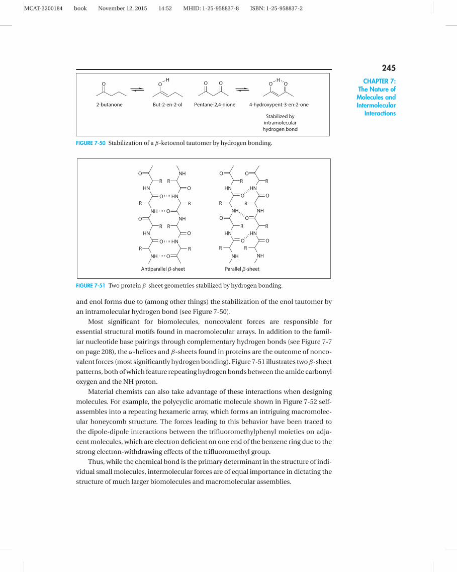
MCAT-3200184 book November 12, 2015 14:52 MHID: 1-25-958837-8 ISBN: 1-25-958837-2
245CHAPTER 7:
The Nature ofMolecules andIntermolecular
InteractionsStabilized by
intramolecular
hydrogen bond
O
2-butanone But-2-en-2-ol Pentane-2,4-dione 4-hydroxypent-3-en-2-one
O O O O OH H
FIGURE 7-50 Stabilization of a β-ketoenol tautomer by hydrogen bonding.
NH
NH
NH
NH
NH
NH
NH
NH
HN HN
HNHN
HN
HN
HN
HN
O O
O
O
O
O
O
O
OO
O
O
R
R
O
O
O
O
R
R
R
R R
R
R
R
R
R
RR
R R
Antiparallel �-sheet Parallel �-sheet
FIGURE 7-51 Two protein β-sheet geometries stabilized by hydrogen bonding.
and enol forms due to (among other things) the stabilization of the enol tautomer by
an intramolecular hydrogen bond (see Figure 7-50).
Most significant for biomolecules, noncovalent forces are responsible for
essential structural motifs found in macromolecular arrays. In addition to the famil-
iar nucleotide base pairings through complementary hydrogen bonds (see Figure 7-7
on page 208), the α-helices and β-sheets found in proteins are the outcome of nonco-
valent forces (most significantly hydrogen bonding). Figure 7-51 illustrates two β-sheet
patterns, both of which feature repeating hydrogen bonds between the amide carbonyl
oxygen and the NH proton.
Material chemists can also take advantage of these interactions when designing
molecules. For example, the polycyclic aromatic molecule shown in Figure 7-52 self-
assembles into a repeating hexameric array, which forms an intriguing macromolec-
ular honeycomb structure. The forces leading to this behavior have been traced to
the dipole-dipole interactions between the trifluoromethylphenyl moieties on adja-
cent molecules, which are electron deficient on one end of the benzene ring due to the
strong electron-withdrawing effects of the trifluoromethyl group.
Thus, while the chemical bond is the primary determinant in the structure of indi-
vidual small molecules, intermolecular forces are of equal importance in dictating the
structure of much larger biomolecules and macromolecular assemblies.

MCAT-3200184 book November 12, 2015 14:52 MHID: 1-25-958837-8 ISBN: 1-25-958837-2
OBu
BuO OBu
OBu
OBu
OBu
BuO
BuO
OBu
F3C
CF3
CF3
OBu
BuO OBu
OBu
OBu
OBu
BuO
BuO
OBu
CF3
CF3
OBu
BuO OBu
OBu
OBu
OBu
BuO
BuO
OBu
F3C
F3C
F3C
CF3
CF3
OBu
BuO OBu
OBu
OBu
OBu
BuO
BuO
OBu
F3C
CF3
CF3
OBu
BuO OBu
OBu
OBu
OBu
BuO
BuO
OBu
F3C
CF3
CF3
OBu
BuO OBu
OBu
OBu
OBu
BuO
BuO
OBu
CF3
CF3
FIGURE 7-52 Molecular self-assembly driven by dipole-dipole interactions.
246