Embed Size (px)
Citation preview
5/13/2013
1
Membrane Structure & Function Chapter 7
You should be able to:
1. Define the following terms: amphipathic
molecules, aquaporins, diffusion
2. Explain how membrane fluidity is influenced by
temperature and membrane composition
3. Distinguish between the following pairs or sets of
terms: peripheral and integral membrane
proteins; channel and carrier proteins; osmosis,
facilitated diffusion, and active transport;
hypertonic, hypotonic, and isotonic solutions
4. Explain how transport proteins facilitate diffusion
5. Explain how an electrogenic pump creates
voltage across a membrane, and name two
electrogenic pumps
6. Explain how large molecules are transported
across a cell membrane
5/13/2013
2
Overview: Life at the Edge
o The plasma membrane is the boundary that separates the living cell from its surroundings
o Membranes are made of Proteins and Lipids
o The plasma membrane exhibits selective permeability, allowing some substances to cross it more easily than others
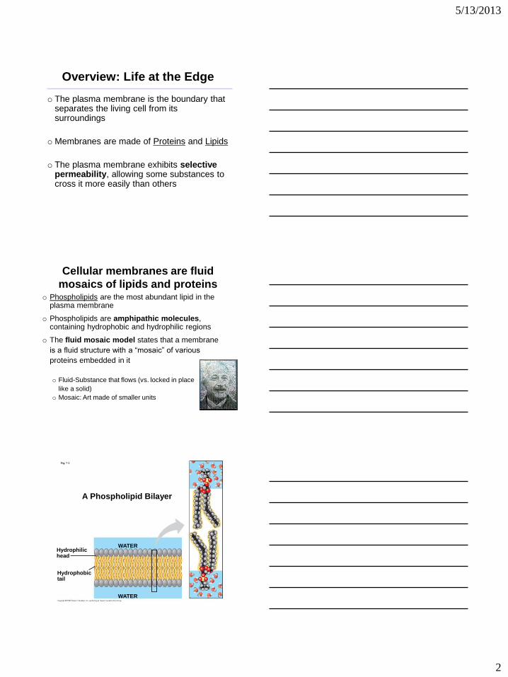
Cellular membranes are fluid
mosaics of lipids and proteins o Phospholipids are the most abundant lipid in the
plasma membrane
o Phospholipids are amphipathic molecules, containing hydrophobic and hydrophilic regions
o The fluid mosaic model states that a membrane
is a fluid structure with a “mosaic” of various
proteins embedded in it
o Fluid-Substance that flows (vs. locked in place
like a solid)
o Mosaic: Art made of smaller units
Fig. 7-2
Hydrophilic head
WATER
Hydrophobic tail
WATER
A Phospholipid Bilayer
5/13/2013
3
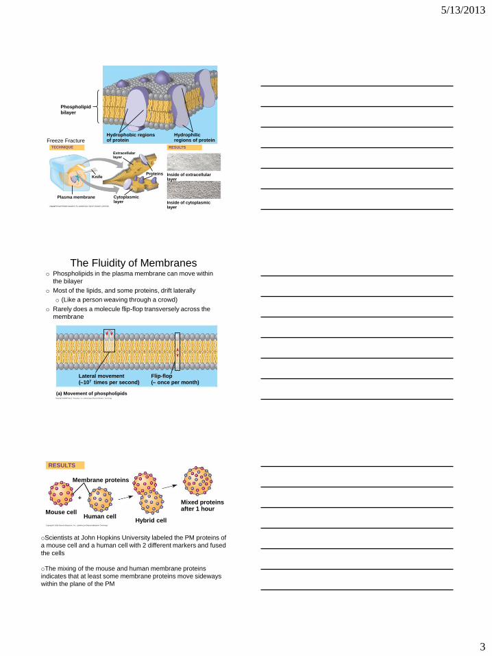
Phospholipid
bilayer
Hydrophobic regions of protein
Hydrophilic regions of protein
TECHNIQUE
Extracellular layer
Knife Proteins Inside of extracellular
layer
RESULTS
Inside of cytoplasmic layer
Cytoplasmic layer
Plasma membrane
Freeze Fracture
The Fluidity of Membranes
(a) Movement of phospholipids
Lateral movement
(107 times per second)
Flip-flop
( once per month)
o Phospholipids in the plasma membrane can move within
the bilayer
o Most of the lipids, and some proteins, drift laterally
o (Like a person weaving through a crowd)
o Rarely does a molecule flip-flop transversely across the
membrane
Fig. 7-6
RESULTS
Membrane proteins
Mouse cell Human cell
Hybrid cell
Mixed proteins after 1 hour
oScientists at John Hopkins University labeled the PM proteins of
a mouse cell and a human cell with 2 different markers and fused
the cells
oThe mixing of the mouse and human membrane proteins
indicates that at least some membrane proteins move sideways
within the plane of the PM
5/13/2013
4
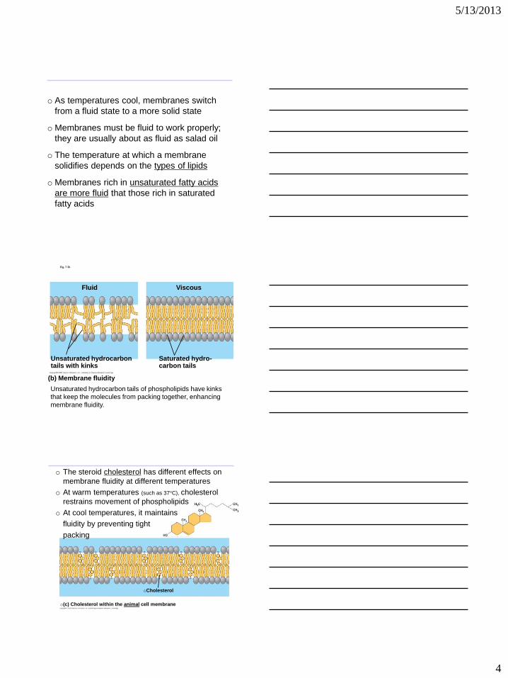
o As temperatures cool, membranes switch
from a fluid state to a more solid state
o Membranes must be fluid to work properly;
they are usually about as fluid as salad oil
o The temperature at which a membrane
solidifies depends on the types of lipids
o Membranes rich in unsaturated fatty acids
are more fluid that those rich in saturated
fatty acids
Fig. 7-5b
(b) Membrane fluidity
Fluid
Unsaturated hydrocarbon tails with kinks
Viscous
Saturated hydro- carbon tails
Unsaturated hydrocarbon tails of phospholipids have kinks
that keep the molecules from packing together, enhancing
membrane fluidity.
oCholesterol
o(c) Cholesterol within the animal cell membrane
o The steroid cholesterol has different effects on
membrane fluidity at different temperatures
o At warm temperatures (such as 37°C), cholesterol
restrains movement of phospholipids
o At cool temperatures, it maintains
fluidity by preventing tight
packing
5/13/2013
5
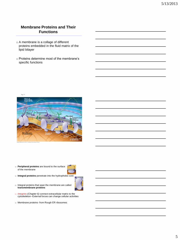
Membrane Proteins and Their
Functions
o A membrane is a collage of different
proteins embedded in the fluid matrix of the
lipid bilayer
o Proteins determine most of the membrane’s
specific functions
Fig. 7-7
Fibers of extracellular matrix (ECM)
Glyco- protein
Microfilaments of cytoskeleton
Cholesterol
Peripheral proteins
Integral protein
CYTOPLASMIC SIDE OF MEMBRANE
Glycolipid
EXTRACELLULAR SIDE OF MEMBRANE
Carbohydrate
o Peripheral proteins are bound to the surface
of the membrane
o Integral proteins penetrate into the hydrophobic core
o Integral proteins that span the membrane are called transmembrane proteins
o Integrins (Chapter 6) connect extracellular matrix to the cytoskeleton--External forces can change cellular activities
o Membrane proteins- from Rough ER ribosomes
5/13/2013
6
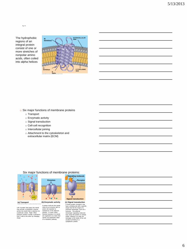
Fig. 7-8
N-terminus
C-terminus
Helix
CYTOPLASMIC SIDE
EXTRACELLULAR SIDE
The hydrophobic
regions of an
integral protein
consist of one or
more stretches of
nonpolar amino
acids, often coiled
into alpha helices
o Six major functions of membrane proteins
o Transport
o Enzymatic activity
o Signal transduction
o Cell-cell recognition
o Intercellular joining
o Attachment to the cytoskeleton and
extracellular matrix (ECM)
(a) Transport (b) Enzymatic activity (c) Signal transduction
ATP
Enzymes
Signal transduction
Signaling molecule
Receptor
Six major functions of membrane proteins:
Left: A protein that spans the memb
may provide a hydrophilic channel
across the memb that is selective for
a particular solute. Right: Other
transport proteins shuttle a substance
from 1 side to the other by changing
shape
A protein built into the memb
many be an enzyme with its
active site exposed to
substances in the adjacent
solution. In some cases.
Several enzymes in a memb
are organized as a team that
carries out sequential steps
of a metabolic pathway
A memb protein (receptor) may
have a binding site with a specific
shape that fits the shape of a
chemical messenger, such as a
hormone. The external
messenger (signaling molecule)
may cause the protein to change
shape, allowing it to relay the
message to the inside of the cell,
usually by binding to a
cytoplasmic protein
5/13/2013
7
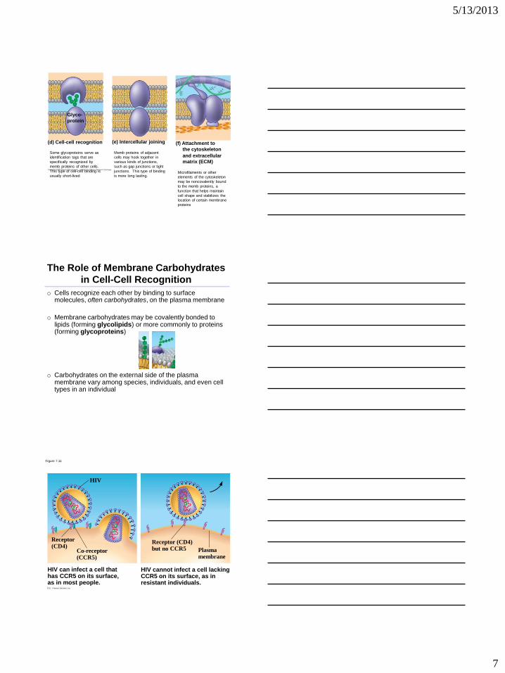
(d) Cell-cell recognition
Glyco-
protein
(e) Intercellular joining (f) Attachment to
the cytoskeleton
and extracellular
matrix (ECM)
Some glycoproteins serve as
identification tags that are
specifically recognized by
memb proteins of other cells.
This type of cell-cell binding is
usually short-lived
Memb proteins of adjacent
cells may hook together in
various kinds of junctions,
such as gap junctions or tight
junctions. This type of binding
is more long lasting. Microfilaments or other
elements of the cytoskeleton
may be noncovalently bound
to the memb proteins, a
function that helps maintain
cell shape and stabilizes the
location of certain membrane
proteins
The Role of Membrane Carbohydrates
in Cell-Cell Recognition
o Cells recognize each other by binding to surface molecules, often carbohydrates, on the plasma membrane
o Membrane carbohydrates may be covalently bonded to lipids (forming glycolipids) or more commonly to proteins (forming glycoproteins)
o Carbohydrates on the external side of the plasma membrane vary among species, individuals, and even cell types in an individual
Figure 7.11
Receptor (CD4)
Co-receptor (CCR5)
HIV
Receptor (CD4) but no CCR5 Plasma
membrane
HIV can infect a cell that has CCR5 on its surface, as in most people.
HIV cannot infect a cell lacking CCR5 on its surface, as in resistant individuals.
5/13/2013
8
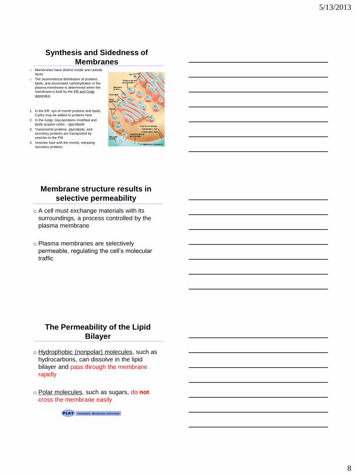
Synthesis and Sidedness of
Membranes o Membranes have distinct inside and outside
faces
o The asymmetrical distribution of proteins,
lipids, and associated carbohydrates in the
plasma membrane is determined when the
membrane is built by the ER and Golgi
apparatus
1. In the ER: syn of memb proteins and lipids.
Carbs may be added to proteins here
2. In the Golgi: Glycoproteins modified and
lipids acquire carbs – glycolipids
3. Transmemb proteins, glycolipids, and
secretory proteins are transported by
vesicles to the PM
4. Vesicles fuse with the memb, releasing
secretory proteins
Membrane structure results in
selective permeability
o A cell must exchange materials with its
surroundings, a process controlled by the
plasma membrane
o Plasma membranes are selectively
permeable, regulating the cell’s molecular
traffic
The Permeability of the Lipid
Bilayer
o Hydrophobic (nonpolar) molecules, such as
hydrocarbons, can dissolve in the lipid
bilayer and pass through the membrane
rapidly
o Polar molecules, such as sugars, do not
cross the membrane easily
Animation: Membrane Selectivity
5/13/2013
9
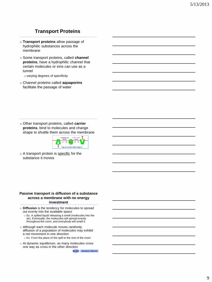
Transport Proteins
o Transport proteins allow passage of
hydrophilic substances across the
membrane
o Some transport proteins, called channel
proteins, have a hydrophilic channel that
certain molecules or ions can use as a
tunnel
o varying degrees of specificity
o Channel proteins called aquaporins
facilitate the passage of water
o Other transport proteins, called carrier
proteins, bind to molecules and change
shape to shuttle them across the membrane
o A transport protein is specific for the
substance it moves
Passive transport is diffusion of a substance
across a membrane with no energy
investment
o Diffusion is the tendency for molecules to spread out evenly into the available space
o Ex. A spilled liquid releasing a smell (molecules into the air). Eventually, the molecules will spread evenly throughout the room, and everybody will smell it.
o Although each molecule moves randomly, diffusion of a population of molecules may exhibit a net movement in one direction
o Ex. From the place of the spill to the rest of the room
o At dynamic equilibrium, as many molecules cross one way as cross in the other direction
Animation: Diffusion
5/13/2013
10
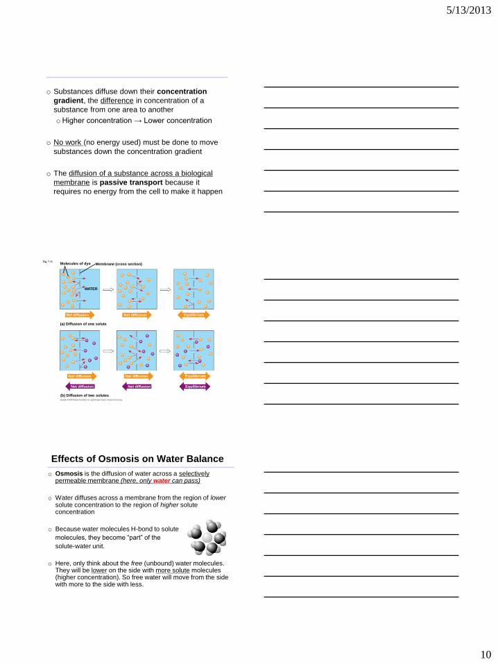
o Substances diffuse down their concentration
gradient, the difference in concentration of a
substance from one area to another
oHigher concentration → Lower concentration
o No work (no energy used) must be done to move
substances down the concentration gradient
o The diffusion of a substance across a biological
membrane is passive transport because it
requires no energy from the cell to make it happen
Fig. 7-11 Molecules of dye Membrane (cross section)
WATER
Net diffusion Net diffusion Equilibrium
(a) Diffusion of one solute
Net diffusion
Net diffusion
Net diffusion
Net diffusion
Equilibrium
Equilibrium
(b) Diffusion of two solutes
Effects of Osmosis on Water Balance
o Osmosis is the diffusion of water across a selectively permeable membrane (here, only water can pass)
o Water diffuses across a membrane from the region of lower solute concentration to the region of higher solute concentration
o Because water molecules H-bond to solute
molecules, they become “part” of the
solute-water unit.
o Here, only think about the free (unbound) water molecules. They will be lower on the side with more solute molecules (higher concentration). So free water will move from the side with more to the side with less.
5/13/2013
11
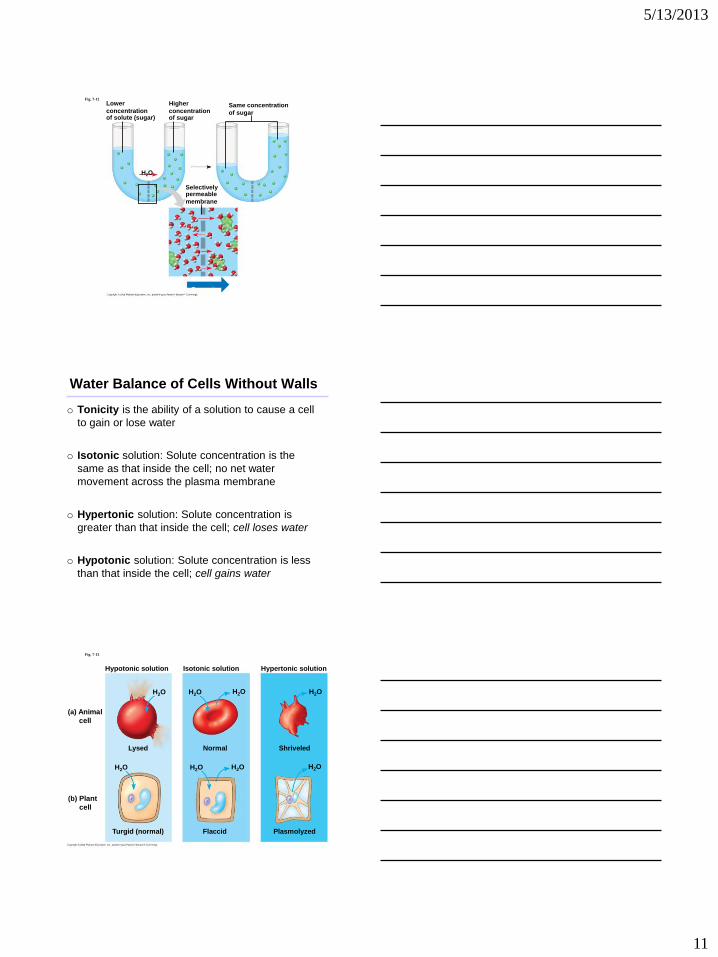
Lower
concentration of solute (sugar)
Fig. 7-12
H2O
Higher
concentration of sugar
Selectively permeable
membrane
Same concentration
of sugar
Osmosis
Water Balance of Cells Without Walls
o Tonicity is the ability of a solution to cause a cell
to gain or lose water
o Isotonic solution: Solute concentration is the
same as that inside the cell; no net water
movement across the plasma membrane
o Hypertonic solution: Solute concentration is
greater than that inside the cell; cell loses water
o Hypotonic solution: Solute concentration is less
than that inside the cell; cell gains water
Fig. 7-13
Hypotonic solution
(a) Animal
cell
(b) Plant
cell
H2O
Lysed
H2O
Turgid (normal)
H2O
H2O
H2O
H2O
Normal
Isotonic solution
Flaccid
H2O
H2O
Shriveled
Plasmolyzed
Hypertonic solution
5/13/2013
12
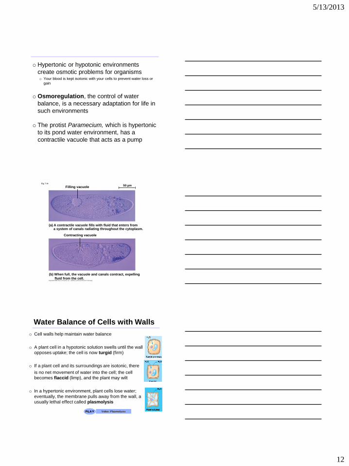
o Hypertonic or hypotonic environments
create osmotic problems for organisms o Your blood is kept isotonic with your cells to prevent water loss or
gain
o Osmoregulation, the control of water
balance, is a necessary adaptation for life in
such environments
o The protist Paramecium, which is hypertonic
to its pond water environment, has a
contractile vacuole that acts as a pump
Fig. 7-14
Filling vacuole 50 µm
(a) A contractile vacuole fills with fluid that enters from a system of canals radiating throughout the cytoplasm.
Contracting vacuole
(b) When full, the vacuole and canals contract, expelling
fluid from the cell.
Water Balance of Cells with Walls
o Cell walls help maintain water balance
o A plant cell in a hypotonic solution swells until the wall
opposes uptake; the cell is now turgid (firm)
o If a plant cell and its surroundings are isotonic, there
is no net movement of water into the cell; the cell
becomes flaccid (limp), and the plant may wilt
o In a hypertonic environment, plant cells lose water;
eventually, the membrane pulls away from the wall, a
usually lethal effect called plasmolysis
Video: Plasmolysis
5/13/2013
13
Facilitated Diffusion: Passive
Transport Aided by Proteins
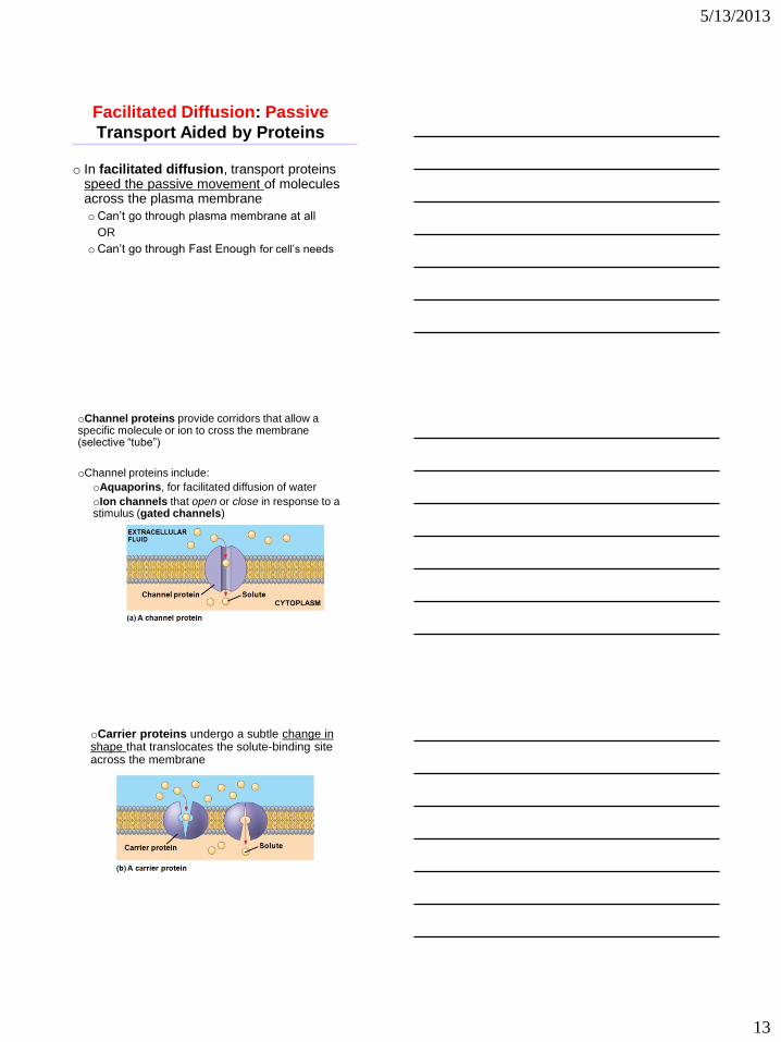
o In facilitated diffusion, transport proteins speed the passive movement of molecules across the plasma membrane
oCan’t go through plasma membrane at all
OR
oCan’t go through Fast Enough for cell’s needs
oChannel proteins provide corridors that allow a specific molecule or ion to cross the membrane (selective “tube”)
oChannel proteins include:
oAquaporins, for facilitated diffusion of water
oIon channels that open or close in response to a stimulus (gated channels)
oCarrier proteins undergo a subtle change in shape that translocates the solute-binding site across the membrane
5/13/2013
14
Active transport uses energy to
move solutes against their gradients
o Facilitated diffusion is still passive, because the solute moves down its concentration gradient
o Some transport proteins can move solutes against their concentration gradients
o Active transport moves substances against their concentration gradient
o Active transport requires energy, usually in the form of ATP
o Like using a pump to move water uphill
o Active transport is performed by specific proteins embedded in the membranes
o Active transport allows cells to maintain
concentration gradients that differ from their
surroundings
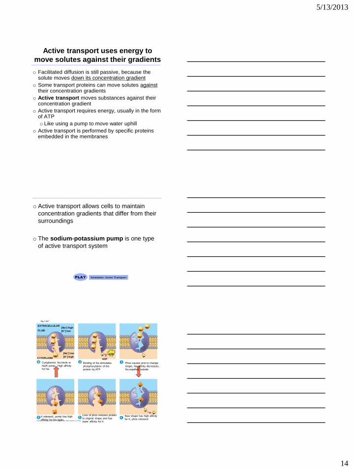
o The sodium-potassium pump is one type
of active transport system
Animation: Active Transport
2
EXTRACELLULAR
FLUID [Na+] high [K+] low
[Na+] low
[K+] high
Na+
Na+
Na+
Na+
Na+
Na+
CYTOPLASM ATP
ADP P
Na+ Na+
Na+
P
3
6 5 4
P P
1
Fig. 7-16-7
Cytoplasmic Na binds to
Na/K pump. High affinity
for Na
Binding of Na stimulates
phosphorylation of the
protein by ATP
Phos causes prot to change
shape, Na affinity decreases,
Na expelled outside
New shape has high affinity
for K, phos released
Loss of phos restores protein
to original shape and has
lower affinity for K
K released, pump has high
affinity for Na again
5/13/2013
15
Passive transport – Substances diffuse
spontaneously down their concentration gradient, crossing a membrance with no expenditure of energy by the cell. The rate of diffusion can be greatky increased by transport proteins in the membrane
Diffusion
Facilitated diffusion
Active transport – Some transport
proteins act as pumps, moving substances across a membrane against their concentration (or electrochemical) gradients. Energy for this work is usually supplied by ATP
ATP
Hydrophobic
molecs & (at a
slow rate)
very sm
uncharged
polar molecs
can diffuse
through the
lipid bilayer
Many hydrophilic
substances diffuse
through membs with
the assistance of
transport proteins,
either channel or
carrier proteins
How Ion Pumps Maintain Membrane Potential
o Membrane potential is the voltage difference across a membrane
o Voltage is created by differences in the distribution of positive and negative ions
o Areas (around a barrier) do not have a “+” or “-” charge (except for electrons and protons). It has a difference. If one side of a barrier (membrane or battery terminal) has more + charges than the other, then the side with more is “+”, and the side with less is negative, “-”, compared to the side with more.
o Cells are usually Negative compared to outside (fewer positive ions) (-50-200 mV)
o 2 combined forces, collectively called the
electrochemical gradient, drive the diffusion of
ions across a membrane:
o A chemical force (the ion’s concentration
gradient)
o An electrical force (the effect of the membrane
potential on the ion’s movement)
o If the components are opposite, ion flow is
slowed
o If the components (chemical and electrical) are
together, flow is very fast and easy!
5/13/2013
16
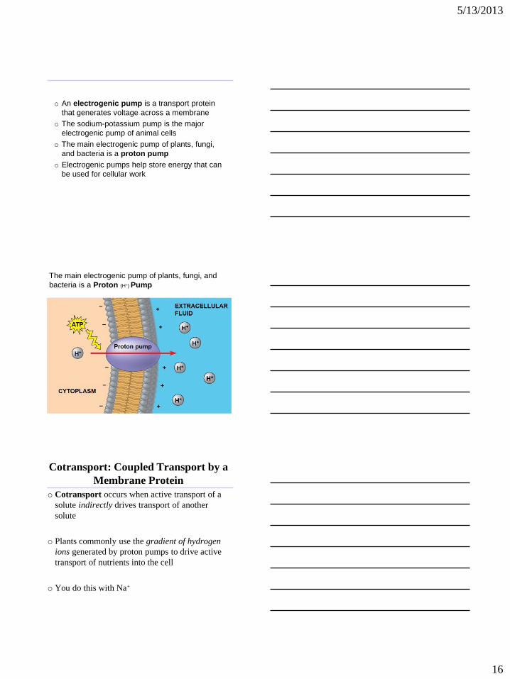
o An electrogenic pump is a transport protein
that generates voltage across a membrane
o The sodium-potassium pump is the major
electrogenic pump of animal cells
o The main electrogenic pump of plants, fungi,
and bacteria is a proton pump
o Electrogenic pumps help store energy that can
be used for cellular work
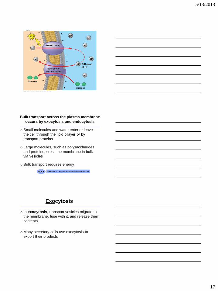
The main electrogenic pump of plants, fungi, and
bacteria is a Proton (H+) Pump
Cotransport: Coupled Transport by a
Membrane Protein
o Cotransport occurs when active transport of a
solute indirectly drives transport of another
solute
o Plants commonly use the gradient of hydrogen
ions generated by proton pumps to drive active
transport of nutrients into the cell
o You do this with Na+
5/13/2013
17
Fig. 7-19
Proton pump
–
–
–
–
–
–
+
+
+
+
+
+
ATP
H+
H+
H+ H+
H+
H+
H+
H+
Diffusion
of H+ Sucrose-H+
cotransporter
Sucrose
Sucrose
Bulk transport across the plasma membrane
occurs by exocytosis and endocytosis
o Small molecules and water enter or leave
the cell through the lipid bilayer or by
transport proteins
o Large molecules, such as polysaccharides
and proteins, cross the membrane in bulk
via vesicles
o Bulk transport requires energy
Animation: Exocytosis and Endocytosis Introduction
Exocytosis
o In exocytosis, transport vesicles migrate to
the membrane, fuse with it, and release their
contents
o Many secretory cells use exocytosis to
export their products
5/13/2013
18
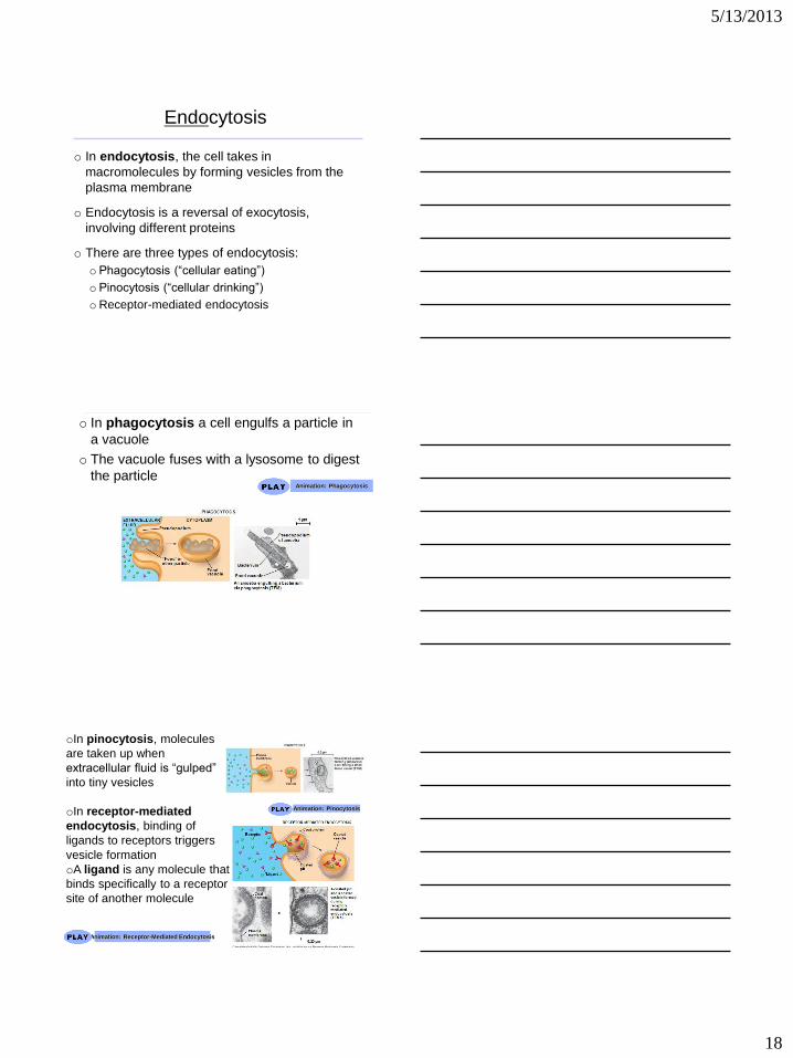
Endocytosis
o In endocytosis, the cell takes in
macromolecules by forming vesicles from the
plasma membrane
o Endocytosis is a reversal of exocytosis,
involving different proteins
o There are three types of endocytosis:
o Phagocytosis (“cellular eating”)
o Pinocytosis (“cellular drinking”)
oReceptor-mediated endocytosis
o In phagocytosis a cell engulfs a particle in
a vacuole
o The vacuole fuses with a lysosome to digest
the particle
Animation: Phagocytosis
oIn pinocytosis, molecules
are taken up when
extracellular fluid is “gulped”
into tiny vesicles
oIn receptor-mediated
endocytosis, binding of
ligands to receptors triggers
vesicle formation
oA ligand is any molecule that
binds specifically to a receptor
site of another molecule
Animation: Pinocytosis
Animation: Receptor-Mediated Endocytosis
5/13/2013
19
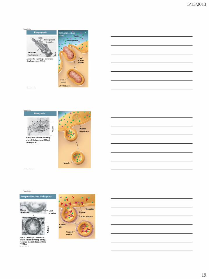
Figure 7.22a
Pseudopodium
Solutes
“Food”
or other
particle
Food
vacuole
CYTOPLASM
EXTRACELLULAR
FLUID
Pseudopodium
of amoeba
Bacterium
Food vacuole
An amoeba engulfing a bacterium
via phagocytosis (TEM).
Phagocytosis
1
m
Figure 7.22b
Pinocytosis vesicles forming
in a cell lining a small blood
vessel (TEM).
Plasma
membrane
Vesicle
0.5
m
Pinocytosis
Figure 7.22c
Top: A coated pit. Bottom: A coated vesicle forming during receptor-mediated endocytosis (TEMs).
Receptor
0.2
5
m
Receptor-Mediated Endocytosis
Ligand
Coat proteins
Coated
pit
Coated
vesicle
Coat
proteins
Plasma
membrane