Embed Size (px)
Citation preview
Chapter 7
Carbohydrates and Glycobiology
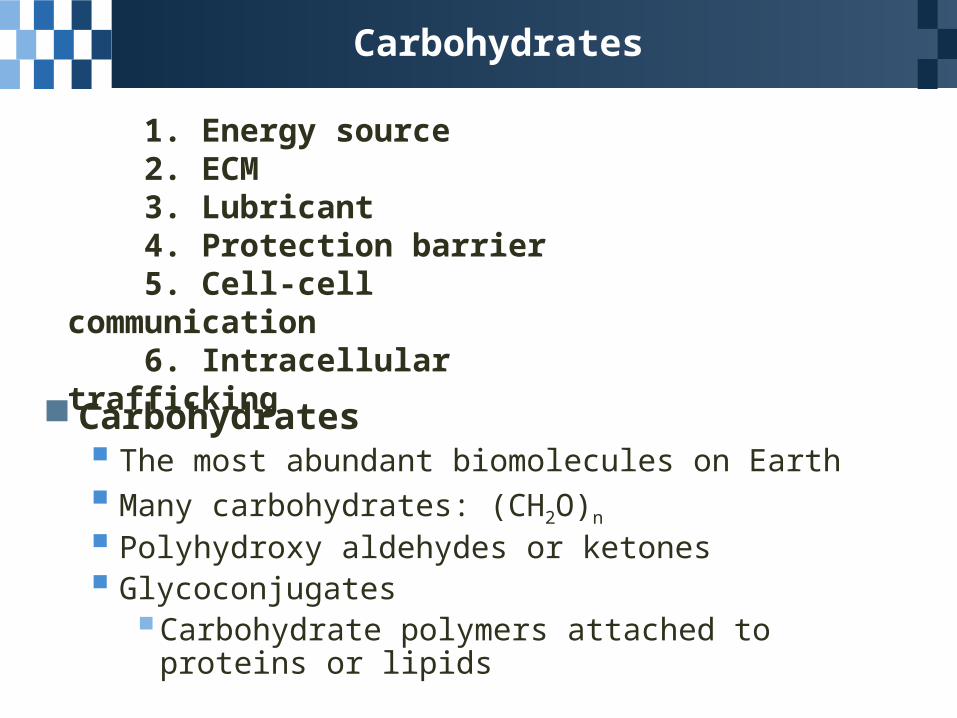
Carbohydrates
Carbohydrates The most abundant biomolecules on Earth Many carbohydrates: (CH2O)n
Polyhydroxy aldehydes or ketones Glycoconjugates
Carbohydrate polymers attached to proteins or lipids
1. Energy source 2. ECM 3. Lubricant 4. Protection barrier 5. Cell-cell communication 6. Intracellular trafficking
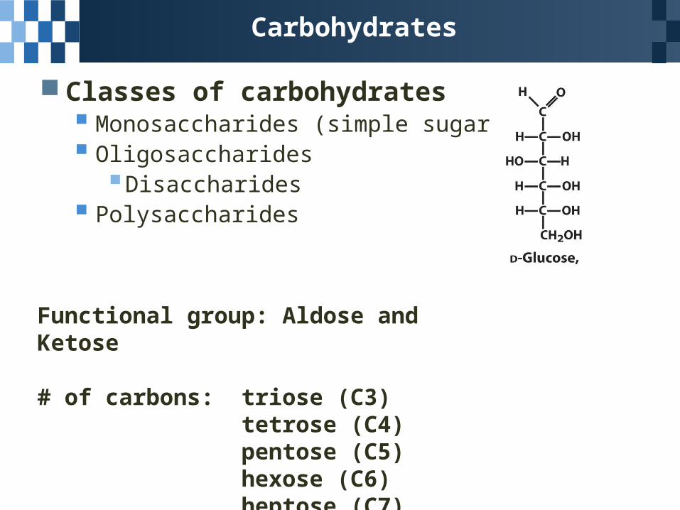
Functional group: Aldose and Ketose
# of carbons: triose (C3)tetrose (C4) pentose (C5) hexose (C6)heptose (C7)
Carbohydrates
Classes of carbohydrates Monosaccharides (simple sugar) Oligosaccharides
Disaccharides Polysaccharides
7.1 Monosaccharides and Disaccharides
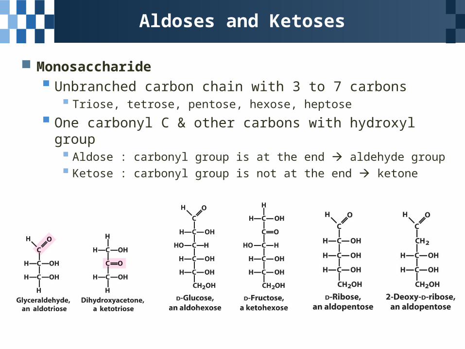
Aldoses and Ketoses
Monosaccharide
Unbranched carbon chain with 3 to 7 carbons Triose, tetrose, pentose, hexose, heptose
One carbonyl C & other carbons with hydroxyl group Aldose : carbonyl group is at the end aldehyde group Ketose : carbonyl group is not at the end ketone
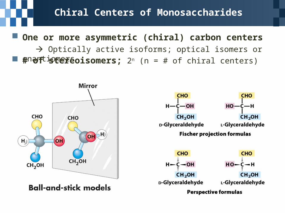
Chiral Centers of Monosaccharides
One or more asymmetric (chiral) carbon centers Optically active isoforms; optical isomers or enantiomers # of stereoisomers; 2n (n = # of chiral centers)
Chiral Centers of Monosaccharides
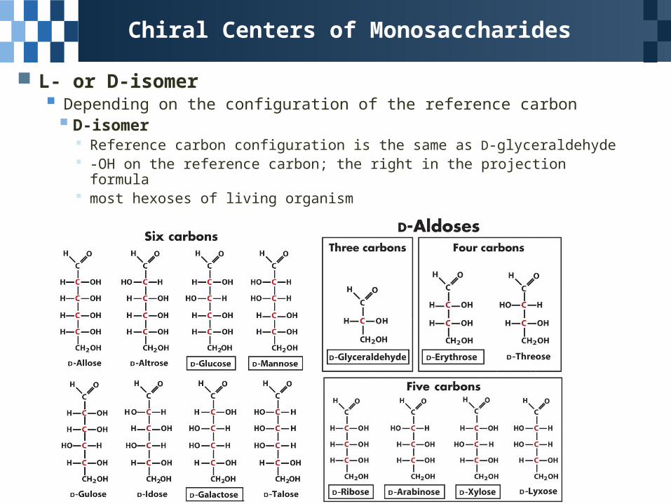
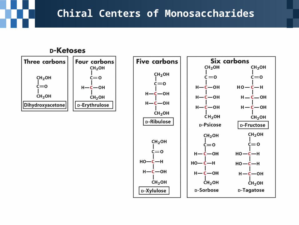
L- or D-isomer Depending on the configuration of the reference carbon D-isomer
Reference carbon configuration is the same as D-glyceraldehyde -OH on the reference carbon; the right in the projection formula most hexoses of living organism
Chiral Centers of Monosaccharides
Chiral Centers of Monosaccharides
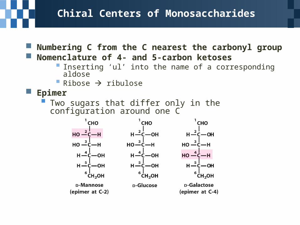
Numbering C from the C nearest the carbonyl group Nomenclature of 4- and 5-carbon ketoses
Inserting ‘ul’ into the name of a corresponding aldose Ribose ribulose
Epimer Two sugars that differ only in the configuration around one C
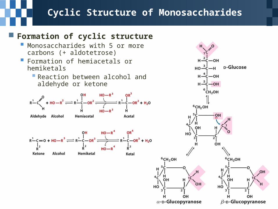
Cyclic Structure of Monosaccharides
Formation of cyclic structure Monosaccharides with 5 or more carbons
(+ aldotetrose) Formation of hemiacetals or hemiketals
Reaction between alcohol and aldehyde or ketone
Cyclic Structure of Monosaccharides
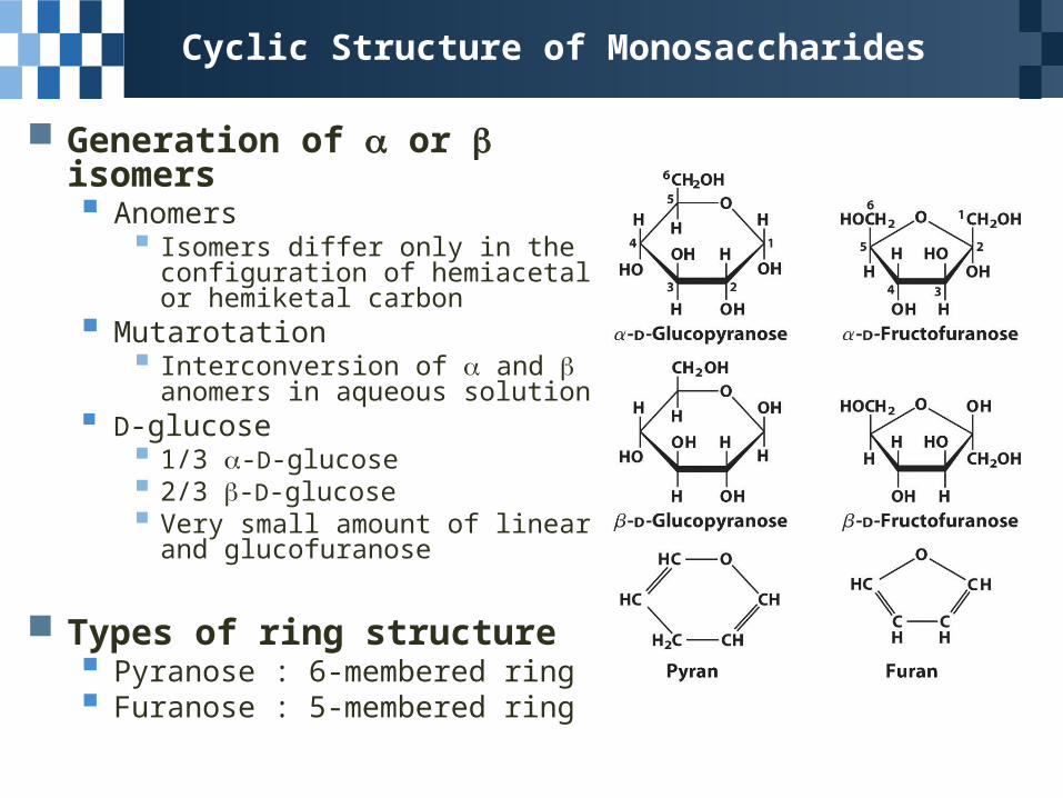
Generation of or isomers Anomers
Isomers differ only in the configuration of hemiacetal or hemiketal carbon
Mutarotation Interconversion of and anomers
in aqueous solution D-glucose
1/3 -D-glucose 2/3 -D-glucose Very small amount of linear and
glucofuranose
Types of ring structure Pyranose : 6-membered ring Furanose : 5-membered ring
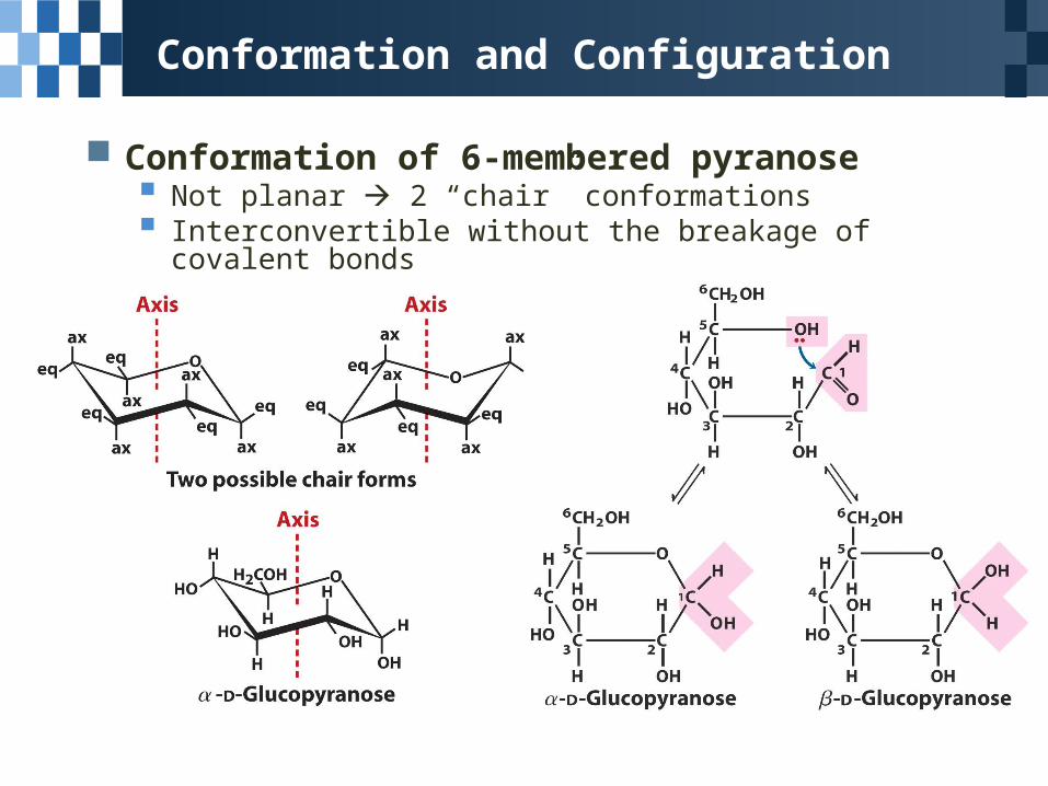
Conformation and Configuration
Conformation of 6-membered pyranose Not planar 2 “chair” conformations Interconvertible without the breakage of covalent bonds
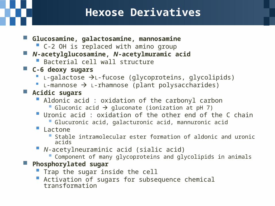
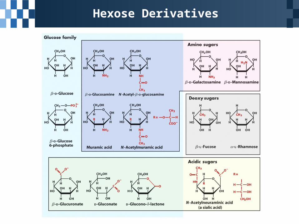
Hexose Derivatives
Glucosamine, galactosamine, mannosamine C-2 OH is replaced with amino group
N-acetylglucosamine, N-acetylmuramic acid Bacterial cell wall structure
C-6 deoxy sugars L-galactose L-fucose (glycoproteins, glycolipids) L-mannose L-rhamnose (plant polysaccharides)
Acidic sugars Aldonic acid : oxidation of the carbonyl carbon
Gluconic acid gluconate (ionization at pH 7) Uronic acid : oxidation of the other end of the C chain
Glucuronic acid, galacturonic acid, mannuronic acid Lactone
Stable intramolecular ester formation of aldonic and uronic acids N-acetylneuraminic acid (sialic acid)
Component of many glycoproteins and glycolipids in animals Phosphorylated sugar
Trap the sugar inside the cell Activation of sugars for subsequence chemical transformation
Hexose Derivatives
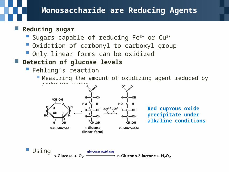
Monosaccharide are Reducing Agents
Reducing sugar Sugars capable of reducing Fe3+ or Cu2+
Oxidation of carbonyl to carboxyl group Only linear forms can be oxidized
Detection of glucose levels Fehling’s reaction
Measuring the amount of oxidizing agent reduced by reducing sugar
Using glucose oxidase
Red cuprous oxide precipitate under alkaline conditions
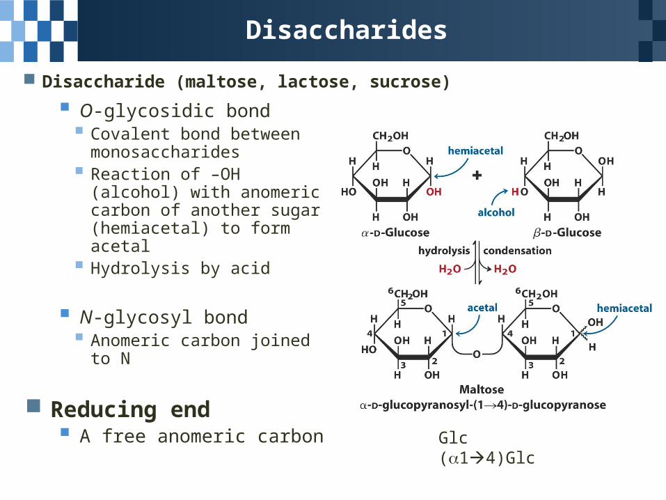
Disaccharides
Disaccharide (maltose, lactose, sucrose)
Glc (14)Glc
O-glycosidic bond Covalent bond between
monosaccharides Reaction of –OH (alcohol) with
anomeric carbon of another sugar (hemiacetal) to form acetal
Hydrolysis by acid
N-glycosyl bond Anomeric carbon joined to N
Reducing end A free anomeric carbon
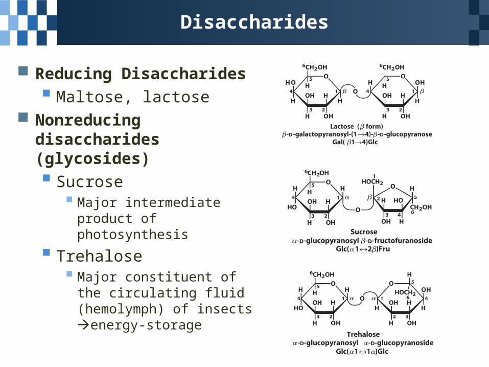
Disaccharides
Reducing Disaccharides
Maltose, lactose Nonreducing disaccharides
(glycosides)
Sucrose Major intermediate product of
photosynthesis
Trehalose Major constituent of the
circulating fluid (hemolymph) of insects energy-storage
7.2 Polysaccharides
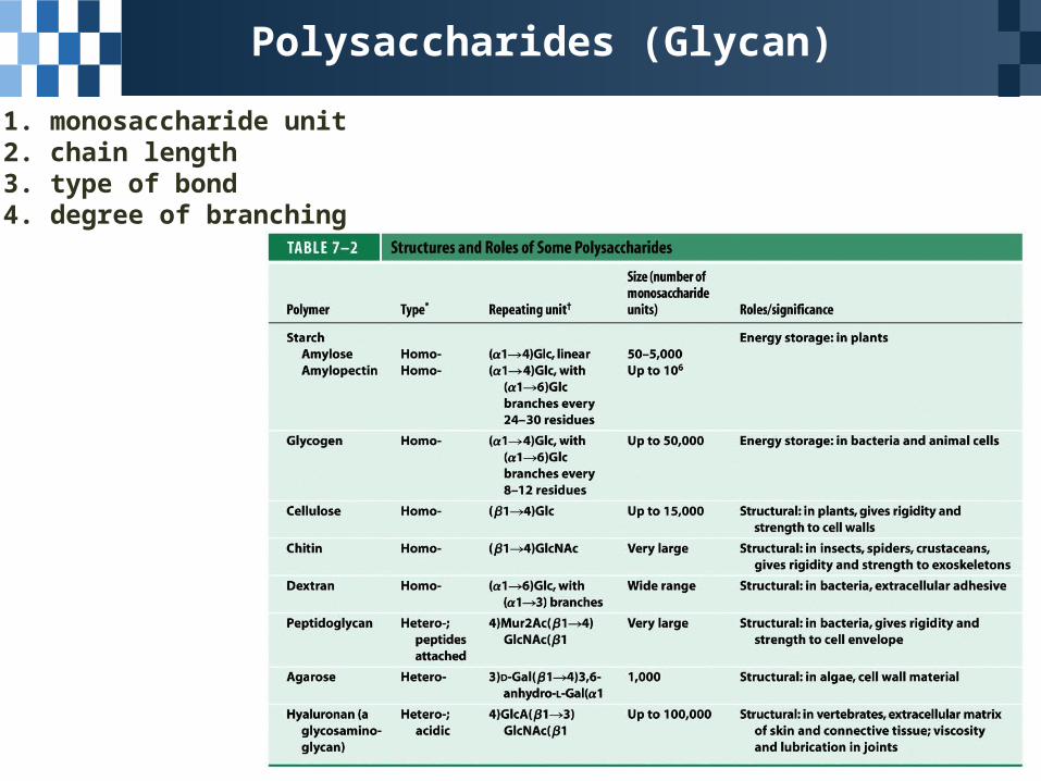
1. monosaccharide unit2. chain length3. type of bond4. degree of branching
Polysaccharides (Glycan)
Polysaccharides (Glycan)
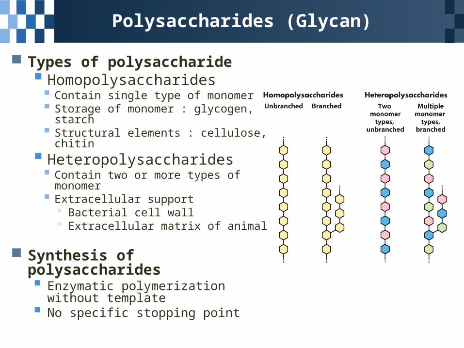
Types of polysaccharide Homopolysaccharides Contain single type of monomer Storage of monomer : glycogen, starch Structural elements : cellulose, chitin
Heteropolysaccharides Contain two or more types of monomer Extracellular support
Bacterial cell wall Extracellular matrix of animal
Synthesis of polysaccharides Enzymatic polymerization without
template No specific stopping point
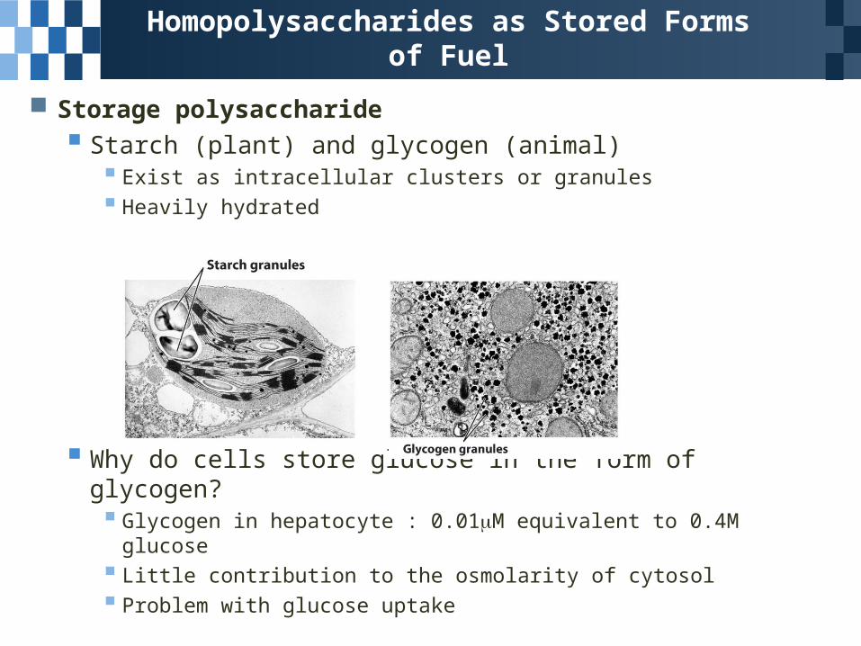
Storage polysaccharide
Starch (plant) and glycogen (animal) Exist as intracellular clusters or granules Heavily hydrated
Why do cells store glucose in the form of glycogen? Glycogen in hepatocyte : 0.01M equivalent to 0.4M glucose Little contribution to the osmolarity of cytosol Problem with glucose uptake
Homopolysaccharides as Stored Forms of Fuel
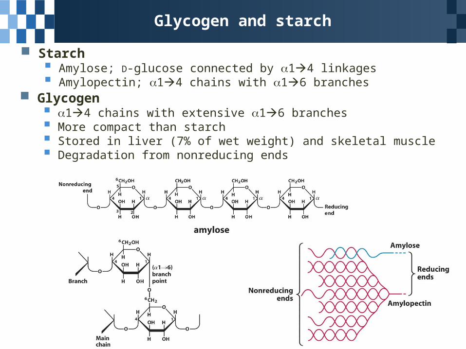
Glycogen and starch
Starch Amylose; D-glucose connected by 14 linkages Amylopectin; 14 chains with 16 branches
Glycogen 14 chains with extensive 16 branches More compact than starch Stored in liver (7% of wet weight) and skeletal muscle Degradation from nonreducing ends
Homopolysaccharides Playing Structural Roles
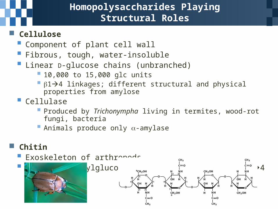
Cellulose Component of plant cell wall Fibrous, tough, water-insoluble Linear D-glucose chains (unbranched)
10,000 to 15,000 glc units 14 linkages; different structural and physical properties from amylose
Cellulase Produced by Trichonympha living in termites, wood-rot fungi, bacteria Animals produce only -amylase
Chitin Exoskeleton of arthropods Linear N-acetylglucosamine chains connected by 14 linkages
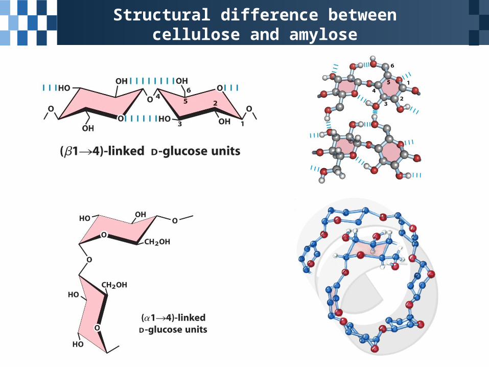
Structural difference between cellulose and amylose
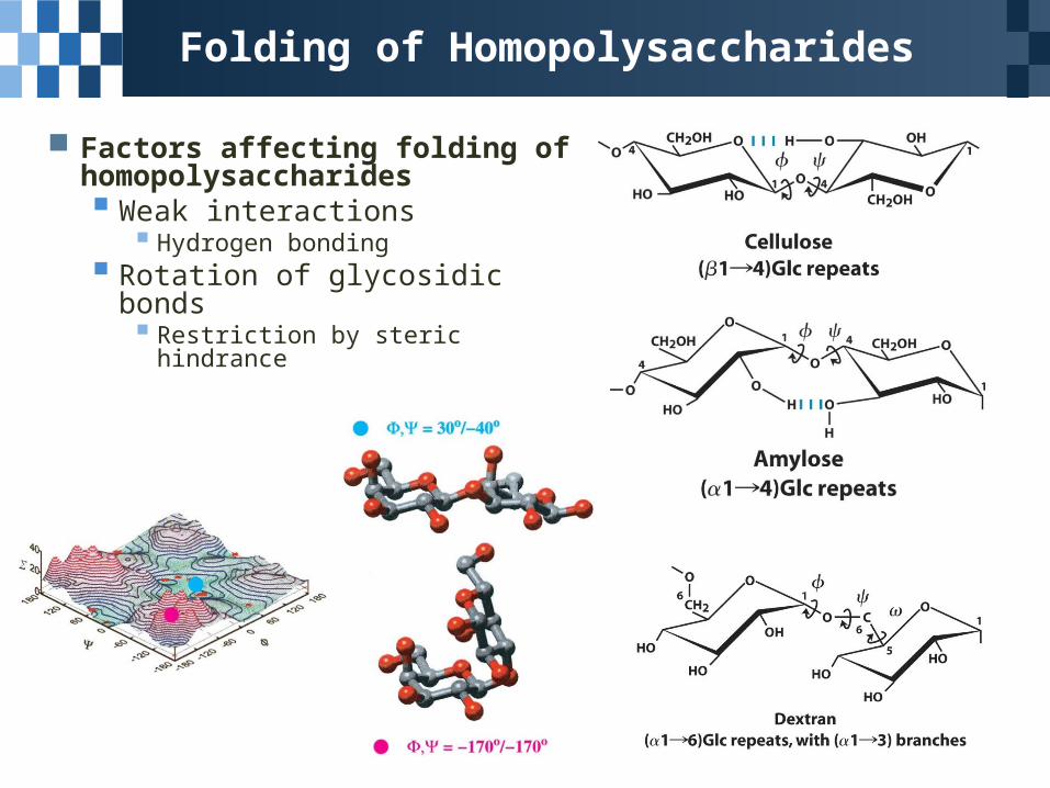
Folding of Homopolysaccharides
Factors affecting folding of homopolysaccharides Weak interactions
Hydrogen bonding Rotation of glycosidic bonds
Restriction by steric hindrance
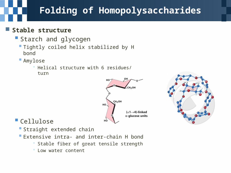
Folding of Homopolysaccharides
Stable structure Starch and glycogen Tightly coiled helix stabilized by H bond Amylose
Helical structure with 6 residues/ turn
Cellulose Straight extended chain Extensive intra- and inter-chain H bond
Stable fiber of great tensile strength Low water content
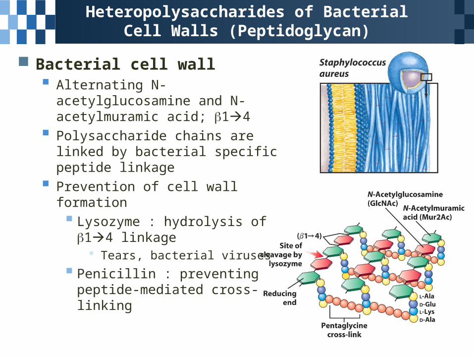
Heteropolysaccharides of Bacterial Cell Walls (Peptidoglycan)
Bacterial cell wall Alternating N-acetylglucosamine and
N-acetylmuramic acid; 14
Polysaccharide chains are linked by bacterial specific peptide linkage
Prevention of cell wall formation Lysozyme : hydrolysis of 14
linkage Tears, bacterial viruses
Penicillin : preventing peptide-mediated cross-linking
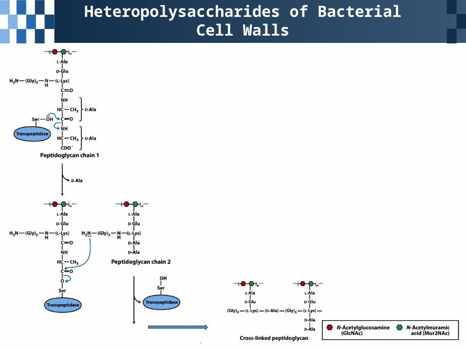
Heteropolysaccharides of Bacterial Cell Walls
Heteropolysaccharides of Algal Cell Walls
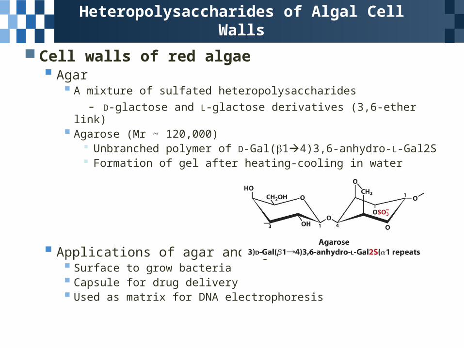
Cell walls of red algae Agar
A mixture of sulfated heteropolysaccharides
- D-glactose and L-glactose derivatives (3,6-ether link) Agarose (Mr ~ 120,000)
Unbranched polymer of D-Gal(14)3,6-anhydro-L-Gal2S Formation of gel after heating-cooling in water
Applications of agar and agarose Surface to grow bacteria Capsule for drug delivery Used as matrix for DNA electrophoresis
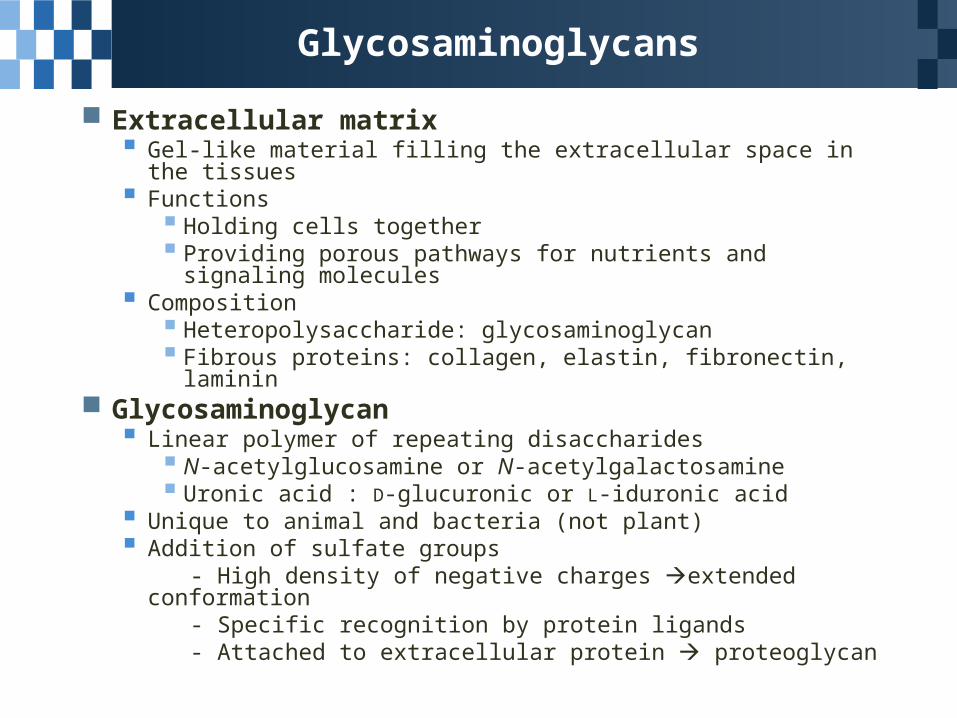
Glycosaminoglycans
Extracellular matrix Gel-like material filling the extracellular space in the tissues Functions
Holding cells together Providing porous pathways for nutrients and signaling
molecules Composition
Heteropolysaccharide: glycosaminoglycan Fibrous proteins: collagen, elastin, fibronectin, laminin
Glycosaminoglycan Linear polymer of repeating disaccharides
N-acetylglucosamine or N-acetylgalactosamine Uronic acid : D-glucuronic or L-iduronic acid
Unique to animal and bacteria (not plant) Addition of sulfate groups - High density of negative charges extended conformation - Specific recognition by protein ligands - Attached to extracellular protein proteoglycan
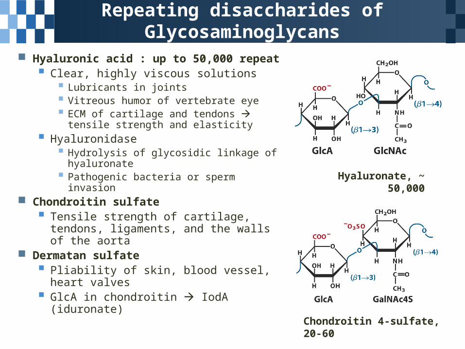
Repeating disaccharides of Glycosaminoglycans
Hyaluronic acid : up to 50,000 repeat Clear, highly viscous solutions
Lubricants in joints Vitreous humor of vertebrate eye ECM of cartilage and tendons
tensile strength and elasticity
Hyaluronidase Hydrolysis of glycosidic linkage of
hyaluronate Pathogenic bacteria or sperm invasion
Chondroitin sulfate Tensile strength of cartilage, tendons,
ligaments, and the walls of the aorta Dermatan sulfate
Pliability of skin, blood vessel, heart valves
GlcA in chondroitin IodA (iduronate)
Hyaluronate, ~ 50,000
Chondroitin 4-sulfate, 20-60
Glycosaminoglycans
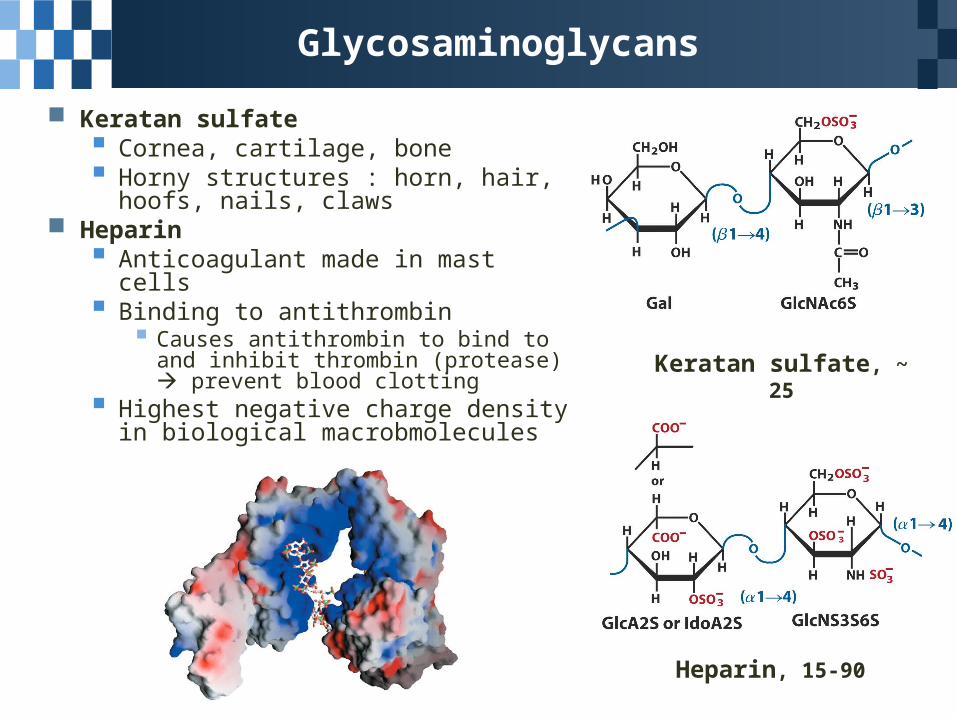
Keratan sulfate Cornea, cartilage, bone Horny structures : horn, hair, hoofs,
nails, claws Heparin
Anticoagulant made in mast cells Binding to antithrombin
Causes antithrombin to bind to and inhibit thrombin (protease) prevent blood clotting
Highest negative charge density in biological macrobmolecules
Keratan sulfate, ~ 25
Heparin, 15-90