Embed Size (px)
Citation preview

1Mosby items and derived items © 2011, 2006 by Mosby, Inc., an affiliate of Elsevier Inc.
Chapter 17
Tuberculosis

2Mosby items and derived items © 2011, 2006 by Mosby, Inc., an affiliate of Elsevier Inc.
Figure 17-1. Tuberculosis. A, Early primary infection. B, Cavitation of a caseous tubercle and new primary lesions developing. C, Further progression and development of cavitations and new primary infections. Note
the subpleural location of some of these lesions. D, Severe lung destruction caused by tuberculosis.
A
C
B
D

3Mosby items and derived items © 2011, 2006 by Mosby, Inc., an affiliate of Elsevier Inc.
Anatomic Alterations of the Lungs
Tuberculosis is classified as either: Primary tuberculosis—also called:
Primary infection stage
Postprimary tuberculosis—also called:
Reactivation TB
Reinfection TB
Secondary TB
Disseminated tuberculosis—also called:
Extrapulmonary TB
Miliary TB
Tuberculosis-disseminated

4Mosby items and derived items © 2011, 2006 by Mosby, Inc., an affiliate of Elsevier Inc.
Anatomic Alterations of the Lungs
(Mainly Postprimary TB)
Alveolar consolidation
Alveolar-capillary destruction
Caseous tubercles or granulomas
Cavity formation
Fibrosis and secondary calcification of the
lung parenchyma
Distortion and dilation of the bronchi
Increased bronchial airway secretions

5Mosby items and derived items © 2011, 2006 by Mosby, Inc., an affiliate of Elsevier Inc.
Etiology
In humans, TB primarily caused by
Mycobacterium tuberculosis
Highly aerobic organisms
Bacilli are acid-fast bacilli
The bacilli are almost exclusively transmitted
within aerosol droplets produced by
coughing, sneezing, or laughing of an
individual with active TB.
6Mosby items and derived items © 2011, 2006 by Mosby, Inc., an affiliate of Elsevier Inc.
Diagnosis
Mantoux tuberculin skin test
Acid-fast Staining
Sputum Culture
QuantiFERON®-TB Gold Test

7Mosby items and derived items © 2011, 2006 by Mosby, Inc., an affiliate of Elsevier Inc.
Diagnosis (Cont’d)
Mantoux tuberculin skin test Injection of purified protein derivative (PPD)
• Wheal less than 5 mm: negative
• Wheal 5 mm to 9 mm: considered suspicious
• Wheal 10 mm or greater: positive

8Mosby items and derived items © 2011, 2006 by Mosby, Inc., an affiliate of Elsevier Inc.
Figure 17-2. The Mantoux test, which consists of an intradermal injection of a small amount of a purified protein derivative (PPD) of the tuberculin bacillus. An induration of 10 mm or greater is
considered positive. A positive reaction is fairly sound evidence of recent or past infection or disease.
9Mosby items and derived items © 2011, 2006 by Mosby, Inc., an affiliate of Elsevier Inc.
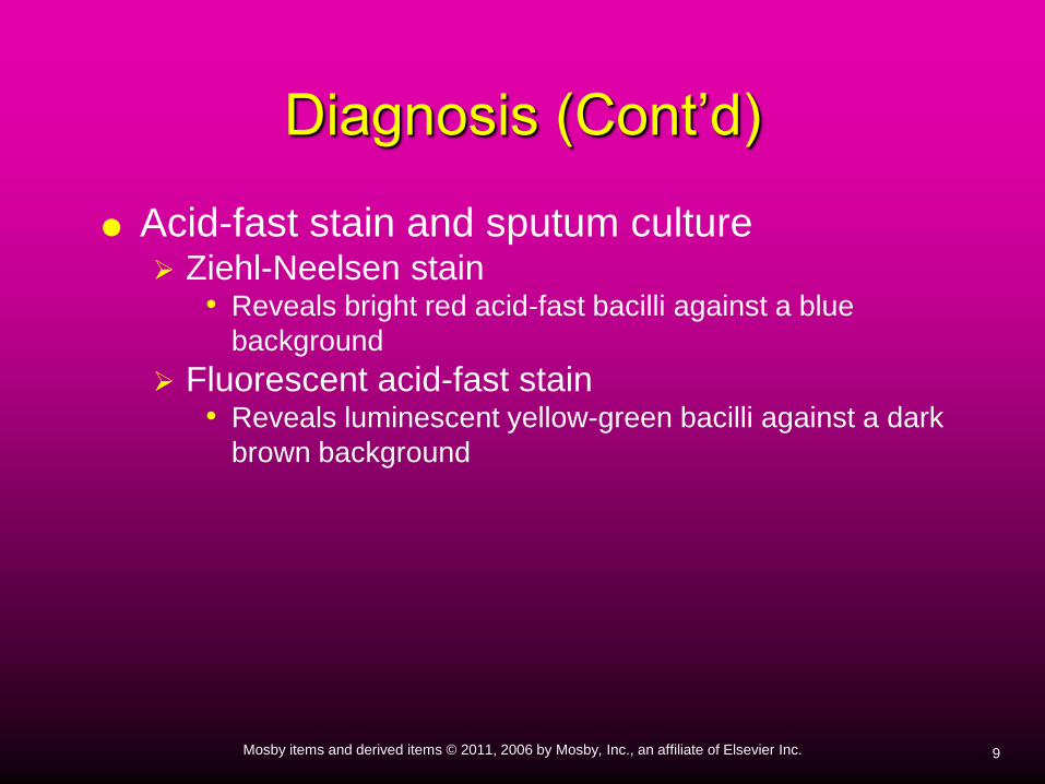
Acid-fast stain and sputum culture Ziehl-Neelsen stain
• Reveals bright red acid-fast bacilli against a blue
background
Fluorescent acid-fast stain• Reveals luminescent yellow-green bacilli against a dark
brown background
Diagnosis (Cont’d)
10Mosby items and derived items © 2011, 2006 by Mosby, Inc., an affiliate of Elsevier Inc.
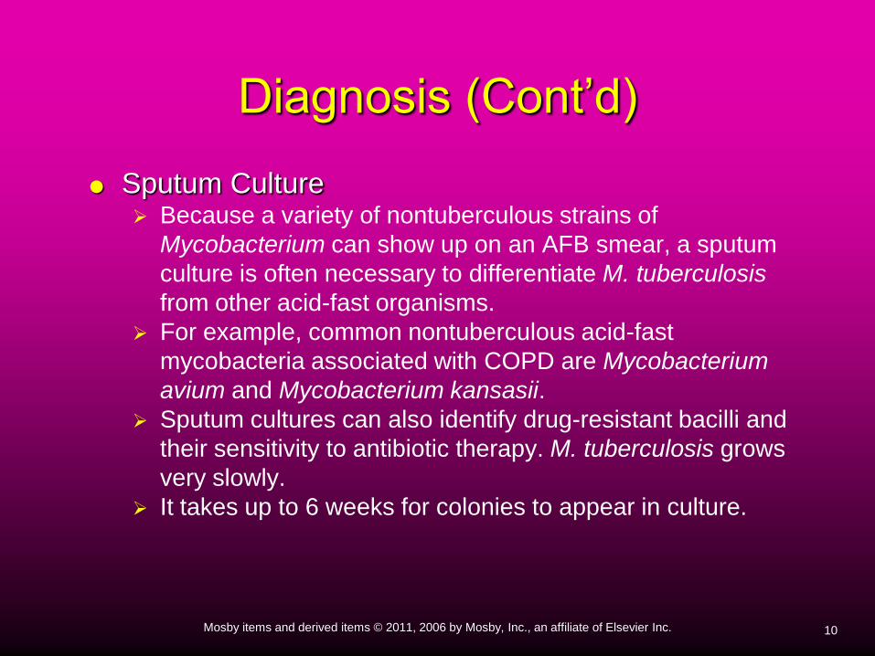
Sputum Culture Because a variety of nontuberculous strains of
Mycobacterium can show up on an AFB smear, a sputum
culture is often necessary to differentiate M. tuberculosis
from other acid-fast organisms.
For example, common nontuberculous acid-fast
mycobacteria associated with COPD are Mycobacterium
avium and Mycobacterium kansasii.
Sputum cultures can also identify drug-resistant bacilli and
their sensitivity to antibiotic therapy. M. tuberculosis grows
very slowly.
It takes up to 6 weeks for colonies to appear in culture.
Diagnosis (Cont’d)

11Mosby items and derived items © 2011, 2006 by Mosby, Inc., an affiliate of Elsevier Inc.
QuantiFERON-TB Gold Test In 2005, the U.S. Food and Drug Administration
(FDA) approved the QuantiFERON-TB Gold test
(QFT-G).
The QFT-G is a whole-blood test used for
diagnosing Mycobacterium tuberculosis infection,
including latent tuberculosis infection.
Results are available after 24 hours
Diagnosis (Cont’d)

12Mosby items and derived items © 2011, 2006 by Mosby, Inc., an affiliate of Elsevier Inc.
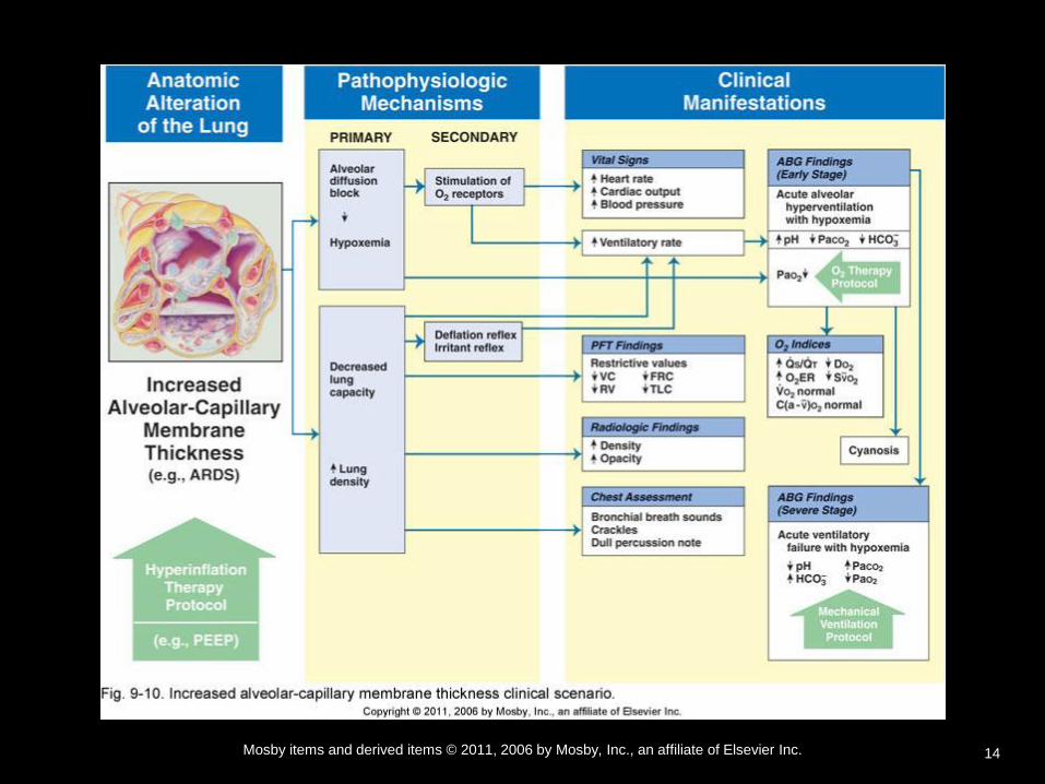
Overview
of the Cardiopulmonary Clinical Manifestations
Associated with
Tuberculosis
The following clinical manifestations result from the
pathophysiologic mechanisms caused (or activated)
by Alveolar Consolidation
Increased Alveolar-Capillary Membrane Thickness

13Mosby items and derived items © 2011, 2006 by Mosby, Inc., an affiliate of Elsevier Inc.
14Mosby items and derived items © 2011, 2006 by Mosby, Inc., an affiliate of Elsevier Inc.

15Mosby items and derived items © 2011, 2006 by Mosby, Inc., an affiliate of Elsevier Inc.
Clinical Data Obtained at the
Patient’s Bedside

16Mosby items and derived items © 2011, 2006 by Mosby, Inc., an affiliate of Elsevier Inc.
The Physical Examination
Vital Signs Increased
• Respiratory rate (Tachypnea)
• Heart rate (pulse)
• Blood pressure

17Mosby items and derived items © 2011, 2006 by Mosby, Inc., an affiliate of Elsevier Inc.
The Physical Examination (Cont’d)
Chest pain/decreased chest expansion
Cyanosis
Digital clubbing
Peripheral edema and venous distention Distended neck veins
Pitting edema
Enlarged and tender liver
Cough, sputum production, and hemoptysis

18Mosby items and derived items © 2011, 2006 by Mosby, Inc., an affiliate of Elsevier Inc.
The Physical Examination (Cont’d)
Chest Assessment Findings
Increased tactile and vocal fremitus
Dull percussion note
Bronchial breath sounds
Crackles, rhonchi, and wheezing
Pleural friction rub • if process extends to pleural surface
Whispered pectoriloquy

19Mosby items and derived items © 2011, 2006 by Mosby, Inc., an affiliate of Elsevier Inc.
Clinical Data Obtained from
Laboratory Tests and Special
Procedures

20Mosby items and derived items © 2011, 2006 by Mosby, Inc., an affiliate of Elsevier Inc.
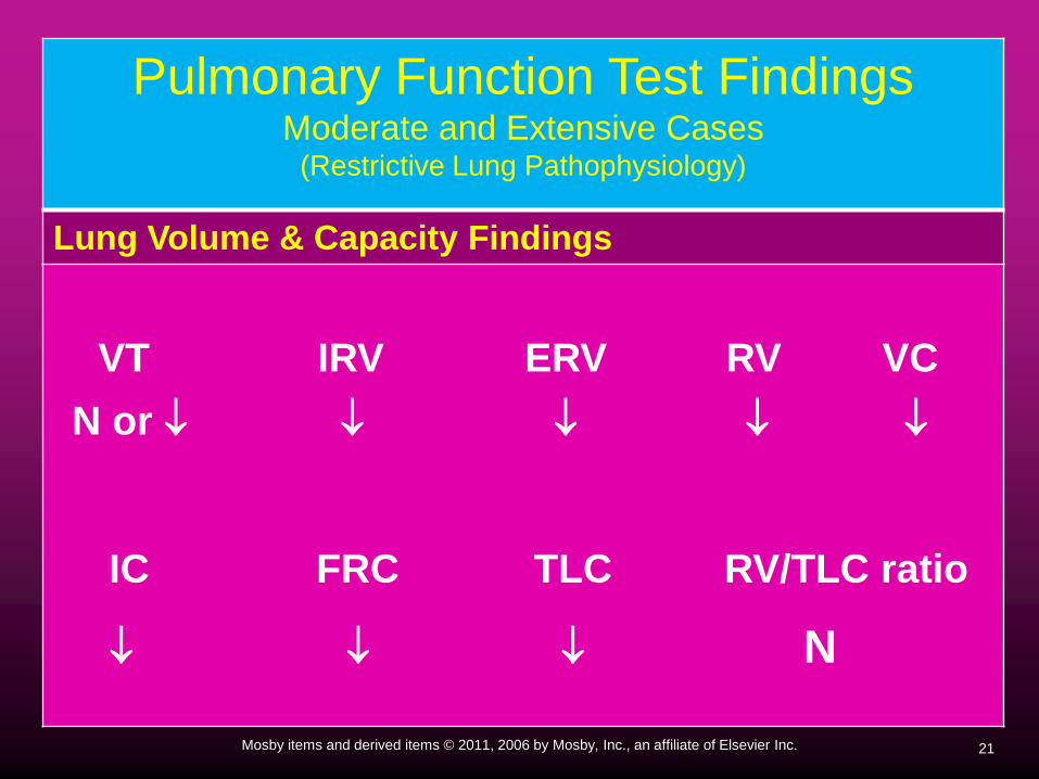
Pulmonary Function Test FindingsModerate and Extensive Cases
(Restrictive Lung Pathophysiology)
Forced Expiratory Flow Rate Findings
FVC FEVT FEV1/FVC ratio FEF25%-75
N or N or N or
FEF50% FEF200-1200 PEFR MVV
N or N or N or N or
21Mosby items and derived items © 2011, 2006 by Mosby, Inc., an affiliate of Elsevier Inc.
Pulmonary Function Test Findings Moderate and Extensive Cases
(Restrictive Lung Pathophysiology)
Lung Volume & Capacity Findings
VT IRV ERV RV VC
N or
IC FRC TLC RV/TLC ratio
N

22Mosby items and derived items © 2011, 2006 by Mosby, Inc., an affiliate of Elsevier Inc.
Arterial Blood GasesModerate Tuberculosis
Acute Alveolar Hyperventilation with Hypoxemia (Acute Respiratory Alkalosis)
pH PaC02 HCO3 Pa02
(slightly)

23Mosby items and derived items © 2011, 2006 by Mosby, Inc., an affiliate of Elsevier Inc.
PaO2 and PaCO2 trends during acute alveolar hyperventilation.

24Mosby items and derived items © 2011, 2006 by Mosby, Inc., an affiliate of Elsevier Inc.
Arterial Blood GasesExtensive Tubeculosis with Pulmonary Fibrosis
Chronic Ventilatory Failure with Hypoxemia (Compensated Respiratory Acidosis)
pH PaC02 HCO3 Pa02
N (Slightly)

25Mosby items and derived items © 2011, 2006 by Mosby, Inc., an affiliate of Elsevier Inc.
PaO2 and PaCO2 trends during acute or chronic ventilatory failure.

26Mosby items and derived items © 2011, 2006 by Mosby, Inc., an affiliate of Elsevier Inc.
Arterial Blood Gases
Acute Ventilatory Changes Superimposed
On
Chronic Ventilatory Failure
Because acute ventilatory changes are frequently seen in
patients with chronic ventilatory failure, the respiratory
care practitioner must be familiar with and alert for the
following: Acute alveolar hyperventilation superimposed on chronic
ventilatory failure
Acute ventilatory failure (acute hypoventilation) superimposed on
chronic ventialtory failure.

27Mosby items and derived items © 2011, 2006 by Mosby, Inc., an affiliate of Elsevier Inc.
Oxygenation IndicesModerate to Severe Stages
QS/QT D02 V02 C(a-v)02 02ER Sv02
N N

28Mosby items and derived items © 2011, 2006 by Mosby, Inc., an affiliate of Elsevier Inc.
Hemodynamic IndicesSevere Stage
CVP RAP PA PCWP CO SV
N N N
SVI CI RVSWI LVSWI PVR SVR
N N N N

29Mosby items and derived items © 2011, 2006 by Mosby, Inc., an affiliate of Elsevier Inc.
Abnormal Laboratory Tests and
Procedures
Positive tuberculosis skin test (PPD)
Positive sputum acid-fast bacillus (AFB) stain test
Positive sputum culture
30Mosby items and derived items © 2011, 2006 by Mosby, Inc., an affiliate of Elsevier Inc.
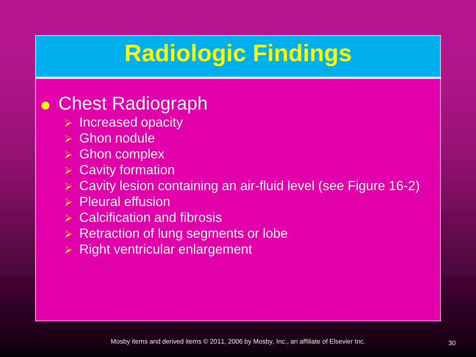
Radiologic Findings
Chest Radiograph Increased opacity
Ghon nodule
Ghon complex
Cavity formation
Cavity lesion containing an air-fluid level (see Figure 16-2)
Pleural effusion
Calcification and fibrosis
Retraction of lung segments or lobe
Right ventricular enlargement

31Mosby items and derived items © 2011, 2006 by Mosby, Inc., an affiliate of Elsevier Inc.
Figure 17-5. Cavitary reactivation tuberculosis showing a left upper lobe cavity and localized pleural
thickening (arrows). (From Hansell DM, Armstrong P, Lynch DA, McAdams HP, eds: Imaging of diseases of
the chest, ed 4, Philadelphia, 2005, Elsevier.)

32Mosby items and derived items © 2011, 2006 by Mosby, Inc., an affiliate of Elsevier Inc.
Figure 17-6. Miliary tuberculosis showing widespread uniformly distributed fine nodulation of the lung.

33Mosby items and derived items © 2011, 2006 by Mosby, Inc., an affiliate of Elsevier Inc.
General Management of
Tuberculosis
Pharmacologic agents
Consists of 2 to 4 drugs for 6 to 9 months 6-month treatment protocol:
• For the first 2 months (call the induction phase), the
patient takes a daily dose of isoniazid (INH), rifampin,
pyrazinamide, and either ethambutol or streptomycin.
• For the next 4 months, the patient takes isoniazid and
rifampin daily or twice weekly.

34Mosby items and derived items © 2011, 2006 by Mosby, Inc., an affiliate of Elsevier Inc.
General Management of
Tuberculosis (Cont’d)
9-month treatment protocol: • For the first 1 to 2 months, the patient takes a daily dose
of isoniazid and rifampin,
• followed by twice-weekly isoniazid and rifampin until the
full 9 month period is completed.

35Mosby items and derived items © 2011, 2006 by Mosby, Inc., an affiliate of Elsevier Inc.
Isoniazid (INH) and rifampin (Rifadin) are first-line
agents prescribed for the entire 9 months.
Isoniazid is considered to be the most effective
first-line antituberculosis agent.
Rifampin is bactericidal and is most commonly
used with isoniazid.
General Management of
Tuberculosis (Cont’d)

36Mosby items and derived items © 2011, 2006 by Mosby, Inc., an affiliate of Elsevier Inc.
Respiratory Care Treatment
Protocols
Oxygen Therapy Protocol
Bronchopulmonary Hygiene Therapy Protocol
Mechanical Ventilation Protocol